


Abstract
Background/Objectives: Metabolic-associated fatty liver disease (MAFLD) has emerged as a significant global health concern, closely linked to metabolic syndrome and characterized by hepatic fat accumulation in the absence of excessive alcohol consumption. Carvedilol (CARV), a non-selective β-blocker, has long been used in the treatment of cardiovascular conditions, but emerging evidence suggests that it also exhibits metabolic regulatory properties, particularly in hepatic lipid metabolism. Apolipoprotein C3 (APOC3) is a key regulator of triglyceride metabolism and has been implicated in the development of hepatic steatosis. This research aimed to investigate the effects of carvedilol on hepatic lipid synthesis and the potential role of APOC3 downregulation in this process. Methods: Through a combination of in vivo and in vitro models. Results:we demonstrate that CARV significantly reduces hepatic lipid accumulation by downregulating APOC3 expression. Mechanistic assays revealed that CARV suppresses de novo lipogenesis and facilitates fatty acid β-oxidation by activating the AMPK signaling pathway in hepatocytes. In HFF-induced MAFLD mice, CARV ameliorated hepatic steatosis, liver injury, lobular inflammation and collagen deposition. Conclusions: Collectively, our findings identify a novel metabolic protective function of carvedilol, providing a potential therapeutic strategy for MAFLD and related metabolic diseases.
1. Introduction
Metabolic-associated fatty liver disease (MAFLD), formerly termed non-alcoholic fatty liver disease (NAFLD), represents one of the fastest-growing chronic liver diseases worldwide. It is tightly intertwined with obesity, insulin resistance, type 2 diabetes mellitus, dyslipidaemia and metabolic syndrome. In this manuscript, we adopt the updated MAFLD nomenclature according to the international consensus statement published in 2020 (PMID: 32278004). Pathologically, MAFLD is characterized by abnormal hepatic fat deposition, manifested as hepatic fat content exceeding 5% of liver wet weight or diffuse fatty degeneration in more than 30% of hepatocytes. Its global prevalence keeps rising alongside the growing obesity pandemic, and the disease has become a major global public-health burden [1,2]. Resmetirom (MGL-3196, Rezdiffra) became the first US-FDA-approved therapeutic agent for NASH in 2024. Nevertheless, therapeutic drugs targeting distinct pathogenic molecules such as APOC3 are still urgently required for MAFLD treatment, motivating us to explore new candidate compounds [3,4,5].
Carvedilol (CARV) is a white or off-white crystalline powder, odorless, soluble in chloroform, insoluble in water, slightly soluble in methanol or ethyl acetate and easily soluble in glacial acetic acid [5]. CARV belongs to the third-generation β-blockers, with adrenergic receptor blocking, antioxidant, smooth muscle cell proliferation inhibition, cardiovascular system remodeling, calcium antagonism and other functions; it is mainly used as a vasodilator and β-adrenergic receptor antagonist, but it has not been reported in the treatment of lipid metabolism diseases [6]. Exploring the application of carvedilol in the treatment of MAFLD is expected to open up a new way for the treatment of this disease.
APOC3 is a relatively small protein that is primarily synthesized in the liver and minimally in the small intestine, where it plays a key role in the metabolism of TG-rich lipoproteins (TRLs) [7,8]. APOC3 has been observed to exhibit elevated expression in disorders with abnormal lipid metabolism, including cardiovascular disease, hypertriglyceridemia and type 2 diabetes [7,8,9]. APOC3 inhibits the clearance of TG-rich lipoprotein particles in hepatocytes, leading to elevated levels of TG and VLDL in the blood [10]. In addition, APOC3 affects triglyceride-rich lipoprotein (TRL) metabolism by inhibiting lipoprotein lipase (LPL) and hepatic lipase (HL). This, in turn, leads to further elevation of TG levels in plasma, which increases the risk of metabolic diseases [11,12,13]. In this study, we hypothesized that inhibiting the expression of APOC3 would slow the development of MAFLD.
This study sought to clarify the protective effect of CARV against MAFLD and its potent inhibitory activity on APOC3 expression, so as to provide experimental and theoretical evidence supporting the repurposing of carvedilol for MAFLD treatment. We first screened CARV as an APOC3 transcriptional inhibitor via a luciferase reporter assay, then validated its lipid-regulating function in FFA-induced steatotic L02 cells with APOC3 overexpression rescue experiments, further clarified AMPK-dependent lipid metabolic pathways in vitro and finally verified its therapeutic efficacy and tissue protection in HFF-diet-fed MAFLD mice.
2. Materials and Methods
2.1. Cell Lines and Cell Culture
Human embryonic kidney cells HEK 293T and human normal hepatocytes L02 were obtained from the cell bank of the Chinese Academy of Sciences. All cells were cultured in high-glucose DMEM supplemented with 10% fetal bovine serum (FBS), 100 μg/mL penicillin and 100 μg/mL streptomycin at 37 °C with 5% CO2. A free fatty acid mixture (FFA, oleic acid–palmitic acid 2:1 molar ratio, purchased from Sigma-Aldrich, St. Louis, MO, USA) was prepared as previously described [14]. To establish an in vitro hepatic steatosis model, L02 cells were treated with 600 μM free fatty acids (FFA) for 48 h.
2.2. Animal Model
C57BL/6 male mice (5–6 weeks old) were purchased from Liaoning Changsheng Biotechnology Co., Benxi, China. All experimental procedures were reviewed and approved by the Animal Research Ethics Committee of Northeast Normal University, China (NENU/IACUC, AP 20191225, approved on 25 December 2019) and conducted in accordance with the ARRIVE guidelines and national institutional guidelines for laboratory animal care. Six to eight mice were assigned to each experimental group. Laboratory conditions were standardized as specific-pathogen-free (SPF). All mice were maintained under a 12 h light/12 h dark cycle (lights on at 6 am and lights off at 6 pm). The standard chow diet was obtained from Changzhou Sykes Biotechnology Co., Ltd., Changzhou, Jiangsu, China. The high-fat-formula (HFF) diet (60% energy from fat, D12492) was purchased from Research Diets, New Brunswick, NJ, USA. MGL-3196 (resmetirom) was purchased from MedChemExpress (MCE, Shanghai, China). Eight-week HFF-diet feeding can induce a robust MAFLD phenotype in C57BL/6 male mice as previously reported [15]. After 7 days of acclimatization feeding, mice were randomly divided into four groups: Normal control group (given a standard pellet feed), Model group (HFF high-fat diet), CARV-treated group (HFF high-fat diet + CARV 10 mg/kg/d) and MGL-3196-treated positive control group (HFF high-fat diet + MGL-3196 10 mg/kg/d). All treatments were administered via intragastric gavage for 8 consecutive weeks. Body weight and cumulative food intake of each mouse were recorded weekly throughout the 8-week intervention period. At the end of the intervention, mice were fasted for 12 h, weighed and anesthetized. Cardiac blood was collected, and whole liver tissues were isolated and weighed for subsequent detection.
2.3. Liposome Transfection
L02/HEK 293T cells were seeded at 2.5 × 10⁵ cells per well in 6-well plates 24 h before transfection. One consistent cell batch was used for all transfection-related experiments. Lipofectamine 2000 (Thermo Fisher Scientific, Waltham, MA, USA) transfection reagent was used for empty pcDNA3.1 plasmid (pc) or pc-APOC3 overexpression plasmid transfection. Typical transfection efficiency reached 60–70% under our experimental conditions. Thirty minutes prior to transfection, complete medium was replaced with serum-free high-glucose DMEM to eliminate serum interference with transfection efficiency. After transfection, cells were incubated in the incubator for 4–6 h, and then the transfection mixture was discarded and replaced with serum-containing complete medium for continuous culture. For rescue experiments, groups including pc, pc-APOC3 and FFA+CARV+pc-APOC3 were set accordingly.
2.4. Luciferase Reporter Screening of APOC3 Transcription Inhibitors
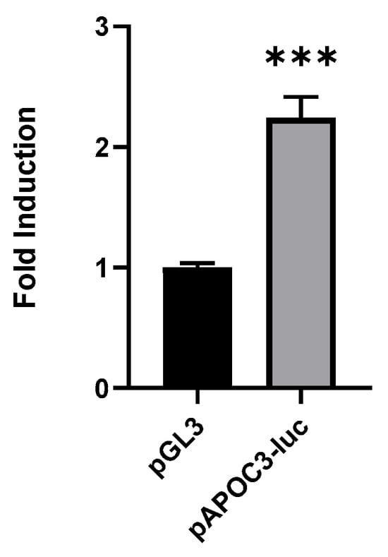
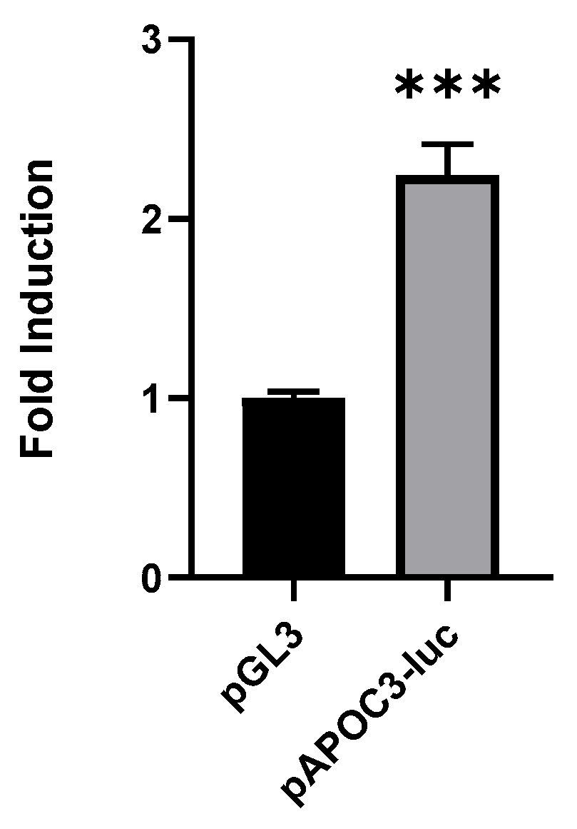
HEK 293T cells were seeded at 1.8 × 10⁵ per well in 24-well plates. After 48 h of plasmid transfection, cellular luciferase activity was quantified using a Dual-Luciferase Reporter Assay Kit (Promega, Madison, WI, USA) according to the manufacturer’s protocol. Luminescence signal was captured by a microplate luminometer, and firefly luciferase signal was normalized to Renilla internal control signal. First, the validity of the pAPOC3-Luc reporter screening system was validated. Empty pGL3 blank vector (Figure S1, Supplementary Materials) and recombinant pAPOC3-Luc plasmid (Figure S2, Supplementary Materials) were separately transfected into HEK 293T cells. After 48 h of incubation, cellular luciferase activity was quantified.
For compound screening, an internal compound library containing >300 small-molecule agents was applied. Screening was performed at a 10 μM compound concentration; vehicle-treated DMSO served as a negative control. Each condition contained three biological replicates. Hit compounds were defined as agents producing a ≥30% reduction of normalized APOC3-luciferase signal. CARV was identified from this primary screening. This screening dataset belongs to our unpublished internal preliminary work.
2.5. MTT Cell Viability Assay
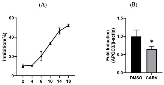
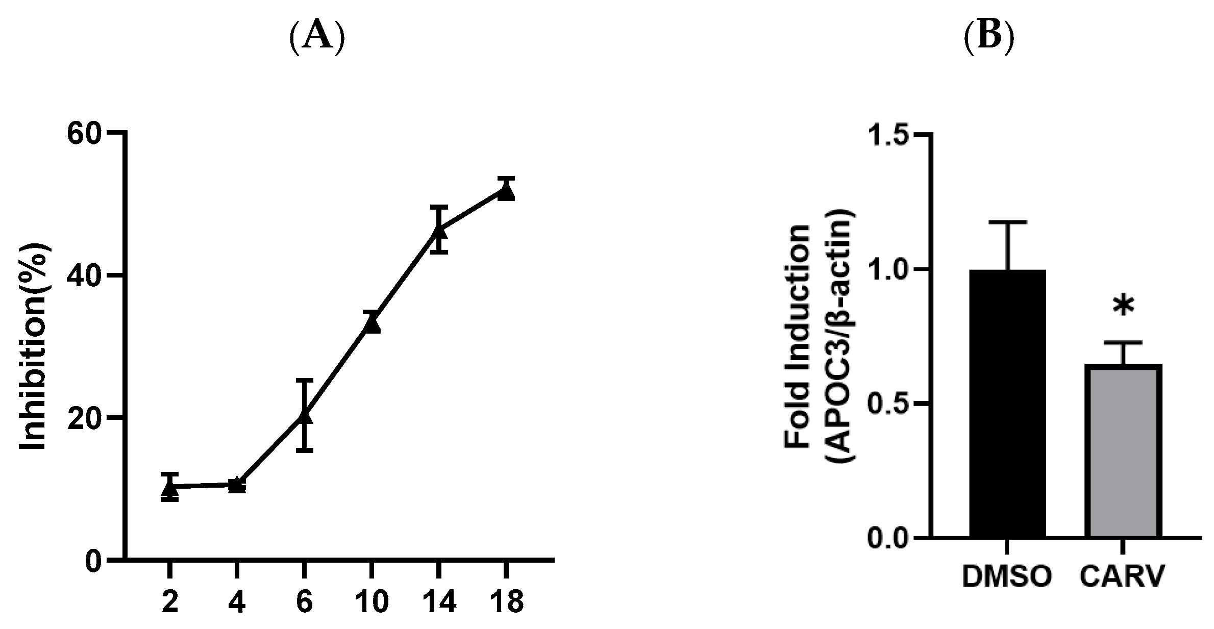
L02 cells were seeded into 96-well plates and cultured until adherent. Original medium was replaced with complete medium containing serially diluted CARV (2, 4, 6, 10, 14, 18 μg/mL), while the vehicle control received an equal volume of DMSO. After 44 h of incubation, 20 μL MTT solution was added to each well, and cells were incubated for another 4 h. Subsequently, 100 μL DMSO was added to dissolve formazan crystals. After shaking for 10 min, absorbance at 492 nm was detected using a microplate reader. Cell viability was calculated as the OD ratio of drug-treated groups versus the vehicle control, and cell viability curves were plotted. The non-toxic concentration of CARV (IC10, the concentration inducing 10% cell growth inhibition) was determined as 2 μg/mL for subsequent functional experiments.
2.6. RNA Extraction and Quantitative Real-Time PCR (RT-qPCR)
Total cellular RNA was extracted using TRIzol reagent (Invitrogen, Carlsbad, CA, USA). RNA was reverse transcribed to complementary cDNA using a reverse transcription kit (All Style Gold, Beijing, China). Three replicates were done for each sample, and RT-qPCR was carried out using the SYBR Supermix kit. Thermal cycling parameters: initial denaturation 95 °C for 3 min, followed by 40 cycles of 95 °C for 10 s and 60 °C for 30 s. Melting-curve analysis was performed after amplification. The primer sequences are listed in Table 1. APOC3 primer sequences were adopted from previously published literature [7]. Finally, results were analyzed using the PikoReal software version 2.0 (Thermo Fisher Scientific). Ct values were calculated for each sample to assess the expression of APOC3 mRNA relative to β-actin mRNA.
Table 1.
The primer sequences involved in RT-qPCR.
2.7. Protein Immunoblot Analysis
Cells or liver tissues were washed with pre-cooled PBS (liver tissues were weighed), followed by washing in pre-cooled lysis buffer for 30 min and centrifugation at 12,000× g rpm to obtain the supernatant. Proteins were separated by SDS-PAGE electrophoresis. Separated proteins were transferred to a polyvinylidene difluoride (PVDF) membrane and sealed with 5% skim milk powder for 1 h at room temperature. The membrane was then incubated with the corresponding primary antibody at 4 °C overnight. The next day, the membrane was incubated with horseradish-peroxidase-labeled secondary antibodies for 1 h at room temperature. Immunocomplexes on the membrane were visualized using enhanced chemiluminescence (ECL) reagents and exposed for imaging with a Bio-Rad gel imaging system. ImageJ (version 1.54t) PikoReal software version 2.0 software was used to quantify the protein blotting results from independent replicate experiments. PVDF membranes were physically cut into several pieces according to molecular-weight markers before primary-antibody incubation for separate primary-antibody hybridization. All images provided are complete raw scans of each individual membrane fragment without subsequent digital cropping after image acquisition.
2.8. Lipid Metabolism and Liver Function Index Testing
Cell samples were ultrasonically disrupted, with each sonication performed for 5 s with an interval of 30 s for a total of five sonications. Cell homogenates were used to determine triglyceride (TG), total cholesterol (TC) and low-density lipoprotein cholesterol (LDL-c) levels according to the manufacturer’s instructions.
After mice were anesthetized, their blood was collected from the heart in clean EP tubes and centrifuged at 3000 rpm for 30 min at 4 °C. The supernatant was divided, and aspartate transaminase (AST) and serum alanine transaminase (ALT) levels were determined according to the manufacturer’s instructions.
Mouse liver tissue was weighed, and nine times the volume of anhydrous ethanol was added according to the ratio of liver weight (g) to volume (mL) of 1:9. Liver tissue was cut using clean surgical scissors and then homogenized on ice, followed by detection of TG, TC and LDL-c levels.
2.9. Oil Red O Staining
Cells were removed from the medium and washed gently with PBS three times, followed by addition of 500 μL of cell fixative for 30 min in the dark. Cells were then stained for 10 min at room temperature by addition of Oil Red O working solution. Sections were differentiated with 500 μL of 60% isopropanol, and excess dye was removed. Finally, sections were blocked with glycerol. Light images of stained sections were captured using a Nikon Eclipse microscope (Nikon, Tokyo, Japan).
Liver tissues were soaked in 4% paraformaldehyde for 24 h and then transferred to 30% sucrose solution for dehydration. After dehydration, liver tissues were embedded with OCT embedding agent in an embedding box and stored at −20 °C in a refrigerator for a short period of time. Before staining, frozen sections were removed from the refrigerator, rewarmed at room temperature for 1 h, after which OCT was removed, and sections were stained with Oil Red O working solution and sealed with glycerol gelatin. Microscopic observation was then recorded.
2.10. Hematoxylin–Eosin (H&E) Staining
Liver tissues were fixed in 10% neutral formalin, after which the tissues were dehydrated using a series of graded ethanol solutions, paraffin-embedded and cut into 5 μm sections. Paraffin sections were then deparaffinized using xylene. Hematoxylin staining was performed for 10 min, followed by eosin staining for 1 min using a slide staining apparatus. Stained sections were dehydrated to make them transparent and covered with coverslips using neutral gum as a sealer. Light images of the stained sections were captured using a Nikon Eclipse microscope (Nikon, Japan).
2.11. Sirius Red Staining
Liver tissue sections (10 μm thick) were deparaffinized, slides were dried with water and Sirius Red staining solution was pipetted onto the tissues for 1 h at room temperature in the dark; the sections were then rinsed with running water to remove floating color. Sections were sealed with neutral gum and then observed and photographed under the microscope.
2.12. Immunohistochemistry (IHC)
The 10% neutral-formalin-fixed, paraffin-embedded tissue samples were cut into 5 μm sections. Paraffin was subsequently removed using xylene. The dewaxed and rehydrated sections were immersed in 0.01 M sodium citrate buffer (pH 6.0) and autoclaved for 3 min. After antigen recovery, slides were incubated with the primary antibody for 4 h at room temperature and then washed with PBS, followed by incubation with the secondary antibody. Antibodies were detected using a DAB staining kit. Hematoxylin was used as the re-staining agent, and slides were blocked with neutral balsam. Light images of the stained sections were captured using a Nikon Eclipse microscope.
2.13. Statistical Analysis
Experiments were repeated three times or more. ImageJ software was used for quantitative analysis; GraphPad Prism 9 software was used for graphing, and statistical analysis was performed using the two-tailed t-test method for comparison of two groups and one-way ANOVA for comparison of three or more groups. * p < 0.05 indicated a statistically significant difference, ** p < 0.01 and *** p < 0.001 indicated a very statistically significant difference and NS indicated no statistically significant difference.
3. Results
3.1. Inhibitors of APOC3 Expression
We first validated the functionality of the pAPOC3-Luc reporter system in HEK 293T cells. Elevated luciferase signal confirmed the reliability of this reporter platform for compound screening (Figure 1). CARV was later identified as an APOC3 transcriptional repressor from compound library screening, and its inhibitory effect was further validated in L02 hepatocytes in the following experiments.
Figure 1.
APOC3 promoter activity analysis. Empty pGL3 vector served as a negative control. Data are presented as mean ± SD, n = 3 biological replicates. *** p < 0.001 vs. control group.
3.2. CARV Downregulates Endogenous APOC3 Expression in L02 Hepatocytes
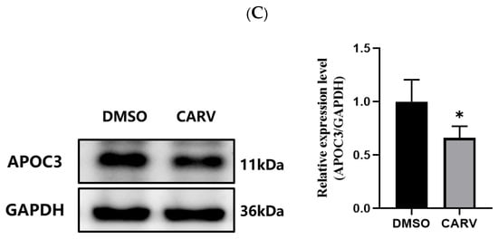
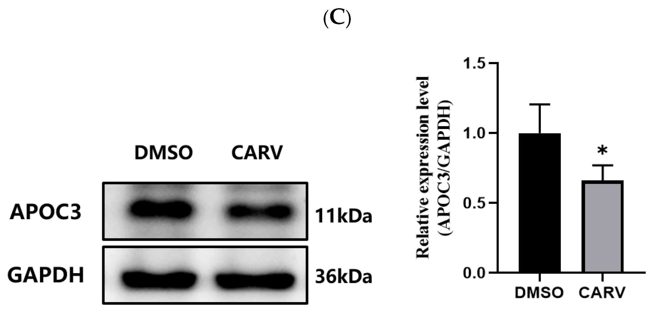
Given CARV’s inhibitory effect on APOC3 promoter activity, we further explored whether CARV represses endogenous APOC3 expression in normal human L02 hepatocytes. The MTT cytotoxicity assay determined IC10 = 2 μg/mL CARV, the concentration inducing an approximately 10% reduction of cell viability, which was selected as the working concentration for subsequent cell functional experiments (Figure 2A). RT-qPCR and western blot results demonstrated that 2 μg/mL CARV significantly suppressed APOC3 expression at both mRNA and protein levels in L02 cells (Figure 2B,C).
Figure 2.
CARV inhibits APOC3 expression in L02 cells. Note: (A) Cell viability of L02 cells treated with different concentrations of CARV detected by MTT assay. (B) Relative mRNA level of APOC3 in L02 cells after CARV treatment. (C) Protein expression of APOC3 in L02 cells detected by western blot. Data are presented as mean ± SD. * p < 0.05, vs. control group.
3.3. CARV Regulates Lipid Disorders in L02 Cells by Inhibiting APOC3 Expression
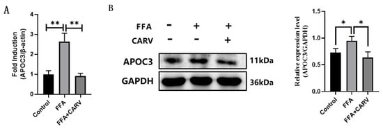
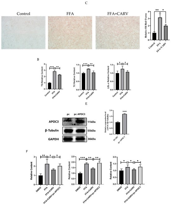
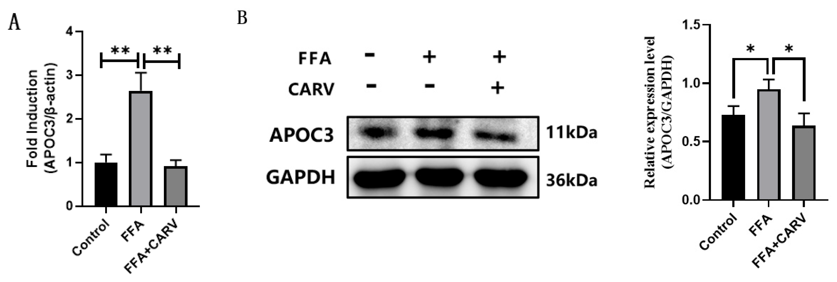
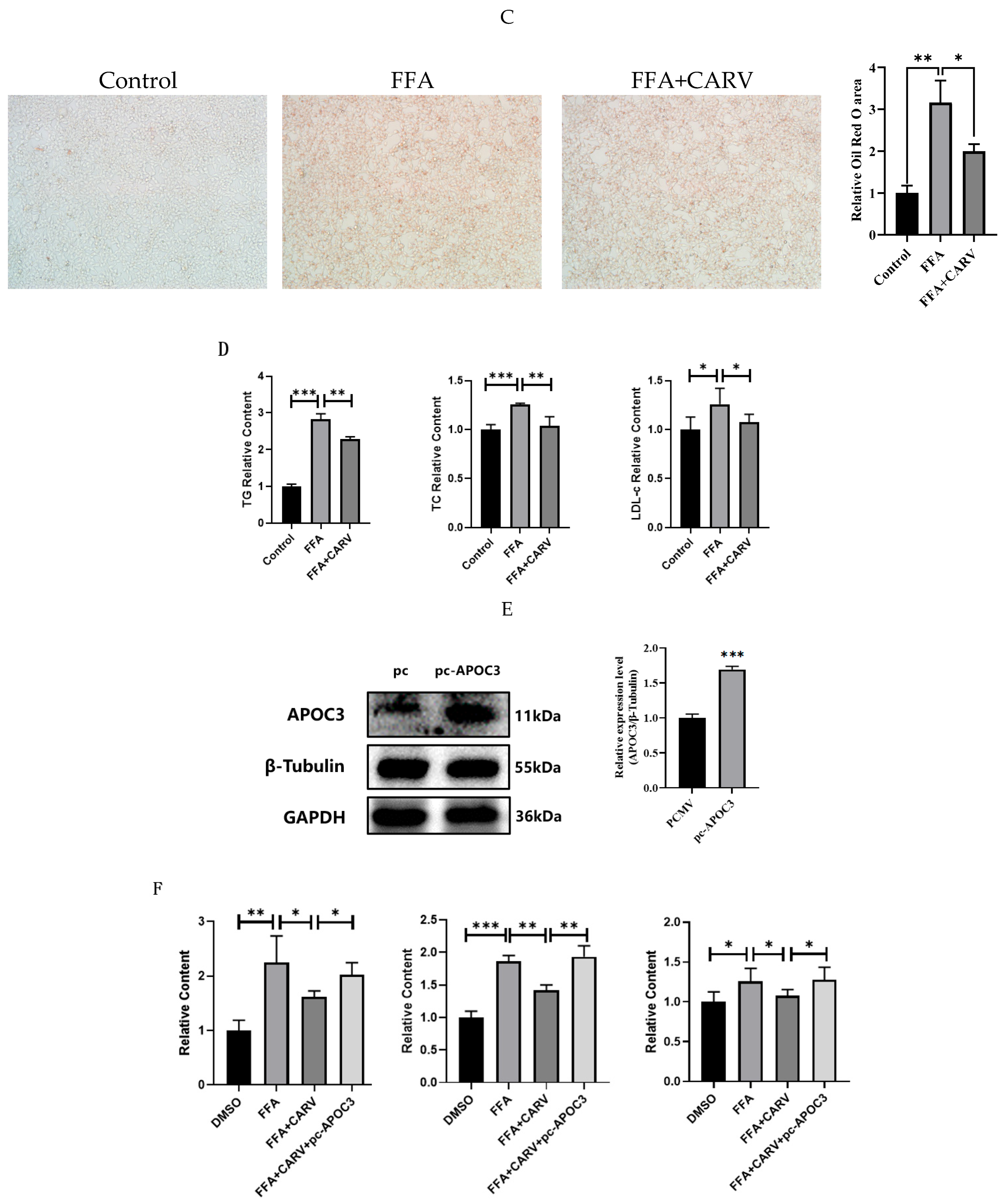
Previous studies have confirmed elevated APOC3 expression in multiple lipid metabolic disorders, and our above data verified that CARV effectively inhibits APOC3 expression. We therefore hypothesized that CARV alleviates disordered hepatic lipid metabolism by targeting APOC3. To validate this hypothesis, L02 cells were stimulated with 600 μM FFA to construct an in vitro hepatic steatosis model. RT-qPCR and western blot results showed that FFA treatment robustly upregulated APOC3 mRNA and protein levels, whereas CARV intervention reversed this upregulation (Figure 3A,B). Oil Red O staining revealed massive intracellular lipid droplet accumulation in FFA-treated cells, which was markedly reduced after CARV co-treatment (Figure 3C).
Figure 3.
CARV alleviates FFA-induced lipid deposition in L02 cells by inhibiting APOC3 expression. Note: (A,B) APOC3 mRNA and protein expression in L02 hepatocytes. (C) Representative Oil Red O staining showing intracellular lipid droplets. (D–F) Quantification of cellular lipid contents and rescue experiment after APOC3 overexpression. Data are presented as mean ± SD. *** p < 0.001. * p < 0.05, ** p < 0.01.
Cellular TG, TC and LDL-c contents are core biomarkers reflecting lipid metabolic dysfunction. Biochemical detection showed that FFA stimulation significantly increased intracellular TG, TC and LDL-c levels, while CARV co-treatment markedly blocked this lipid accumulation (Figure 3D).
To further confirm that CARV’s lipid-lowering function fully relies on APOC3 inhibition, we performed APOC3 overexpression rescue experiments. Forced overexpression of APOC3 completely abrogated CARV-mediated lipid reduction and restored cellular TG, TC and LDL-c levels to the model level (Figure 3E,F). Collectively, these data demonstrate that CARV ameliorates hepatocellular lipid metabolic disorders mainly through downregulating APOC3 expression.
3.4. CARV Regulates Hepatocyte Lipid Metabolism Through Activating the AMPK Signaling Axis (In Vitro)
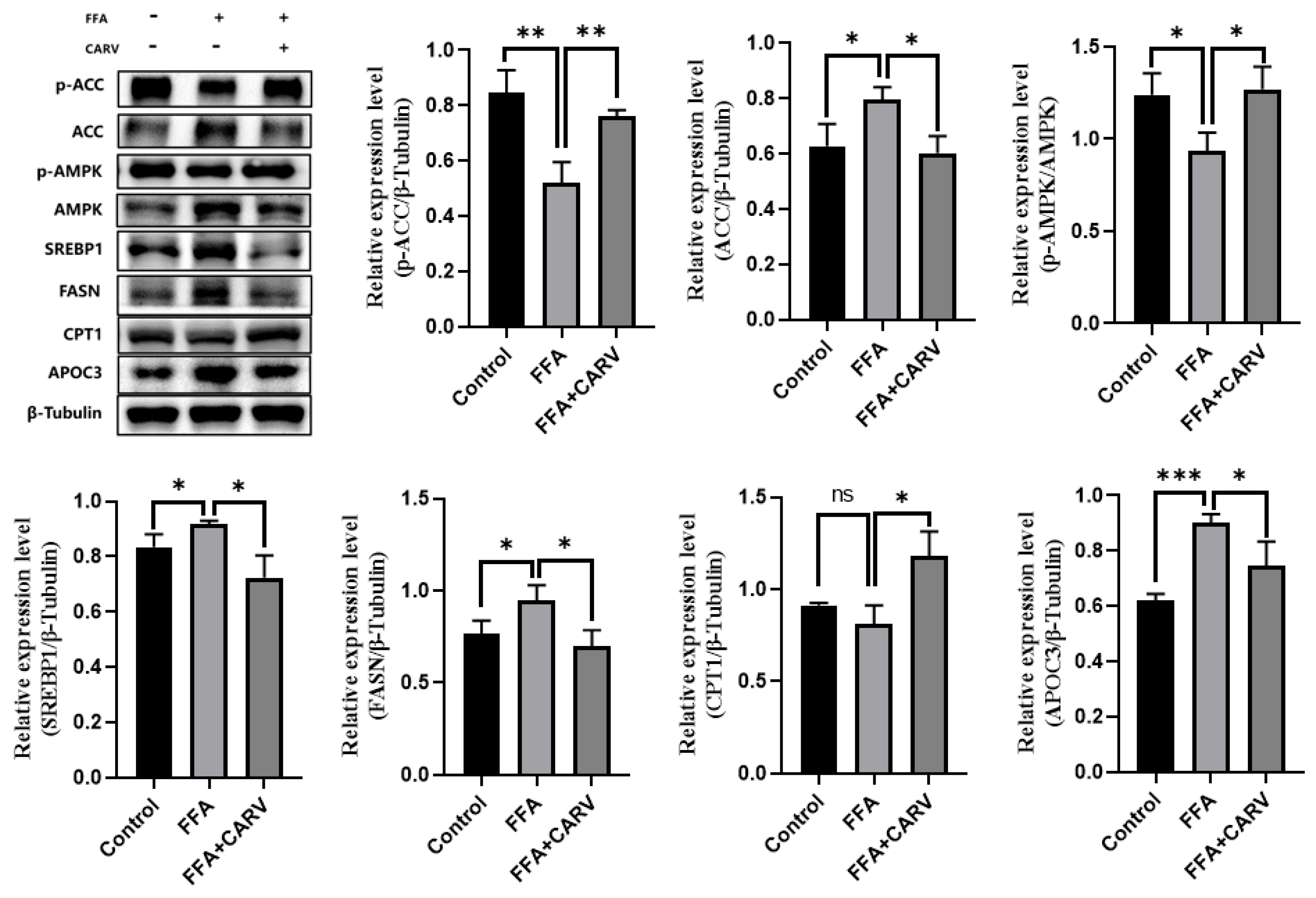
AMPK is a master cellular energy sensor that governs hepatic lipid homeostasis. Activated AMPK phosphorylates ACC1/ACC2 to reduce ACC enzymatic activity, inhibiting malonyl-CoA synthesis and relieving malonyl-CoA-mediated CPT1 suppression, thereby promoting mitochondrial fatty acid β-oxidation. Western blot analysis showed that FFA treatment decreased AMPK and ACC phosphorylation, upregulated APOC3 and downregulated CPT1 expression. CARV co-treatment fully reversed all these FFA-induced alterations, significantly elevating CPT1 protein abundance in L02 cells (Figure 4).
Figure 4.
CARV modulates lipid metabolism in L02 cells via AMPK pathway activation. Note: Protein levels of p-AMPK, AMPK, p-ACC, ACC, CPT1 and APOC3. Cropped blots from different parts of the same blot are clearly separated by dividing lines. Data are presented as mean ± SD. *** p < 0.001. * p < 0.05, ** p < 0.01.
In addition, SREBP1 acts as a key transcription factor driving hepatic de novo fatty acid synthesis by activating downstream FASN expression, while AMPK activation suppresses SREBP1 maturation. Western blot results confirmed that FFA induction reduced AMPK phosphorylation and elevated SREBP1 and FASN expression. CARV intervention significantly upregulated phosphorylated AMPK and suppressed SREBP1 and FASN expression. These in vitro findings indicate that CARV inhibits hepatic de novo lipogenesis and accelerates fatty acid β-oxidation by activating the AMPK–ACC and AMPK–SREBP1–FASN signaling pathways.
3.5. CARV Ameliorates Hepatic Lipid Deposition and Liver Injury in MAFLD Mice
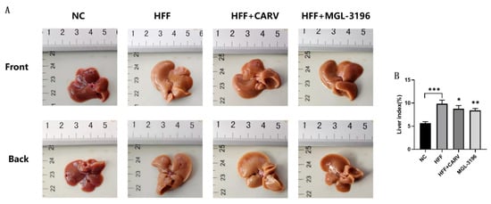
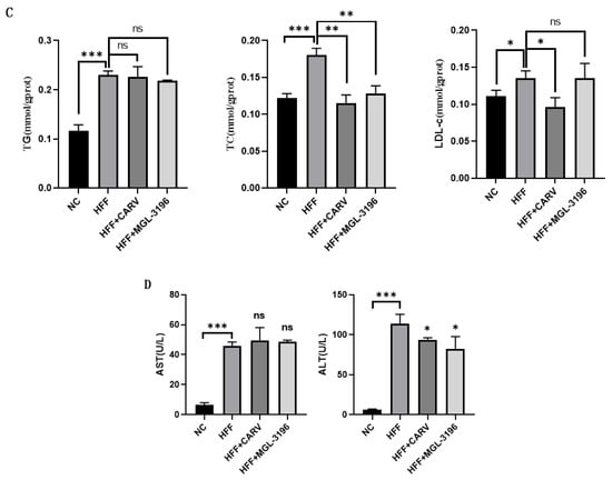
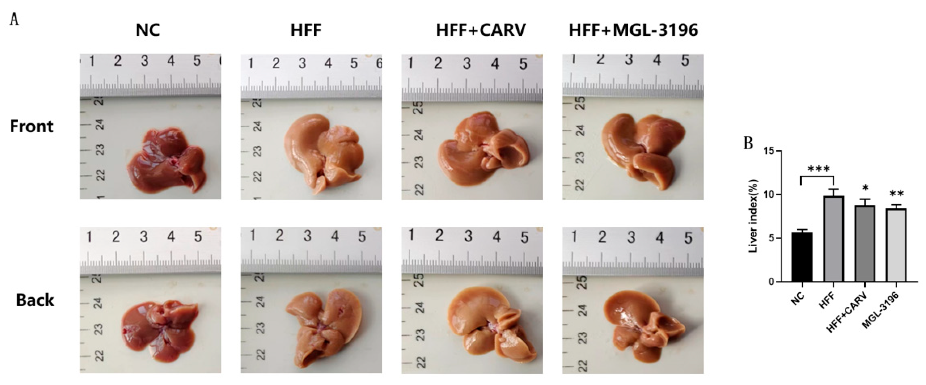
In order to further investigate the therapeutic effect of CARV on fatty liver disease, we used a mouse model of MAFLD. The livers of mice were collected to assess the morphological changes (Figure 5A). The livers of mice in the control group were bright reddish brown in color, with a smooth and glossy surface. In contrast, the livers of mice in the model group were yellowish, with a rough surface lacking luster. For comparison, the livers of mice in the CARV group, as well as in the positive drug group, were slightly redder than those of the model group, with a smooth surface and a certain luster. Nevertheless, the color of their livers did not completely return to the color observed in normal mouse livers. Overall, these results suggest that CARV treatment can improve liver tissue lesions to a certain extent.
Figure 5.
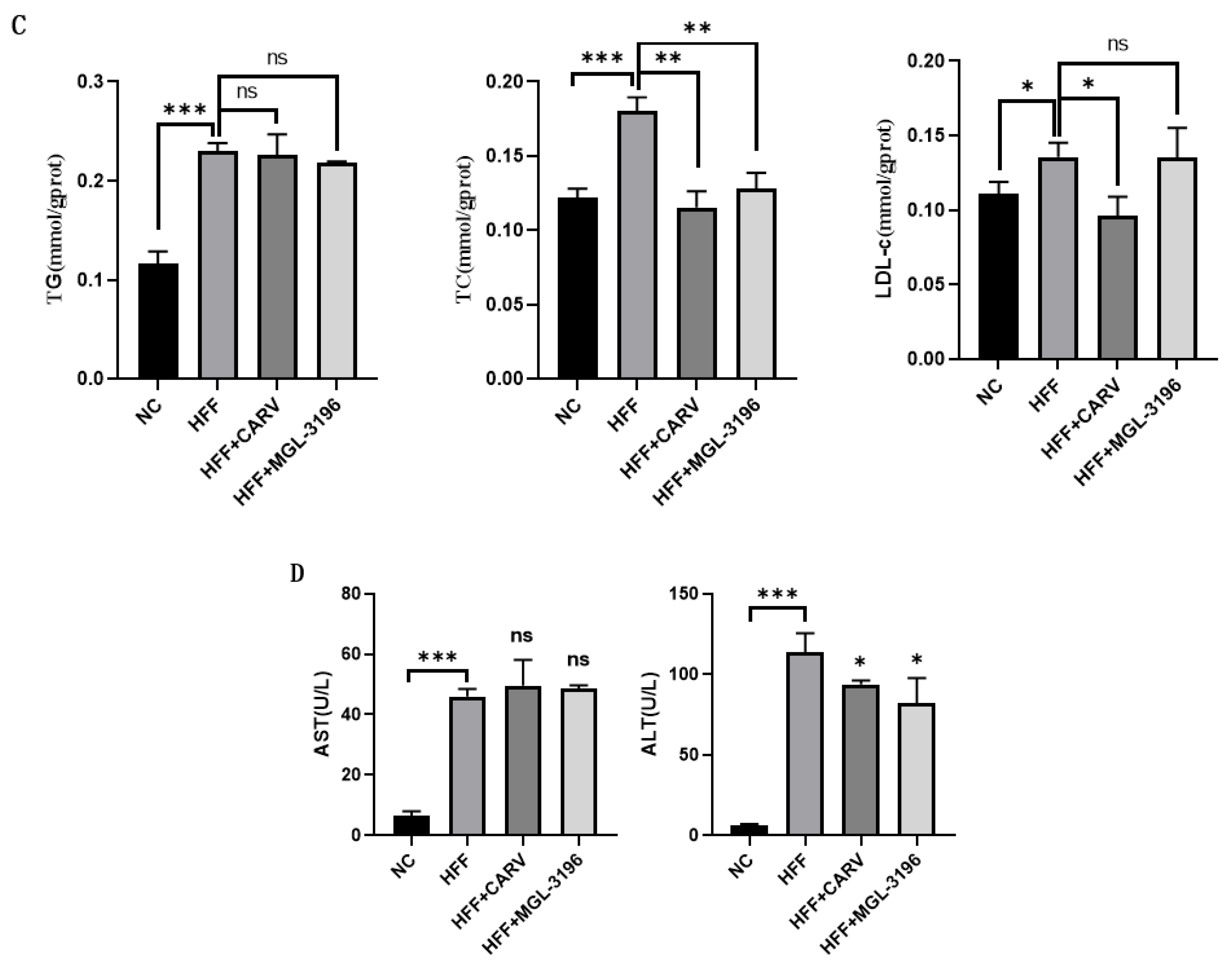
CARV ameliorates hepatic lipid deposition and liver injury in MAFLD mice. Note: (A) Representative gross morphology of mouse liver tissues. (B) Liver index of mice in each group. (C) Hepatic TG, TC and LDL-c levels. (D) Serum ALT and AST levels. Data are presented as mean ± SD. * p < 0.05, ** p < 0.01, *** p < 0.001 vs. HFF model group.
Weekly monitored body weight and cumulative food intake showed no statistically significant differences among the HFF, HFF+CARV and HFF+MGL-3196 groups, excluding the possibility that CARV improves hepatic pathological phenotypes merely by reducing food consumption or inducing body-weight loss.
In addition, MAFLD mice have increased liver weights due to lipid accumulation; therefore, we also calculated the liver coefficient in mice, comparing the liver weight with the body weight. These results (Figure 5B) showed that the liver coefficient in the model group reached about 10%, which was significantly higher than that of the control group. This indicates that lipid deposition in these livers led to an increase in liver weight, and the liver coefficients of the CARV group and the positive drug group were reduced. The liver coefficient was significantly reduced in the CARV group, as well as in the positive drug group, indicating that both CARV and the positive drug significantly improved the liver coefficient in MAFLD mice.
Subsequently, TG, TC and LDL-c contents in the livers of mice in each group were examined. These results (Figure 5C) showed that the TG and TC contents of mice in the model group were significantly higher. The TC content of the CARV-treated group was significantly lower than that of the model group. The TG and TC contents of the positive drug group were decreased, and the difference in TC content was significant compared with that of the model group; the TG content was also decreased, but the difference was not significant. Furthermore, there was no significant difference in the LDL-c content between the model and positive drug groups, but the LDL-c content was decreased significantly in the CARV group compared with the model group. These results indicated that CARV treatment ameliorated the HFF-diet-induced blockage of hepatic lipid transport function in MAFLD mice.
Results of liver enzyme levels are shown in Figure 5D. Compared with the control group, serum levels of ALT and AST in the model group were significantly increased. Moreover, levels of ALT in the CARV group were decreased significantly. The levels of ALT in the positive drug group were significantly decreased, whereas the levels of AST were abnormally increased. These results indicate that CARV significantly improves liver injury in mice.
3.6. Effect of CARV on the Pathologic Changes of Liver Tissue in MAFLD Mice
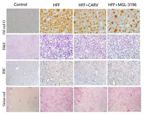
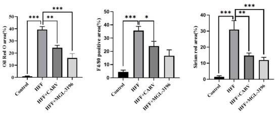
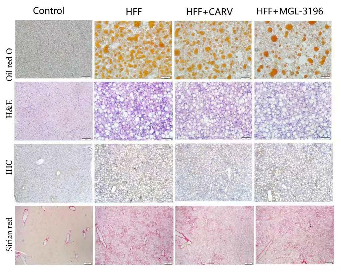
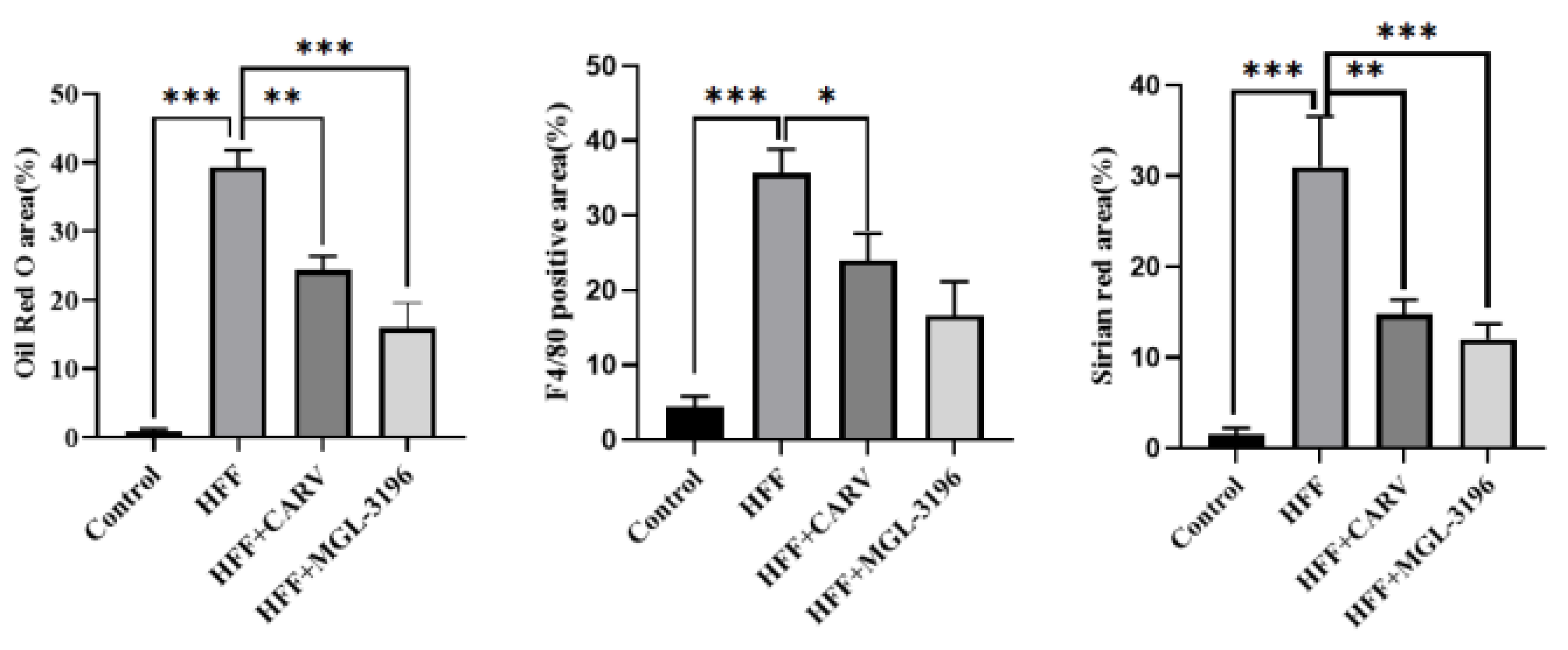
The degree of lipid accumulation in the liver of these mice was detected by frozen sectioning and Oil Red O staining. Results (Figure 6) showed that there were no obvious lipid droplets in the liver tissues of mice in the normal group. The number of lipid droplets in the model group was the highest, indicating that there was a large amount of lipid deposition. On the other hand, the number of lipid droplets in the CARV-treated group, as well as in the positive drug group, was significantly reduced, indicating that CARV effectively attenuates the degree of lipid accumulation in the liver tissue.
Figure 6.
Effect of CARV on the pathologic changes of liver tissue in MAFLD mice. Note: Liver tissues were subjected to Oil Red O staining, H&E staining, F4/80 immunohistochemistry and Sirius Red staining. CARV treatment significantly reduced lipid accumulation, inflammatory infiltration, macrophage activation and collagen dep. *** p < 0.001. * p < 0.05, ** p < 0.01. Scale: 200×.
Hepatocellular inflammatory infiltration is a basic feature of the inflammatory response in MAFLD, which is manifested by accumulation of inflammatory cells within the liver lobules and in the interstitium. Histopathological changes were observed by H&E staining of mouse liver tissues. Livers in the control group showed a normal state. In the model group of mice, however, there was a large infiltration of inflammatory cells, and compared to the model group, there was a significant improvement in inflammation in both the CARV-treated and positive drug groups.
Macrophages are one of the major players in the hepatic inflammatory response. When livers are injured, macrophages are activated and migrate to the damaged site, and these activated macrophages release a variety of inflammatory mediators and cytokines, which further exacerbate the hepatic inflammatory response. Therefore, the number of macrophages usually reflects the degree and extent of liver inflammation. In the livers of these mice, the number of macrophages was immunohistochemically determined by using the F4/80 antibody, with representative results shown in Figure 6. Compared with normal mice, a large area of positive cell staining was seen in the livers of the mice in the HFF group, whereas the number of macrophages in the livers of mice treated with CARV was significantly reduced. These results indicate that both CARV and the positive drug reduce inflammation in the livers of these mice.
The severity of liver fibrosis is an important index for histological assessment of NASH. Sirius Red staining indicated that no fibrosis was seen in the livers of mice under normal dietary conditions, whereas the livers of mice in the model group showed a large number of collagen fibers. Compared with the model group, both the CARV-treated and positive drug groups showed significantly lower liver fibrosis. These results indicate that CARV reduces the deposition of collagen fibers and inhibits liver fibrosis in these mice.
3.7. CARV Suppresses MAFLD Progression by Downregulating APOC3 and Regulating the AMPK-Related Signaling Pathway (In Vivo)
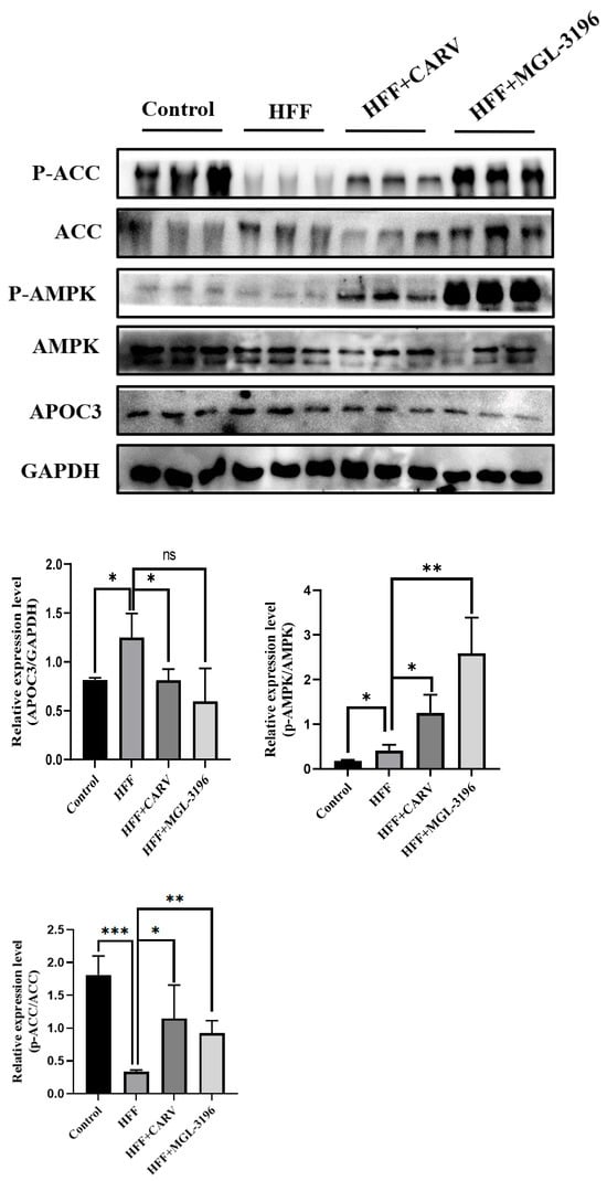
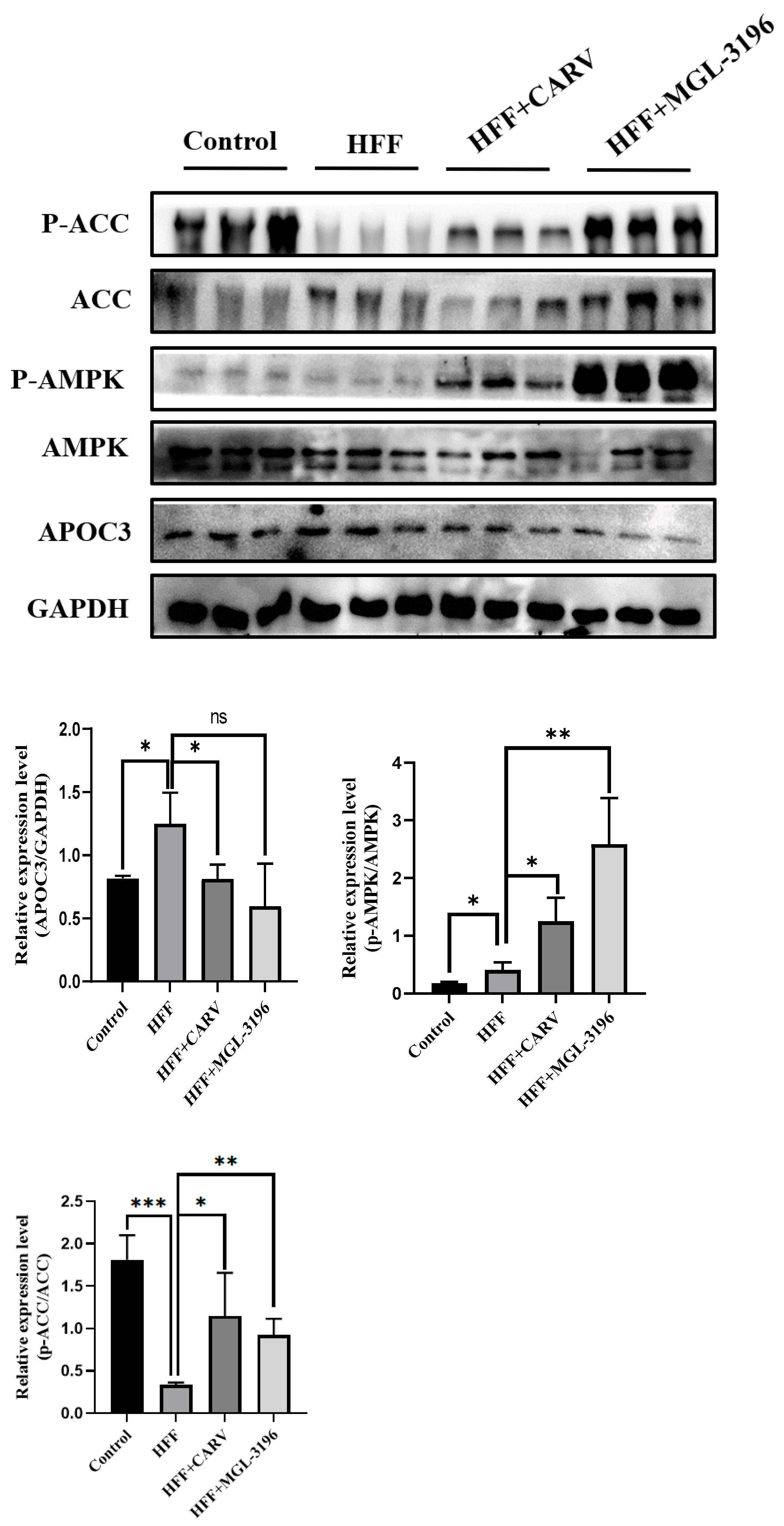
We further validated the AMPK-dependent regulatory mechanism of CARV in mouse liver tissue. Western blot results showed that the HFF diet did not significantly alter total AMPK expression levels but markedly reduced ACC phosphorylation and upregulated ACC and APOC3 expression. CARV treatment significantly increased AMPK and ACC phosphorylation, while reducing ACC and APOC3 expression (Figure 7). These in vivo data confirm that CARV activates hepatic AMPK signaling to suppress lipogenic gene expression and ameliorate MAFLD pathological phenotypes.
Figure 7.
CARV blocks MAFLD progression by activating hepatic AMPK signaling pathways in mice. Note: Protein levels of p-AMPK, AMPK, p-ACC, ACC and APOC3 in mouse liver tissues. Data are presented as mean ± SD. * p < 0.05, ** p < 0.01, *** p < 0.001 vs. HFF group.
4. Discussion
CARV exerts comprehensive protective effects against MAFLD, which are fully supported by our in vitro and in vivo experimental results: reducing intrahepatic lipid accumulation, restoring disrupted lipid homeostasis, alleviating hepatocellular injury, suppressing hepatic lobular inflammatory cell infiltration, lowering macrophage recruitment and retarding collagen fiber deposition [3]. Mechanistically, CARV represses lipogenic gene expression and balances fatty acid anabolism and catabolism to resolve excessive hepatic lipid overload. Its anti-inflammatory and anti-fibrotic capacities originate from inhibited inflammatory cell recruitment and reduced collagen overproduction in liver tissue, consistent with well-established pathological mechanisms of MAFLD progression [16,17].
Our research workflow started with APOC3 promoter luciferase screening (Figure 1), which identified CARV as a potent transcriptional suppressor of APOC3 among over 300 candidate compounds. Subsequent cellular experiments validated this inhibitory effect at endogenous mRNA and protein levels (Figure 2). The IC10 value of 2 μg/mL from the MTT assay confirmed this concentration produces only a mild 10% growth inhibition without obvious hepatotoxicity, which guaranteed the reliability of downstream lipid metabolism functional tests.
APOC3 is liver-enriched and functions as a central modulator of TRL metabolism [7,8]. Excess APOC3 protein inhibits LPL and HL activity, blocks hepatic triglyceride clearance and accelerates hepatocellular steatosis development [4,18]. Our APOC3 overexpression rescue experiment (Figure 3E,F) provides direct causal evidence: the lipid-lowering activity of CARV is fully dependent on APOC3 suppression; ectopic APOC3 expression completely abolished CARV-mediated clearance of intracellular TG, TC and LDL-c in FFA-induced steatotic L02 cells. This result establishes APOC3 as the core target through which CARV improves hepatocellular lipid disorder, which matches the elevated APOC3 phenotype in the FFA-stimulated L02 model.
AMPK serves as the core energy-sensing kinase maintaining hepatic lipid homeostasis [19]. Western blot results of FFA-treated L02 cells demonstrated that CARV activates two parallel downstream branches of AMPK signaling in isolated hepatocytes (Figure 2). On the AMPK–ACC–CPT1 branch: FFA exposure suppressed phosphorylation of AMPK and ACC, raising ACC activity to boost malonyl-CoA synthesis. As malonyl-CoA represses CPT1 (the rate-limiting enzyme of mitochondrial fatty acid β-oxidation), CPT1 expression declined and fatty acid catabolism was blocked. CARV treatment elevated p-AMPK and p-ACC, reduced malonyl-CoA production and upregulated CPT1 to accelerate fatty acid oxidation. On the AMPK–SREBP1–FASN lipogenic branch: FFA-induced AMPK dephosphorylation released inhibitory control over the transcription factor SREBP1, leading to upregulation of the downstream lipogenic enzyme FASN and excessive de novo fatty acid synthesis. CARV-mediated AMPK activation suppressed SREBP1 maturation and decreased FASN expression, further restraining lipid anabolism. In single-cell culture without systemic metabolic feedback, CARV exerts dual anti-steatotic effects via both CPT1-dependent fatty acid oxidation promotion and SREBP1/FASN-dependent lipogenesis inhibition. However, these two downstream axes do not respond to CARV treatment in intact mouse liver tissue, which we interpret in combination with the in vivo molecular data below. Several potential mechanisms may account for this in-vitro–in-vivo mechanistic uncoupling. Isolated L02 hepatocytes lack whole-body hormonal, neuronal and inter-organ metabolic feedback signals, so CARV can directly modulate CPT1, SREBP1 and FASN within single hepatocytes. In living mice, oral intragastric administration leads to complex pharmacokinetic profiles, and actual drug exposure within liver parenchymal cells differs from direct drug incubation in cell culture. Moreover, the intact liver consists of multiple heterogeneous cell populations besides hepatocytes, and endogenous compensatory metabolic circuits may counteract AMPK-dependent modulation of SREBP1/FASN/CPT1 in vivo.
Our HFF-diet feeding successfully constructed a mouse MAFLD model recapitulating core human pathological phenotypes, including fatty discolored liver, increased liver coefficient, elevated hepatic lipids, liver enzyme leakage, intrahepatic inflammation and early fibrosis. All quantitative indexes confirmed CARV exhibits superior therapeutic efficacy relative to the positive drug MGL-3196 (Figure 5). Macroscopically, HFF model mice displayed pale-yellow, dull livers with prominent hepatomegaly; CARV intervention partially restored normal reddish liver morphology and significantly lowered the liver coefficient reflecting lipid overload (Figure 5A,B). For hepatic lipid biochemistry, CARV significantly lowered hepatic TC, while hepatic TG showed only a decreasing trend without reaching statistical significance. Hepatic LDL-c was also significantly decreased in the CARV-treated group, while MGL-3196 only produced a significant decrease in TC with no obvious improvement in TG and LDL-c (Figure 5C). In terms of liver injury markers, serum ALT was markedly elevated in HFF mice and significantly rescued by CARV without abnormal AST elevation; by contrast, MGL-3196 reduced ALT but triggered AST upregulation, indicating better liver safety of CARV in vivo (Figure 5D).
Four complementary histological staining methods further validated CARV’s multi-pathological protective effects against MAFLD progression (Figure 6). Oil Red O frozen-section staining showed massive neutral lipid deposition in HFF mouse liver, which was markedly attenuated after CARV gavage. H&E staining revealed severe lobular inflammatory cell infiltration in model mice, with obvious alleviation in the CARV group. F4/80 immunohistochemistry targeting hepatic macrophages demonstrated abundant macrophage recruitment in HFF liver, while CARV significantly reduced macrophage accumulation, a key driver of hepatic inflammatory cascades. Sirius Red collagen staining showed extensive collagen fiber deposition in model liver, and CARV intervention remarkably suppressed collagen production to delay early liver fibrosis. Macrophage-mediated inflammation and progressive collagen accumulation are critical triggers for the transition from simple steatosis to irreversible NASH and cirrhosis [17,20]. Our histological data confirm CARV simultaneously ameliorates three core MAFLD pathological hallmarks, i.e., hepatic steatosis, inflammatory infiltration and early collagen deposition.
In vivo western blot detection of mouse liver tissue clarified the single functional branch of AMPK signaling activated by CARV, which is distinctly different from the dual regulatory pattern observed in L02 cells (Figure 7). After 8 weeks of HFF feeding, hepatic p-AMPK and p-ACC levels were significantly decreased in model mice, accompanied by elevated total ACC and APOC3 protein expression. Oral CARV treatment effectively restored AMPK and ACC phosphorylation, downregulating ACC and APOC3 to suppress hepatic de novo lipid synthesis. Critically, two AMPK downstream axes responsive to CARV in vitro showed no significant expression changes in mouse liver. CPT1 protein abundance remained unchanged between the HFF model and CARV treatment groups, meaning CARV fails to promote fatty acid oxidation via CPT1 upregulation in vivo. The protein levels of the lipogenic transcription factor SREBP1 and its downstream effector FASN exhibited no statistically significant differences after CARV intervention, indicating the AMPK–SREBP1–FASN axis is not involved in CARV’s anti-steatotic effect in mice.
Several limitations of the current study should be acknowledged to guide follow-up research. First, all mechanistic and therapeutic data are derived from in vitro human hepatocytes and mouse MAFLD models; no human liver biopsy samples or clinical cohort data were included, so the clinical efficacy and long-term safety of CARV for MAFLD patients require large-scale clinical trials for verification. Second, although we confirmed CARV targets APOC3 and activates hepatic AMPK–ACC signaling, the upstream molecular linkage between CARV stimulation and APOC3 transcriptional repression remains unclear. Further transcription-factor screening and chromatin immunoprecipitation assays are needed to identify intermediate signaling molecules bridging CARV and the APOC3 promoter. Third, only a single daily oral dose (10 mg/kg/d) of CARV was applied to mice; gradient-dose experiments are required to define the optimal therapeutic concentration and dose–effect curve. In addition, long-term chronic toxicity evaluation of CARV in metabolic-disorder animal models was not performed in this work. Future research will focus on uncovering the upstream regulatory network of CARV on APOC3, carrying out multi-dose animal efficacy studies and launching clinical observational trials to evaluate the translational value of CARV as a repurposed MAFLD therapeutic agent.
In addition, our in vitro functional experiments mainly adopted a single CARV concentration (2 μg/mL, IC10). Although 2.5 μg/mL and 4 μg/mL are still close to the IC10 range, higher concentrations bring elevated cytotoxic stress, and multi-gradient drug concentrations will be included in future cell-based investigations. We note that the MTT assay only reflects cellular metabolic activity and cannot fully exclude subtle cell stress or apoptosis; further cell-injury-related assays will be needed in follow-up work. Our luciferase assay demonstrates that CARV suppresses APOC3-driven promoter activity and downregulates APOC3 mRNA level, but we cannot distinguish whether this effect originates from direct promoter binding or indirect modulation of upstream transcription factors. Further promoter-truncation and transcription-factor screening experiments are required to resolve this precise regulatory mechanism.
In summary, integrated in vitro and in vivo experiments establish a clear mechanistic model for CARV against MAFLD. In isolated L02 hepatocytes, CARV activates two AMPK downstream branches (AMPK–ACC–CPT1 and AMPK–SREBP1–FASN) alongside APOC3 downregulation to simultaneously inhibit lipogenesis and accelerate fatty acid oxidation. In HFF-induced MAFLD mice, however, CARV only activates the AMPK–ACC signaling cascade and represses APOC3 expression, with no regulatory effects on CPT1, SREBP1 or FASN protein levels. Despite the absence of CPT1 and SREBP1/FASN modulation in vivo, CARV still effectively alleviates hepatic lipid accumulation, hepatocellular injury, lobular inflammation and early liver fibrosis in mice. Few existing marketed drugs can simultaneously improve steatosis, inflammation and fibrosis without severe liver adverse reactions. As a mature, clinically widely used cardiovascular drug, carvedilol has prominent repurposing potential for MAFLD intervention and deserves further translational research.
5. Conclusions
This study demonstrates that CARV can be developed as a therapeutic agent for the treatment of MAFLD. In vitro cellular experiments and in vivo mouse model results collectively confirm that carvedilol effectively reduces hepatic lipid deposition, corrects lipid metabolic disorders, decreases the liver coefficient, repairs impaired liver function and alleviates hepatic lobular inflammation and liver fibrosis. The core molecular mechanism is that carvedilol downregulates APOC3 expression and activates the AMPK signaling pathway to suppress hepatic de novo lipid synthesis. This work provides a novel therapeutic target and candidate drug for MAFLD intervention. Further investigations are warranted to uncover the precise molecular regulatory network of carvedilol and validate its clinical application value in human MAFLD treatment.
Supplementary Materials
The following supporting information can be downloaded at: https://www.mdpi.com/article/10.3390/biomedicines14112525/s1, Figure S1: The Diagram of pGL3-Basic; Figure S2: The Diagram of pAPOC3-Luc.
Author Contributions
Conceptualization, Z.L. and Y.B.; methodology, Z.L.; investigation, J.G., T.L., H.Y., Q.Y., X.X., S.W., D.Z. and L.Z.; formal analysis, J.G.; data curation, J.G.; writing—original draft preparation, Z.L.; writing—review and editing, Z.L. and Y.B.; visualization, J.G.; supervision, Z.L. and Y.B.; project administration, Z.L.; funding acquisition, Z.L. All authors have read and agreed to the published version of the manuscript.
Funding
This work was supported by the Science and Technology Development Plan Project of Jilin Province, China (grant no. 20220204015YY). The APC was funded by the Science and Technology Development Plan Project of Jilin Province, China (grant no. 20220204015YY).
Institutional Review Board Statement
The animal study protocol was approved by the Animal Research Ethics Committee of Northeast Normal University, China (NENU/IACUC, AP 20191225, approved on 25 December 2019). All animal experiments were performed according to the ARRIVE guidelines and national institutional guidelines for laboratory animal care.
Informed Consent Statement
Not applicable.
Data Availability Statement
The datasets generated and/or analyzed during the current study are available from the corresponding author on reasonable request. All raw western blot images, quantitative PCR data and biochemical measurements are included in the Supplementary Information.
Acknowledgments
The authors thank the instrument platform of Changchun Normal University for technical support.
Conflicts of Interest
The authors declare no conflicts of interest. The funders had no role in the design of the study; in the collection, analyses, or interpretation of data; in the writing of the manuscript; or in the decision to publish the results.
References
- Zhou, F.; Zhou, J.H.; Wang, W.X.; Zhang, X.-J.; Ji, Y.-X.; Zhang, P.; She, Z.-G.; Zhu, L.H.; Cai, J.J.; Li, H.L. Unexpected rapid increase in the burden of MAFLD in China from 2008 to 2018: A systematic review and meta-analysis. Hepatology 2019, 70, 1119–1133. [Google Scholar] [CrossRef] [Scilit] [PubMed]
- Zhou, J.H.; Zhou, F.; Wang, W.X.; Zhang, X.-J.; Ji, Y.-X.; Zhang, P.; She, Z.-G.; Zhu, L.H.; Cai, J.J.; Li, H.L. Epidemiological features of MAFLD from 1999 to 2018 in China. Hepatology 2020, 71, 1851–1864. [Google Scholar] [CrossRef] [Scilit] [PubMed]
- Riazi, K.; Azhari, H.; Charette, J.H.; Underwood, F.E.; King, J.A.; Afshar, E.E.; Swain, M.G.; Congly, S.E.; Kaplan, G.G.; Shaheen, A.A. The prevalence and incidence of MAFLD worldwide: A systematic review and meta-analysis. Lancet Gastroenterol. Hepatol. 2022, 7, 851–861. [Google Scholar] [CrossRef] [Scilit] [PubMed]
- Younossi, Z.M.; Golabi, P.; Paik, J.M.; Henry, A.; van Dongen, C.; Henry, L. The global epidemiology of nonalcoholic fatty liver disease (MAFLD) and nonalcoholic steatohepatitis (NASH): A systematic review. Hepatology 2023, 77, 1335–1347. [Google Scholar] [CrossRef] [Scilit] [PubMed]
- Hamed, R.; Awadallah, A.; Sunoqrot, S.; Tarawneh, O.; Nazzal, S.; AlBaraghthi, T.; Sayyad, J.A.; Abbas, A. pH-Dependent Solubility and Dissolution Behavior of Carvedilol--Case Example of a Weakly Basic BCS Class II Drug. AAPS PharmSciTech 2016, 17. Available online: https://pubmed.ncbi.nlm.nih.gov/26202065/ (accessed on 16 October 2026). [CrossRef] [Scilit] [PubMed] [PubMed Central]
- Hafez, M.A.; Mahmoud, M.F.; Ibrahim, I.A. Carvedilol: New pharmacological approaches. Zagazig J. Pharm. Sci. 2024, 33, 1–21. [Google Scholar] [CrossRef] [Scilit]
- Björnson, E.; Adiels, M.; Taskinen, M.R.; Packard, C.J.; Borén, J. Apolipoprotein C-III: Risk-factor, regulator of triglyceride-rich lipoprotein metabolism and therapeutic target. Curr. Atheroscler. Rep. 2026, 28, 22. [Google Scholar] [CrossRef] [Scilit] [PubMed]
- Ramms, B.; Gordts, P.L.S.M. Apolipoprotein C-III in triglyceride-rich lipoprotein metabolism. Curr. Opin. Lipidol. 2018, 29, 171–179. [Google Scholar] [CrossRef] [Scilit] [PubMed]
- West, G.; Rodia, C.; Li, D.; Johnson, Z.; Dong, H.; Kohan, A.B. Key differences between apoC-III regulation and expression in intestine and liver. Biochem. Biophys. Res. Commun. 2017, 491, 747–753. [Google Scholar] [CrossRef] [Scilit] [PubMed]
- Shapiro, M.D.; Nasir, K. Silencing the noise: APOC3 inhibition and the long-overdue reckoning with triglycerides. Am. J. Prev. Cardiol. 2026, 27, 101551. [Google Scholar] [CrossRef] [Scilit] [PubMed]
- Biolo, M.; Galimberti, F.; Portinari, C.; Bertocco, S.; Tosin, P.; Casula, M.; Previato, L.; Zambon, S.; Simioni, P.; Zambon, A. ApoC-III as therapeutic target: Is it primetime for clinical use? Curr. Atheroscler. Rep. 2025, 27, 72. [Google Scholar] [CrossRef] [Scilit] [PubMed]
- Packard, C.J.; Pirillo, A.; Tsimikas, S.; Ference, B.A.; Catapano, A.L. Exploring apolipoprotein C-III: Pathophysiological and pharmacological relevance. Cardiovasc. Res. 2023, 119, 2843–2857. [Google Scholar] [CrossRef] [Scilit] [PubMed]
- Tomlinson, B.; Wu, Q.Y.; Zhong, Y.M.; Li, Y.H. Advances in dyslipidaemia treatments: Focusing on APOC3 and ANGPTL3 inhibitors. J. Lipid Atheroscler. 2023, 13, 2. [Google Scholar] [CrossRef] [Scilit] [PubMed]
- Yang, F.; Wu, Y.; Chen, Y.; Xi, J.; Chu, Y.; Jin, J.; Yan, Y. Human umbilical cord mesenchymal stem cell-derived exosomes ameliorate liver steatosis by promoting fatty acid oxidation and reducing fatty acid synthesis. JHEP Rep. 2023, 5, 100746. [Google Scholar] [CrossRef] [Scilit] [PubMed]
- Sun, Y.; Zhou, W.; Zhu, M. Serum metabolomics uncovers the mechanisms of inulin in preventing non-alcoholic fatty liver disease. Pharmaceuticals 2024, 17, 895. [Google Scholar] [CrossRef] [Scilit] [PubMed]
- Machado, M.V.; Diehl, A.M. Pathogenesis of nonalcoholic steatohepatitis. Gastroenterology 2016, 150, 1769–1777. [Google Scholar] [CrossRef] [Scilit] [PubMed]
- Ibrahim, S.H.; Hirsova, P.; Gores, G.J. Non-alcoholic steatohepatitis pathogenesis: Sublethal hepatocyte injury as a driver of liver inflammation. Gut 2018, 67, 963–972. [Google Scholar] [CrossRef] [Scilit] [PubMed]
- Fouad, Y.; Waked, I.; Bollipo, S.; Gomaa, A.; Ajlouni, Y.; Attia, D. What’s in a name? renaming ‘MAFLD’ to ‘MAFLD’. Liver Int. 2020, 40, 1254–1261. [Google Scholar] [CrossRef] [Scilit] [PubMed]
- Musso, G.; Cassader, M.; Gambino, R. Non-alcoholic steatohepatitis: Emerging molecular targets and therapeutic strategies. Nat. Rev. Drug Discov. 2016, 15, 249–274. [Google Scholar] [CrossRef] [Scilit] [PubMed]
- Parthasarathy, G.; Revelo, X.; Malhi, H. Pathogenesis of nonalcoholic steatohepatitis: An overview. Hepatol. Commun. 2020, 4, 478–492. [Google Scholar] [CrossRef] [Scilit] [PubMed]

Disclaimer/Publisher’s Note: The statements, opinions and data contained in all publications are solely those of the individual author(s) and contributor(s) and not of MDPI and/or the editor(s). MDPI and/or the editor(s) disclaim responsibility for any injury to people or property resulting from any ideas, methods, instructions or products referred to in the content. |
© 2026 by the authors. Licensee MDPI, Basel, Switzerland. This article is an open access article distributed under the terms and conditions of the Creative Commons Attribution (CC BY) license.