


Abstract
Background: This study aimed to characterize the histopathological spectrum of surgically biopsied oral and maxillofacial lesions and their distribution according to anatomical location, tissue involvement, sex, age, and diagnostic status in a single-center setting. Methods: In this retrospective single-center study, 295 consecutive biopsy records from March 2021 to February 2024 were screened, and 279 eligible patients were included. Demographic data, anatomical locations, and histopathological diagnoses were obtained from institutional records. Diagnostic categories were compared according to sex and age using the Fisher–Freeman–Halton exact test with Monte Carlo estimation. Results: Thirty-four distinct histopathological diagnoses were identified. The most frequent were radicular cysts (27.6%), dentigerous cysts (17.9%), and periapical granulomas (12.9%). Jaw cysts represented the largest diagnostic category (54.1%), and most lesions involved bone (72.8%). The mandible was the most frequently affected site (43.4%). Overall, 257 lesions (92.1%) were classified as benign, 20 (7.2%) as oral potentially malignant disorders, and two (0.7%) as malignant. The overall distribution of diagnostic categories differed significantly according to sex (p < 0.001) and age group (p = 0.002). Subgroup patterns were interpreted descriptively because of small category sizes. Conclusions: Jaw cysts, particularly radicular and dentigerous cysts, predominated in this institutional biopsy series. The findings describe the histopathological distribution of biopsied lesions at a single center and should not be interpreted as estimates of population-level disease prevalence.
1. Introduction
Oral and maxillofacial lesions comprise a heterogeneous group of pathological conditions involving both soft tissues and the maxillofacial skeleton, ranging from inflammatory and reactive processes to cystic, neoplastic, and potentially malignant disorders. Their clinical presentation may overlap considerably, and lesions with different biological characteristics may exhibit similar clinical features, making definitive diagnosis challenging in some cases [1,2]. Histopathological examination of biopsy specimens therefore remains a fundamental component of the diagnostic assessment of oral and maxillofacial lesions and plays an important role in guiding appropriate clinical management [3,4].
The distribution of histopathologically diagnosed oral and maxillofacial lesions varies across populations and clinical settings according to demographic characteristics, anatomical site, referral patterns, and the spectrum of lesions subjected to biopsy [2,5,6]. Previous retrospective biopsy-based studies have demonstrated substantial differences in the relative distribution of cystic, inflammatory, soft-tissue, and neoplastic lesions among different institutional populations [7,8,9,10]. These variations highlight the value of institution-based histopathological series in characterizing the spectrum of lesions encountered in clinical practice, while recognizing that biopsy-derived distributions reflect referral and biopsy-selection patterns rather than population-level disease prevalence.
Although previous retrospective studies have described the distribution of oral and maxillofacial lesions, differences in study populations, lesion inclusion criteria, and institutional settings may limit direct comparisons. The present study provides a contemporary institutional overview of both bone and soft-tissue biopsy specimens, examining their histopathological distribution alongside demographic and anatomical characteristics. Accordingly, the present study aimed to characterize the histopathological spectrum of surgically biopsied oral and maxillofacial lesions and to examine whether their distribution differed according to anatomical location, tissue involvement, age, sex, and diagnostic status in a tertiary oral and maxillofacial surgery setting.
2. Materials and Methods
2.1. Study Design and Ethical Approval
This single-center retrospective study included patients who underwent biopsy procedures at the Department of Oral and Maxillofacial Surgery, Faculty of Dentistry, Ankara Yıldırım Beyazıt University, between March 2021 and February 2024. Ethical approval was obtained from the Health Sciences Ethics Committee of Ankara Yıldırım Beyazıt University (26 December 2024; Decision No. 10/1020). The study was conducted in accordance with the principles of the Declaration of Helsinki. Informed consent for the biopsy procedures was obtained from patients and/or their parents or legal guardians as part of routine clinical care. Research data extraction was initiated after ethical approval had been obtained.
2.2. Eligibility Criteria and Data Collection
All consecutive biopsy records from March 2021 to February 2024 were retrospectively retrieved from the hospital’s electronic medical record system and institutional archives. The investigators screened the records according to the predefined inclusion and exclusion criteria. Original histopathology reports were examined to extract the documented diagnoses and assess record completeness and eligibility. No independent histopathological reassessment was performed. All histopathological reports were issued by the pathology department of Ankara Yıldırım Beyazıt University. Patients were eligible for inclusion when the histopathological diagnosis was clearly documented and information regarding age, sex, and anatomical location of the biopsied lesion was available. Cases were excluded if they represented repeated biopsies of the same lesion, had unclear or incomplete histopathological findings, lacked demographic or anatomical location data, or involved lesions outside the oral and maxillofacial region. Each included patient contributed one biopsy record corresponding to one distinct lesion.
For each included case, age, sex, histopathological diagnosis, and anatomical location of the biopsied lesion were recorded. Anatomical sites included the gingiva, tongue, buccal mucosa, labial mucosa, floor of the mouth, palatal mucosa, maxilla, and mandible. Age was categorized by decade for descriptive and comparative analyses.
2.3. Histopathological Classification
Cysts and neoplastic lesions of the oral and maxillofacial region were classified with reference to the fifth edition of the World Health Organization (WHO) Classification of Head and Neck Tumours [11]. Other histopathological diagnoses were categorized based on Neville’s Oral and Maxillofacial Pathology, 4th edition [12]. For consistency across the study period, the diagnoses documented in the original pathology reports were retrospectively mapped to the corresponding diagnostic categories using these classification frameworks. This process was performed solely for analytical categorization without modifying the original histopathological diagnoses.
For analysis, lesions were classified according to tissue involvement as either bone or soft-tissue lesions. Each lesion was assigned to a single, mutually exclusive diagnostic category based on its documented histopathological diagnosis. The complete mapping of individual histopathological diagnoses to their diagnostic categories, tissue involvement, and diagnostic status is provided in Supplementary Table S1. Reactive soft-tissue lesions included fibroepithelial hyperplasia, pyogenic granuloma, and peripheral ossifying fibroma. Peripheral giant cell granulomas were classified as soft-tissue lesions, whereas central giant cell granuloma and simple bone cyst were classified as bone lesions. For comparisons according to sex and age, these lesions were grouped under the broader category of “Giant cell lesions and bone cysts.”
For descriptive analysis, lesions were additionally grouped according to diagnostic status as benign, oral potentially malignant disorders (OPMDs), or malignant. OPMD classification was based on the documented diagnoses in the original pathology reports, with reference to established OPMD nomenclature [13].
2.4. Statistical Analysis
Statistical analyses were performed using SPSS (version 27.0; IBM Corp., Armonk, NY, USA). Categorical variables were summarized as frequencies and percentages, and continuous variables were summarized using mean and standard deviation where applicable. Given the small number of observations in several diagnostic categories, global differences in the distribution of diagnostic categories according to sex and age groups were assessed using the Fisher–Freeman–Halton extension of Fisher’s exact test with Monte Carlo estimation based on 250,000 samples, implemented in SciPy version 1.17.0 (Python version 3.13.5). No category-specific post hoc comparisons were performed, and no adjustment for multiple comparisons was applied because the analyses were exploratory. Statistical significance was defined as p < 0.05, and subgroup patterns were interpreted descriptively. The normality of age distribution was assessed using the Shapiro–Wilk test.
3. Results
3.1. Study Population
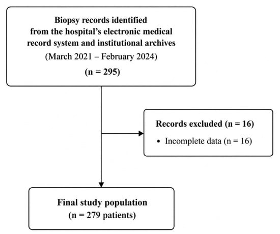
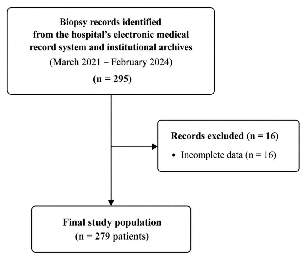
A total of 295 consecutive biopsy records were screened during the study period. Sixteen records were excluded due to incomplete or insufficient information required for the analysis. The final study population comprised 279 patients, each contributing one distinct lesion to the analysis (Figure 1). Of these, 130 (46.6%) were male and 149 (53.4%) were female, corresponding to a male-to-female ratio of 0.87. The mean age was 40.05 ± 18.60 years (range, 6–90 years), with mean ages of 39.62 ± 19.22 years in males and 40.43 ± 18.11 years in females. The largest age group was 21–30 years (n = 53, 19.0%). The distribution of sex did not differ significantly across age groups (p = 0.484). The age distribution deviated significantly from normality (Shapiro–Wilk test, W = 0.977, p < 0.001), with a median age of 41 years (interquartile range, 25–56 years).
Figure 1.
Flowchart of the selection process of the study population.
3.2. Histopathological Spectrum and Anatomical Distribution
A total of 34 distinct histopathological diagnoses were identified, and their frequencies and classifications are presented in Supplementary Table S1. The most frequent diagnoses were radicular cyst (n = 77, 27.6%), dentigerous cyst (n = 50, 17.9%), periapical granuloma (n = 36, 12.9%), fibroepithelial hyperplasia (n = 20, 7.2%), oral lichen planus (n = 14, 5.0%), odontogenic keratocyst (n = 13, 4.7%), nasopalatine duct cyst (n = 9, 3.2%), and peripheral giant cell granuloma (n = 8, 2.9%). The remaining diagnoses collectively accounted for 18.6% of the cases.
Bone lesions accounted for 203 (72.8%) cases and soft-tissue lesions for 76 (27.2%). Jaw cysts were the most frequent diagnostic group (n = 151, 54.1%), followed by pulpal and periapical diseases (n = 38, 13.6%), reactive soft-tissue lesions (n = 31, 11.1%), and dermatological diseases (n = 16, 5.7%) (Table 1).
Table 1.
Description of the lesions according to the involvement of the tissues.
The mandible was the most frequently involved anatomical site (n = 121, 43.4%), followed by the maxilla (n = 82, 29.4%), gingiva (n = 33, 11.8%), and buccal mucosa (n = 21, 7.5%). Among all lesions, 257 (92.1%) were classified as benign, 20 (7.2%) as oral potentially malignant disorders, and 2 (0.7%) as malignant. All bone lesions were classified as benign. Among soft-tissue sites, the buccal mucosa contained the largest number of oral potentially malignant disorders (n = 15) (Table 2). Of the 20 lesions classified as OPMDs, 13 were oral lichen planus, four were epithelial dysplasia, two were oral lichenoid lesions, and one was lupus erythematosus.
Table 2.
Description of the diagnostic status of the lesions according to the involvement of the anatomical areas.
3.3. Distribution According to Sex and Age
The overall distribution of diagnostic categories differed significantly according to sex (Fisher–Freeman–Halton exact test with Monte Carlo estimation, p < 0.001). Descriptively, jaw cysts constituted a greater proportion of lesions among males, whereas dermatological diseases, reactive soft-tissue lesions, and pulpal and periapical diseases showed higher proportions among females (Table 3).
Table 3.
Distribution of diagnostic categories according to sex.
The overall distribution of diagnostic categories also differed significantly across age groups (p = 0.002). However, several diagnostic categories contained small numbers of cases, limiting the interpretation of individual age-specific patterns (Table 4).
Table 4.
Distribution of Diagnostic Categories Across Age Groups.
Two malignant lesions were identified: a secretory carcinoma of the salivary gland involving the palatal mucosa in a 47-year-old male and a squamous cell carcinoma of the tongue in a 51-year-old female.
4. Discussion
The present study characterized the histopathological spectrum and demographic distribution of 279 surgically biopsied oral and maxillofacial lesions over a three-year period. The principal findings were the predominance of bone lesions, particularly jaw cysts, and the marked contribution of radicular and dentigerous cysts to the overall biopsy spectrum. The mandible was the most frequently involved anatomical site. The overall distribution of diagnostic categories differed according to sex, with descriptive differences observed in the relative representation of several diagnostic groups. Most lesions were benign, whereas oral potentially malignant disorders and malignant lesions represented relatively small proportions of the biopsy series. By including both bone and soft-tissue lesions, the present study provides a comprehensive overview of the histopathological spectrum encountered in our institutional biopsy population. Nevertheless, these findings should be interpreted in the context of institution-specific biopsy and referral practices rather than as representative of the general population.
Jaw cysts constituted more than half of all biopsied lesions in the present series, with radicular cysts, dentigerous cysts, and odontogenic keratocysts being the most frequently identified cystic lesions. Radicular and dentigerous cysts have similarly been among the most common odontogenic cysts reported in large retrospective biopsy series from different populations [8,14,15]. More recent histopathology-based series have also demonstrated that cystic and inflammatory lesions account for a substantial proportion of oral and maxillofacial biopsy specimens [9,10]. Comparisons with previous institutional studies should, however, be interpreted cautiously because differences in inclusion criteria, age ranges, biopsy indications, specimen-submission practices, and referral patterns may influence the observed lesion distribution.
Pulpal and periapical diseases constituted the second most frequent diagnostic group, with periapical granuloma accounting for most cases within this category. Periapical granulomas have also been frequently identified in previous histopathological biopsy series, although their relative contribution varies considerably among institutions [5,14,16]. Such variation may partly reflect differences in clinical management and specimen-submission practices, as not all inflammatory periapical lesions undergo surgical biopsy and histopathological examination. Consequently, their frequency within a biopsy series should not be interpreted as an estimate of their occurrence in the underlying population.
Bone lesions were considerably more frequent than soft-tissue lesions in the present cohort, and the mandible was the most commonly involved anatomical site. A predominance of mandibular involvement has also been reported in several retrospective studies of intraosseous and odontogenic lesions [17,18,19]. Nevertheless, direct comparisons between studies are complicated by differences in lesion classification and anatomical grouping. Some series are restricted to intraosseous lesions, whereas others include both bone and soft-tissue specimens, as in the present study. Therefore, the observed predominance of mandibular lesions should primarily be interpreted within the context of the biopsy population evaluated.
The overall distribution of diagnostic categories differed according to sex. Descriptively, jaw cysts represented a larger proportion of lesions among males, whereas dermatological diseases, reactive soft-tissue lesions, and pulpal and periapical diseases showed higher proportions among females. Previous retrospective studies have also reported sex-related differences in the distribution of odontogenic cysts [8,14,15,20]. Oral lichen planus, which was the most frequent dermatological diagnosis in the present series, was predominantly identified in women, consistent with systematic reviews reporting a higher prevalence of oral lichen planus among women [21,22]. Nevertheless, because several diagnostic categories contained relatively small numbers of cases and no category-specific post hoc comparisons were performed, these descriptive patterns should not be interpreted as evidence of sex-specific disease risk.
Oral potentially malignant disorders accounted for 7.2% of the biopsy specimens. Given the relatively small number of OPMDs in the present series, this finding should be interpreted descriptively. Oral potentially malignant disorders comprise a heterogeneous group of conditions associated with varying risks of progression to oral cancer, and their recognition and appropriate clinical follow-up remain important [13]. However, the present study did not evaluate longitudinal outcomes; therefore, no conclusions regarding malignant transformation or individual progression risk can be drawn from these data.
Only two malignant lesions were identified in the present cohort: one squamous cell carcinoma of the tongue and one secretory carcinoma of the salivary gland. The small number of malignant lesions precludes meaningful analysis of demographic or anatomical patterns of malignancy. Large biopsy-based studies have identified squamous cell carcinoma as the predominant oral malignancy [2,14], but direct comparison of malignancy proportions across institutional biopsy series should be approached cautiously because referral pathways and case selection may differ substantially. Detailed information regarding referral pathways for patients with suspected malignancies was unavailable. Therefore, the potential influence of referral practices on the low number of malignant lesions in this cohort could not be determined.
The findings of this study should therefore be interpreted as a description of the histopathological spectrum encountered among patients undergoing surgical biopsy at a single oral and maxillofacial surgery center rather than as estimates of population-level disease prevalence. Histopathological biopsy series remain useful for characterizing the range of lesions encountered in clinical practice and for identifying patterns in the specimens submitted for pathological examination. The predominance of cystic and inflammatory lesions in this cohort, together with the diversity of soft-tissue diagnoses, also illustrates the broad pathological spectrum encountered in oral and maxillofacial surgical practice.
Several limitations should be acknowledged. First, the retrospective and single-center design limits the generalizability of the findings and makes the observed distribution susceptible to referral and biopsy-selection bias. Detailed information regarding institutional referral pathways was not systematically available, preventing an assessment of their potential contribution to the predominance of odontogenic cysts. Furthermore, lesions managed without biopsy were not represented; therefore, the reported frequencies cannot be extrapolated to the general population.
Second, the analysis was limited to demographic, anatomical, and histopathological variables. Potentially relevant clinical factors, including symptoms, lesion duration, systemic diseases, and tobacco and alcohol exposure, were not evaluated.
Third, clinical and radiological provisional diagnoses were not assessed, precluding evaluation of diagnostic concordance. Additionally, histopathological diagnoses were extracted from the original pathology reports without independent reassessment. This limitation is particularly relevant to rare malignancies, such as salivary gland secretory carcinoma, for which expert pathological review may provide additional diagnostic confidence [23].
Finally, several diagnostic categories contained relatively few cases, particularly in age-stratified analyses. The demographic comparisons were exploratory, and no category-specific post hoc analyses were performed. Therefore, the observed subgroup patterns should be interpreted as descriptive rather than confirmatory.
5. Conclusions
In this single-center retrospective study, surgically biopsied oral and maxillofacial lesions predominantly involved bone, with jaw cysts representing the most frequent diagnostic category. Radicular and dentigerous cysts were the most commonly identified diagnoses, and the mandible was the most frequently involved anatomical site. These findings characterize the histopathological distribution of biopsied lesions at our institution and should be interpreted in the context of potential referral and biopsy-selection biases rather than as estimates of population-level disease prevalence.
Supplementary Materials
The following supporting information can be downloaded at https://www.mdpi.com/article/10.3390/biomedicines14112466/s1. Table S1: Detailed histopathological diagnoses and classification of oral and maxillofacial biopsies.
Author Contributions
S.K.S.: Methodology, Formal analysis, Writing—original draft. F.P.: Investigation, Data curation. K.T.: Investigation, Data curation. S.A.T.: Writing—review and editing. E.Y.: Writing—review and editing, Methodology, Conceptualization, Supervision. All authors have read and agreed to the published version of the manuscript.
Funding
This research received no external funding.
Institutional Review Board Statement
Ethical approval was obtained from the Health Sciences Ethics Committee of Ankara Yıldırım Beyazıt University (26 December 2024; Decision No. 10/1020).
Informed Consent Statement
Informed consent for the biopsy procedures was obtained from patients and/or their parents or legal guardians as part of routine clinical care.
Data Availability Statement
The data supporting the findings of this study are available from the corresponding author upon reasonable request. The data are not publicly available due to patient privacy and ethical restrictions.
Acknowledgments
AI-assisted language editing was used to improve the clarity and readability of the manuscript. No AI tools were used for data analysis or interpretation.
Conflicts of Interest
The authors declare no conflicts of interest.
References
- Guedes, M.M.; Albuquerque, R.; Monteiro, M.; Lopes, C.; Amaral, J.; Pacheco, J.; Monteiro, L. Oral soft tissue biopsies in Oporto, Portugal: An eight year retrospective analysis. J. Clin. Exp. Dent. 2015, 7, e640–e648. [Google Scholar] [CrossRef] [Scilit]
- Dovigi, E.A.; Kwok, E.Y.; Eversole, L.R.; Dovigi, A.J. A retrospective study of 51,781 adult oral and maxillofacial biopsies. J. Am. Dent. Assoc. 2016, 147, 170–176. [Google Scholar] [CrossRef] [Scilit]
- Abati, S.; Bramati, C.; Bondi, S.; Lissoni, A.; Trimarchi, M. Oral Cancer and Precancer: A Narrative Review on the Relevance of Early Diagnosis. Int. J. Environ. Res. Public Health 2020, 17, 9160. [Google Scholar] [CrossRef] [Scilit]
- Sindi, A.M.; Aljohani, K. Agreement Between Clinical and Histopathological Diagnoses of Oral and Maxillofacial Lesions and Influencing Factors: A Five-Year Retrospective Study. Clin. Cosmet. Investig. Dent. 2024, 16, 273–282. [Google Scholar] [CrossRef] [Scilit]
- Kelloway, E.; Ha, W.N.; Dost, F.; Farah, C.S. A retrospective analysis of oral and maxillofacial pathology in an Australian adult population. Aust. Dent. J. 2014, 59, 215–220. [Google Scholar] [CrossRef] [Scilit]
- Monteiro, L.S.; Albuquerque, R.; Paiva, A.; de la Peña-Moral, J.; Amaral, J.B.; Lopes, C.A. A comparative analysis of oral and maxillofacial pathology over a 16-year period, in the north of Portugal. Int. Dent. J. 2017, 67, 38–45. [Google Scholar] [CrossRef] [Scilit]
- Alhindi, N.A.; Sindi, A.M.; Binmadi, N.O.; Elias, W.Y. A retrospective study of oral and maxillofacial pathology lesions diagnosed at the Faculty of Dentistry, King Abdulaziz University. Clin. Cosmet. Investig. Dent. 2019, 11, 45–52. [Google Scholar] [CrossRef] [Scilit]
- Hosgor, H.; Tokuc, B.; Kan, B.; Coskunses, F.M. Evaluation of biopsies of oral and maxillofacial lesions: A retrospective study. J. Korean Assoc. Oral Maxillofac. Surg. 2019, 45, 316–323. [Google Scholar] [CrossRef] [Scilit]
- Shabir, H.; Irshad, M.; Durrani, S.H.; Sarfaraz, A.; Arbab, K.N.; Khattak, M.T. First comprehensive report on distribution of histologically confirmed oral and maxillofacial pathologies; a nine-year retrospective study. J. Pak. Med. Assoc. 2022, 72, 685–690. [Google Scholar] [CrossRef] [Scilit]
- Alzahrani, S.; Wazzan, T.; Almaghrabi, A.; Alkhudran, A.; Aljereb, H.; Elsayed, S.; Alolayan, A.B. The Prevalence and Diagnostic Patterns of Oral and Maxillofacial Lesions: A Seven-Year, Retrospective, Single-Center Cone Beam Computed Tomography and Histopathology Study in Saudi Arabia. J. Clin. Med. 2024, 13, 7774. [Google Scholar] [CrossRef] [Scilit]
- Vered, M.; Wright, J.M. Update from the 5th Edition of the World Health Organization Classification of Head and Neck Tumors: Odontogenic and Maxillofacial Bone Tumours. Head Neck Pathol. 2022, 16, 63–75. [Google Scholar] [CrossRef] [Scilit]
- Neville, B.W.; Damm, D.D.; Allen, C.M.; Chi, A.C. Oral and Maxillofacial Pathology, 4th ed.; Elsevier Health Sciences: St. Louis, MO, USA, 2015. [Google Scholar]
- Warnakulasuriya, S.; Kujan, O.; Aguirre-Urizar, J.M.; Bagan, J.V.; González-Moles, M.Á.; Kerr, A.R.; Lodi, G.; Mello, F.W.; Monteiro, L.; Ogden, G.R.; et al. Oral potentially malignant disorders: A consensus report from an international seminar on nomenclature and classification, convened by the WHO Collaborating Centre for Oral Cancer. Oral Dis. 2021, 27, 1862–1880. [Google Scholar] [CrossRef] [Scilit]
- Jones, A.V.; Franklin, C.D. An analysis of oral and maxillofacial pathology found in adults over a 30-year period. J. Oral Pathol. Med. 2006, 35, 392–401. [Google Scholar] [CrossRef] [Scilit]
- Tekkesin, M.S.; Olgac, V.; Aksakalli, N.; Alatli, C. Odontogenic and nonodontogenic cysts in Istanbul: Analysis of 5088 cases. Head Neck 2012, 34, 852–855. [Google Scholar] [CrossRef] [Scilit]
- Peker, E.; Öğütlü, F.; Karaca, İ.R.; Gültekin, E.S.; Çakır, M. A 5 year retrospective study of biopsied jaw lesions with the assessment of concordance between clinical and histopathological diagnoses. J. Oral Maxillofac. Pathol. 2016, 20, 78–85. [Google Scholar] [CrossRef] [Scilit]
- Ali, M.A. Biopsied jaw lesions in Kuwait: A six-year retrospective analysis. Med. Princ. Pract. 2011, 20, 550–555. [Google Scholar] [CrossRef] [Scilit]
- Johnson, N.R.; Savage, N.W.; Kazoullis, S.; Batstone, M.D. A prospective epidemiological study for odontogenic and non-odontogenic lesions of the maxilla and mandible in Queensland. Oral Surg. Oral Med. Oral Pathol. Oral Radiol. 2013, 115, 515–522. [Google Scholar] [CrossRef] [Scilit]
- Jaafari-Ashkavandi, Z.; Akbari, B. Clinicopathologic Study of Intra- Osseous Lesions of the Jaws in Southern Iranian Population. J. Dent. 2017, 18, 259–264. [Google Scholar]
- Tamiolakis, P.; Thermos, G.; Tosios, K.I.; Sklavounou-Andrikopoulou, A. Demographic and Clinical Characteristics of 5294 Jaw Cysts: A Retrospective Study of 38 Years. Head Neck Pathol. 2019, 13, 587–596. [Google Scholar] [CrossRef] [Scilit]
- Li, C.; Tang, X.; Zheng, X.; Ge, S.; Wen, H.; Lin, X.; Chen, Z.; Lu, L. Global Prevalence and Incidence Estimates of Oral Lichen Planus: A Systematic Review and Meta-analysis. JAMA Dermatol. 2020, 156, 172–181. [Google Scholar] [CrossRef] [Scilit]
- González-Moles, M.Á.; Warnakulasuriya, S.; González-Ruiz, I.; González-Ruiz, L.; Ayén, Á.; Lenouvel, D.; Ruiz-Ávila, I.; Ramos-García, P. Worldwide prevalence of oral lichen planus: A systematic review and meta-analysis. Oral Dis. 2021, 27, 813–828. [Google Scholar] [CrossRef] [Scilit]
- Filippini, D.M.; Carosi, F.; Querzoli, G.; Sabbioni, S.; Fermi, M.; Chiari, F.; Gili, R.; Pantaleo, M.A.; Succo, G.; Molteni, G.; et al. Challenges in pathological diagnosis of rare head and neck cancers: National survey and retrospective study. Eur. Arch. Otorhinolaryngol. 2025, 282, 6465–6475. [Google Scholar] [CrossRef] [Scilit]

Disclaimer/Publisher’s Note: The statements, opinions and data contained in all publications are solely those of the individual author(s) and contributor(s) and not of MDPI and/or the editor(s). MDPI and/or the editor(s) disclaim responsibility for any injury to people or property resulting from any ideas, methods, instructions or products referred to in the content. |
© 2026 by the authors. Licensee MDPI, Basel, Switzerland. This article is an open access article distributed under the terms and conditions of the Creative Commons Attribution (CC BY) license.