


Abstract
Background/Objectives: Dysphagia is common in Alzheimer’s disease (AD), but reported estimates vary with disease severity, outcome definitions, and assessment methods. This review aimed to synthesize evidence on the dysphagia phenotype in AD and to examine whether dysphagia estimates differ according to disease severity and assessment method. Methods: Following a prospectively registered protocol (PROSPERO CRD420261480514) and PRISMA 2020, six databases (PubMed/MEDLINE, Embase, Scopus, Web of Science, CENTRAL, PsycINFO) were searched for studies evaluating swallowing function in adults with clinically diagnosed AD. Risk of bias was appraised with JBI checklists. Proportions were pooled with random-effects (REML) models after Freeman–Tukey transformation; odds ratios (ORs) were pooled for the severity comparison; heterogeneity was explored with subgroup, stage-stratified, sensitivity, and meta-regression analyses; and certainty was rated with GRADE. Results: Twenty-eight studies were included, and 12 contributed to at least one meta-analysis. In 11 studies (n = 886), the pooled proportion with dysphagia was 47% (95% CI 26–68%) (I2 = 97.4%; prediction interval almost 0–100%). Estimates differed across assessment-method categories (p = 0.008; questionnaire/screening, one study), but clinical and instrumental assessments did not differ significantly (p = 0.37). More advanced AD may be associated with higher odds of dysphagia (7 studies; OR 5.79, 95% CI 2.12–15.81); pooled proportions were 20% (95% CI 6–40%) in less advanced and 54% (95% CI 25–81%) in more advanced stage groups, whereas study-level characteristics did not explain the heterogeneity. Exploratory pooled proportions were 9% for aspiration, 30% for laryngeal penetration, and 48% for penetration and/or aspiration. Certainty was very low for all outcomes, including the severity association. Conclusions: Dysphagia is frequently reported in AD, but its estimated frequency depends strongly on the sample, disease stage, and assessment method, so the pooled value should not be interpreted as a population prevalence. Phase-specific findings suggest a provisional stage-related pattern, from oral-phase slowing in early disease to pharyngeal residue and laryngeal penetration in advanced disease, but derive from few small studies. Standardized definitions, consistent staging, and harmonized assessment protocols are needed.
1. Introduction
Alzheimer’s disease (AD) is a chronic neurodegenerative disease characterized by progressive cognitive and neurodegenerative deterioration, leading to significant functional loss and care requirements in the elderly population. Disease progression is not limited to impairment in memory and other cognitive functions, but also causes significant changes in motor control, behavioral functions, and activities of daily living. Within this multifaceted neurodegenerative process, impaired swallowing function is one of the important but often overlooked complications that can arise throughout the clinical course of the disease. While it has long been known that dysphagia becomes particularly prominent in the later stages of Alzheimer’s disease, there is evidence suggesting that swallowing disorders can also be detected in earlier stages [1,2]. In the study by Seçil et al. [2], dysphagia was detected in 75% of Alzheimer’s patients, and it was reported that electrophysiological disorders related to swallowing can be seen even in the early stages of the disease.
Swallowing is a complex sensorimotor process involving the coordinated work of oral preparation, oral, pharyngeal, and esophageal phases. Therefore, swallowing impairment in Alzheimer’s disease is not limited to a single clinical finding but may involve various biomechanical disorders such as chewing and bolus preparation, oral bolus control and transport, oral residue, delayed initiation of swallowing, pharyngeal bolus transport, and pharyngeal residue. Previous studies have shown impairments in different components of the oral and pharyngeal phases in Alzheimer’s patients [2,3,4,5]. In addition, it has been reported that early impairments in cortical swallowing control may appear before more advanced clinical swallowing disorders in Alzheimer’s disease [6]. These findings suggest that dysphagia in Alzheimer’s disease should be considered not merely a late-stage complication, but a multidimensional clinical phenotype that can affect different components throughout the disease process.
The clinical significance of dysphagia is not limited to symptoms that occur during swallowing. Impaired swallowing safety is particularly important in terms of laryngeal penetration and aspiration. Aspiration, characterized by the passage of bolus or material into the airway below the vocal cords, is a significant risk factor for aspiration pneumonia. In Alzheimer’s patients, aspiration pneumonia has been identified as one of the major causes of morbidity and mortality, especially in advanced disease stages [7]. However, it has been noted that the risk of aspiration cannot be explained solely by the presence of dysphagia; mobility, nutritional status, level of consciousness, oral health, and other clinical factors may also play a role [1]. Therefore, in evaluating swallowing disorders in Alzheimer’s patients, it is not sufficient to diagnose general dysphagia alone; it is also important to separately evaluate indicators of airway safety such as penetration and aspiration.
The onset and severity of dysphagia in Alzheimer’s disease appear to be related to the clinical progression of the disease. As the disease progresses, impairment of cognitive and motor control, loss of postural control, regression in oral motor skills, and impairment of voluntary processes related to swallowing can negatively affect swallowing function. Edahiro et al. [8], while examining factors affecting nutritional independence in Alzheimer’s patients, showed that swallowing and oral functions are related to the functional course of the disease. Similarly, Takagi et al. [9] drew attention to the relationship between skeletal muscle mass and swallowing function. Subsequent studies have also shown that the impairment in swallowing function can differ between stages of Alzheimer’s disease [10,11,12]. Parlak et al. [10] reported that dysphagia, residual, penetration, and aspiration findings differed among the clinical stages of Alzheimer’s disease. Therefore, disease severity should be considered an important clinical variable in evaluating the dysphagia phenotype in Alzheimer’s patients.
However, reported prevalences of dysphagia and swallowing disorder characteristics in Alzheimer’s patients show significant differences between studies. One important reason for this difference is the heterogeneity of methods used to assess swallowing function. While clinical swallowing tests, bedside assessments, and screening tools are practical and applicable, they may have limitations in directly visualizing the biomechanical components of swallowing, as well as penetration and aspiration. In contrast, videofluoroscopic swallowing study (VFSS) and fiberoptic endoscopic evaluation of swallowing (FEES) allow for a more detailed assessment of different phases of swallowing and airway safety. The use of different assessment methods in Alzheimer’s disease can lead to the reporting of different prevalences for the same clinical phenotype. Indeed, previous systematic reviews in the dementia population have reported significant heterogeneity in terms of study design, assessment method, and outcome measures, limiting quantitative synthesis [13].
This difference in assessment methods is particularly important in Alzheimer’s patients. For example, while clinical or electrophysiological swallowing tests can be used to determine the presence of dysphagia, VFSS and FEES allow for the direct assessment of specific impairments in the oral and pharyngeal phases, as well as indicators of airway safety such as penetration and aspiration. Parlak et al. [10] evaluated swallowing function in different stages of Alzheimer’s disease patients using the bedside water swallow test, EAT-10, and FEES and reported that the measurements differed according to the disease stage. Therefore, the reported prevalence of dysphagia in Alzheimer’s patients should be interpreted not only based on the clinical characteristics of the studies but also considering the assessment methods used.
Although there are a significant number of studies on dysphagia in Alzheimer’s patients in the current literature, the evidence has mostly focused on a single swallowing component, a specific assessment method, or a particular disease stage. Previous reviews have evaluated oropharyngeal swallowing disorders in different types of dementia together and have shown that methodological heterogeneity between studies limits quantitative synthesis [13]. While subsequent literature has expanded the evidence regarding dysphagia management, screening methods, and treatment approaches in Alzheimer’s disease, there remains a need for a comprehensive quantitative synthesis that considers Alzheimer’s-specific oral and pharyngeal swallowing disorders, penetration and aspiration findings, disease severity, and assessment methods together within the same analytical framework. Current studies also emphasize the importance of using screening tools such as EAT-10 in conjunction with objective assessments in advanced Alzheimer’s disease, supporting the potential impact of assessment methods on outcomes [14].
Therefore, this systematic review and meta-analysis aimed to characterize the dysphagia phenotype in individuals with Alzheimer’s disease by synthesizing—quantitatively where comparable data permitted and narratively otherwise—the prevalence of overall dysphagia, laryngeal penetration, aspiration, and combined penetration and/or aspiration, together with the evidence on specific oral- and pharyngeal-phase abnormalities, silent aspiration, and swallowing apraxia. In addition, we examined whether the odds of dysphagia differed according to the clinical severity of Alzheimer’s disease and whether dysphagia estimates differed according to the swallowing assessment method used.
Research Questions
Specifically, the review addressed the following questions:
- –
- What proportion of individuals with Alzheimer’s disease are classified as having dysphagia, and how variable are the reported estimates?
- –
- What proportions of individuals with Alzheimer’s disease show laryngeal penetration and aspiration on instrumental assessment?
- –
- Which oral- and pharyngeal-phase abnormalities have been reported in Alzheimer’s disease, and have silent aspiration and swallowing apraxia been quantified?
- –
- Are more advanced stages of Alzheimer’s disease associated with higher odds of dysphagia, and do dysphagia estimates differ according to the assessment method (clinical, instrumental, or questionnaire/screening)?
2. Methods
This study was designed as a systematic review and meta-analysis and is reported in accordance with the PRISMA 2020 statement [15]. The protocol was prospectively registered in the International Prospective Register of Systematic Reviews (PROSPERO; registration number CRD420261480514) on 10 June 2026 and is publicly available at https://www.crd.york.ac.uk/PROSPERO/view/CRD420261480514 (accessed on 28 September 2026). The protocol was registered prospectively, before data extraction began. Deviations from the registered protocol, including those introduced during peer review, are listed in Supplementary Table S2.
2.1. Search Strategy
The following information sources were searched from inception: PubMed/MEDLINE (National Library of Medicine), Embase (Embase.com), Scopus, Web of Science Core Collection, the Cochrane Library (CENTRAL), and APA PsycINFO via EBSCOhost (the database referred to as ‘EBSCO’ in the submitted version). All databases were last searched on 1 August 2026 (searches were run between 1 July and 1 August 2026, without date or language limits); the platform and full strategy for each source are reported in Supplementary Table S1. Searches were conducted independently by the first and second researchers using the same predetermined search strategy, and the records obtained were evaluated independently. Disagreements regarding the suitability of studies were discussed and resolved through consensus, with the opinion of a third researcher consulted when necessary.
The search strategy was as follows: Medical Subject Headings (MeSH) terms and database-specific controlled subject headings were used to generate the results, along with free-text keywords related to Alzheimer’s disease and swallowing disorders. Search terms were combined using AND and OR Boolean operators and adapted to each database’s indexing system and search features.
The basic screening strategy is structured around two main concepts:
Terms associated with Alzheimer’s disease:
(“Alzheimer Disease”[MeSH] OR “Alzheimer disease” OR “Alzheimer’s disease” OR “Alzheimer dementia” OR “Alzheimer-type dementia” OR “dementia of the Alzheimer type” OR “AD”)
AND
Terms associated with swallowing disorders:
(“Deglutition Disorders”[MeSH] OR dysphagia OR “swallowing disorder*” OR “swallowing dysfunction*” OR “swallowing impairment*” OR “deglutition disorder*” OR “oropharyngeal dysphagia” OR “oral dysphagia” OR “pharyngeal dysphagia” OR “oral phase” OR “oral-phase dysfunction” OR “pharyngeal phase” OR “pharyngeal-phase dysfunction” OR “oral transit” OR “pharyngeal transit” OR “swallowing apraxia” OR “delayed swallow*” OR “swallow initiation” OR “oral residue” OR “pharyngeal residue” OR penetration OR aspiration OR “silent aspiration” OR “laryngeal penetration”)
The search strategy was designed to be broad, encompassing not only studies explicitly using the term “dysphagia,” but also those evaluating different components of the dysphagia phenotype in Alzheimer’s disease. Accordingly, terms describing specific swallowing abnormalities such as oral and pharyngeal phase disorders, disorders related to swallowing initiation and transit time, oral and pharyngeal residue, penetration, aspiration, silent aspiration, and swallowing apraxia were included in the search strategy.
Terms relating to specific assessment methods such as videofluoroscopic swallowing study (VFSS), fiberoptic endoscopic evaluation of swallowing (FEES), and clinical swallowing assessment were not used as a mandatory search block. This aims to prevent the overlooking of studies that do not explicitly state the assessment method in their title, abstract, or keywords but are relevant to the research question.
The strategy was adapted to the controlled vocabulary, field codes, and syntax of each database. The complete strategies for every database, including PubMed/MEDLINE, are reported in Supplementary Table S1.
In addition to electronic database searches, the bibliographies of existing systematic reviews and meta-analyses on the subject, as well as the reference lists of all primary studies included in this review, were manually searched to identify additional suitable studies. Furthermore, forward citation tracking was performed for primary studies where appropriate to identify studies that may have been overlooked in electronic searches.
Nine additional records were identified through citation searching (reference lists of included studies and relevant reviews, and forward citation tracking, as described above) and were screened against the same eligibility criteria. Conference abstracts and presentations, master’s and doctoral theses, preprints, letters to the editor, editorials, commentaries, case reports, and other publications that had not undergone peer review were excluded. Only primary studies that provided sufficient data on swallowing disorders or specific swallowing abnormalities in individuals with Alzheimer’s disease, were accessible in full text, and had undergone peer review were included in the systematic review. Studies providing sufficient quantitative data were included in the meta-analysis.
2.2. Eligibility Criteria
Studies included in this systematic review and meta-analysis were determined according to the PICOS (Population, Exposure, Comparison, Outcomes, Study Design) framework. This study examined quantitative research evaluating the characteristics of dysphagia and oral and pharyngeal phase swallowing disorders, as well as penetration and aspiration findings, in individuals with Alzheimer’s disease.
Participants (P): Studies including adult and elderly individuals with a clinical diagnosis of Alzheimer’s disease were included. The diagnosis of Alzheimer’s disease was based on the DSM, NINCDS-ADRDA, NIA-AA, or other standard clinical diagnostic criteria accepted at the time of the study. Studies including individuals in mild, moderate, or advanced/severe clinical stages of Alzheimer’s disease were considered. Studies reporting a clinically defined disease stage or severity category using validated instruments such as the Clinical Dementia Rating (CDR), Global Deterioration Scale (GDS), Functional Assessment Staging Test (FAST), or comparable approaches were considered eligible for analyses according to disease severity.
Mixed-sample studies including Alzheimer’s disease and other types of dementia were included only if data from participants with Alzheimer’s disease were available separately. Studies where results regarding Alzheimer’s disease could not be derived separately from other types of dementia were excluded. Studies including only vascular dementia, Lewy body dementia, frontotemporal dementia, Parkinson’s disease dementia, or other neurodegenerative diseases were not included.
Exposure/Condition (E): In this review, the primary clinical condition is defined as Alzheimer’s disease. Studies must have evaluated swallowing function or specific swallowing disorders in individuals with Alzheimer’s disease. To investigate the potential impact of disease severity on the dysphagia phenotype, the clinical severity or stage of Alzheimer’s disease was considered a secondary exposure/moderator variable and was analysed in the studies that reported stage-specific dysphagia counts (Section 2.5).
Comparison (C): Since the primary aim of the study was to define the dysphagia phenotype in Alzheimer’s disease, the inclusion of a healthy control group was not mandatory for the inclusion of appropriate studies. Single-group observational studies that quantitatively report the frequency or severity of a specific swallowing abnormality in individuals with Alzheimer’s disease were also considered. However, studies comparing different clinical stages of Alzheimer’s disease (e.g., mild, moderate, and severe Alzheimer’s disease) and studies comparing Alzheimer’s patients to healthy controls or other comparison groups were included, provided that data relating to the Alzheimer’s group could be extracted separately. Differences between stages were evaluated with a comparative meta-analysis of stage-specific data (Section 2.5).
Outcome (O): The primary outcomes were the overall prevalence of dysphagia and the specific swallowing abnormalities constituting the dysphagia phenotype in individuals with Alzheimer’s disease. Specific outcomes included oral- and pharyngeal-phase swallowing disorders, laryngeal penetration, aspiration, silent aspiration, and swallowing apraxia. Oral-phase abnormalities included chewing disorders, impaired bolus preparation and control, lingual movement disorders, prolonged or delayed oral transit, and oral residue. Pharyngeal-phase abnormalities included delayed swallowing initiation, impaired pharyngeal transit, and pharyngeal residue.
Study Design (S): This systematic review and meta-analysis included cross-sectional, case–control, cohort, and longitudinal observational studies. Pre-intervention data from intervention studies were considered eligible when participants were not selected on the basis of pre-existing dysphagia and when baseline quantitative swallowing data were available. Intervention studies in which dysphagia or abnormal swallowing was an eligibility criterion were not included in prevalence meta-analyses because of the potential for selection bias. In the final dataset, no intervention or experimental study contributed to a meta-analysis (Table 1). Case reports, case series, qualitative studies, systematic reviews, meta-analyses, scoping reviews, narrative reviews, editorial articles, commentary articles, letters, and publications reporting only study protocols were excluded. Case series were defined as descriptive reports of selected individual patients; a prospective series in which all enrolled participants were assessed with a standardized instrumental swallowing protocol was considered a single-group observational study and was eligible (one study [3]; Supplementary Table S2). Each included report was assigned, according to these rules, to one or more evidence streams: (i) overall prevalence; (ii) stage comparison; (iii) airway invasion (penetration and/or aspiration); (iv) phenotype-specific narrative synthesis; and (v) narrative context only (e.g., intervention studies that selected participants because of dysphagia, studies in which participants were selected for normal swallowing, and mechanistic studies). The stream(s) to which each study contributed and the Alzheimer-specific data extracted are shown in Table 1 and Supplementary Table S3.
Table 1.
Characteristics of the included studies, Alzheimer-specific data extracted, and contribution to each synthesis.
Publications in English or Turkish that were peer-reviewed and for which the full text was accessible were eligible. No restriction was imposed on year of publication. Studies published only as abstracts or for which the full text was unavailable were excluded.
All studies meeting the predefined eligibility criteria were included in the systematic review. Quantitative meta-analyses were conducted separately for individual outcomes when at least two studies provided sufficiently comparable quantitative data. Analyses based on two to four studies were designated exploratory.
Exclusion Criteria
Studies that did not address the research question or did not meet the predefined PICOS eligibility criteria were excluded. Studies conducted in populations without a clinical diagnosis of Alzheimer’s disease were excluded. Studies including Alzheimer’s disease together with other types of dementia were excluded when data for participants with Alzheimer’s disease could not be reported or extracted separately.
Studies that did not provide quantitative data for a specific outcome of interest were excluded from the corresponding meta-analysis, rather than from the systematic review as a whole. Thus, a study could be included in the systematic review but not contribute to a particular meta-analysis when the required quantitative data for that outcome were unavailable. Studies reporting only general dysphagia without data on specific oral- or pharyngeal-phase abnormalities, penetration, or aspiration were therefore eligible for the overall dysphagia prevalence analysis when appropriate quantitative data were available, but were not included in phenotype-specific meta-analyses for outcomes that they did not report.
Studies evaluating only consequences or correlates of dysphagia, such as nutritional status, malnutrition, weight loss, dehydration, pneumonia, mortality, tube feeding, or quality of life, without reporting quantitative data on swallowing function or specific swallowing abnormalities, were excluded.
Studies in which stroke, head and neck cancer, neuromuscular diseases, Parkinson’s disease, or other neurological or clinical conditions that could independently substantially affect swallowing function constituted the primary study population were excluded. In mixed clinical populations, studies were eligible only when data for participants with Alzheimer’s disease could be identified and extracted separately.
Case reports, case series (as defined above), qualitative studies, systematic reviews, meta-analyses, scoping reviews, narrative reviews, editorials, commentaries, letters, conference abstracts or proceedings, theses, preprints, and publications reporting only study protocols were excluded. Studies for which the full text could not be obtained were also excluded.
2.3. Study Selection and Data Extraction
Studies were selected following a multi-stage process in accordance with the PRISMA 2020 (Preferred Reporting Items for Systematic Reviews and Meta-Analyses) guidelines. All records obtained from electronic database searches were transferred to a reference management program, and duplicate records were removed, first through the software and then manually by the researchers.
Following the removal of duplicate records, the titles and abstracts of the remaining studies were independently evaluated by the first and second researchers. In the first screening phase, the suitability of the studies was examined according to the predetermined PICOS criteria. Full texts of potentially suitable studies were accessed, and a detailed suitability assessment was performed. Disagreements that arose during the full-text assessment were discussed with the participation of a third researcher and resolved through consensus. The processes of identifying, screening, suitability assessment, and inclusion of studies are shown in the PRISMA 2020 flowchart.
Data extraction was performed using a standardized data extraction form, created to address the research questions of this systematic review and meta-analysis, based on standard data extraction tools recommended by the Joanna Briggs Institute (JBI). Data were independently extracted by two researchers, and the resulting data were then compared. If differences were found between researchers, the relevant study was re-examined, and in cases of unresolved disagreements, a third researcher’s opinion was sought to reach a final decision. This approach was implemented to reduce potential errors in the data extraction process and increase inter-researcher consistency. For every meta-analytic estimate, the outcome definition, assessment method and diagnostic threshold, event count, denominator, relevant subgroup or stage group, and the location of the data in the source report were recorded (Supplementary Table S3). Information not reported in the original publication was recorded as ‘not reported’, to distinguish it from information that had not been captured during extraction.
For each study, the first author, publication year, country of study, study design, sample size, mean age of participants, gender distribution, criteria used in the diagnosis of Alzheimer’s disease, duration of disease, and clinical stage of Alzheimer’s disease (if applicable) were recorded. Data from assessment tools used to evaluate disease severity or cognitive impairment, such as the Mini-Mental State Examination (MMSE), Clinical Dementia Rating (CDR), Global Deterioration Scale (GDS), Functional Assessment Staging Test (FAST), or similar assessment tools, were also extracted.
The methods used to assess swallowing function were recorded in detail. This includes videofluoroscopic swallowing studies (VFSS), fiberoptic endoscopic evaluation of swallowing (FEES), clinical swallowing assessments, and standardized swallowing assessment tools with proven validity and reliability. Furthermore, the assessment protocol used, the consistency and volume of the food or liquid tested, and the grading systems used during the assessment were transferred to the data extraction form as reported by the studies.
Regarding primary outcomes, quantitative data on oral and pharyngeal phase swallowing disorders, as well as penetration and aspiration findings, were extracted. Within the oral phase, data on oral preparation disorder, chewing disorder, bolus generation and control disorder, lingual movement disorder, delayed or prolonged oral transit, and oral residue were recorded; within the pharyngeal phase, data on delayed initiation of swallowing, pharyngeal transit disorder, and pharyngeal residue were recorded. For penetration and aspiration, event counts, prevalence/rates, and, where available, Penetration–Aspiration Scale (PAS) scores were extracted.
Secondary outcomes included data on silent aspiration, swallowing apraxia, and other specific swallowing physiology disorders reported by the studies. In studies reporting separate outcomes according to clinical stages of Alzheimer’s disease, data for mild, moderate, and severe/progressive Alzheimer’s patients were extracted separately. These stage-specific data were used in the severity meta-analysis and in the stage-stratified pooled proportions (Section 2.5).
For the meta-analysis, reported quantitative data such as the number of participants showing each specific swallowing abnormality and the total number of participants evaluated (n/N), prevalence or rate, mean, standard deviation, median and interquartile range, effect estimates, and 95% confidence intervals were recorded. For comparative studies, group-based sample sizes and relevant outcome data were extracted separately. Results reported in different formats were converted to common effect measures (proportions and odds ratios) when the published data allowed. When a study reported more than one dysphagia measure, the measure designated by the original authors as their primary definition of dysphagia was used for the prevalence analysis. In two studies [23,27], the authors’ primary definition of dysphagia was airway invasion on instrumental assessment (penetration and/or aspiration); because overall swallowing dysfunction and PAS-defined airway invasion are distinct constructs, a sensitivity analysis excluding these two studies was performed (Section 3.3.3). Because dysphagia necessarily followed each study’s own operational definition and threshold (Supplementary Table S3), studies were grouped a priori by assessment method (clinical, instrumental, or questionnaire/screening) rather than harmonized to a single threshold.
If multiple publications were identified from the same study population, publications were compared in terms of study center, sample characteristics, data collection dates, and other relevant features to prevent participants from being represented multiple times in the meta-analysis. In cases of overlapping samples, the publication providing the most comprehensive sample and most detailed quantitative data for the relevant outcome was prioritized; other publications were only used if they provided unique and complementary data.
No additional data were obtained from study authors; all analyses are based on data reported in the published articles. Studies that did not provide the necessary quantitative data and did not allow calculation of the relevant effect size or event rate from the published information were not included in the quantitative synthesis but were evaluated in the qualitative synthesis if they met the other eligibility criteria.
After the data extraction process was completed, all data were checked one last time by the lead researcher. After evaluating the accuracy, consistency, and possible data entry errors, the verified data were combined into a single dataset for the meta-analysis. This multi-stage process was implemented to improve the accuracy and consistency of the data included in the systematic review and meta-analysis, and to minimize potential data extraction errors.
2.4. Methodological Quality Assessment
Risk of bias was appraised using the Joanna Briggs Institute (JBI) critical appraisal checklists appropriate to each design [33]: analytical cross-sectional studies, cohort studies, case–control studies, quasi-experimental (non-randomized intervention) studies, and randomized controlled trials. Because these checklists contain different numbers of items (8–13), item judgements were recorded on a harmonized nine-domain framework (Q1–Q9) derived from the checklists; the definition of each domain and its mapping to the items of each design-specific checklist are given in Supplementary Table S5.
Methodological quality assessment was performed independently by two researchers. If there was a difference of opinion between the researchers’ assessments, the relevant study was re-examined; unresolved differences were discussed with the participation of a third researcher and a final decision was reached through consensus.
The nine domains were: Q1, clear definition of the sample and selection using appropriate methods; Q2, clearly specified inclusion criteria; Q3, diagnosis of Alzheimer’s disease using valid and reliable criteria; Q4, appropriate measurement of exposure and clinical characteristics, including disease stage; Q5, assessment of swallowing function using valid and reliable methods; Q6, identification and, where appropriate, control of confounding variables; Q7, valid and reliable outcome measurement; Q8, adequacy and completeness of follow-up (where applicable); and Q9, appropriateness of the statistical analysis. Each domain was rated ‘Yes’, ‘No’, ‘Unclear’, or ‘Not applicable’.
In line with the research question of this systematic review, particular attention was paid to how the diagnosis of Alzheimer’s disease was confirmed and which methods were used to assess swallowing disorders constituting the dysphagia phenotype. In this context, the use of videofluoroscopic swallowing studies (VFSS), fiberoptic endoscopic evaluation of swallowing (FEES), standardized clinical swallowing assessments, and other validated assessment tools was considered. The use of standardized classification systems such as the Penetration–Aspiration Scale (PAS) in penetration and aspiration assessments was also noted.
The overall judgement was based on three pre-specified critical domains—valid confirmation of the AD diagnosis (Q3), valid and reliable assessment of swallowing function (Q5), and valid and reliable outcome measurement (Q7)—rather than on a quality score or on the proportion of favourable judgements: a study was rated at high risk of bias if any critical domain was rated ‘No’ or if two or more critical domains were rated ‘Unclear’; at moderate risk if one critical domain was rated ‘Unclear’; and at low risk if all critical domains were rated ‘Yes’. ‘Unclear’ denotes insufficient reporting, whereas ‘No’ denotes a demonstrably inadequate method. Domains that do not apply to a design (follow-up, Q8, in cross-sectional studies and case series) were rated ‘Not applicable’, in line with the mapping in Supplementary Table S5. The number of ‘Yes’ judgements is shown in Table 2 for information only. Throughout the manuscript, ‘high’ refers to high risk of bias, i.e., lower methodological quality. Risk-of-bias ratings were not used to exclude studies; they informed a sensitivity analysis restricted to studies at low risk of bias and the GRADE judgements. A dedicated prevalence-specific appraisal (JBI checklist for prevalence studies [34]) was not performed; the setting and recruitment of each study—the main determinants of representativeness for prevalence estimates—are reported in Table 1 and were considered in the GRADE ratings.
Table 2.
Risk of bias of the included studies: harmonized JBI-based domains (Q1–Q9) and overall rating.
The certainty of evidence for each meta-analysed outcome was assessed with GRADE. For prevalence (event-rate) outcomes, we used the GRADE approach for rating confidence in estimates of event rates, in which evidence from observational studies starts at high certainty [35]; for the association between disease severity and dysphagia, evidence from predominantly cross-sectional observational comparisons started at low certainty. Certainty was rated down for risk of bias, inconsistency, indirectness, imprecision, and publication bias, and could be rated up for a large magnitude of association only when the relative risk was large (approximately > 2) and plausible confounding or selection bias could be excluded [36]. Judgements were made independently by two reviewers and agreed by consensus.
The outcomes rated were overall dysphagia, aspiration, laryngeal penetration, penetration and/or aspiration, and the association with disease severity. Certainty was classified as high, moderate, low, or very low; outcome-level judgements with the reason for every downgrade or upgrade are reported in Section 3.5. Oral- and pharyngeal-phase abnormalities, silent aspiration, and swallowing apraxia could not be pooled and were therefore not rated.
2.5. Statistical Analysis
All reported estimates were produced from a single verified event/denominator dataset (Supplementary Table S3) with a scripted implementation in Python 3.15.2 (Python Software Foundation, Wilmington, DE, USA) using the NumPy version 2.5, SciPy version 1.18.1, and Matplotlib version 3.10 libraries, which also generated all forest plots. The implementation was validated by showing that, when applied to the input data of the submitted version, it reproduced exactly the REML estimates obtained with StataNow 19.5 (StataCorp LLC, College Station, TX, USA); these historical estimates were used only for this validation and are not reported as results, and the data corrections made since submission are listed in Supplementary Table S2. The analysis dataset, the executable code, the software versions and dependencies, and step-by-step instructions to reproduce every estimate, table, and figure are provided in Supplementary File S1. All values in the Abstract, text, tables, and figures derive from this single final analysis.
For each prevalence outcome, the number of participants with the outcome and the number assessed were extracted. Proportions were pooled with random-effects models after Freeman–Tukey double-arcsine transformation, t = arcsin√[x/(n + 1)] + arcsin√[(x + 1)/(n + 1)], with sampling variance 1/(n + 0.5) [37]. This transformation was prespecified because several studies reported proportions close to 0 or 1 (e.g., 31/32 and 1/35), which it accommodates without a continuity correction. The between-study variance (τ2) was estimated by restricted maximum likelihood (REML), and Wald-type 95% confidence intervals (CIs) were calculated. Pooled values and their confidence limits were back-transformed to the proportion scale with Miller’s inverse, using the harmonic mean of the study sample sizes [38]. Study-specific 95% CIs are exact (Clopper–Pearson), and all prevalence forest plots are displayed on the proportion (0–1) scale.
Because the Freeman–Tukey back-transformation can be misleading when sample sizes differ [39], each prevalence analysis was repeated with a binomial–normal generalized linear mixed model (GLMM; random-intercept logistic model fitted by maximum likelihood with adaptive Gauss–Hermite quadrature) [40] and with logit-transformed proportions (REML; 0.5 continuity correction for 0 or n events). Separate random-effects meta-analyses were performed for laryngeal penetration, aspiration, and combined penetration and/or aspiration, using each study’s definition. Analyses including two to four studies were designated exploratory.
To explore whether assessment method contributed to heterogeneity, studies were categorized by the primary method used to define dysphagia: clinical swallowing assessment, instrumental assessment (videofluoroscopic swallowing study [VFSS] or fiberoptic endoscopic evaluation of swallowing [FEES]), or questionnaire/screening. Differences were tested with the Q-test for heterogeneity between subgroups (Q_b), which is an omnibus comparison across the three categories; a post hoc pairwise comparison of clinical versus instrumental assessment is reported separately. Because assessment method was confounded with disease stage, setting, referral status, bolus protocol, and diagnostic threshold, subgroup findings were interpreted as exploratory associations.
Further exploration of heterogeneity in the primary prevalence analysis was added during peer review (Supplementary Table S2) and used only variables already extracted for every contributing study (Table 1 and Supplementary Table S3). First, stage-stratified pooled proportions were calculated separately for the less advanced and the more advanced groups of the studies that reported stage-specific counts, using the stage mapping of the severity analysis (Supplementary Table S4). Second, univariable random-effects meta-regression (REML, Freeman–Tukey scale) was performed with three study-level covariates: the proportion of participants in the most advanced stage category reported by each study, the natural logarithm of the sample size, and the publication year. Because the number of studies was small, the Knapp–Hartung adjustment was applied to standard errors and tests [41,42], the proportion of between-study variance explained was summarized as R2 = (τ2_null − τ2_residual)/τ2_null (truncated at 0), and multivariable models were not fitted. Third, subgroup analyses by risk of bias (low vs. moderate), by whether the sample included participants with severe or advanced disease, and by sample size (≥50 vs. <50) were performed, together with sensitivity analyses excluding the study that defined dysphagia with a symptom questionnaire (EAT-10 ≥ 3) and the study that classified any Penetration–Aspiration Scale score above 1 as dysphagia. Two further sensitivity analyses excluded the studies whose primary definition of dysphagia was penetration and/or aspiration [23,27] and the prospective series described above [3]. These analyses were regarded as hypothesis-generating.
The association between disease severity and dysphagia was examined with a meta-analysis of odds ratios (ORs) comparing more advanced with less advanced Alzheimer’s disease. Participants were classified as ‘less advanced’ (the mildest stage group[s] for which dysphagia counts were reported) or ‘more advanced’ (all higher stages combined); where the original study reported only two combined groups, that grouping was retained. The stage mapping, counts, and rationale for each study are given in Supplementary Table S4. Unadjusted ORs were calculated from the reported 2 × 2 counts (adjusted estimates were not pooled because adjustment sets differed between studies), and 0.5 was added to all four cells of any table containing a zero cell (one study). Each study contributed a single, independent comparison. Log ORs were pooled with a REML random-effects model. Because dysphagia was common in several samples, a pooled risk ratio (RR; REML, log scale; 0.5 added to the zero cell) was also calculated to assess the GRADE large-effect criterion on a relative-risk scale.
Between-study heterogeneity was assessed using Cochran’s Q statistic and the I2 statistic. I2 values of approximately 25%, 50%, and 75% were interpreted as representing low, moderate, and high heterogeneity, respectively. The between-study variance (τ2) was reported on the analysis scale, and 95% prediction intervals were calculated for analyses with at least three studies using a t-distribution with k−2 degrees of freedom [43]. Because substantial clinical and methodological heterogeneity was anticipated owing to differences in participant characteristics, disease severity, definitions of dysphagia, swallowing assessment methods, and assessment protocols, random-effects models were used throughout the primary meta-analyses.
Sensitivity analyses comprised (i) the alternative models described above; (ii) leave-one-out analyses; (iii) restriction to studies at low risk of bias; and, for the severity comparison, (iv) exclusion of the study with a zero cell, (v) exclusion of the two studies in which moderate disease fell in the less advanced group, and (vi) addition of stage-specific counts from one study that did not contribute to the primary severity analysis; the sensitivity analyses of the prevalence outcome are described in the preceding paragraph.
Small-study effects were assessed only for the primary prevalence analysis (k = 11), the only analysis that met the prespecified threshold of at least 10 studies, using a funnel plot, Egger’s regression test [44], and Begg’s rank correlation test [45]; trim-and-fill (L0 estimator) [46] was used as an exploratory sensitivity analysis rather than as a correction. Because funnel-plot methods perform poorly for proportions and under extreme heterogeneity [47], these results were interpreted cautiously, and asymmetry was not regarded as proof of publication bias.
All statistical tests were two-sided, and a p-value < 0.05 was considered statistically significant. Meta-analysis results were reported as pooled prevalence estimates or effect estimates (ORs), with corresponding 95% CIs, measures of heterogeneity, and p-values where appropriate.
3. Results
3.1. Study Selection
Figure 1 shows the study selection process according to the PRISMA 2020 flow diagram [15]. A total of 747 records were identified: 738 from the database searches (Supplementary Table S1) and 9 through citation searching. After removal of 213 duplicates, 534 records were screened by title and abstract, of which 494 were excluded. Of the 40 reports sought for full-text retrieval, 4 could not be retrieved, and 36 were assessed for eligibility (Figure 1).
Figure 1.
PRISMA 2020 flow diagram illustrating the study identification, screening, eligibility assessment, and inclusion process. * PubMed/MEDLINE, Embase, Scopus, Web of Science Core Collection, Cochrane Library (CENTRAL), and APA PsycINFO; database-specific search strategies are reported in Supplementary Table S1. ** Records excluded by two independent reviewers at title/abstract screening; no automation tools were used.
Eight reports were excluded after full-text assessment: six because Alzheimer-specific data could not be separated from mixed dementia populations and two because quantitative or methodological information was insufficient for data extraction. One of these, Rösler et al. [48], was included in the submitted version but was excluded during revision because, although 24 of its 161 participants had AD, results by dementia type were not reported. These reports and the four reports that could not be retrieved are summarized in Supplementary Table S6. Consequently, 28 studies were included in the systematic review, of which 12 contributed to at least one meta-analysis: 11 to the overall prevalence analysis, 7 to the severity comparison, and 6 to the airway-invasion analyses (Table 1, column ‘Syntheses’). Intervention studies that enrolled participants because of dysphagia or an abnormal water-swallowing test [20,21,25] and the longitudinal study that enrolled only participants with normal baseline swallowing [29] did not contribute to any prevalence analysis.
Reporting of participant and methodological characteristics varied considerably among the included studies (Table 1). To limit the influence of incompletely reported studies on the pooled estimates, quantitative synthesis was restricted to the 12 studies for which the event count, the denominator, the operational definition and threshold of the outcome, and the assessment method could be extracted unambiguously from the published report; the location of each value in the source report is given in Supplementary Table S3. The remaining 16 studies—including all 12 studies judged at high risk of bias—contributed only to the narrative synthesis and did not influence any pooled estimate.
The included studies were published between 1994 and 2026 and evaluated outcomes such as swallowing function, dysphagia, penetration and aspiration, swallowing performance according to disease stage, and clinical features associated with dysphagia in individuals with Alzheimer’s disease. Significant differences were observed among the studies in terms of sample characteristics, clinical stage of Alzheimer’s disease, swallowing assessment methods, and outcome definitions.
The included studies used various clinical and instrumental assessment methods, including videofluoroscopic swallowing study (VFSS), fiberoptic endoscopic evaluation of swallowing (FEES), Eating Assessment Tool-10 (EAT-10), Repetitive Saliva Swallowing Test (RSST), Timed Water Swallowing Test (TWST), Modified Water Swallowing Test (MWST), Penetration–Aspiration Scale (PAS), and Dysphagia Outcome and Severity Scale (DOSS). The baseline characteristics of the included studies are presented in Table 1, and methodological quality assessments are presented in Table 2.
3.2. Studies and Participant Characteristics
This systematic review evaluated 28 studies examining swallowing function, dysphagia, aspiration risk, eating function, and related clinical factors in Alzheimer’s disease (AD). The included studies were published between 1994 and 2026, demonstrating that the literature, spanning approximately thirty years, addresses swallowing disorders in Alzheimer’s disease using different methods and disease stages [3,4,16,32].
The methodological designs of the studies show significant heterogeneity. The literature includes cross-sectional and observational studies, as well as comparative studies, case–control studies, prospective and longitudinal studies, and intervention studies aimed at improving swallowing function. For example, Shirobe et al. [29] evaluated changes in swallowing function with a one-year longitudinal follow-up, Arslan-Sarımehmetoğlu and Bazancir-Apaydin [30] compared individuals with early-stage AD with age-matched healthy controls, Ribeiro et al. [28] investigated the acute effect of neuromuscular electrical stimulation (NMES) with a prospective experimental design, and Ye et al. [25] evaluated the effectiveness of stepwise swallowing training in a multicenter randomized controlled trial. Therefore, the current evidence is not limited to observational studies.
Participant characteristics and the severity of Alzheimer’s disease differed between studies. While some studies focused only on individuals with early- or mild-stage AD [30], others evaluated mild, moderate, and advanced/severe stages together [2,8,9,10,18,19,23,31,32]. The Clinical Dementia Rating (CDR) has been widely used to determine disease stage. For example, 12 of the 30 patients examined by Ribeiro et al. [28] were at CDR 1, 12 at CDR 2, and 6 at CDR 3, whereas the study by Manay and Bengisu [31] included 15 participants in each of the mild, moderate, and severe AD groups.
Sample sizes also varied significantly between studies. Alzheimer-specific sample sizes ranged from 13 participants in Humbert et al. [6] to 232 participants in Takagi et al. [9]. Other examples included 34 participants in Dias et al. [22], 40 AD patients in Seçil et al. [2], 45 participants in Manay and Bengisu [31], 55 in Oliveira et al. [32], 63 in Shirobe et al. [29], 76 in Özsürekci et al. [23], 93 in Ye et al. [25], 66 in Güner et al. [11], and 35 in Parlak et al. [10]. Some studies compared Alzheimer’s patients with healthy elderly individuals or other clinical groups [6,11,17,27,30].
The methods used to evaluate swallowing function were also quite diverse. Among objective/instrumental assessments, videofluoroscopic swallowing study (VFSS) was commonly used [22,23,28,32]. Tanrıverdi et al. [27] examined swallowing using fiberoptic endoscopic evaluation of swallowing (FEES). The Penetration–Aspiration Scale (PAS) and Dysphagia Outcome and Severity Scale (DOSS) were used to classify swallowing safety and dysphagia severity [28,32]. In addition, clinical and screening tools such as the Modified Water Swallowing Test (MWST), Water Swallowing Test, Timed Water Swallowing Test (TWST), 10-mL and 100-mL water swallowing tests, Repetitive Saliva Swallowing Test (RSST), and Eating Assessment Tool-10 (EAT-10) were used [2,9,10,18,29,30,31].
In general, studies show a tendency for swallowing function to deteriorate with the progression of Alzheimer’s disease. Sato et al. [18] reported that the dysphagia rate was 12.8% in the mild/moderate AD group and 41.0% in the severe AD group. Simões et al. [24] similarly found dysphagia in 91.8% of participants with severe AD, compared with 16.7% of those with mild or moderate AD. Seçil et al. [2] detected dysphagia in 75% of AD patients and showed that the rate increased from 45.5% in the mild stage to 89% in the severe stage. Similarly, Manay and Bengisu [31] reported that EAT-10 scores increased as CDR scores increased (r = 0.816), while SWAL-QOL, TWST, and RSST results worsened. Oliveira et al. [32] showed that all participants with dysphagia according to DOSS were in the moderate or severe dementia group, and that the relationship between dysphagia and dementia severity was significant (p = 0.047).
However, the frequency and nature of dysphagia vary significantly depending on the assessment method used. Oliveira et al. [32] found dysphagia in 12.7% of participants according to DOSS in VFSS assessment, and penetration/aspiration with PAS ≥ 2 in 10.9%. In contrast, some studies using clinical screening or different definitions have reported higher rates [2,11,27]. Arslan-Sarımehmetoğlu and Bazancir-Apaydin [30] reported that the 10-mL water swallowing time was longer in the early-stage AD group compared with healthy controls (4.1 vs. 3.0 s; p = 0.019) and that functional dysphagia was observed in 24% of the AD group but not in the control group.
Studies have shown that swallowing disorders are associated not only with disease stage but also with various clinical and functional characteristics. Özsürekci et al. [23] identified sarcopenia and polypharmacy as independently associated factors with dysphagia, while Takagi et al. [9] reported a relationship between advanced dementia severity, low body mass index, low calf circumference, impaired swallowing function, and low skeletal muscle mass. Ye et al. [26] identified age and medical comorbidities as factors associated with Standardized Swallowing Assessment scores in hospitalized patients with AD. In a one-year follow-up by Shirobe et al. [29], 16 out of 63 AD patients (25.4%) who initially had normal MWST performance experienced a decline in swallowing function; refusal of oral care, tongue coating, and unclear/ka/articulation were found to be independently associated with this decline. Manay and Bengisu [31] showed that as disease severity increased, not only swallowing performance but also nutritional status and swallowing-related quality of life deteriorated.
Intervention studies also indicate that swallowing function may be a modifiable clinical outcome. Chen et al. [20] reported improvement in swallowing and feeding-related indicators following a feeding intervention, while Tang et al. [21] observed greater improvement in swallowing and feeding outcomes in the group treated with NMES and electromyographic biofeedback. Ye et al. [25] reported that four weeks of nurse-led stepwise swallowing training improved swallowing and feeding outcomes. In contrast, Ribeiro et al. [28] evaluated the acute effect of NMES in a single application and showed that motor NMES affected hyoid bone displacement in pudding consistency but did not produce a significant change in penetration/aspiration or pharyngeal transit time.
In conclusion, the included studies reveal that swallowing impairment in Alzheimer’s disease is a multidimensional clinical problem that becomes more pronounced as the disease progresses. However, significant differences in study designs, AD stages, dysphagia definitions, and assessment tools used are important sources of methodological heterogeneity that should be considered when making direct comparisons between studies and combining results meta-analytically. Detailed characteristics of the included studies and participants are presented in Table 1.
3.3. Meta-Analysis
3.3.1. Overall Dysphagia
Eleven studies (n = 886) contributed to the analysis of overall dysphagia. The pooled proportion of participants classified as having dysphagia was 47% (95% CI 26–68%). Study estimates ranged from 7% to 97%, heterogeneity was extreme (I2 = 97.44%; τ2 = 0.502 on the transformed scale; Q(10) = 375.89; p < 0.001), and the 95% prediction interval spanned almost the entire possible range (0–100%; 2–98% in the logit-scale model). The GLMM (47% (95% CI 24–70%)) and logit-REML model (46% (95% CI 24–70%)) gave similar estimates. In leave-one-out analyses, the pooled estimate ranged from 41% to 52%, and restriction to the nine studies at low risk of bias gave 39% (95% CI 18–63%) (Supplementary Table S7). Because the contributing studies differed in setting, recruitment, disease stage, and dysphagia definition, this value summarizes the reported estimates rather than the prevalence of dysphagia in the AD population (Figure 2). Data for Parlak et al. [10] were re-extracted from the original report during revision (Supplementary Table S2).
Figure 2.
Proportion of participants with dysphagia in studies of Alzheimer’s disease (k = 11; N = 886). Random-effects model (REML) with Freeman–Tukey double-arcsine transformation and Miller back-transformation, shown on the proportion scale; study-specific exact 95% CIs; the black bar indicates the 95% prediction interval. Studies: Horner et al. [3], Sato et al. [18], Seçil et al. [2], Takagi et al. [9], Özsürekci et al. [23], Simões et al. [24], Parlak et al. [10], Güner et al. [11], Tanrıverdi et al. [27], Arslan-Sarımehmetoğlu and Bazancir-Apaydin [30], and Oliveira et al. [32].
Small-study effects. For the primary analysis, the funnel plot showed no clear asymmetry (Supplementary Figure S1); neither Egger’s test (p = 0.117) nor Begg’s test (p = 0.435) was significant, and trim-and-fill imputed no studies. Given the extreme heterogeneity and the limitations of these methods for proportions, these results do not exclude small-study effects.
3.3.2. Subgroup Analysis by Dysphagia Assessment Method
Estimates differed across the three assessment-method categories (omnibus test for subgroup differences: Q_b = 9.55, df = 2, p = 0.008). The pooled proportion was 34% (95% CI 14–59%; I2 = 97.05%; τ2 = 0.35) in six studies using clinical swallowing tests (n = 632) and 58% (95% CI 15–95%; I2 = 97.34%; τ2 = 0.82) in four studies using instrumental assessment (n = 188); the single questionnaire/screening study reported 76% (95% CI 65–85%). The post hoc comparison of clinical versus instrumental assessment was not statistically significant (Q_b = 0.81, df = 1, p = 0.37), so the omnibus result does not establish a difference between these two methods and appears to be driven by the single questionnaire-based study. Residual heterogeneity remained extreme within both main subgroups, and assessment method was confounded with disease stage, setting, referral status, bolus protocol, and diagnostic threshold; these differences are therefore exploratory associations rather than an effect of the assessment method itself (Figure 3).
Figure 3.
Proportion of participants with dysphagia stratified by assessment method (clinical k = 6, n = 632; instrumental k = 4, n = 188; questionnaire/screening k = 1, n = 66). Random-effects models (REML) with Freeman–Tukey transformation, shown on the proportion scale. The test for subgroup differences is an omnibus comparison across the three categories. Studies: Sato et al. [18], Seçil et al. [2], Takagi et al. [9], Simões et al. [24], Parlak et al. [10], Arslan-Sarımehmetoğlu and Bazancir-Apaydin [30], Horner et al. [3], Özsürekci et al. [23], Tanrıverdi et al. [27], Oliveira et al. [32], and Güner et al. [11].
3.3.3. Further Exploration of Heterogeneity
Because residual heterogeneity remained extreme within the assessment-method subgroups, further sources of variability were examined (Table 3). The clearest contrast was observed within studies: in the seven studies that reported stage-specific counts, the pooled proportion with dysphagia was 20% (95% CI 6–40%; I2 = 92.9%; n = 339) in the less advanced and 54% (95% CI 25–81%; I2 = 97.2%; n = 429) in the more advanced stage groups. The same pattern was observed when the stage-specific counts of Parlak et al. [10] were added (17% vs. 51%) and when the analysis was restricted to the four studies that used clinical swallowing tests (15% vs. 58%). In contrast, none of the study-level characteristics examined explained the between-study variance. In univariable meta-regression, the proportion of participants in the most advanced stage category (k = 10; p = 0.77), publication year (k = 11; p = 0.46), and log sample size (k = 11; p = 0.20) were not associated with the proportion with dysphagia and explained 0%, 0%, and 9% of τ2, respectively (residual I2 ≥ 97%). Pooled proportions did not differ significantly between samples that did and did not include participants with severe or advanced disease (46% vs. 51%; p = 0.87) or between studies with ≥50 and <50 participants (34% vs. 63%; p = 0.20). Studies at moderate risk of bias yielded higher proportions than studies at low risk of bias (78% vs. 39%; p = 0.002); however, the two moderate-risk studies were the questionnaire-based study and the oldest videofluoroscopic series of patients with moderate-to-severe disease, so this contrast is confounded with assessment method and disease stage. Excluding the questionnaire-based study (44%; 95% CI 22–66%), the study that classified any PAS score above 1 as dysphagia (41%; 95% CI 22–61%), or both (37%; 95% CI 18–57%) lowered the pooled estimate, but heterogeneity remained extreme (I2 96.7–97.5%). Excluding the two studies that defined dysphagia solely by penetration and/or aspiration [23,27] gave 42% (95% CI 21–64%; I2 = 97.2%), and excluding the prospective series of Horner et al. [3] gave 43% (95% CI 22–65%; I2 = 97.5%). Overall, disease stage within studies was the most consistent correlate of the reported proportion, whereas the between-study heterogeneity was largely unexplained by the available study-level characteristics.
Table 3.
Exploration of heterogeneity in the proportion of participants with dysphagia.
3.3.4. Association Between Alzheimer’s Disease Severity and Dysphagia
Seven studies (n = 768) provided stage-specific counts (stage mapping in Supplementary Table S4). More advanced Alzheimer’s disease was associated with higher odds of dysphagia than less advanced disease (OR 5.79, 95% CI 2.12–15.81; p < 0.001). Heterogeneity was substantial (I2 = 78.57%; τ2 = 1.35; Q(6) = 30.23; p < 0.001), and the 95% prediction interval (0.22–151.08) included the null. All seven point estimates exceeded 1, although that of Özsürekci et al. was close to the null (OR 1.06, 0.38–2.95). Expressed as a risk ratio, the pooled estimate was considerably smaller (RR 2.28, 95% CI 1.40–3.73; I2 = 72.9%; 95% prediction interval 0.51–10.16). The association persisted in leave-one-out analyses (OR range 3.45–8.04), after exclusion of the zero-cell study (OR 5.57, 1.87–16.56), after exclusion of the two studies in which moderate disease was classified as less advanced (OR 3.15, 1.38–7.20), and after addition of stage-specific counts from Parlak et al. [10] (OR 6.04, 2.36–15.47); omitting Simões et al., the most extreme estimate, reduced the OR to 3.45 (1.80–6.63) and heterogeneity to I2 = 37.8% (Supplementary Table S7). Because the comparisons are unadjusted and mostly cross-sectional, they indicate an association, not progression or causation (Figure 4).
Figure 4.
Odds of dysphagia in more advanced versus less advanced Alzheimer’s disease (k = 7; N = 768). Columns show participants with (Yes) and without (No) dysphagia in each group. Random-effects model (REML) on the log odds ratio scale; 0.5 added to all cells of the table with a zero cell; the black bar indicates the 95% prediction interval. Studies: Sato et al. [18], Seçil et al. [2], Takagi et al. [9], Özsürekci et al. [23], Simões et al. [24], Güner et al. [11], and Oliveira et al. [32].
3.3.5. Penetration and Aspiration (Exploratory Analyses)
Aspiration was reported in four studies (n = 130) in which the denominator was the whole instrumentally assessed AD sample (Supplementary Table S3); in at least one study (Oliveira et al.), videofluoroscopic examinations performed in routine geriatric care were analysed, so instrumental assessment may have been clinically indicated; these values are proportions among instrumentally assessed patients rather than population prevalences. Study estimates ranged from 2.9% to 24.0%. The exploratory pooled proportion was 9% (95% CI 2–20%) (GLMM 9% (95% CI 4–20%)), with moderate heterogeneity (I2 = 61.98%; τ2 = 0.053; Q(3) = 7.58; p = 0.056) and a wide prediction interval (0–64%) (Figure 5). Excluding the prospective series of Horner et al. [3] gave 5% (95% CI 1–11%; I2 = 0%).
Figure 5.
Proportion of participants with aspiration on instrumental assessment (k = 4; N = 130; exploratory). Model and display as in Figure 2. Studies: Horner et al. [3], Suh et al. [17], Parlak et al. [10], and Oliveira et al. [32].
Laryngeal penetration was reported in three studies (n = 105). Estimates ranged from 5.5% (Oliveira et al.) to 73.3% (Suh et al.). The exploratory pooled proportion was 30% (95% CI 1–73%) (GLMM 27% (95% CI 6–69%)), with considerable heterogeneity (I2 = 94.46%; τ2 = 0.537; Q(2) = 29.41; p < 0.001); the prediction interval covered the full 0–100% range, so no informative summary value can be given (Figure 6).
Figure 6.
Proportion of participants with laryngeal penetration on instrumental assessment (k = 3; N = 105; exploratory). Model and display as in Figure 2. Studies: Suh et al. [17], Parlak et al. [10], and Oliveira et al. [32].
Three studies (n = 163) reported penetration and/or aspiration (PAS ≥ 2 or equivalent): Özsürekci et al. in 24/76 participants (32%; 95% CI 21–43%), Tanrıverdi et al. in 31/32 (97%; 95% CI 84–100%), and Oliveira et al. in 6/55 (11%; 95% CI 4–22%). The data of Tanrıverdi et al., whose definition of oropharyngeal dysphagia was any PAS score above 1, were added to this analysis during revision. The pooled proportion was 48% (95% CI 2–96%) (GLMM 53% (95% CI 7–95%)), with extreme heterogeneity (I2 = 98.24%; Q(2) = 86.31; p < 0.001) and a prediction interval covering the full 0–100% range; the pooled value is therefore uninformative and is presented as an exploratory, descriptive summary only (Figure 7).
Figure 7.
Proportion of participants with laryngeal penetration and/or aspiration (k = 3; N = 163; exploratory). Model and display as in Figure 2. Studies: Özsürekci et al. [23], Tanrıverdi et al. [27], and Oliveira et al. [32].
3.4. Oral- and Pharyngeal-Phase Abnormalities, Silent Aspiration, and Swallowing Apraxia
Phase-specific outcomes could not be pooled because each was reported by few studies using non-comparable definitions, protocols, and metrics (e.g., transit times in seconds, residue on FEES, electrophysiological parameters). Table 4 summarizes this evidence. Oral-phase abnormalities—most consistently prolonged oral transit—were reported from the mild stage onward [4,30], were more pronounced for liquids in AD than in vascular dementia [17], and oral transit time increased with CDR stage [22]. Pharyngeal-phase findings included a delayed swallowing reflex that became more frequent with disease stage [23], prolonged pharyngeal response [4], pharyngeal residue in about three-quarters of participants on FEES with increasing frequency across stages [10], and electrophysiological swallowing parameters that worsened with severity [2]. Early deficits in cortical swallowing control were shown with functional imaging [6]. No included study reported silent aspiration or swallowing apraxia as a quantitative outcome; these components of the phenotype therefore remain unquantified in AD.
Table 4.
Narrative evidence on phase-specific swallowing abnormalities in Alzheimer’s disease (not pooled).
Table 5 integrates the pooled, stage-stratified, and narrative findings by swallowing component. In less advanced disease, the reported abnormalities were mainly temporal and oral-phase changes (prolonged oral transit and swallow duration) and altered cortical activation during swallowing [4,6,22,30]. In more advanced disease, pharyngeal-phase findings and airway invasion were reported more often: in the only study that reported both by stage, pharyngeal residue increased from 58.3% in mild to 90.9% in advanced AD and laryngeal penetration from 8.3% to 54.5% [10]. Where penetration and aspiration were reported for the same instrumentally assessed sample, penetration was at least as frequent as aspiration (Suh et al.: 11/15 vs. 2/15; Parlak et al.: 9/35 vs. 1/35; Oliveira et al.: 3/55 vs. 3/55) [10,17,32], and in the study that also reported residue, residue (26/35) was more frequent than penetration or aspiration [10]. Silent aspiration and swallowing apraxia were not quantified by any included study.
Table 5.
Integrated, stage-related summary of the dysphagia phenotype in Alzheimer’s disease.
3.5. Risk of Bias
Risk of bias was appraised with the harmonized JBI-based domains (Table 2; domain definitions and mapping in Supplementary Table S5). Applying the critical-domain rule, 10 studies were at low, 6 at moderate, and 12 at high risk of bias. The domains most often rated ‘Unclear’ were the diagnosis of AD in older studies (Q3), outcome measurement (Q7), and the statistical analysis (Q9); no domain was rated ‘No’, so all ‘Unclear’ judgements reflect insufficient reporting rather than demonstrably inadequate methods.
The four intervention or experimental studies [20,21,25,28] were judged to be at high risk of bias, as were eight observational studies in which two or more critical domains were unclear [4,5,6,12,16,19,22,26]; none of the high-risk studies contributed to a meta-analysis. Of the 11 studies in the primary prevalence analysis, nine were at low and two at moderate risk of bias, as before the revision of the rule, so the sensitivity analysis restricted to studies at low risk of bias (39%, 95% CI 18–63%) and the GRADE risk-of-bias judgements were unchanged.
The methodological limitations identified among the included studies should be considered when interpreting the combined estimates. There is significant clinical and methodological heterogeneity, particularly in terms of the severity of Alzheimer’s disease in participants, the swallowing assessment methods used, the definition of outcomes, and study designs. Therefore, findings from meta-analyses should be interpreted taking into account the methodological diversity of the available evidence. The certainty of the evidence for each meta-analysed outcome is summarized in Table 6.
Table 6.
Summary of findings and certainty of evidence (GRADE).
4. Discussion
This systematic review and meta-analysis synthesized, on an Alzheimer-specific basis, the evidence on the frequency of dysphagia, its phenotype, airway safety, disease severity, and assessment method. Approximately half of the participants in the contributing studies were classified as having dysphagia, but estimates varied too widely for this value to represent a single population prevalence; estimates differed across assessment-method categories in an exploratory analysis; and more advanced AD was associated with higher odds of dysphagia. Laryngeal penetration and aspiration were also documented, although the corresponding analyses were based on few small studies. Together with the narrative evidence on oral- and pharyngeal-phase abnormalities, these findings support the view that dysphagia in AD is not a single entity or solely a late-stage complication, but a heterogeneous disorder that may change throughout the disease course [49].
4.1. Overall Prevalence of Dysphagia in Alzheimer’s Disease
In a meta-analysis of eleven studies, the pooled proportion of participants with dysphagia was 47% (95% CI 26–68%). The extreme heterogeneity (I2 = 97.4%) and a prediction interval covering almost the entire 0–100% range indicate that this value should not be interpreted as the prevalence of dysphagia in the AD population or applied to any particular setting; it summarizes estimates from mostly selected clinical samples that differed in setting, stage, and dysphagia definition.
However, the magnitude of our pooled proportion is similar to that reported in a recent meta-analysis. A meta-analysis of 18 studies involving 12,532 elderly individuals with different dementia subtypes, published in 2024 by Putri et al. [50], reported a prevalence of swallowing disorders in the general dementia population of 58%, while the prevalence specific to Alzheimer’s dementia was calculated as 46.5%. The similarity between the 46.5% reported in this study and the 47% estimate obtained in the current meta-analysis is consistent with the view that dysphagia is a fairly common clinical problem in Alzheimer’s disease, despite methodological differences between studies. Because the two reviews may include overlapping primary studies, however, this agreement cannot be regarded as independent confirmation.
Our findings also quantitatively expand upon the results of the Alzheimer’s disease-specific systematic review conducted by Mira et al. [49]. In the aforementioned review, 26 studies were evaluated, and it was stated that swallowing disorders in AD may begin in the early stages, especially with oral phase disorders; and that pharyngeal disorders and swallowing apraxia may become more pronounced as the disease progresses. However, the authors emphasized that current evidence is insufficient to establish a reliable prevalence estimate [49]. The present study therefore adds quantitative, stage-stratified estimates to this previously qualitative description, while showing that a single summary prevalence cannot be estimated reliably from the current evidence.
The wide prevalence differences among the individual studies included in our study also indicate that this estimate should be interpreted with caution. While Seçil et al. [2] reported dysphagia in 75% of individuals with AD, Parlak et al. [10] reported swallowing difficulty in 20% of participants on the bedside water swallow test. Arslan-Sarımehmetoğlu and Bazancir-Apaydin [30] detected functional dysphagia in 24% of the early-stage AD group. Similarly, Sato et al. [18] reported the dysphagia rate as 12.8% in mild/moderate AD and 41% in severe AD. Therefore, it is more appropriate to consider the pooled estimate of 47% as a summary indicating the significant clinical burden of dysphagia in AD, rather than as a constant rate for all disease stages and assessment methods.
4.2. Assessment Method and Reported Dysphagia Proportions: An Exploratory Association
Dysphagia estimates also differed across the three assessment-method categories (Q_b = 9.55, df = 2, p = 0.008): 34% (95% CI 14–59%) with clinical swallowing tests, 58% (95% CI 15–95%) with instrumental assessment, and 76% in the single questionnaire-based study. This omnibus test does not establish a difference between clinical and instrumental methods, which was not statistically significant in a post hoc comparison (p = 0.37). Because the questionnaire/screening category comprised a single study and the clinical–instrumental comparison was not significant, these data describe differences in the reported proportions only; they do not indicate that any assessment method is diagnostically superior, and bedside tests, questionnaires, and instrumental examinations measure different constructs.
It is noteworthy that these results are quite similar to the meta-analysis published by Putri et al. [50]. In that study, a dysphagia prevalence of 58% was reported with instrumental assessment and 39% with clinical assessment for AD. Obtaining similar rates in two meta-analyses that may share primary studies is compatible with the assessment method contributing to the variability in the reported prevalence of dysphagia in AD.
This difference is also clinically significant because clinical assessments and instrumental examinations do not measure exactly the same dimensions of swallowing. While bedside tests and symptom-based tools are more effective in identifying observable or reported swallowing problems, methods such as VFSS and FEES allow for direct assessment of swallowing physiology, residue, penetration, and aspiration. Symptom questionnaires additionally depend on the respondent’s awareness of, and ability to report, swallowing difficulties, which decline as cognitive impairment progresses. In the only questionnaire-based study, dysphagia was defined as an EAT-10 score ≥ 3 in participants with mild-to-moderate AD [11]. In another study of AD, EAT-10 scores were not correlated with Penetration–Aspiration Scale scores and showed limited diagnostic performance, and the appropriate source of information (patient or caregiver) remained uncertain [14]. Questionnaire-based estimates are therefore likely to be least applicable in moderate-to-advanced disease.
A study by Wang et al. [51] in hospitalized individuals with Alzheimer’s disease and related dementias (ADRD) also demonstrates that different assessment methods can yield different results. In total, 75% of patients referred for swallowing assessment were classified as dysphagic at bedside assessment; subsequently, dysphagia was confirmed in 92% of those who underwent VFSS [51]. However, these rates are specific to a selected hospitalized group and should not be interpreted as general AD prevalence.
On the other hand, the finding of a higher prevalence in the instrumental group in the current meta-analysis should not be interpreted as instrumental methods determining a higher rate of dysphagia in every case. For example, Oliveira et al. [32] reported dysphagia in only 12.7% of cases according to DOSS in VFSS assessment, while different clinical and instrumental studies reported much higher rates [2,27]. The heterogeneity, which remains very high in both clinical and instrumental subgroups, indicates that the assessment method explains only a portion of the interstudy differences.
Factors such as bolus volume and consistency, diagnostic thresholds, AD stage, the operational definition of dysphagia, and sample selection may also affect the reported prevalence. Because assessment method was confounded with these factors in the included studies, the subgroup differences should be regarded as exploratory associations, and the assessment method and diagnostic criteria should be considered explicitly when interpreting the prevalence of dysphagia in AD.
4.3. Clinical Severity of Alzheimer’s Disease and Dysphagia
Another important finding of this meta-analysis is the association between the clinical severity of AD and dysphagia. Across seven studies with stage-specific data, more advanced AD was associated with higher odds of dysphagia than less advanced disease (OR 5.79, 95% CI 2.12–15.81; p < 0.001). An OR of this size denotes increased odds rather than a six-fold increase in risk; because dysphagia was common in several samples, the corresponding risk ratio was considerably smaller (pooled RR 2.28, 95% CI 1.40–3.73). Consistent with this, the stage-stratified pooled proportions were 20% in less advanced and 54% in more advanced stage groups (Table 3).
This result is consistent with both primary studies and recent systematic reviews. Mira et al. [49] described dysphagia in AD as a progressive continuum; reported that oral phase disorders become more evident earlier, and pharyngeal disorders and swallowing apraxia become more evident as the disease progresses. Similarly, Putri et al. [50] showed moderate–severe Clinical Dementia Rating values among the factors explaining the heterogeneity in the prevalence of swallowing disorders in individuals with dementia.
However, it also appears that clinically significant swallowing disorders do not only occur in the advanced stages. O’Neill et al. [52], in their systematic review examining eating, drinking and swallowing difficulties in early-stage dementia, identified delayed oral transit, oral dysfunction, sarcopenia and polypharmacy as features that may indicate early swallowing difficulties. The fact that four of the six studies included in that review included participants with AD supports that dysphagia should not be considered only as an end-stage complication.
Individual studies included in the current review are also in a similar direction. Seçil et al. [2] reported that the prevalence of dysphagia increased from 45.5% in the mild phase to 89% in the severe phase; Parlak et al. [10] showed that bedside and FEES findings, including residue and penetration, worsened with advancing disease stage. Manay and Bengisu [31] reported that as the CDR score increases, EAT-10 scores increase, while SWAL-QOL, TWST and RSST results worsen. Oliveira et al. [32] also showed that participants with dysphagia according to DOSS were concentrated in the moderate or severe dementia group.
There may be more than one mechanism underlying this relationship. As AD progresses, impairments in cognitive control, voluntary motor planning, attention, sensory-motor integration and oral motor functions may negatively affect the swallowing process. Early impairments in cortical swallowing control have been demonstrated previously [6]. In addition, impairments in oral function and skeletal muscle status may also contribute to the development of dysphagia [9,12].
However, the substantial heterogeneity (I2 = 78.57%) and a prediction interval that includes the null (0.22–151.08) must be taken into account. The studies used different staging systems and cut-offs, the intermediate stage was grouped differently across studies (Supplementary Table S4), and all ORs were unadjusted. The association was nevertheless robust in sensitivity analyses (OR range 3.15–8.04). Because most of the evidence is cross-sectional, the finding indicates an association between more advanced disease and dysphagia; it does not show that dysphagia develops as a consequence of progression within individuals, nor does it establish causality. Notably, the stage contrast was evident within studies but not between them: the proportion of participants in the most advanced stage category did not explain between-study variance in meta-regression (Table 3). This is plausible because stage categories were defined differently across studies and because associations based on study-level averages are susceptible to aggregation (ecological) bias [41]; within-study comparisons are therefore the more informative evidence on the role of disease severity.
4.4. Laryngeal Penetration, Aspiration, and Airway Safety
The present study did not evaluate dysphagia solely as present or absent but also analyzed airway-safety outcomes separately. The exploratory pooled proportions were 9% (95% CI 2–20%) for aspiration, 30% (95% CI 1–73%) for laryngeal penetration, and 48% (95% CI 2–96%) for penetration and/or aspiration. These estimates are based on two to four small studies with wide confidence and prediction intervals and should be regarded as descriptive; they nevertheless indicate that impaired airway protection is a recognizable component of dysphagia in AD.
However, aspiration and aspiration pneumonia should not be considered as the same clinical outcome. Yoshimatsu et al. [53], in their systematic reviews examining the diagnosis of aspiration pneumonia in elderly individuals, showed that there were significant differences in diagnostic approaches and that aspiration pneumonia was generally assessed in conjunction with dysphagia or evidence of aspiration, as well as factors such as advanced age, frailty, and comorbidities. This reveals that the relationship between dysphagia, aspiration, and pneumonia is not linear and deterministic.
Similarly, Nativ-Zeltzer et al. [54], in their study following 689 patients who underwent VFSS, showed that the development of pneumonia was not solely associated with swallowing findings. Along with swallowing-related features such as vallecula residue, clinical factors such as chronic obstructive pulmonary disease and tracheostomy were also found to be associated with the risk of pneumonia. Although this study did not include only AD patients, it supports the need to assess the risk of aspiration within a broader clinical context.
This point is particularly important for AD, because advanced age, frailty, nutritional deficiencies, mobility limitations, poor oral health, multimorbidity, and cognitive impairment can all coexist in the same individual. Therefore, the aspiration prevalence determined in the present study does not directly indicate the risk of aspiration pneumonia, but rather the frequency of impaired airway safety.
The high heterogeneity in laryngeal penetration analysis should also be carefully considered. Bolus consistency and volume, PAS threshold values, disease severity, and instrumental assessment protocols can significantly influence penetration and aspiration findings. In future studies, it is important to report penetration and aspiration separately and, whenever possible, use standardized classifications such as the Penetration–Aspiration Scale.
4.5. Heterogeneous Structure of Dysphagia Phenotype
The high heterogeneity observed throughout the meta-analysis should not be considered merely a statistical problem. This also reflects the actual clinical and methodological diversity of dysphagia accompanying Alzheimer’s disease. Swallowing disorders can affect different components individually or in combination, such as mastication, bolus preparation, oral transit, initiation of swallowing, pharyngeal transit, residue, penetration, and aspiration [3,4,5,6,22,49].
The additional analyses performed during revision clarify, but do not resolve, this heterogeneity (Table 3). Assessment method, risk of bias, sample size, publication year, and the stage composition of the samples left high residual heterogeneity in every subgroup or model with more than two studies (I2 87.7–98.2%), and the only factor consistently associated with the proportion of participants with dysphagia was disease stage within studies. The remaining variability is therefore likely to reflect characteristics that the primary studies did not report consistently or that could not be harmonized, such as the clinical setting and reason for referral, bolus volume and consistency, the operational threshold for dysphagia, and assessor training. Until such characteristics are reported in a standardized way, further meta-regression is unlikely to explain the heterogeneity, and pooled proportions will remain imprecise.
Integrating the phase-specific, airway-safety, and stage-related findings (Table 4 and Table 5) supports a provisional, stage-related dysphagia phenotype in AD rather than a single uniform profile. Early or mild disease was characterized mainly by temporal and oral-phase changes, such as prolonged oral transit and swallow duration, together with altered cortical activation during swallowing [4,6,22,30], and approximately one in five participants in the less advanced stage groups met the dysphagia criteria of the primary studies. In more advanced disease, impairments of pharyngeal efficiency, such as a delayed swallowing reflex and pharyngeal residue, were reported more often [2,10,23], and laryngeal penetration became more frequent [10]. Penetration was at least as frequent as aspiration in every study that reported both, and in the only study that also reported residue, impaired efficiency (residue) was more frequent than impaired safety (penetration or aspiration) [10]. This pattern corresponds to the distinction between the efficacy and the safety of swallowing used in the assessment of oropharyngeal dysphagia in older adults [55] and suggests that, in AD, efficiency-related changes may precede or exceed overt airway invasion. Two components often attributed to AD-related dysphagia, swallowing apraxia and silent aspiration [49], could not be evaluated because no included study quantified them. The phenotype described here should therefore be regarded as a hypothesis derived from few, small, mostly cross-sectional studies that requires confirmation in longitudinal studies using standardized instrumental protocols.
Current quantitative evidence also supports this approach. Putri et al. [50] reported Alzheimer’s dementia, moderate-to-severe CDR level, delayed oral and pharyngeal phases, and impaired tongue movements as contributing factors to heterogeneity in swallowing disorders associated with dementia. Therefore, the variability in reported dysphagia rates reflects both disease-related factors and which component of swallowing physiology is being assessed.
The prospective cohort study conducted by Makhnevich et al. [56] is important in this respect. Improvement in swallowing function was observed in a significant proportion of hospitalized ADRD patients with dysphagia during reassessment; this improvement was also seen in some individuals with advanced dementia. This finding demonstrates that swallowing function in AD is not always static and that acute clinical conditions, in addition to the underlying neurodegenerative process, can affect swallowing.
Therefore, future studies should not treat all dysphagia cases in AD as a homogeneous clinical outcome; they should describe the disease stage, clinical setting, affected swallowing phase, assessment method, and airway safety findings in detail.
4.6. Certainty of the Evidence and Implications for Interpretation
The risk-of-bias and GRADE assessments have direct consequences for how the pooled estimates can be used. For overall dysphagia, certainty was very low: the true proportion of people with AD who have dysphagia in a given setting may be substantially different from 47%, and the estimate should not be used as a benchmark for service planning or for counselling individual patients and families. The main reasons were inconsistency that could not be explained by any of the study-level characteristics examined (Table 3), the predominance of convenience or clinic-based samples, and heterogeneous operational definitions of dysphagia.
Two features of the risk-of-bias profile are particularly relevant. First, although nine of the 11 studies in the prevalence analysis were at low risk of bias, the domains most frequently rated “unclear” (confirmation of the AD diagnosis, outcome measurement, and statistical analysis) bear directly on the validity of a prevalence estimate, and a prevalence-specific appraisal tool was not applied. Second, restricting the analysis to studies at low risk of bias lowered the pooled proportion from 47% to 39%, and the two studies at moderate risk of bias, one of which was questionnaire-based, yielded high proportions (84% and 76%). Less rigorous or symptom-based ascertainment may therefore inflate estimates, and the pooled value is more likely to overestimate than to underestimate the proportion that would be found with standardized instrumental assessment.
For the association with disease severity, certainty was very low: the evidence started at low certainty, was rated down for inconsistency, and was not rated up for a large association. Although the pooled OR was approximately 6, the ORs were unadjusted, the samples were selected in different ways, and confounding by age, comorbidity, frailty, and setting is plausible; moreover, because dysphagia was common in several samples, the corresponding pooled risk ratio was considerably smaller (RR 2.28, 95% CI 1.40–3.73) and its prediction interval included the null (0.51–10.16), so the large-effect criterion [36] was not met. The direction of the association was consistent—all seven point estimates exceeded 1, and the association persisted in every sensitivity analysis—which supports the qualitative conclusion that dysphagia is identified more often in more advanced AD. Its magnitude, however, is uncertain (prediction interval 0.22–151.08), the odds ratios were unadjusted, and the comparisons were cross-sectional. In line with GRADE guidance on informative statements [57], this finding is therefore expressed in the Abstract and Conclusions as “may be associated”, together with an explicit statement that the evidence is very uncertain, without a precise multiplier. For aspiration, laryngeal penetration, and their combination, certainty was very low and the evidence came from two to four small, possibly referral-based samples; these estimates show that impaired airway protection occurs in AD but cannot be used to estimate the risk of aspiration pneumonia.
These certainty ratings limit the strength of the clinical recommendations that can be drawn. The evidence supports clinical vigilance for swallowing difficulties throughout the course of AD and explicit consideration of the assessment method when reported rates are interpreted, but it does not support stage-specific screening thresholds, the preferential use of a particular screening instrument, or prevalence-based resource allocation. The implications in Section 4.7 should be read in this light.
4.7. Clinical and Research Implications
The current findings have several implications for clinical practice. First, the pooled estimate of 47% in the present study, considered together with the 46.5% Alzheimer-specific estimate reported by Putri et al. [50], suggests that dysphagia is frequently encountered in clinical AD populations and is a relevant consideration in AD care. Given the very low certainty of this estimate, however, it indicates that dysphagia is common in the clinical samples studied rather than quantifying its burden in the wider AD population.
Second, swallowing assessment should not be limited to advanced disease stages. Evidence synthesized by Mira et al. [49] and O’Neill et al. [52] suggests that oral phase and other eating-drinking-swallowing disorders may begin in earlier periods of cognitive decline. Findings such as prolonged oral transit, loss of oral function, sarcopenia, and polypharmacy, especially when present together, may necessitate a more detailed swallowing assessment. However, this review did not evaluate the benefits, harms, optimal interval, or diagnostic pathway of a screening programme, so these findings support clinical vigilance across disease stages rather than a specific screening policy.
Third, clinical screening and instrumental assessment should be considered complementary, not interchangeable, approaches. Clinical assessments are important because they are accessible and reproducible, while VFSS and FEES provide additional information about swallowing physiology and airway invasion. The numerical difference between clinical and instrumental estimates in both the current meta-analysis (not statistically significant in our data) and the study by Putri et al. [50] suggests that reported dysphagia rates should not be interpreted without specifying the assessment method.
Re-evaluation after acute illness may also be important, because dysphagia improved on reassessment in a substantial proportion of hospitalized patients with ADRD (Section 4.5) [56].
Similarly, it should not be assumed that interventions aimed at reducing aspiration automatically lead to better clinical outcomes. Makhnevich et al. [58] examined the association of concentrated fluid use with clinical outcomes in hospitalized individuals with AD and related dementias and dysphagia, noting that reducing aspiration in swallowing assessment may not always translate into improved patient-centered clinical outcomes. This highlights the importance of balancing airway safety with hydration, nutrition, acceptability, and quality of life.
Evidence on dysphagia treatment in individuals with dementia is generally limited. A systematic review by Chan et al. [59] evaluated compensatory, rehabilitative, stimulation-based, and pharmacological approaches; however, it concluded that the available evidence is insufficient to determine which intervention is more effective. The authors stated that larger sample size randomized controlled trials and longer follow-up periods are needed. Therefore, although some intervention studies included in the current review reported improvements in swallowing or feeding outcomes [20,21,25], the current evidence does not support a single, universally effective dysphagia treatment strategy in AD.
In practice, these findings support a patient-centred approach: recognizing swallowing difficulties, attending to nutrition and fluid intake, supporting caregivers, and arranging specialist assessment, including instrumental examination, when concerns arise. They do not show that routine stage-specific screening improves patient outcomes.
Future studies should utilize standardized definitions of dysphagia, harmonized classification of disease severity, detailed swallowing protocols, and common diagnostic criteria for penetration/aspiration. Furthermore, it is important to differentiate between persistent swallowing disorders associated with neurodegenerative processes and potentially reversible changes associated with acute illness, drug overload, nutritional deficiencies, or frailty.
4.8. Strengths and Limitations
One of the key strengths of this systematic review and meta-analysis is its adoption of an approach specific to Alzheimer’s disease. Some previous reviews have evaluated different dementia subtypes together [13,50], while others have largely qualitatively described the progression of dysphagia in AD [49]. This study, however, synthesizes the overall prevalence of dysphagia quantitatively, as well as separately examining disease severity, assessment method, laryngeal penetration, aspiration, and the combined outcome of penetration/aspiration.
This disease-specific approach is important because Putri et al. [50] have shown that the prevalence of swallowing difficulties can vary among different dementia subtypes. Co-evaluating AD with vascular dementia, Lewy body dementia, Parkinsonian-associated dementia, or mixed dementia may mask disease-specific patterns.
Another strength is that the quantitative synthesis is limited to results for which sufficiently comparable and verifiable data are available. Studies with fundamentally different definitions or outcome structures were not forcibly included in the same analysis. This reduced the risk of producing statistically significant but clinically misleading results. Heterogeneity was also explored with subgroup, stage-stratified, sensitivity, and meta-regression analyses, which identified the within-study stage contrast, rather than study-level characteristics, as the most consistent correlate of the reported proportions.
However, there are several limitations. First, a limited number of studies contributed to penetration, aspiration, and penetration/aspiration analyses. Therefore, confidence intervals are wide, and the resulting combined estimates should be interpreted with caution. These analyses were therefore designated exploratory.
Second, there is very high clinical and methodological heterogeneity in the primary prevalence analysis and some secondary analyses. Sample characteristics, disease severity, study setting, diagnostic criteria, bolus characteristics, swallowing assessment methods, and outcome thresholds differ significantly between studies. The high heterogeneity reported in other recent meta-analyses of swallowing disorders associated with dementia indicates that this problem is not unique to the present study [50]. Moreover, eligibility was restricted to peer-reviewed publications in English or Turkish. Relevant studies published in other languages—for example, in Japanese, Chinese, or Portuguese, languages of countries that contributed several included studies—may therefore have been missed; because the direction of any resulting bias cannot be predicted, this restriction adds to the uncertainty of the pooled estimates. In addition, with 10–11 studies the meta-regression analyses had limited power, stage categories were not harmonized across studies, and the available study-level covariates explained almost none of the between-study variance; much of the heterogeneity therefore remains unexplained.
Third, studies in which dysphagia was a prerequisite for enrollment were not included in the baseline prevalence meta-analysis to avoid artificially overestimating prevalence. However, these studies contributed to the qualitative synthesis of the systematic review or other appropriate outcome analyses.
Fourth, it cannot be assumed that questionnaire-based, bedside clinical, and instrumental dysphagia definitions measure the same construct, particularly when airway invasion on instrumental assessment was used as the overall definition of dysphagia [23,27]. This is particularly important in individuals with cognitive impairment, as a person’s capacity to report symptoms, level of awareness, and communication skills can be affected by disease severity. Therefore, the assessment method was not disregarded in the present study and was analyzed as a separate source of heterogeneity.
Fifth, much of the current evidence is cross-sectional. While a consistent association has been found between advanced AD and dysphagia, further longitudinal research is needed to demonstrate how the oral and pharyngeal swallowing components change at the individual level from early to advanced stages. Such studies could also help differentiate persistent swallowing impairment due to neurodegenerative progression from potentially reversible impairments due to acute disease or other clinical factors.
Finally, the overall risk-of-bias categories were derived with a critical-domain rule defined during revision and applied to harmonized domains rather than to the original items of each JBI checklist, a prevalence-specific appraisal tool was not applied, and the certainty of evidence was very low for all outcomes, including the severity association. Phase-specific outcomes could be synthesized only narratively, and no included study quantified silent aspiration or swallowing apraxia, so these phenotype-related aims could be addressed only partially.
In conclusion, current findings indicate that dysphagia is a common but clinically and methodologically inhomogeneous problem in Alzheimer’s disease. Disease severity, assessment method, affected swallowing phase, airway safety, and the individual’s overall clinical condition should be considered together. Future studies incorporating standardized, longitudinal, and instrumental assessments will contribute to more accurate prevalence estimates, identification of at-risk individuals, and improvement of clinical management.
5. Conclusions
This systematic review and meta-analysis shows that dysphagia is frequently reported in Alzheimer’s disease but that its estimated frequency depends strongly on the sample, disease stage, and the definition and assessment method used. Across 11 studies, the pooled proportion was 47% (95% CI 26–68%), but heterogeneity was extreme and the prediction interval spanned almost the entire possible range, so this value should not be read as a population prevalence (very low certainty of evidence). Estimates differed across clinical, instrumental, and questionnaire/screening categories in an exploratory analysis, whereas clinical and instrumental methods did not differ significantly; the questionnaire/screening category comprised a single study, and these comparisons do not indicate that any assessment method is diagnostically superior.
More advanced Alzheimer’s disease may be associated with higher odds of dysphagia than less advanced disease (OR 5.79; 95% CI 2.12–15.81); in stage-stratified analyses, 20% of participants in less advanced and 54% in more advanced stage groups were classified as having dysphagia, whereas study-level characteristics did not explain the between-study heterogeneity. This association was consistent in direction across sensitivity analyses but derives mainly from unadjusted, cross-sectional comparisons (very low certainty of evidence).
Exploratory pooled proportions were 9% for aspiration, 30% for laryngeal penetration, and 48% for penetration and/or aspiration, based on two to four small studies with wide confidence intervals (very low certainty). Oral- and pharyngeal-phase abnormalities were described in several studies and suggest a provisional stage-related pattern, from predominantly oral-phase and temporal changes in early disease to pharyngeal residue and laryngeal penetration in advanced disease, whereas silent aspiration and swallowing apraxia were not quantified.
Overall, dysphagia in Alzheimer’s disease cannot be reduced to a single clinical finding and should be interpreted in light of disease severity, the assessment method, and the phase of swallowing affected. In clinical practice, swallowing function is a relevant consideration throughout the course of the disease, and referral for instrumental assessment may be appropriate when clinical concerns arise; whether routine screening at specific stages improves outcomes was not evaluated in this review and requires dedicated studies. Future research using standardized dysphagia definitions, comparable staging, and harmonized clinical and instrumental protocols will improve the comparability of the evidence and the reliability of prevalence estimates.
Supplementary Materials
The following supporting information can be downloaded at: https://www.mdpi.com/article/10.3390/healthcare14223997/s1, Table S1: Information sources and database-specific search strategies; Table S2: Deviations from the registered protocol (PROSPERO CRD420261480514); Table S3: Study-level extraction for every meta-analytic estimate; Table S4: Harmonization of Alzheimer’s disease stage for the severity comparison; Table S5: Risk-of-bias domains (Q1–Q9), mapping to the design-specific JBI checklists, and decision rule; Table S6: Reports excluded at full-text assessment (n = 8) and reports not retrieved (n = 4); Table S7: Sensitivity analyses and small-study-effect assessments; Figure S1: Funnel plot for the primary prevalence analysis; File S1: Analysis dataset and code.
Author Contributions
Conceptualization, A.Y.K. and A.N.O.; methodology, A.Y.K. and A.N.O.; software, R.R.; validation, A.Y.K., A.N.O. and R.R.; formal analysis, A.Y.K. and R.R.; investigation, A.Y.K. and A.N.O.; data curation, A.Y.K. and A.N.O.; writing—original draft preparation, A.Y.K. and A.N.O.; writing—review and editing, A.Y.K., A.N.O., R.R. and A.Y.; visualization, R.R.; supervision, A.Y.; project administration, A.Y.K. All authors have read and agreed to the published version of the manuscript.
Funding
This research received no external funding.
Institutional Review Board Statement
Not applicable. This study is a systematic review and meta-analysis of previously published studies and did not involve the collection of new data from human participants.
Informed Consent Statement
Not applicable.
Data Availability Statement
All data analyzed in this study were extracted from previously published articles cited in the reference list. The study-level extraction dataset and the analysis code supporting the reported results are provided in the Supplementary Materials (Table S3 and File S1); File S1 contains the analysis dataset, executable code, dependency information, and instructions for reproducing all reported analyses.
Conflicts of Interest
The authors declare no conflicts of interest.
References
- Chouinard, J. Dysphagia in Alzheimer disease: A review. J. Nutr. Health Aging 2000, 4, 214–217. [Google Scholar] [PubMed]
- Seçil, Y.; Arıcı, Ş.; Kurt İncesu, T.; Gürgör, N.; Beckmann, Y.; Ertekin, C. Dysphagia in Alzheimer’s disease. Neurophysiol. Clin. 2016, 46, 171–178. [Google Scholar] [CrossRef] [Scilit] [PubMed]
- Horner, J.; Alberts, M.J.; Dawson, D.V.; Cook, G.M. Swallowing in Alzheimer’s disease. Alzheimer Dis. Assoc. Disord. 1994, 8, 177–189. [Google Scholar] [CrossRef] [Scilit] [PubMed]
- Priefer, B.A.; Robbins, J. Eating changes in mild-stage Alzheimer’s disease: A pilot study. Dysphagia 1997, 12, 212–221. [Google Scholar] [CrossRef] [Scilit] [PubMed]
- Correia, S.M.; Morillo, L.S.; Jacob Filho, W.; Mansur, L.L. Swallowing in moderate and severe phases of Alzheimer’s disease. Arq. Neuro-Psiquiatr. 2010, 68, 855–861. [Google Scholar] [CrossRef] [Scilit] [PubMed]
- Humbert, I.A.; McLaren, D.G.; Kosmatka, K.; Fitzgerald, M.; Johnson, S.; Porcaro, E.; Kays, S.; Umoh, E.-O.; Robbins, J. Early deficits in cortical control of swallowing in Alzheimer’s disease. J. Alzheimer’s Dis. 2010, 19, 1185–1197. [Google Scholar] [CrossRef] [Scilit] [PubMed]
- Kalia, M. Dysphagia and aspiration pneumonia in patients with Alzheimer’s disease. Metabolism 2003, 52, 36–38. [Google Scholar] [CrossRef] [Scilit] [PubMed]
- Edahiro, A.; Hirano, H.; Yamada, R.; Chiba, Y.; Watanabe, Y.; Tonogi, M.; Yamane, G.Y. Factors affecting independence in eating among elderly with Alzheimer’s disease. Geriatr. Gerontol. Int. 2012, 12, 481–490. [Google Scholar] [CrossRef] [Scilit] [PubMed]
- Takagi, D.; Hirano, H.; Watanabe, Y.; Edahiro, A.; Ohara, Y.; Yoshida, H.; Kim, H.; Murakami, K.; Hironaka, S. Relationship between skeletal muscle mass and swallowing function in patients with Alzheimer’s disease. Geriatr. Gerontol. Int. 2017, 17, 402–409. [Google Scholar] [CrossRef] [Scilit] [PubMed]
- Parlak, M.M.; Babademez, M.A.; Tokgöz, S.A.; Bizpınar, Ö.; Saylam, G. Evaluation of swallowing function according to the stage of Alzheimer’s disease. Folia Phoniatr. Logop. 2022, 74, 186–194. [Google Scholar] [CrossRef] [Scilit] [PubMed]
- Güner, M.; Baş, A.O.; Ceylan, S.; Kahyaoğlu, Z.; Çöteli, S.; Ünsal, P.; Çavuşoğlu, Ç.; Özsürekci, C.; Doğu, B.B.; Cankurtaran, M.; et al. Dysphagia is closely related to frailty in mild-to-moderate Alzheimer’s disease. BMC Geriatr. 2023, 23, 304. [Google Scholar] [CrossRef] [Scilit] [PubMed]
- Shirobe, M.; Edahiro, A.; Motokawa, K.; Morishita, S.; Motohashi, Y.; Matsubara, C.; Iwasaki, M.; Watanabe, Y.; Hirano, H. Feasibility of oral function evaluation according to dementia severity in older adults with Alzheimer’s disease. Nutrients 2024, 16, 992. [Google Scholar] [CrossRef] [Scilit] [PubMed]
- Alagiakrishnan, K.; Bhanji, R.A.; Kurian, M. Evaluation and management of oropharyngeal dysphagia in different types of dementia: A systematic review. Arch. Gerontol. Geriatr. 2013, 56, 1–9. [Google Scholar] [CrossRef] [Scilit] [PubMed]
- Parlak, M.M.; İnceoğlu, P.; Tokgöz, S.A.; Bizpınar Munis, Ö.; Saylam, G. Use of EAT-10 in individuals with Alzheimer’s disease: Who should be the source of information? Dysphagia 2025, 40, 668–679. [Google Scholar] [CrossRef] [Scilit] [PubMed]
- Page, M.J.; McKenzie, J.E.; Bossuyt, P.M.; Boutron, I.; Hoffmann, T.C.; Mulrow, C.D.; Shamseer, L.; Tetzlaff, J.M.; Akl, E.A.; Brennan, S.E.; et al. The PRISMA 2020 statement: An updated guideline for reporting systematic reviews. BMJ 2021, 372, n71. [Google Scholar] [CrossRef] [Scilit] [PubMed]
- Wada, H.; Nakajoh, K.; Satoh-Nakagawa, T.; Suzuki, T.; Ohrui, T.; Arai, H.; Sasaki, H. Risk factors of aspiration pneumonia in Alzheimer’s disease patients. Gerontology 2001, 47, 271–276. [Google Scholar] [CrossRef] [Scilit] [PubMed]
- Suh, M.K.; Kim, H.; Na, D.L. Dysphagia in patients with dementia: Alzheimer versus vascular. Alzheimer Dis. Assoc. Disord. 2009, 23, 178–184. [Google Scholar] [CrossRef] [Scilit] [PubMed]
- Sato, E.; Hirano, H.; Watanabe, Y.; Edahiro, A.; Sato, K.; Yamane, G.; Katakura, A. Detecting signs of dysphagia in patients with Alzheimer’s disease with oral feeding in daily life. Geriatr. Gerontol. Int. 2014, 14, 549–555. [Google Scholar] [CrossRef] [Scilit] [PubMed]
- Goes, V.F.; Mello-Carpes, P.B.; de Oliveira, L.O.; Hack, J.; Magro, M.; Bonini, J.S. Evaluation of dysphagia risk, nutritional status and caloric intake in elderly patients with Alzheimer’s. Rev. Lat.-Am. Enferm. 2014, 22, 317–324. [Google Scholar] [CrossRef] [Scilit] [PubMed]
- Chen, L.-L.; Li, H.; Lin, R.; Zheng, J.-H.; Wei, Y.-P.; Li, J.; Chen, P.; Chen, H.-Y. Effects of a feeding intervention in patients with Alzheimer’s disease and dysphagia. J. Clin. Nurs. 2016, 25, 699–707. [Google Scholar] [CrossRef] [Scilit] [PubMed]
- Tang, Y.; Lin, X.; Lin, X.J.; Zheng, W.; Zheng, Z.K.; Lin, Z.M.; Chen, J.H. Therapeutic efficacy of neuromuscular electrical stimulation and electromyographic biofeedback on Alzheimer’s disease patients with dysphagia. Medicine 2017, 96, e8008. [Google Scholar] [CrossRef] [Scilit] [PubMed]
- Dias, M.d.C.; Vicente, L.C.C.; Friche, A.A.d.L.; Ribeiro, E.G.; Motta, A.R. Tempo de trânsito oral na demência de Alzheimer [Oral transit time in Alzheimer’s disease]. Audiol. Commun. Res. 2018, 23, e1900. [Google Scholar] [CrossRef] [Scilit]
- Özsürekci, C.; Arslan, S.S.; Demir, N.; Çalışkan, H.; Şengül Ayçiçek, G.; Kılınç, H.E.; Yaşaroğlu, Ö.F.; Kızılarslanoğlu, C.; Tuna Doğrul, R.; Balcı, C.; et al. Timing of dysphagia screening in Alzheimer’s dementia. J. Parenter. Enter. Nutr. 2020, 44, 516–524. [Google Scholar] [CrossRef] [Scilit] [PubMed]
- Simões, A.L.S.; Oliva Filho, A.; Hebling, E. Signs for early detection of dysphagia in older adults with severe Alzheimer’s disease. J. Nutr. Health Aging 2020, 24, 659–664. [Google Scholar] [CrossRef] [Scilit] [PubMed]
- Ye, J.; Wu, C.; Chen, J.; Wang, H.; Pan, Y.; Huang, X.; Wu, J.; Zhong, X.; Zhou, H.; Wang, W.; et al. Effectiveness of nurse-delivered stepwise swallowing training on dysphagia in patients with Alzheimer’s disease: A multi-center randomized controlled trial. Int. J. Nurs. Stud. 2024, 150, 104649. [Google Scholar] [CrossRef] [Scilit] [PubMed]
- Ye, J.; Pan, Y.; Zhou, T.; Liu, F.; Wei, Y.; Chen, J.; Wang, W.; Zheng, X.; Liu, D.; Wu, S.; et al. Evaluating dysphagia in Alzheimer’s disease: The significance of age and medical comorbidities, a cross-sectional study from a tertiary psychiatric hospital in Guangzhou, China. Front. Psychiatry 2024, 15, 1482951. [Google Scholar] [CrossRef] [Scilit]
- Tanrıverdi, M.; Osmanoğlu, E.; Gelişin, Ö.; Çalım, Ö.F.; Soysal, P. The comparison of oropharyngeal dysphagia in Alzheimer’s disease versus older adults with presbyphagia. Dysphagia 2025, 40, 792–800. [Google Scholar] [CrossRef] [Scilit] [PubMed]
- Ribeiro, E.G.; Oliveira, C.M.d.S.; Mourão, A.M.; Vicente, L.C.C.; Motta, A.R.; Honório, H.M.; Berretin-Felix, G. Immediate effect of neuromuscular electrical stimulation on swallowing in elderly people with Alzheimer’s dementia. Int. Arch. Otorhinolaryngol. 2025, 29, 1–7. [Google Scholar] [CrossRef] [Scilit] [PubMed]
- Shirobe, M.; Edahiro, A.; Motokawa, K.; Morishita, S.; Ohara, Y.; Watanabe, Y.; Iwasaki, M.; Hirano, H. Predictive factors for swallowing function decline in older adults with Alzheimer’s disease: A 1-year longitudinal study. J. Oral Rehabil. 2025, 52, 375–383. [Google Scholar] [CrossRef] [Scilit] [PubMed]
- Arslan-Sarımehmetoğlu, E.; Bazancir-Apaydin, Z. Comparison of swallowing dysfunction, endurance, and dysphagia symptom severity in early-stage Alzheimer’s disease and age-matched controls. Folia Phoniatr. Logop. 2025, 77, 506–516. [Google Scholar] [CrossRef] [Scilit] [PubMed]
- Manay, B.; Bengisu, S. Investigating swallowing performance and swallowing-related quality of life across Alzheimer’s stages. Turk. J. Ear Nose Throat 2025, 35, 111–121. [Google Scholar] [CrossRef] [Scilit]
- Oliveira, P.M.; Vicente, L.C.C.; Oliveira, G.D.; Bicalho, M.A.C.; Friche, A.A.L. Swallowing disorders in older people with dementia due to Alzheimer’s disease: Videofluoroscopic findings. Audiol. Commun. Res. 2026, 31, e3026. [Google Scholar] [CrossRef] [Scilit]
- Moola, S.; Munn, Z.; Tufanaru, C.; Aromataris, E.; Sears, K.; Sfetcu, R.; Currie, M.; Qureshi, R.; Mattis, P.; Lisy, K.; et al. Chapter 7: Systematic reviews of etiology and risk. In JBI Manual for Evidence Synthesis; Aromataris, E., Munn, Z., Eds.; JBI: Adelaide, Australia, 2020. [Google Scholar] [CrossRef] [Scilit]
- Munn, Z.; Moola, S.; Lisy, K.; Riitano, D.; Tufanaru, C. Methodological guidance for systematic reviews of observational epidemiological studies reporting prevalence and cumulative incidence data. Int. J. Evid.-Based Healthc. 2015, 13, 147–153. [Google Scholar] [CrossRef] [Scilit] [PubMed]
- Iorio, A.; Spencer, F.A.; Falavigna, M.; Alba, C.; Lang, E.; Burnand, B.; McGinn, T.; Hayden, J.; Williams, K.; Shea, B.; et al. Use of GRADE for assessment of evidence about prognosis: Rating confidence in estimates of event rates in broad categories of patients. BMJ 2015, 350, h870. [Google Scholar] [CrossRef] [Scilit] [PubMed]
- Guyatt, G.H.; Oxman, A.D.; Sultan, S.; Glasziou, P.; Akl, E.A.; Alonso-Coello, P.; Atkins, D.; Kunz, R.; Brozek, J.; Montori, V.; et al. GRADE guidelines: 9. Rating up the quality of evidence. J. Clin. Epidemiol. 2011, 64, 1311–1316. [Google Scholar] [CrossRef] [Scilit] [PubMed]
- Barendregt, J.J.; Doi, S.A.; Lee, Y.Y.; Norman, R.E.; Vos, T. Meta-analysis of prevalence. J. Epidemiol. Community Health 2013, 67, 974–978. [Google Scholar] [CrossRef] [Scilit] [PubMed]
- Miller, J.J. The inverse of the Freeman–Tukey double arcsine transformation. Am. Stat. 1978, 32, 138. [Google Scholar] [CrossRef] [Scilit]
- Schwarzer, G.; Chemaitelly, H.; Abu-Raddad, L.J.; Rücker, G. Seriously misleading results using inverse of Freeman-Tukey double arcsine transformation in meta-analysis of single proportions. Res. Synth. Methods 2019, 10, 476–483. [Google Scholar] [CrossRef] [Scilit] [PubMed]
- Stijnen, T.; Hamza, T.H.; Özdemir, P. Random effects meta-analysis of event outcome in the framework of the generalized linear mixed model with applications in sparse data. Stat. Med. 2010, 29, 3046–3067. [Google Scholar] [CrossRef] [Scilit] [PubMed]
- Thompson, S.G.; Higgins, J.P.T. How should meta-regression analyses be undertaken and interpreted? Stat. Med. 2002, 21, 1559–1573. [Google Scholar] [CrossRef] [Scilit] [PubMed]
- Knapp, G.; Hartung, J. Improved tests for a random effects meta-regression with a single covariate. Stat. Med. 2003, 22, 2693–2710. [Google Scholar] [CrossRef] [Scilit] [PubMed]
- IntHout, J.; Ioannidis, J.P.A.; Rovers, M.M.; Goeman, J.J. Plea for routinely presenting prediction intervals in meta-analysis. BMJ Open 2016, 6, e010247. [Google Scholar] [CrossRef] [Scilit] [PubMed]
- Egger, M.; Davey Smith, G.; Schneider, M.; Minder, C. Bias in meta-analysis detected by a simple, graphical test. BMJ 1997, 315, 629–634. [Google Scholar] [CrossRef] [Scilit] [PubMed]
- Begg, C.B.; Mazumdar, M. Operating characteristics of a rank correlation test for publication bias. Biometrics 1994, 50, 1088–1101. [Google Scholar] [CrossRef] [Scilit]
- Duval, S.; Tweedie, R. Trim and fill: A simple funnel-plot-based method of testing and adjusting for publication bias in meta-analysis. Biometrics 2000, 56, 455–463. [Google Scholar] [CrossRef] [Scilit] [PubMed]
- Hunter, J.P.; Saratzis, A.; Sutton, A.J.; Boucher, R.H.; Sayers, R.D.; Bown, M.J. In meta-analyses of proportion studies, funnel plots were found to be an inaccurate method of assessing publication bias. J. Clin. Epidemiol. 2014, 67, 897–903. [Google Scholar] [CrossRef] [Scilit] [PubMed]
- Rösler, A.; Pfeil, S.; Lessmann, H.; Höder, J.; Befahr, A.; von Renteln-Kruse, W. Dysphagia in dementia: Influence of dementia severity and food texture on the prevalence of aspiration and latency to swallow in hospitalized geriatric patients. J. Am. Med. Dir. Assoc. 2015, 16, 697–701. [Google Scholar] [CrossRef] [Scilit] [PubMed]
- Mira, A.; Gonçalves, R.; Rodrigues, I.T. Dysphagia in Alzheimer’s disease: A systematic review. Dement. Neuropsychol. 2022, 16, 261–269. [Google Scholar] [CrossRef] [Scilit] [PubMed]
- Putri, A.R.; Chu, Y.-H.; Chen, R.; Chiang, K.-J.; Banda, K.J.; Liu, D.; Lin, H.-C.; Niu, S.-F.; Chou, K.-R. Prevalence of swallowing disorder in different dementia subtypes among older adults: A meta-analysis. Age Ageing 2024, 53, afae037. [Google Scholar] [CrossRef] [Scilit] [PubMed]
- Wang, S.; Gustafson, S.; Deckelman, C.; Sampene, E.; Daggett, S.; Loosen, J.; Robison, R.; Pulia, M.S.; Knigge, M.; Thibeault, S.; et al. Dysphagia profiles among inpatients with dementia referred for swallow evaluation. J. Alzheimer’s Dis. 2022, 89, 351–358. [Google Scholar] [CrossRef] [Scilit] [PubMed]
- O’Neill, M.; Duffy, O.; Henderson, M.; Kernohan, W.G. Identification of eating, drinking and swallowing difficulties for people living with early-stage dementia: A systematic review. Int. J. Lang. Commun. Disord. 2023, 58, 1994–2007. [Google Scholar] [CrossRef] [Scilit] [PubMed]
- Yoshimatsu, Y.; Melgaard, D.; Westergren, A.; Skrubbeltrang, C.; Smithard, D.G. The diagnosis of aspiration pneumonia in older persons: A systematic review. Eur. Geriatr. Med. 2022, 13, 1071–1080. [Google Scholar] [CrossRef] [Scilit] [PubMed]
- Nativ-Zeltzer, N.; Nachalon, Y.; Kaufman, M.W.; Seeni, I.C.; Bastea, S.; Aulakh, S.S.; Makkiyah, S.; Wilson, M.D.; Evangelista, L.; Kuhn, M.A.; et al. Predictors of aspiration pneumonia and mortality in patients with dysphagia. Laryngoscope 2022, 132, 1172–1176. [Google Scholar] [CrossRef] [Scilit] [PubMed]
- Rofes, L.; Arreola, V.; Almirall, J.; Cabré, M.; Campins, L.; García-Peris, P.; Speyer, R.; Clavé, P. Diagnosis and management of oropharyngeal dysphagia and its nutritional and respiratory complications in the elderly. Gastroenterol. Res. Pract. 2011, 2011, 818979. [Google Scholar] [CrossRef] [Scilit] [PubMed]
- Makhnevich, A.; Perrin, A.; Porreca, K.; Lee, J.Y.; Sison, C.; Gromova, V.; Accardi, K.; David, I.; Burch, L.; Chua, V.; et al. Oropharyngeal dysphagia in hospitalized older adults with dementia: A prospective cohort study. J. Am. Med. Dir. Assoc. 2024, 25, 105267. [Google Scholar] [CrossRef] [Scilit] [PubMed]
- Santesso, N.; Glenton, C.; Dahm, P.; Garner, P.; Akl, E.A.; Alper, B.; Brignardello-Petersen, R.; Carrasco-Labra, A.; De Beer, H.; Hultcrantz, M.; et al. GRADE guidelines 26: Informative statements to communicate the findings of systematic reviews of interventions. J. Clin. Epidemiol. 2020, 119, 126–135. [Google Scholar] [CrossRef] [Scilit] [PubMed]
- Makhnevich, A.; Perrin, A.; Talukder, D.; Liu, Y.; Izard, S.; Chiuzan, C.; D’Angelo, S.; Affoo, R.; Rogus-Pulia, N.; Sinvani, L. Thick liquids and clinical outcomes in hospitalized patients with Alzheimer disease and related dementias and dysphagia. JAMA Intern. Med. 2024, 184, 778–785. [Google Scholar] [CrossRef] [Scilit] [PubMed]
- Chan, C.T.W.; Wu, T.Y.; Cheng, I. A systematic review on dysphagia treatments for persons living with dementia. Eur. Geriatr. Med. 2024, 15, 1573–1585. [Google Scholar] [CrossRef] [Scilit] [PubMed]
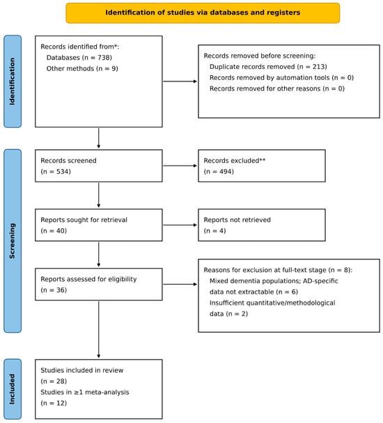
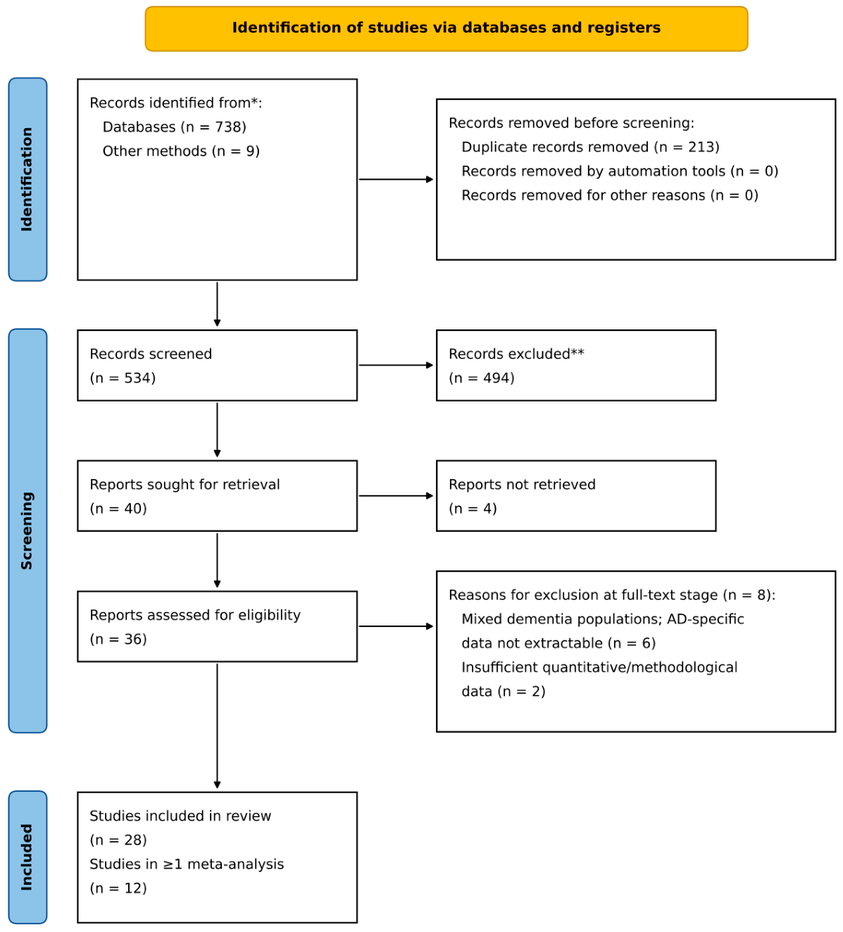
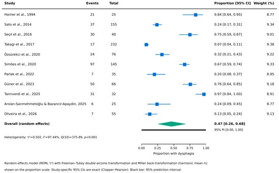
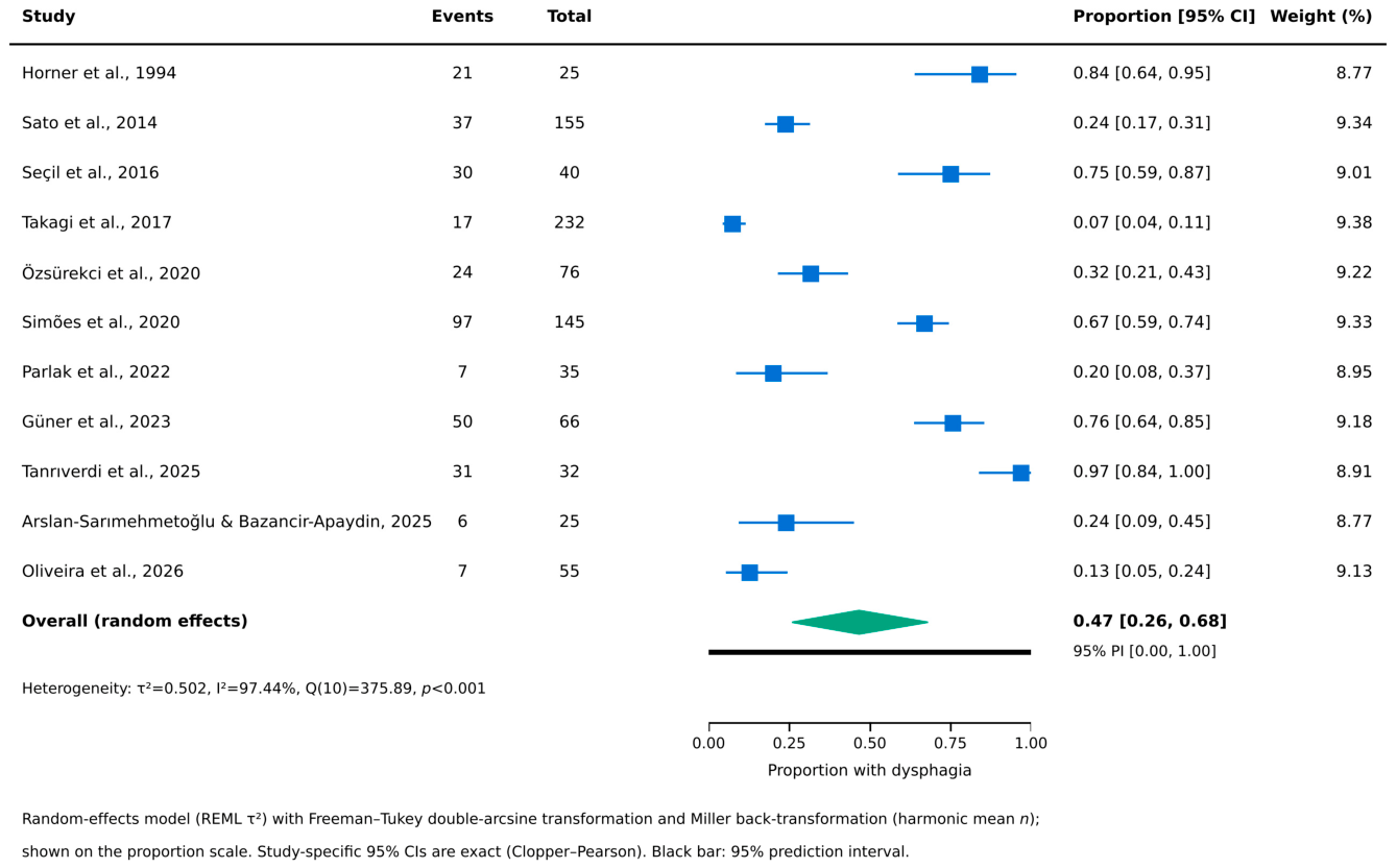
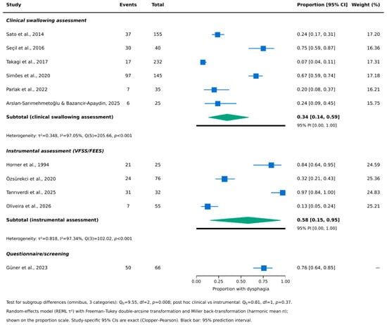
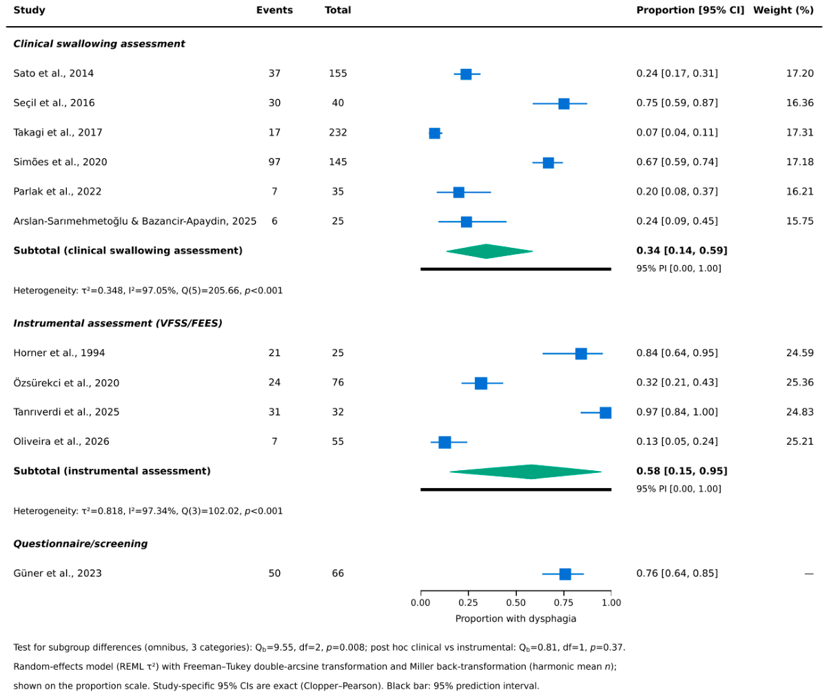
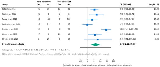
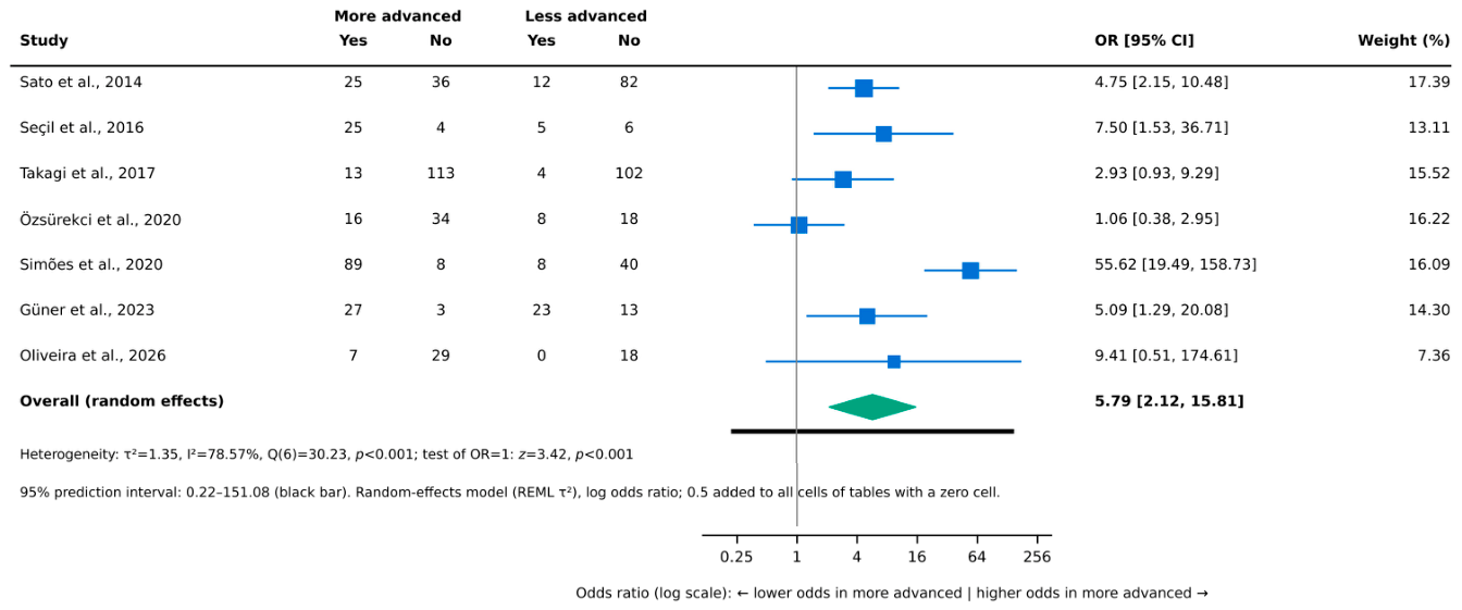
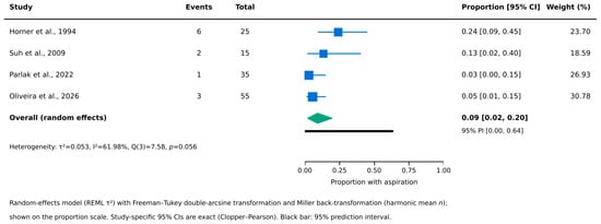
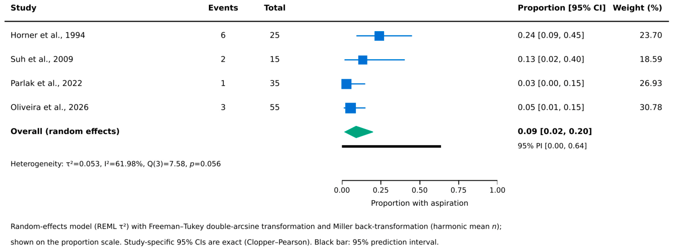
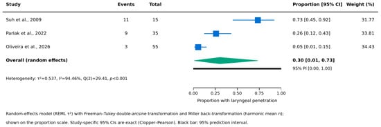
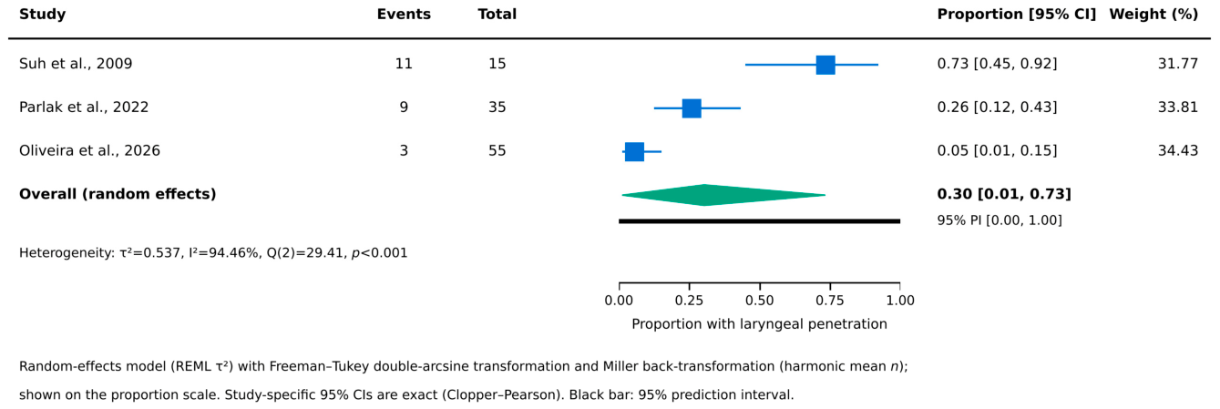
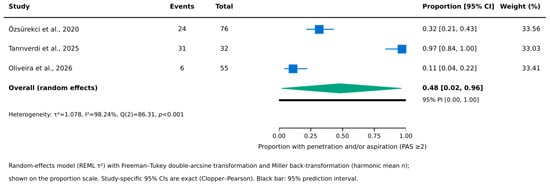
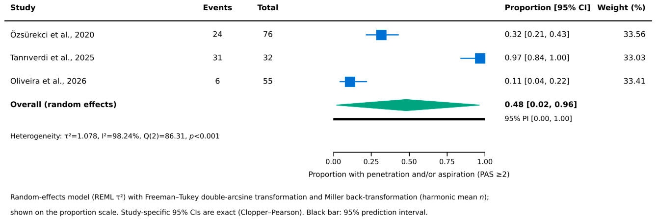

Disclaimer/Publisher’s Note: The statements, opinions and data contained in all publications are solely those of the individual author(s) and contributor(s) and not of MDPI and/or the editor(s). MDPI and/or the editor(s) disclaim responsibility for any injury to people or property resulting from any ideas, methods, instructions or products referred to in the content. |
© 2026 by the authors. Licensee MDPI, Basel, Switzerland. This article is an open access article distributed under the terms and conditions of the Creative Commons Attribution (CC BY) license.