


Highlights
What are the main findings?
- The first three modules of a structured breast ultrasound curriculum were delivered to 55 imaging learners at a Ugandan referral hospital. Self-reported confidence was numerically higher in all eight domains in the February 18–19 subset. Three exploratory Welch comparisons had unadjusted p < 0.05; none met the Bonferroni threshold of 0.00625. Anonymous, potentially overlapping samples preclude valid independent-sample inference.
- Among 29 post-course respondents from February 18–19, 2026, satisfaction ratings were favorable.
What are the implications of the main findings?
- Pilot implementation of a structured breast ultrasound curriculum was feasible within a resource-limited referral hospital.
- This pilot provides a potential framework for breast imaging workforce development using targeted breast ultrasound education.
Abstract
Background: Prompt evaluation of breast symptoms is important for breast cancer care, but breast ultrasound diagnostic imaging capacity remains limited in many low- and middle-income countries, where mammography and breast MRI are often unavailable. Breast ultrasound can support assessment of palpable abnormalities and other symptoms, although its clinical utility depends on trained personnel capable of image acquisition, lesion characterization, and appropriate recommendations. We evaluated the feasibility and early learner response to a structured breast ultrasound curriculum in Uganda as an initial component of workforce development for diagnostic breast imaging services. Methods: We conducted a prospective pre–post educational feasibility study at Mulago National Referral Hospital in Kampala, Uganda. Fifty-five diagnostic radiology residents, radiographers, sonographers, and radiography students participated in a four-hour lecture series covering breast ultrasound fundamentals, breast imaging fundamentals, and BI-RADS lexicon, assessment, and management. Anonymous pre-course and post-course surveys assessed self-reported confidence across breast ultrasound domains using a five-point Likert scale. Mean scores, independent sample comparisons, and Cohen’s d effect sizes were calculated. Results: Of 55 attendees, 38 pre-course and 29 post-course responses were dated 18–19 February 2026. Anonymous surveys could not be linked; exploratory between-group comparisons therefore have important dependence limitations. Three unadjusted Welch comparisons yielded p < 0.05, but none met the Bonferroni threshold of 0.00625. Potential overlap between anonymous respondent groups limits inferential interpretation. Conclusions: Pilot implementation of a structured breast ultrasound curriculum was feasible within a resource-limited referral hospital and was associated with small numerical differences without statistically significant changes in self-reported confidence in clinically relevant interpretation domains including lesion recognition and standardized image interpretation. This pilot provides a potential framework for workforce development using targeted breast ultrasound education as a preliminary step toward evaluating breast imaging workforce training in resource-limited settings.
1. Introduction
Breast cancer is the most common cancer among women worldwide and remains a major cause of cancer death in low- and middle-income countries (LMICs), where delayed presentation and limited access to diagnostic services contribute to poor outcomes [1,2]. Restricted availability of diagnostic imaging, pathology, specialty referral, and trained breast imaging personnel can delay evaluation of symptomatic breast disease [3]. These barriers can affect whether patients with a breast complaint can receive an appropriate diagnostic assessment and progress toward treatment. In settings with limited access to subspecialty breast imaging services, developing the clinical imaging workforce is therefore one component of improving the breast care pathway.
Breast ultrasound is useful in these settings because it is portable, relatively inexpensive, and can be deployed in facilities where mammography or breast MRI is unavailable. Prior studies have shown that hand-held ultrasound can detect breast malignancy with good sensitivity and specificity when used as an independent diagnostic tool [4,5]. In many LMICs, ultrasound serves as the primary imaging modality for evaluating women with breast complaints. Beyond detecting abnormalities, ultrasound can help guide clinical care: ultrasound findings may help characterize a palpable abnormality, identify lesions requiring additional evaluation, and communicate the need for follow-up or tissue diagnosis.
Breast ultrasound depends on both technical image acquisition and accurate lesion interpretation. Unlike mammography, ultrasound requires real-time lesion targeting, optimization of transducer positioning, and immediate assessment of suspicious findings, making diagnostic performance highly operator-dependent. Formal breast ultrasound education remains limited in many LMICs, where providers often rely on general ultrasound experience without dedicated breast imaging instruction [6]. Workforce preparation and consistency of clinical reporting can influence how imaging information is incorporated into patient management.
Structured instruction in scanning techniques and lesion interpretation may help address these gaps. To meet this need, breast imaging fellowship-trained radiologists and diagnostic radiology residents developed a breast ultrasound curriculum using radiologic–pathologic correlation cases, peer-reviewed educational materials, and guidance from the American College of Radiology Breast Imaging Reporting and Data System (BI-RADS) Atlas and relevant professional educational resources [7]. The purpose of this study was to evaluate the feasibility and early educational effect of piloting the first three modules among healthcare imaging learners at Mulago National Referral Hospital in Kampala, Uganda. The pilot assessed training feasibility and learner response, not clinical performance or patient outcomes.
For patients presenting with breast symptoms, imaging is one step in a sequence of clinical decisions. A palpable finding may require targeted ultrasound, diagnostic mammography where available, tissue sampling, and appropriate referral based on the level of suspicion. The ability to describe a finding consistently and recommend appropriate follow-up is particularly important when multiple professionals contribute to the pathway. Teaching a standardized ultrasound approach may help learners communicate findings and make clinical recommendations. Whether this training changes patient care remains to be studied.
Uganda-specific studies highlight the need for accessible diagnostic breast imaging education. A situational analysis of breast cancer early detection and diagnostic capacity identified constraints in trained personnel, imaging and pathology services, and referral pathways [8]. Among patients receiving care at the Uganda Cancer Institute, 41.9% had stage III and 34.7% had IV disease at diagnosis, and the median interval from first consultation to diagnosis was 5.6 months [9]. Earlier breast ultrasound educational work in Uganda demonstrated improvements in interpretation following focused instruction using a condensed BI-RADS framework [10]. Training initiatives in rural South Africa have also evaluated breast ultrasound education for nonradiologist providers, including hands-on teaching and longitudinal mentorship [11]. Together, these studies support the need for locally adapted training while underscoring the importance of evaluating objective skills in subsequent phases.
2. Materials and Methods
2.1. Ethics Review
This study was reviewed by the Vanderbilt University Medical Center Institutional Review Board and determined to meet criteria for exempt educational research. Participation in all surveys was voluntary and anonymous. The lecture series and associated survey collection were approved by the Department of Radiology at Mulago National Referral Hospital in Kampala, Uganda. The educational activities and survey administration were conducted under the oversight of senior radiologists at Mulago, with institutional permission obtained prior to implementation. The initiative was conducted as an educational program evaluation rather than a clinical research intervention; therefore, separate local ethics committee review was not sought.
2.2. Study Design and Setting
We conducted a prospective pre–post educational feasibility study at Mulago National Referral Hospital in Kampala, Uganda. The study was performed as part of an educational initiative focused on improving breast imaging knowledge and diagnostic capacity in a resource-limited clinical setting. The hospital setting provided an opportunity to introduce a shared approach to breast ultrasound education among personnel with different clinical imaging responsibilities. The lecture series and associated pre- and post-course surveys (see File S1) were conducted at Mulago National Referral Hospital in Kampala, Uganda, on 18–19 February 2026. Dr. Rita Nassanga, a senior radiologist at Mulago National Referral Hospital and Director of the Radiology Residency Program at Makerere University College of Health Sciences, reviewed the educational lectures and survey questions prior to implementation to ensure their relevance and appropriateness for the local training environment. The lecture series and survey administration were conducted with departmental approval and under the oversight of senior radiologists at Mulago National Referral Hospital. This was an educational pilot, with no changes to patient-facing services. No changes in patient pathways or clinical outcomes were prospectively measured.
Healthcare learners involved in medical imaging education and clinical ultrasound practice were invited to participate. Participants included diagnostic radiology residents, radiographers, sonographers, and radiography students. A total of 55 learners attended the educational sessions. Participation in the pre-course and post-course surveys was voluntary and anonymous; respondent-level pairing was not performed.
2.3. Educational Curriculum
The first three modules of the structured breast ultrasound curriculum were delivered in English by Dr. Matthew Burger during two in-person teaching sessions totaling four hours at Mulago National Referral Hospital. The educational curriculum was adapted to the local resource setting by emphasizing breast ultrasound as the primary imaging modality, with limited discussion of mammography and breast MRI given their restricted availability. Content focused on practical ultrasound techniques, lesion characterization, and BI-RADS assessment applicable to the local clinical environment. The curriculum was developed by breast imaging fellowship-trained radiologists and diagnostic radiology residents using radiologic–pathologic correlation cases, peer-reviewed educational resources, and ACR breast imaging guidelines. The breast ultrasound course is also available through the online Learning Center of RAD-AID International, a nonprofit organization that works to expand access to medical imaging in underserved communities through education, training, and local partnerships.
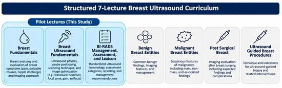
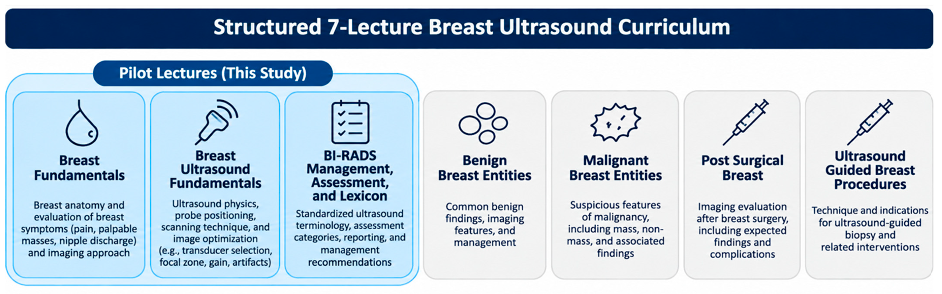
The pilot curriculum addressed three major content areas: breast anatomy, common presenting breast symptoms, and imaging evaluation; foundational breast ultrasound physics and scanning technique; and standardized lesion description, assessment, and management using BI-RADS terminology. Lectures incorporated annotated ultrasound images, case-based examples, and structured teaching slides derived from clinical breast imaging cases and open-access educational materials. We selected content relevant to decisions made during clinical breast assessment: identifying the area of concern, obtaining interpretable images, distinguishing benign from suspicious features, and communicating findings in a standardized way. These skills are used in diagnostic care, but lectures alone cannot establish proficiency. “Breast Fundamentals” discussed breast anatomy and evaluation of breast symptoms, including breast pain, palpable masses, nipple discharge, and imaging approaches based on American College of Radiology Appropriateness Criteria [12]. “Breast Ultrasound Fundamentals” introduced ultrasound physics, probe positioning, scanning technique, and image optimization, including transducer selection, focal zone placement, gain adjustment, and common artifacts. “BI-RADS Management, Assessment, and Lexicon” introduced standardized terminology for ultrasound findings, assessment categories, reporting, and management recommendations. A summary of curriculum content is provided in Figure 1; the three piloted lectures are highlighted in blue.
Figure 1.
Breast Ultrasound Curriculum.
The pilot included only the first three modules of the seven-part curriculum. Subsequent modules address additional clinical entities and procedures but were not delivered or evaluated in this pilot. The scope of the training is important when considering its clinical use: a brief targeted pilot education program can introduce diagnostic concepts, whereas independent clinical performance requires practice, supervision, quality assurance, and appropriate institutional authorization. This pilot was not a certification or competency assessment.
2.4. Survey Assessment
Participants completed surveys immediately before and after the educational intervention to assess perceived breast ultrasound knowledge and self-reported confidence. The pre-course survey collected participant role, prior ultrasound experience, frequency of ultrasound performance, and prior formal ultrasound or breast imaging training. Participants also rated baseline comfort across multiple breast ultrasound domains using a five-point Likert scale. The post-course survey repeated the confidence assessment after completion of the educational modules and included a course evaluation component addressing perceived effectiveness of the curriculum, instructional materials, and likelihood of recommending the course to colleagues. Both surveys also included open-ended questions designed to capture learner expectations, perceived barriers to breast ultrasound training, and post-course feedback. Survey assessment domains are summarized in Table 1. These were self-reported perceptions; no objective knowledge examination was administered.
Table 1.
Survey Assessment Categories.
The pre- and post-course questionnaires were developed by the study team to assess learners’ self-reported confidence in breast ultrasound-related tasks and their perceptions of the educational intervention. The questionnaires were administered in English and reviewed by senior Ugandan radiologist Dr. Rita Nassanga for relevance to the local clinical and educational setting. Formal pilot testing or psychometric validation of the survey instruments was not performed. Each confidence domain was analyzed separately rather than combined into a composite scale; therefore, internal consistency was not assessed using Cronbach’s alpha.
2.5. Qualitative Analysis
Five open-ended survey questions were evaluated using an exploratory descriptive content-analysis approach. The two pre-course questions asked about desired skills and barriers to breast ultrasound training; the three post-course questions addressed perceived learning, valuable course components, and desired future topics. Only responses dated 18–19 February 2026 were included. Responses were counted separately for each question. Answers consisting only of “None” were excluded from thematic categorization, and percentages were calculated using the number of substantive responses to each question as the denominator.
Responses were reviewed and grouped into descriptive categories based on recurring topics identified in participants’ answers. Individual responses could be assigned to multiple categories when more than one topic was mentioned. Theme frequencies were reported as the number and percentage of substantive responses mentioning each category. Selected anonymized quotations were included to illustrate common themes. This was an exploratory descriptive analysis rather than a formally validated thematic analysis, and independent coding or intercoder agreement was not assessed. Dr. Matthew Burger coded the open-ended responses. A second reviewer did not independently verify the coding.
2.6. Statistical Analysis
Likert-scale confidence responses were converted to numerical values from 1 to 5, with higher scores representing greater confidence (1 = very uncomfortable, 5 = very comfortable). Mean values, standard deviations, and 95% confidence intervals were calculated for each educational domain.
Because surveys were anonymous and could not be linked, comparisons were exploratory. Respondent groups may overlap, violating the independence assumption of Welch t-tests and Mann–Whitney U tests. We report descriptive means and sample SDs, Welch two-sided p-values, ordinal Mann–Whitney U sensitivity p-values, Cohen’s d calculated as (post mean − pre mean)/pooled sample SD, where pooled SD = √{[(n_pre − 1)SD_pre2 + (n_post − 1)SD_post2]/(n_pre + n_post − 2)}. Approximate 95% confidence intervals for d use a normal standard-error approximation and should be interpreted cautiously. Eight domains were evaluated using unadjusted exploratory p-values; statistical significance was also assessed against a Bonferroni-adjusted threshold of 0.00625 (0.05/8). Role-stratified descriptive results were calculated for radiology residents. Only 18–19 February 2026 responses were included.
3. Results
3.1. Participant Characteristics
A total of 55 learners attended the Uganda lecture series on 18–19 February 2026. During data cleaning, we found that the post-course survey export included 12 responses from diagnostic radiology residents who attended a separate virtual lecture series at Komfo Anokye Teaching Hospital in Kumasi, Ghana, in November 2025. We then restricted the analysis to surveys dated 18–19 February 2026. Of the 42 post-course responses in the export, we excluded the 12 responses from the Ghana series and one response dated 20 February 2026, leaving 29 post-course responses for analysis (53% of attendees). We retained 38 pre-course responses (69% of attendees). Diagnostic residents represented 26/38 (68%) pre-course respondents and 19/29 (66%) post-course respondents, summarized in Table 2. Because responses were anonymous, individual participants could not be matched.
Table 2.
Participant Professional Roles.
Participants represented a multidisciplinary imaging cohort with a broad range of prior ultrasound experience. Among pre-course respondents, ultrasound experience was <6 months in 14/38, 6–12 months in 6/38, 1–3 years in 16/38, and >3 years in 2/38. Eighteen of 38 reported performing breast ultrasound occasionally (<1/week), 18/38 regularly (1–5/week), and 2/38 never. Prior formal breast ultrasound training was reported by 22/38, informal instruction by 6/38, and none by 10/38.
3.2. Confidence Outcomes
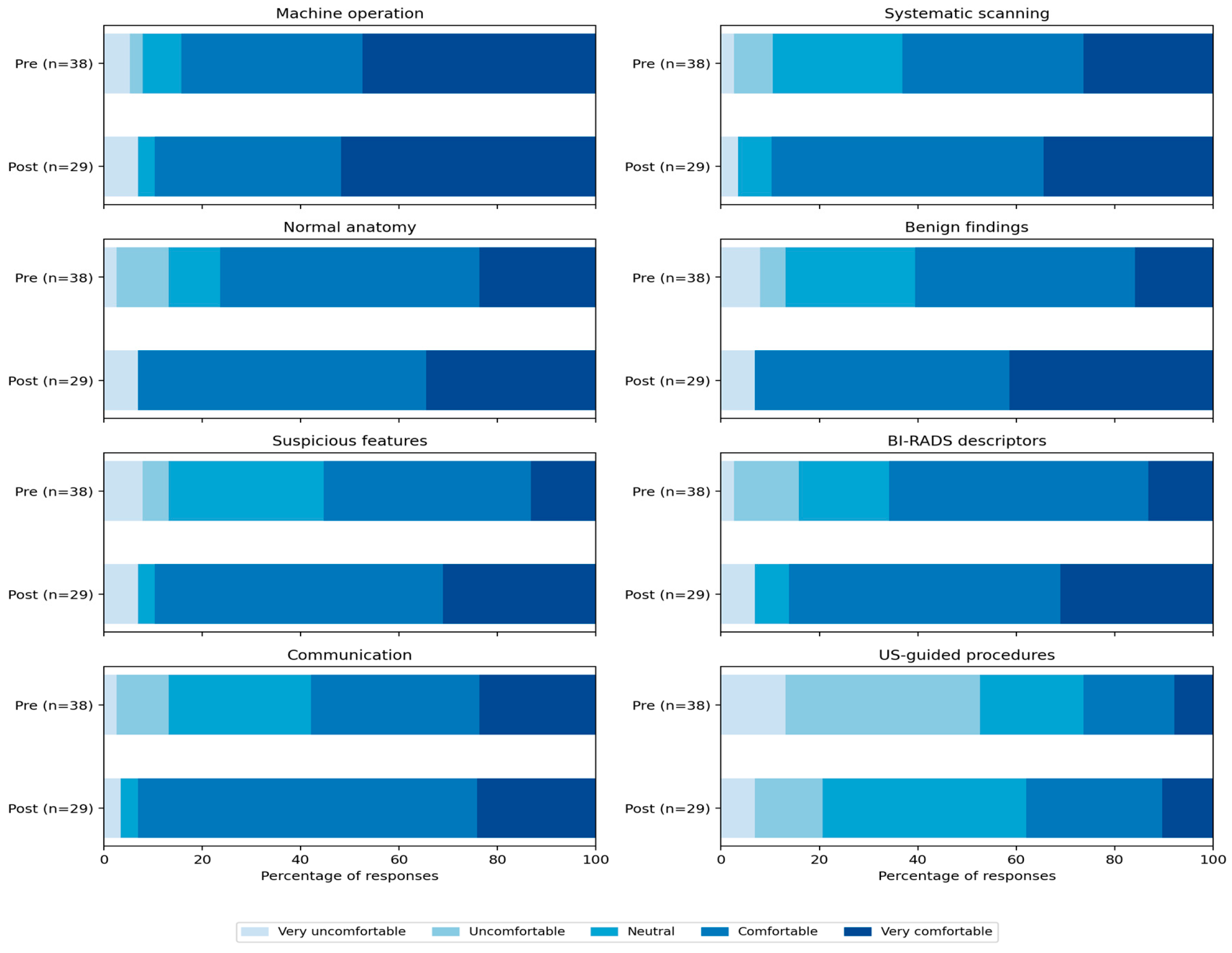
Across eight domains, post-course mean confidence was numerically higher than pre-course mean confidence (pre n = 38; post n = 29). Comparisons are descriptive because respondents could not be paired and the groups may overlap. Table 3 provides item-level n, means, SDs, exploratory effect sizes, and tests and Figure 2 provides a breakdown of the individual self-reported confidence domain by Likert scores. Subgroup descriptive analyses by professional role are reported in Table 4.
Table 3.
Pre- and Post-course Confidence Scores Across Evaluated Ultrasound Educational Domains (n, mean, SD, exploratory effect sizes and tests).
Table 4.
Role-stratified exploratory confidence outcomes. Subgroups are unpaired and potentially overlapping. “Other learners” combines sonographers, imaging technologists, radiographers, and students; this is not a homogeneous professional category. Values are descriptive means on a 1–5 scale; no between-role causal inference is supported.
Figure 2.
Distribution of five-point confidence responses for each domain (pre n = 38; post n = 29). Percentages are descriptive; anonymous respondents could not be linked.
3.3. Course Evaluation and Qualitative Feedback
Course evaluations were favorable among the 29 post-course respondents. All 29 (100%) rated the course as effective or very effective in meeting learning expectations, while 27 (93%) gave favorable ratings for the instructional materials and 27 (93%) for recommending the course to colleagues. The recommendation question used effectiveness rather than likelihood response options, which limits interpretation.
Five open-ended questions assessed learner needs, barriers, and course feedback. Before the course, 33/38 (87%) respondents identified skills they wanted to improve, most commonly scanning techniques (18/33, 55%), lesion identification (13/33, 39%), and BI-RADS or reporting (11/33, 33%). Of the 25 respondents describing barriers, the most common were limited case exposure (8/25, 32%), insufficient practice or training time (7/25, 28%), and limited supervision (4/25, 16%).
All 29 post-course respondents answered questions about perceived learning and valuable course components. Reported changes included greater confidence or knowledge (17/29, 59%), improved lesion recognition (13/29, 45%), and greater familiarity with BI-RADS or reporting (9/29, 31%). The most valued components were BI-RADS instruction (11/29, 38%), lesion recognition (10/29, 34%), and case-based examples (9/29, 31%). Among 28 respondents suggesting future topics, the most common breast imaging related requests were additional interpretation cases (11/28, 39%), breast MRI (6/28, 21%), and ultrasound-guided procedures (5/28, 18%).
Representative comments included “Few cases” and “Lack of knowledge and supervision” when describing training barriers. One participant identified “Putting images to the features of the diagnoses” as particularly valuable, while another requested additional instruction on image-guided biopsy techniques. These findings reflect learners’ perceptions rather than objectively measured improvements.
Learner feedback identified practical gaps that would need attention before the curriculum could be incorporated into clinical practice. Limited case exposure can restrict opportunities to recognize the range of benign and suspicious findings. Inadequate supervision can make it difficult to confirm whether a scanning or interpretation approach is correct. Requests for hands-on and procedural teaching further suggest that learners saw a need for practical training beyond the initial lecture series. These concerns came from learner feedback and were not independently assessed during this pilot study.
4. Discussion
4.1. Principal Findings and Comparison with Previous Studies
We found that a brief structured breast ultrasound curriculum could be delivered in a resource-limited academic hospital and was associated with modest changes in learner confidence across most evaluated domains. The largest numerical gains occurred in lesion recognition and BI-RADS-based interpretation, but no domain reached statistical significance, specifically no Welch comparison met the threshold for significance. The findings support feasibility and learner acceptance but do not establish improved clinical competency. This makes an educational intervention potentially useful as one component of a broader healthcare workforce development strategy.
In the date-restricted subset, exploratory unadjusted Welch comparisons suggested differences in benign finding recognition, suspicious feature recognition, and communication of findings (p = 0.014, 0.022, and 0.049). None met the Bonferroni-adjusted threshold, and the pre- and post-course respondents could overlap. These findings suggest that the curriculum was feasible and well received, although the observed differences in confidence should be interpreted cautiously.
This pilot should be considered in the context of prior regional training initiatives. Scheel et al. evaluated a condensed BI-RADS educational intervention among Ugandan healthcare workers using case-based interpretation assessments [10]. Dickerson et al. assessed a longer breast ultrasound training program in rural South Africa [11]. In Uganda, a cross-sectional study of diagnostic intervals has also emphasized the importance of connecting ultrasound assessment with timely diagnostic follow-up [9]. In contrast, the present study evaluated the initial delivery and learner response to the first three modules of a seven-part curriculum among a multidisciplinary group at Mulago National Referral Hospital.
4.2. Clinical and Educational Implications
Breast ultrasound is a primary diagnostic imaging tool for symptomatic breast evaluation in many LMICs, where access to mammography, MRI, subspecialty breast radiologists, and pathology services remains limited. In these settings, the quality of ultrasound assessment depends on operator technique, recognition of suspicious imaging features, and the ability to communicate appropriate next steps. Inconsistent descriptions or uncertainty about assessment categories can complicate clinical handoffs and decisions about further evaluation. Standardized instruction may help establish a common vocabulary among radiologists, radiographers, and sonographers, but the present study did not evaluate reporting quality, referral appropriateness, or timeliness of diagnosis.
The multidisciplinary nature of the curriculum is particularly relevant in settings where imaging responsibilities are shared across professional roles. However, the educational needs of these groups may differ. Radiology residents may benefit from more advanced case-based interpretation and reporting exercises, while sonographers and radiographers may require additional supervised scanning and image acquisition training.
The curriculum was also adapted to the resources available at Mulago National Referral Hospital, emphasizing ultrasound with limited discussion of mammography and MRI. Ultrasound access is only one part of the diagnostic pathway. An ultrasound examination may identify a suspicious abnormality, but timely care also requires access to image-guided sampling or another appropriate biopsy method, pathology interpretation, communication of results, and finally clinical treatment [2]. This approach may be useful in other resource-limited settings, although educational recommendations must reflect locally available imaging, biopsy, and referral services.
4.3. Study Limitations
This single-site pilot had several limitations. The sample sizes were modest (pre n = 38; post n = 29), and anonymous responses could not be linked between surveys. Because the respondent groups may overlap, the independence assumptions of the Welch t-tests and Mann–Whitney U tests were not satisfied. The statistical comparisons should therefore be considered exploratory. Eight domains were evaluated, and none of the Welch p-values met the Bonferroni-adjusted threshold of 0.00625.
Baseline confidence was moderate to high across most domains (3.47–4.18), creating potential ceiling effects. Approximately two-thirds of respondents were radiology residents, and some introductory material may have reinforced concepts they were already familiar with. Although resident representation was similar between surveys (68% pre-course versus 66% post-course), the small subgroup sizes and heterogeneous composition of other professional roles limited meaningful comparisons.
Confidence in ultrasound-guided procedures also increased despite this topic not being covered in the three piloted modules. This suggests that some of the observed differences may reflect nonspecific factors, including response shift, social desirability, a general increase in confidence following the lectures, or differences in respondent composition (d = 0.47).
Finally, the study assessed self-reported confidence rather than objective knowledge acquisition, interpretation accuracy, scanning competency, changes in clinical practice, or patient outcomes. Greater confidence does not necessarily mean greater accuracy [13]. A learner may feel more comfortable assigning BI-RADS descriptors without demonstrating improved sensitivity, specificity, or agreement with an expert reader. The study also did not evaluate long-term knowledge retention or the sustainability of the educational intervention.
4.4. Future Directions
Future evaluations should incorporate anonymous self-generated linking codes to permit paired pre- and post-course comparisons while preserving participant anonymity. Objective assessments, including standardized case interpretation exercises and supervised scanning evaluations, would provide a more direct measure of educational effectiveness. Role-specific assessments could also help determine whether different professional groups benefit from different components of the curriculum. Subsequent curricula may need role-specific objectives, including acquisition competencies for personnel who perform examinations and interpretation or reporting competencies for clinicians responsible for clinical decisions.
A longer-term educational approach could combine short didactic modules with supervised scanning, curated interpretation cases, regular case conferences, and feedback from experienced breast imagers. Local faculty involvement in curriculum delivery and ongoing mentorship would be important for developing a sustainable training model rather than relying on individual educational visits. Implementation would need to account for staff availability, equipment access, and local referral pathways.
Future evaluation could proceed in stages, beginning with training feasibility and then assessing clinical performance. The first stage would assess participation, acceptability, and practical delivery of the curriculum. A second stage would measure objective interpretation performance and supervised acquisition competency. A later clinical stage could evaluate reporting consistency, referral completion, and diagnostic intervals, with attention to the availability of biopsy and pathology services. This approach would help determine whether improvements in individual skills translate into measurable changes in healthcare processes, while avoiding the assumption that education by itself is sufficient to improve breast cancer outcomes.
Future research could progress from educational feasibility to objective clinical performance and ultimately to diagnostic service outcomes. Subsequent studies could evaluate reporting consistency, referral completion, diagnostic intervals, and access to biopsy and pathology services. Future clinical evaluations should consider unintended consequences. Additional detection of indeterminate findings can increase demand for repeat imaging, biopsy, and specialist consultation which may strain a resource limited healthcare system. If downstream capacity is overloaded, this may introduce new delays even when initial ultrasound access improves. A future service evaluation should consider the balance between identifying suspicious findings, avoiding unnecessary referrals, and ensuring that patients with concerning results can complete the recommended workup. Learner confidence surveys cannot answer these questions; future studies will need to examine them directly.
5. Conclusions
Pilot implementation of the first three modules of a structured breast ultrasound curriculum at Mulago National Referral Hospital was feasible and associated with small, non-significant numerically higher post-course ratings in self-reported confidence across most assessed domains. The intervention addresses skills relevant to breast diagnostic service delivery, but its effect on clinical competency and patient outcomes will need to continue to be studied.
Supplementary Materials
The following supporting information can be downloaded at: https://www.mdpi.com/article/10.3390/healthcare14223994/s1, File S1: Introduction to Breast Ultrasound Pre-Course Survey and Introduction to Breast Ultrasound Post-Course Survey.
Author Contributions
Conceptualization, M.B., A.J., Z.A., J.R.S. and E.P.; Methodology, M.B., A.J., Z.A., J.R.S. and E.P.; Formal Analysis, M.B.; Writing—Original Draft Preparation, M.B., A.J. and Z.A.; Writing—Review & Editing, M.B., A.J., Z.A., A.M., J.R.S. and E.P.; Supervision, A.M., J.R.S. and E.P. All authors have read and agreed to the published version of the manuscript.
Funding
This research received no external funding.
Institutional Review Board Statement
The study was conducted in accordance with the Declaration of Helsinki. Ethical review and approval were waived by the Vanderbilt University Medical Center Institutional Review Board because the project met criteria for exempt, minimal-risk educational research involving anonymous survey responses (IRB 251499).
Informed Consent Statement
Participation was voluntary, and completion of the anonymous pre-course or post-course survey was considered implied informed consent. No personally identifiable information was collected.
Data Availability Statement
The original data presented in the study are openly available in FigShare at DOI: 10.6084/m9.figshare.35040922.
Acknowledgments
Genevieve Abbey, Mann Dai, Arjun Patel, Megan Carrillo, and Daniel Malek for their contributions to the development of the educational lecture materials used in this study. These individuals were not directly involved in the implementation or evaluation of the project. Rita Nassanga, Radiology Residency Program at Makerere University College of Health Sciences and Mulago National Referral Hospital, for her assistance with local coordination in Uganda, review of the educational materials and survey instruments, and support in facilitating the lecture series. RAD-AID International for supporting the breast ultrasound course by making it available through the RAD-AID Learning Center.
Conflicts of Interest
The authors declare no conflicts of interest.
References
- Chanakira, E.Z.; Thomas, C.V.; Balen, J.; Mandrik, O. A systematic review of public health interventions to address breast cancer inequalities in low- and middle-income countries. Syst. Rev. 2024, 13, 195. [Google Scholar] [CrossRef] [Scilit] [PubMed] [PubMed Central]
- World Health Organization. Global Breast Cancer Initiative Implementation Framework: Assessing, Strengthening and Scaling Up Services for the Early Detection and Management of Breast Cancer; World Health Organization: Geneva, Switzerland, 2023; Available online: https://www.who.int/publications/i/item/9789240065987 (accessed on 7 July 2026).
- Anderson, B.O.; Ilbawi, A.M.; El Saghir, N.S. Breast cancer in low- and middle-income countries (LMICs): A shifting tide in global health. Breast J. 2015, 21, 111–118. [Google Scholar] [CrossRef] [Scilit] [PubMed]
- Sood, R.; Rositch, A.F.; Shakoor, D.; Ambinder, E.; Pool, K.L.; Pollack, E.; Mollura, D.J.; Mullen, L.A.; Harvey, S.C. Ultrasound for breast cancer detection globally: A systematic review and meta-analysis. J. Glob. Oncol. 2019, 5, 1–17. [Google Scholar] [CrossRef] [Scilit] [PubMed]
- Iacob, R.; Iacob, E.R.; Stoicescu, E.R.; Ghenciu, D.M.; Cocolea, D.M.; Constantinescu, A.; Ghenciu, L.A.; Manolescu, D.L. Evaluating the role of breast ultrasound in early detection of breast cancer in low- and middle-income countries: A comprehensive narrative review. Bioengineering 2024, 11, 262. [Google Scholar] [CrossRef] [Scilit] [PubMed]
- Ginsburg, A.S.; Liddy, Z.; Khazaneh, P.T.; May, S.; Pervaiz, F. A survey of barriers and facilitators to ultrasound use in low- and middle-income countries. Sci. Rep. 2023, 13, 3322. [Google Scholar] [CrossRef] [Scilit] [PubMed]
- Leung, J.W.T.; Baker, J.A.; Hooley, R.; Loving, V.A.; Rapelyea, J.A. Ultrasound. In ACR BI-RADS® v2025 Manual; American College of Radiology: Reston, VA, USA, 2025. [Google Scholar]
- Scheel, J.R.; Giglou, M.J.; Segel, S.; Orem, J.; Tsu, V.; Galukande, M.; Okello, J.; Nakigudde, G.; Mugisha, N.; Muyinda, Z.; et al. Breast cancer early detection and diagnostic capacity in Uganda. Cancer 2020, 126, 2469–2480. [Google Scholar] [CrossRef] [Scilit] [PubMed] [PubMed Central]
- Achan, J.; Kasujja, F.X.; Opito, R.; Wabinga, H.; Orach, C.G.; Mwaka, A.D. Factors associated with diagnostic and pre-treatment intervals among breast cancer patients attending care at the Uganda Cancer Institute: A cross-sectional study. Cancer Med. 2023, 12, 19701–19713. [Google Scholar] [CrossRef] [Scilit] [PubMed] [PubMed Central]
- Scheel, J.R.; Peacock, S.; Orem, J.; Bugeza, S.; Muyinda, Z.; Porter, P.L.; Wood, W.C.; Comis, R.L.; Lehman, C.D. Improving Breast Ultrasound Interpretation in Uganda Using a Condensed Breast Imaging Reporting and Data System. Acad. Radiol. 2016, 23, 1271–1277. [Google Scholar] [CrossRef] [Scilit] [PubMed]
- Dickerson, L.K.; Rositch, A.F.; Lucas, S.; Harvey, S.C. Pilot Educational Intervention and Feasibility Assessment of Breast Ultrasound in Rural South Africa. J. Glob. Oncol. 2017, 3, 502–508. [Google Scholar] [CrossRef] [Scilit] [PubMed]
- American College of Radiology. ACR Appropriateness Criteria®: Palpable Breast Masses. Revised 2022; American College of Radiology: Reston, VA, USA, 2022; Available online: https://acsearch.acr.org/docs/69495/Narrative (accessed on 7 July 2026).
- Davis, D.A.; Mazmanian, P.E.; Fordis, M.; Van Harrison, R.; Thorpe, K.E.; Perrier, L. Accuracy of physician self-assessment compared with observed measures of competence: A systematic review. JAMA 2006, 296, 1094–1102. [Google Scholar] [CrossRef] [Scilit] [PubMed]

Disclaimer/Publisher’s Note: The statements, opinions and data contained in all publications are solely those of the individual author(s) and contributor(s) and not of MDPI and/or the editor(s). MDPI and/or the editor(s) disclaim responsibility for any injury to people or property resulting from any ideas, methods, instructions or products referred to in the content. |
© 2026 by the authors. Licensee MDPI, Basel, Switzerland. This article is an open access article distributed under the terms and conditions of the Creative Commons Attribution (CC BY) license.