


Abstract
Background: Implant restoration of single narrow edentulous sites still represents a challenging clinical scenario. This study aimed to evaluate the clinical and radiographic outcomes of category 2 narrow-diameter implants (NDIs) used to restore single missing maxillary lateral incisors. Methods: Patients with a single-unit implant restoration were enrolled in this prospective, single-arm observational study as part of a maintenance programme. The primary outcome was marginal bone loss (MBL), while secondary outcomes included the prevalence of prosthetic and biological complications and esthetic outcomes (pink and white esthetic score, PES/WES). Seventeen patients received 19 category 2 NDIs in the upper lateral incisor position and were followed for 24 months after prosthesis delivery. Results: Mean MBL at 24 months was 0.31 ± 0.20 mm (patient-clustered 95% CI, 0.22 to 0.41). No implant showed more than 1.0 mm of bone loss. Peri-implant mucositis occurred in two implants in two patients (implant level 10.5%, patient level 11.8%). No peri-implantitis or implant failures were recorded, and overall implant survival was 100%. Mean PES/WES was 16.5 ± 2.7. In the exploratory multivariable analysis, higher scores were associated with a thick preoperative mucosal phenotype and greater mesio-distal width. Conclusions: Category 2 NDIs demonstrated a high survival rate for both implants and prosthetic restorations. Long-term outcomes and the stability of implant rehabilitations with category 2 NDIs require further investigation. Larger comparative cohorts should examine long-term biologic and prosthetic complications.
1. Introduction
The implant-prosthetic rehabilitation of patients with congenital absence or loss of maxillary lateral incisors represents a clinical challenge and requires a multidisciplinary approach [1]. The aim is to achieve optimal implant placement in sites where bone volumes are often limited, provide a prosthetic restoration that harmonizes with the adjacent teeth and the contralateral lateral incisor, and establish an emergence profile that ensures optimal aesthetics and predictable long-term outcomes [2]. In recent years, scientific advances have contributed to improving implant placement procedures through the development of computer-guided implant surgery. The use of surgical guides allows for more accurate implant positioning. This is particularly relevant in the rehabilitation of maxillary lateral incisors, especially considering the proximity and inclination of adjacent tooth roots, which may restrict the available interradicular space and increase the risk of root damage, bone fenestrations, and dehiscences [3].
The scientific literature agrees that an inter-implant distance of at least 3 mm and a tooth-to-implant distance of at least 1.5 mm must be observed to preserve hard and soft tissues [4]. In addition, it has been recommended that a minimum buccal bone thickness of 1.5 mm should be preserved to reduce the risk of bone loss as well as an adequate volume of keratinized mucosa has been suggested to avoid soft tissue recession [5,6]. However, the placement of standard-diameter implants (>3.5 mm) is not always feasible due to the limited mesiodistal space available in the maxillary lateral incisor region. To overcome this limitation, the use of narrow-diameter implants has been proposed and described in the literature as a valid treatment option for sites with limited mesiodistal space [2,7,8]. Beyond their anatomical advantages, narrow-diameter implants may offer additional clinical benefits in selected cases, including a reduced need for bone augmentation procedures and the possibility of adopting a less invasive surgical approach. Furthermore, they may provide a fixed implant-supported rehabilitation option in situations where the limited mesiodistal space would otherwise favour alternative prosthetic solutions, such as resin-bonded fixed dental prostheses [9].
According to the literature NDIs can be classified as follows:
- -
- Category 1: Implants with a diameter < 2.5 mm (‘mini-implants’), described primarily for severely resorbed edentulous ridges and for single teeth in the anterior region.
- -
- Category 2: Implants with a diameter of 2.5 mm to <3.3 mm, for the restoration of a single tooth in the anterior region (mainly to replace maxillary or mandibular lateral incisors).
- -
- Category 3: Implants with a diameter of 3.3 mm to 3.5 mm, described for all regions, including maxillary or mandibular single posterior teeth [10].
In 2022, Roccuzzo et al., in a 1-year prospective, controlled clinical study, investigated the clinical reliability of narrow-diameter implants in the rehabilitation of patients with congenitally missing maxillary lateral incisors. The study included two groups: a test group comprising 50 implants with a diameter of 2.9 mm and a control group comprising 50 implants with a diameter of 3.3 mm. The authors evaluated implant survival, changes in marginal bone levels, aesthetic outcomes, biological and technical complications, and patient-reported outcomes (PROMs). The overall implant survival rate was 99%, with no statistically significant differences between the two groups. Both implant diameters showed favorable outcomes in terms of marginal bone stability, aesthetics, complications, and patient satisfaction after one year of follow-up [2].
The decision to rehabilitate a maxillary lateral incisor site using a narrow-diameter implant should take into account not only the anatomical conditions of the recipient site but also biomechanical considerations. Indeed, implant diameter influences the distribution of stresses in the peri-implant bone, particularly at the cortical level [11]. In this regard, Qiu et al. (2024), in a systematic review of finite element analysis studies, highlighted that implant diameter is a key factor influencing bone stress distribution and preventing excessive stress concentrations at the crestal bone level [12]. Therefore, the choice of implant diameter should not depend solely on the available space but should also consider the biomechanical conditions under which the implant will function [12]. Despite their clinical advantages, the reduced diameter of NDIs may represent a biomechanical limitation, as the smaller cross-sectional area can reduce implant resistance to functional loading and increase mechanical stress at the implant–abutment connection. Potential technical complications include abutment, screw or implant fracture and screw loosening, although their occurrence may vary according to implant design, prosthetic configuration, implant location, and occlusal loading. Biological complications, such as marginal bone loss and peri-implant soft tissue recession, should also be considered when evaluating the long-term performance of these implants [10,13].
Several studies in the literature have compared the clinical outcomes of narrow-diameter implants and regular-diameter implants used for the rehabilitation of the anterior region. Among these, the 2021 systematic review and meta-analysis by Cruz et al. included 484 narrow-diameter implants and 359 regular-diameter implants. The results showed no statistically significant differences between the two groups in terms of implant survival, marginal bone loss, or biological and mechanical complications. However, the authors highlighted the need for further investigations to strengthen the current evidence [14]. Despite the encouraging findings reported in the literature, further prospective clinical evidence is needed to better characterize the clinical and radiographic performance of category 2 narrow-diameter implants. In particular, data focusing exclusively on 2.9-mm implants used for the rehabilitation of single missing maxillary lateral incisors remain limited. Therefore, the present prospective study aimed to evaluate the clinical and radiographic outcomes of 2.9-mm category 2 narrow-diameter implants in this specific clinical indication, with a 2-year follow-up.
2. Materials and Methods
This prospective, single-arm observational study was performed in compliance with the revised Declaration of Helsinki [15]. The study was approved by the Lazio Area 3 Territorial Ethics Committee, under protocol no. 0001121/24, dated 27 June 2024. The study design and reporting adhered to the STROBE (Strengthening the Reporting of Observational Studies in Epidemiology) recommendations for observational research [16]. The study was registered on ClinicalTrials.gov under the following registration number: NCT06528587. All enrolled patients provided written informed consent prior to their inclusion in the study.
Patients rehabilitated with a single-unit implant restoration were prospectively followed from prosthesis delivery for the maintenance programme at the Department of Prosthodontics of the Catholic University of the Sacred Heart of Rome. Patients were enrolled between July 2024 and October 2024. Definitive prostheses were delivered between July 2024 and October 2024, and the 24-month follow-up evaluations started from prosthesis delivery between July 2026 and October 2026.
Inclusion criteria were as follows:
- Patients receiving an implant-supported rehabilitation of a single maxillary lateral incisor;
- ASA I and ASA II patients;
- Age > 18 years;
- Non-smokers or smokers of ≤5 cigarettes/day;
- Achievement of FMPS and FMBS ≤ 15%;
- Patients who had declined a single-retainer all-ceramic resin-bonded prosthesis;
- Written informed consent to participate in the study was obtained.
Exclusion criteria were as follows:
- Severe general medical conditions;
- Immunodeficiency states;
- Radiotherapy in the head and neck region;
- Uncontrolled diabetes or hypertension;
- Smokers of >5 cigarettes/day;
- Pregnancy or breastfeeding;
- Inability to perform consistent and continuous follow-up;
- Inability to provide written informed consent.
2.1. Clinical Procedures
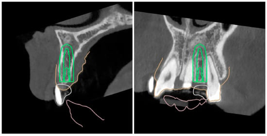
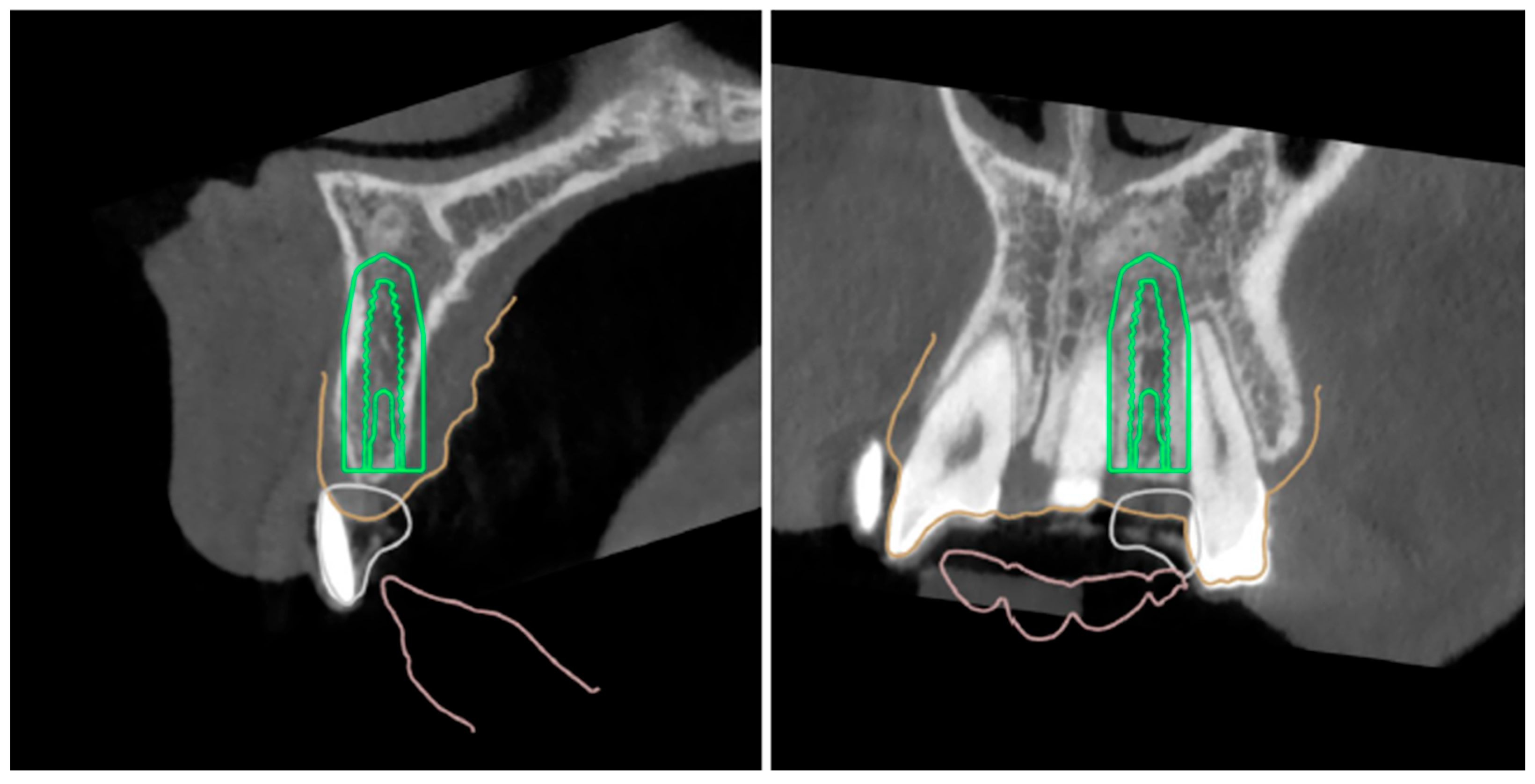
An experienced clinician performed all surgeries (P.D.A.) using standardized procedures and recorded intraoperative measurements while the prosthetic rehabilitation was performed by the same two trained operators (P.F.M, E.R.). Implants were placed following a guided surgery protocol; thus, an intraoral digital impression was taken, matched with a CBCT exam, and following a digital evaluation of the case a 3D-printed guide was made for the static computer-guided surgery (Figure 1).
Figure 1.
Digital planning of the implant placement.
The guide was tried before starting the surgery, as it was imperative to have a clear and precise fit.
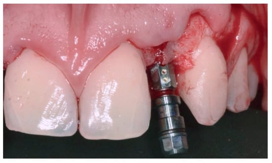
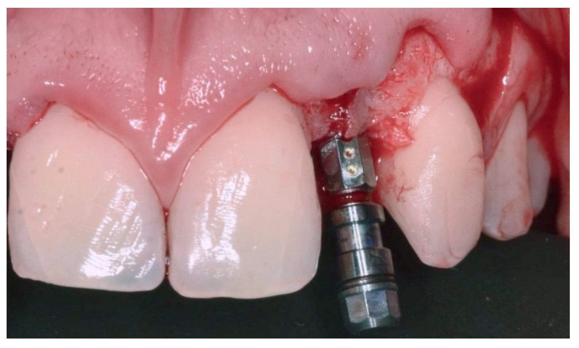
Before surgery, patients were given antibiotic treatment (two doses of 1 g amoxicillin-clavulanate). The skin around the mouth was disinfected using sterile gauze attached to a Klemmer clamp and dipped in povidone–iodine solution. The patients were then covered with TNT cloths and the mucous membranes were cleaned using gauze soaked in 0.2% chlorhexidine solution. The procedure was conducted under local anaesthesia (using 4% articaine with epinephrine at a ratio of 1:100,000). A horizontal incision was made in the centre of the ridge in the edentulous area, starting intrasulcular on the distal tooth and again intrasulcular on the mesial tooth (the distal tooth was the canine, and the mesial tooth was the central incisor). Release incisions were made only when required. A full-thickness flap was elevated, exposing the bone. Following the manufacturer’s instructions, bone-level implants between 10 and 14 mm in length and 2.9 mm (Bone level tapered implant, Straumann®) in diameter were carefully placed (Figure 2 and Figure 3).
Figure 2.
Horizontal incision.
Figure 3.
Implant placement.
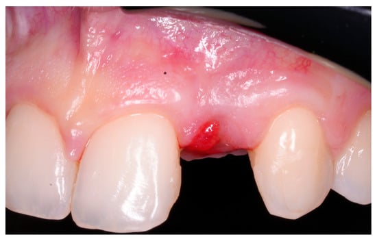
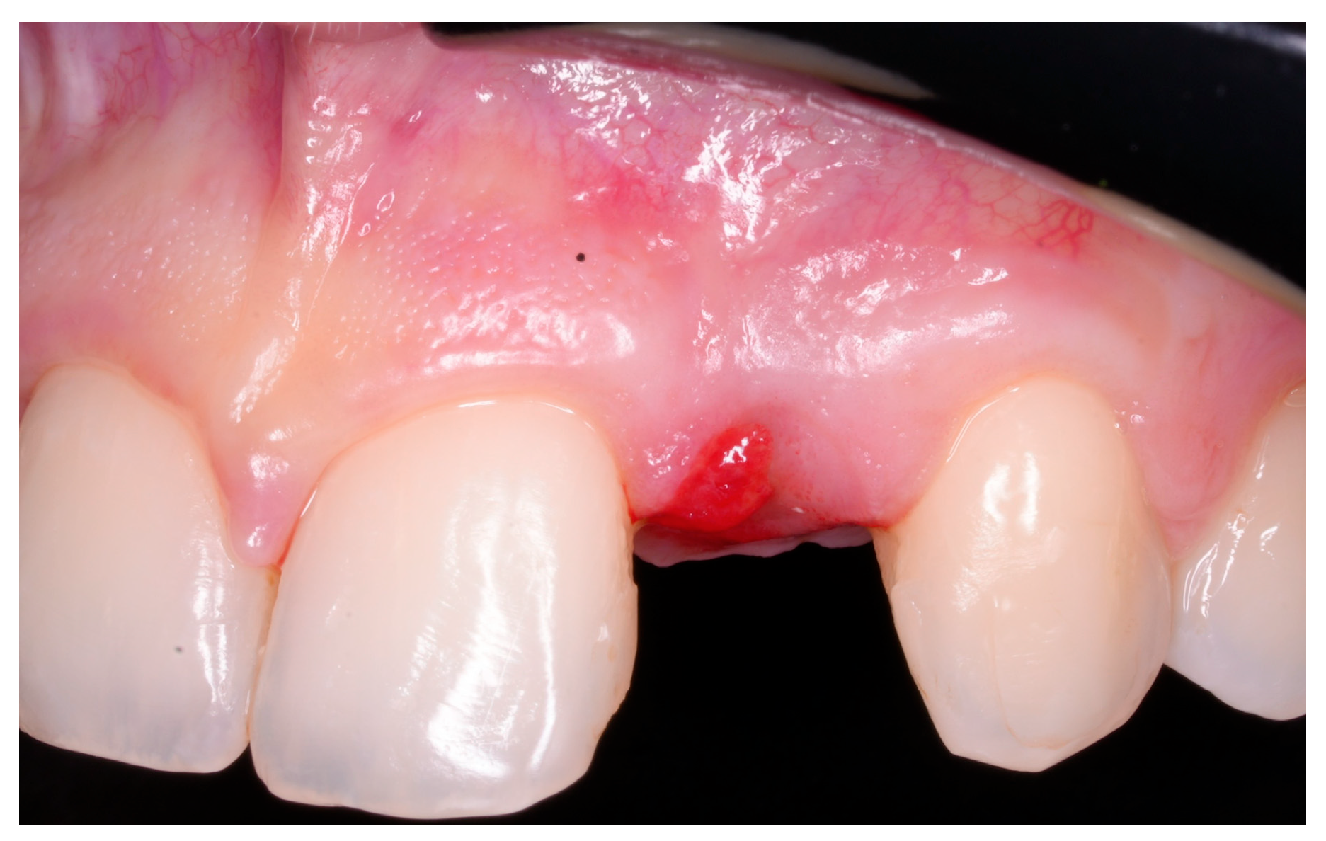
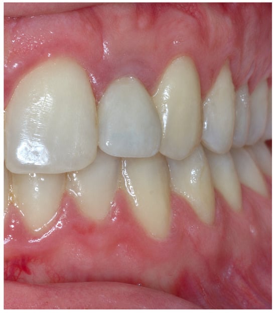
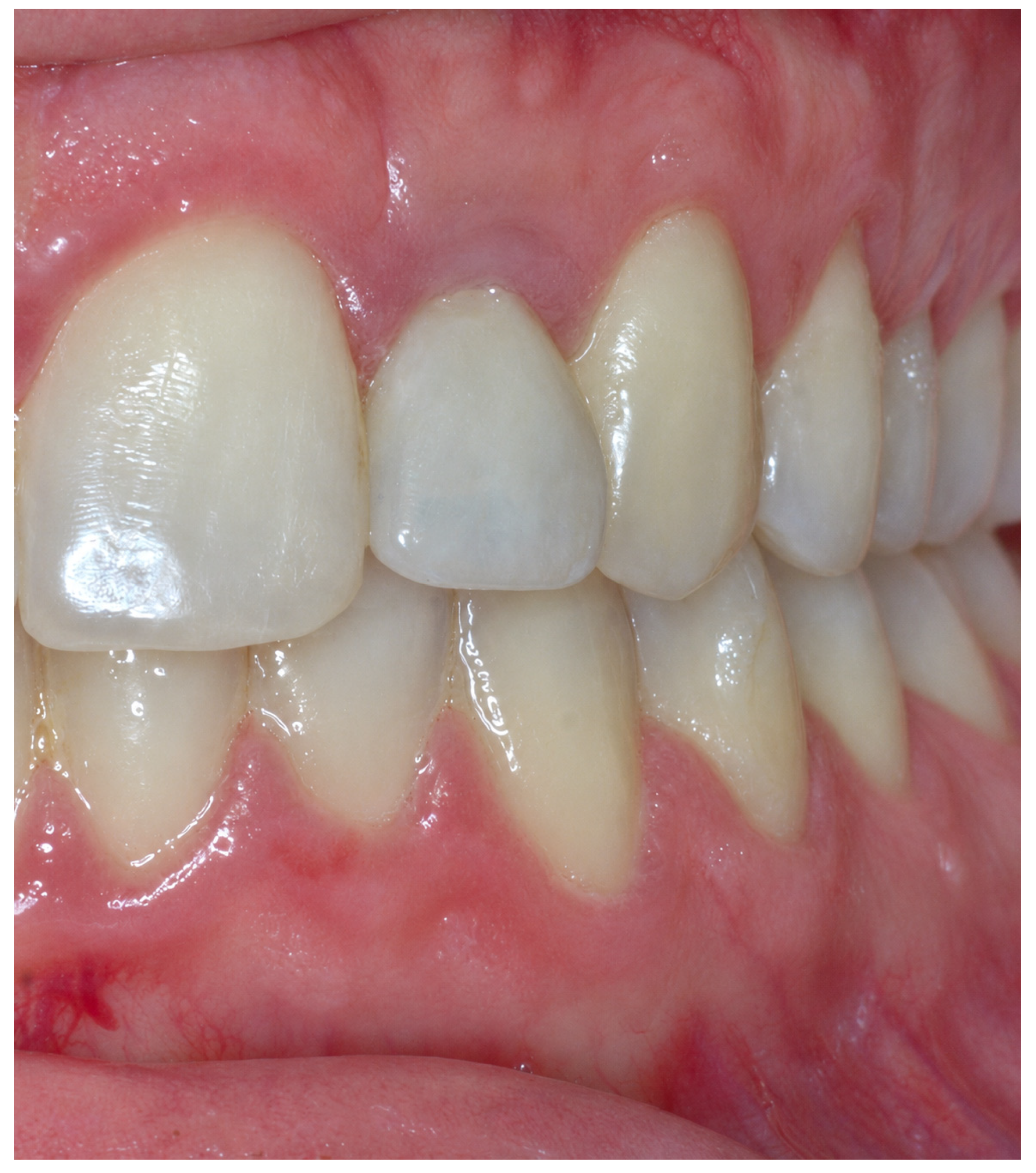
Primary stability was assessed at implant placement by measuring insertion torque and performing a manual examination. Insertion torque ranged from 15 to 40 N·cm, and at least 1 mm of bone thickness was preserved circumferentially around each implant. The crestal incision was closed with single sutures. Patients were advised to rinse their mouths twice daily with 0.2% chlorhexidine mouthwash for seven days. In addition, analgesics (ketoprofen 80 mg) were prescribed for the next three days, depending on individual needs. Sutures were removed seven days after surgery and all patients were included in a maintenance treatment programme. The second-stage surgery was carried out between three and six months following implant placement. Once soft tissue healing had been achieved, a digital workflow incorporating an intraoral scan was employed to fabricate a provisional restoration via 3D printing. This provisional served two purposes: shaping the peri-implant soft tissues and allowing functional and esthetic assessment before the definitive restoration was delivered. The definitive restoration consisted of a monolithic or buccally veneered zirconia crown. At definitive crown delivery, occlusal contacts were assessed in maximum intercuspation using 8-µm shimstock foil. The foil could be withdrawn freely during light closure. Fifteen restorations were screw-retained and four were cement-retained (Figure 4). All the patients received a nightguard and were enrolled in a supportive implant therapy based on each patient’s risk profile.
Figure 4.
Two-year follow-up of the implant-supported restoration.
2.2. Data Collection
All clinical and radiographic outcomes were evaluated by an independent and calibrated examiner from the prosthesis delivery. Biological complications were categorized as either mucositis or peri-implantitis based on the 2017 World Workshop classification of peri-implant diseases and conditions [17], which considers probing pocket depth, bleeding on probing, suppuration, and peri-implant marginal bone loss as diagnostic parameters. The thickness of the soft tissues was measured using an endodontic K-file, piercing the vestibular mucosa on the line which connects the mucosal zenith of the two adjacent teeth, thereby allowing the definition of a thin (marginal mucosal thickness < 1 mm) and a thick (marginal mucosal thickness ≥ 1 mm) phenotype. Marginal bone loss around implants was assessed radiographically through standardized periapical radiographs obtained using the parallel technique. A patient-specific registration index was used to reproduce the projection and each radiograph was calibrated using the length of the implant as an internal reference. MBL was calculated as the difference between the implant shoulder-to-first bone-to-implant contact distance measured at prosthesis delivery and at the 24-month follow-up. The highest value between the mesial and distal measurements was used for analysis. The baseline radiograph was taken at prosthesis delivery and the follow-up radiograph two years after delivery. Intra-examiner reliability of the radiographic measurements was assessed using an absolute-agreement intraclass correlation coefficient (ICC). Implant and prosthesis survival were both defined by the continued presence in situ of the respective implant or prosthesis two years after the prosthesis delivery. Implant and prosthesis success were defined as absence of any type of complications of the respective implant or prosthesis two years after prosthesis delivery. The implant health was assessed via peri-implant clinical examination performed with a UNC-15 periodontal probe, by which probing pocket depth (PPD) and bleeding on probing (BOP) were assessed at six sites per implant 2 years after the prosthesis delivery [18].
Technical complications were analyzed at the 24-month follow-up and classified into two categories: minor and major. Minor complications encompassed those that could be resolved directly at chairside, without need for laboratory intervention. Major complications, by contrast, were those necessitating additional laboratory work and/or component replacement.
Patient-related outcomes were evaluated after prosthesis delivery using a questionnaire in which patient satisfaction in relation to the treatment received was evaluated: the degree of satisfaction was recorded using a visual analog scale (VAS) (0–10), where 0 was considered “not at all satisfied” and 10 was “totally satisfied”.
Esthetic outcomes were assessed at the 24-month follow-up using the pink and white esthetic score (PES/WES; total score 0–20, with PES and WES each ranging from 0 to 10) [19]. The mesio-distal width of the edentulous space was measured using the CBCT scan.
The peri-implant soft tissues were diagnosed using the classification of peri-implant soft tissue dehiscence/deficiency (PSTD) by Zucchelli et al. in terms of soft tissue levels [20]. The classification of PSTD was formulated in four classes based on the location of the mucosal margin and 3 subcategories based on the features of the peri-implant papilla.
2.3. Sample Size Calculation
Based on the literature regarding the use of narrow-diameter implants in the anterior sectors, using marginal bone loss as the primary endpoint, hypothesizing a marginal bone loss value of 0.6 mm for narrow-diameter implants, compared to a marginal bone loss of 1.3 ± 0.7 for standard-diameter implants, with a type 1 error of 0.05 and a power of 95%, 16 subjects were estimated to be sufficient under the stated assumptions [21]. The present study included a single cohort. The initial calculation therefore does not establish the precision of this estimate.
2.4. Statistical Analysis
The sample was described in its demographic, clinical, and instrumental variables through descriptive statistical techniques. Qualitative variables were expressed as absolute and relative percent frequency, with exact 95% confidence intervals (CIs) for the main proportions (implant survival, peri-implant mucositis, complications and success), while quantitative variables were described through mean and standard deviation (SD) with 95% CI, median, and interquartile range (IQR). Normality was assessed with the Shapiro–Wilk test. The implant was used as the unit of analysis. Two patients contributed bilateral implants. For mean MBL and the coefficients of the multivariable regression models, we calculated cluster-robust standard errors using the patient as the clustering unit. We applied a finite-sample correction and used a t distribution with 16 degrees of freedom to calculate 95% confidence intervals and two-sided p-values for the regression coefficients. The effect of the mucosal phenotype (thin vs. thick) and of connective tissue grafting (CTG vs. no CTG) on MBL and PES/WES was assessed with Welch’s t-test, which does not assume equal variances, reporting mean differences with 95% CI. Correlations between the mesio-distal width of the edentulous space, MBL, and PES/WES were assessed with Spearman’s rank correlation coefficient (ρ), with 95% CI. The adjusted association of mesio-distal width, phenotype, and CTG with PES/WES and MBL was explored with multivariable linear regression. Influential observations were identified with Cook’s distance, and a sensitivity analysis excluding the most influential site was performed. In a separate sensitivity analysis, we retained one implant per patient and repeated the analyses for all four possible combinations of sites from the two bilateral patients. Statistical significance was set for two-sided p-values <0.05. All analyses were conducted with Python, version 3.13 (SciPy 1.18 and statsmodels 0.15).
3. Results
A total of 17 patients with a mean age of 37.1 ± 16.9, who ranged from 25 to 74, were enrolled in this study and received a total of 19 implants. Four were males and 13 were females. All the patients completed the follow-up (Table 1).
Table 1.
Baseline features and main outcomes.
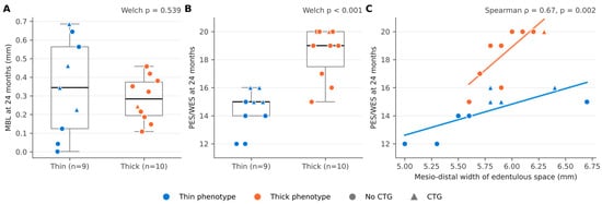
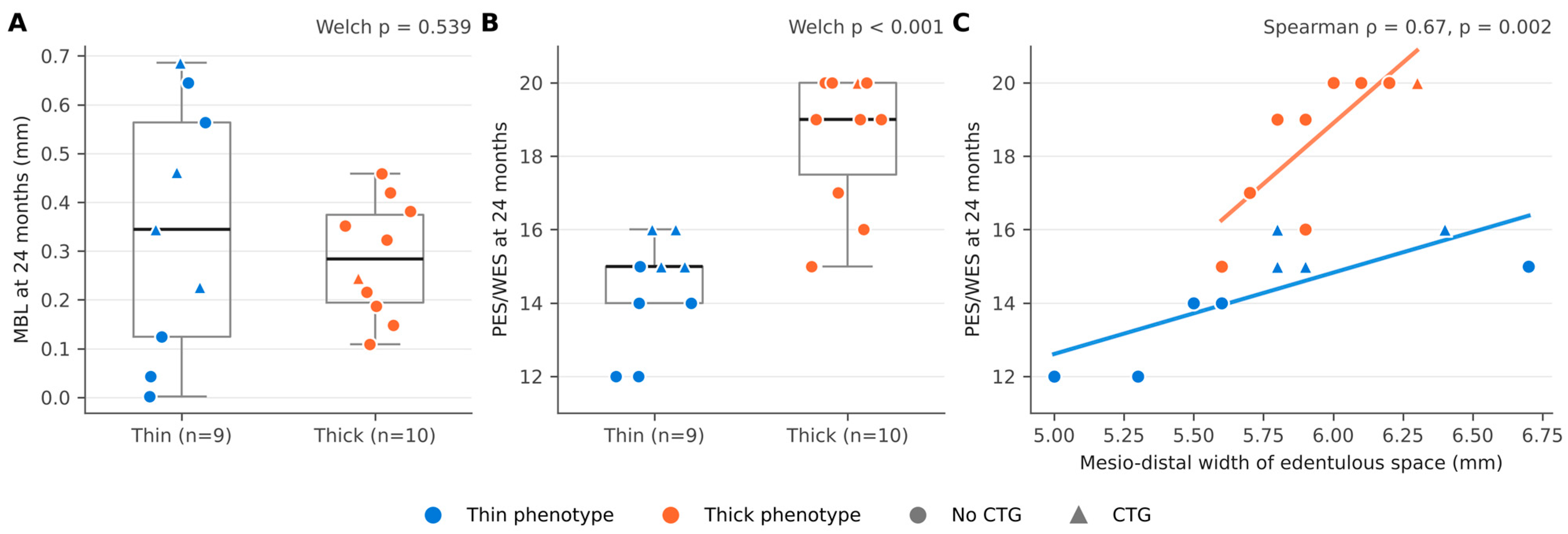
All the implants were category 2 NDIs and were placed in the upper lateral incisor position. Two patients received bilateral restorations due to bilateral agenesis. Twelve patients were restored due to agenesis, two patients were restored due to destructive carious lesions, and one patient was restored due to fracture, while one had a root resorption and one was treated for periodontal disease. Fourteen patients received the NDI due to insufficient mesio-distal space (≤6.3 mm) while three patients received it because of insufficient bone volume and unwillingness to undergo adjunct regenerative procedures. The mean mesio-distal width of the edentulous space was 5.86 ± 0.39 mm (median 5.90 mm, IQR 5.65–6.05; range 5.0–6.7). Nine sites (47.4%) were preoperatively classified with a thin phenotype while ten (52.6%) were classified with a thick phenotype; the mesio-distal width did not differ between phenotypes (p = 0.407). Four patients (five sites, 26.3%) received an additional soft tissue grafting procedure to correct the buccal volumetric deficiency using a subepithelial connective tissue graft harvested from the palate during the second surgery. Since CTG was indicated in sites presenting a buccal volumetric deficiency, it was performed mainly in the thin phenotype sites and atrophic sites (thin-phenotype sites 44.4% versus thick-phenotype sites 10.0%). No implant failures were recorded with an overall implant survival rate of 100% (19/19; 95% CI 82.4–100%). Peri-implant mucositis was observed in two implants on two patients. The rate of peri-implant mucositis at implant level was 10.5% (95% CI 1.3–33.1%) while at patient level was 11.8% (95% CI 1.5–36.4%). Major biological complications such as peri-implantitis were not observed (0/19; 95% CI 0–17.6%). The mean PPD was 4.16 ± 1.01 mm. At the two-year follow-up, the mean marginal bone loss was 0.31 ± 0.20 mm (patient-clustered 95% CI 0.22–0.41; median 0.32 mm, IQR 0.17–0.44; range 0.00–0.69), and data were normally distributed (Shapiro–Wilk test, p = 0.798). No implant showed a marginal bone loss > 1.0 mm. The mean MBL in the Thin group was 0.34 ± 0.26 mm (median 0.34, IQR 0.12–0.56) while in the Thick group it was 0.28 ± 0.12 mm (median 0.28, IQR 0.19–0.37). The difference of 0.06 mm (95% CI −0.15 to 0.27) was not statistically significant (p = 0.539). At the 24-month follow-up, the mean full-mouth plaque score (FMPS) and full-mouth bleeding score (FMBS) were 16% and 13%, respectively. Implant success, defined as the absence of any biological complication, was 89.5% (17/19; 95% CI 66.9–98.7%), the two complications being the cases of peri-implant mucositis.
Despite the guided approach, fifteen sites (78.9%) received screw-retained restorations, whereas four (21.1%) received cement-retained restorations. Five implants (26.3%) received a zirconia monolithic restoration, whereas 14 implants (73.7%) received a buccally veneered zirconia restoration based on the adjacent teeth features.
Minor technical complications consisted of a titanium base decementation on a patient with an increased vertical dimension (1/19 implants, 5.3%; 95% CI 0.1–26.0%). At 24 months no cases of chipping were recorded. No major complications were recorded. All prostheses remained in function throughout the observation period, resulting in a prosthesis survival rate of 100% (19/19; 95% CI 82.4–100%). Since the only technical complication (titanium base decementation) was resolved chairside, prosthesis success was 94.7% (18/19; 95% CI 74.0–99.9%). Overall, 16 of 19 implant-supported restorations (considering both implants and prostheses) (84.2%; 95% CI 60.4–96.6%) were free of any biological or technical complication at 24 months.
The peri-implant soft tissues were classified in terms of soft tissue levels as class one in 13 sites (68.4%) and class two in 6 sites (31.6%) and subclass a in all the cases, according to Zucchelli et al. [20]. Patient satisfaction was 8.8 ± 1.01. At 24 months, the mean PES was 8.0 ± 1.6 (range, 5–10), and the mean WES was 8.5 ± 1.2 (range, 6–10). The mean combined PES/WES was 16.5 ± 2.7 (range, 12–20). Exploratory analyses were conducted. Sites with a preoperative thick phenotype showed a significantly higher PES/WES than sites with a preoperative thin phenotype (p < 0.001) (Figure 5B). Grafted sites had a buccal volume deficiency, and most had been classified preoperatively as having a thin mucosal phenotype. At 24 months, their mean esthetic score was similar to that of non-grafted sites (16.4 ± 2.1 vs. 16.6 ± 3.0; difference −0.2 points, 95% CI −2.9 to 2.5; p = 0.891). Descriptively, the observed PES/WES difference between grafted thin-phenotype sites and thick-phenotype sites was smaller than that between non-grafted thin-phenotype sites and thick-phenotype sites; a causal reduction attributable to CTG cannot be established (Figure 5B). The mesio-distal width of the edentulous space showed a positive correlation with PES/WES (Spearman’s ρ = 0.67, 95% CI 0.23–0.94; p = 0.002), which was consistent within both the thin (ρ = 0.82, p = 0.006) and the thick phenotype (ρ = 0.88, p < 0.001) (Figure 5C). Conversely, no significant correlation was found between the mesio-distal width and the marginal bone loss two years after the prosthesis delivery (ρ = 0.01, 95% CI −0.54 to 0.53; p = 0.980), nor between MBL and PES/WES (ρ = 0.01; p = 0.957). In the exploratory multivariable model, each additional millimetre of mesio-distal width was associated with a 2.46-point higher PES/WES (patient-clustered 95% CI, 0.69 to 4.22). The adjusted difference between thick and thin preoperative phenotypes was 4.14 points (95% CI, 2.71 to 5.56), and the adjusted difference between grafted and non-grafted sites was 1.07 points (95% CI, −0.08 to 2.22) (Table 2).
Figure 5.
Marginal bone loss (A) and PES/WES (B) at 24 months according to mucosal phenotype, and relationship between the mesio-distal width of the edentulous space and PES/WES (C). Boxes show median and IQR; whiskers show range. Triangles indicate sites treated with a connective tissue graft (CTG); lines in (C) are least-squares fits within each phenotype.
Table 2.
Multivariable linear regression models for PES/WES and marginal bone loss (MBL) at 24 months (n = 19 implants). B, unstandardized regression coefficient. Confidence intervals and two-sided p-values were calculated using patient-clustered standard errors with a finite-sample correction and 16 degrees of freedom. CTG, connective tissue graft.
After exclusion of the most influential site (mesio-distal width, 6.7 mm; Cook’s distance, 1.75), the adjusted association between mesio-distal width and PES/WES remained positive. The estimated difference in PES/WES per additional millimetre of width increased from 2.46 to 4.38 points. The corresponding estimate for thick versus thin preoperative phenotype was 2.96 points. The adjusted difference between grafted and non-grafted sites changed from +1.07 to −0.19 points. Across the four analyses retaining one implant per patient, mean MBL ranged from 0.304 to 0.317 mm. In the PES/WES model, the coefficients ranged from 2.42 to 2.45 points per millimetre for mesio-distal width, from 3.99 to 4.12 points for thick versus thin preoperative phenotype, and from 0.90 to 1.23 points for CTG versus no CTG.
4. Discussion
The primary aim of this prospective study was to assess peri-implant marginal bone loss (MBL) around 2.9-mm category 2 narrow-diameter implants used for the replacement of maxillary lateral incisors. In recent years, many studies have shown that the use of NDIs yields predictable clinical outcomes [7,22].
More recently, attention has shifted towards the long-term biological response and stability of narrow-diameter implants. Available evidence suggests that NDIs used to replace single anterior teeth in sites with limited mesio-distal space can achieve biological outcomes comparable to those of standard-diameter implants, including similar marginal bone loss and medium-term success rates [22]. NDIs may also be considered a therapeutic alternative in cases of horizontal bone deficiency, provided that the manufacturer’s indications, the clinical conditions, and the planned prosthetic rehabilitation are carefully considered. Schiegnitz et al. reported similar clinical outcomes for 3.3-mm implants placed in pristine bone and 4.1-mm implants placed in conjunction with horizontal bone augmentation, after a mean follow-up exceeding three years [23]. However, the retrospective design limits conclusions regarding comparative effectiveness. Furthermore, these findings should not be directly extrapolated to category 2 NDIs, including 2.9-mm implants, for which comparative evidence remains limited and applicability must be assessed against the specific clinical indications established by the manufacturer [23].
Implant survival rate tends to be very high, ranging between 95.65 and 100% with rare failures related to conditions like peri-implantitis or mechanical problems [24,25,26,27]. From an esthetic point of view, evaluated with PES/WES (Pink/White Esthetic Score), the results were not inferior to those of standard implants [27]. The study by Pieri et al. [22] reported soft tissue stability with a slow but progressive increase in papillary volume and an average esthetic score of 14.1/18.
In the present cohort, mean MBL after two years of function was 0.31 ± 0.20 mm, with no implant exceeding 1.0 mm, a value well below the 0.6 mm hypothesised a priori and consistent with the short-term bone stability reported for 2.9 mm bone-level implants [25,27]. No statistically significant association of MBL with mucosal phenotype or mesio-distal width was detected. Although thin sites showed a more heterogeneous bone response, as reflected by their greater variance, the mean difference was small (0.06 mm) and its confidence interval was wide (−0.15 to 0.27 mm); thus, the present data cannot establish equivalence between phenotypes or identify the principal determinants of bone response.
In the exploratory multivariable model, higher PES/WES values were associated with a thick preoperative mucosal phenotype and greater mesio-distal width. Thick-phenotype sites scored on average 4 points higher than thin sites. A thicker peri-implant mucosa is known to mask the greyish shine-through of titanium components, to support a more stable mucosal margin and to provide the soft tissue volume required to shape the emergence profile [5,27,28]. The effect of the mesio-distal width can be interpreted geometrically: with a 2.9 mm implant ideally centred in the edentulous space, the tooth-to-implant distance on each side ranges from approximately 1.05 mm at 5.0 mm to 1.9 mm at 6.7 mm, and the 1.5 mm threshold commonly recommended to preserve the interproximal bone and papilla [6] is reached only when the space is ≈5.9 mm, which corresponds to the median of our sample. Below this value, the reduced interproximal room limits both papilla support and the development of a harmonious emergence profile [27,29,30,31]. The estimated association was 2.46 PES/WES points per additional millimetre of mesio-distal width.
The PES/WES model included three predictors and its estimates were sensitive to individual observations. In particular, the adjusted difference between grafted and non-grafted sites changed from +1.07 to −0.19 points after exclusion of the most influential site. All five grafted sites had a buccal volume deficiency, and four had a thin preoperative mucosal phenotype. This confounding by indication limits interpretation of the subgroup comparison. The findings are exploratory and do not establish whether CTG improved esthetic outcomes [29]. Finally, the main benefit of soft tissue augmentation is expected to emerge as long-term stability of the mucosal margin [29], which a 24-month follow-up cannot fully capture. Clinically, category 2 narrow-diameter implants may present some prosthetic and biological challenges. Owing to the reduced implant and prosthetic dimensions, platform switching may be absent or present only to a limited extent, potentially affecting the development of the implant supracrestal complex and the long-term stability of peri-implant hard and soft tissues [32]. Moreover, reduced mesio-distal space may further limit the emergence profile design, making maintenance procedures and esthetic outcomes more challenging [27,28,29]. As mentioned earlier, these implants are often used in young patients where early placement, associated with continued maxillary growth and potential infraocclusion over time, poses challenges for clinicians [33]. Similarly, given their reduced diameter, they could theoretically be associated with a higher risk of fracture. However, from a biomechanical perspective, implant fracture was not observed in the study sample, possibly as a consequence of the high mechanical strength associated with the alloy these implants are made of (Roxolid®, Straumann®) which offers excellent structural strength.
The in-vitro study by Alberti et al. used finite element analysis (FEM) to compare the mechanical behaviour of 2.9 mm and 3.3 mm morse-taper implants [34]. The results showed that, although the 3.3 mm implant had slightly higher load and fatigue limits, the difference was considered clinically negligible. The conical connection effectively distributes stress, reducing it to the implant collar and increasing fracture resistance. These laboratory data support the clinical reliability of even the narrowest diameters, providing that a stable implant-prosthetic connection is present.
The connection of NDIs used in this sample is a friction-fit with mechanical locking. It features a conical-cylindrical 15° Small CrossFit® connection (Straumann) with four internal grooves, providing long-term stability under loading conditions with a reduced risk of screw loosening.
In certain clinical scenarios, especially in the esthetic area and in the younger patients, there are minimally invasive alternatives to single implants with reduced patient morbidity such as the resin-bonded fixed dental prostheses with a single retainer. Evidence supports single-retainer zirconia resin-bonded fixed dental prostheses as a treatment option for replacing missing anterior teeth [35]. Unlike implant failure, the most frequent problems—detachment or chipping—are easily treated and do not cause the loss of soft or hard tissues. Thus, a single-retainer all-ceramic resin-bonded fixed dental prosthesis (RBFDP) may be considered a minimally invasive alternative fixed dental prostheses for the replacement of single incisors in young patients with limited mesio-distal space or atrophic site in order to guarantee tissue preservation and optimal esthetics. However, comparative evidence remains insufficient to establish the superiority of either treatment, and treatment selection should follow a careful multidisciplinary assessment. Treatment costs, the number and invasiveness of procedures, the number of visits required, and patient age may influence this decision. Bawa et al. compared tooth-supported resin-bonded fixed dental prostheses with implant-supported single crowns and reported lower complication and failure rates in the implant-supported group [36]. However, the non-randomized design, preferential use of RBFDPs in narrower spaces, and heterogeneity of the restorations limit causal interpretation and the generalizability of these findings.
From a clinical standpoint, these exploratory data suggest that limited mesio-distal space and a thin mucosal phenotype may be associated with a less favourable esthetic scenario, without establishing a validated clinical threshold. In such cases, orthodontic space management, soft tissue augmentation with a connective tissue graft or a single-retainer RBFDP should be carefully considered.
Among the strengths of the study is the sample homogeneity, as the rehabilitation of the upper lateral incisors acts as a selection criterion. The study has some limitations that may affect internal and external validity. The follow-up did not allow for a full assessment of long-term complications of the implant restoration. Moreover, an additional limitation is the small sample size and the lack of a control group due to the specific clinical scenario selected. Two patients contributed bilateral implants. We accounted for within-patient clustering when estimating confidence intervals for mean MBL and the regression coefficients. With only 17 patients, these intervals should be interpreted cautiously. The secondary analyses were exploratory, and no adjustment was made for multiple comparisons. Finally, the mucosal phenotype was assessed only preoperatively and esthetics only at 24 months, so the effect of grafting on final mucosal thickness and long-term esthetic stability could not be evaluated. These limitations underscore the need for further studies with different experimental designs, larger samples, and more extensive follow-up in order to validate and expand the current available evidence base.
5. Conclusions
Category 2 narrow-diameter implants demonstrated a high survival rate for both implants and prosthetic restorations, with evidence of biological and esthetic integration in the absence of major technical complications. In exploratory analyses, higher PES/WES values were associated with a thick preoperative mucosal phenotype and greater mesio-distal width. No clear associations with MBL were identified. However, given the limited available evidence and the short follow-up, these findings should be interpreted with caution, and further long-term studies are needed to confirm their clinical reliability.
Author Contributions
P.D.A.: Conceptualization; Investigation; Methodology; Writing—Original Draft; Writing—Review & Editing. D.P.: Conceptualization; Investigation; Methodology; Writing—Original Draft; Writing—Review & Editing. E.R.: Formal Analysis; Methodology; Validation; Visualization. G.D.R.: Methodology; Writing—Review & Editing. A.S.: Methodology; Data Curation; Visualization; Writing—Review & Editing. G.G.: Formal Analysis; Writing—Review & Editing. L.R.: Supervision; Methodology; Resources; Writing—Review & Editing. P.F.M.: Supervision; Methodology; Resources; Writing—Review & Editing. All authors have read and agreed to the published version of the manuscript.
Funding
This research received no external funding.
Institutional Review Board Statement
Ethical approval was obtained from the institutional review board of the Lazio Area 3 Territorial Ethics Committee of the Catholic University of the Sacred Heart of Rome (N. 0001121/24), approval date 21 June 2024.
Informed Consent Statement
Informed consent was obtained from all subjects involved in the study.
Data Availability Statement
Data are available upon reasonable request due to privacy. The authors used OpenAI (GPT-6) in order to perform grammar checking. The authors reviewed and edited the content as needed and take full responsibility for the content of the published article.
Conflicts of Interest
The authors declare no conflicts of interest.
References
- Kavadia, S.; Papadiochou, S.; Papadiochos, I.; Zafiriadis, L. Agenesis of Maxillary Lateral Incisors: A Global Overview of the Clinical Problem. Orthodontics (Chic.) 2011, 12, 296–317. [Google Scholar] [PubMed]
- Roccuzzo, A.; Imber, J.C.; Lempert, J.; Hosseini, M.; Jensen, S.S. Narrow Diameter Implants to Replace Congenital Missing Maxillary Lateral Incisors: A 1-Year Prospective, Controlled, Clinical Study. Clin. Oral Implants Res. 2022, 33, 844–857. [Google Scholar] [CrossRef] [Scilit] [PubMed]
- Khaohoen, A.; Powcharoen, W.; Sornsuwan, T.; Chaijareenont, P.; Rungsiyakull, C.; Rungsiyakull, P. Accuracy of Implant Placement with Computer-Aided Static, Dynamic, and Robot-Assisted Surgery: A Systematic Review and Meta-Analysis of Clinical Trials. BMC Oral Health 2024, 24, 359. [Google Scholar] [CrossRef] [Scilit] [PubMed]
- Tarnow, D.P.; Cho, S.C.; Wallace, S.S. The Effect of Inter-Implant Distance on the Height of Inter-Implant Bone Crest. J. Periodontol. 2000, 71, 546–549. [Google Scholar] [CrossRef] [Scilit] [PubMed]
- Herrera-Pérez, P.; García-De-La-Fuente, A.M.; Andia-Larrea, E.; Marichalar-Mendia, X.; Aguirre-Urizar, J.M.; Aguirre-Zorzano, L.A. Clinical Analysis of the Tooth-Implant Papilla for Two Narrow-Diameter Titanium-Zirconium Implants in the Anterior Area: Prospective Controlled Clinical Study. BMC Oral Health 2024, 24, 310. [Google Scholar] [CrossRef] [Scilit] [PubMed]
- De Angelis, P.; Manicone, P.F.; Rella, E.; Liguori, M.G.; De Angelis, S.; Tancredi, S.; D’aDdona, A. The Effect of Soft Tissue Augmentation on the Clinical and Radiographical Outcomes Following Immediate Implant Placement and Provisionalization: A Systematic Review and Meta-Analysis. Int. J. Implant Dent. 2021, 7, 86. [Google Scholar] [CrossRef] [Scilit] [PubMed]
- González-Valls, G.; Roca-Millan, E.; Céspedes-Sánchez, J.M.; González-Navarro, B.; Torrejon-Moya, A.; López-López, J. Narrow Diameter Dental Implants as an Alternative Treatment for Atrophic Alveolar Ridges: Systematic Review and Meta-Analysis. Materials 2021, 14, 3234. [Google Scholar] [CrossRef] [Scilit] [PubMed]
- Davarpanah, M.; Martinez, H.; Tecucianu, J.F.; Celletti, R.; Lazzara, R. Small-Diameter Implants: Indications and Contraindications. J. Esthet. Dent. 2000, 12, 186–194. [Google Scholar] [CrossRef] [Scilit] [PubMed]
- Kern, M. Single-Retainer Resin-Bonded Fixed Dental Prostheses as an Alternative to Orthodontic Space Closure (and to Single-Tooth Implants). Quintessence Int. 2018, 49, 789–798. [Google Scholar] [CrossRef] [Scilit] [PubMed]
- Schiegnitz, E.; Al-Nawas, B. Narrow-Diameter Implants: A Systematic Review and Meta-Analysis. Clin. Oral Implants Res. 2018, 29, 21–40. [Google Scholar] [CrossRef] [Scilit] [PubMed]
- Chou, H.Y.; Müftü, S.; Bozkaya, D. Combined Effects of Implant Insertion Depth and Alveolar Bone Quality on Periimplant Bone Strain Induced by a Wide-Diameter, Short Implant and a Narrow-Diameter, Long Implant. J. Prosthet. Dent. 2010, 104, 293–300. [Google Scholar] [CrossRef] [Scilit] [PubMed]
- Qiu, P.; Cao, R.; Li, Z.; Fan, Z. A Comprehensive Biomechanical Evaluation of Length and Diameter of Dental Implants Using Finite Element Analyses: A Systematic Review. Heliyon 2024, 10, e26876. [Google Scholar] [CrossRef] [Scilit] [PubMed]
- Yeh, Y.T.; Chiou, L.L.; Chen, H.H.; Lin, G.H.; Kao, R.T.; Curtis, D.A. Influence of Dental Implant Diameters on Prosthesis Complications: A Systematic Review and Meta-Analysis. Int. J. Oral Maxillofac. Implants 2025, 40, 529–546. [Google Scholar] [CrossRef] [Scilit] [PubMed]
- Cruz, R.S.; Lemos, C.A.A.; de Batista, V.E.S.; Yogui, F.C.; Oliveira, H.F.F.; Verri, F.R. Narrow-Diameter Implants versus Regular-Diameter Implants for Rehabilitation of the Anterior Region: A Systematic Review and Meta-Analysis. Int. J. Oral Maxillofac. Surg. 2021, 50, 674–682. [Google Scholar] [CrossRef] [Scilit] [PubMed]
- World Medical Association. World Medical Association Declaration of Helsinki: Ethical Principles for Medical Research Involving Human Subjects. JAMA 2013, 310, 2191–2194. [Google Scholar] [CrossRef] [Scilit] [PubMed]
- von Elm, E.; Altman, D.G.; Egger, M.; Pocock, S.J.; Gøtzsche, P.C.; Vandenbroucke, J.P.; STROBE Initiative. The Strengthening the Reporting of Observational Studies in Epidemiology (STROBE) Statement: Guidelines for Reporting Observational Studies. BMJ 2007, 335, 806–808. [Google Scholar] [CrossRef] [Scilit] [PubMed]
- Berglundh, T.; Armitage, G.; Araujo, M.G.; Avila-Ortiz, G.; Blanco, J.; Camargo, P.M.; Chen, S.; Cochran, D.; Derks, J.; Figuero, E.; et al. Peri-Implant Diseases and Conditions: Consensus Report of Workgroup 4 of the 2017 World Workshop on the Classification of Periodontal and Peri-Implant Diseases and Conditions. J. Clin. Periodontol. 2018, 45, S286–S291. [Google Scholar] [CrossRef] [Scilit] [PubMed]
- De Angelis, P.; Liguori, M.G.; Rella, E.; Piccirillo, D.; Tescione, A.D.; Staffieri, A.; Manicone, P.F. Retrospective Clinical Evaluation of Non-Engaging Abutments Used for Multi-Unit Screw-Retained Fixed Prosthesis. Dent. J. 2025, 13, 525. [Google Scholar] [CrossRef] [Scilit] [PubMed]
- Belser, U.C.; Grütter, L.; Vailati, F.; Bornstein, M.M.; Weber, H.P.; Buser, D. Outcome Evaluation of Early Placed Maxillary Anterior Single-Tooth Implants Using Objective Esthetic Criteria: A Cross-Sectional, Retrospective Study in 45 Patients with a 2- to 4-Year Follow-Up Using Pink and White Esthetic Scores. J. Periodontol. 2009, 80, 140–151. [Google Scholar] [CrossRef] [Scilit] [PubMed]
- Zucchelli, G.; Tavelli, L.; Stefanini, M.; Barootchi, S.; Mazzotti, C.; Gori, G.; Wang, H. Classification of Facial Peri-Implant Soft Tissue Dehiscence/Deficiencies at Single Implant Sites in the Esthetic Zone. J. Periodontol. 2019, 90, 1116–1124. [Google Scholar] [CrossRef] [Scilit] [PubMed]
- Cho, S.C.; Froum, S.; Tai, C.H.; Cho, Y.S.; Elian, N.; Tarnow, D.P. Immediate Loading of Narrow-Diameter Implants with Overdentures in Severely Atrophic Mandibles. Pract. Proced. Aesthet. Dent. 2007, 19, 167–174. [Google Scholar] [PubMed]
- Pieri, F.; Siroli, L.; Forlivesi, C.; Corinaldesi, G. Clinical, Esthetic, and Radiographic Evaluation of Small-Diameter (3.0-mm) Implants Supporting Single Crowns in the Anterior Region: A 3-Year Prospective Study. Int. J. Periodontics Restor. Dent. 2014, 34, 825–832. [Google Scholar] [CrossRef] [Scilit] [PubMed]
- Schiegnitz, E.; Kämmerer, P.W.; Hellwich, P.; König, J.; Sagheb, K.; Al-Nawas, B. Treatment Concepts of Horizontally Deficient Ridges—A Retrospective Study Comparing Narrow-Diameter Implants in Pristine Bone with Standard-Diameter Implants in Augmented Bone. Clin. Oral Implants Res. 2021, 32, 1159–1167. [Google Scholar] [CrossRef] [Scilit] [PubMed]
- Peña-Cardelles, J.F.; Akhondi, S.; Markovic, J.; Pala, K.; Lanis, A.; Gallucci, G.O. Survival Rate and Marginal Bone Loss in Titanium vs. Titanium-Zirconia Single-Unit Narrow-Diameter Implants: A Systematic Review and Meta-Analysis of Prospective Studies. Med. Oral Patol. Oral Cir. Bucal 2025, 30, e499–e511. [Google Scholar] [CrossRef] [Scilit] [PubMed]
- Walter, C.; Sagheb, K.; Blatt, S.; Klein, M.O.; Herrmann, J.; Kleinheinz, J.; Al-Nawas, B. Evaluation of the Clinical Safety and Performance of a Narrow Diameter (2.9 mm) Bone-Level Implant: A 1-Year Prospective Single-Arm Multicenter Study. Int. J. Implant Dent. 2023, 9, 32. [Google Scholar] [CrossRef] [Scilit] [PubMed]
- Roccuzzo, A.; Imber, J.C.; Lempert, J.; Jensen, S.S. Clinical, Radiographic, and Aesthetic Outcomes at Two Narrow-Diameter Implants to Replace Congenital Missing Maxillary Lateral Incisors: A 3-Year Prospective Clinical Study. Clin. Implant Dent. Relat. Res. 2024, 26, 777–786. [Google Scholar] [CrossRef] [Scilit] [PubMed]
- Roccuzzo, A.; Imber, J.C.; Lempert, J.; Mancini, L.; Jensen, S.S. Five-Year Clinical, Radiographic, and Soft Tissue Profilometric Outcomes at Two Narrow-Diameter Implants to Replace Missing Maxillary Lateral Incisors. Clin. Oral Implants Res. 2025, 36, 1412–1423. [Google Scholar] [CrossRef] [Scilit] [PubMed]
- Mattheos, N.; Vergoullis, I.; Janda, M.; Miseli, A. The Implant Supracrestal Complex and Its Significance for Long-Term Successful Clinical Outcomes. Int. J. Prosthodont. 2021, 34, 88–100. [Google Scholar] [CrossRef] [Scilit] [PubMed]
- De Souza, A.B.; Sukekava, F.; Tolentino, L.; César-Neto, J.B.; Garcez-Filho, J.; Araújo, M.G. Narrow- and Regular-Diameter Implants in the Posterior Region of the Jaws to Support Single Crowns: A 3-Year Split-Mouth Randomized Clinical Trial. Clin. Oral Implants Res. 2018, 29, 100–107. [Google Scholar] [CrossRef] [Scilit] [PubMed]
- Kan, J.Y.K.; Rungcharassaeng, K.; Deflorian, M.; Weinstein, T.; Wang, H.L.; Testori, T. Immediate Implant Placement and Provisionalization of Maxillary Anterior Single Implants. Periodontol. 2000, 77, 197–212. [Google Scholar] [CrossRef] [Scilit] [PubMed]
- Esquivel, J.; Meda, R.G.; Blatz, M.B. The Impact of 3D Implant Position on Emergence Profile Design. Int. J. Periodontics Restor. Dent. 2021, 41, 79–86. [Google Scholar] [CrossRef] [Scilit] [PubMed]
- Momberger, N.; Mukaddam, K.; Zitzmann, N.U.; Bornstein, M.A.; Filippi, A.; Kühl, S. Esthetic and Functional Outcomes of Narrow-Diameter Implants Compared in a Cohort Study to Standard-Diameter Implants in the Anterior Zone of the Maxilla. Quintessence Int. 2022, 53, 502–509. [Google Scholar] [CrossRef] [Scilit] [PubMed]
- Bohner, L.; Hanisch, M.; Kleinheinz, J.; Jung, S. Dental Implants in Growing Patients: A Systematic Review. Br. J. Oral Maxillofac. Surg. 2019, 57, 397–406. [Google Scholar] [CrossRef] [Scilit] [PubMed]
- Alberti, A.; Corbella, S.; Francetti, L. Mechanical Resistance of a 2.9-mm-Diameter Dental Implant with a Morse-Taper Implant-Abutment Connection. J. Oral Implantol. 2023, 49, 323–329. [Google Scholar] [CrossRef] [Scilit] [PubMed]
- Kern, M.; Passia, N.; Sasse, M.; Yazigi, C. Ten-Year Outcome of Zirconia Ceramic Cantilever Resin-Bonded Fixed Dental Prostheses and the Influence of the Reasons for Missing Incisors. J. Dent. 2017, 65, 51–55. [Google Scholar] [CrossRef] [Scilit] [PubMed]
- Bawa, A.; Seth-Johansen, C.; Jensen, S.S.; Gotfredsen, K. Resin-Bonded Fixed Dental Prosthesis versus Implant-Supported Single Crowns in the Anterior Region. Clin. Implant Dent. Relat. Res. 2023, 25, 1197–1206. [Google Scholar] [CrossRef] [Scilit] [PubMed]

Disclaimer/Publisher’s Note: The statements, opinions and data contained in all publications are solely those of the individual author(s) and contributor(s) and not of MDPI and/or the editor(s). MDPI and/or the editor(s) disclaim responsibility for any injury to people or property resulting from any ideas, methods, instructions or products referred to in the content. |
© 2026 by the authors. Licensee MDPI, Basel, Switzerland. This article is an open access article distributed under the terms and conditions of the Creative Commons Attribution (CC BY) license.