


Abstract
Objectives: Osteoarthritis (OA) of the medial compartment in varus knee is a common degenerative condition. High tibial osteotomy (HTO) has been recognized as a primary joint-sparing procedure. The aim of this systematic review was to investigate functional and radiological outcomes, complications, and the progression of knee OA in patients undergoing medial opening wedge HTO. Methods: A total of 18 studies were included. Patients’ demographics, Knee Injury and Osteoarthritis Outcome Score (KOOS), number and types of complications, conversion in arthroplasty surgery rate, survival rate, and radiological evaluation were recorded. Results: A total of 2683 patients were evaluated. The frequency-weighted mean follow-up was 168.6 ± 54.6 months. The mean preoperative KOOS score was 46.5 ± 19, while postoperatively the mean score was 66.4 ± 22.9 (p < 0.001). The pre- and postoperative mTFA angle was −5.7° ± 2.8°, and 1.4° ± 2.4°, respectively; with a statistically significant improvement (p = 0.002). A total of 12.5% of patients had a conversion to a total knee arthroplasty and 1.5% had a conversion to unicompartmental knee arthroplasty, while 1.3% of the patients had a revision surgery unrelated to arthroplasty. The survival rate after 10 years was 86.3%. The nonunion rate was 1.3% and the infection rate was 0.9%. Conclusions: Patients who underwent medial opening wedge HTO achieved a significant correction of mTFA associated with an improvement in functional outcomes. An 86% survivorship rate at 10 years was observed, while a 14% conversion rate to knee arthroplasty for OA progression was reported after a mean follow-up of 14 years.
1. Introduction
Osteoarthritis (OA) of the medial compartment of the knee is a common degenerative condition that severely affects patients’ quality of life due to chronic pain and functional loss [1]. This pathology is often associated with a varus deformity of the lower limb, which leads to a shift in the weight-bearing distribution to the medial compartment of the knee, thus contributing to the accelerated wear of the articular cartilage [2]. High tibial osteotomy (HTO) has long been recognized as a primary joint-sparing procedure, especially in young and active patients who wish to delay total knee arthroplasty (TKA) [3,4]. The basic biomechanical concept of HTO is the transfer of weight-bearing forces from the affected medial compartment to the less affected lateral compartment by means of the correction of the mechanical axis [5]. Traditionally, the lateral closing wedge high tibial osteotomy was recognized as the gold standard for several decades [6]. However, medial opening wedge high tibial osteotomy has recently gained popularity because of its technical accuracy and the reduced risk of peroneal nerve injury [7]. Recent studies have also analysed the biomechanical consequences of medial opening wedge HTO. Beyond the coronal plane correction, this procedure can influence knee kinematics, joint line alignment, and load distribution. The accuracy of the correction during the surgery is one of the factors that influence postoperative functional outcomes and survivorship. The restoration of the mechanical axis can be considered one of the major factors that ensure unloading of the medial compartment and may contribute to preventing the development of the disease [8]. Another problem is the progression of the patellofemoral arthritis. Despite the successful correction of the varus deformity, the complexity of the biomechanical changes may not completely prevent the degenerative changes of the patellofemoral compartment [9]. These findings suggest that radiographic progression may occur despite satisfactory clinical improvement, highlighting the complexity of the relationship between alignment correction and long-term joint preservation.
In young and active patients, especially those under the age of 45, HTO has shown excellent functional results and long-term clinical stability [10]. However, it has been observed that, despite these early successes, the survival rates of the procedure tend to decline over time [11]. The relationship between alignment correction, radiographic progression, and long-term clinical outcomes remains incompletely understood despite the improvement in pain and function [12]. It has been observed that certain demographic factors, such as a higher body mass index (BMI) and higher ages at the time of surgery, are independent negative predictors of survival rates [13]. An emerging challenge in HTO is the management of residual intra-articular varus [14]. Since HTO is an extra-articular procedure, it may not completely correct intra-articular deformities. In fact, these deformities, as assessed through the joint line convergence angle, may not be completely corrected through HTO [2]. This discrepancy has resulted in OA progression despite a correct limb alignment. Given these complexities, a rigorous analysis of the current evidence is essential if we are to refine patient selection and define the biological and mechanical limits of the procedure [15].
The aim of this systematic review was to investigate functional and radiological outcomes, complications, and the progression of knee osteoarthritis in patients undergoing medial opening wedge high tibial osteotomy.
2. Materials and Methods
2.1. Search Strategy
A systematic review of the published literature was conducted and reported according to the Preferred Reporting Items for Systematic Reviews and Meta-Analyses statement. The study protocol was registered in PROSPERO (CRD420261387290). The PubMed, MEDLINE, Scopus, and Cochrane Central databases were searched in January 2026. The search terms used to retrieve relevant articles were “tibial osteotomy” AND “varus knee” AND “osteoarthrosis” AND “survival”, AND “outcome”, OR ‘‘results”. Two authors (S.C. and F.P.) independently screened the titles and abstracts to identify articles for inclusion, contacting a third senior author (M.M.) in cases of major discrepancies. The reference lists of each included article, as well as the grey literature available at our institution (including technical reports, theses, institutional documents, and other non-peer-reviewed materials), were screened for potential additional articles.
2.2. Inclusion Criteria and Study Selection
The inclusion criteria were applied during title, abstract, and full-text screening according to the PICO: (1) Population—patients who underwent tibial osteotomy for varus knee; (2) Intervention—studies on tibial osteotomy for varus knee reporting >5 surgically treated cases; (3) Comparator—all studies were included, irrespective of the presence or absence of comparator or control groups; (4) Outcome—articles written in English reporting outcomes and/or complications tibial osteotomy for varus knee with a minimum mean follow-up of 12 months. Other reviews, case reports, and articles without outcomes or results, cadaveric or biomechanical studies, technical notes, editorials, letters to the editor, and expert opinions were excluded from the analysis but considered for the Section 4. We excluded studies that do not report explicit clinical, functional, or radiological outcomes, as well as those evaluating alternative surgical procedures.
2.3. Data Extraction and Quality Assessment
Two knee surgeons (M.M. and E.C.) examined the included studies and extracted the data. The first author, journal name, year of publication, scientific level, type of surgery, and patient demographics were recorded for each article. Data extracted for quantitative analysis included the Knee Injury and Osteoarthritis Outcome Score (KOOS), the number and types of complications, the conversion in arthroplasty surgery rate, and the survival rate.
Radiologic evaluations of the knee were reported, including assessments of the Kellgren–Lawrence OA classification, the medial Tibiofemoral Angle (mTFA), the medial Lateral Distal Femoral Angle (mLDFA), and the Medial Proximal Tibial Angle (MPTA).
A methodological quality assessment was conducted independently by two authors (M.M. and E.C.); cohort studies were assessed using the Modified Newcastle–Ottawa Quality Assessment Scale [16]. Randomized controlled trials were assessed with version 2 of the risk of bias tool [17], as recommended by the Cochrane Collaboration. Details of the quality assessment are shown in Table 1.
2.4. Statistical Analysis
The quantitative data were organized for statistical analysis; all data were collected, measured, and reported with 1-decimal accuracy. Weighted means and standard deviations were calculated for data concerning demographic characteristics and outcomes. When standard deviations were not directly provided, they were calculated with the equation [max range − min range/4] to allow for statistical aggregation. The normality of data distribution was evaluated using the Shapiro–Wilk test. Continuous variables representing paired pre- and postoperative data were analysed using the non-parametric Wilcoxon signed-rank test to minimize the risk of type-II errors. Proportions were analysed using 2 × 2 contingency tables [18]. All tests were performed with SPSS Statistics software (version 25.0; IBM Corp., Armonk, NY, USA) and GraphPad Prism (version 7.0; GraphPad Software Inc., San Diego, CA, USA). Confidence intervals were set at 95%, and a p value less than 0.05 was considered significant.
Table 1.
Quality assessment of included studies according to the Modified Newcastle–Ottawa Quality Assessment Scale.
3. Results
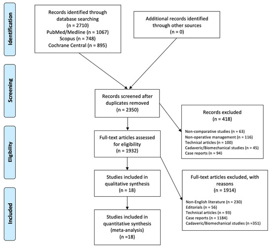
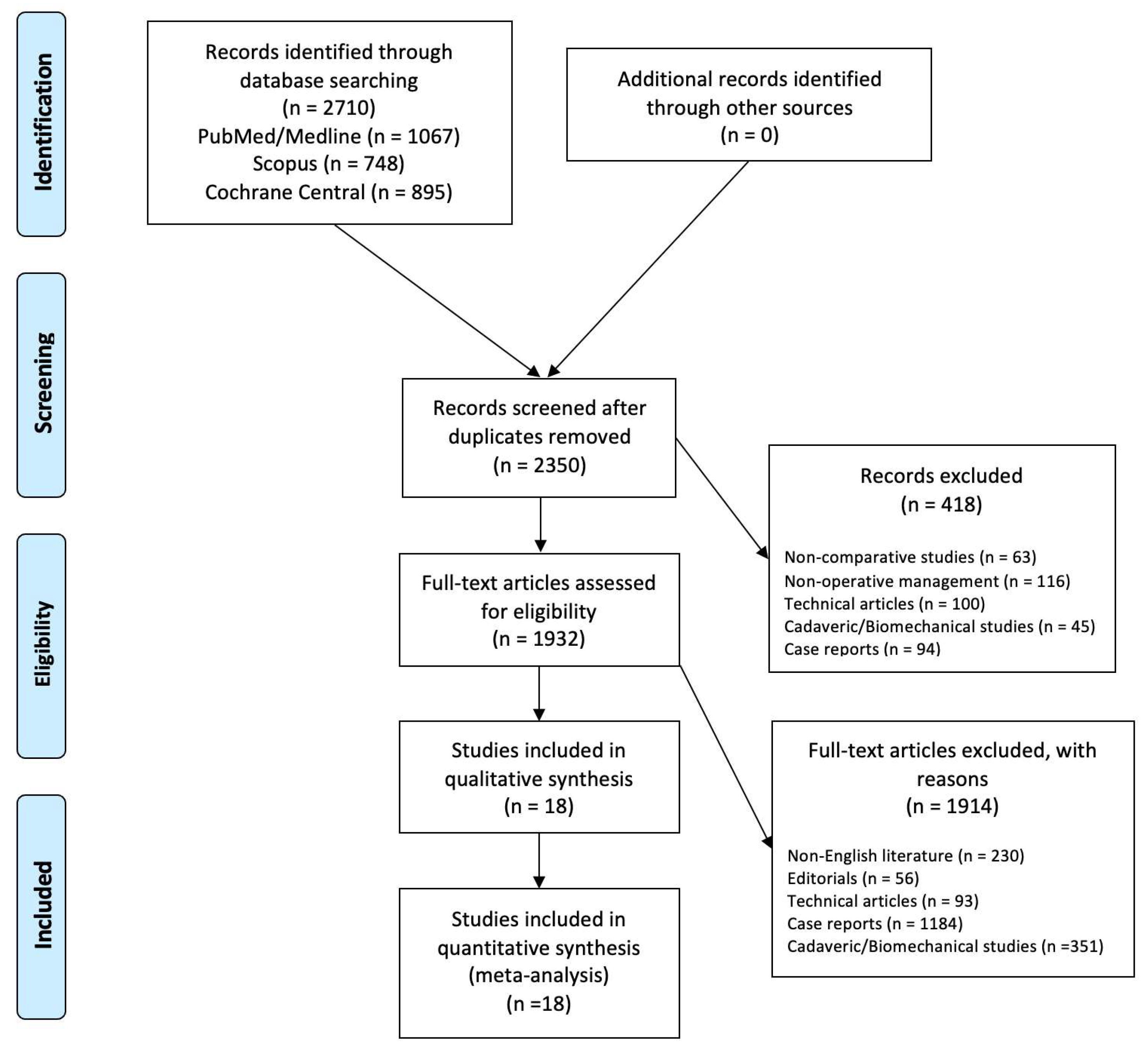
A total of 2710 relevant articles were identified through the initial search, 2350 abstracts were screened, and 1932 full-text articles were assessed for eligibility based on our inclusion criteria, resulting in 18 studies that were eligible for the systematic review (Figure 1).
Figure 1.
Preferred Reporting Items for Systematic Review and Meta-Analysis (PRISMA) flowchart for the searching and identification of included studies. Source: Moher et al. For more information, visit www.prisma-statement.org.
A total of 2683 patients were identified; overall, male patients accounted for 1628 (60.7%) of the cases. The frequency-weighted mean age at the time of the operation was 49.9 ± 11.9 years, and the frequency-weighted mean follow-up was 168.6 ± 54.6 months. The weighted mean BMI was 27.7 ± 5.4 kg/m2. The characteristics of the included studies are reported in Table 2.
Table 2.
Characteristics of included studies.
3.1. Functional Outcomes
The pre- and postoperative KOOS scores were measured in three studies with 703 patients, with mean weighted values of 46.5 ± 19 and 66.4 ± 22.9, respectively; a statistically significant improvement was found (p < 0.001).
3.2. Radiological Evaluation
The preoperative and postoperative radiological evaluations are shown in Table 3.
Table 3.
Pre- and postoperative radiographical evaluations.
According to the Kellgren–Lawrence classification, a total of 6 patients had Grade 0 (0.7%), 78 patients (8.9%) had Grade 1, 324 patients (36.8%) had Grade 2, 307 patients (34.9%) had Grade 3, and 165 patients (18.7%) had Grade 4.
The pre- and postoperative mTFA angles were measured in four studies with 798 patients, with mean weighted values of −5.7° ± 2.8° and 1.4° ± 2.4°, respectively; a statistically significant improvement was found (p = 0.002).
The pre- and postoperative mLDFA angles were measured in three studies with 816 patients, with mean weighted values of 88.8° ± 1.9° and 88.7° ± 2°, respectively (p = 0.3).
The pre- and postoperative MPTA angles were measured in four studies with 848 patients, with mean weighted values of 90.7° ± 7.5° and 90.8° ± 3.3°, respectively (p = 0.722).
3.3. Revision, Conversion to Arthroplasty, and Survivorship
The characteristics of surgery are shown in Table 4. A total of 335 patients (12.5%) had a revision surgery with a conversion to a total knee arthroplasty (TKA), while a total of 41 (1.5%) had a conversion to unicompartmental knee arthroplasty (UKA).
Table 4.
Characteristics of surgery of the included studies.
A total of nine studies reported the survival rate after 5 years, with a mean rate of 94.3% (1843 patients).
A total of 10 studies reported the survival rate after 10 years, with a mean rate of 86.3% (1828 patients).
3.4. Complications
Nonunion rate was 1.3%, while peroneal nerve palsy rate was 1.3%. The infection rate was 0.9% (25 patients). Superficial wound infection was reported in 26 patients, comprising 1%. Finally, deep venous thrombosis was reported in 22 patients (0.8%). The results are shown in Table 5.
Table 5.
Complications.
4. Discussion
Medial opening wedge high tibial osteotomy achieved a statistically significant correction of the mTFA with improvements in the functional outcomes. The procedure demonstrated a 10-year survival rate of 86%, with a 14% conversion rate to total or unicompartmental knee arthroplasty for OA progression after a mean follow-up of 14 years.
HTO is still considered a primary indication for the treatment of medial compartment knee OA in young and active patients, by acting as a joint-preserving strategy that maintains native joint kinematics [15]. Unlike arthroplasty, HTO preserves native ligaments and bone stock, maintaining proprioception and allowing higher postoperative activity levels with a more physiological knee perception. Several studies have also shown that opening wedge HTO remains an effective option even in patients with advanced OA, challenging the traditional consideration that this procedure should be used for early-stage OA [7,15,19,31].
Correction of the mTFA remains the main biomechanical determinant of HTO success, and its loss over time represents one of the most relevant mechanisms underlying long-term failure. Our results move from −5.7° to 1.4°, which is consistent with the recommendations of Koshino et al. [14], who emphasized that achieving and maintaining a valgus alignment is the most significant factor in preventing the recurrence of varus deformity and OA progression. Also, maintaining stable joint kinematics is important to avoiding complications in further surgical interventions [7,32]. The survival of HTO appears to depend not only on the degree of correction achieved but also on the long-term maintenance of both coronal and sagittal alignment. In fact, Dal Fabbro et al. [7] discussed the importance of preserving the posterior tibial slope (PTS) because a change in the slope may adversely affect joint biomechanics and accelerate the progression of OA [33]. Similarly, Choi et al. [22] reported a progressive relapse to varus alignment after 15 years; the average Tibiofemoral Angle varies from 187° to 170°, suggesting that the loss of correction may contribute to long-term failure. This mechanism may partially explain the 14% osteotomy failure rate observed in our review, consistent with the 14% revision rate reported by Flecher et al. [23].
The radiological correction obtained is associated with the enhanced functional outcomes, which are consistent findings across the literature. Mabrouk et al. [28] and Ahrend et al. [19] reported high patient satisfaction and significant improvements in quality-of-life scores, maintained over 10 years postoperatively. In our study, KOOS significantly improved from 46.5 ± 19 preoperatively to 66.4 ± 22.9 postoperatively (p < 0.001), with a mean increase of 19.9 ± 4 points, consistently exceeding the Minimal Clinically Important Difference (MCID) threshold of 10 points [34]. These results are particularly relevant considering the relatively young age of our cohort (49.9 years), as they represent a population that prioritizes the ability to remain active [35]. The partial dissociation between radiographic results and patient-reported outcomes was found. Some studies have found patients who have shown a sustained improvement clinically while having radiographic progression. It suggests that the alleviation of symptoms achieved by HTO may be driven by biomechanical unloading of the medial compartment rather than complete arrest of the degenerative process.
In this context, patient selection remains a crucial factor that influences the success of HTO. Several studies demonstrated that younger age, lower BMI, and limited cartilage degeneration are associated with higher outcomes and survivorship. Higher BMI may increase mechanical load on the osteotomy site, making accurate correction more challenging. These findings underline the importance of careful preoperative assessment in optimizing patient selection, which remains one of the most important determinants of long-term success [36,37].
In our study, the survival rate at 10 years was 86.3%. Michaela et al. [30] reported a lower survival rate of 79.9% at 10 years, which dropped significantly to 54.1% after 18 years. In contrast, Flecher et al. [23] demonstrated high longevity with 85% survival at 20 years, consistent with the data reported by Akizuki et al. [20], who also confirmed stable outcomes in their 10-to-20-year follow-up period. These observations suggest that the precision of the correction may be more critical than the specific surgical approach used. The variability in survivorship reported across the included studies reflects the differences in patient selection, correction targets, and OA severity at the time of surgery. Studies including younger patients with lower BMI and minor cartilage degeneration generally reported higher long-term survival rates.
Despite these favourable outcomes, HTO should still be considered a time-buying procedure. In our cohort, 14% of patients required a conversion to knee arthroplasty, which demonstrates the necessity of using this approach as a bridge. This transition to TKA remains a critical aspect. The meta-analysis of Loke et al. [38] (which reviewed more than 550,000 patients) found that, although HTO and subsequent TKA result in similar survival and functional results to those of primary TKA, it is associated with significantly higher complication rates. This emphasizes the importance of the surgical technique during the initial HTO, particularly regarding hardware placement and preservation of posterior tibial slope, in order to reduce technical difficulties during eventual conversion to TKA [7].
Finally, our findings align with the evidence provided by Ge et al. [39], who confirmed that HTO results in approximately 14 years of survivorship for young patients with active lifestyles. However, this finding should be interpreted within the context of the relatively young age of the treated population. Delaying arthroplasty by more than a decade may represent a clinically meaningful achievement, potentially reduce the lifetime risk of revision arthroplasty while enabling patients to maintain higher activity levels during the most active years of their lives.
Although the survivorship, HTO remains associated with relevant complications. In our study, nonunion and peroneal nerve palsy were both observed in 1.3% of cases, while the overall infection rate was 0.9%. Revision surgery unrelated to arthroplasty conversion was required in 1.3% of patients. These findings are similar to the complication rate reported in the previous reviews. In the systematic review by Miltenberg et al. [40], which analysed 7836 patients, an overall postoperative complication rate of 6.9% was reported; with superficial wound infection representing the most common one (2.2%), followed by nonunion (1.9%). The authors also described an overall reoperation rate of 15.5%.
Bone healing complications remain the most clinically relevant adverse event, as the instability at the osteotomy site may contribute to delayed union, loss of correction, or fixation failure. Neurological and thromboembolic complications, although relatively uncommon, may also impact postoperative recovery and patient morbidity. In our cases, the complication rates are in line with the literature, suggesting that meticulous surgical technique, stable fixation, and postoperative management are essential for minimization.
This study has several limitations. First, only English language studies were considered, potentially contributing to publication bias; moreover, although four recommended databases were used for the search, we cannot exclude the possibility that additional articles could have been found by searching other databases. Second, heterogeneity of the technique across the included cohorts with different fixation devices makes it difficult to generalize specific complication rates. Finally, the lack of detailed data on modifiable biological factors, such as smoking status, limits the possibility of a more precise risk stratification.
These findings mean that HTO is not seen just as a temporary procedure, but rather a long-term strategy that effectively redefines patients’ surgical timelines, offering a reliable decade of high-level knee function.
5. Conclusions
This systematic review showed that patients who underwent medial opening wedge high tibial osteotomy achieved a significant correction of mTFA associated with an improvement in the functional outcomes. An 86% survivorship rate at 10 years was observed, while a 14% conversion rate to knee arthroplasty for OA progression was reported after a mean follow-up of 14 years. Low incidence rates for nonunion (1.3%) and infection (0.9%) were found. The results of the current study may be of interest to patients, clinicians, and health professionals involved in the management of under-50 and active patients with medial compartment OA in a varus knee.
Author Contributions
Conceptualization, M.M., F.F.F. and E.C.; methodology, M.M., U.G.L. and E.C.; data curation, M.M., F.P., S.C. and E.C.; writing—original draft preparation, E.C. and M.M.; writing—review and editing, M.M., F.P., E.C., S.C., O.G., F.F.F., U.G.L. and A.C.; supervision, M.M., U.G.L., O.G., F.F.F. and G.G.; project administration, M.M. and E.C. All authors have read and agreed to the published version of the manuscript.
Funding
This research received no external funding.
Institutional Review Board Statement
Not applicable.
Informed Consent Statement
Not applicable.
Data Availability Statement
No new data were created or analysed in this study.
Conflicts of Interest
The authors declare no conflicts of interest.
References
- Kanakamedala, A.C.; Hurley, E.T.; Manjunath, A.K.; Jazrawi, L.M.; Alaia, M.J.; Strauss, E.J. High Tibial Osteotomies for the Treatment of Osteoarthritis of the Knee. JBJS Rev. 2022, 10, e21. [Google Scholar] [CrossRef] [Scilit]
- Ji, W.; Luo, C.; Zhan, Y.; Xie, X.; He, Q.; Zhang, B. A Residual Intra-Articular Varus after Medial Opening Wedge High Tibial Osteotomy (HTO) for Varus Osteoarthritis of the Knee. Arch. Orthop. Trauma. Surg. 2019, 139, 743–750. [Google Scholar] [CrossRef] [Scilit] [PubMed]
- Marti, R.K.; Verhagen, R.A.; Kerkhoffs, G.M.; Moojen, T.M. Proximal Tibial Varus Osteotomy. Indications, Technique, and Five to Twenty-One-Year Results. J. Bone Jt. Surg. Am. 2001, 83, 164–170. [Google Scholar]
- Mercurio, M.; Gasparini, G.; Galasso, O.; Familiari, F.; Cofano, E.; Sanzo, V.; Ciolli, G.; Corona, K.; Cerciello, S. Lateral versus Medial Approach for Total Knee Arthroplasty for Valgus Knee Deformity Shows Comparable Functional Outcomes, Hip–Knee–Ankle Angle Values, and Complication Rates: A Meta-Analysis of Comparative Studies. Arch. Orthop. Trauma. Surg. 2023, 144, 869–878. [Google Scholar] [CrossRef] [Scilit] [PubMed]
- Hernigou, P.; Medevielle, D.; Debeyre, J.; Goutallier, D. Proximal Tibial Osteotomy for Osteoarthritis with Varus Deformity. A Ten to Thirteen-Year Follow-up Study. J. Bone Jt. Surg. Am. 1987, 69, 332–354. [Google Scholar]
- Coventry, M.B.; Ilstrup, D.M.; Wallrichs, S.L. Proximal Tibial Osteotomy. A Critical Long-Term Study of Eighty-Seven Cases. J. Bone Jt. Surg. 1993, 75, 196–201. [Google Scholar] [CrossRef] [Scilit] [PubMed]
- Dal Fabbro, G.; Balboni, G.; Paolo, S.D.; Varchetta, G.; Grassi, A.; Marcheggiani Muccioli, G.M.; Zaffagnini, S. Lateral Closing Wedge High Tibial Osteotomy Procedure for the Treatment of Medial Knee Osteoarthritis: Eleven Years Mean Follow up Analysis. Int. Orthop. 2025, 49, 1655–1666. [Google Scholar] [CrossRef] [Scilit] [PubMed]
- Kurien, T.; East, J.; Mandalia, V. The Effects of Open Wedge High Tibial Osteotomy for Knee Osteoarthritis on the Patellofemoral Joint: A Systematic Review. Knee 2022, 40, 201–219. [Google Scholar] [CrossRef] [Scilit] [PubMed]
- Effect of Medial Open Wedge High Tibial Osteotomy on Progression of Patellofemoral Osteoarthritis-PubMed. Available online: https://pubmed.ncbi.nlm.nih.gov/36274182/ (accessed on 25 June 2026).
- Hantes, M.E.; Natsaridis, P.; Koutalos, A.A.; Ono, Y.; Doxariotis, N.; Malizos, K.N. Satisfactory Functional and Radiological Outcomes Can Be Expected in Young Patients under 45 Years Old after Open Wedge High Tibial Osteotomy in a Long-Term Follow-Up. Knee Surg. Sports Traumatol. Arthrosc. 2018, 26, 3199–3205. [Google Scholar] [CrossRef] [Scilit] [PubMed]
- Van Wulfften Palthe, A.F.Y.; Clement, N.D.; Temmerman, O.P.P.; Burger, B.J. Survival and Functional Outcome of High Tibial Osteotomy for Medial Knee Osteoarthritis: A 10–20-Year Cohort Study. Eur. J. Orthop. Surg. Traumatol. 2018, 28, 1381–1389. [Google Scholar] [CrossRef] [Scilit] [PubMed]
- Coakley, A.; McNicholas, M.; Biant, L.; Tawy, G. A Systematic Review of Outcomes of High Tibial Osteotomy for the Valgus Knee. Knee 2023, 40, 97–110. [Google Scholar] [CrossRef] [Scilit] [PubMed]
- Naudie, D.; Bourne, R.B.; Rorabeck, C.H.; Bourne, T.J. The Install Award. Survivorship of the High Tibial Valgus Osteotomy. A 10- to -22-Year Followup Study. Clin. Orthop. Relat. Res. 1999, 367, 18–27. [Google Scholar]
- Koshino, T.; Yoshida, T.; Ara, Y.; Saito, I.; Saito, T. Fifteen to Twenty-Eight Years’ Follow-up Results of High Tibial Valgus Osteotomy for Osteoarthritic Knee. Knee 2004, 11, 439–444. [Google Scholar] [CrossRef] [PubMed]
- Gkekas, N.K.; Komnos, G.A.; Mylonas, T.; Chalatsis, G.; Koutalos, A.A.; Hantes, M.E. Medial Open Wedge High Tibial Osteotomy Is a Viable Option in Young Patients with Advanced Arthritis in a Long-term Follow-up. Knee Surg. Sports Traumatol. Arthrosc. 2025, 33, 1025–1032. [Google Scholar] [CrossRef] [Scilit] [PubMed]
- Wells, G.; Shea, B.; O’Connell, D.; Peterson, J.; Wekch, V.; Losos, M. The Newcastle-Ottawa Scale (NOS) for Assessing the Quality of Nonrandomised Studies in Meta-Analyses; Ottawa Hospital Research Institute: Ottawa, ON, Canada, 2000. [Google Scholar]
- Higgins, J.P.T.; Altman, D.G.; Gotzsche, P.C.; Juni, P.; Moher, D.; Oxman, A.D.; Savovic, J.; Schulz, K.F.; Weeks, L.; Sterne, J.A.C.; et al. The Cochrane Collaboration’s Tool for Assessing Risk of Bias in Randomised Trials. BMJ 2011, 343, d5928. [Google Scholar] [CrossRef] [Scilit] [PubMed]
- Mercurio, M.; Cerciello, S.; Corona, K.; Guerra, G.; Simonetta, R.; Familiari, F.; Galasso, O.; Gasparini, G. Factors Associated with a Successful Return to Performance After Anterior Cruciate Ligament Reconstruction: A Multiparametric Evaluation in Soccer Players. Orthop. J. Sports Med. 2024, 12, 23259671241275663. [Google Scholar] [CrossRef] [Scilit] [PubMed]
- Ahrend, M.; Petzold, D.; Histing, T.; Ihle, C.; Schröter, S.; Herbst, M. High Survival Rates and Patient Satisfaction 12 Years after Medial Open Wedge High Tibial Osteotomy Surgery: A Prospective Cohort Study. Knee Surg. Sports Traumatol. Arthrosc. 2025, 33, 4344–4352. [Google Scholar] [CrossRef] [Scilit] [PubMed]
- Akizuki, S.; Shibakawa, A.; Takizawa, T.; Yamazaki, I.; Horiuchi, H. The Long-Term Outcome of High Tibial Osteotomy: A Ten- to 20-Year Follow-Up. J. Bone Jt. Surg. Br. 2008, 90, 592–596. [Google Scholar] [CrossRef] [Scilit] [PubMed]
- Benzakour, T.; Hefti, A.; Lemseffer, M.; El Ahmadi, J.D.; Bouyarmane, H.; Benzakour, A. High Tibial Osteotomy for Medial Osteoarthritis of the Knee: 15 Years Follow-Up. Int. Orthop. (SICOT) 2010, 34, 209–215. [Google Scholar] [CrossRef] [Scilit] [PubMed]
- Choi, H.-R.; Hasegawa, Y.; Kondo, S.; Shimizu, T.; Ida, K.; Iwata, H. High Tibial Osteotomy for Varus Gonarthrosis: A 10- to 24-Year Follow-up Study. J. Orthop. Sci. 2001, 6, 493–497. [Google Scholar] [CrossRef] [Scilit] [PubMed]
- Flecher, X.; Parratte, S.; Aubaniac, J.-M.; Argenson, J.-N.A. A 12–28-Year Followup Study of Closing Wedge High Tibial Osteotomy. Clin. Orthop. Relat. Res. 2006, 452, 91–96. [Google Scholar] [CrossRef] [Scilit] [PubMed]
- Michaela, G.; Florian, P.; Michael, L.; Christian, B. Long-Term Outcome after High Tibial Osteotomy. Arch. Orthop. Trauma Surg. 2007, 128, 111–115. [Google Scholar] [CrossRef] [Scilit] [PubMed]
- Howells, N.R.; Salmon, L.; Waller, A.; Scanelli, J.; Pinczewski, L.A. The Outcome at Ten Years of Lateral Closing-Wedge High Tibial Osteotomy: Determinants of Survival and Functional Outcome. Bone Jt. J. 2014, 96-B, 1491–1497. [Google Scholar] [CrossRef] [Scilit] [PubMed]
- Hui, C.; Salmon, L.J.; Kok, A.; Williams, H.A.; Hockers, N.; Van Der Tempel, W.M.; Chana, R.; Pinczewski, L.A. Long-Term Survival of High Tibial Osteotomy for Medial Compartment Osteoarthritis of the Knee. Am. J. Sports Med. 2011, 39, 64–70. [Google Scholar] [CrossRef] [Scilit] [PubMed]
- Leutloff, D.; Tobian, F.; Perka, C. High Tibial Osteotomy for Valgus and Varus Deformities of the Knee. Int. Orthop. 2001, 25, 93–96. [Google Scholar] [CrossRef] [Scilit] [PubMed]
- Mabrouk, A.; Risebury, M.; Yasen, S. High Survivorship and Low Complication Rate in a Single-centre Series of 651 Medial Opening Wedge High Tibial Osteotomy Cases with a Mean Follow-up of 13 Years. Knee Surg. Sports Traumatol. Arthrosc. 2024, 32, 736–749. [Google Scholar] [CrossRef] [Scilit] [PubMed]
- Pfahler, M.; Lutz, C.; Anetzberger, H.; Maier, M.; Hausdorf, J.; Pellengahr, C.; Refior, H.J. Long-Term Results of High Tibial Osteotomy for Medial Osteoarthritis of the Knee. Acta Chir. Belg. 2003, 103, 603–606. [Google Scholar] [CrossRef] [Scilit] [PubMed]
- Valenti, J.R.; Calvo, R.; Lopez, R.; Cañadell, J. Long Term Evaluation of High Tibial Valgus Osteotomy. Int. Orthop. 1990, 14, 347–349. [Google Scholar] [CrossRef] [Scilit] [PubMed]
- Cerciello, S.; Proietti, L.; Mocini, F.; Corona, K.; Venosa, M.; Mercurio, M.; Neyret, P. Current Concepts of Medial Unicompartmental Knee Replacement: Part 2-Hot Topics and Further Directions. Arch. Orthop. Trauma. Surg. 2026, 146, 28. [Google Scholar] [CrossRef] [Scilit] [PubMed]
- Mercurio, M.; Gasparini, G.; Familiari, F.; Castioni, D.; Galasso, O. Outcomes of Bi-Unicompartmental Versus Total Knee Arthroplasty for the Treatment of Medial and Lateral Knee Osteoarthritis: A Systematic Review and Meta-Analysis of Comparative Studies. Indian. J. Orthop. 2022, 56, 963–972. [Google Scholar] [CrossRef] [Scilit] [PubMed]
- Erquicia, J.I.; Gil-Gonzalez, S.; Ibañez, M.; Leal-Blanquet, J.; Combalia, A.; Monllau, J.C.; Pelfort, X. A Lower Starting Point for the Medial Cut Increases the Posterior Slope in Opening-Wedge High Tibial Osteotomy: A Cadaveric Study. J. Exp. Orthop. 2022, 9, 124. [Google Scholar] [CrossRef] [Scilit] [PubMed]
- Birmingham, T.B.; Primeau, C.A.; Moyer, R.F.; Bryant, D.M.; Ma, J.; Leitch, K.M.; Wirth, W.; Degen, R.; Getgood, A.M.; Litchfield, R.B.; et al. High Tibial Osteotomy for Medial Compartment Knee Osteoarthritis: A Randomized Trial with Parallel Preference Arm. Ann. Intern. Med. 2025, 178, 1238–1248. [Google Scholar] [CrossRef] [Scilit] [PubMed]
- Ciolli, G.; Proietti, L.; Mercurio, M.; Corona, K.; Maccauro, G.; Schiavone Panni, A.; Cerciello, S. Return to Sport Following Distal Femur Osteotomy: A Systematic Review. Orthop. Rev. 2022, 14, 33774. [Google Scholar] [CrossRef] [Scilit] [PubMed]
- Huang, T.; Kang, K.; Qiao, Q.; Li, T.; Liu, T.; Ji, C.; Gao, S. Muti-Factor Analysis of Sport Activity Level after High Tibial Osteotomy. J. Orthop. Surg. Res. 2023, 18, 813. [Google Scholar] [CrossRef] [Scilit] [PubMed]
- Nicolini, A.P.; Christiano, E.S.; Abdalla, R.J.; Cohen, M.; de Carvalho, R.T. Return to Sports After High Tibial Osteotomy Using the Opening Wedge Technique. Rev. Bras. Ortop. 2021, 56, 313–319. [Google Scholar] [CrossRef] [Scilit] [PubMed]
- Loke, R.W.K.; Tan, B.W.L.; Tan, E.Y.E.K.; Hui, J.H.P. Total Knee Arthroplasty after Prior High Tibial Osteotomy Results in Comparable Survival and Clinical Outcomes but Significantly Higher Complication Rates Compared to Primary Total Knee Arthroplasty: A Systematic Review and Meta-Analysis of 550,000 Patients. Eur. J. Orthop. Surg. Traumatol. 2025, 35, 119. [Google Scholar] [CrossRef] [Scilit] [PubMed]
- Ge, Y.; Häner, M.; Borges, J.S.; Stöckle, U.; Petersen, W. Long-Term Survivorship after Medial Open-Wedge High Tibial Osteotomy: A Systematic Review and Single-Arm Meta-Analysis with Regional Subgroup Analysis. Knee Surg. Sports Traumatol. Arthrosc. 2026, 34, 1390–1404. [Google Scholar] [CrossRef] [Scilit] [PubMed]
- Miltenberg, B.; Puzzitiello, R.N.; Ruelos, V.C.B.; Masood, R.; Pagani, N.R.; Moverman, M.A.; Menendez, M.E.; Ryan, S.P.; Salzler, M.J.; Drager, J. Incidence of Complications and Revision Surgery After High Tibial Osteotomy: A Systematic Review. Am. J. Sports Med. 2024, 52, 258–268. [Google Scholar] [CrossRef] [Scilit] [PubMed]

Disclaimer/Publisher’s Note: The statements, opinions and data contained in all publications are solely those of the individual author(s) and contributor(s) and not of MDPI and/or the editor(s). MDPI and/or the editor(s) disclaim responsibility for any injury to people or property resulting from any ideas, methods, instructions or products referred to in the content. |
© 2026 by the authors. Licensee MDPI, Basel, Switzerland. This article is an open access article distributed under the terms and conditions of the Creative Commons Attribution (CC BY) license.