


Abstract
Electrospun composite nanofibers represent an emerging strategy for the development of efficient fertilizer systems, as they enable modulation of the structural properties of the nanofibrous network and, consequently, the transport and release processes of nutrients. In this study, polyvinyl alcohol (PVA) nanofibers loaded with two different urea contents (0.09 g and 0.36 g) were fabricated and characterized to investigate how urea incorporation modifies the nanofiber morphology and influences urea release kinetics. SEM and EDS analyses confirmed that increasing urea content promotes surface roughnes and reduced nanofiber diameters, whereas XRD and FTIR demonstrated a decrease in crystallinity and the formation of hydrogen-bonded interactions between PVA chains and urea molecules, indicating that urea is incorporated within the PVA network rather than being superficially adsorbed on the nanofiber surface. These structural changes govern water retention and release kinetics: the 0.36 g formulation exhibited a 100-h induction period followed by multiphase diffusion, while the 0.09 g system displayed immediate release but lower final concentrations. Kinetic modeling revealed excellent fitting to the Higuchi and second-order models, confirming diffusion-controlled urea release modulated by internal interactions. The nanofiber network thus behaves as an active regulator of nitrogen mobility, overcoming the limitations of conventional coating-based fertilizers. These findings demonstrate the potential of PVA/urea nanofibers as scalable platforms for sustainable nitrogen delivery in agriculture, bridging morphology-driven polymer design with environmental performance.
1. Introduction
Urea is a nitrogen-based compound of great importance in agriculture due to its high nitrogen content (46%), easy water solubility, and low cost, which makes it one of the most widely used fertilizers worldwide [1]. However, its direct application presents limitations such as losses through leaching and volatilization, which decrease nitrogen use efficiency (NUE) and generate environmental problems [2]. It has been estimated that more than 40% of applied urea nitrogen can be lost through volatilization and leaching, severely reducing its agricultural effectiveness and causing groundwater contamination [3].
Despite the high precision and morphological control offered by conventional single-needle electrospinning configurations, their practical application in widespread agronomic setups remains constrained by inherently low mass-production rates (typically 0.1–1.0 mL/h). To enhance industrial relevance and support large-scale agricultural deployment, needleless electrospinning architectures—most notably bubble electrospinning—have emerged as a pivotal technological alternative [4]. This method relies on aerodynamic or pneumatic mechanisms to generate gas bubbles at the surface of a free polymer solution. When exposed to a high-voltage electrostatic field, the thin film of each bubble experiences Maxwell stresses that overcome the solution’s surface tension, triggering the simultaneous ejection of multiple polymeric micro-jets from the bubble’s surface [5]. Consequently, this technique significantly escalates the nanofiber production throughput by several orders of magnitude compared to needle-based systems, while maintaining desirable web porosity and preserving the structural homogeneity required for controlled composite entrapment. Therefore, the development of sustainable and efficient delivery systems for nitrogen fertilizers has become a priority.
On the other hand, polymeric nanofibers have emerged as promising systems for the encapsulation and controlled release of active compounds [6]. Among them, polyvinyl alcohol (PVA) nanofibers stand out for being biodegradable, biocompatible, non-toxic, and having excellent fiber-forming capacity through electrospinning [7]. Electrospinning allows the production of nanofibers with nanometric diameters, high surface area-to-volume ratios, and elevated porosity, characteristics that facilitate the incorporation of functional molecules such as drugs, bioactive compounds, or fertilizers [8]. Composite and hollow nanostructures, such as core–sheath or double-layered yarns, further expand the possibilities for controlled release, since the external barrier layers can modulate diffusion and significantly prolong the release of urea or other actives [3].
Electrospun nanofibers have emerged as promising platforms in agricultural applications due to their high surface area, porosity, and ability to encapsulate and control the release of active compounds [9]. These systems have been explored for seed coatings, plant protection, and controlled delivery of fertilizers and agrochemicals, enabling improved nutrient efficiency and reduced environmental losses [10]. Moreover, nanofiber-based technologies offer advantages such as relatively low production cost, simplicity of fabrication, and the possibility of scalable manufacturing, particularly with advances in electrospinning techniques. As a result, they represent a viable and potentially cost-effective alternative to conventional agricultural delivery systems, combining functional performance with environmental sustainability [11].
Nanofiber morphology is highly dependent on electrospinning solution properties, including viscosity, conductivity, and surface tension [12]. Changes in these parameters may induce the formation of bead-like structures, which can modify water uptake, diffusion pathways, and the release behavior of encapsulated compounds [13]. Therefore, evaluating the effect of urea incorporation on bead formation is relevant for the development of controlled-release fertilizer systems.
In recent literature, several authors have modified PVA nanofibers through the incorporation of urea as a structural agent, mainly to improve the solubility, wettability, or chemical interaction of the PVA. It has been reported that the incorporation of urea increased hydrophilicity and improved drug release [14], while Mohamed [1] demonstrated that carbonized PVA nanofibers with urea can function as catalytic supports. Furthermore, Li [15] demonstrated that PVA-based hybrid nanofibers incorporating titanium dioxide and urea not only enhanced the physicochemical stability of the nanofibers but also offered a synergistic effect for controlled nutrient delivery, combining photocatalytic activity with fertilizer encapsulation. These studies highlight the advantages of urea in modifying fiber properties such as wettability, porosity, and ionic conductivity.
In addition to modifying the physicochemical properties of PVA nanofibers, morphological changes have also been associated with controlled-release performance. Previous studies reported that reductions in fiber diameter and improvements in morphological uniformity can enhance sustained release behavior by increasing the specific surface area and promoting greater structural disorder within the composite [2]. These observations suggest that morphology plays a key role in regulating nutrient transport and release from electrospun systems.
Previous studies have shown that variations in material composition can significantly alter physicochemical behavior, transport phenomena, and interaction mechanisms with water. Accordingly, modifying urea loading within electrospun PVA composite nanofibers is expected to influence structural organization, water absorption capacity, and nutrient release performance. Since water uptake governs polymer swelling, molecular mobility, and diffusion pathways, understanding the relationship between urea loading and composite hydration becomes essential for the rational design of controlled-release fertilizer systems [16].
Nevertheless, some researchers have pointed out that the incorporation of urea into PVA may not be favorable in certain contexts, since its excessive plasticizing effect can reduce the mechanical strength of the nanofibers or accelerate their degradation in aqueous media, thereby compromising the structural stability of the material [7,14]. Urea incorporation may also increase the mobility of polymer chains and facilitate jet stretching during the electrospinning process, promoting the formation of fibers with smaller diameters. Recent studies also suggest that hybrid formulations, such as PVA combined with gelatin, silica-based bioactive glass, or metal oxide nanoparticles, can overcome these drawbacks by reinforcing mechanical stability and enhancing water interaction, making them highly promising for agricultural applications [15,17].
However, despite the growing interest in electrospun PVA/urea systems, the relationship between urea incorporation, structural reorganization of the PVA composite, and nutrient release behavior remains insufficiently understood. In particular, it is still unclear how urea is integrated within the PVA nanofibrous network, how different urea loadings alter the physicochemical and morphological properties of the nanofibers, and which diffusion or interaction mechanisms ultimately govern nitrogen release. Therefore, the present study seeks to elucidate the structural and kinetic mechanisms underlying urea-loaded electrospun PVA nanofibers, addressing key questions such as:
- How does urea integrate into the PVA polymeric matrix?
- How does urea loading influence the release kinetics of the nanofibrous system?
- What diffusion and PVA–urea interaction mechanisms govern urea release from the nanofibers?
Finally, this manuscript is organized as follows. Section 2 describes the materials and electrospinning methodology. Section 3 presents the physicochemical, morphological, and spectroscopic analyses carried out to evaluate the influence of urea incorporation. Finally, Section 4 summarizes the main conclusions of the study and highlights the potential of electrospun PVA/urea nanofibers.
2. Materials and Methods
2.1. Materials
Polyvinyl alcohol (PVA) (molecular weight: 125,000; degree of polymerization: 1700–1800; hydrolysis degree: 98–99%) and urea (U5378, ≥98% purity, BioReagent, Molecular Biology grade) were purchased from Sigma-Aldrich (St. Louis, MO, USA).
2.2. Fabrication of Urea-Loaded PVA Nanofibers
A 10% (w/v) PVA solution was prepared by dissolving 3.0 g of PVA in 30 mL of distilled water under continuous magnetic stirring and heating. Once the temperature reached 70 °C, the mixture was maintained under these conditions for 1 h to ensure complete dissolution of the PVA. Subsequently the system was stirred for an additional hour to obtain a homogeneous and aggregate-free dispersion.
The resulting solution was divided into three formulations: a PVA control without urea, a low-urea-loading formulation, and a high-urea-loading formulation. For the urea-loaded formulations, 0.09 g and 0.36 g of urea were separately added to 1 mL aliquots of the 10% (w/v) PVA solution. Each solution was prepared in triplicate.
The prepared solutions were subsequently processed by electrospinning using a custom-built electrospinning system manufactured by Giga IE (Querétaro, Mexico), equipped with a 25 kV high-voltage power supply. The PVA jet was collected on circular collectors with a diameter of 10 cm, where the nanofibrous were formed and deposited for subsequent characterization and release studies.
The Table 1 summarizes the electrospinning conditions and the average environmental parameters recorded during the fabrication of PVA and PVA/urea nanofibrous mats.
Table 1.
Average electrospinning parameters and resulting thickness of PVA/urea nanofibrous mats under controlled processing conditions.
The formulations were selected to evaluate the influence of different nutrient-loading levels on fiber morphology, urea incorporation capacity, and the release behavior of the system. The selected urea contents were defined based on agronomically relevant nitrogen-loading levels, representing low- and high-loading conditions. The resulting nanofibrous membranes were conceived as individual fertilizer units intended for controlled nitrogen delivery.
To bridge the gap between materials synthesis and practical field management, a quantitative agronomic evaluation of the effective nitrogen (N) payload within the dry nanofibrous composites was calculated. Pure crystalline urea possesses a standard theoretical nitrogen content of 46.7% w/w. Based on the dry-mass formulations utilized during the electrospinning process (), the theoretical effective nitrogen concentrations embedded within the monolithic composite mats were determined as follows: for the PVA/urea 0.09 g formulation (comprising 47.37 wt.% urea in the PVA), the effective nitrogen content is approximately 22.12% w/w; for the PVA/urea 0.36 g formulation (comprising 78.26 wt.% urea in the PVA), the effective nitrogen content scales to approximately 36.54% w/w. These values provide a direct baseline for agronomic dosing and field scaling calculations.
2.3. Structural and Morphological Analysis
X-ray diffraction (XRD), scanning electron microscopy coupled with energy-dispersive X-ray spectroscopy (SEM/EDS), FTIR spectroscopy analyses were performed at the Center for Applied Physics and Advanced Technology (CFATA) of the National Autonomous University of Mexico (UNAM), Querétaro campus.
XRD measurements were conducted using an XRDynamic 500 X-ray diffractometer (Anton Paar GmbH, Graz, Austria). SEM/EDS analyses were performed using a Hitachi SU8230 ultra-high-resolution cold field emission scanning electron microscope (Hitachi High-Tech Corporation, Tokyo, Japan).
2.4. Determination of Fiber Diameter by SEM Image Analysis
The fiber diameter was determined from SEM micrographs using the ImageJ software (version 1.54g, National Institutes of Health, Bethesda, MD, USA). Each image was calibrated to a 5 µm scale, and 25 nanofibers were randomly selected to obtain the average diameter. Based on these measurements, histograms and diameter distribution curves were generated to analyze the morphological characteristics of the electrospun nanofibers.
2.5. Water Retencion Percentage
The determination of the water retention percentage was carried out using a fiber sample with an area of 1 cm2. First, the fiber was weighed in its dry state, and then moistened using a Pasteur pipette. The excess surface water was carefully removed with absorbent paper to avoid overestimation due to free moisture. Finally, the fiber was weighed again in its wet state. The procedure was performed in triplicate for the two urea loading and for the urea-free (control) fiber. The Table 2 summarizes the time points used for the water retention measurements.
Table 2.
Time intervals used for water retention measurements.
Equation (1) was used to quantify the water retention percentage at each time interval.
Table 3 summarizes the average mass and area of the PVA and PVA/urea nanofibrous membranes used in the water retention experiments. These measurements were recorded prior to hydration to ensure consistency among samples and to provide a reference for the subsequent evaluation of water retention behavior. The results indicate that the membranes exhibited similar dimensions and masses, allowing meaningful comparison of their moisture retention performance over time.
Table 3.
Average mass of PVA/urea nanofibrous membranes used in the water retention experiments.
2.6. Release Profile
The nanofibers used in the urea release experiments were prepared according to the electrospinning procedure described in Section 2.2. The average mass and area of the membranes evaluated are presented in Table 4. The membranes were placed inside pre-hydrated cellulose dialysis tubing with a molecular weight cut-off (MWCO) of 1 kDa. Subsequently, the dialysis bags were immersed in 9 mL of distilled water, which served as the release medium.
Table 4.
Average mass of PVA/urea nanofibrous membranes used in the urea release experiments.
To each system, 150 µL of phenol red [18] and 150 µL of urease were added as indicator reagents. Subsequently, a 2 mL aliquot of the release medium was collected and analyzed using a GENESYS 10S UV–Vis Thermo Fisher Scientific (Waltham, MA, USA) spectrophotometer at 560 nm. Measurements were carried out daily over a period of 42 days to monitor the urea release profile.
The release experiments were conducted without continuous agitation. However, prior to each aliquot withdrawal, the medium was gently homogenized to ensure a uniform distribution of the released urea. The temperature was monitored throughout the experiments to ensure that all measurements were performed within a range of 22–25 °C.
The selected MWCO allowed the diffusion of released urea molecules (60 Da) while preventing the passage of the high-molecular-weight PVA composite, ensuring that the measured signal corresponded exclusively to urea released from the nanofibrous system.
2.7. Calibration Curve
The quantification of the urea loaded into the electrospun PVA nanofibers was performed using a calibration curve prepared with ten standard loading ranging from 0.1 M to 1.0 M.
From each loading, 3 mL of solution were taken and mixed with 50 µL of phenol red and 50 µL of urease.
The resulting mixtures were analyzed using a GENESYS 10S UV–Vis spectrophotometer (Thermo Fisher Scientific, Waltham, MA, USA) at 560 nm, establishing a linear relationship between absorbance and urea loading.
2.8. Urea Release Percentage in Electrospun Nanofibers
In controlled-release systems based on electrospinning, the release percentage (RP%) corresponds to the fraction of urea that is continuously transferred from the PVA composite into the surrounding aqueous medium over time, relative to the initial nutrient mass loaded into the precursor solution. To mathematically that define Release Percentage is, Equation (2) and was adjusted as follows:
where:
represents the cumulative mass of urea released into the surrounding medium at a specific time interval t.
represents the total initial mass of urea loaded into the PVA solution prior to the electrospinning process.
3. Results
To elucidate how urea loading modulates both the internal structure and functional performance of the electrospun composite nanofibers, a comprehensive multiscale characterization was conducted. Morphological and elemental analyses by SEM and EDS were first employed to correlate fiber morphology and chemical composition with the amount of urea incorporated, while XRD and FTIR provided insight into changes in crystallinity and molecular interactions within the PVA network. These structural findings were then linked to the water retention capacity of the nanofibers and to the urea release kinetics quantified by UV–Vis spectrophotometry, allowing the identification of the dominant transport mechanisms through classical kinetic models. Together, these complementary techniques establish a coherent picture of how microstructure, hydration behavior, and diffusion processes are coupled in PVA/urea nanofibers designed for controlled nutrient delivery.
3.1. SEM and EDS Mapping
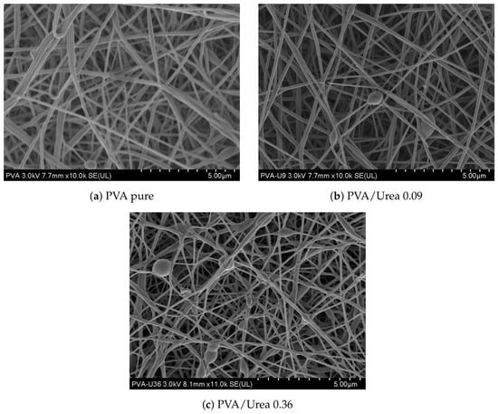
SEM micrographs revealed distinct morphological differences among the samples. In Figure 1a, pure PVA nanofibers displayed continuous, smooth surfaces with uniform diameters and no detectable defects such as beads or fractures, consistent with the well–known fiber–forming ability of PVA due to its hydroxyl functionality and hydrogen–bonding capacity [17] For PVA with 0.09 g urea (Figure 1b), nanofibers appeared thinner and more homogeneous, with a marked reduction in bead formation. In contrast, PVA containing 0.36 g of urea (Figure 1c) exhibited irregular morphologies characterized by variable diameters, rougher surfaces, and partial fiber fusion. These observations suggest that excessive urea alters the solution properties and promotes greater structural heterogeneity. Similar effects have been previously reported for urea-loaded electrospun PVA systems [1,19].
Figure 1.
Scanning electron microscopy (SEM) micrograph of PVA nanofibers at 5 μm.
Overall, the results suggest that the morphology of the electrospun nanofibers is influenced by the amount of incorporated urea. While the PVA/U0.09 formulation exhibited a more homogeneous fiber distribution, the PVA/U0.36 formulation showed a greater presence of morphological irregularities, indicating that higher urea loadings may compromise fiber uniformity and structural stability.
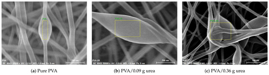
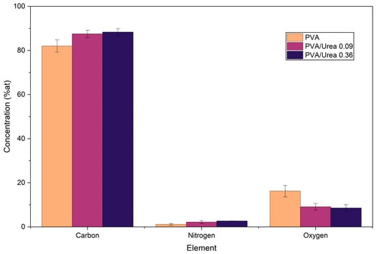
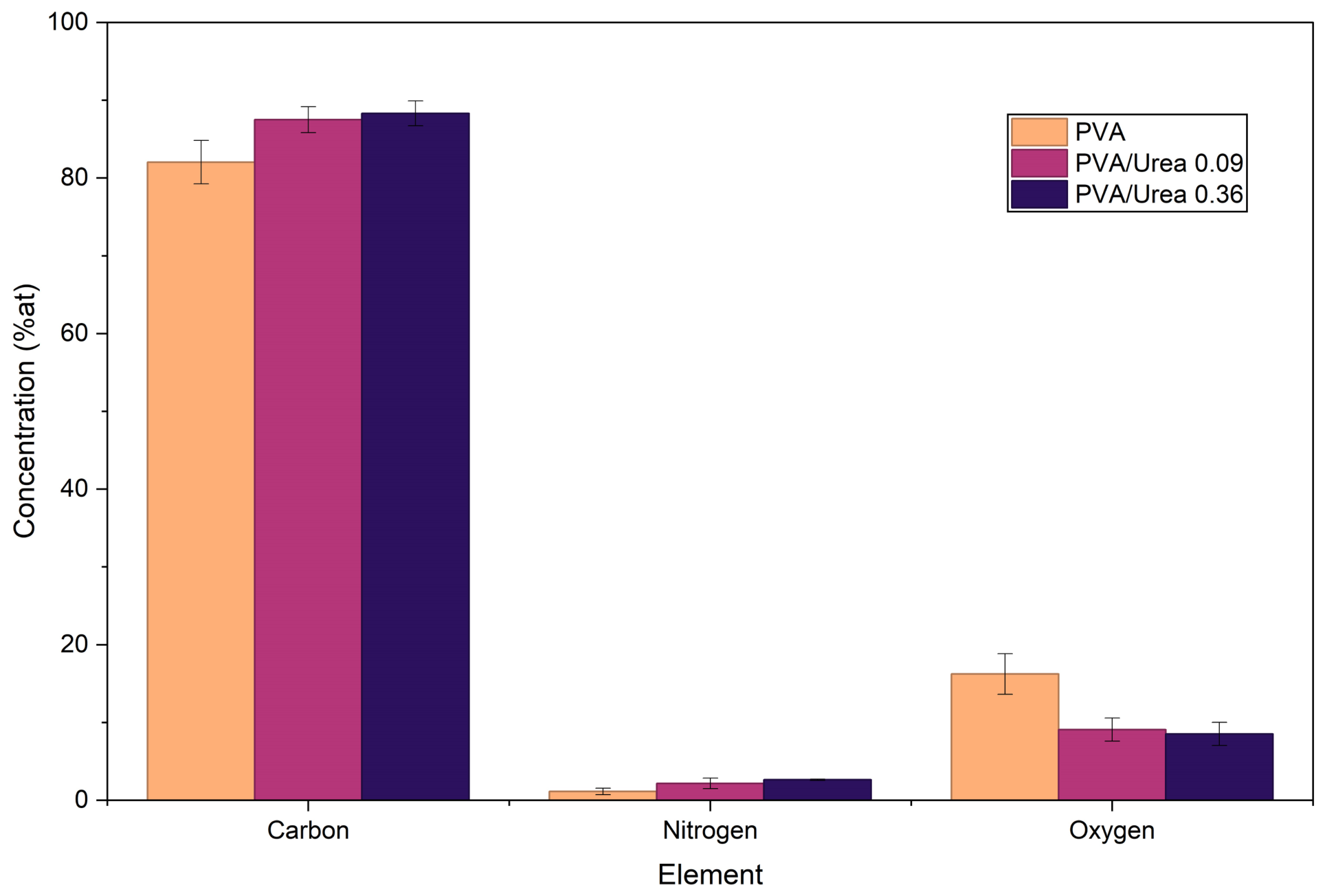
Figure 2 shows the energy-dispersive spectroscopy (EDS) analysis and show the presence of enlarged regions along some fibers, characterized by an elongated morphology and a diameter greater than that of the adjacent fibers. These structures are dispersed throughout the nanofibrous network and indicate morphological variations associated with the fiber formation process. This study provided evidence of the modifications induced by the incorporation of urea into the electrospun PVA nanofibers. Pure PVA exhibited a characteristic elemental profile dominated by carbon (82.04 wt.%) and oxygen (16.24 wt.%) reported in Table 1, which is mainly constituted by carbon backbones and hydroxyl functional groups. Upon the addition of urea, significant changes in elemental composition were detected: the carbon content increased progressively up to 88.32 wt.% in the PVA/0.36 g urea sample, while oxygen decreased to 8.53 wt.%. These variations can be attributed to the partial substitution of oxygenated groups in the PVA environment by urea–derived functionalities, which possess a lower oxygen/carbon ratio. In parallel, nitrogen, a distinctive element of urea absent in pure PVA, appeared at consistent levels (1.14–1.13 wt.%), serving as a clear compositional fingerprint of the molecular incorporation. Within the analysis of nitrogen content in the pure PVA sample, a value of 1.14% is observed, which is attributed to trace contamination, likely originating from environmental residues, solvents, or sample handling. However, due to its low percentage, this value is considered not significant and does not represent a real presence of nitrogen in the PVA structure.This trend not only confirms the effective embedding of urea within the nanofiber network but suggests the establishment of specific physicochemical interactions, between the amide groups of urea and the hydroxyl groups of PVA. Such interactions may play a crucial role in altering the physicochemical behavior of the nanofibers, particularly in terms of solubility, swelling dynamics, and the kinetics of nutrient release, as previously described in polymer–urea hybrid systems. Furthermore, the observed decrease in oxygen content with increasing urea loading contrasts, with some reports in the literature where enhanced oxygen ratios were associated with surface oxidation during fiber processing; in this case, the dominance of urea incorporation appears to override such effects [20]. Taken together, these results highlight that urea loading induces a systematic and reproducible modification of the elemental composition of PVA nanofibers, generating a unique chemical signature that underpins their potential use as controlled release systems.
Figure 2.
EDS elemental analysis micrographs of electrospun PVA nanofibers with different urea contents.
Pure PVA exhibited an elemental composition dominated by carbon (82.04 ± 2.78 wt.%) and oxygen (16.24 ± 2.61 wt.%), as shown in Table 3, which is consistent with the chemical structure of PVA, mainly composed of carbon backbones and hydroxyl functional groups. Following urea incorporation, significant changes in elemental composition were observed. Carbon content increased from 82.04 wt.% in neat PVA to 87.50 and 88.32 wt.% in the PVA/U0.09 and PVA/U0.36 formulations, respectively, while oxygen content decreased from 16.24 wt.% to 9.09 and 8.53 wt.%. Statistical analysis revealed significant differences in carbon and oxygen contents between neat PVA and the urea–loaded nanofibers (p < 0.05).
Nitrogen content also increased with urea loading, reaching 2.17 ± 0.67 wt.% in PVA/U0.09 and 2.64 ± 0.08 wt.% in PVA/U0.36. Although a low-nitrogen signal was detected in the neat PVA sample (1.145 ± 0.41 wt.%), this value was significantly lower than that observed for the highest urea-loaded formulation and may be associated with background contamination, environmental residues, or instrumental limitations commonly encountered in EDS analysis. The progressive increase in nitrogen content following urea incorporation provides compositional evidence supporting the successful incorporation of urea into the nanofibrous composite.
Table 5 presents the quantitative summary corresponding to Figure 3, where the percentages of carbon, oxygen, and nitrogen obtained by EDS analysis are reported for pure PVA nanofibers and urea-loaded PVA nanofibers. These results allow comparison of the changes in elemental composition associated with the incorporation of urea into the PVA, evidencing chemical and structural modifications within the electrospun system.
Table 5.
Elemental composition of PVA and PVA/urea nanofibers determined by EDS analysis. Values are expressed as mean ± standard deviation (n = 3). Different superscript letters within the same row indicate significant differences according to Tukey’s test (p < 0.05).
Figure 3.
Statistical analysis of EDS data of electrospun PVA nanofibers with different urea content.
The compositional changes are consistent with the findings reported by Kondal [21] and Beig [22], who demonstrated that the incorporation of nitrogen–containing compounds into PVA composite nanofibers leads to a decrease in oxygen content and an increase in total carbon, reflecting the replacement of oxygenated groups by C–N bonds.
Table 5 also presents the elemental composition determined by EDS, expressed as mean ± standard deviation (n = 3), together with the statistical analysis performed using Tukey’s multiple comparison test (p < 0.05). Different superscript letters indicate significant differences among treatments for the same element. The results show that urea incorporation significantly modified the elemental composition of the nanofibers, particularly with respect to nitrogen content.
Nitrogen was the most relevant element for confirming the incorporation of urea into the PVA. Although a nitrogen signal was detected in the neat PVA sample (1.14 ± 0.42 wt.%), which may be attributed to environmental contamination, processing residues, or inherent limitations of the EDS technique, the urea–loaded formulations exhibited an increase in nitrogen content. The PVA/U0.36 formulation showed the highest nitrogen content (2.65 ± 0.09 wt.%), which was significantly different from that of neat PVA (p < 0.05). These results reveal an increasing trend in nitrogen content with increasing urea concentration, providing compositional evidence for the presence of the fertilizer within the electrospun nanofibers.
Overall, the elemental composition obtained by EDS confirms the successful incorporation of urea into the electrospun nanofibers, as well as its direct influence on the chemical reorganization of the PVA network, resulting in a structure richer in carbon and nitrogen and with lower oxygen content. Although EDS is an elemental characterization technique, the results suggest that urea is not merely entrapped within the fiber core but rather integrated throughout the composite, forming a homogeneous polymer–urea system.
Therefore, the reported values correspond to representative measurements rather than replicated quantitative analyses, and standard deviation or error bars are not typically included unless multiple independent areas are systematically evaluated.
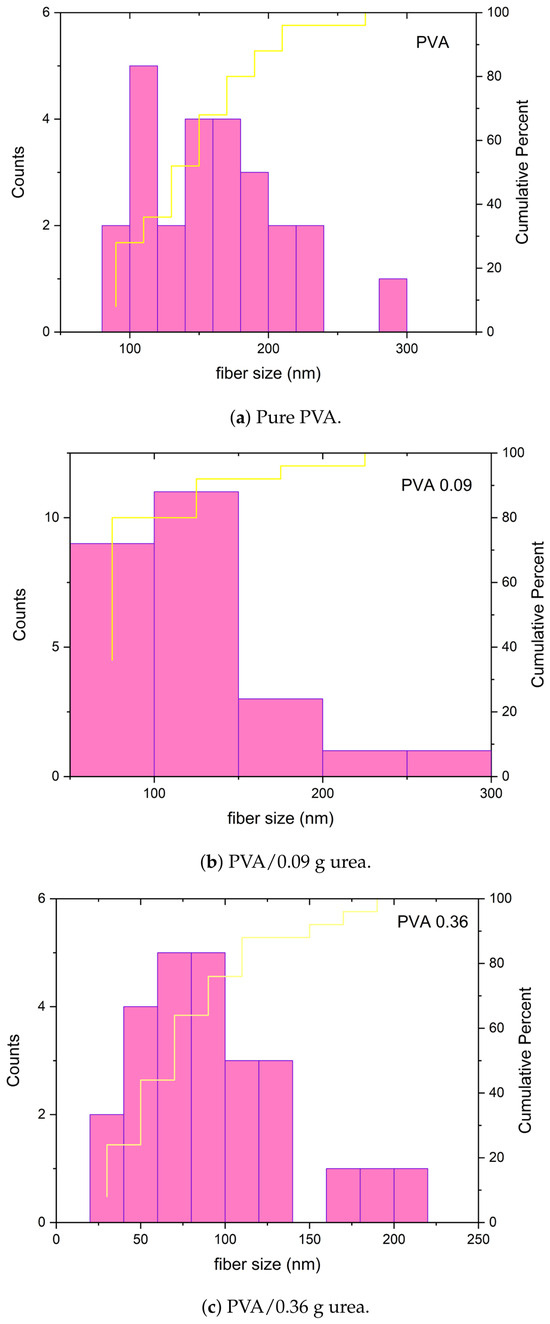
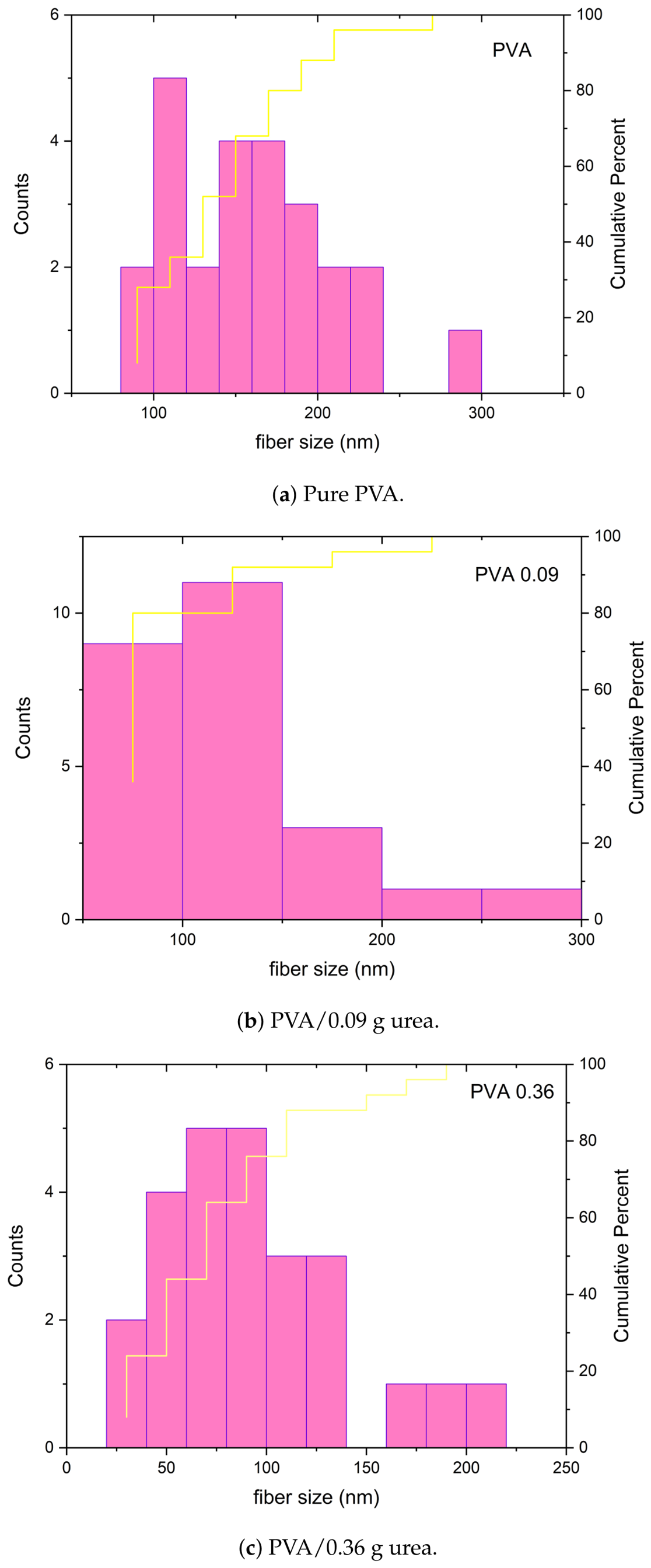
The average diameters of the nanofibers were determined through scanning electron microscopy (SEM) analysis. Table 6 presents the average fiber diameters obtained, whereas Figure 4 shows the corresponding diameter distribution, with values of 161.4 nm for pure PVA, 124.6 nm for PVA with 0.09 g of urea, and 95.4 nm for PVA with 0.36 g of urea. This systematic reduction in fiber diameter with increasing urea loading demonstrates that the molecule acts as a structural modulator during the electrospinning process. The findings are consistent with previous reports on PVA/urea systems modified with other additives, where the incorporation of urea increased the conductivity of the solution and enhanced the efficiency of electrostatic stretching, resulting in thinner and more uniform nanofibers [19]. Furthermore, the partial disruption of PVA–PVA hydrogen bonding and the formation of new PVA–urea interactions (–NH2–OH) reduce the viscosity and rigidity of the solution, facilitating fiber thinning, in agreement with prior observations in PVA–based composites designed for controlled release [23].
Table 6.
Average fiber diameter of PVA and PVA/urea nanofibers.
Figure 4.
Diameter distribution of electrospun PVA and PVA/urea nanofibers.
The trend observed in our work not only confirms the modulatory effect of urea on fiber morphology but also supports its potential to optimize composite nanofibers for agricultural and environmental applications where precise control of nutrient release is required.
The polydispersity index (PDI) shows that pure PVA exhibits the highest uniformity (0.092), while the incorporation of urea progressively increases the diameter dispersion (0.187 and 0.236), indicating greater heterogeneity in the nanofibers. This behavior is attributed to changes in the solution properties and to the interactions between PVA and urea, which affect the stability of the jet during electrospinning. The PDI values remained within the range of 0.092–0.236, well below the threshold of 0.30 commonly associated with narrow size distributions and high morphological uniformity [24]. This finding demonstrates that the electrospinning process produced homogeneous and well-defined nanofibers while maintaining adequate control over fiber morphology, a desirable characteristic for controlled-release applications.
3.2. XRD Analysis
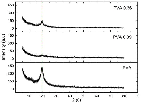
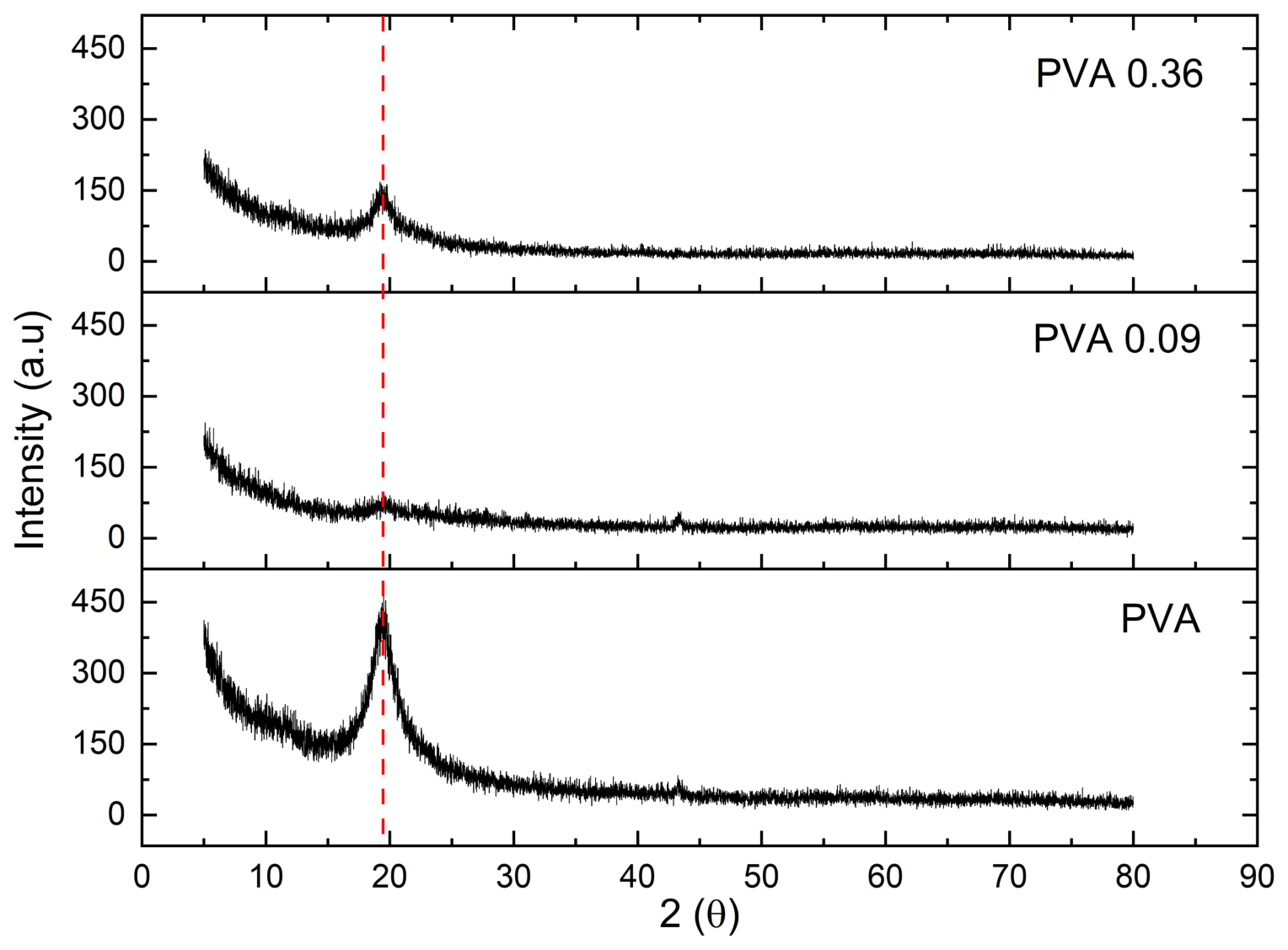
The X-ray diffraction patterns of PVA, PVA/U0.09, and PVA/U0.36 nanofibers (Figure 5) show, for the three formulations, a broad halo centered around 19–20°, characteristic of the semicrystalline packing of electrospun PVA [25]. Upon incorporation of urea (0.09 and 0.36 g), the maximum intensity decreases markedly and the peak becomes broader, while the 2θ position remains practically unchanged. This behavior indicates a reduction of the crystalline fraction due to the disruption of PVA–urea interactions (–NH2⋯OH), which promote a more disordered composite. The absence of additional sharp reflections in the urea-containing samples suggests that urea does not crystallize and that its distribution is predominantly amorphous within the polymeric network. The progressive decrease in the intensity of the characteristic PVA diffraction peak with increasing urea content suggests a disruption of the ordered arrangement of the polymer chains. This behavior indicates that urea is not merely dispersed within the fibers but rather interacts with the polymer network, interfering with the intermolecular interactions that contribute to the formation of crystalline domains. Therefore, the reduction in peak intensity supports the hypothesis of effective urea incorporation within the PVA composite, leading to modifications in the internal organization of the nanofibers.
Figure 5.
X-ray diffraction (XRD) patterns of PVA and PVA/urea nanofibers with different urea loading. The red dashed vertical line indicates the characteristic diffraction of PVA at 2θ ≈ 19.5–20°, used as a reference to compare changes in intensity and broadening after urea incorporation.
3.3. FTIR Analysis
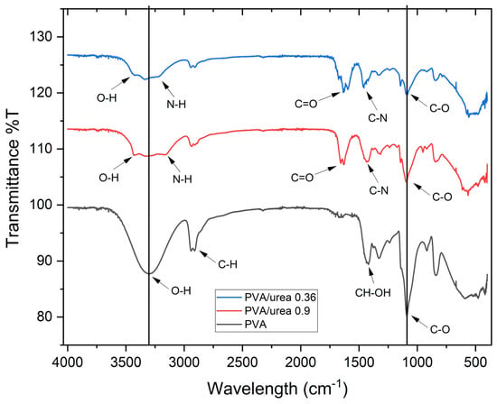
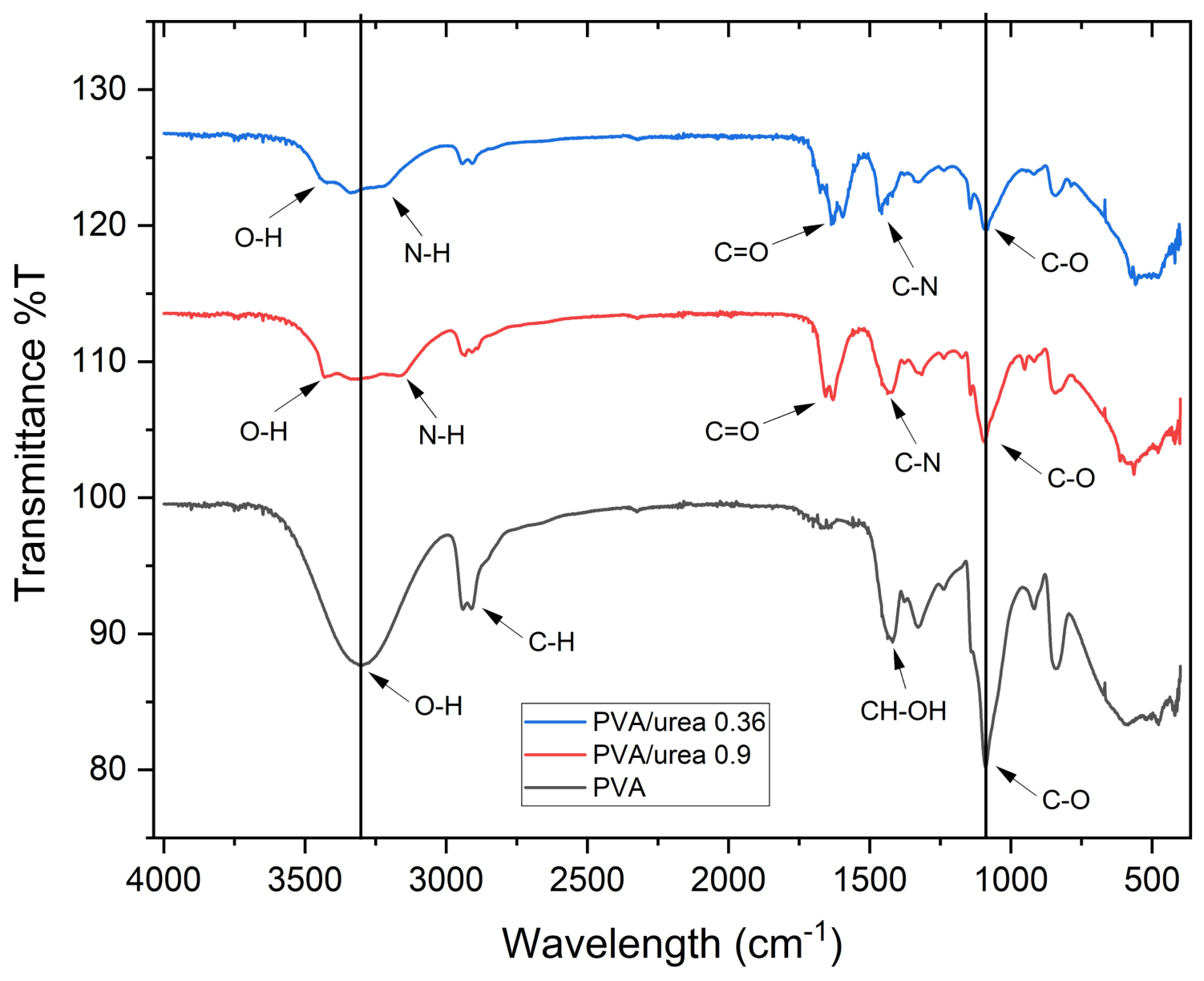
The FTIR spectra of pure PVA and PVA/urea nanofibers with different loadings (0.09 and 0.36 g) provide clear evidence of molecular-level interactions between PVA and urea (Figure 6). In all samples, the broad band around 3300–3400 cm−1 corresponds to O–H stretching vibrations of PVA hydroxyl groups. With urea incorporation, this band becomes less intense and slightly broader, indicating the rupture of PVA hydrogen bonding and the establishment of new interactions. These molecular interactions are correlated with the reduced crystallinity observed in XRD, confirming that urea induces a more amorphous polymeric compound.
Figure 6.
FTIR spectra of PVA and PVA/urea nanofibers with different urea loading.
Peaks at 2930–2940 cm−1 (C–H stretching) remain present in all spectra but exhibit a slight reduction in intensity with increasing urea, suggesting that chain mobility is modified as hydrogen-bonded networks become less rigid. More importantly, the spectra of PVA/urea samples reveal characteristic urea-associated bands: the C=O stretching at approximately 1670 cm−1 and the N–H bending vibrations around 1600 cm−1, which are absent in pure PVA [26]. Their progressive increase with urea loading confirms successful molecular incorporation, in agreement with the nitrogen signal detected by EDS.
Additional changes occur in the characteristic absorption region (800–1200 cm−1), where the C–O stretching of PVA overlaps with N–C–N vibrations of urea [27]. The relative enhancement of these signals in the PVA/0.36 g sample suggests a stronger contribution of urea-derived functionalities, reinforcing the idea of a polymer–additive hybrid network rather than a physical mixture.
The FTIR analysis demonstrates that urea not only becomes incorporated within the PVA but also establishes specific interactions, primarily through hydrogen bonding and other intermolecular interactions with the PVA chains. These interactions modify the vibrational modes of the functional groups involved, as evidenced by the changes observed in the FTIR spectra, and contribute to changes in the structural organization of the nanofibrous composite, as supported by the XRD results. Although FTIR does not provide spatial information regarding the distribution of these interactions, the observed spectral and structural changes suggest that the effect is associated with the polymer network rather than being restricted exclusively to bead regions. These interactions explain the morphological and structural effects observed: thinner nanofibers (SEM), modified composition (EDS), and decreased crystallinity (XRD). More importantly, such chemical reorganization at the molecular scale provides a mechanistic basis for the improved sustained release performance expected in PVA/urea systems, since a less crystalline, more hydrogen-bonded composite promotes water uptake, swelling, and gradual nutrient diffusion.
Table 7 summarizes the main vibrations identified in the FTIR spectra and their corresponding functional assignments, allowing a clearer visualization of the structural and chemical modifications that occur when urea is incorporated into the PVA. The comparison between pure PVA and urea-loaded nanofibers reveals significant changes in the intensity, position, and broadening of several characteristic bands, particularly those associated with O–H, N–H, and C=O groups.
Table 7.
FTIR band assignments of PVA and PVA/urea nanofibers.
3.4. Urea Release Profile
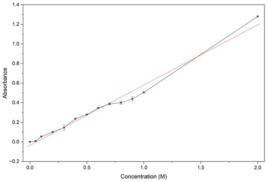
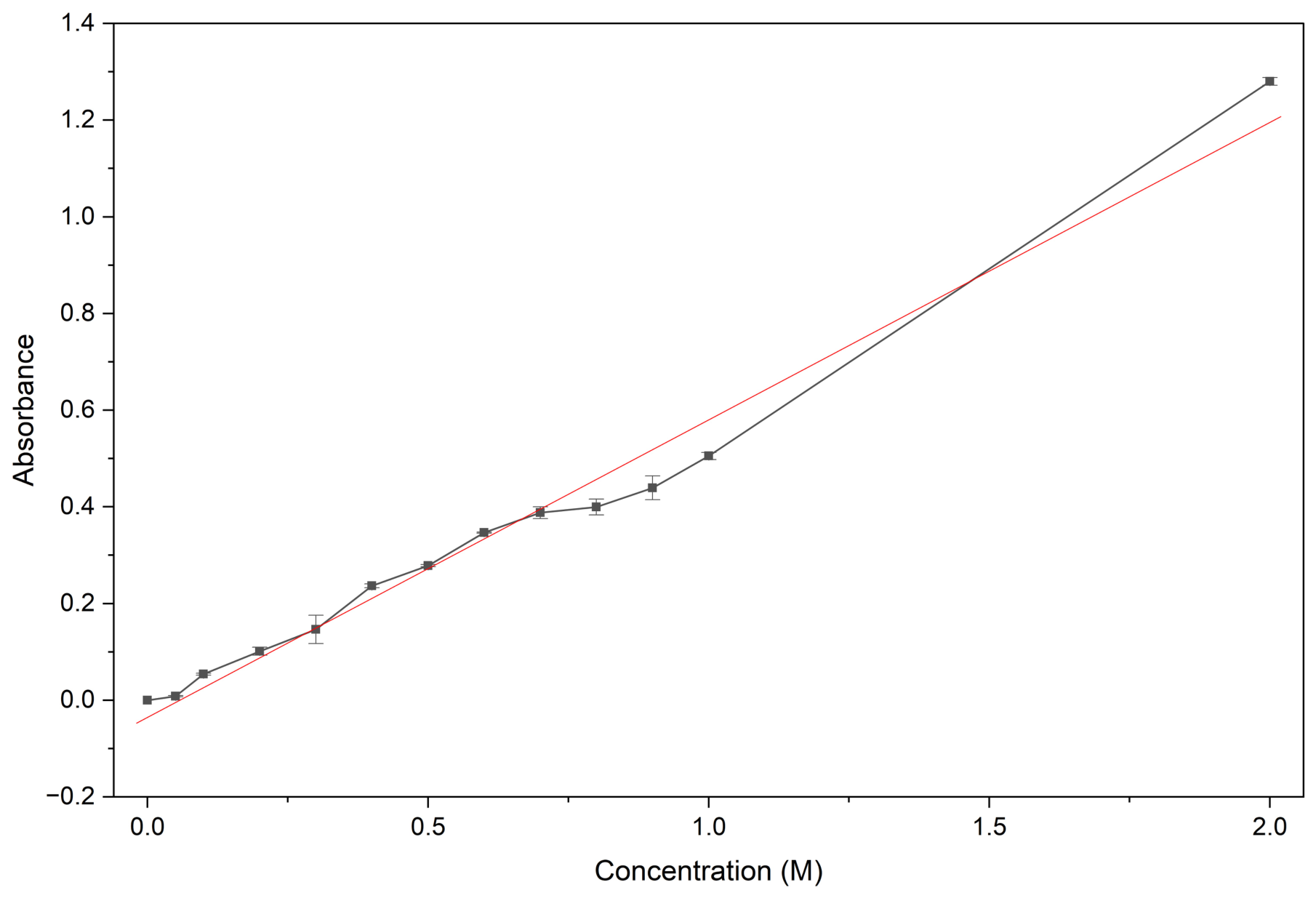
The resulting curve (Figure 7) exhibited a linear behavior, described by the linear regression coefficient (R2 = 0.98), indicating an excellent correlation between the variables.
Figure 7.
Urea calibration curve obtained by UV–Vis spectrophotometry at 560 nm. y = 0.6154x − 0.0355, R2 = 0.98.
These values were used to calculate the urea loading released as a function of time during the dissolution kinetics assays. The linearity observed in the calibration curve validates the application of the proposed UV–Vis spectrophotometric method, allowing for accurate quantification of the analyte.
3.5. Urea Release Behavior from PVA/Urea Nanofiber
The release profiles obtained demonstrate a prolonged supply of urea from the PVA nanofibers, consistent with the expected behavior of polymeric systems designed to deliver an analyte in a controlled manner over time. These electrospun composite nanofibers exhibit a high surface-area-to-volume ratio and a porous microstructure that facilitates molecular transport through the PVA network, modulating the diffusion of the encapsulated compound and preventing abrupt bursts of release [3].
It is essential to note that, in these formulations, urea is not merely adsorbed on the fiber surface but is instead distributed within the PVA. Upon hydration, PVA absorbs water and forms partially swollen domains in which molecular mobility increases, enabling diffusive gradients that act as driving forces for solute migration toward the external medium. This phenomenon allows release to occur even in the absence of erosion processes and primarily responds to transport mechanisms governed by the polymeric permeability and physical state [32]. Additionally, the bead structures observed in the urea-containing nanofibers may influence the release behavior. The morphological heterogeneity introduced by these structures can modify water penetration and diffusion pathways within the PVA nanofibrous composite, thereby affecting the transport mechanisms responsible for urea release.
Furthermore, several studies have shown that electrospun nanofibers function as regulatory barriers that slow down the release of hydrophilic agents through spatial constraints and confinement effects within the polymeric composite, thereby prolonging the release kinetics and preventing burst effects typical of non-structured systems [33]. Consequently, the interaction between urea and the hydrated PVA network explains the existence of a sustained diffusional profile, in which the release rate is determined by the water retention capacity of PVA, the local reorganization of polymer chains, and the gradual migration of the analyte from internal regions toward the exposed surface.
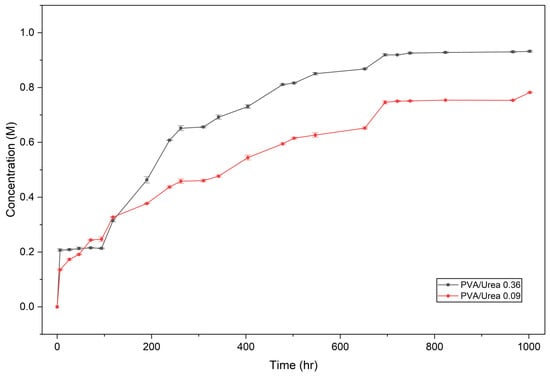
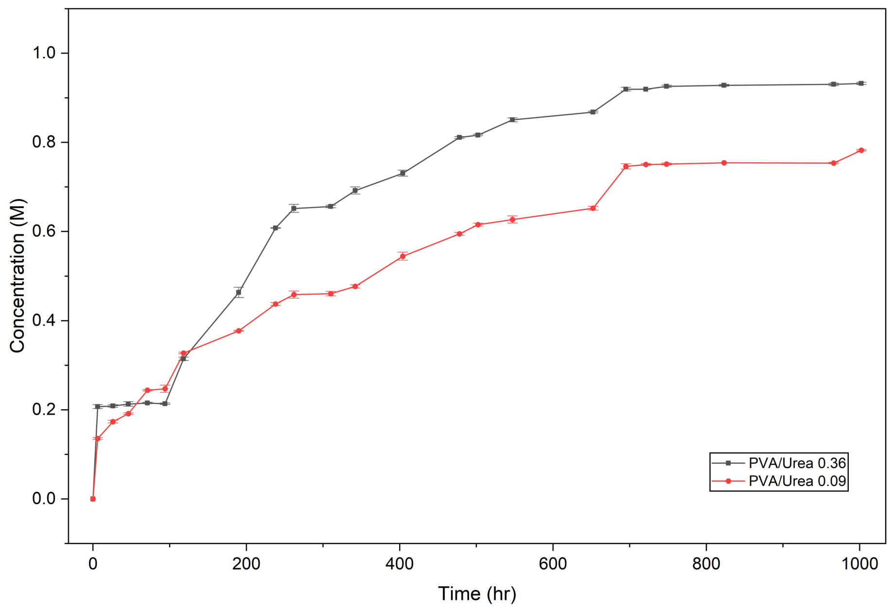
As shown in Figure 8, the urea release profiles corresponding to the formulations containing 0.36 g and 0.09 g exhibit distinct kinetic behaviors depending on the initial amount incorporated into the polymeric composite. These profiles allow for the identification of different phases within the release process and demonstrate the direct influence of the analyte loading on both the release rate and the time required to reach equilibrium.
Figure 8.
Urea release.
Table 8 presents the sampling times used during the urea release experiments, together with the measured values for the PVA/U0.09 and PVA/U0.36 nanofibrous composite. Measurements were conducted over a period of 1002 h (approximately 42 days) to monitor the urea release behavior from the nanofibers. The selected sampling intervals enabled evaluation of both the initial release stage and the subsequent stabilization phase, providing the experimental data used for concentration calculations and kinetic modeling.
Table 8.
Sampling times and absorbance values obtained during the urea release experiments.
In the system containing 0.36 g of urea, the curve exhibits a particular behavior: during the first 100 h, virtually no release is detected, indicating an initial hydration and diffusion period within the composite before urea can migrate toward the external medium. Subsequently, between 100 and 150 h, an abrupt increase in concentration is observed, evidencing an accelerated release attributable to the establishment of diffusion pathways within the polymeric network. After this phase, the slope gradually decreases, and the process transitions into a regime governed by internal diffusion, reaching a quasi-steady state between 750 and 800 h with a maximum concentration close to 0.95 M. This stepwise behavior is characteristic of hydrophilic electrospun composite nanofibers, in which release depends on the ingress and reorganization of water within the polymer, as well as on the subsequent diffusion of the encapsulated analyte [33].
In contrast, the system containing 0.09 g of urea exhibits an immediate release from the beginning, reflecting a lower diffusional resistance due to the reduced amount of analyte present in the composite. However, although the delay observed in the more concentrated formulation is absent, the slope of the profile is less pronounced, indicating a slower release process over time. This behavior leads to an earlier stabilization, occurring approximately between 650 and 700 h, with a final concentration close to 0.75 M. The reduced concentration gradient diminishes the driving force for urea transport and shortens the duration of the process, although it does not modify the release mechanism, which remains predominantly diffusion controlled.
Taken together, both systems demonstrate that the initial amount of urea influences the release kinetics and the time required to reach equilibrium but does not alter the fundamental release mechanism, which is essentially governed by hydration phenomena, diffusion, and reorganization of the polymeric composite.
The comparison of the urea release profiles obtained for both loading concentrations shows that, although the overall trend of the process is upward in both systems, their initial behavior differs significantly. In the formulation containing 0.36 g, the release profile exhibits an initial lag phase, during which no appreciable increase in the released concentration is observed during the first hours, confirming the absence of a burst release and suggesting that the urea remains confined within the polymeric network until the PVA reaches a critical hydration level capable of generating internal transport channels. In contrast, the 0.09 g formulation displays an earlier and more gradual increase, indicating that the lower urea content reduces the internal resistance to transport, allowing a faster establishment of the chemical gradient responsible for diffusion. This experimental result supports the idea that the release kinetics are strongly conditioned by the relationship between osmotic loading, degree of encapsulation, and structural reorganization of the composite and that the system does not respond solely to a simple diffusive process.
3.6. Kinetic Fittings
To better understand the mechanisms governing urea release from PVA nanofibers, the experimental data were fitted to different kinetic models commonly used in controlled-release systems. The zero-order, first-order, second-order, Higuchi, and Korsmeyer–Peppas models were selected because each describes distinct transport mechanisms and allows identification of the predominant process responsible for nutrient release.
The zero-order model assumes a constant release rate that is independent of the concentration of the incorporated compound. The first-order model considers that the release rate depends on the amount of solute still retained within the composite. The second-order model is commonly applied to systems in which release may be influenced by more complex interactions between the active compound and the polymeric composite. In contrast, the Higuchi model is based on Fickian diffusion through a solid composite and is widely used for polymer-based delivery systems. Finally, the Korsmeyer–Peppas model enables identification of the dominant transport mechanism through the release exponent (n), allowing differentiation between Fickian diffusion, anomalous transport, and composite relaxation-controlled mechanisms.
The evaluation of multiple kinetic models is necessary because polymeric systems often exhibit more than one release mechanism simultaneously. Therefore, comparing the goodness-of-fit parameters of different models makes it possible to determine which model best describes the experimental behavior and provides insight into the physical processes controlling urea release from the nanofibrous composites.
3.6.1. Analysis of Urea Release Kinetics in PVA/Urea 0.36 Nanofibers
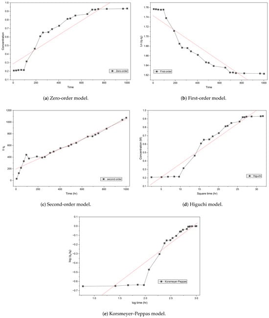
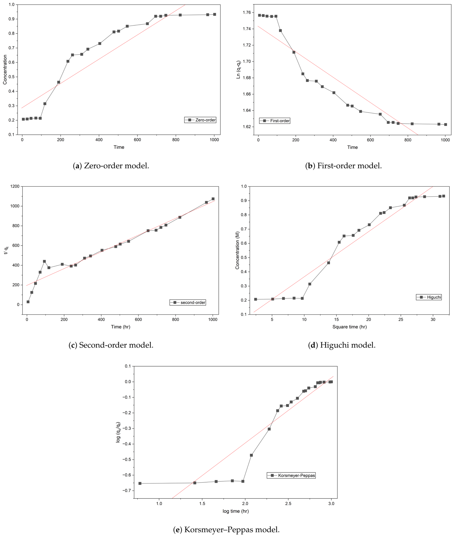
The kinetic fittings obtained for the five models evaluated in the PVA/urea 0.36 nanofiber system are presented in Figure 9. Similarly, Table 9 summarizes the main parameters obtained from fitting different kinetic models applied to urea release from PVA/Urea nanofibers. The R2 values, linear equations, and associated kinetic constants allow comparison of the fitting quality of each model and provide insight into the dominant mechanisms governing the release process. Through this analysis, it is possible to identify whether nutrient transport is mainly controlled by diffusion, polymer composite relaxation, or physicochemical interactions between urea and PVA. Furthermore, the comparison among models provides relevant information regarding the sustained-release behavior and the influence of the nanofibrous structure on the kinetics of the system.
Figure 9.
Comparative kinetic modeling of urea release from electrospun PVA nanofibers loaded with 0.36 g of urea using different mathematical models: (a) zero-order, (b) first-order, (c) second-order, (d) Higuchi, and (e) Korsmeyer–Peppas. The red solid line represents the linear fitting/regression of each kinetic model, while the black symbols correspond to the experimental urea release data.
Table 9.
Kinetic model fitting parameters for urea release from PVA/urea 0.36 nanofibers.
For the zero-order (R2 = 0.8488) and first-order (R2 = 0.8489) models, they exhibit poor fitting, ruling out that the release is governed exclusively by time or by the remaining solute concentration. Likewise, the Korsmeyer–Peppas model shows a low correlation (R2 = 0.5731) and an exponent n = 0.0283, well below the Fickian diffusion threshold (n < 0.45), indicating highly restricted diffusion and confirming that this model does not adequately describe the system.
In contrast, the Higuchi model presents a significantly higher coefficient of determination (R2 = 0.9379, kh = 0.0317), indicating that diffusion through a hydrated polymeric composite is a relevant mechanism in the release process. This behavior is consistent with previous reports on electrospun systems, where the high surface-area-to-volume ratio and fibrous microstructure generate tortuous pathways that promote Fickian transport of the analyte [34].
Nevertheless, the second-order kinetic model provides the best statistical fit (R2 = 0.9463, k2 = 0.8439), emerging as the dominant model for this formulation. This superiority indicates that the release is not governed solely by the concentration gradient but also by specific interactions between urea and the PVA composite. In this context, the process is controlled both by transport within the polymer network and by the energy required to disrupt molecular interactions, particularly hydrogen bonding between urea molecules and PVA hydroxyl groups, as well as by the progressive reorganization of hydrated domains and the formation of transport pathways.
It is important to explicitly note that, although the Higuchi model shows a strong fit, it assumes that the polymeric composite behaves as an inert medium where transport is governed exclusively by Fickian diffusion. However, the superior fitting of the second-order model demonstrates that this assumption is insufficient to fully describe the system. In particular, the formation of hydrogen bonds between urea molecules and PVA hydroxyl groups introduces an additional energetic barrier that must be overcome for release to occur, which is not accounted for in the Higuchi model. Consequently, the predominance of the second-order model reflects a hybrid mechanism in which diffusion is coupled with interaction-controlled release.
The structural basis for this behavior is supported by X-ray diffraction results, which show a reduction in the intensity of the characteristic semicrystalline peak of PVA (19–20° 2θ) upon urea incorporation. This decrease in crystallinity indicates a transition toward a more amorphous structure, where previously confined functional groups become more accessible, enhancing interaction with the solute. From a kinetic perspective, the second-order constant (k2 = 0.8439) reflects this combined effect of diffusion and interaction, establishing a direct relationship between the kinetic parameter and the structural properties of the material, such as reduced crystallinity and increased availability of active sites within the amorphous phase.
3.6.2. Analysis of Urea Release Kinetics in PVA/Urea 0.09 Nanofibers
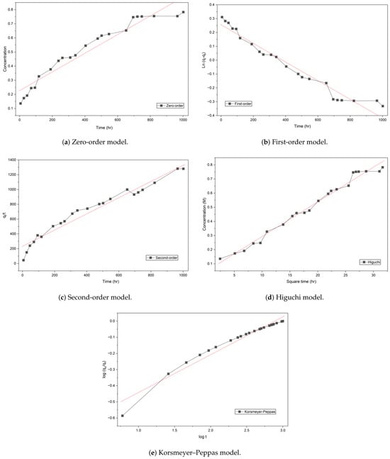
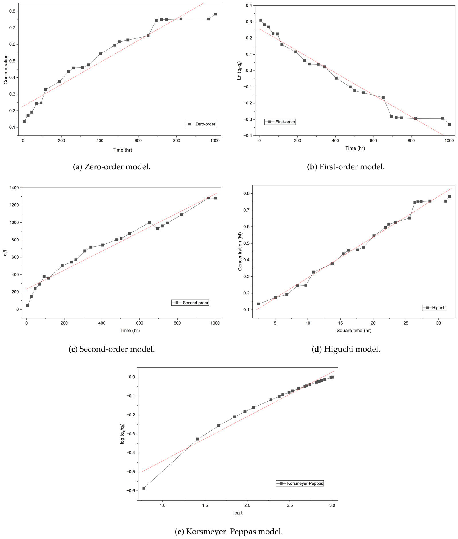
On the other hand, for the higher loading formulation (Figure 10, the kinetic behavior shows important differences. The zero-order model presents a relatively high fit (R2 = 0.9275), suggesting a partially time-dependent trend, while the first-order model improves the fit (R2 = 0.9545), indicating that the release depends to some extent on the remaining solute concentration. The second-order model also shows a high fit (R2 = 0.9562), suggesting the presence of physicochemical interactions between urea and the polymeric composite. Table 10 shows the urea release profile corresponding to the PVA/urea system with a loading of 0.36 g, allowing observation of the temporal evolution of the released concentration and the different kinetic regimes involved in the process. The obtained behavior demonstrates that the release does not occur immediately or linearly but rather through successive stages associated with polymer composite hydration, diffusion channel formation, and the progressive transport of urea toward the external medium.
Figure 10.
Comparative kinetic modeling of urea release from electrospun PVA nanofibers loaded with 0.09 g of urea using different mathematical models: (a) zero-order, (b) first-order, (c) second-order, (d) Higuchi, and (e) Korsmeyer–Peppas. The red solid line represents the linear fitting/regression of each kinetic model.
Table 10.
Kinetic model fitting parameters for urea release from PVA/Urea 0.09 nanofibers.
However, in this case, the Higuchi model provides the best fit (R2 = 0.9836, kh = 0.0245), demonstrating that the release is predominantly governed by a diffusion mechanism through the hydrated polymeric composite. This behavior indicates that, at higher urea loadings, the availability of the analyte within the composite is sufficient for the process to be dominated by dissolution and diffusion into the surrounding medium, reducing the relative impact of molecular interactions on the overall kinetics.
Additionally, the Korsmeyer–Peppas model shows a good fit (R2 = 0.9578); however, since the Higuchi model provides the highest correlation, the release is primarily interpreted as diffusion-controlled.
3.6.3. Comparative Analysis of Release Mechanisms in PVA
Overall, the comparison between both formulations reveals a clear transition in the release mechanism as a function of urea loading. In the lower loading system (0.09 g), the process is governed by a hybrid mechanism in which diffusion is coupled with physicochemical interactions and structural reorganization. In contrast, in the higher loading system (0.36 g), the behavior is predominantly diffusive, with the polymer composite acting mainly as a physical barrier that regulates analyte release.
This behavior is consistent with the nature of electrospun composite nanofibers, where polymer hydration, chain reorganization, and solute–composite interactions determine release kinetics [33].
While classical semi-empirical models (such as the Higuchi and Korsmeyer–Peppas frameworks) successfully classify the macro-scale transport behavior in this study, they treat the diffusion domain as a continuous, homogeneous Euclidean space. However, electrospun nanofibrous mats possess an intrinsically disordered, hierarchically porous, and tortuous morphology. The transport of urea within this composite is governed by structural features across two distinct physical dimensions: the sub–micron scale of the individual fiber core (where solid-state interaction and composite relaxation restrict molecular mobility) and the macro-porous scale of the interconnected fibrous network (where fluid percolation occurs). To describe this anomalous, non-linear diffusion with elevated mathematical rigor, a fractal Fick model based on the two-scale fractal derivative presents a compelling advanced framework.
Under this theoretical perspective, the standard space-time coordinates are transformed using a fractional or fractal metric tensor to account for the spatial constraints and structural tortuosity of the porous medium. The generalized fractal diffusion equation can be expressed as:
where and represent the fractal derivatives with respect to time (t) and space (x), respectively, capturing the memory effects of the polymer-solute interactions and the non-Euclidean path of the permeating fluid. Specifically, applying a two–scale fractal derivative allows the decoupling of transport phenomena occurring at the micro-structural level (intra–fiber boundary layers) from those occurring at the macro–structural level (inter–fiber voids). Incorporating this fractal calculus approach into future work will enable the formulation of unified predictive equations that accurately capture local concentration gradients and anomalous transport exponents without relying on empirical fitting parameters [35].
In conclusion, the release kinetics in PVA/urea nanofibers cannot be described by a single universal mechanism, but rather depend critically on the material structure and analyte loading. The lower loading formulation exhibits a highly interactive and structurally dynamic system, whereas the higher loading formulation follows a predominantly classical diffusive regime, where analyte availability governs the release process.
3.7. Urea Release Percentage
The release behavior of urea from electrospun PVA nanofibers was evaluated in terms of release percentage (RP), which represents the fraction of urea effectively transferred from the polymeric composite into the surrounding medium relative to the initial loading (Equation (2)).
The formulation containing 0.36 g of urea exhibits a final RP value of 15.5% (Table 11), indicating that a large proportion of the nutrient remains confined within the nanofibrous composite. This behavior suggests the presence of strong physicochemical interactions between urea and PVA chains, as well as possible localization of urea within less accessible domains, such as semi-crystalline regions or densely packed fiber structures, which limit mass transport into the aqueous medium.
Table 11.
Encapsulation efficiency of urea in electrospun PVA nanofiber.
In contrast, the 0.09 g formulation reaches a significantly higher RP value of 52.0%, reflecting a more efficient release process. This can be attributed to a more homogeneous distribution of urea within the polymer composite, reduced structural density, and shorter and less tortuous diffusion pathways, which facilitate the migration of urea molecules toward the external medium.
These results clearly demonstrate that the release percentage is strongly dependent on the initial urea loading. Higher loadings promote retention due to increased composite densification and intermolecular interactions, which impose an additional barrier to diffusion. Conversely, lower loadings promote enhanced release by reducing these constraints.
From an application perspective, the 0.36 g system behaves as a highly retentive reservoir suitable for prolonged nutrient delivery, while the 0.09 g formulation achieves a more favorable balance between nutrient availability and controlled release. This highlights the critical role of composite design and composition in governing nutrient delivery efficiency and optimizing agronomic performance.
4. Discussion
The results obtained in this study demonstrate that urea loading acts as a key design parameter controlling the transport behavior of electrospun PVA nanofibers. Rather than functioning solely as a nutrient reservoir, the incorporated urea modifies the physicochemical organization of the composite, generating release profiles that can be tuned through formulation design. This finding highlights the potential of electrospinning as a morphology-engineering strategy for the development of controlled-release agricultural materials.
Compared with conventional controlled-release systems, including starch/PVA/biochar coatings [36], hydrophobic PVA systems modified with fluorosilanes [37], chitosan/PVP membranes [38], lignin–PVA composite coatings [39], and starch/PVA nanocomposites [40], the electrospun systems developed in this work regulate nutrient release through the structural characteristics of the nanofibrous network rather than solely through the presence of an external diffusion barrier. As summarized in Table 12, the interconnected porous morphology of electrospun fibers provides an alternative route for controlling nutrient transport while avoiding additional coating steps that may increase processing complexity. In addition, the bead structures observed in the urea-containing nanofibers may introduce local morphological heterogeneities within the fibrous network. These features could influence water penetration and diffusion pathways by generating regions with different hydration and transport characteristics, potentially contributing to the release behavior observed in the present study.
Table 12.
Comparative overview of PVA-based urea release systems.
From an agronomic perspective, although the nitrogen content of the nanofibrous mats is lower than that of bulk granular fertilizers, their value lies in the ability to regulate nutrient availability over extended periods. Conventional fertilizers frequently exhibit low nitrogen use efficiency due to ammonia volatilization and nitrate leaching losses, which can account for a substantial fraction of the applied nitrogen [22]. By providing a more gradual nutrient supply, controlled-release systems may improve synchronization between nitrogen availability and crop demand, thereby contributing to enhanced nitrogen use efficiency and reduced environmental impact. Similar improvements in nutrient management have been reported for PVA-based controlled-release formulations and starch/PVA composite systems [36,40].
Furthermore, the fabrication strategy employed in this work is compatible with emerging high-throughput electrospinning technologies such as bubble electrospinning, which may facilitate the large-scale production of morphology-engineered fertilizers. The ability to modulate release behavior through structural design rather than through extensive chemical modification represents a significant advantage for future agricultural applications.
Finally, the anomalous and multiphase transport behavior observed in these nanofibrous systems suggests that future studies may benefit from advanced theoretical approaches capable of describing diffusion in hierarchical porous materials. Fractal Fick-law-based models, recently applied to moisture diffusion and permeability in porous concrete, provide a useful conceptual framework for interpreting transport processes governed by pore-size distribution, tortuosity, and multiscale transport pathways. Therefore, fractal diffusion analysis could be incorporated in future work to quantify scale-dependent urea transport within electrospun PVA/urea nanofibrous networks [35].
Overall, the findings demonstrate that electrospun PVA/urea nanofibers constitute a promising platform for controlled nutrient delivery, where release behavior can be tailored through structural design of the nanofibrous composite, offering new opportunities for the development of sustainable fertilization technologies.
5. Conclusions
The results obtained demonstrate that urea loading plays a dual role in electrospun PVA nanofibers, acting not only as the nutrient source but also as a structural modifier. By altering the internal organization of the nanofibers, urea directly influences the mechanisms governing nutrient transport and release. Consequently, urea concentration emerges as a key design parameter for controlling release behavior, providing a foundation for the development of fertilizers capable of being tailored to the nutritional requirements of different agricultural systems.
Author Contributions
Conceptualization, M.G.G.-B., A.U.C.-R., J.R.-R. and V.V.-B.; methodology, C.E.P.-G., A.A.F.-P. and A.Á.-L.; software, M.G.G.-B. and J.R.-R.; validation, M.G.G.-B., A.U.C.-R. and A.Á.-L.; formal analysis, M.G.G.-B., A.U.C.-R. and C.E.P.-G.; investigation, M.G.G.-B., A.U.C.-R., C.E.P.-G., A.A.F.-P. and A.Á.-L.; resourses, J.R.-R., A.A.F.-P. and A.Á.-L.; data curation, A.A.F.-P., A.U.C.-R., C.E.P.-G., A.Á.-L. and V.V.-B.; writting—original draft preparation, M.G.G.-B., C.E.P.-G. and A.Á.-L.; writing—review and editing, J.R.-R., C.E.P.-G. and A.Á.-L.; administration, J.R.-R. and A.A.F.-P.; funding acquisition, J.R.-R., A.A.F.-P., A.Á.-L. and V.V.-B. All authors have read and agreed to the published version of the manuscript.
Funding
This research received no external funding.
Institutional Review Board Statement
Not applicable.
Informed Consent Statement
Not applicable.
Data Availability Statement
Data will be made available upon reasonable request.
Acknowledgments
The author wishes to express her sincere gratitude to the Secretariat of Science, Humanities, Technology and Innovation (SECIHTI) for the support provided for the completion of this work, under the fellowship registration number CVU 926190. The authors acknowledge the use of artificial intelligence-based tools to assist with language refinement, grammatical correction, writing improvement, and enhancement of the manuscript’s clarity. All ideas, experimental design, analyses, interpretation of results, and conclusions presented in this work are the sole responsibility of the authors. The use of these tools was strictly limited to improving the quality and readability of the manuscript and did not influence the scientific development, methodology, data analysis, or conclusions of the research.
Conflicts of Interest
The authors declare no conflicts of interest.
References
- Cruz-Navarro, A.; Rivera, J.M.; Durán-Hernández, J.; Castillo-Blum, S.; Flores-Parra, A.; Sánchez, M.; Hernández-Ahuactzi, I.; Colorado-Peralta, R. Luminescence properties and DFT calculations of lanthanide (III) complexes (Ln = La, Nd, Sm, Eu, Gd, Tb, Dy) with 2,6-bis(5-methyl-benzimidazol-2-yl) pyridine. J. Mol. Struct. 2018, 1164, 209–216. [Google Scholar] [CrossRef] [Scilit]
- Govindasamy, P.; Muthusamy, S.K.; Bagavathiannan, M.; Mowrer, J.; Jagannadham, P.T.K.; Maity, A.; Halli, H.M.; Sujayananad, G.K.; Vadivel, R.; Das, T.K.; et al. Nitrogen Use Efficiency—A Key to Enhance Crop Productivity under a Changing Climate. Front. Plant Sci. 2023, 14, 1121073. [Google Scholar] [CrossRef] [Scilit] [PubMed]
- Javazmi, L.; Low, T.; Ash, G.; Young, A. Investigation of slow release of urea from biodegradable single- and double-layered hollow nanofibre yarns. Sci. Rep. 2020, 10, 19619. [Google Scholar] [CrossRef] [Scilit] [PubMed]
- Liu, G.L.; Zhang, Y.M.; Tian, D.; Zhou, B.Z.; Lu, Z.Q.; Wang, C.X. Last patents on bubble electrospinning. Recent Pat. Nanotechnol. 2020, 14, 5–9. [Google Scholar] [CrossRef] [Scilit] [PubMed]
- Wan, L.Y. Bubble electrospinning and bubble-spun nanofibers. Recent Pat. Nanotechnol. 2020, 14, 10–13. [Google Scholar] [CrossRef] [Scilit] [PubMed]
- Huesca-Urióstegui, K.; García-Valderrama, E.J.; Gutierrez-Uribe, J.A.; Antunes-Ricardo, M.; Guajardo-Flores, D. Nanofiber Systems as Herbal Bioactive Compounds Carriers: Current Applications in Healthcare. Pharmaceutics 2022, 14, 191. [Google Scholar] [CrossRef] [Scilit] [PubMed]
- Walteros-León, M.; Álvarez Láinez, M.L. Colloidal and rheological properties of natural rubber latex concentrated with hydroxyethyl cellulose and sodium dodecyl sulphate. J. Appl. Polym. Sci. 2022, 139, 52034. [Google Scholar] [CrossRef] [Scilit]
- Yin, X.; Zhang, T.; Zhao, T.; Wang, K.; Xu, Z.; Zhao, Y. Cellulose-based, flexible polyurethane polyHIPEs with quasi-closed-cell structures and high stability for thermal insulation. Carbohydr. Polym. 2023, 302, 120385. [Google Scholar] [CrossRef] [Scilit] [PubMed]
- Colín-Orozco, J.; Colín-Orozco, E.; Valdivia-Barrientos, R. Production of Nanofibers by Electrospinning as Carriers of Agrochemical. Fibers 2024, 12, 64. [Google Scholar] [CrossRef] [Scilit]
- Badgar, K.; Abdalla, N.; El-Ramady, H.; Prokisch, J. Sustainable Applications of Nanofibers in Agriculture and Water Treatment: A Review. Sustainability 2022, 14, 464. [Google Scholar] [CrossRef] [Scilit]
- Meraz-Dávila, S.; Pérez-García, C.E.; Feregrino-Perez, A.A. Challenges and advantages of electrospun nanofibers in agriculture: A review. Mater. Res. Express 2021, 8, 042001. [Google Scholar] [CrossRef] [Scilit]
- Yin, J.Y.; Boaretti, C.; Lorenzetti, A.; Martucci, A.; Roso, M.; Modesti, M. Effects of Solvent and Electrospinning Parameters on the Morphology and Piezoelectric Properties of PVDF Nanofibrous Membrane. Nanomaterials 2022, 12, 962. [Google Scholar] [CrossRef] [Scilit] [PubMed]
- Gong, W.; Yang, W.; Zhou, J.; Zhang, S.; Yu, D.G.; Liu, P. Engineered Beads-on-a-String Nanocomposites for an Improved Drug Fast-Sustained Bi-Stage Release. Nanocomposites 2024, 10, 228–241. [Google Scholar] [CrossRef] [Scilit]
- Inayah, N.N.; Kusumaatmaja, A.; Murtafi’atin, R. Slow-release fertilizer behavior of polyvinyl alcohol (PVA)/urea/TiO2 nanofiber membrane. Polym. Adv. Technol. 2024, 35, e6472. [Google Scholar] [CrossRef] [Scilit]
- Li, X.; Shen, J.; Sun, Z.; Liu, Y.; Zhang, W.; Wu, B.; Ma, F.; Gu, Q. Degradation of 2,4-dinitrotoluene using ferrous activated persulfate: Kinetics, mechanisms, and effects of natural water matrices. J. Environ. Chem. Eng. 2021, 9, 106048. [Google Scholar] [CrossRef] [Scilit]
- Ángeles, H.M.; Moreno, J.G.R.; Rubio, C.A.N.; Castillo, R.R.V.; Ronquillo-Lomeli, G.; Carrillo-Serrano, R.V.; Perea, M.T. Effect of humidity incorporation on the development of a ground enhancement material. Sci. Rep. 2025, 15, 22049. [Google Scholar] [CrossRef] [Scilit] [PubMed]
- Temel-Soylu, T.M.; Kececiler-Emir, C.; Rababah, T.; Ozel, C.; Yucel, S.; Basaran-Elalmis, Y.; Altan, D.; Kirgiz, O.; Secinti, I.E.; Kaya, U.; et al. Green electrospun poly(vinyl alcohol)/gelatin-based nanofibrous membrane by incorporating 45S5 bioglass nanoparticles and urea for wound dressing applications: Characterization and in vitro and in vivo evaluations. ACS Omega 2024, 9, 21187–21203. [Google Scholar] [CrossRef] [Scilit] [PubMed]
- Pih, G.Y.; Noh, J.H.; Ahn, J.Y.; Han, G.S.; Jung, H.S.; Jung, H.Y.; Kim, J.M. Objective Interpretation of the Rapid Urease Test for Helicobacter pylori Infection Using Colorimetry. J. Korean Med. Sci. 2022, 37, e226. [Google Scholar] [CrossRef] [Scilit] [PubMed]
- Zhan, F.; Yan, X.w.; Li, J.; Sheng, F.; Li, B. Encapsulation of tangeretin in PVA/PAA crosslinking electrospun fibers by emulsion-electrospinning: Morphology characterization, slow-release, and antioxidant activity assessment. Food Chem. 2021, 337, 127763. [Google Scholar] [CrossRef] [Scilit] [PubMed]
- Lo, C.T.; Lin, K.W.; Wang, T.P.; Huang, S.M.; Lee, C.L. Differentiating between the Effects of Nitrogen Plasma and Hydrothermal Treatment on Electrospun Carbon Fibers Used as Supercapacitor Electrodes. Electrochim. Acta 2021, 381, 138255. [Google Scholar] [CrossRef] [Scilit]
- Kondal, R.; Kalia, A.; Krejcar, O.; Kuča, K.; Sharma, S.P.; Luthra, K.; Dheri, G.S.; Vikal, Y.; Taggar, M.S.; Abd-Elsalam, K.A.; et al. Chitosan-urea nanocomposite for improved fertilizer applications: The effect on the soil enzymatic activities and microflora dynamics in N cycle of potatoes (Solanum tuberosum L.). Polymers 2021, 13, 2887. [Google Scholar] [CrossRef] [Scilit] [PubMed]
- Beig, B.; Niazi, M.B.K.; Jahan, Z.; Hussain, A.; Zia, M.H.; Mehran, M.T. Coating materials for slow release of nitrogen from urea fertilizer: A review. J. Plant Nutr. 2020, 43, 1510–1533. [Google Scholar] [CrossRef] [Scilit]
- Adeel, M.; Ren, L.F.; Li, J.; Shao, J.; Jawad, A.; Su, C.; Wang, Y.; Guo, L.; He, Y. Enhanced mechanical properties of PDMS/PMMA composite membrane using MWCNTs and its application in phenol separation from saline wastewater. J. Appl. Polym. Sci. 2019, 136, 47123. [Google Scholar] [CrossRef] [Scilit]
- Chudzińska-Skorupińska, J.; Wawrzyńczak, A.; Feliczak-Guzik, A. Ethosome-Based Colloidal Systems for Transdermal Delivery: The Role of Biosurfactant in Enhancing Stability and Efficacy. Materials 2025, 18, 5355. [Google Scholar] [CrossRef] [Scilit] [PubMed]
- Alwan, T.J.; Toma, Z.A.; Kudhier, M.A.; Ziadan, K.M. Preparation and characterization of the PVA nanofibers produced by electrospinning. Madridge J. Nanotechnol. Nanosci. 2016, 1, 1–3. [Google Scholar] [CrossRef] [Scilit]
- Nandiyanto, A.B.D.; Ragadhita, R.; Fiandini, M. Interpretation of Fourier Transform Infrared Spectra (FTIR): A Practical Approach in the Polymer/Plastic Thermal Decomposition. Indones. J. Sci. Technol. 2023, 8, 113–126. [Google Scholar] [CrossRef] [Scilit]
- Gáplovská, K.; Šimonovičová, A.; Halko, R.; Okenicová, L.; Žemberyová, M.; Čerňanskỳ, S.; Brandeburová, P.; Mackulǎk, T. Study of the Binding Sites in the Biomass of Aspergillus niger Wild-Type Strains by FTIR Spectroscopy. Chem. Pap. 2018, 72, 2283–2288. [Google Scholar] [CrossRef] [Scilit]
- Singh, W.I.; Sinha, S.S.y.; Devi, N.A.; Nongthombam, S.; Laha, S.; Swain, B.P. Investigation of chemical bonding and electronic network of rGO/PANI/PVA electrospun nanofiber. Polym. Bull. 2021, 78, 6613–6629. [Google Scholar] [CrossRef] [Scilit]
- Eddarai, E.M.; El Mouzahim, M.; Boussen, R.; Bellaouchou, A.; Guenbour, A.; Zarrouk, A. Chitosan-kaolinite clay composite as durable coating material for slow release NPK fertilizer. Int. J. Biol. Macromol. 2022, 195, 424–432. [Google Scholar] [CrossRef] [Scilit] [PubMed]
- Abhiram, G. Contributions of nano-nitrogen fertilizers to sustainable development goals (SDGs): A comprehensive review. Nitrogen 2023, 4, 397–415. [Google Scholar] [CrossRef] [Scilit]
- Wang, C.; Tsai, C.W.; Hashimoto, T. Poly(vinyl alcohol) fibrils with highly oriented amorphous chains developed in electrospun nanofibers. Macromolecules 2024, 57, 2191–2204. [Google Scholar] [CrossRef] [Scilit]
- Weng, L.; Xie, J. Smart electrospun nanofibers for controlled drug release: Recent advances and new perspectives. Curr. Pharm. Des. 2015, 21, 1944–1959. [Google Scholar] [CrossRef] [Scilit] [PubMed]
- Piccirillo, G.; Carvajal Berrio, D.A.; Laurita, A.; Pepe, A.; Bochicchio, B.; Schenke-Layland, K.; Hinderer, S. Controlled and tuneable drug release from electrospun fibers and a non-invasive approach for cytotoxicity testing. Sci. Rep. 2019, 9, 3446. [Google Scholar] [CrossRef] [Scilit] [PubMed]
- Paarakh, M.P.; Jose, P.A.A.; Setty, C.M.; Peterchristoper, G.V. Release kinetics–concepts and applications. Int. J. Pharm. Res. Technol. 2018, 8, 12–20. [Google Scholar]
- He, C.H.; Liu, H.W.; Liu, C. A fractal-based approach to the mechanical properties of recycled aggregate concretes. Facta Univ. Ser. Mech. Eng. 2024, 22, 329–342. [Google Scholar] [CrossRef] [Scilit]
- Zhao, C.; Xu, J.; Bi, H.; Shang, Y.; Shao, Q. A slow-release fertilizer of urea prepared via biochar-coating with nano-SiO2-starch-polyvinyl alcohol: Formulation and release simulation. Environ. Technol. Innov. 2023, 32, 103264. [Google Scholar] [CrossRef] [Scilit]
- Shi, Y.; Zhao, R.; Tang, J.; Mao, Z.; Chen, S. Flexible PVDF-based dielectric composites prepared by surface modification of BaTiO3 with fluorosilanes of varying alkane chain lengths. Ceram. Int. 2025, 51, 15914–15922. [Google Scholar] [CrossRef] [Scilit]
- Lusiana, R.A.; Khabibi, K.; Nuryanto, R.; Al Aziz, M.R.S. Development and Characterization of a Chitosan and Polyvinyl Alcohol (CS/PVP)-Based Slow-Release Urea Fertilizer Membrane. J. Kim. Sains Apl. 2024, 27, 470–476. [Google Scholar] [CrossRef] [Scilit]
- Liu, Y.S.Y.; Cao, L.G.; Wang, L.; Qi, Y.; Zhao, Y.; Lu, H.G.; Lu, L.; Zhang, D.; Wang, Z.; Zhang, H. Preparation and application of degradable lignin/poly(vinyl alcohol) polymers as urea slow-release coating materials. Molecules 2024, 29, 1699. [Google Scholar] [CrossRef] [Scilit] [PubMed]
- Zafar, N.; Niazi, M.B.K.; Sher, F.; Khalid, U.; Jahan, Z.; Shah, G.A.; Zia, M. Starch and polyvinyl alcohol encapsulated biodegradable nanocomposites for environment friendly slow release of urea fertilizer. Chem. Eng. J. Adv. 2021, 7, 100123. [Google Scholar] [CrossRef] [Scilit]

Disclaimer/Publisher’s Note: The statements, opinions and data contained in all publications are solely those of the individual author(s) and contributor(s) and not of MDPI and/or the editor(s). MDPI and/or the editor(s) disclaim responsibility for any injury to people or property resulting from any ideas, methods, instructions or products referred to in the content. |
© 2026 by the authors. Licensee MDPI, Basel, Switzerland. This article is an open access article distributed under the terms and conditions of the Creative Commons Attribution (CC BY) license.