


Abstract
Pathogenesis-related protein 1 (PR1) plays important roles in plant responses to both biotic and abiotic stresses; however, its role in mediating defense against pine wood nematode in Pinus massoniana remains unclear. In this study, a total of 63 PR1 family members were identified in P. massoniana using bioinformatics approaches and were named PmPR1-1 to PmPR1-63 based on their phylogenetic relationships. Phylogenetic analysis showed that these members were distributed among four of the six subfamilies. Most of the encoded proteins were hydrophilic, with lengths ranging from 131 to 406 amino acids. Their promoter regions contained multiple cis-acting elements associated with phytohormone signaling and stress responses, and some members formed gene clusters on chromosomes 2, 5, and 9. qRT-PCR (quantitative reverse transcription polymerase chain reaction) analysis showed that the clustered genes PmPR1-46, PmPR1-55, PmPR1-56, and PmPR1-61 were significantly upregulated in the early stage of pine wood nematode inoculation in both resistant and susceptible P. massoniana plants, with higher expression levels in resistant plants. Transient overexpression of PmPR1-61 increased SOD and PPO activities as well as proline content while decreasing CAT activity. These results suggest that the PmPR1 family may be involved in the defense response of P. massoniana against pine wood nematode. Among them, PmPR1-55, PmPR1-56, and PmPR1-61 represent candidate resistance genes worthy of further investigation and provide valuable gene resources for elucidating resistance mechanisms and supporting molecular breeding in P. massoniana.
1. Introduction
Pinus massoniana Lamb. is an evergreen arbor species belonging to the genus Pinus in the family Pinaceae. Owing to its strong adaptability, drought tolerance, and tolerance to poor soils, it is a pioneer tree species widely used for afforestation of barren hills in southern China. It is extensively utilized in pulp and paper production, construction, and resin tapping, and plays an essential role in the establishment of fast-growing timber forests and oleoresin-producing forest bases in China [1]. However, the pine industry is currently facing a serious threat from pine wilt disease caused by Bursaphelenchus xylophilus. According to an announcement issued by the National Forestry and Grassland Administration in 2025, this disease has spread to 18 provinces (autonomous regions and municipalities) in China, causing severe economic and ecological losses. Although chemical control and the removal of infected wood have, to some extent, slowed the spread of the disease, breeding resistant varieties is still regarded as the most economical and sustainable strategy for the effective control of pine wilt disease in P. massoniana in the long term [2]. Therefore, identifying key genes involved in resistance to pine wilt disease and elucidating their molecular mechanisms are of great significance for resistance breeding in P. massoniana.
Pathogenesis-related proteins (PRs), as important defensive proteins in plants, are widely distributed in higher plants. At present, 17 PR families have been identified, and different PR family members play distinct yet coordinated roles in plant defense responses [3,4]. For example, PR-2 exhibits β-1,3-glucanase activity, PR-3 exhibits chitinase activity, PR-5 exerts dual defensive functions by forming transmembrane pores or interfering with fungal signaling pathways, PR-9 exhibits peroxidase activity, PR-10 functions as a defensive protein in plant responses to adverse environmental stresses, and PR-12 to PR-14 confer broad-spectrum antimicrobial activity by disrupting pathogen membrane structures [5,6,7]. These findings indicate that PR family members participate in plant immune responses through multiple mechanisms, including degradation of pathogen cell walls, disruption of membrane structures, production of antimicrobial metabolites, and activation of systemic defense signaling. Among them, the PR1 family has attracted considerable attention because of its high sequence conservation and broad inducible expression characteristics. PR1 proteins usually contain a typical CAP domain, which is considered to be closely associated with their antimicrobial activity [8], suggesting that PR1 plays an important role in plant defense responses.
A large number of studies have demonstrated the functional diversity of PR1 (Pathogenesis-Related protein 1) genes in plant defense responses. In terms of disease resistance, overexpression of PR1 genes can significantly enhance plant resistance to various pathogens, including fungi, bacteria, and viruses [8]. For example, recombinant maize PR1 protein exhibits significant antifungal activity against Fusarium oxysporum [9]; in tomato (Solanum lycopersicum), SlPR1 enhances immune responses by blocking the translocation of the effector protein FolSvp2 [10]; and in watermelon, overexpression of ClPR1 significantly improves the resistance of susceptible cultivars to zucchini yellow mosaic virus (ZYMV) [11]. In addition, PR1 genes are also involved in responses to abiotic stresses such as high salinity, drought, and low temperature [12]. For instance, in upland cotton (Gossypium hirsutum), PR1 expression is upregulated under both fungal infection and drought stress [13], whereas AsPR1-4 expression is significantly downregulated under salt, low-temperature, heavy metal, and drought stresses [8]. These results indicate that PR1 genes play multiple roles in plant adaptation to complex environments.
Although the functions and regulatory mechanisms of PR1 genes have been relatively well studied in many herbaceous plants, such as pepper, tomato, rice, and soybean, as well as in woody plants such as poplar and citrus [14,15,16,17,18,19], research on these genes in coniferous species remains very limited. As an important coniferous timber species in China, P. massoniana possesses a large and highly complex genome, which has to some extent hindered the systematic identification and functional characterization of related genes.
Based on this, the present study systematically identified members of the PmPR1 gene family using the whole-genome data of P. massoniana and analyzed their physicochemical properties, phylogenetic relationships, chromosomal distribution, gene clusters, and conserved motifs through bioinformatics approaches. Furthermore, qRT-PCR was used to examine the expression patterns of these genes in resistant and susceptible plants at different time points after infection by pine wood nematode, and transient overexpression assays were performed to preliminarily verify the defensive function of the candidate gene PmPR1-61. This study aims to provide a theoretical basis for further revealing the roles of P. massoniana PR1 proteins in defense against pine wilt disease.
2. Results
2.1. Identification and Physicochemical Property Analysis of the PmPR1 Gene Family
In this study, PR1 family member sequences from Arabidopsis thaliana, Populus trichocarpa, Oryza sativa, and Ginkgo biloba were used as query sequences to screen the P. massoniana genome [19,20]. The candidate sequences were then further verified for the presence of the CAP domain (PF00188), and sequences lacking the CAP domain or containing incomplete domains were removed. Ultimately, 63 PmPR1 family members were identified and named according to their phylogenetic relationships (Table 1). The number of family members was significantly higher than that in A. thaliana, O. sativa, and other species.
Table 1.
Basic characteristics, physicochemical properties, and subcellular localization prediction of the identified proteins.
Analysis of the physicochemical properties of the 63 PmPR1 genes revealed that the encoded proteins ranged from 131 to 406 amino acids in length. Their molecular weights varied from 14,284.02 to 45,797.16 Da, with PmPR1-59 encoding the smallest protein and PmPR1-45 the largest. The theoretical isoelectric points (pI) ranged from 4.28 to 9.46, with basic proteins accounting for approximately 63% of the total. The instability index values ranged from 28.53 to 55.59, and 38 proteins were predicted to be unstable, representing about 60% of the family members. The aliphatic index ranged from 55.35 to 93.42, with PmPR1-25 showing the lowest value and PmPR1-63 the highest. GRAVY analysis showed that, except for PmPR1-63, all other members had GRAVY values below 0 (ranging from −0.716 to −0.08), indicating that nearly all PmPR1 proteins are hydrophilic. In addition, approximately 83% (52 proteins) of the PmPR1 proteins were predicted to contain signal peptides. TMHMM-2.0 analysis further showed that, except for PmPR1-45, none of the PmPR1 proteins contained transmembrane helices. Collectively, these results suggest that PmPR1 proteins are secreted soluble proteins rather than membrane-bound proteins. Subcellular localization prediction indicated that most PmPR1 proteins were localized to the chloroplast and extracellular space, with approximately 40% predicted to localize to the extracellular space, 41% to the chloroplast, and 6.3% to the vacuole, whereas only a few were predicted to localize to the nucleus or cytoplasm.
2.2. Phylogenetic Analysis of the PR1 Gene Family
To further clarify the evolutionary relationship of the PmPR1s, this study constructed a phylogenetic tree using MEGA12 software (Version 12.0.11)with 63 PR1 proteins identified from P. massoniana, 18 PR1 proteins from A. thaliana, 16 protein sequences from G. biloba, and 17 protein sequences from P. trichocarpa (Figure 1). Based on phylogenetic relationship analysis, all PR1 proteins were clustered into six groups, designated Group I to Group VI. Among them, Group I, Group II, Group III, and Group V contained members from P. massoniana, whereas Group IV and Group VI were composed exclusively of PR1 proteins from non-P. massoniana species and did not include any PmPR1 members. The number of PmPR1 family members varied considerably across these groups. Group I contained 32 PmPR1 proteins (PmPR1-1 to PmPR1-32); Group II was composed of 12 PmPR1 proteins (PmPR1-33 to PmPR1-44) and clustered with AT4G25780.1, indicating a close relationship. Previous studies have shown that AT4G25780.1 is closely related to the basic chitinase HCHIB, which has antifungal activity and is associated with the JA/ET (Jasmonic acid, Ethylene) mediated signaling pathway. Therefore, it is speculated that the PmPR1 members clustered with it may have similar functions. Group III consisted of 16 PmPR1 proteins (PmPR1-45 to PmPR1-60); Groups IV and VI were composed exclusively of PR1 proteins from A. thaliana and P. trichocarpa; Group V included 3 PmPR1 proteins (PmPR1-61 to PmPR1-63), which clustered with AT5G66590.1. Previous studies have shown that AT5G66590.1 interacts with PR4, which is highly related to the resistance of grape to downy mildew and is associated with the JA/ET signaling pathway [21]. Based on this, it is speculated that PmPR1-61 to PmPR1-63 may have certain functional associations in defense responses, although this hypothesis still requires further verification.
2.3. Analysis of the Gene Structure, Conserved Domains and Conserved Motifs of the PmPR1 Gene Family
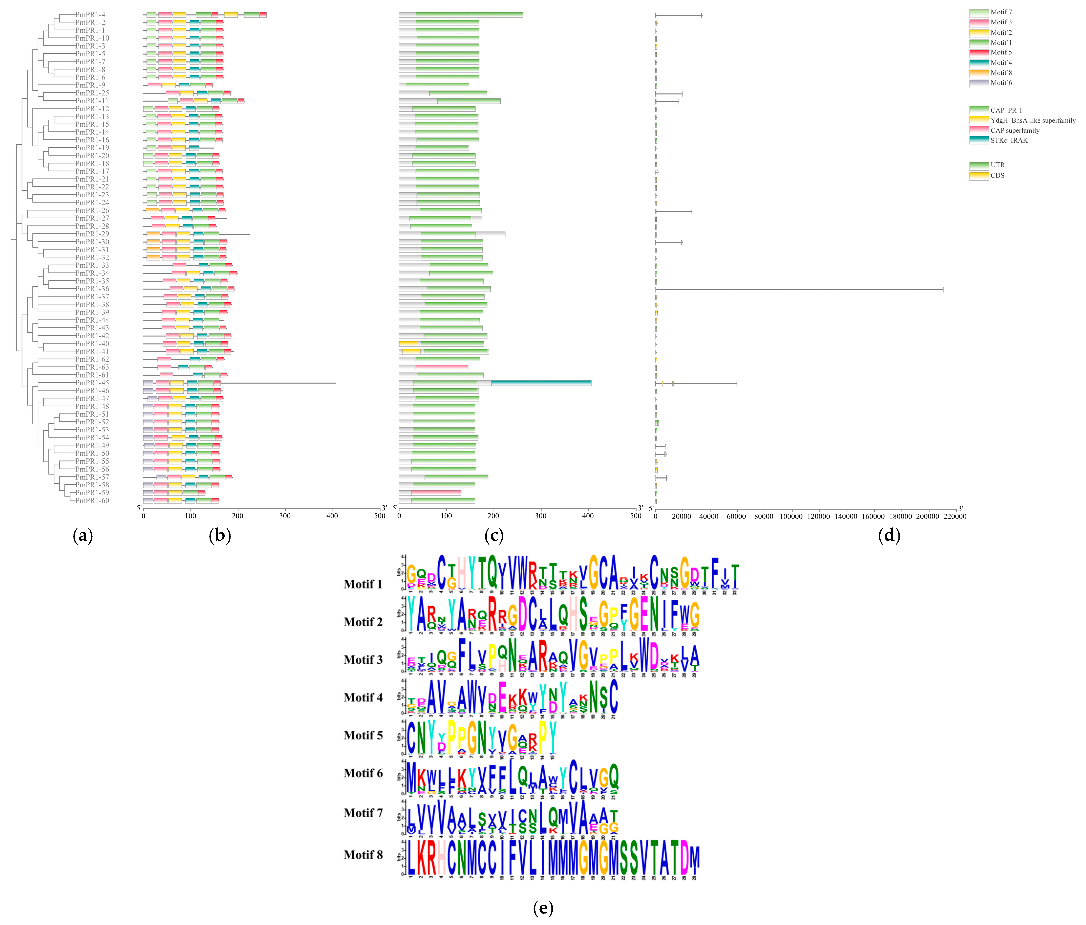
Conserved motif analysis of the PmPR1 proteins was conducted using MEME (Version 5.5.9, https://meme-suite.org/meme/tools/meme, accessed on 3 April 2026) and Tbtools (Version 2.466), as shown in Figure 2a. Eight conserved motifs were identified in the PmPR1 proteins, among which five motifs (motif 1, motif 2, motif 3, motif 4, and motif 5) were consistently present in most family members, indicating high conservation. Additionally, motif 6 was mainly found in PmPR1-45 to PmPR1-60, motif 7 was mainly present in PmPR1-1 to PmPR1-23, and motif 8 was mainly observed in PmPR1-29 to PmPR1-32. The arrangement of these conserved motifs in the 63 PmPR1 protein members highlights the significant conservation within the PmPR1 gene family. The results of the conserved domain analysis are shown in Figure 2b. All 63 PmPR1 proteins contain the CAP_PR-1 domain, which reinforces the conclusion that all members are part of the CAP superfamily.
The gene structures of the PmPR1s were analyzed using bioinformatics tools, and the results are shown in Figure 2c. Most of the genes are single-exon genes, with only 13 genes belonging to the multi-exon structure type. Among them, PmPR1-1, PmPR1-19, PmPR1-29, PmPR1-32, PmPR1-40, PmPR1-41, PmPR1-42, and PmPR1-47, these 8 members lack untranslated regions (UTRs). UTRs play important roles in the regulation of mRNA stability, transport, and translation in eukaryotes [22]. Therefore, the absence of UTR annotations in these genes may be related to limitations in the current annotation results, and their potential biological significance still requires further investigation.
Figure 1.
Phylogenetic tree of the PR1 gene family. Note: At: Arabidopsis thaliana; Gbi: Ginkgo biloba; Pm: Pinus massoniana; Potri: Populus trichocarpa. Branches in different colors represent distinct subfamilies. Green: Group I; Red: Group II; Orange: Group III; Blue: Group IV; Purple: Group V; Pink: Group VI. Bootstrap values (based on 1000 replicates) are shown at the nodes.
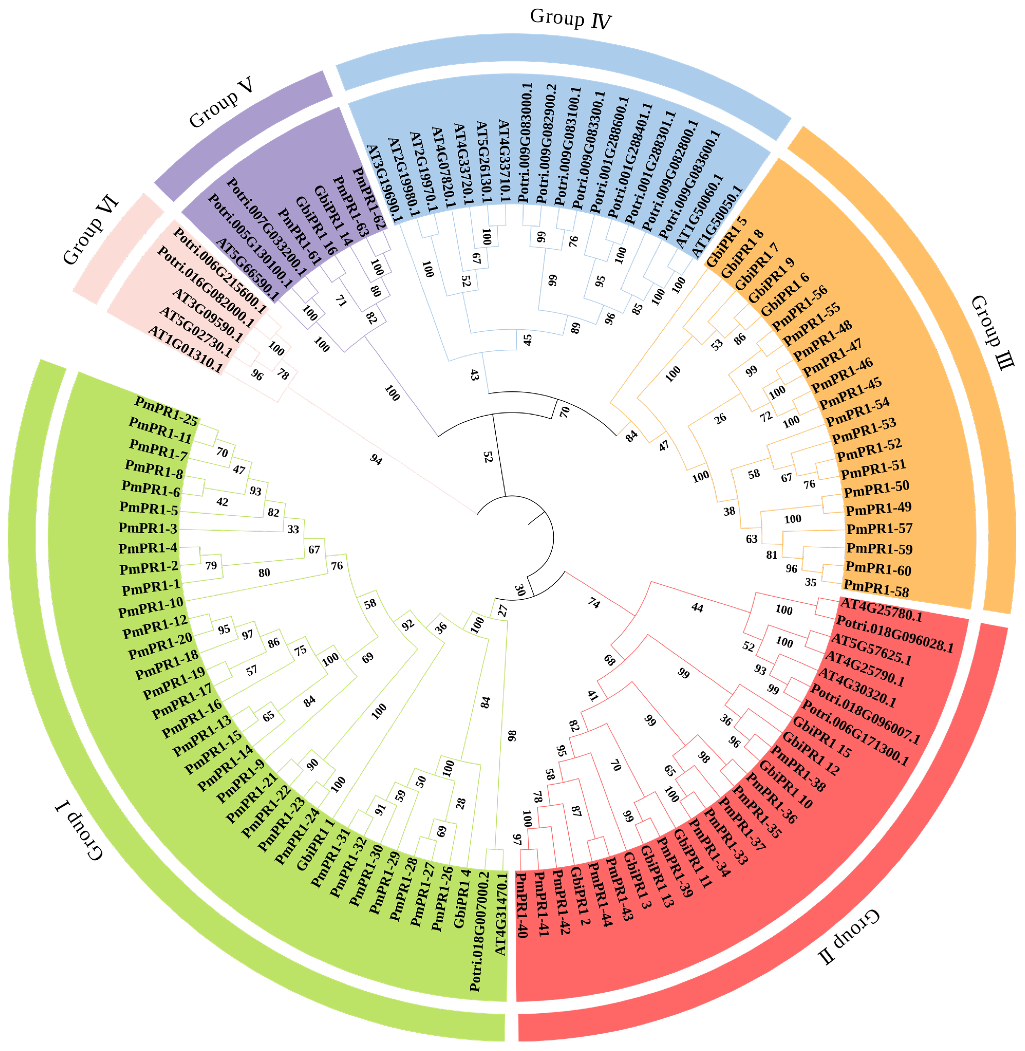
Figure 2.
Gene structure and protein conserved motif distribution of PmPR1. Note: (a): Phylogenetic tree; (b): Conserved motifs of the protein encoded by the PmPR1 gene; (c): Conserved domain architecture of PmPR1; (d): Gene structure of PmPR1; (e): Sequence logos of the eight motifs shown in panel (b).
2.4. Prediction of Cis-Acting Elements in the Promoter Regions of the PmPR1 Gene Family
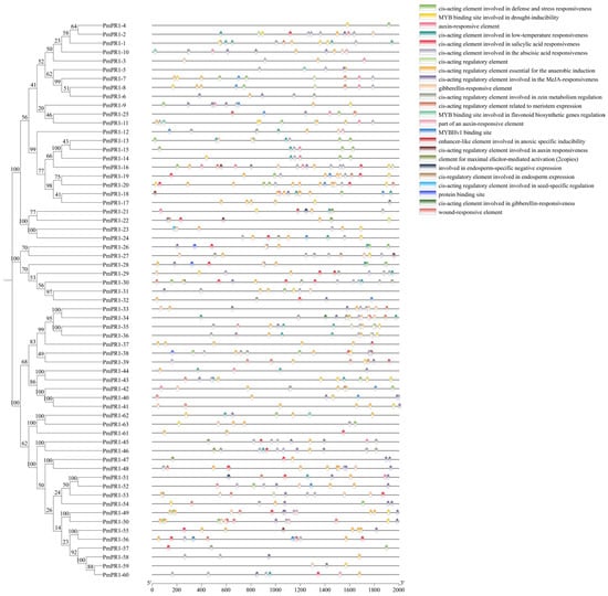
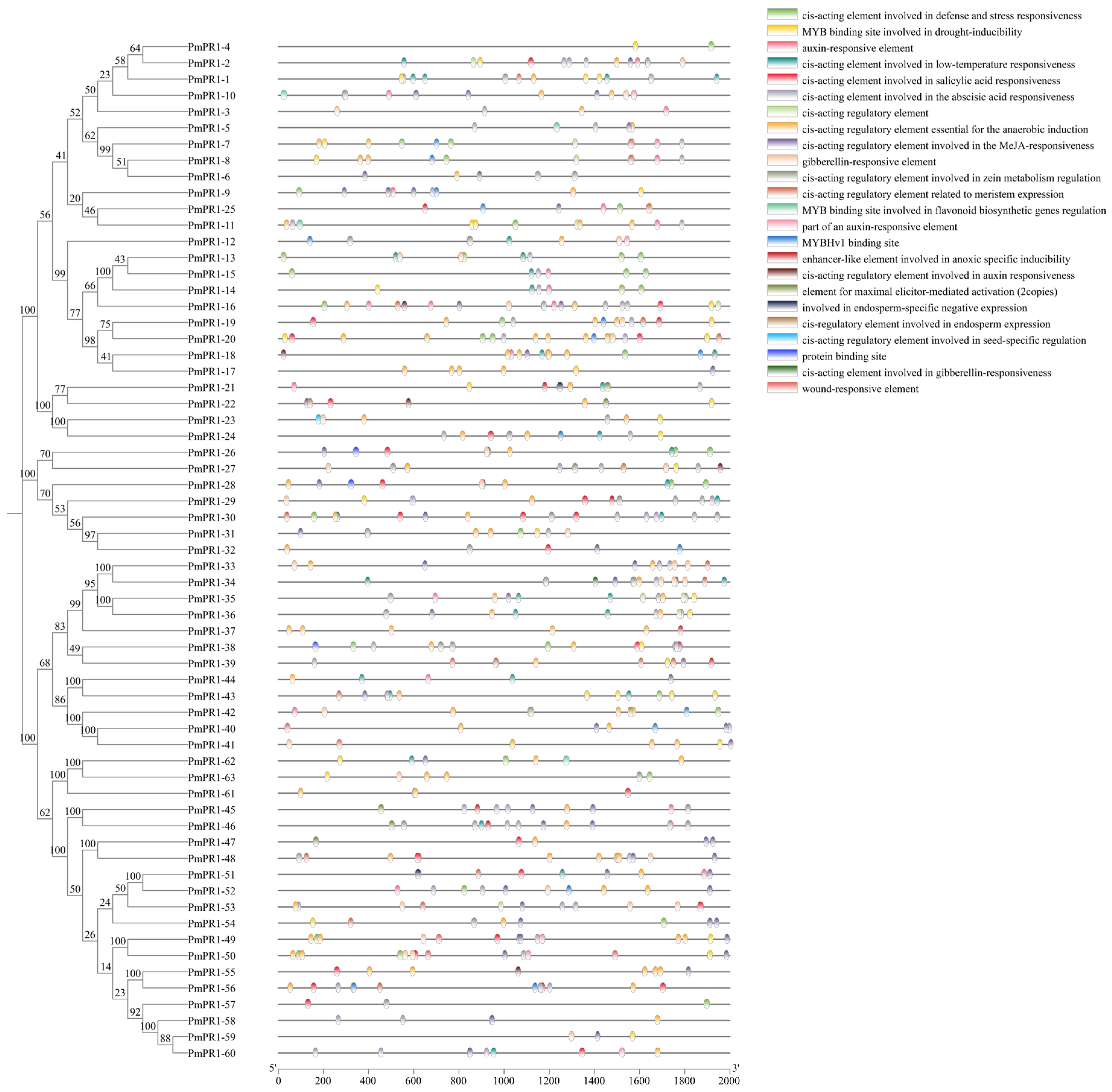
To further investigate the potential biological processes and signaling pathways involved in the PmPR1s, this study extracted the 2000 bp upstream promoter sequences of the PmPR1s to analyze their cis-element characteristics. The results indicated that the promoter regions of the PmPR1s were enriched with a series of elements related to biotic and abiotic stress responses (defense and stress response, anaerobic induction, hypoxia-specific induction, drought induction, cold response, mechanical injury response), plant hormone interactions, and elements associated with plant growth and development (Figure 3).
Figure 3.
Analysis of cis-acting elements in the PmPR1 gene family.Note: The 2-kb sequences upstream of the start codon were analyzed. Different colors represent different types of cis-acting elements.
Firstly, in terms of stress response, a total of 241 elements were identified to be involved in biotic and abiotic stress responses. Among them, elements related to defense and stress response were the most widely distributed, present in the promoters of 27 genes including PmPR1-4, PmPR1-7, PmPR1-8, and PmPR1-9, and these genes were evenly distributed across the four subfamilies of Group I, Group II, Group III, and Group IV, indicating that this function is relatively conserved in evolution. Additionally, specific elements such as mechanical injury response elements were mainly concentrated in the promoters of genes PmPR1-40, PmPR1-41, PmPR1-49, and PmPR1-50 in the Group II and Group III subfamilies. Secondly, the largest number of elements were related to hormone responses, totaling 322, including elements for salicylic acid (SA), methyl jasmonate (MeJA), gibberellin (GA), abscisic acid (ABA), and auxin response. Notably, the presence of SA and MeJA response elements further confirmed the potential role of PmPR1s in plant stress resistance. For instance, Salicylic acid response elements are present in the promoters of 24 genes, including PmPR1-2, PmPR1-16, and PmPR1-22, mainly concentrated in the Group I and Group III subfamilies. Additionally, 36 elements related to plant growth and development were identified, such as auxin response elements, zein metabolism regulatory elements, seed-specific regulatory elements, and endosperm expression elements. The presence of these elements suggests that the PmPR1s, in addition to responding to stress, may also be involved in the growth and development of P. massoniana, performing diverse regulatory functions.
2.5. Mining of PmPR1s’ Gene Cluster in P. massoniana
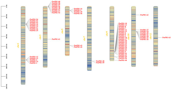
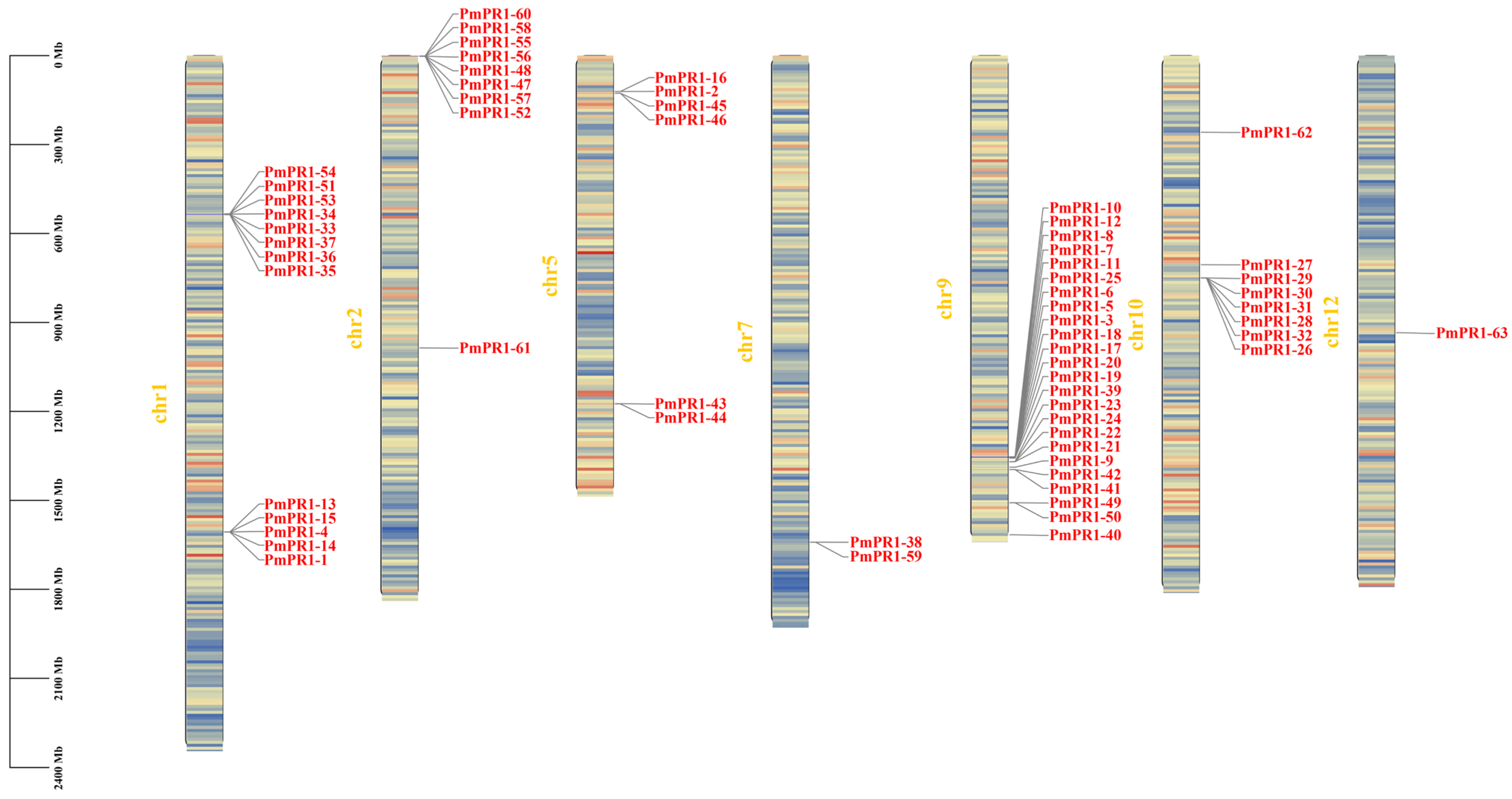
PR1, as an important component of the plant defense system, participates in the co-evolution of plants and pathogens and can form gene clusters through evolutionary events such as tandem and segmental duplications. Members of gene clusters can not only respond rapidly to pathogen infection through coordinated expression but also enhance plant disease resistance through functional differentiation. For instance, during the early and middle stages of Fusarium oxysporum infection in Aleurites montana, the Aleurites montana PGK3-TPS10-CYP726 gene cluster was continuously highly expressed in the roots [23]; in soybeans, different members of the RGC2 family have distinct functions and show different expression-related phenotypes [24]. Based on the genome annotation information of P. massoniana, the chromosomal localization of PmPR1s and the gene clusters they belong to were explored. The results are shown in Figure 4. PmPR1s are unevenly distributed on seven chromosomes of the P. massoniana genome, namely chromosomes 1, 2, 5, 7, 9, 10, and 12. Among them, chromosome 9 has the largest number of genes, with 24 genes, followed by chromosome 1 with 13 genes, and chromosome 12 has the fewest genes, with only the PmPR1-63 located on it.
Figure 4.
Chromosomal localization of the PmPR1 gene family.Note: Red regions indicate high gene density, and blue regions indicate low gene density.
The PmPR1s are located on multiple chromosomes, including chromosome 1, chromosome 2, chromosome 7 and chromosome 9. A large number of PmPR1s are arranged in series, forming gene clusters. Among them, the PmPR1s located on chromosome 1 and belonging to the Group I subfamily form two clusters: the cluster composed of PmPR1-4, PmPR1-13, PmPR1-14 and PmPR1-15 has a total span of 72.8 kb, and the cluster composed of PmPR1-51 and PmPR1-54 has a total span of 63 kb. On chromosome 2, the PmPR1s belonging to the Group III subfamily also form two clusters: the cluster composed of PmPR1-47, PmPR1-48 and PmPR1-52 has a span of 291.7 kb, while the cluster composed of PmPR1-58 and PmPR1-60 has a span of 48.1 kb. The PmPR1-10 and PmPR1-12 gene cluster is located on chromosome 9, with a total span of 41.6 kb, and all members belong to the Group I subfamily. The PmPR1-38 and PmPR1-59 gene cluster is located on chromosome 7, with a total span of 327.8 kb. PmPR1-38 belongs to the Group II subfamily, and PmPR1-59 belongs to the Group III subfamily. This indicates that after forming gene clusters, PR1 has undergone functional differentiation and participates in different disease resistance processes.
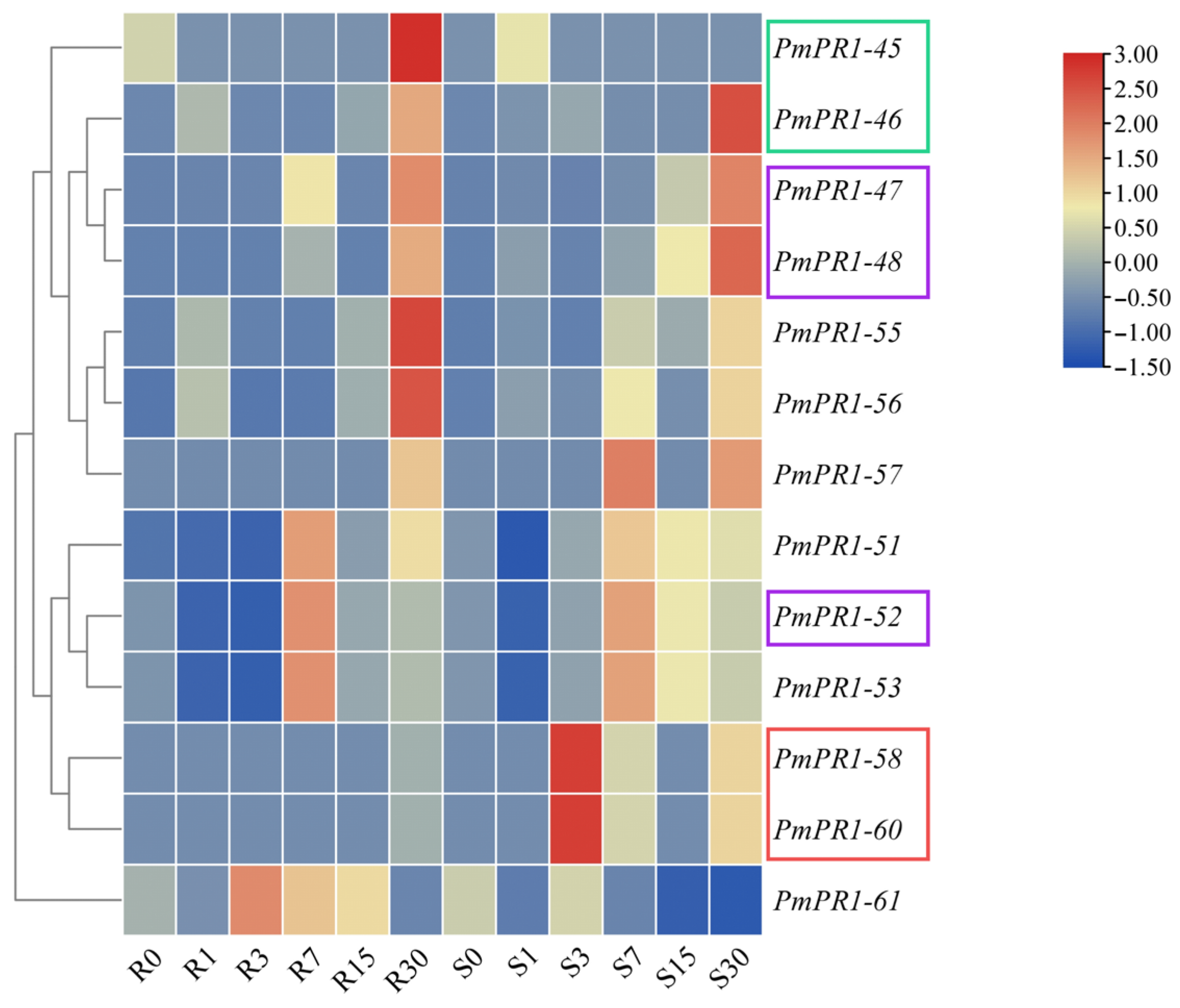
Based on the transcriptome data of high-resistant and susceptible P. massoniana at different infection stages obtained in the previous study of our research group, further analysis of the expression patterns of PmPR1s and gene clusters in high-resistant and susceptible P. massoniana at different infection nodes revealed that the expression of PmPR1-45, PmPR1-46, PmPR1-48, PmPR1-51, PmPR1-52, PmPR1-53, PmPR1-55, PmPR1-56, PmPR1-58, PmPR1-60, and PmPR1-61 was particularly significant in resistant and susceptible plants. Moreover, PmPR1-45 and PmPR1-46 were clustered on chromosome 5, while PmPR1-58 and PmPR1-60 were clustered on chromosome 2. Additionally, PmPR1-47, PmPR1-48, and PmPR1-52 were also clustered on chromosome 2 (Figure 5), indicating that their induction and activation can lead to rapid cluster expression [25].
As shown in Figure 6, different gene clusters exhibit distinct expression patterns in response to invasion in P. massoniana. The PmPR1-58 and PmPR1-60 gene clusters show the same trend of gene expression after infection by B. xylophilus, indicating their co-expression within the cluster; while the PmPR1-45 and PmPR1-46 gene clusters, as well as the PmPR1-47, PmPR1-48 and PmPR1-52 gene clusters, display specific expression in response to the invasion of B. xylophilus.
Figure 5.
Schematic of the PmPR1s’ gene cluster. Note: Boxes in different colors represent different gene clusters: red (chromosome 2, 1.47–1.52 Mb), purple (chromosome 2, 2.01–2.34 Mb), and green (chromosome 5, 126.88–127.02 Mb). Genes within boxes of the same color belong to the same gene cluster. The direction of the arrow in each box indicates gene orientation, and the length of each box is proportional to gene length. Chromosomal coordinates are shown in bp.
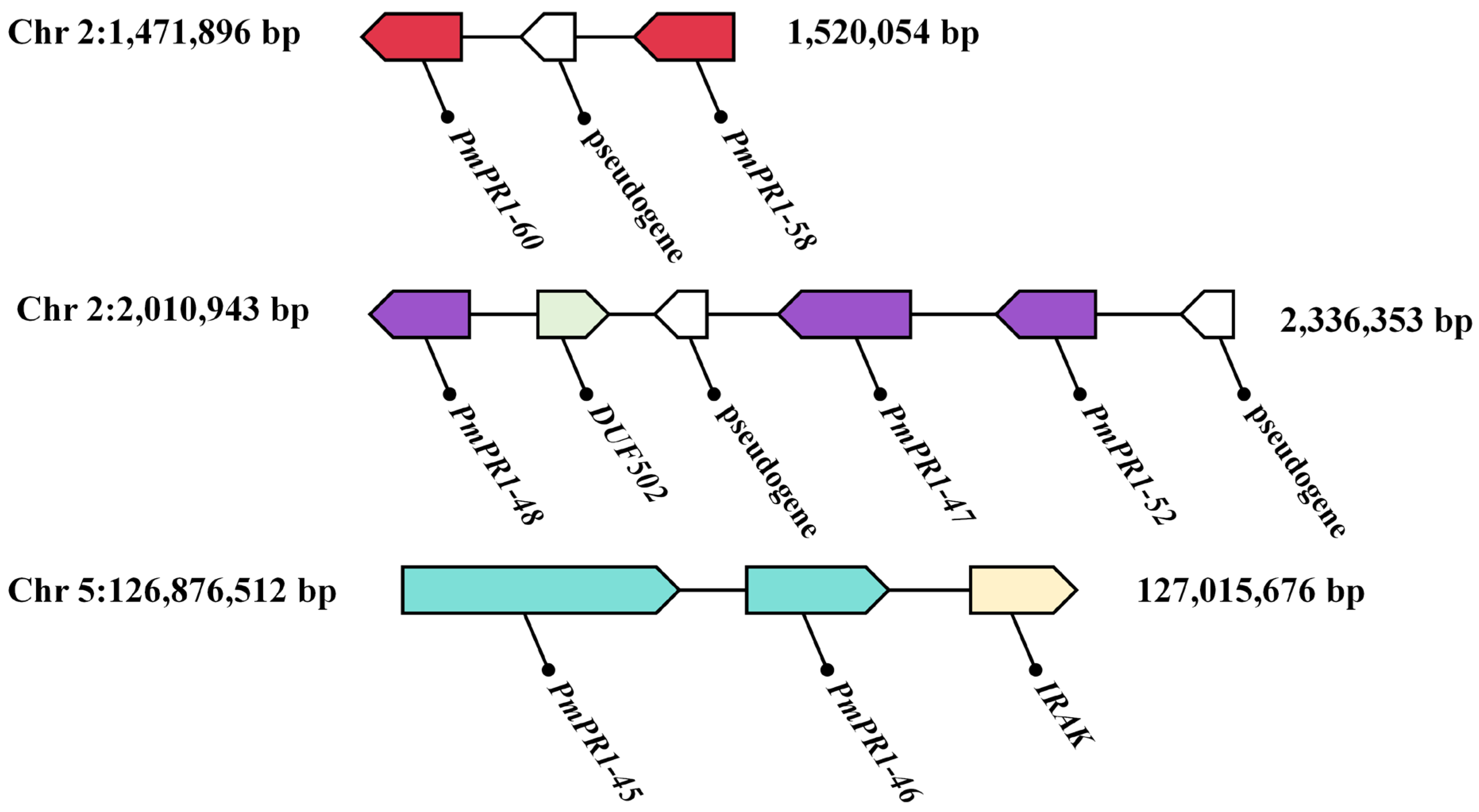
Figure 6.
Transcript profiling of PmPR1s based on RNA-seq analysis. Note: Borders of the same color represent the same gene cluster. The red and purple borders indicate two different gene clusters on chromosome 2, whereas the green border indicates the gene cluster on chromosome 5.
2.6. Expression Analysis of PmPR1s in Resistant and Susceptible P. massoniana at Different Periods After Nematode Inoculation
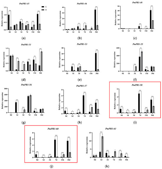
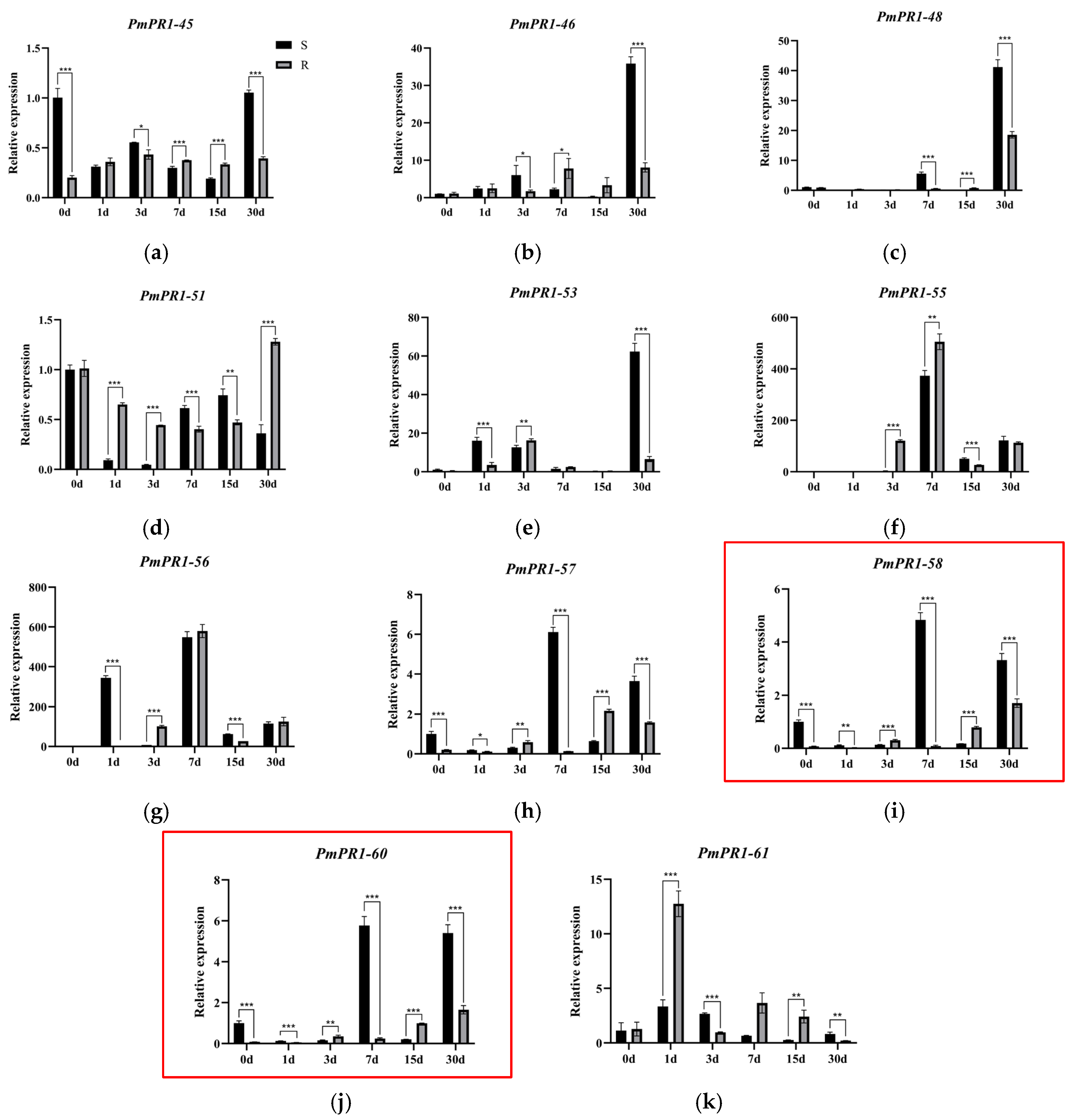
To elucidate the expression patterns of PmPR1s in highly resistant (R) and susceptible (S) P. massoniana following inoculation with B. xylophilus over a 30-day period, key genes and gene clusters responsive to nematode infection in resistant and susceptible pines were selected for qRT-PCR analysis. Using 0, 1, 3, 7, 15, and 30 days post-inoculation as time points, Quantitative Real-Time PCR was performed on the 11 selected genes (Figure 7). The results revealed differential expression patterns among these genes, suggesting potential functional divergence. Using susceptible pines at 0 d post-inoculation (S0) as the control, distinct expression trends were observed for PmPR1-45 and PmPR1-46. PmPR1-45 exhibited an initial increase followed by stabilization in resistant plants after inoculation. In susceptible plants, its expression was significantly higher than in resistant plants at 0 d and 30 d post-inoculation, while no significant differences were observed at other time points. PmPR1-46 reached peak expression in resistant plants at 7 d post-inoculation, whereas its induction in susceptible plants was delayed, with significantly higher expression than in resistant plants at 30 d post-inoculation. PmPR1-58 and PmPR1-60 showed similar expression trends, Synergistic response to pine wood nematode infection, with significant upregulation in susceptible clones at 7 d and 30 d post-inoculation. At 7 d post-inoculation, expression levels were approximately 5–6 times higher than at 0 d, representing a substantial increase. Specifically, at both time points, transcript levels in susceptible plants were significantly higher (approximately 2–5 times) than those in resistant plants. PmPR1-48 showed no significant expression changes during early inoculation but was upregulated in both resistant and susceptible plants after 30 days, with significantly higher expression in susceptible plants. PmPR1-51 displayed a downregulation-followed-by-upregulation pattern in both resistant and susceptible pines after inoculation, with higher expression in resistant plants. PmPR1-55, PmPR1-56, and PmPR1-61 all exhibited an initial increase followed by a decrease in resistant plants, though with distinct response kinetics. At 1 d post-inoculation, PmPR1-61 was rapidly induced in resistant plants, with expression levels approximately 3.7 times higher than in susceptible plants. PmPR1-55 and PmPR1-56 responded slightly later, showing rapid upregulation in resistant plants at 3 d and 7 d post-inoculation, respectively, with significantly higher expression than in susceptible plants. These findings indicate that the three PR1 family genes (PmPR1-55, PmPR1-56, and PmPR1-61) may play critical roles in activating defense responses against B. xylophilus in P. massoniana. Given that PmPR1-61 is rapidly induced during early resistance, potentially functioning upstream in defense signaling, it was selected for subsequent transient transformation experiments to validate its function.
Figure 7.
Quantitative real-time PCR (qRT-PCR) analysis of PmPR1s’ expression. Note: The expression of PmPR1 genes was validated by quantitative reverse transcription PCR (qRT-PCR). Panels (a–k) show the real-time fluorescence quantitative results for 11 different genes. The red box indicates genes that are located in the same gene cluster and show consistent expression trends. Relative expression levels were calculated using susceptible plants at 0 days post-inoculation (S0) as the control. Data are presented as mean ± standard deviation (SD) of three biological replicates. Asterisks indicate significant expression differences between resistant and susceptible plants (* p < 0.05; ** p < 0.01; *** p < 0.001; independent samples t-test).
2.7. Transient Transformation of PmPR1-61 in Callus of P. massoniana
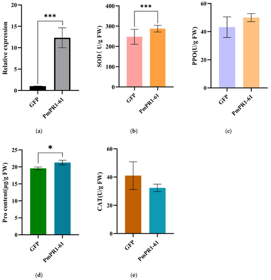
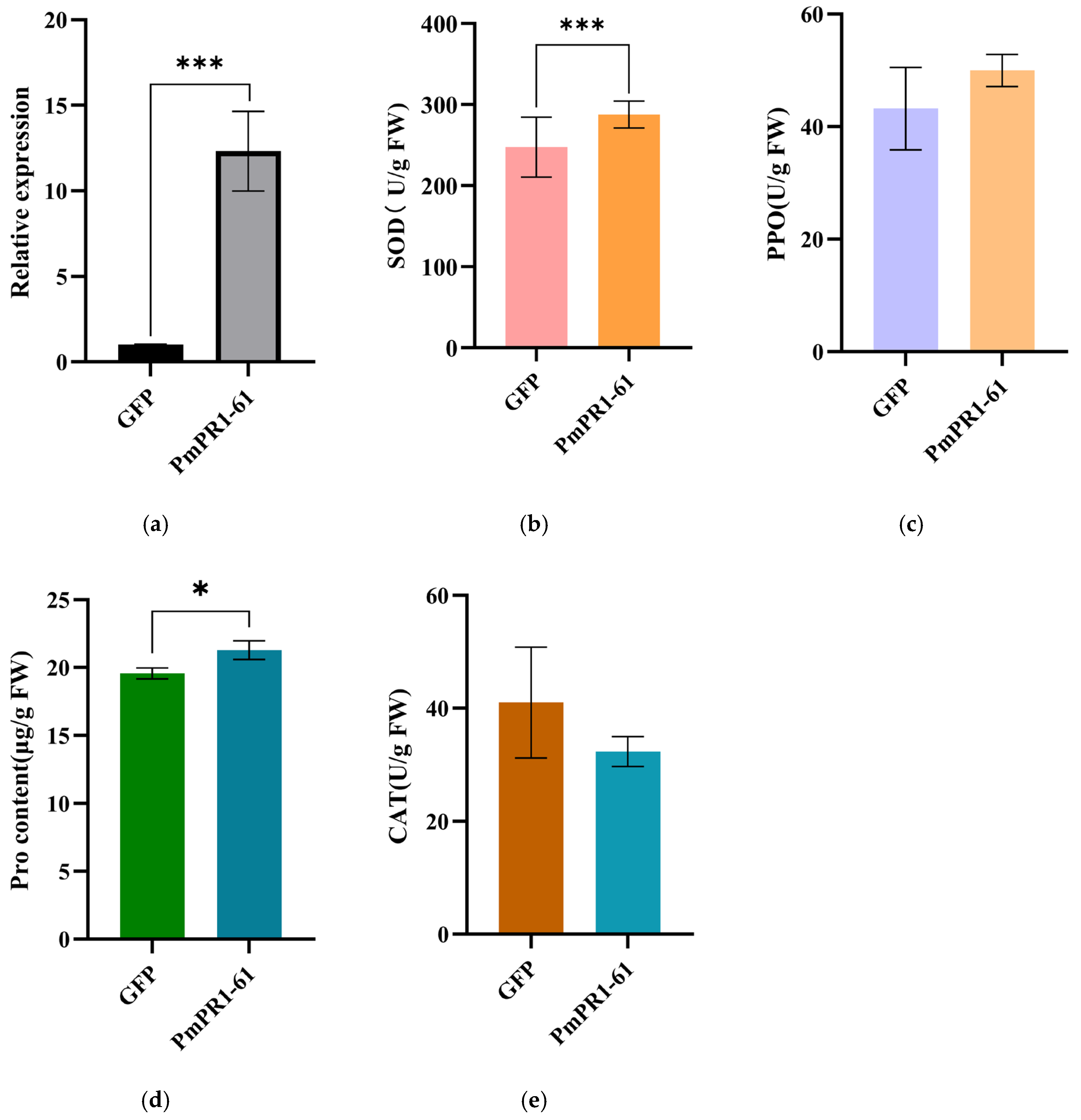
To further explore the role of the PmPR1-61 in activating the defense response of P. massoniana against B. xylophilus, an overexpression vector of this gene was constructed and transiently transformed into P. massoniana callus. The qRT-PCR results (Figure 8a) indicated that the relative expression level of the gene in the callus transiently transformed with pCAMBIA1300-PmPR1-61-EGFP was significantly higher than that in the control callus transiently transformed with the empty vector, approximately 12 times that of the control, suggesting the successful transient transformation of the P. massoniana callus.
Figure 8.
Physiological indicators of P. massoniana callus after transient overexpression of PmPR1-61. Note: (a): Expression analysis in P. massoniana callus after transient overexpression of PmPR1-61; (b): Superoxide dismutase (SOD) activity; (c): Polyphenol oxidase (PPO) activity; (d): Proline (PRO) content; (e): Catalase (CAT) activity. Data are presented as the mean ± standard deviation (SD) of three biological replicates. Asterisks indicate significant differences between the control (GFP) and transgenic (PmPR1-61) plants (* p < 0.05; *** p < 0.001; independent samples t-test).
This study further examined the activities of defense-related enzymes and the metabolite contents in overexpressing calli (Figure 8b–e). The results showed that, compared with the control, transient overexpression of PmPR1-61 significantly increased superoxide dismutase (SOD) activity and proline (PRO) content (p < 0.05) while also enhancing polyphenol oxidase (PPO) activity; in contrast, catalase (CAT) activity decreased. SOD is a key enzyme responsible for scavenging superoxide anions, and its increased activity usually reflects the activation of the cellular antioxidant response. As one of the main enzymes involved in hydrogen peroxide (H2O2) decomposition, the reduced activity of CAT may indicate suppressed H2O2-scavenging capacity, thereby promoting H2O2 accumulation to activate downstream defense responses. Alternatively, it may reflect enzyme inactivation caused by oxidative damage, inhibition by post-translational modification, or an overall disruption of cellular redox homeostasis. Since H2O2 content was not directly measured in this study, these possibilities cannot currently be distinguished. The increases in PPO and PRO may indicate the establishment of an antimicrobial barrier and enhanced osmotic regulation capacity. In summary, transient overexpression of PmPR1-61 altered the antioxidant enzyme system of the calli and promoted the accumulation of PPO and PRO. These changes may synergistically enhance the defense potential of P. massoniana, although the underlying mechanisms still require further verification through direct measurement of H2O2 levels and enzyme activity inhibition assays.
3. Discussion
PR-1 proteins are among the most abundantly synthesized proteins during plant immune responses and play a crucial role in SAR [26]. Although the PR1 family has been systematically identified in many angiosperms, related studies in Pinaceae species remain lacking. In this study, a total of 63 PmPR1 genes were systematically identified for the first time in the conifer species P. massoniana. This number is markedly higher than that reported in Populus deltoides [20], Pyrus betulaefolia Bunge [27], and Medicago sativa [28], indicating that the PR1 family has undergone substantial expansion in P. massoniana. Combined with the chromosome localization results, PmPR1s were found to be tandemly distributed across multiple chromosomal regions, suggesting that tandem duplication may have been an important driving force for the expansion of this family in P. massoniana.
Most members of the PmPR1 family lacked introns, a structural feature consistent with reports of PR1 genes in poplar and other plant species [19,20]. Previous studies have shown that intronless or intron-poor genes are often more conducive to rapid transcriptional responses under stress conditions [25,29]. Therefore, the predominance of single-exon genes in the PmPR1 family may provide a structural basis for the rapid activation of defense responses in P. massoniana during the early stage of pine wood nematode infection. However, such a structural advantage does not necessarily translate directly into stronger disease resistance, because whether a gene can be induced in a timely manner is also influenced by multiple factors, including the composition of cis-regulatory elements, chromatin accessibility, and the dynamic regulation of upstream transcription factors [5]. Therefore, the differential expression of PmPR1 genes between resistant and susceptible plants is more likely the result of the combined effects of structural characteristics and regulatory networks.
Promoter analysis revealed that PmPR1s contain numerous cis-regulatory elements associated with SA, MeJA, ABA, and MYB/MYC, suggesting that the regulation of this family may be linked to multiple phytohormonal and stress-related signaling pathways [5,30]. It is generally accepted that the SA pathway is mainly associated with SAR-related resistance, whereas the JA/ET pathway is more commonly involved in responses to mechanical injury and insect stress [31]. Pine wood nematode infection is usually accompanied by multiple changes, including mechanical damage, tissue necrosis, and disruption of oxidative balance. Therefore, the coexistence of SA and MeJA responsive elements in PmPR1 promoters suggests that members of this family may be coordinately regulated by multiple hormonal signals and participate in the crosstalk among different defense pathways [32]. Taken together, the differential expression of different PmPR1 members may be associated with the defense demands at different stages of infection as well as changes in cellular physiological status during the infection process.
This study found that PmPR1-61 was rapidly induced at 1 day after inoculation in highly resistant materials, whereas PmPR1-55 and PmPR1-56 were significantly upregulated at 3–7 days after inoculation. In contrast, PmPR1-58 and PmPR1-60 showed relatively high expression mainly at the late stage of infection in susceptible materials. The members that were rapidly upregulated at the early stage may be involved in the initiation of early defense responses, whereas those highly expressed at later stages in susceptible materials may be associated with stress responses or defense imbalance as the disease progresses [33,34,35]. These findings suggest that the PmPR1 family may have undergone evident functional differentiation during resistance to nematode infection, with some members tending to function in early immune activation, while others may be involved in late defense maintenance or stress adaptation.
Studies on the relationship between PR1 proteins and reactive oxygen species (ROS) signaling have shown that ROS not only serve as important early defense signals during pathogen infection, but also act as major factors causing cellular oxidative damage [36,37]. In this study, transient overexpression of PmPR1-61 led to increased SOD and PPO activities, elevated proline content, and reduced CAT activity. These results suggest that the role of PmPR1-61 may extend beyond serving merely as a marker of defense responses, and that it may participate in the regulation of ROS homeostasis. In addition, the enhancement of PPO activity and the increase in proline accumulation after PmPR1-61 overexpression indicate that its function is not limited to a single-antioxidant enzyme system. PPO is generally associated with the oxidation of phenolic compounds, tissue lignification, and the formation of physical barriers, whereas proline plays important roles in osmotic adjustment, membrane structure stabilization, and buffering against oxidative stress [38,39]. Therefore, PmPR1-61 is more likely to enhance the defense capacity of P. massoniana through multiple mechanisms, including regulation of ROS signaling, reinforcement of cell wall defense, and maintenance of stress homeostasis. However, the decrease in CAT activity may also indicate an increase in local oxidative pressure, and thus the possibility that PmPR1-61 enhances defense while simultaneously aggravating oxidative stress cannot yet be completely excluded. Further studies integrating in situ H2O2 staining, MDA determination, and related analyses are still needed to determine whether PmPR1-61 enhances resistance mainly by regulating the dynamic changes of H2O2.
During the long-term co-evolution between plants and pathogens, the formation of disease-resistance gene clusters, such as PR-1 and R genes, represents a key genomic adaptation strategy. Evolutionary events including tandem duplication and segmental duplication provide the genetic basis for the generation of duplicated genes. These duplicated genes can subsequently undergo functional divergence through neofunctionalization and subfunctionalization, thereby supplying abundant genetic resources for plant disease resistance [40,41,42]. Studies have shown that, in rice, different members within NLR gene clusters exhibit specific resistance to different races of Magnaporthe oryzae [43]. In A. thaliana, genes in the RPW8/HR cluster confer broad-spectrum disease resistance, whereas RPP7 members provide race-specific resistance [44]. These findings indicate that functional differentiation within gene clusters is of great biological significance, manifesting either as broad-spectrum resistance or as specialized recognition and defense against specific pathogens.
Members of the PR1 gene cluster in P. massoniana are distributed among different evolutionary branches. For example, PmPR1-38 and PmPR1-59 belong to Group II and Group III subfamilies, respectively, and may participate in defense against different pathogens in P. massoniana. Functional differentiation within gene clusters is reflected not only in nucleotide and amino acid sequences, but also in expression patterns. The key candidate disease-resistance gene PmPR1-52, together with PmPR1-47 and PmPR1-48, forms a clustered distribution spanning 291.7 kb on chromosome 2. These genes display coordinated or specific expression patterns in different resistant materials and at different infection stages, suggesting that the gene cluster may regulate defense through coordinated action or functional divergence. Genes within the cluster may share similar combinations of promoter elements, such as SA, MeJA, and W-box-related binding sites, and are therefore likely to be co-regulated by the same class of defense-related transcription factors [5,20,30]. In addition, following pathogen infection, the chromatin in this genomic region may become more accessible, thereby facilitating the binding of relevant transcription factors and promoting the rapid induction of genes within the cluster [45,46].
In summary, members of the PmPR1 gene family may exhibit both coordinated responses and functional differentiation during the resistance of P. massoniana to pine wood nematode, among which PmPR1-55, PmPR1-56, and PmPR1-61 appear to be potential key resistance-related genes.
4. Materials and Methods
4.1. Materials
4.1.1. Plant Materials
The experimental materials were collected from 8-year-old P. massoniana clonal trial forests in Linhai Nursery, Linhai City, Zhejiang Province. Three plants each of the highly resistant clone (1144-2) and the susceptible clone (9-5) were selected, and their current-year tender branches were inoculated with pine wood nematodes. The control group consisted of the susceptible masson pine clones inoculated 0 days ago. The pine wood nematodes used for inoculation were the highly pathogenic “Guangde 3B” and a mixed population of nematodes isolated from diseased masson pine trees in the wild. Each tender branch of P. massoniana was inoculated with 10,000 pine wood nematodes suspended in 200 μL. Three biological replicates of the highly resistant and susceptible P. massoniana clones inoculated with pine wood nematodes were collected at different time points (0, 1, 3, 7, 15, and 30 days), and immediately placed in liquid nitrogen and stored at −80 °C for subsequent experimental analysis.
4.1.2. Data Sources
The genomic data of P. massoniana used in this study was obtained from the publicly available data of Guangxi Academy of Forestry Sciences (PRJNA1240911). The transcriptome data were the sequencing data from the previous work of our research group.
4.1.3. Preparation of Culture Medium and Resuspension Solution
LB medium (1 L): 10 g NaCl + 10 g Tryptone + 5 g Yeast extract + 15 g Agar;
Co-culture medium (1 L): 4.48 g M519 + 30 g sucrose + 7.5 g agar, pH 5.8;
Resuspension solution: 10 mM/L MgCl2 + 10 mM/L MES + 200 μM/L AS.
4.2. Method
4.2.1. Identification and Physicochemical Property Analysis of PmPR1 Gene Family Members
The protein sequence of the pathogenesis-related protein PR1 of A. thaliana was downloaded from the TAIR database (Version 10.0, https://www.arabidopsis.org/, accessed on 3 December 2025), the protein sequences of PR1 in O. sativa and P. trichocarpa were downloaded from the NCBI official website (https://www.ncbi.nlm.nih.gov/gene/, accessed on 3 December 2025), and the PR1 protein sequence of G. biloba was obtained from published papers. Using the PR1 protein sequences of A. thaliana, O. sativa, P. trichocarpa and the gymnosperm G. biloba as search objects, BLASTP analysis was performed on the P. massoniana genome data through Tbtools, with strict thresholds (E value ≤ 1× 10 −10). To verify the existence of domains, the hidden Markov model file of the PR1 protein (PF00188) was downloaded from Pfam (Version 38.0, https://www.ebi.ac.uk/interpro/, accessed on 3 December 2025), and then HMM Search was conducted using the TBtools software (Version 2.466) to initially screen out candidate gene family members. The intersection of the results obtained by the above methods was taken, and the Batch CD-Search tool of NCBI (https://www.ncbi.nlm.nih.gov/Structure/bwrpsb/bwrpsb.cgi, accessed on 3 December 2025) was used to verify the domains of these candidate genes. Further, the physicochemical properties of the PR1 family proteins of P. massoniana were analyzed using TBtools, including molecular weight, isoelectric point, instability index, hydrophobicity coefficient and total average hydrophilicity. Signal peptides were predicted using SignalP-6.0 (https://services.healthtech.dtu.dk/services/SignalP-6.0/, accessed on 6 April 2026), and transmembrane domains were predicted using TMHMM-1.0 (https://services.healthtech.dtu.dk/services/DeepTMHMM-1.0/, accessed on 6 April 2026). Finally, protein subcellular localization was predicted using the WoLF PSORT online platform with the plant mode selected (https://wolfpsort.hgc.jp/, accessed on 4 December 2025).
4.2.2. Phylogenetic Analysis of PmPR1 Gene Family
The PR1 proteins of P. massoniana, A. thaliana, G. biloba and P. tomentosa were subjected to multiple sequence alignment using MEGA12 (Version 12.0.11) software. The evolutionary tree was constructed by the Maximum Likelihood Estimation (ML). The parameters were set as follows: the amino acid substitution model was JTT, Gaps/Missing Data was set to Use all sites, Tree Inference Options was set to NNI + Automatic initial tree, Number of Threads was set to 6, and bootstrap analysis was performed with 1000 replicates. Finally, the phylogenetic tree was visualized and annotated using the iTOL online tool (version 7.5.1, https://itol.embl.de/, accessed on 3 April 2026).
4.2.3. Analysis of the Gene Structure Characteristics and Conserved Domains of the PmPR1s
The nucleic acid and protein sequences of the PmPR1 family members were extracted from the genome annotation file of P. massoniana, including their complete genomic sequences, coding sequences (CDS), and corresponding protein sequences. On this basis, the gene structure diagrams were visualized using TBtools (Version 2.466), and the number and length of exons of each member were counted. To further explore the functional conservation, the conserved domain analysis was conducted using the Conserved domains software of NCBI (https://www.ncbi.nlm.nih.gov/Structure/bwrpsb/bwrpsb.cgi, accessed on 5 December 2025), and the conserved motif analysis was performed using MEME (Version 5.5.9, https://meme-suite.org/meme/tools/meme, accessed on 3 April 2026), with the parameters set to identify a maximum of 10 motifs and the distribution option set to zero or one occurrence per sequence (zoops), while all other parameters were kept at their default settings. Finally, the identified motifs were visualized using TBtools.
4.2.4. Prediction of Cis-Acting Elements in the Promoter Regions of the PmPR1 Gene Family
The 2000 bp sequence upstream of the start codon of the PmPR1s was extracted using the GFF3 sequence extraction tool in TBtools software (Version 2.466)and submitted to the PlantCare online website (https://bioinformatics.psb.ugent.be/webtools/plantcare/html/, accessed on 13 December 2025) to predict cis-acting elements. The cis-acting elements of the predicted promoter were visualized and analyzed using TBtools.
4.2.5. Chromosome Mapping and Gene Cluster Mining of the PmPR1 Gene Family
Based on the GFF annotation file of the P. massoniana genome data, the chromosomal location information of the PmPR1 was obtained, and the visualization plot was made using the TBtools software. Adjacent PmPR1 genes that are closely linked on the chromosome and belong to the same phylogenetic branch were defined as members of the same gene cluster. Gene location information was extracted from the genome GFF file and then visualized. The differentially expressed PR1 genes in susceptible and resistant P. massoniana at different infection periods were screened out based on the transcriptome data, and the heat map of the differentially expressed genes in the PmPR1 gene family of P. massoniana was drawn and analyzed.
4.2.6. Analysis of the Expression Patterns of PmPR1s
To further investigate the functions of PmPR1s during infection by B. xylophilus, qRT-PCR analysis was conducted to verify the genes with significant differences in expression levels based on the gene cluster results and transcriptome data. Total RNA was extracted from the young branches of highly resistant and susceptible P. massoniana inoculated with B. xylophilus for different days using the Aidlab RNA extraction kit. The Aidlab reverse transcription kit was used to reverse transcribe 700 ng of total RNA into cDNA, which was then diluted three times for use. The thermal cycling program was as follows: 95 °C for 2 min; 95 °C for 15 s, 60 °C for 34 s, for 40 cycles. Specific primers were designed using the online software Primer3 plus (Version 3.2.0, https://www.primer3plus.com/, accessed on 24 November 2025) and synthesized. EF2 was used as the internal reference gene, and the 2−∆∆CT method was used for data analysis. Three biological replicates were conducted. The PCR products were sent to a biological company for sequencing to verify the primer specificity. Data analysis and graphing were performed using IBM SPSS Statistics (Version 27.0.1) and GraphPad prism software (Version 10.1.2). The primers used for detection are shown in Table 2.
Table 2.
Primers used for qRT-PCR.
4.2.7. Transient Transformation of PmPR1-61 in P. massoniana Callus
The PmPR1-61 was cloned and ligated to the pCAMBIA1300-35S-EGFP expression vector through homologous recombination. The Agrobacterium GV3101 was transformed with the empty vector and the constructed pCAMBIA1300-PmPR1-61-EGFP plasmid, and then cultured in LB medium at 28 °C until the OD600 reached 0.6–0.8. After centrifugation, the bacteria were resuspended in a resuspension solution to an OD600 of 0.1. The P. massoniana callus was immersed in the infection solution and vacuum filtered for 10 min, and the bacterial solution was filtered through a sterile cell sieve. The material was then dried with filter paper and placed on a solid co-culture medium for 48 h in the dark. The callus was collected and immediately frozen in liquid nitrogen and stored at −80 °C.
The content of the plant physiological indicator proline (Pro), and the activities of superoxide dismutase (SOD), catalase (CAT), and polyphenol oxidase (PPO) are classic parameters for evaluating plant disease resistance responses and oxidative damage. The corresponding determination methods (provided by Nanjing Convinced-test Technology Co., Ltd. (Nanjing, China)) were used to measure these indicators. A total of three biological replicates were conducted, and the data were analyzed and graphed using SPSS (Version 27.0.1) and GraphPad prism software (Version 10.1.2).
5. Conclusions
This study systematically identified 63 members of the PmPR1 family in P. massoniana and found that this family has undergone a notable expansion in this species while generally exhibiting relatively conserved structural features but markedly diversified expression patterns. Most PmPR1 genes lacked introns, which may facilitate the rapid activation of defense-related transcription during the early stage of B. xylophilus infection. In contrast, differences in conserved motifs, protein properties, and expression patterns suggest that functional diversification accompanied the expansion of this gene family. Several PmPR1 members formed gene clusters on chromosomes and showed coordinated or specific expression patterns in different resistant materials and at different infection stages, indicating that these gene clusters may play regulatory roles in rapid response and functional specialization. Among them, PmPR1-55, PmPR1-56, and PmPR1-61 may serve as important candidate genes involved in the response to pine wood nematode infection, and PmPR1-61, in particular, may participate in the early establishment of resistance by regulating ROS homeostasis and enhancing cell wall defense and osmotic protection. This study provides a foundation for elucidating the molecular mechanisms underlying pine wood nematode resistance in P. massoniana and for mining candidate resistance genes.
This study still has certain limitations. At present, only the preliminary functional validation of PmPR1-61 has been conducted, and systematic analyses of H2O2 accumulation, MDA content, and other related oxidative damage indicators are still lacking. The underlying mechanisms therefore require further verification through stable transformation, protein interaction analyses, and additional physiological and biochemical experiments.
Author Contributions
Conceptualization, H.Z.; methodology, H.Z., Q.C., S.H., Z.Z. and K.G.; software, H.Z., Q.C., S.H. and K.G.; validation, H.Z., Q.C. and S.H.; formal analysis, H.Z.; investigation, H.Z.; resources, B.L.; data curation, H.Z.; writing—original draft preparation, H.Z.; writing—review and editing, B.L., Q.L., Z.Z. and K.G.; visualization, H.Z.; supervision, B.L., Q.L., Z.Z. and K.G.; project administration, B.L., Q.L. and Z.Z.; funding acquisition, B.L., Q.L. and Z.Z. All authors have read and agreed to the published version of the manuscript.
Funding
This work was supported by Project of the 2030 Initiative for Scientific and Technological Innovation (2022ZD04016); Project Funded by the Natural Science Foundation of Zhejiang Province (LZ25C160002); and National Key Research and Development Program of China, Grant/Award Number: 2022YFD2200202.
Data Availability Statement
All the necessary data to evaluate the conclusions in thepaper can be found in the paper itself. Further inquiries can be directed to the corresponding author.
Conflicts of Interest
The authors declare no conflicts of interest.
Abbreviations
The following abbreviations are used in this manuscript:
| SOD | Superoxide dismutase |
| PPO | Polyphenol oxidase |
| CAT | Catalase |
| Pro | Proline |
References
- Ji, K.S.; Xu, L.A.; Wang, D.B.; Ni, Z.; Wang, Z. Progresses and achievements of genetic improvement on Masson pine (Pinus massoniana) in China. J. Nanjing For. Univ. (Nat. Sci. Ed.) 2022, 46, 10–22. [Google Scholar] [CrossRef]
- Ye, J.R. Epidemic Status of Pine Wilt Disease in China and Its Prevention and Control Techniques and Counter Measures. Sci. Silvae Sin. 2019, 55, 1–10. [Google Scholar]
- Zhang, M.; Song, Y.L.; Lin, M.M.; Wang, R.; Li, Y.K.; Sun, Y.X.; Fang, J.B.; Su, Y.P.; Sun, L.M.; Qi, X.J. Cloning and Functional Analysis of the Pathogenesis-Related Protein PR-1 Gene in Kiwifruit. J. Fruit Sci. 2024, 41, 1524–1533. [Google Scholar] [CrossRef]
- Dos Santos, C.; Franco, O.L. Pathogenesis-Related Proteins (PRs) with Enzyme Activity Activating Plant Defense Responses. Plants 2023, 12, 2226. [Google Scholar] [CrossRef] [Scilit]
- Islam, M.M.; El-Sappah, A.H.; Ali, H.M.; Zandi, P.; Huang, Q.; Soaud, S.A.; Alazizi, E.M.Y.; Wafa, H.A.; Hossain, M.A.; Liang, Y. Pathogenesis-Related Proteins (PRs) Countering Environmental Stress in Plants: A Review. S. Afr. J. Bot. 2023, 160, 414–427. [Google Scholar] [CrossRef] [Scilit]
- Ali, S.; Ganai, B.A.; Kamili, A.N.; Bhat, A.A.; Mir, Z.A.; Bhat, J.A.; Tyagi, A.; Islam, S.T.; Mushtaq, M.; Yadav, P.; et al. Pathogenesis-Related Proteins and Peptides as Promising Tools for Engineering Plants with Multiple Stress Tolerance. Microbiol. Res. 2018, 212–213, 29–37. [Google Scholar] [CrossRef] [Scilit] [PubMed]
- Yang, T.; Wang, Y. Research Progress on Plant Pathogenesis-Related Protein PR-10. Plant Physiol. J. 2017, 53, 2057–2068. [Google Scholar] [CrossRef]
- Zheng, Y.Y.; Guan, F.Y.; Fan, J.; Qiu, H.L.; Mi, X.Y.; Wang, B.W.; Gao, B.W.; Wang, X.H. Identification and Expression Analysis of Pathogenesis-Related Protein 1 Family Genes in Aquilaria sinensis. Acta Pharm. Sin. 2025, 60, 2890–2900. [Google Scholar] [CrossRef]
- Saleem, M.Z.; Babar, Z.U.; Saif, S.; Saleem, A.; Ahmad, N.; Zulfiqar, A.; Shah, A.A.; Iftikhar, M.; Shaffique, S.; Okla, M.K.; et al. In Silico Characterization, Physiochemical Analysis, and Antifungal Evaluation of the Zea Mays PR-1 Protein. Sci. Rep. 2025, 15, 31523. [Google Scholar] [CrossRef] [Scilit]
- Akbudak, M.A.; Yildiz, S.; Filiz, E. Pathogenesis Related Protein-1 (PR-1) Genes in Tomato (Solanum lycopersicum L.): Bioinformatics Analyses and Expression Profiles in Response to Drought Stress. Genomics 2020, 112, 4089–4099. [Google Scholar] [CrossRef] [Scilit] [PubMed]
- Hao, X.; Liu, F.; Liu, L.; Wu, H.; Liang, Z.; Zhao, W.; Wang, Y.; Gu, Q.; Kang, B. Zucchini Yellow Mosaic Virus-Induced Hypersensitive Response Is Associated with Pathogenesis-Related 1 Protein Expression and Confers Resistance in Watermelon. Plant Cell Rep. 2024, 43, 277. [Google Scholar] [CrossRef] [Scilit]
- Heng, Y.Q.; You, X.L.; Wang, Y. Heterologous Expression of the Pathogenesis-Related Protein SfPR1a Gene from Salsola ferganica Enhances Resistance to Drought, Salt, and Leaf Spot Disease in Tobacco. Acta Agron. Sin. 2020, 46, 503–512. [Google Scholar] [CrossRef] [Scilit]
- Liu, T.; Chen, T.; Kan, J.; Yao, Y.; Guo, D.; Yang, Y.; Ling, X.; Wang, J.; Zhang, B. The GhMYB36 Transcription Factor Confers Resistance to Biotic and Abiotic Stress by Enhancing PR1 Gene Expression in Plants. Plant Biotechnol. J. 2022, 20, 722–735. [Google Scholar] [CrossRef] [Scilit]
- Dang, F.F.; Lin, J.H.; Chen, C.C.; Chen, Y.P.; Huang, G.D.; Guan, D.Y.; He, S.L. Overexpression of Pepper CaNPR1 Enhances Resistance to Ralstonia solanacearum in Tobacco. Plant Sci. J. 2012, 30, 494–500. [Google Scholar] [CrossRef] [Scilit]
- He, W.; Zhang, H.; Wang, J.L.; Huang, Y.; Wang, X.L.; Shen, X.Y. Genome-Wide Identification of the PR-1 Gene Family in Pepper and Analysis of Their Expression Patterns in Response to Phytophthora capsici. J. Agric. Biotechnol. 2025, 33, 1192–1205. [Google Scholar]
- Yan, G.; Liu, Y.; Lan, J.; Zhang, T.; Wang, T.; Li, L.; Liu, G.; Dou, S. The Rapid Induction of OsPR1A Protein Is Crucial in Xa21-Mediated Rice Bacterial Blight Resistance. J. Plant Pathol. 2022, 104, 969–978. [Google Scholar] [CrossRef] [Scilit]
- Su, W.H.; Zhao, Z.H.; Qi, M.N.; Sun, T.J.; Wang, D.M.; Zhang, J. Cloning of the Pathogenesis-Related Protein Gene GmPR1-6 and Preliminary Functional Analysis during Soybean Resistance to SMV Infection. J. Hebei Agric. Univ. 2023, 46, 8–15. [Google Scholar] [CrossRef]
- Estrella-Maldonado, H.; Díaz-Martínez, R.; Chan-León, A.; Santillán-Mendoza, R.; Matilde-Hernández, C.; Flores-de La Rosa, F.R. Transcriptome-Wide Analysis of Pathogenesis-Related Protein (PR) Genes from Citrus Latifolia Tan. Reveals Its Critical Role during Huanglongbing Disease. Physiol. Mol. Plant Pathol. 2025, 140, 102961. [Google Scholar] [CrossRef] [Scilit]
- Wang, P.; Zhou, J.; Sun, W.; Li, H.; Li, D.; Zhuge, Q. Characteristics and Function of the Pathogenesis-Related Protein 1 Gene Family in Poplar. Plant Sci. 2023, 336, 111857. [Google Scholar] [CrossRef] [Scilit]
- Wei, S.; Zheng, B.; Wang, S.; Yang, X.; Chen, Y.; Yin, T. Integrated Analysis of Populus Deltoides PR1 Genes Uncovered a PdePR1 as a Defense Marker against Foliar Rust. Plant Physiol. Biochem. 2025, 222, 109769. [Google Scholar] [CrossRef] [Scilit] [PubMed]
- Liu, Q.; Xue, Q. Computational Identification of Novel PR-1-Type Genes in Oryza sativa. J. Genet. 2006, 85, 193–198. [Google Scholar] [CrossRef] [Scilit]
- Volkhin, I.A.; Paremskaia, A.I.; Dashian, M.A.; Smeshnova, D.S.; Pavlov, R.E.; Mityaeva, O.N.; Volchkov, P.Y.; Deviatkin, A.A. Selection of UTRs in mRNA-Based Gene Therapy and Vaccines. Biochemistry 2025, 90, 725–753. [Google Scholar] [CrossRef] [Scilit] [PubMed]
- Wang, Y.H.; Gao, M.; Zhao, Y.X.; Wang, Y.D.; Li, W.; Chen, Y.C. Mining of the Species-Specific PGK3-TPS10-CYP726 Gene Cluster in Vernicia montana and Analysis of Its Evolutionary and Expression Characteristics. J. Beijing For. Univ. 2025, 47, 1–9. [Google Scholar]
- Meyer, J.D.F.; Silva, D.C.G.; Yang, C.; Pedley, K.F.; Zhang, C.; Van De Mortel, M.; Hill, J.H.; Shoemaker, R.C.; Abdelnoor, R.V.; Whitham, S.A.; et al. Identification and Analyses of Candidate Genes for Rpp4 -Mediated Resistance to Asian Soybean Rust in Soybean. Plant Physiol. 2009, 150, 295–307. [Google Scholar] [CrossRef] [Scilit]
- Chen, J.; Gao, T.; Wan, S.; Zhang, Y.; Yang, J.; Yu, Y.; Wang, W. Genome-Wide Identification, Classification and Expression Analysis of the HSP Gene Superfamily in Tea Plant (Camellia sinensis). Int. J. Mol. Sci. 2018, 19, 2633. [Google Scholar] [CrossRef] [Scilit] [PubMed]
- Van Loon, L.C.; Rep, M.; Pieterse, C.M.J. Significance of Inducible Defense-Related Proteins in Infected Plants. Annu. Rev. Phytopathol. 2006, 44, 135–162. [Google Scholar] [CrossRef] [Scilit]
- Wufuerjiang, A.; Sai, J.; Wen, Y.; Wang, L.; Chen, C.; Li, X.; Tian, J. Genome-Wide Identification of the PR-1 Gene Family in Pyrus Betulaefolia Bunge and Its Expression Analysis Under Fire Blight Stress. Int. J. Mol. Sci. 2025, 26, 5074. [Google Scholar] [CrossRef] [Scilit]
- Li, Y.; Sui, C.; Wei, N.; Zhang, W.; Liu, Z.; Xie, W.; Liu, W. Genome-wide Identification of the PR-1 Gene Family and Functional Analysis of MsPR-1-12 under Drought Stress in Alfalfa (Medicago sativa L.). Crop Sci. 2025, 65, e70078. [Google Scholar] [CrossRef] [Scilit]
- Zhang, Q.; Guo, N.; Zhang, Y.; Yu, Y.; Liu, S. Genome-Wide Characterization and Expression Analysis of Pathogenesis-Related 1 (PR-1) Gene Family in Tea Plant (Camellia sinensis (L.) O. Kuntze) in Response to Blister-Blight Disease Stress. Int. J. Mol. Sci. 2022, 23, 1292. [Google Scholar] [CrossRef] [Scilit]
- Cao, Y.; Li, K.; Li, Y.; Zhao, X.; Wang, L. MYB Transcription Factors as Regulators of Secondary Metabolism in Plants. Biology 2020, 9, 61. [Google Scholar] [CrossRef] [Scilit]
- Pieterse, C.M.J.; Spoel, S.; Ton, J.; van Wees, S.C.M.; van Pelt, J.A.; van Loon, L.C. Cross-Talk between Salicylate- and Jasmonate-Dependent Induced Defenses in Arabidopsis. In Biology of Plant-Microbe Internactions; Utrecht University: Utrecht, The Netherlands, 2002; pp. 107–112. [Google Scholar]
- Ali, S.; Mir, Z.A.; Bhat, J.A.; Tyagi, A.; Chandrashekar, N.; Yadav, P.; Rawat, S.; Sultana, M.; Grover, A. Isolation and Characterization of Systemic Acquired Resistance Marker Gene PR1 and Its Promoter from Brassica Juncea. 3 Biotech 2018, 8, 10. [Google Scholar] [CrossRef] [Scilit]
- Lu, S.; Edwards, M.C. Molecular Characterization and Functional Analysis of PR-1-Like Proteins Identified from the Wheat Head Blight Fungus Fusarium graminearum. Phytopathology 2018, 108, 510–520. [Google Scholar] [CrossRef] [Scilit]
- El Oirdi, M.; El Rahman, T.A.; Rigano, L.; El Hadrami, A.; Rodriguez, M.C.; Daayf, F.; Vojnov, A.; Bouarab, K. Botrytis cinerea Manipulates the Antagonistic Effects between Immune Pathways to Promote Disease Development in Tomato. Plant Cell 2011, 23, 2405–2421. [Google Scholar] [CrossRef] [Scilit]
- Mitsuhara, I.; Iwai, T.; Seo, S.; Yanagawa, Y.; Kawahigasi, H.; Hirose, S.; Ohkawa, Y.; Ohashi, Y. Characteristic Expression of Twelve Rice PR1 Family Genes in Response to Pathogen Infection, Wounding, and Defense-Related Signal Compounds (121/180). Mol. Genet. Genom. 2008, 279, 415–427. [Google Scholar] [CrossRef] [Scilit]
- Guo, Z.G.; Wang, M.X.; Cui, L.; Han, B.Y. Research Progress on the Regulatory Mechanisms of Six Defense Enzymes in Plant Responses to Insect Pest Stress. Chin. J. Appl. Ecol. 2018, 29, 4248–4258. [Google Scholar] [CrossRef]
- Qi, J.; Wang, J.; Gong, Z.; Zhou, J.-M. Apoplastic ROS Signaling in Plant Immunity. Curr. Opin. Plant Biol. 2017, 38, 92–100. [Google Scholar] [CrossRef] [Scilit]
- Jose, J.; Hamow, K.Á.; Éva, C.; Moncsek, B.; Kyrpa, T.; Gamarra Reinoso, L.; Bozsó, Z.; Bakonyi, J.; Balázs, E.; Sági, L. CRISPR/Cas-Mediated Polyphenol Oxidase Gene Knockout in Potato Reveals Divergent Roles in Resistance to Bacterial Wilt and Late Blight. Plant Sci. 2026, 364, 112944. [Google Scholar] [CrossRef] [Scilit] [PubMed]
- Luo, H.; Wu, B.; Amin, B.; Li, J.; Chen, Z.; Shi, J.; Huang, W.; Fang, Z. Amino Acid Regulation in Rice: Integrated Mechanisms and Agricultural Applications. Rice 2025, 18, 73. [Google Scholar] [CrossRef] [Scilit] [PubMed]
- Kuang, H.; Woo, S.-S.; Meyers, B.C.; Nevo, E.; Michelmore, R.W. Multiple Genetic Processes Result in Heterogeneous Rates of Evolution within the Major Cluster Disease Resistance Genes in Lettuce. Plant Cell 2004, 16, 2870–2894. [Google Scholar] [CrossRef] [Scilit]
- Deng, L.Y.; Da, C.Y.; Chen, J.H.; Zou, W.H.; Liu, Z.X.; Chen, Y.G. Phylogeny of the Halophilic Bacterium JSM 1682029 from a Non-Saline Environment and Analysis of Salt-Tolerance-Related Gene Clusters in Related Genera and Species. Life Sci. Res. 2025, 29, 339–349. [Google Scholar] [CrossRef]
- Wang, W.; Chen, L.; Fengler, K.; Bolar, J.; Llaca, V.; Wang, X.; Clark, C.B.; Fleury, T.J.; Myrvold, J.; Oneal, D.; et al. A Giant NLR Gene Confers Broad-Spectrum Resistance to Phytophthora Sojae in Soybean. Nat. Commun. 2021, 12, 6263. [Google Scholar] [CrossRef] [Scilit]
- Liu, M.; Kang, H.; Xu, Y.; Peng, Y.; Wang, D.; Gao, L.; Wang, X.; Ning, Y.; Wu, J.; Liu, W.; et al. Genome-wide Association Study Identifies an NLR Gene That Confers Partial Resistance to Magnaporthe oryzae in Rice. Plant Biotechnol. J. 2020, 18, 1376–1383. [Google Scholar] [CrossRef] [Scilit] [PubMed]
- Barragan, C.A.; Wu, R.; Kim, S.-T.; Xi, W.; Habring, A.; Hagmann, J.; Van De Weyer, A.-L.; Zaidem, M.; Ho, W.W.H.; Wang, G.; et al. RPW8/HR Repeats Control NLR Activation in Arabidopsis Thaliana. PLoS Genet. 2019, 15, e1008313. [Google Scholar] [CrossRef] [Scilit]
- Jin, L.; Zhang, Y.; Guo, J.; Liu, X.; Lai, Y.; Huang, X.; Zou, Y.; Yan, S.; Dai, X.; Zhong, Z. Epigenetically Poised Chromatin States Regulate PRR and NLR Genes in Soybean. aBIOTECH 2025, 6, 411–423. [Google Scholar] [CrossRef] [Scilit] [PubMed]
- Zhang, X.; Ménard, R.; Li, Y.; Coruzzi, G.M.; Heitz, T.; Shen, W.-H.; Berr, A. Arabidopsis SDG8 Potentiates the Sustainable Transcriptional Induction of the Pathogenesis-Related Genes PR1 and PR2 During Plant Defense Response. Front. Plant Sci. 2020, 11, 277. [Google Scholar] [CrossRef] [Scilit] [PubMed]

Disclaimer/Publisher’s Note: The statements, opinions and data contained in all publications are solely those of the individual author(s) and contributor(s) and not of MDPI and/or the editor(s). MDPI and/or the editor(s) disclaim responsibility for any injury to people or property resulting from any ideas, methods, instructions or products referred to in the content. |
© 2026 by the authors. Licensee MDPI, Basel, Switzerland. This article is an open access article distributed under the terms and conditions of the Creative Commons Attribution (CC BY) license.