


Abstract
The olive tree (Olea europaea L.) is one of the oldest known cultivated trees worldwide and an iconic species within the Mediterranean Basin. This study evaluated the impact of three bacterial strains, Bacillus subtilis D3, Paenibacillus tundrae M4, and Streptomyces tricolor HM10, on the mortality of the following four mite pests: Oxycenus niloticus, Tegolophus hassani, Aceria olivi, and Tetranychus urticae. B. subtilis D3 confirmed the highest efficacy, causing 91.84–85.36% mortality in laboratory tests and 88.90–84.12% in field trials after five days. In addition, P. tundrae M4 ranked second, achieving 90.49–84.26% mortality in the lab and 87.87–83.81% in the field after one week. S. tricolor HM10 produced 80.06–74.09% mortality in laboratory assays and 76.73–73.36% under the field conditions. Effects on the predatory mites Agistemus exsertus and Amblyseius swirskii were minimal, with mortality ranging from 13.28 to 18.55% in the lab work and 12.46–16.74% in the field experiment. Genome analysis of strain HM10 revealed a biosynthetic gene cluster with predicted terpenes production. Terpenes can cause chemo-osmotic stress and broad membrane-disrupting capabilities. These results highlight the promise of microbial agents for sustainable mite management and provide a foundation for further optimization of bacterial biocontrol strategies.
1. Introduction
The olive tree (Olea europaea L.) is a subtropical evergreen tree that grows in temperate climates and can be cultivated in arid areas. It is native to the Mediterranean Basin, known for its silvery-green leaves and gnarled trunk. In Saudi Arabia, more than 21.5 million olive trees produce 351,000 tonnes annually. This significant number is a result of the application of sustainable agricultural practices and the expansion of cultivated areas [1]. However, olive trees are prone to being attacked by several phytophagous mites, including the olive bud mite Oxycenus niloticus (Zaher and Abou-Awad), the olive rust mite Tegolophus hassani Keifer, the olive mite Aceria olivi Zaher and Abou-Awad (Acari: Eriophyidae), and the spider mite, Tetranychus urticae Koch (Acari: Tetranychidae), resulting in serious damage and significant reductions in yield [2,3]. The heavy use of chemical pesticides, including carbamates and organophosphates, has led to significant economic losses and risks to environmental and human health and adversely affected natural enemies, the presence of pesticide residues in crops, increasing resistance among pests, making them less effective over time, causing soil degradation, abatement of biodiversity as well as the negative impacts on life health. This has raised concerns about the sustainability of chemical control methods in agriculture [4,5,6,7]. These concerns mainly require the development of alternative pest control techniques, such as microbial control agents for the management of mites infesting olive trees. The creation of biopesticides is an alternative strategy to reduce reliance on these dangerous chemicals [8,9]. The use of biological pesticides provides the best substitute that is easy to adopt, cost-effective, safe, and successful against different pathogens and pests [10]. The Environmental Protection Agency (EPA) of the United States of America states that microbial biopesticides are pest pathogenic microbiomes that were substances derived from organisms and possess the capacity to eliminate pests through infection [11]. Biopesticides attained from the organisms (fungi, bacteria, nematodes, protozoan, and viruses) can be broadly utilized to control the population of mites, insects, plant pathogens, nematodes, and weeds [12,13,14,15,16]. Approximately 74% of biopesticides produced globally come from bacterial microbiomes [17]. Numerous entomopathogenic bacteria species have the capacity to cause infection as well as harm to mites and insects [12,18]. Applications of entomopathogenic bacteria have been utilized under the IPM program, which is frequently applied for the management of diseases and pests [19]. Numerous insect and mite killing compounds, namely avermectin, abamectin, emamectin, spinosyns, polynactins, and milbemycin, were isolated from Actinomycetes and have the potential to cope with mite and insect pest problems in agriculture [20].
Numerous studies have confirmed that biopesticides are selective and safe for non-targeted organisms as well as for the environment [21]. In addition, biopesticides are cheap and economical, easy to produce, eco-friendly, very effective, and could control the pest population and show various strategies to cope with pest resistance [22].
In Saudi Arabia, several investigations were conducted on the biological control of the citrus rust mite Phyllocoptruta oleivora (Ashmead) by three bacterial preparations of Streptomyces tricolor strain HM10, Streptomyces thinghirensis strain HM3, and Bacillus subtilis E5 under field and laboratory conditions [12]. In addition, other studies were carried out on the biological control of the spider mite, T. urticae, by three bacterial preparations of Acinetobacter sp., B. qassimus, and Bacillus subtilis [23]. Nevertheless, no study to date has examined the possibility of Bacillus subtilis D3, Paenibacillus tundrae strain M4, and Streptomyces tricolor strain HM10 on phytophagous mites infesting olive trees. B. subtilis D3 is a soil-dwelling, Gram-positive, rod-shaped bacterium, producing endospores with dimensions of 2–6 µm × <1 µm. Its robust peptidoglycan cell wall and spore structures underpin its survival in diverse and harsh environments; it is known for its beneficial effects on plant health and its role as a biological nematicide in soil, suppressing numerous plant-parasitic nematodes, including cyst nematodes and root-knot nematodes [24,25,26]. Paenibacillus species have been known as a pathogen of nematodes, mollusks, and lepidopteran insects [27,28].
Paenibacillus tundrae is categorized as a Gram-positive, spore-forming bacterium, rod-shaped, encircled with flagella, and can develop and grow in both aerobic and anaerobic environments. Its morphology supports resilience in cold, acidic, and variable-oxygen environments. Streptomyces tricolor is a Gram-positive, filamentous Actinobacterium characterized by branching substrate and aerial hyphae, spore-chain formation, and pigmented, mycelium-rich colonies. Its morphology aligns with the classical Streptomyces developmental cycle, including hyphal differentiation and sporulation, and reflects its adaptation to soil environments. The bacterium S. tricolor is well-known for its biological control activity and ability to generate a variety of secondary metabolites, including siderophores, which are crucial for promoting plant growth [29]. The interest in employing plant-growth-promoting bacteria (PGPB) as biological control agents is significantly growing due to the discovery of new properties of certain beneficial bacteria in safeguarding crops, resulting in a significant decrease in the use of chemical pesticides. Therefore, the purpose of the current study was to evaluate the impact of the following three bacterial species: B. subtilis, P. tundrae, and S. tricolor against mites infesting olive trees, and their impact on predatory mites A. exsertus and A. swirskii under laboratory and field conditions.
2. Results
2.1. Effect of B. subtilis, P. tundrae, and S. tricolor on the Mobile Stages and Eggs of O. niloticus, T. hassani, A. olive and T. urticae
The three bacterial species B. subtilis D3, P. tundrae M4 and S. tricolor HM10 were tested against the eggs and mobile stages of Oxycenus niloticus, Tegolophus hassani, Aceria olivi and Tetranychus urticae under field and laboratory conditions, besides their detrimental effects on the predatory mites, Agistemus exsertus and Amblyseius swirskii.
The highest effectiveness rate on O. niloticus, T. hassani, A. olivi and T. urticae was observed with B. subtilis sprays. Five days after treatment, mite mortality was 91.84, 90.27, 89.75 and 85.36 under lab conditions and 88.90, 89.51, 88.24, and 84.12% under field conditions, followed by P. tundrae, which demonstrated miticidal action against O. niloticus, T. hassani, A. olivi and T. urticae. One week after treatment, mite mortality reached 90.49, 89.24, 86.37, and 84.26 under lab conditions and 87.87, 88.17, 85.10, and 83.81% under field conditions. Moreover, the mortality rates were noticeably lower, 80.06, 78.10, 75.60, and 74.09; 76.73, 77.50, 74.23, and 73.36% using S. tricolor under lab and field conditions, seven days after treatment (Table 1, Table 2, Table 3 and Table 4).
Table 1.
Corrected mortality (in % calculated with Henderson–Tilton equation) of the olive bud mite, Oxycenus niloticus treated with the following three bacterial species: Bacillus subtilis D3, Paenibacillus tundrae M4, Streptomyces tricolor HM10 under laboratory and field conditions.
Table 2.
Corrected mortality (in % calculated with Henderson–Tilton equation) of the olive rust mite, Tegolophus hassani treated with the following three bacterial species: B. subtilis D3, P. tundrae, S. tricolor under laboratory and field conditions.
Table 3.
Corrected mortality (in % calculated with Henderson–Tilton equation) of the olive mite, Aceria olivi treated with the following three bacterial species: B. subtilis D3, P. tundrae, S. tricolor under laboratory and field conditions.
Table 4.
Corrected mortality (in % calculated with Henderson–Tilton equation) of the spider mite, Tetranychus urticae treated with the following three bacterial species: B. subtilis D3, P. tundrae, S. tricolor under laboratory and field conditions.
The findings of acaricidal activity of the three bacterial species suggest that the application of B. subtilis D3, P. tundrae strain M4, and S. tricolor strain HM10 be regarded as a potentially effective control agent against the mobile stages of O. niloticus, T. hassani, A. olivi, and T. urticae. The current investigation documented the following diagnostic characteristics, which are thought to be general features of bacterial infection: incapacity to move, food discontinuation, low rate of excretion following feeding stop, deterioration of the external cuticle, discharge of diarrhea-like feces, noticeable color variations (dark gray tinged with black), and dead mites become light black and soft as a result of the bacterial infection. In the case of P. tundrae strain M4, the infected mites become milky as the bacterium expands all over the body. By the end of the infection phase the milky color turned to light gray.
2.2. The Effects of Three Bacterial Preparations on the Predacious Mites, Agistemus exsertus and Amblyseius swirskii
Despite the eriophyid and spider mite being adversely impacted by being exposed to the three bacterial species, the predatory mites, A. exsertus and A. swirskii, were less impacted. The number of mobile stages was calculated daily for one week to observe the cumulative decrease in A. exsertus and A. swirskii. Mortality rates continue to rise for the next five days for B. subtilis D3, P. tundrae strain M4; whereas, for the S. tricolor strain HM10 treatment, the fatality rate continued to rise until day seven. A statistical analysis revealed that the mortality rates of A. exsertus and A. swirskii populations showed no discernible differences across the three treatments (Table 5 and Table 6). The results showed that the predacious mites, A. exsertus and A. swirskii, showed a greater ability for tolerance than phytophagous mites to the three bacterial species (p < 0.001). The corrected mortality percentages were 15.35, 14.70, and 13.28%; 18.55, 16.85, and 15.33% under lab conditions and 14.75, 13.29, and 12.46%; 16.74, 15.42, and 14.26% under field conditions after one week of exposure with B. subtilis, P. tundrae, and S. tricolor, respectively.
Table 5.
Corrected mortality (in % calculated with Henderson–Tilton equation) of the predacious mite, Agistemus exsertus treated with the following three bacterial species: B. subtilis D3, P. tundrae, S. tricolor under laboratory and field conditions.
Table 6.
Corrected mortality (in % calculated with Henderson–Tilton equation) of the predacious mite, Amblyseius swirskii treated with the following three bacterial species: B. subtilis D3, P. tundrae, S. tricolor under laboratory and field conditions.
The following are the pathological symptoms of the predatory mite that were observed throughout this investigation: before they died, individuals of A. exsertus and A. swirskii had a characteristic dark-gray gut blockage and became so pale that they were translucent.
2.3. Assessment of the Three Bacterial Species on O. niloticus, T. hassani, A. olivi, and T. urticae Egg Hatchability
The results of this study showed that the hatchability of the eggs in the untreated control increased exponentially, ranging from 96.95 to 98.28%, by day 5 of the observation period, which was higher than the hatch rates stated in the treatment groups, as shown in Table 7.
Table 7.
Number of protonymphs and larvae hatching from eggs of O. niloticus, T. hassani, A. olivi, and T. urticae, treated with three bacterial species: B. subtilis D3, P. tundrae, S. tricolor under laboratory conditions.
Conversely, B. subtilis D3, P. tundrae strain M4 and S. tricolor strain HM10 demonstrated a successful inhibition of egg hatchability, which was reduced to 27.56, 36.31, and 44.21%; 30.54, 35.76 and 45.87%; 31.17, 32.88 and 42.46 and 35.32, 37.41 and 46.48 of laid eggs by O. niloticus, T. hassani, A. olive and T. urticae, respectively, after the period of five days which is significantly lower as compared to control (98.28, 96.95, 97.84 and 98.14), indicating inhibition of hatching after being exposed to these strains. The rates of egg hatchability in the control group and the groups treated with B. subtilis, P. tundrae, and S. tricolor strains showed significant differences. This distinction highlights the pronounced acaricidal efficacy of three bacterial strains, thus underscoring their potential as a useful tool in reducing O. niloticus, T. hassani, A. olivi, and T. urticae egg hatchability. The following are the pathological symptoms of the eggs that were observed throughout this investigation: before they failed to hatch, eggs of O. niloticus, T. hassani, A. olivi had a shrinking of the eggshell with the egg turning a light gray color, which eventually turns black over time. Meanwhile, in the case of T. urticae eggs, the color of the eggs gradually changed from transparent to light yellow and then to gray.
2.4. Phylogenetic Tree and Genome Analysis
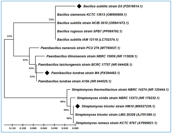
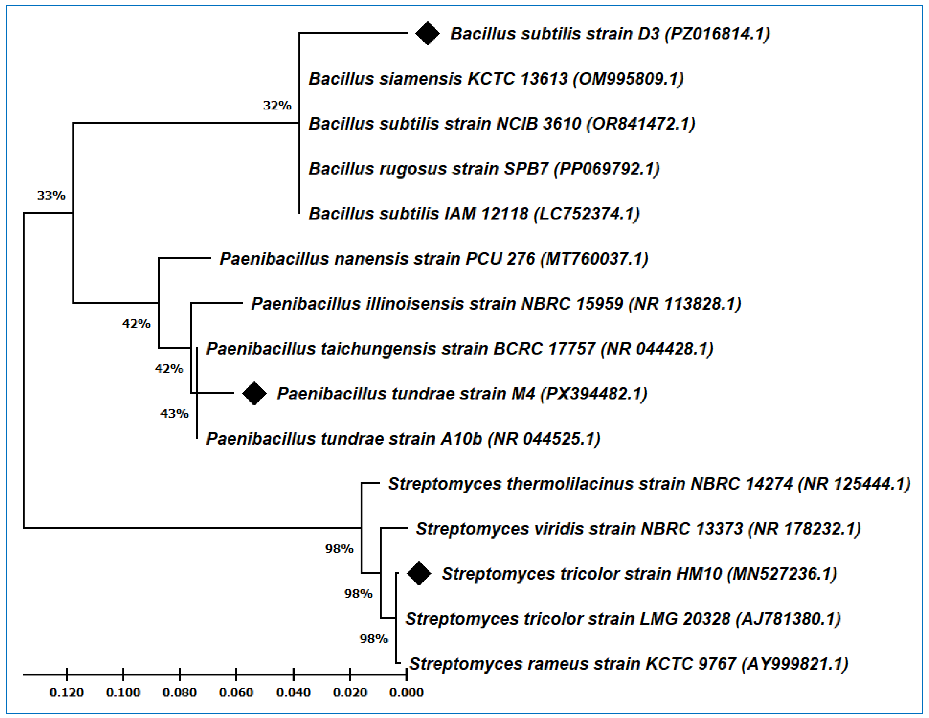
The phylogenetic tree was constructed using the 16S rRNA gene sequences from the identified strains, with their closely related strains in the GenBank. The BLAST (version: 2.17.0) search of strain Paenibacillus tundrae M4 displayed 97.68% identity with the type strain Paenibacillus tundrae A10b, whereas Bacillus subtilis strain D3 revealed 96.96% similarity with the type strain Bacillus subtilis 168. In addition, strain HM10 exhibited a close relationship with the type strain Streptomyces tricolor LMG 20328 and showed the highest degree of similarity, reaching 99.69%. When the 16S rRNA sequences from the three applied strains and their related sequences from the GenBank were aligned and used to construct the phylogenetic tree, P. tundrae M4 branched off separately with their related Paenibacillus strains in one group (Figure 1). Furthermore, B. subtilis strain D3 followed the same direction and grouped with other Bacillus strains, whereas S. tricolor HM10 clustered with its closer strains from the genus Streptomyces.
Figure 1.
The molecular phylogenetic tree of Paenibacillus tundrae M4, Bacillus subtilis D3 and Streptomyces tricolor HM10 with their closely related strains in the GenBank based on the 16S rRNA sequences. (♦) symbol pointed to the strains under study.
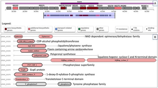
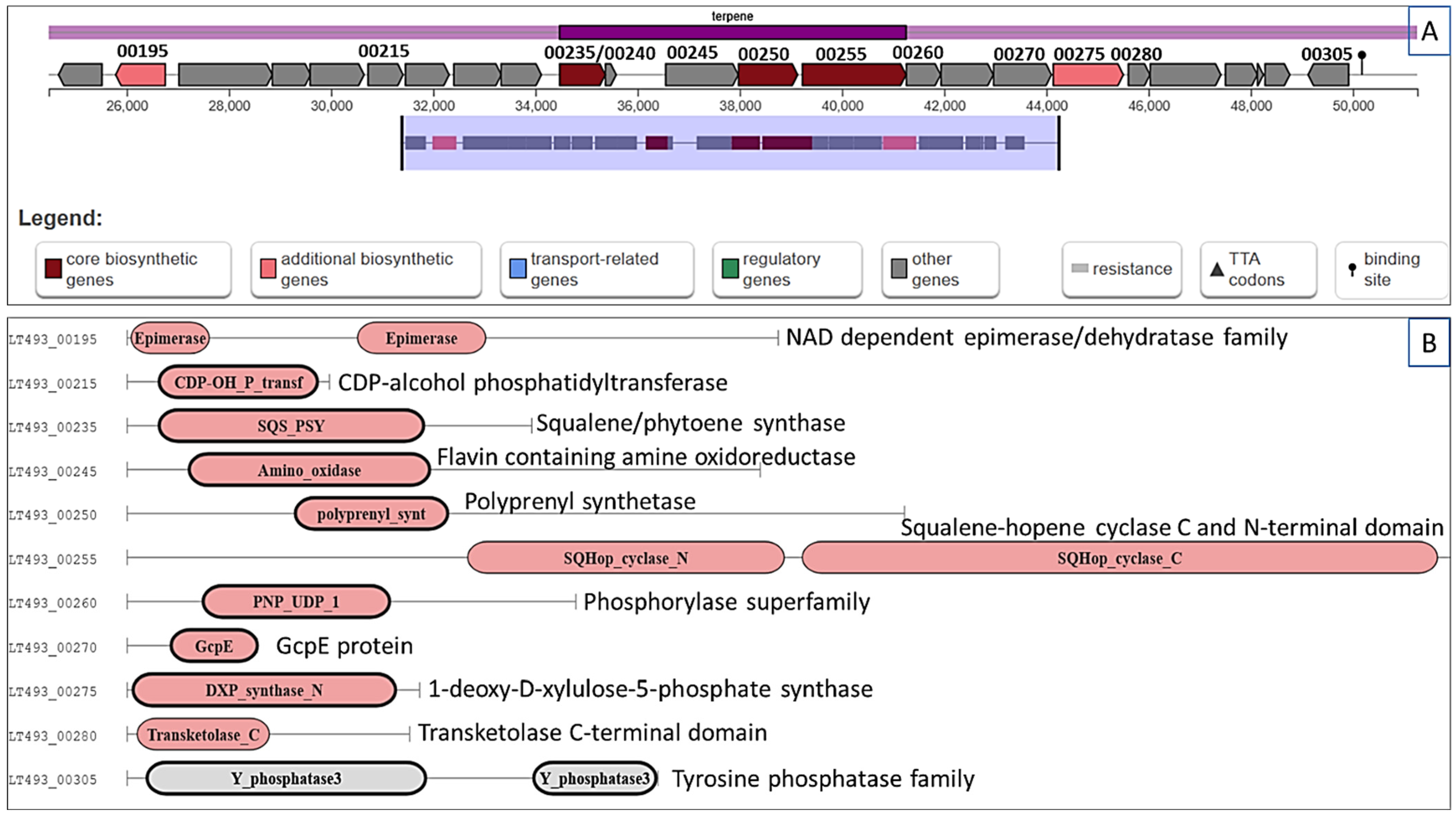
To predict the potential secondary metabolites related to the mite species control, the whole genome of S. tricolor HM10 was subjected to antiSMASH analysis. A potential biosynthetic gene cluster related to terpene biosynthesis was identified inside S. tricolor HM10 genome (Figure 2). This cluster locates in region 2.1 among nucleotides 24,474 and 51,258 (total: 26,785 nt in size). The proposed gene cluster presented a similarity score of 0.55 to the terpene (hopene) biosynthetic gene cluster from Streptomyces coelicolor A3(2). This biosynthetic gene cluster (BGC) segment had up to 24 genes and most of them are involved in terpene biosynthesis (Figure 2A). The main putative genes in the biosynthesis process are UDP-glucose 4-epimerase GalE (LT493_00195), which is predicted to catalyze the reversible interconversion of UDP-glucose and UDP-galactose. In addition, squalene synthase HpnD (LT493_00240) is supposed to catalyze the formation of presqualene diphosphate (PSPP) from two molecules of farnesyl diphosphate (FPP), whereas squalene synthase HpnC (LT493_00235) will function in converting presqualene diphosphate (PSPP) to hydroxysqualene (HSQ). Hydroxysqualene dehydroxylase HpnE (LT493_00245) is predicted to reduce hydroxysqualene (HSQ) to squalene (SQ). On the other hand, polyprenyl synthetase family protein (LT493_00250) is proposed to elongate the linear hydrocarbon backbones (prenyl diphosphates) from five-carbon isoprene units (isopentenyl diphosphate and dimethylallyl diphosphate), whereas squalene hopene cyclase (LT493_00255) works in inverting the linear precursor squalene into pentacyclic triterpenes through the cyclization process. Furthermore, 1-hydroxy-2-methyl-2-butenyl 4-diphosphate reductase (LT493_00260) will convert 1-hydroxy-2-methyl-2-(E)-butenyl 4-diphosphate into DMAPP (dimethylallyl diphosphate) and IPP (isopentenyl diphosphate). Additionally, 1-deoxy-D-xylulose-5-phosphate synthase (LT493_00275) and transketolase C-terminal domain (LT493_00280) are supposed to be involved in the biosynthesis of 1-deoxy-D-xylulose 5-phosphate (DXP) from pyruvate.
Figure 2.
Terpene (hopene) biosynthetic gene cluster in S. tricolor HM10. (A) The proposed terpene gene cluster in region 2.1 from HM10 genome; (B) the predicted genes involved in terpene biosynthesis based on Pfam domain annotation. The main genes in terpene biosynthesis are: squalene synthase HpnC (LT493_00235), squalene synthase HpnD (LT493_00240), hydroxysqualene dehydroxylase HpnE (LT493_00245), polyprenyl synthetase (LT493_00250), squalene hopene cyclase (LT493_00255), 1-hydroxy-2-methyl-2-butenyl 4-diphosphate reductase (LT493_00260), and 1-deoxy-D-xylulose-5-phosphate synthase (LT493_00275).
3. Discussion
Microorganisms produce useful natural products, including bioactive substances and natural antibiotics that have a variety of uses. These microorganisms, such as pathogenic bacteria, provide a much safer and environmentally friendly alternative to the commercially available chemical pesticides [30]. Streptomyces species, which are actinobacteria, have produced a wide range of secondary metabolites with significant commercial value. The detection of the avermectins from Streptomyces avermitilis as strong acaricides and antiparasitic agents accelerated the search for more novel pesticidal [31].
In continuation of that, the acaricidal potential of Streptomyces tricolor strain HM10, Bacillus subtilis D3, and Paenibacillus tundrae strain M4 was assessed in the current study against O. niloticus, T. hassani, A. olivi, and T. urticae under lab and field conditions. In comparison to control, significantly higher mobile stage mortality and lower egg hatching were noted after treatment with these three strains. In 2025, Al-Azzazy et al. [12] stated that S. tricolor HM10 reduced egg hatching in the citrus rust mite Phyllocoptruta oleivora (Ashmead) to 36.74% and induced mobile stages mortality to 83.66% after six days. In another report, Kaur et al. [32] displayed that culture filtrates of Streptomyces hydrogenans strain DH16 were found to be more lethal than to Meloidogyne incognita because 90% of J2 mortality occurred, and only less than 1% egg hatching in the time period of 72 h. Similar results were obtained by Al-Azzazy et al. [23], who reported that the direct application of Bacillus qassimus, Bacillus subtilis, and Acinetobacter sp. on two spotted spider mite, Tetranychus urticae, caused a mite mortality of 70.74%, 72.22%, and 84.11%, respectively, under laboratory conditions.
Pathogenic bacteria invade their hosts primarily through the alimentary tract and mouth parts. After that, tripartite Tc toxin complexes of pathogenic bacteria pierce the midguts of the host and introduce toxic enzymes as soluble proteins inside the host cell. Due to the midgut’s high pH, the poisons dissolve and are broken down by gut proteolytic enzymes into their toxic moieties [33,34]. In other instances, pathogenic bacteria enter via the integument, which damages the exoskeleton’s essential function in the hosts, leading to fatality [35,36]. Bacillus subtilis D3 is demonstrated to possess strong nematocidal properties; this is therefore of interest to scientists nowadays. Jurat-Fuentes and Jackson [37] reported that the soil bacterium B. firmus was described as pathogenic against larvae of the narcissus moth, Eligma narcissus Cramer (Lepidoptera: Nolidae). Dead larvae become black and soft, and discharged gut contents contain the rod-shaped bacterium and symptomatic of septicemia. In addition, Bacillus species exhibits a certain amount of toxicity by the production of endotoxins during sporulation; the receptors on intestinal epithelial cells in the midgut react with crystalliferous inclusions to release δ-endotoxins. Then, the pest’s midgut is harmed by the proteinaceous toxins produced by bacteria during the late infectious stage, with the ensuing osmotic imbalance leading to rupture of cells, jeopardizing the gut epithelium’s integrity, resulting in the death of the mite and different insect pests [23,38,39].
In the present study, numerous elements may have played a part in the mortality of O. niloticus, T. hassani, A. olivi, and T. urticae. In another study, Wilson et al. declared that external toxins called bacterial hemolysins target the membranes of cells, resulting in bodily distension and cell tears. These hemolysins might also be a factor in the pathogenicity of S. tricolor strain HM10, B. subtilis D3 and P. tundrae strain M4 against O. niloticus, T. hassani, A. olivi, and T. urticae, especially given the rapid death rate monitored.
Paenibacillus tundrae belongs to the genus Paenibacillus, which was divided from Bacillus in 1993 due to its unique genetic and behavioral traits. Members of this genus have been known as an obligatory pathogen of nematodes, insects, mollusks, phytopathogenic bacteria, and fungi [40,41]. Paenibacillus popilliae was discovered to infect Japanese beetle Popillia japonica Newman and was registered as the first microbial control agent in US [42]. According to Grady et al. [43], these bacteria create the enzyme chitinase, which hydrolyzes chitin, a structural polysaccharide of insect exoskeleton and stomodeum and proctodeum, as well as other structures., resulting in insect death. Numerous studies have emphasized the enormous application potential of Paenibacillus in several fields, including plant growth promotion, nitrogen fixation, hormone generation, phosphate solubilization, bioremediation of xenobiotics, and biocontrol activities [44].
Compared to other ecosystem interventions, the threat to natural enemies should be evaluated while assessing the pathogenic bacteria’s safety and environmental impact [45]. The three bacterial species showed preferential toxicity towards the phytophagous mites while saving the predatory mites. The obtained mortality values for predacious mites are significantly different and were five to six times higher than the corresponding ones for phytophagous mites. This could be attributed to the fact that predatory mites possess a thicker body wall with shields on the ventral and dorsal sides [46] that may obstruct the penetration of the bacteria into their bodies and decrease the received dose of bacteria compared to the thin-walled body of plant-feeding mites [12].
Our study revealed that the three bacterial species tested inhibit egg hatching under laboratory conditions. These impacts align with other findings, against citrus rust mite P. oleivora, including those of the Streptomyces tricolor strain HM10 that totally inhibits egg hatching to 36.74% [12]. In 2019, d’Errico et al. [24] stated that B. firmus has shown a lethal effect on nematodes such as Meloidogyne incognita, Tylenchulus semipenetrans, Heterodera sp., Ditylenchus sp., Xiphinema index, Pseudopyrenochaeta lycopersici and Rodopholus similis, where the toxins created by B. subtilis cause damage to the gall-forming nematodes’ outer egg pellicle, thus avoiding the hatching of plant parasitic nematode eggs. Furthermore, S. hydrogenans strain DH16 was found to be more lethal to M. incognita because of only less than 1% egg hatching in the period of three days [32].
When going forward and analyzing the S. tricolor MH10 genome, a predicted pathway was identified related to terpenes biosynthesis. Terpenes are natural products that have a wide range of structural variations, such as carbocyclic skeletons or linear hydrocarbons. Terpenes are widespread in the genus Streptomyces and have antiviral, antifungal, antibacterial, and antiparasitic properties [47]. They are employed as insecticides to store agricultural products [48,49]. Li et al. [50] evaluated the effectiveness of six terpenes that are frequently present in essential oils (carvacrol, eugenol, geraniol, citral, terpinen-4-ol, and linalool) against Sarcoptes scabiei eggs. They found that significant ovicidal actions are exhibited by carvacrol, eugenol, and geraniol, which make them promising ovicidal drugs for scabies treatment. In addition, combinations from three terpenes (carvacrol, thymol, and menthol) were assessed on the poultry red mite (Dermanyssus gallinae) under in vitro and in-field assays.
This terpene-based combination demonstrated encouraging miticidal action under in vitro and field application [51,52]. Wei et al. [53] used the highly pure chemical xenocoumacin 1 (purified from the supernatant of the Xenorhabdus nematophila CB6) to investigate its acaricidal effects on T. urticae and its predator Neoseiulus californicus. According to a number of research, monoterpenes mainly target insects’ nervous systems to produce their insecticidal effects [54]. Thymol (monoterpenoid) exhibited acaricidal activity through reducing wing-beat frequency and flight muscle impulses by interfering with the energy metabolism, electrical activity of the dorsal longitudinal flight muscles and nerve conduction of the mites [55,56,57]. Eugenol has neuroinhibitory effects, whereas linalool possesses neuroexcitatory characteristics [58]. Within the insect nervous system, monoterpenes may target ion channels like γ-aminobutyric acid type A receptors (GABAARs), nicotinic acetylcholine receptors (nAChRs), tyramine (TA) receptors, octopamine (OA) receptors, transient receptor potential (TRP) channels, in addition to enzymes such as acetylcholinesterase (AChE) and Na+/K+-ATPase [59,60].
4. Materials and Methods
4.1. Bacterial Strains and Culture Conditions
The previously isolated and identified Streptomyces tricolor strain HM10 (Accession, MN527236) [61] was used in this study, in addition to new isolates (B. subtilis D3 and P. tundrae M4 isolated from the soil), which were grown in glucose soyabean meal broth (GSB) and Nutrient Broth (NB) media, respectively. The three bacterial strains were inoculated on the appropriate medium and incubated at 30 °C with shaking at 150 rpm for 3 days. The bacterial cells (approximately with OD600 = 1.12) were removed from the culture filtrates through the Whatman Filter Paper (Grade 1), and the produced filtrates were implemented in either the lab or field against four mite species, O. niloticus, T. hassani, A. olivi, and T. urticae.
4.2. Bacterial Identification and Phylogenetic Tree
The bacterial genomic DNA from D3 and M4 isolates was extracted according to the method of Cook and Meyers [62]. DreamTaq PCR Master Mix (Thermo Scientific, Vilnius, Lithuania) was used to amplify the 16S rRNA region using general bacterial identification primers (27F and 1492R). The resulting PCR product was purified, sequenced (Macrogen, Seoul, Republic of Korea), assembled (BioEdit V7.7.1), and deposited in the GenBank under the following accession numbers: PZ016814 and PX394482. Using ClustalW built in MEGA X software (version: 10.2.6) [63], the 16S rRNA sequences of Bacillus subtilis strain D3, Paenibacillus tundrae strain M4, and Streptomyces tricolor strain HM10 were blasted and aligned with closely related species in the GenBank. The Tamura–Nei model, Maximum Likelihood, and Bootstrap method deduced the phylogenetic tree using the sequences obtained from the BLAST search in the NCBI database.
4.3. Genome Analysis and Potential Gene Cluster Identification
The available genome sequence of S. tricolor HM10 (GenBank: JA-JREA000000000.1) was subjected to analysis by antiSMASH (bacterial version, antiSMASH 7.0, accessible on 9 March 2025) [64]. In addition, unknown secondary metabolite biosynthesis gene clusters were identified using PRISM (Prediction Informatics for Secondary Metabolomes), in addition to their projected chemical structures [65]. Furthermore, the MIBiG (Minimum Information about a Biosynthetic Gene cluster) was applied for cluster comparison with identified known gene clusters in NCBI GenBank [66].
4.4. Lab Experiments—Residual Effects on Moving Stages and Eggs of O. niloticus, T. hassani, A. olivi, and T. urticae
All experiments were conducted at the laboratory of entomology, Department of Plant Protection, College of Agriculture and Food, Qassim University, Saudi Arabia. Naturally infested olive leaves were obtained from the agricultural research and experimental station of Qassim University, Saudi Arabia. Leaves were infected with O. niloticus, T. hassani, A. olivi, and T. urticae. Pre-treatment counts all laid eggs, and the mite’s mobile individuals were noted before treatment. Olive leaves were directly sprayed with 100 milliliters of the bacterial preparations of Bacillus subtilis D3, Paenibacillus tundrae strain M4, and Streptomyces tricolor strain HM10 using a high-pressure hand pump sprayer. Only tap water was sprayed on the control group. Olive leaves were placed upside down; the upper surfaces were placed face down on cotton wool that had been moistened (0.9 cm thick) in Petri dishes (16 cm in diameter × 2.5 cm high), one leaf for each Petri dish. A ring of Vaseline was applied to the leaf margins to prevent mites from escaping. Every day, 10 milliliters of water were given to the Petri dish to keep the cotton moist. Translucent plastic boxes (L 30 × W 25 × H 20 cm) were used to hold the Petri dishes and glass and were placed in an incubator at 30 ± 1 °C, 55 ± 5% RH, and a photoperiod of 14 h light (14 L)/10 h dark (10D). For each bacterial species, three replicates were performed. Olive leaves were checked daily, and the number of dead mites was recorded during seven days. The hatching of laid eggs was documented every day, where hatched protonymphs of experimental species were counted daily until the end of hatching in the untreated control, and hatched protonymphs were removed daily.
4.5. Laboratory Experiments—Direct Impacts on Predatory Mites, Agistemus Exsertus and Amblyseius swirskii
Colonies of predatory mites, A. exsertus and A. swirskii, were established with individuals collected from unsprayed olive orchards in Al Mulayda, Al-Qassim region (26.299138° N, 43.78896° E), Saudi Arabia. To obtain age-matched egg cohorts, eighty gravid females of each predator species extracted from the stock colonies were positioned in rearing units made of common bean leaves, Phaseolus vulgaris L. (Fabaceae), with about 800 two-spotted spider mites, Tetranychus urticae, as prey to each predator species. These mite-rearing units were maintained in an incubator at 32 ± 1 °C and 50 ± 5% RH. After two days, eggs laid by each predator were carefully moved separately to new rearing units, and the newly hatched larvae were provided with immature stages of spider mite, T. urticae as prey; suitable numbers of prey nymphs (20 T. urticae individuals) were added every day to the rearing units. Forty identically aged adult females of each predator species were carefully transferred to a bean leaf disk (4 cm in diameter), positioned face down in a Petri dish (8 cm in diameter × 2.5 cm high) on cotton that has been moistened with distilled water and sprayed with 10 mL of the bacterial preparations of Bacillus subtilis D3, Paenibacillus tundrae strain M4 and Streptomyces tricolor strain HM10 utilizing a tiny pressure spray bottle. For each bacterial species, three replicates were performed, and 30 adult females of each predator species were considered per replicate. Female mortality was noted 72 h after spraying. The control groups received merely tap water spraying. The bacterial incident was checked by stereo microscope inspection of A. exsertus and A. swirskii cadavers.
4.6. Field Experiment Impacts on Phytophagous and Predatory Mites
This study was conducted at the unsprayed olive orchards in Al Mulayda, Al-Qassim region (26.299138° N, 43.78896° E), Saudi Arabia. Four groups, each made up of three young olive trees (Olea europaea L.) (3 years old) of similar size and shape with a history of O. niloticus, T. hassani, A. olivi, and T. urticae infestations, were chosen for the study. Individual trees (replicates) were completely randomized as part of the experimental research design. For every bacterial species and untreated control, three replicates were employed. Three different species of bacteria, B. subtilis D3, P. tundrae strain M4 and S. tricolor strain HM10, were recently prepared from the stock culture (750 mL each with OD600 = 1.12) and immediately sprayed following filtration (750 mL from each strain culture) on olive trees (approximately 250 mL for each olive tree) using a one-liter volume hand atomizer for testing all mobile stages of O. niloticus, T. hassani, A. olivi and T. urticae and compared with the control, as well as their side impacts on the predacious mites, A. exsertus and A. swirskii. Tap water was sprayed onto the untreated control.
Twenty olive leaves per tree were randomly collected in all directions (60 leaves per treatment; 240 leaves per sampling). Leaves were placed in labeled plastic bags and transported to the acarology laboratory, where they were examined under a stereoscopic microscope. To ascertain the initial density and distribution of phytophagous and predacious mites, a pre-spray count was conducted for every replicate. Observations were conducted daily for seven consecutive days post-treatment.
4.7. Statistical Analysis
The proportion reduction in the mean populations of phytophagous and predacious mites and the mean proportion of the number of protonymphs (in the case of eriophyid mites) and larvae (in the case of T. urticae) hatching from eggs of O. niloticus, T. hassani, A. olivi, and T. urticae were determined using Henderson and Tilton’s equation 1955.
where Cb is the number of mites or laid eggs in the untreated control before spray, Ca is the number of hatched larvae and protonymphs or mites in the untreated control after spray, Tb is the number of mites or laid eggs in treatment before spray, and Ta is the number of hatched larvae and protonymphs or mites in treatment after spray.
The mortalities of O. niloticus, T. hassani, A. olivi, T. urticae, A. swirskii, and A. exsertus were determined by direct inspection. One-way analysis of variance (ANOVA) was used to statistically analyze the collected data for all variables. Tukey’s Honestly Significant Difference (HSD) test in Excel was applied to ascertain which specific group means are different from each other (p < 0.05).
5. Conclusions
The present study is the first investigation into acaricidal properties of B. subtilis D3, P. tundrae strain M4 against mites infesting olive trees. The current investigation revealed that B. subtilis D3, P. tundrae strain M4 are effective in managing O. niloticus, T. hassani, A. olivi, and T. urticae populations under laboratory and field conditions, while Streptomyces tricolor strain HM10 produces a moderate level of control. Consequently, pathogenic bacteria could be applied as a possible microbial control on O. niloticus, T. hassani, A. olivi, and T. urticae in the olive orchards, providing a practical substitute for traditional synthetic acaricides. Also, the three bacterial species reduced egg hatching around 53.52–64.68% in laboratory bioassays. Nevertheless, in addition to their mite-killing capabilities and inhibitory impacts on the eggs hatching, the three bacterial species exhibited a slight contact toxicity on the predatory mites, A. exsertus and A. swirskii, under laboratory and field conditions. Additionally, complementary studies should be carried out to assess the viability of these bacterial species in mite pest management plans as a means of satisfying the growing need for nonchemical control methods.
Author Contributions
Conceptualization, M.M.A.-A., S.S.A. and M.R.; methodology M.M.A.-A., S.S.A. and M.R.; software, M.R.; formal analysis, M.M.A.-A.; investigation, M.M.A.-A., S.S.A. and M.R.; writing—original draft preparation, M.M.A.-A., S.S.A. and M.R.; writing—review and editing, M.M.A.-A. and M.R.; funding acquisition, S.S.A. All authors have read and agreed to the published version of the manuscript.
Funding
This research was funded by Qassim University (QU-APC-2026).
Data Availability Statement
Data are contained within the article.
Acknowledgments
The researchers would like to thank the Deanship of Graduate Studies and Scientific Research at Qassim University for financial support (QU-APC-2026).
Conflicts of Interest
The authors declare no conflicts of interest.
References
- Saudi Press Agency. On World Olive Tree Day, Saudi Arabia’s Olive Production Surpasses 351,000 Tons; Saudi Press Agency: Riyadh, Saudi Arabia, 2025.
- Al-Azzazy, M.M. Studies on Mites Associated with Olive Trees. Master’s Thesis, Al-Azhar University, Cairo, Egypt, 2002. [Google Scholar]
- Alazzazy, M.; Alhewairini, S. A Life Table Analysis to Evaluate Biological Control of Four Mite Species Associated with Olive Trees Using the Predatory Mite Phytoseius plumifer (Acari: Phytoseiidae) in Saudi Arabia. Pak. J. Agric. Sci. 2019, 57, 299–305. [Google Scholar] [CrossRef]
- Raj, A.; Dubey, A.; Malla, M.A.; Kumar, A. Pesticide Pestilence: Global Scenario and Recent Advances in Detection and Degradation Methods. J. Environ. Manag. 2023, 338, 117680. [Google Scholar] [CrossRef] [Scilit] [PubMed]
- Alhewairini, S.S.; Al-Azzazy, M.M. Side Effects of Abamectin and Hexythiazox on Seven Predatory Mites. Braz. J. Biol. 2023, 83, e251442. [Google Scholar] [CrossRef] [Scilit]
- Shekhar, C.; Khosya, R.; Thakur, K.; Mahajan, D.; Kumar, R.; Kumar, S.; Sharma, A.K. A Systematic Review of Pesticide Exposure, Associated Risks, and Long-Term Human Health Impacts. Toxicol. Rep. 2024, 13, 101840. [Google Scholar] [CrossRef] [Scilit] [PubMed]
- Zhou, W.; Li, M.; Achal, V. A Comprehensive Review on Environmental and Human Health Impacts of Chemical Pesticide Usage. Emerg. Contam. 2025, 11, 100410. [Google Scholar] [CrossRef] [Scilit]
- Thakur, N.; Kaur, S.; Kaur, T.; Tomar, P.; Devi, R.; Thakur, S.; Tyagi, N.; Thakur, R.; Mehta, D.K.; Yadav, A.N. 25—Organic Agriculture for Agro-Environmental Sustainability. In Trends of Applied Microbiology for Sustainable Economy; Soni, R., Suyal, D.C., Yadav, A.N., Goel, R., Eds.; Academic Press: Cambridge, MA, USA, 2022; pp. 699–735. [Google Scholar]
- Butu, M.; Stef, R.; Grozea, I.; Corneanu, M.; Butnariu, M. Biopesticides: Clean and Viable Technology for Healthy Environment. In Bioremediation and Biotechnology: Sustainable Approaches to Pollution Degradation; Hakeem, K.R., Bhat, R.A., Qadri, H., Eds.; Springer International Publishing: Cham, Switzerland, 2020; pp. 107–151. [Google Scholar]
- Tomar, P.; Thakur, N.; Jhamta, S.; Chowdhury, S.; Kapoor, M.; Singh, S.; Shreaz, S.; Rustagi, S.; Rai, P.K.; Rai, A.K.; et al. Bacterial Biopesticides: Biodiversity, Role in Pest Management and Beneficial Impact on Agricultural and Environmental Sustainability. Heliyon 2024, 10, e31550. [Google Scholar] [CrossRef] [Scilit]
- Thakur, N.; Kaur, S.; Tomar, P.; Thakur, S.; Yadav, A.N. Chapter 15—Microbial Biopesticides: Current Status and Advancement for Sustainable Agriculture and Environment. In New and Future Developments in Microbial Biotechnology and Bioengineering; Rastegari, A.A., Yadav, A.N., Yadav, N., Eds.; Elsevier: Amsterdam, The Netherlands, 2020; pp. 243–282. [Google Scholar]
- Al-Azzazy, M.M.; Alhewairini, S.S.; Rehan, M. Biological Control of Citrus Rust Mite Phyllocoptruta oleivora by Three Bacterial Species. Sci. Rep. 2025, 15, 37452. [Google Scholar] [CrossRef] [Scilit]
- Perumal, V.; Kannan, S.; Alford, L.; Pittarate, S.; Mekchay, S.; Reddy, G.V.P.; Elangovan, D.; Marimuthu, R.; Krutmuang, P. Biocontrol Effect of Entomopathogenic Fungi Metarhizium anisopliae Ethyl Acetate-Derived Chemical Molecules: An Eco-Friendly Anti-Malarial Drug and Insecticide. Arch. Insect Biochem. Physiol. 2023, 114, e22037. [Google Scholar] [CrossRef] [Scilit]
- Chinnasamy, R.; Govindasamy, B.; Venkatesh, M.; Magudeeswaran, S.; Dhanarajan, A.; Devarajan, N.; Willie, P.; Perumal, V.; Mekchay, S.; Krutmuang, P. Bio-Efficacy of Insecticidal Molecule Emodin against Dengue, Filariasis, and Malaria Vectors. Environ. Sci. Pollut. Res. 2023, 30, 61842–61862. [Google Scholar] [CrossRef] [Scilit]
- Salimi, F.; Hamedi, J. Biopesticides: Microbes for Agricultural Sustainability. In Soil Microbiomes for Sustainable Agriculture: Functional Annotation; Yadav, A.N., Ed.; Springer International Publishing: Cham, Switzerland, 2021; pp. 471–501. [Google Scholar]
- Singh, A.K.; Kumar, M.; Ahuja, A.; Vinay, B.K.; Kommu, K.K.; Thakur, S.; Paschapur, A.U.; Jeevan, B.; Mishra, K.K.; Meena, R.P.; et al. Chapter 6—Entomopathogenic Nematodes: A Sustainable Option for Insect Pest Management. In Biopesticides; Woodhead Publishing: Cambridge, UK, 2022; pp. 73–92. [Google Scholar]
- Thakore, Y. The Biopesticide Market for Global Agricultural Use. Ind. Biotechnol. 2006, 2, 194–208. [Google Scholar] [CrossRef] [Scilit]
- Chaudhary, R.; Nawaz, A.; Khattak, Z.; Butt, M.A.; Fouillaud, M.; Dufossé, L.; Munir, M.; Haq, I.u.; Mukhtar, H. Microbial bio-control agents: A comprehensive analysis on sustainable pest management in agriculture. J. Agric. Food Res. 2024, 18, 101421. [Google Scholar] [CrossRef] [Scilit]
- Shukla, P.; Bankar, D.; Kumar, A.; Suhaib, M.; Mishra, P.; Raghuvanshi, H.; Gayithri, M. Advancements in the Use of Entomopathogenic Microbes for Pest and Disease Management—A Review. Int. J. Environ. Clim. Change 2023, 13, 945–953. [Google Scholar] [CrossRef] [Scilit]
- Khan, R.; Naveed, M. Evaluation of Comparative Toxicity of Different Insecticides against Fruit Fly, Bactrocera zonata Saunders (Diptera: Tephritidae). Pak. J. Zool. 2017, 49, 375–377. [Google Scholar] [CrossRef] [Scilit]
- Cheng, X.L.; Liu, C.J.; Yao, J.W. The Current Status, Development Trend and Strategy of the Bio-Pesticide Industry in China. Hubei Agric. Sci. 2010, 49, 2287–2290. [Google Scholar]
- Tripathi, Y.N.; Divyanshu, K.; Kumar, S.; Jaiswal, L.K.; Khan, A.; Birla, H.; Gupta, A.; Singh, S.P.; Upadhyay, R.S. Biopesticides: Current Status and Future Prospects in India. In Bioeconomy for Sustainable Development; Keswani, C., Ed.; Springer: Singapore, 2020; pp. 79–109. [Google Scholar]
- Al-Azzazy, M.M.; Alsohim, A.S.; Yoder, C.E. Biological Effects of Three Bacterial Species on Tetranychus urticae (Acari: Tetranychidae) Infesting Eggplant under Laboratory and Greenhouse Conditions. Acarologia 2020, 60, 587–594. [Google Scholar] [CrossRef] [Scilit]
- d’Errico, G.; Marra, R.; Crescenzi, A.; Davino, S.W.; Fanigliulo, A.; Woo, S.L.; Lorito, M. Integrated Management Strategies of Meloidogyne incognita and Pseudopyrenochaeta lycopersici on Tomato Using a Bacillus firmus-Based Product and Two Synthetic Nematicides in Two Consecutive Crop Cycles in Greenhouse. Crop Prot. 2019, 122, 159–164. [Google Scholar] [CrossRef] [Scilit]
- Arakere, U.C.; Jagannath, S.; Krishnamurthy, S.; Chowdappa, S.; Konappa, N. Chapter 12—Microbial Bio-Pesticide as Sustainable Solution for Management of Pests: Achievements and Prospects. In Biopesticides; Rakshit, A., Meena, V.S., Abhilash, P.C., Sarma, B.K., Singh, H.B., Fraceto, L., Parihar, M., Singh, A.K., Eds.; Woodhead Publishing: Cambridge, UK, 2022; pp. 183–200. [Google Scholar]
- Huang, M.; Bulut, A.; Shrestha, B.; Matera, C.; Grundler, F.M.W.; Schleker, A.S.S. Bacillus firmus I-1582 Promotes Plant Growth and Impairs Infection and Development of the Cyst Nematode Heterodera schachtii over Two Generations. Sci. Rep. 2021, 11, 14114. [Google Scholar] [CrossRef] [Scilit]
- Sharma, S.B.; Sayyed, R.Z.; Trivedi, M.H.; Gobi, T.A. Phosphate Solubilizing Microbes: Sustainable Approach for Managing Phosphorus Deficiency in Agricultural Soils. Springerplus 2013, 2, 587. [Google Scholar] [CrossRef] [Scilit] [PubMed]
- Ruiu, L.; Satta, A.; Floris, I. Emerging Entomopathogenic Bacteria for Insect Pest Management. Bull. Insectology 2013, 66, 181–186. [Google Scholar]
- Nazari, M.T.; Schommer, V.A.; Braun, J.C.A.; dos Santos, L.F.; Lopes, S.T.; Simon, V.; Machado, B.S.; Ferrari, V.; Colla, L.M.; Piccin, J.S. Using Streptomyces spp. as Plant Growth Promoters and Biocontrol Agents. Rhizosphere 2023, 27, 100741. [Google Scholar] [CrossRef] [Scilit]
- Zeng, Q.; Huang, H.; Zhu, J.; Fang, Z.; Sun, Q.; Bao, S. A New Nematicidal Compound Produced by Streptomyces albogriseolus HA10002. Antonie Van Leeuwenhoek 2013, 103, 1107–1111. [Google Scholar] [CrossRef] [Scilit]
- Cerna-Chávez, E.; Rodríguez-Rodríguez, J.F.; García-Conde, K.B.; Ochoa-Fuentes, Y.M. Potential of Streptomyces avermitilis: A Review on Avermectin Production and Its Biocidal Effect. Metabolites 2024, 14, 374. [Google Scholar] [CrossRef] [Scilit]
- Kaur, T.; Jasrotia, S.; Ohri, P.; Manhas, R.K. Evaluation of In Vitro and In Vivo Nematicidal Potential of a Multifunctional Streptomycete, Streptomyces hydrogenans Strain DH16 against Meloidogyne incognita. Microbiol. Res. 2016, 192, 247–252. [Google Scholar] [CrossRef] [Scilit]
- Gatsogiannis, C.; Lang, A.E.; Meusch, D.; Pfaumann, V.; Hofnagel, O.; Benz, R.; Aktories, K.; Raunser, S. A Syringe-like Injection Mechanism in Photorhabdus luminescens Toxins. Nature 2013, 495, 520–523. [Google Scholar] [CrossRef] [Scilit]
- Sheets, J.J.; Hey, T.D.; Fencil, K.J.; Burton, S.L.; Ni, W.; Lang, A.E.; Benz, R.; Aktories, K. Insecticidal Toxin Complex Proteins from Xenorhabdus nematophilus: Structure and Pore Formation. J. Biol. Chem. 2011, 286, 22742–22749. [Google Scholar] [CrossRef] [Scilit]
- Poinar, G.; Poinar, R. Parasites and Pathogens of Mites. Annu. Rev. Entomol. 1998, 43, 449–469. [Google Scholar] [CrossRef] [Scilit]
- Akpor, O.B.; Akinwusi, O.D.; Ogunnusi, T.A. Production, Characterization and Pesticidal Potential of Bacillus Species Metabolites against Sugar Ant (Camponotus consobrinus). Heliyon 2021, 7, e08447. [Google Scholar] [CrossRef] [Scilit]
- Jurat-Fuentes, J.L.; Jackson, T.A. Chapter 8—Bacterial Entomopathogens. In Insect Pathology, 2nd ed.; Vega, F.E., Kaya, H.K., Eds.; Academic Press: San Diego, CA, USA, 2012; pp. 265–349. [Google Scholar]
- Khan, M.A.; Paul, B.; Ahmad, W.; Paul, S.; Aggarwal, C.; Khan, Z.; Akhtar, M.S. Potential of Bacillus thuringiensis in the Management of Pernicious lepidopteran Pests. In Plant, Soil and Microbes: Volume 2: Mechanisms and Molecular Interactions; Hakeem, K.R., Akhtar, M.S., Eds.; Springer International Publishing: Cham, Switzerland, 2016; pp. 277–301. [Google Scholar]
- Fabrick, J.A.; Wu YiDong, W.Y. Roles of Insect Midgut Cadherin in Bt Intoxication and Resistance. In Bt Resistance: Characterization and Strategies for GM Crops Producing Bacillus thuringiensis Toxins; CABI: Oxfordshire, UK, 2015; pp. 69–86. [Google Scholar] [CrossRef] [Scilit]
- Nelson, D.M.; Glawe, A.J.; Labeda, D.P.; Cann, I.K.O.; Mackie, R.I. Paenibacillus tundrae sp. Nov. and Paenibacillus xylanexedens sp. Nov., Psychrotolerant, Xylan-Degrading Bacteria from Alaskan Tundra. Int. J. Syst. Evol. Microbiol. 2009, 59, 1708–1714. [Google Scholar] [CrossRef] [Scilit] [PubMed]
- Koppenhöfer, A.M.; Jackson, T.A.; Klein, M.G. Bacteria for Use against Soil-Inhabiting Insects. In Manual of Techniques in Invertebrate Pathology; Academic Press: San Diego, CA, USA, 2012; pp. 129–149. [Google Scholar]
- Arthurs, S.; Dara, S.K. Microbial Biopesticides for Invertebrate Pests and Their Markets in the United States. J. Invertebr. Pathol. 2019, 165, 13–21. [Google Scholar] [CrossRef] [Scilit] [PubMed]
- Grady, E.N.; MacDonald, J.; Liu, L.; Richman, A.; Yuan, Z.-C. Current Knowledge and Perspectives of Paenibacillus: A Review. Microb. Cell Fact. 2016, 15, 203. [Google Scholar] [CrossRef] [Scilit] [PubMed]
- Patowary, R.; Deka, H. Chapter 17—Paenibacillus. In Beneficial Microbes in Agro-Ecology; Amaresan, N., Senthil Kumar, M., Annapurna, K., Kumar, K., Sankaranarayanan, A., Eds.; Academic Press: San Diego, CA, USA, 2020; pp. 339–361. [Google Scholar]
- Hoy, M.A.; Jeyaprakash, A. Symbionts, Including Pathogens, of the Predatory Mite Metaseiulus occidentalis: Current and Future Analysis Methods. Exp. Appl. Acarol. 2008, 46, 329–347. [Google Scholar] [CrossRef] [Scilit]
- Zannou, I.; Moraes, G.; Ueckermann, E.; Oliveira, A.; Yaninek, J.; Hanna, R. Phytoseiid Mites of the Subtribe Amblyseiina (Acari: Phytoseiidae: Amblyseiini) from Sub-Saharan Africa. Zootaxa 2007, 1550, 1. [Google Scholar] [CrossRef] [Scilit]
- Rehan, M.; Gueddou, A.; Alharbi, A.; Ben Abdelmalek, I. In Silico Prediction of Secondary Metabolites and Biosynthetic Gene Clusters Analysis of Streptomyces thinghirensis HM3 Isolated from Arid Soil. Fermentation 2023, 9, 65. [Google Scholar] [CrossRef] [Scilit]
- Abdallah, I.I.; Quax, W.J. A Glimpse into the Biosynthesis of Terpenoids. KnE Life Sci. 2017, 3, 81–98. [Google Scholar] [CrossRef] [Scilit]
- Ninkuu, V.; Zhang, L.; Yan, J.; Fu, Z.; Yang, T.; Zeng, H. Biochemistry of Terpenes and Recent Advances in Plant Protection. Int. J. Mol. Sci. 2021, 22, 5710. [Google Scholar] [CrossRef] [Scilit] [PubMed]
- Li, M.; Liu, S.; Yin, Z.; Bernigaud, C.; Guillot, J.; Fang, F. Activity of Terpenes Derived from Essential Oils against Sarcoptes Scabiei Eggs. Parasit. Vectors 2021, 14, 600. [Google Scholar] [CrossRef] [Scilit]
- Tabari, M.A.; Jafari, A.; Jafari, M.; Youssefi, M.R. Laboratory and Field Efficacy of Terpene Combinations (Carvacrol, Thymol and Menthol) against the Poultry Red Mite (Dermanyssus gallinae). Vet. Parasitol. 2023, 313, 109842. [Google Scholar] [CrossRef] [Scilit]
- Sparagano, O.; Khallaayoune, K.; Duvallet, G.; Nayak, S.; George, D. Comparing Terpenes from Plant Essential Oils as Pesticides for the Poultry Red Mite (Dermanyssus gallinae). Transbound. Emerg. Dis. 2013, 60, 150–153. [Google Scholar] [CrossRef] [Scilit]
- Wei, J.; Yan, H.; Ren, J.; Li, G.; Zhang, B.; Xu, X. Acaricidal Effect of the Antimicrobial Metabolite Xenocoumacin 1 on Spider Mite Control. J. Integr. Agric. 2024, 23, 948–959. [Google Scholar] [CrossRef] [Scilit]
- Oliveira, J.A.C.; Fernandes, L.A.; Figueiredo, K.G.; Corrêa, E.J.A.; Lima, L.H.F.; Alves, D.S.; Bertolucci, S.K.V.; Carvalho, G.A. Effects of Essential Oils on Biological Characteristics and Potential Molecular Targets in Spodoptera frugiperda. Plants 2024, 13, 1801. [Google Scholar] [CrossRef] [Scilit]
- Waliwitiya, R.; Belton, P.; Nicholson, R.A.; Lowenberger, C.A. Effects of the Essential Oil Constituent Thymol and Other Neuroactive Chemicals on Flight Motor Activity and Wing Beat Frequency in the Blowfly Phaenicia sericata. Pest Manag. Sci. 2010, 66, 277–289. [Google Scholar] [CrossRef] [Scilit]
- Qasim, M.; Islam, W.; Rizwan, M.; Hussain, D.; Noman, A.; Khan, K.A.; Ghramh, H.A.; Han, X. Impact of Plant Monoterpenes on Insect Pest Management and Insect-Associated Microbes. Heliyon 2024, 10, e39120. [Google Scholar] [CrossRef] [Scilit]
- Zhou, Y.; Liao, F.; Fu, J.; Weng, J.; Mo, Q.; Xu, R.; Zhang, Y.; Sun, W.; Yue, D.; Ren, Z. In Vitro Acaricidal Activity of the Thymol against Sarcoptes scabiei and Regulating Effects on Enzyme Activity. J. Vet. Sci. Technol. 2018, 9, 554. [Google Scholar] [CrossRef]
- Gaire, S.; Scharf, M.E.; Gondhalekar, A.D. Toxicity and Neurophysiological Impacts of Plant Essential Oil Components on Bed Bugs (Cimicidae: Hemiptera). Sci. Rep. 2019, 9, 3961. [Google Scholar] [CrossRef] [Scilit]
- Abdelgaleil, S.A.M.; Gad, H.A.; Ramadan, G.R.M.; El-Bakry, A.M.; El-Sabrout, A.M. Monoterpenes: Chemistry, Insecticidal Activity against Stored Product Insects and Modes of Action—A Review. Int. J. Pest Manag. 2024, 70, 267–289. [Google Scholar] [CrossRef] [Scilit]
- Souto, A.L.; Sylvestre, M.; Tölke, E.D.; Tavares, J.F.; Barbosa-Filho, J.M.; Cebrián-Torrejón, G. Plant-Derived Pesticides as an Alternative to Pest Management and Sustainable Agricultural Production: Prospects, Applications and Challenges. Molecules 2021, 26, 4835. [Google Scholar] [CrossRef] [Scilit]
- Rehan, M.; ALSohim, A.S.; Abidou, H.; Rasheed, Z.; Abdulmonem, W.A.L. Isolation, Identification, Biocontrol Activity, and Plant Growth Promoting Capability of a Superior Streptomyces tricolor Strain HM10. Pol. J. Microbiol. 2021, 70, 245–256. [Google Scholar] [CrossRef] [Scilit]
- Cook, A.E.; Meyers, P.R. Rapid Identification of Filamentous Actinomycetes to the Genus Level Using Genus-Specific 16S RRNA Gene Restriction Fragment Patterns. Int. J. Syst. Evol. Microbiol. 2003, 53, 1907–1915. [Google Scholar] [CrossRef] [Scilit] [PubMed]
- Kumar, S.; Stecher, G.; Li, M.; Knyaz, C.; Tamura, K. MEGA X: Molecular Evolutionary Genetics Analysis across Computing Platforms. Mol. Biol. Evol. 2018, 35, 1547–1549. [Google Scholar] [CrossRef] [Scilit] [PubMed]
- Blin, K.; Shaw, S.; Augustijn, H.E.; Reitz, Z.L.; Biermann, F.; Alanjary, M.; Fetter, A.; Terlouw, B.R.; Metcalf, W.W.; Helfrich, E.J.N.; et al. AntiSMASH 7.0: New and Improved Predictions for Detection, Regulation, Chemical Structures and Visualisation. Nucleic Acids Res. 2023, 51, W46–W50. [Google Scholar] [CrossRef] [Scilit] [PubMed]
- Skinnider, M.A.; Johnston, C.W.; Gunabalasingam, M.; Merwin, N.J.; Kieliszek, A.M.; MacLellan, R.J.; Li, H.; Ranieri, M.R.M.; Webster, A.L.H.; Cao, M.P.T.; et al. Comprehensive Prediction of Secondary Metabolite Structure and Biological Activity from Microbial Genome Sequences. Nat. Commun. 2020, 11, 6058. [Google Scholar] [CrossRef] [Scilit] [PubMed]
- Kautsar, S.A.; Blin, K.; Shaw, S.; Navarro-Muñoz, J.C.; Terlouw, B.R.; van der Hooft, J.J.J.; van Santen, J.A.; Tracanna, V.; Suarez Duran, H.G.; Pascal Andreu, V.; et al. MIBiG 2.0: A Repository for Biosynthetic Gene Clusters of Known Function. Nucleic Acids Res. 2020, 48, D454–D458. [Google Scholar] [CrossRef] [Scilit] [PubMed]

Disclaimer/Publisher’s Note: The statements, opinions and data contained in all publications are solely those of the individual author(s) and contributor(s) and not of MDPI and/or the editor(s). MDPI and/or the editor(s) disclaim responsibility for any injury to people or property resulting from any ideas, methods, instructions or products referred to in the content. |
© 2026 by the authors. Licensee MDPI, Basel, Switzerland. This article is an open access article distributed under the terms and conditions of the Creative Commons Attribution (CC BY) license.