


Abstract
This review aims to explore the green synthesis of metal and metal oxide nanoparticles using various species of the genus Artemisia. The synthesis processes commonly involve aqueous or organic extracts of plant parts (e.g., leaves, stems, and roots), which react with metal salt solutions (e.g., AgNO3, Zn(NO3)2, HAuCl4, Cu(NO3)2) under controlled parameters, including pH, temperature, and light exposure. The synthesized nanoparticles are characterized using techniques such as UV–Visible spectroscopy, transmission electron microscopy (TEM), scanning electron microscopy (SEM), Fourier transform infrared spectroscopy (FTIR), X-ray diffraction (XRD), dynamic light scattering (DLS), and zeta potential analysis. These approaches provide information on nanoparticle size, morphology, crystallinity, surface chemistry and charge, which are significantly influenced by synthesis parameters and the specific Artemisia species used. The biosynthesized nanoparticles have demonstrated promising multifunctional applications, including broad-spectrum antimicrobial activity against bacterial and fungal strains, antioxidant capacity, anticancer potential, as well as applications in agriculture and environmental remediation.
1. Introduction
Different mitigation approaches are employed to enhance global agricultural crop productivity and assist environmental remediation [1]. Nanotechnology encompasses the design, synthesis, and manipulation of materials at the nanometric scale for the fabrication, characterization, and application of a wide range of nanoparticles (NPs). In recent years, metal nanoparticles have attracted considerable interest due to their unique physicochemical properties dependent on their small size, shape, and specific surface area [2,3]. While achieving high monodispersity is a critical goal for diverse applications (e.g., targeted drug delivery, electronics), the uniformity of particles depends significantly on the synthesis method [4]. Physical methods, such as laser ablation, offer the advantage of high purity but often require high energy consumption and can result in broad size distributions (polydispersity) depending on the experimental conditions [5]. In contrast, chemical fabrication methods generally provide superior control over nucleation and growth steps, allowing improved monodispersity and higher yields compared with physical techniques. However, conventional chemical routes often involve hazardous reducing agents and stabilizers, raising environmental and safety concerns [6].
Over the past decade, the principles of green chemistry have been intensively explored in the context of sustainable development [7]. Consequently, increasing efforts have been directed toward the production of non-toxic NPs using environmentally friendly biological approaches. The biosynthesis of nanoparticles employing plant extracts represents a viable alternative to conventional chemical and physical methods. This approach typically requires the use of an eco-friendly solvent (most commonly water), a non-toxic reducing agent, and a suitable stabilizing agent [8]. Beyond the environmental advantages, plant-mediated synthesis offers significant economic benefits. Many plant sources are abundant, renewable, and can be harvested at a low cost, providing a “waste-to-wealth” pathway for large-scale NP production. Moreover, the presence of diverse primary and secondary metabolites in plant extracts can contribute to improved NP stability and may reduce agglomeration and toxicity [9].
Accordingly, the field of nanotechnology is undergoing rapid advancement, and there is increasing attention toward the development of green synthesis approaches for producing metal and metal oxide nanoparticles using plant-based resources. The genus Artemisia is taxonomically classified within the family Asteraceae (Compositae), including over 500 species [10]. Members of this genus are predominantly adapted to temperate climates and are widely distributed across Europe, East Asia, North America, North Africa, and Australia. The highest species diversity is observed in Asia, particularly in China, where approximately 150 species have been recorded, while Europe hosts about 57 species [10,11]. Based on global distribution data, the majority of Artemisia species are concentrated in the Northern Hemisphere, whereas only about 25 species are found in the Southern Hemisphere [12].
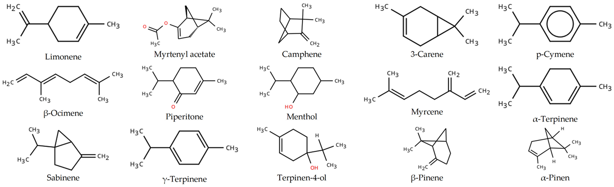
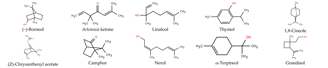
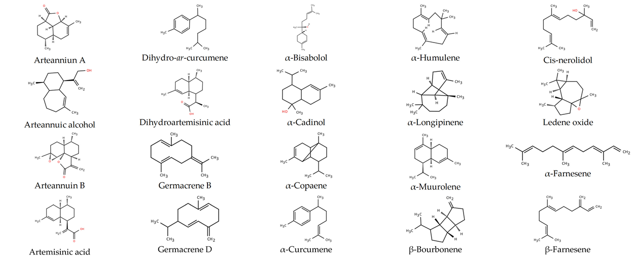
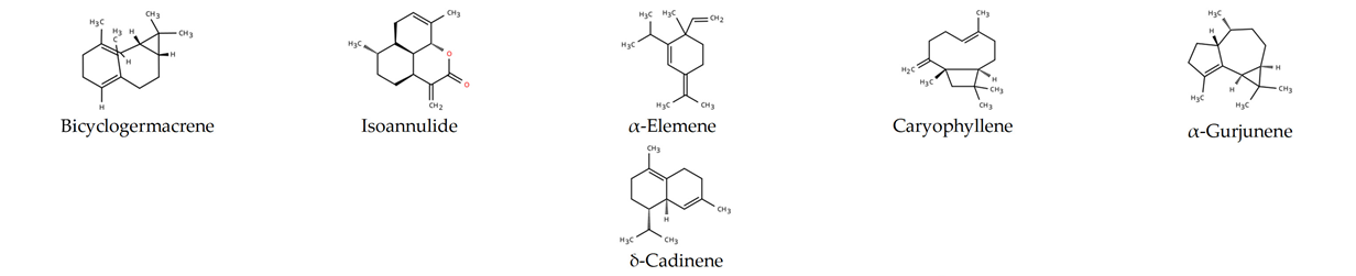
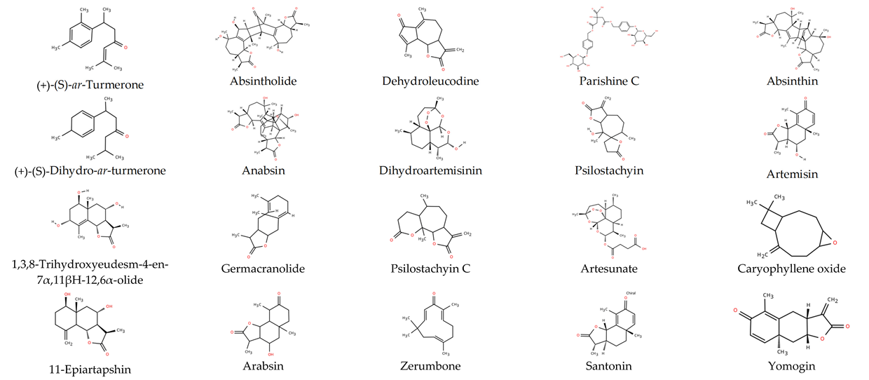
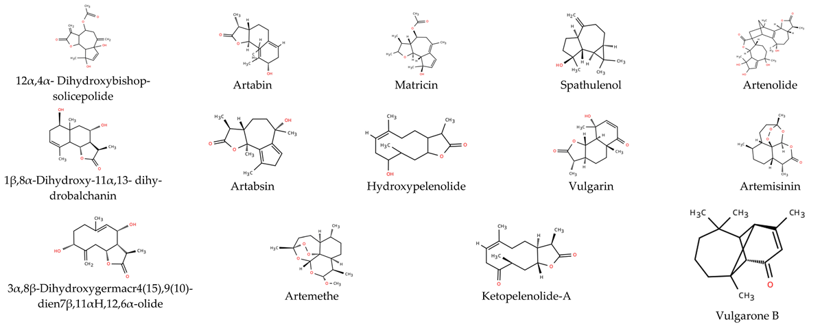
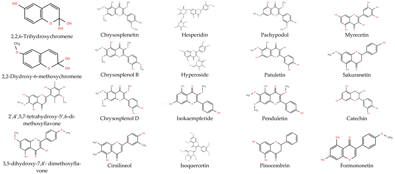
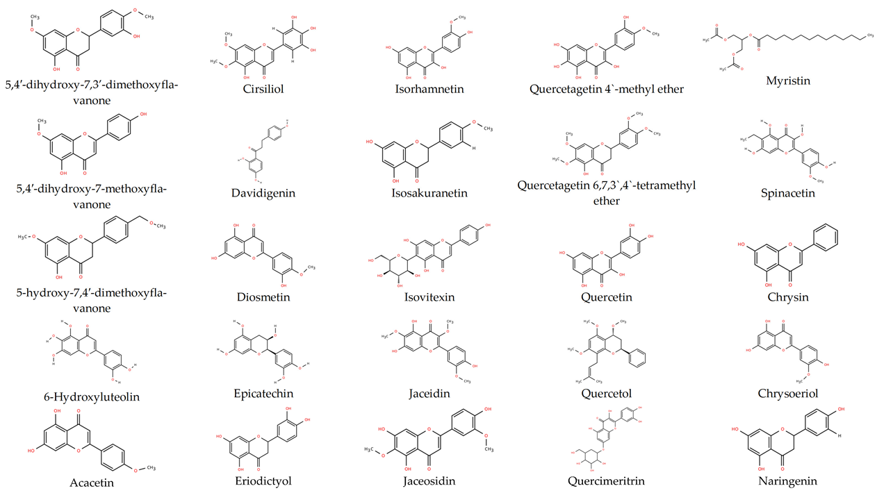
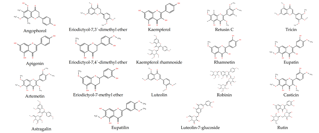
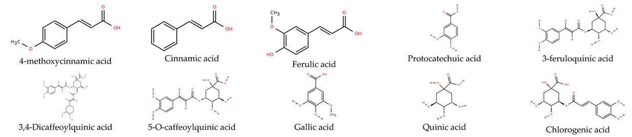
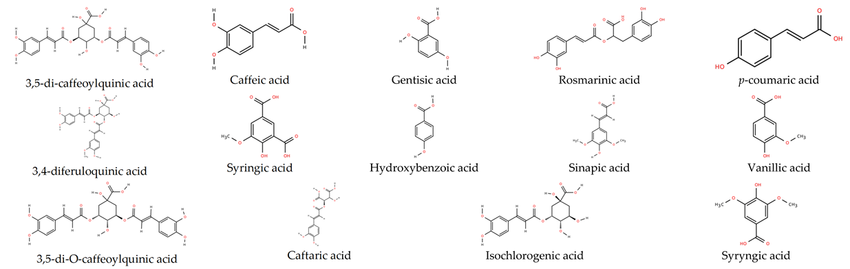
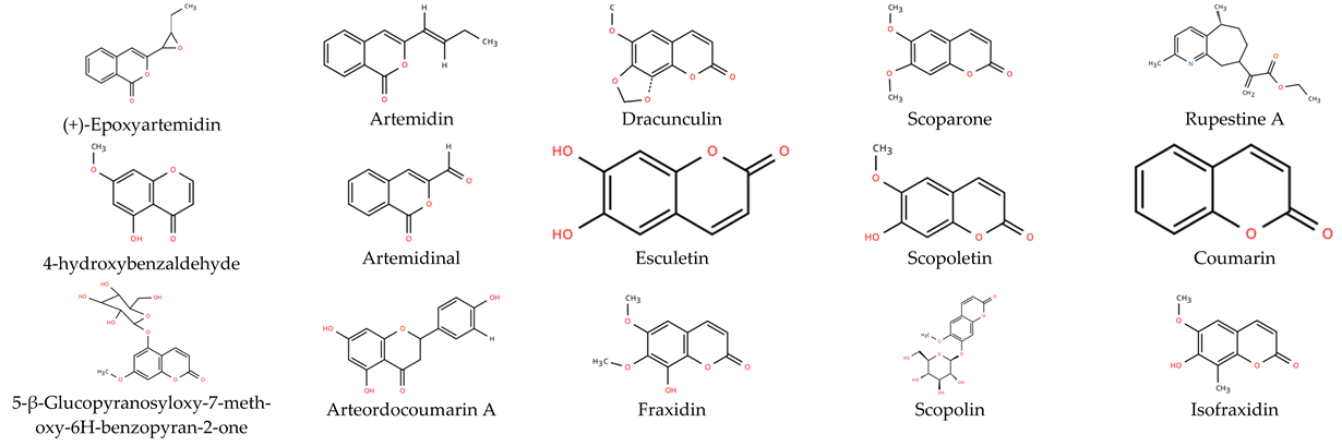
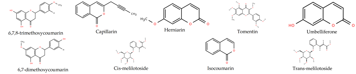
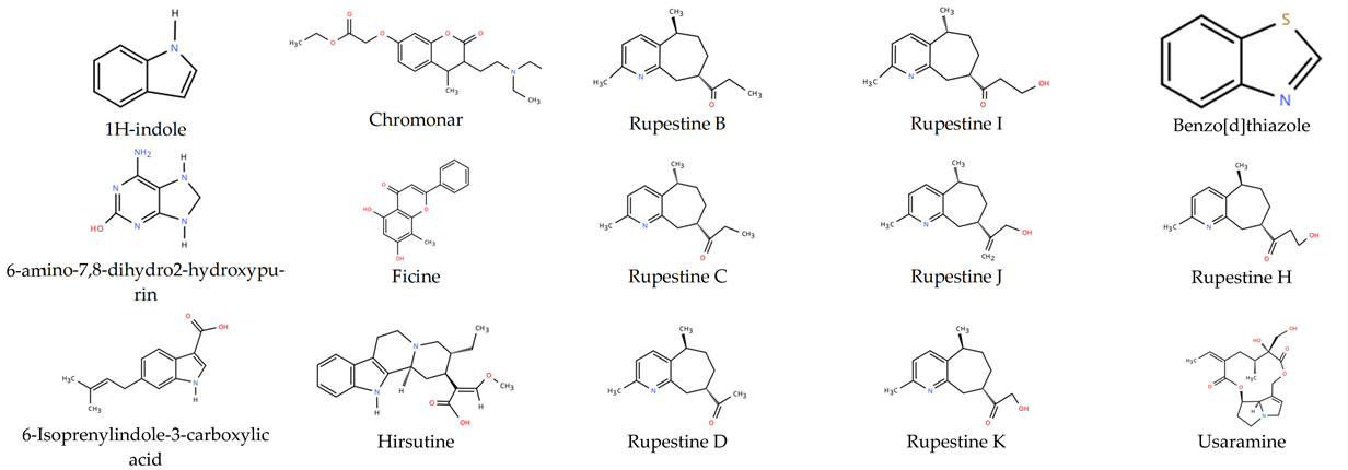
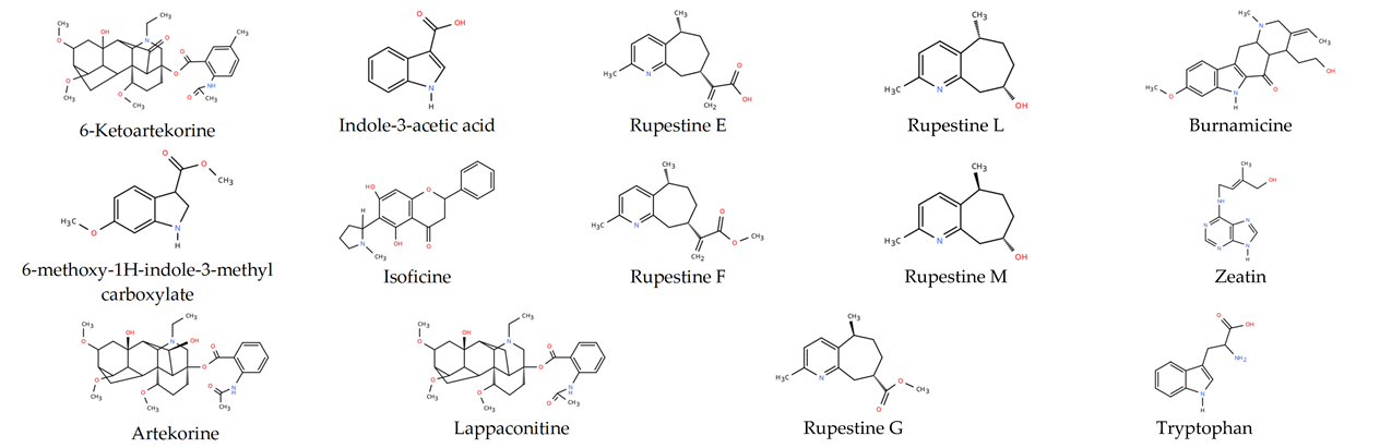
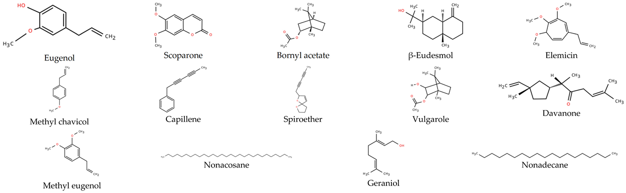
Plants belonging to the genus Artemisia are well known for their richness in bioactive compounds, many of which contribute to their significant medicinal value and diverse biological activities. To date, over 260 Artemisia species have been investigated for their essential oil composition and secondary metabolite profiles. Various classes of bioactive compounds have been identified, including terpenoids, flavonoids, coumarins, phenolic acids, alkaloids, essential oils, and sterols (Table A1) [13]. Due to their rich phytochemical content, Artemisia species extracts have emerged as promising candidates for the biosynthesis of NPs. These compounds serve as both reducing agents (converting metal ions to NPs) and capping agents (stabilizing the NPs) [14,15].
Research suggests that the Artemisia genus is a valuable resource for the synthesis of multifunctional NPs with applications across various sectors. Among the nanoparticles synthesized using Artemisia extracts, silver nanoparticles (AgNPs) are the most studied. These NPs have demonstrated effective antimicrobial properties against both Gram-positive and Gram-negative bacterial strains and fungal species [16,17], as well as antioxidant activity [18] and antitumoral properties [19]. Furthermore, they show significant potential in environmental remediation [20], agriculture, and food safety [18,21].
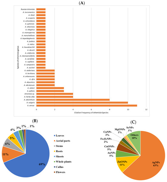
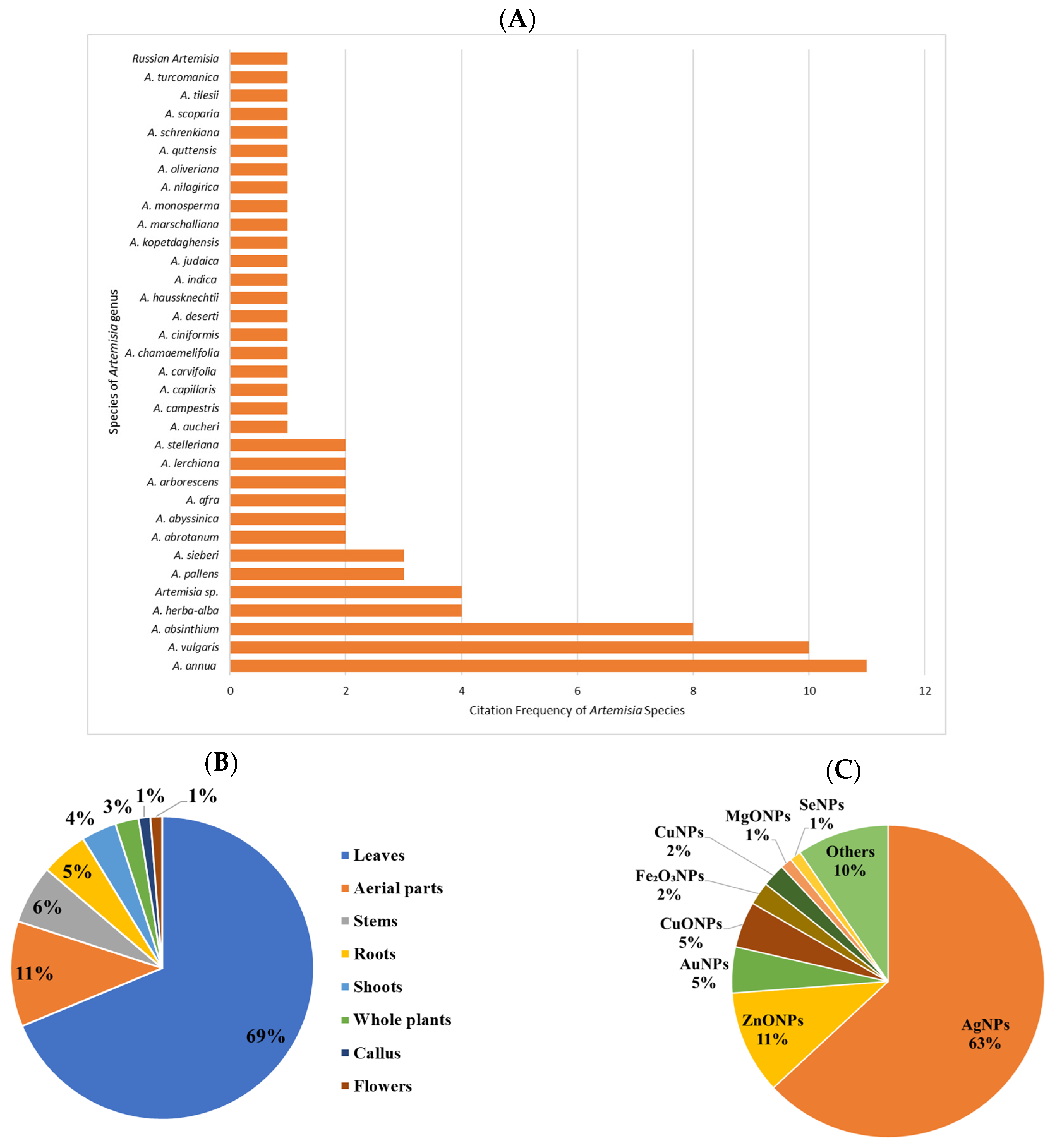
According to the literature reviewed, among the 34 documented Artemisia species used for nanoparticles synthesis, the most common were A. annua, A. vulgaris, and A. absinthium (Figure 1A). Furthermore, while phytochemicals are distributed in different plant organs (e.g., leaves, stems, flowers, fruits and roots), the majority of studies (69%) utilized leaves, followed by aerial parts (11%), for plant-mediated nanoparticles (Figure 1B).
Figure 1.
(A) The most mentioned Artemisia species in terms of number involved in the biosynthesis of NPs. (B) Different parts of the plant and (C) types of metal/metal oxide-based NPs synthesized using Artemisia species extracts.
The consulted literature highlights the synthesis and characterization of diverse Artemisia-derived NPs, including AgNPs, silver oxide nanoparticles (Ag2ONPs), copper nanoparticles (CuNPs), copper oxide nanoparticles (CuONPs), gold nanoparticles (AuNPs), titanium dioxide nanoparticles (TiO2NPs), selenium nanoparticles (SeNPs), zinc oxide nanoparticles (ZnONPs), and tin oxide nanoparticles (TONPs). Of all nanoparticles synthesized via green methods using this genus, most were AgNPs (63%), followed by ZnONPs (11%) and AuNPs (5%) (Figure 1C).
Despite the proliferation of individual studies on the genus Artemisia, a significant knowledge gap exists regarding comparative analyses that evaluate the efficacy of different species and plant parts against various metal precursors. The novelty of this paper lies in addressing this gap through a comprehensive analysis of metal and metal oxide NP synthesis using extracts from various parts of Artemisia species. Additionally, this work details the synthesis methods, physicochemical characteristics (size, shape, zeta potential), and diverse applications, ranging from medical and antimicrobial uses to environmental remediation and food safety.
2. Synthesis of Metal and Metal Oxide NPs
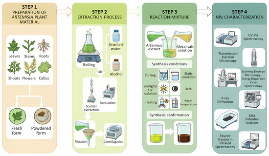
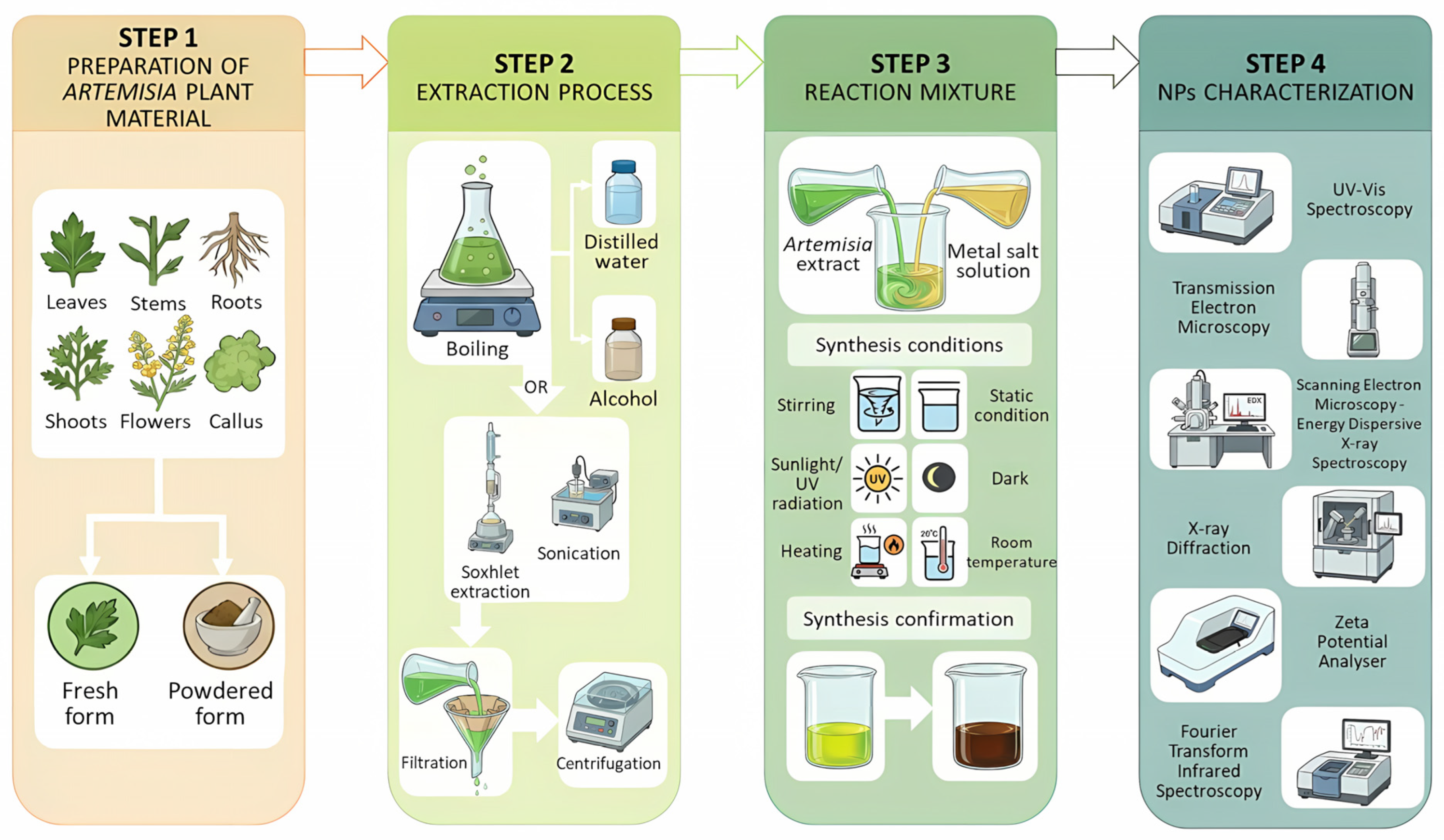
According to the literature, various methods for NP synthesis using extracts from genus Artemisia have been documented, usually following four main steps (Figure 2). The initial step involves the collection and preparation of plant material, which may include leaves [22,23,24], stems [25], roots [26,27], aerial parts [28,29], flowers [30], callus [31] or the whole plant [32]. This material can be utilized either fresh [20,33,34] or in powdered form [26,35]. Next, the plant material undergoes an extraction process, mostly through boiling in organic or inorganic solvents (e.g., distilled water [15,36], ethanol [37,38], or methanol [39,40]). Other methods include Soxhlet extraction [41,42] or sonication [30], followed by filtration and centrifugation. The chemical composition of the extract significantly influences the efficiency of NP synthesis. Secondary metabolites, including flavonoids, alkaloids, terpenoids, and phenolic acids, play a crucial role in the reduction and stabilization of metal ions into NPs [39]. Furthermore, specific functional groups, such as amino (-NH2), carboxyl (-COOH), carbonyl (-CO), hydroxyl (-OH), and thiol (-SH), enhance the reducing and capping capacity of phytoconstituents, facilitating the formation of stable, non-agglomerated NPs [23]. Metal salt solutions, such as silver nitrate (AgNO3), chloroauric acid (HAuCl4), copper nitrate (Cu(NO3)2), zinc acetate (Zn(CH3COO)2), and iron (III) chloride (FeCl3), are prepared at varying concentrations. To induce synthesis, the reaction mixtures are maintained under either continuous stirring [43,44,45] or static conditions while being subjected to specific environmental parameters. These parameters often include exposure to sunlight/UV radiation [31,35,46,47] or dark conditions [48,49], heating [38,50,51] or maintenance at room temperature [44,52]. The formation of NPs is typically indicated by a visible color change in the reaction solution. Depending on the metal and species used, the solution may transform from colorless, yellow, blue, orange, or pale green to final shades of pink, violet, reddish-brown, grey-black, or dark brown. This optical shift is attributed to surface plasmon resonance (SPR), which is directly influenced by the size and shape of the synthesized NPs [14,53,54,55].
Figure 2.
Schematic representation of the workflow for the green synthesis of nanoparticles using Artemisia species.
Among the phytoconstituent classes present in species of the Artemisia genus, terpenoids and flavonoids are notably abundant (Table A1) [56]. These bioactive compounds participate in numerous chemical processes, including the synthesis of NPs. During NP formation, functional groups such as -OH and -CO from flavonoids and other antioxidants donate electrons to metal ions (e.g., Ag+, Au3+, Cu2+, Zn2+), reducing them to metallic or metal oxide nanoparticles [15,20,23]. Simultaneously, these phytochemicals adsorb onto NP surfaces, acting as capping and stabilizing agents that prevent aggregation and help control particle size and morphology [57].
Fourier transform infrared spectroscopy (FTIR) has confirmed the interaction of functional groups in metal reduction and stabilization processes through specific absorption bands [14]. As summarized in Table 1, key functional groups include O–H stretching of phenols and alcohols (broad peaks between 3206 and 3472 cm−1), C–H stretching of aromatic and methylene groups (2853–3078 cm−1; 2072 cm−1), and the C=O carbonyl stretching of amide and phenolic compounds (1635–1661 cm−1). The spectra also reveal C=C stretching of aromatic rings (~1607 cm−1), carboxylate –COO− groups (~1580 cm−1), N–O stretching of nitro compounds (1319–1504 cm−1), C≡C stretching (2118 cm−1), and C–O/C–O–C stretching (1026–1243 cm−1; 588 cm−1) [14,17,20,27,45]. Furthermore, the successful synthesis of metal and metal oxide nanoparticles is definitively proven by the emergence of characteristic metal–oxygen vibration bands (e.g., Cu–O at 1385 cm−1, Ag–O at 506.55 cm−1, and Zn–O at 661.84 cm−1), confirming that these organic constituents effectively reduce metal ions and stabilize the resulting nanostructures [14,20,45].
The yield of NP synthesis is affected by various factors, including pH, temperature, and the concentration of both plant extract and metallic salt solution. For instance, Avitabile et al. reported that approximately 60% of initial silver was converted into AgNPs after six centrifugation cycles [58]. Similarly, a precursor salt concentration of 0.1 M resulted in the highest production of CuONPs (58%), whereas higher concentrations inhibited synthesis [14]. Regarding reactant ratios, Ali et al. found that an extract to AgNO3 ratio of 6:4 (v/v) achieved the highest conversion efficiency [59]. Furthermore, Kobylinska et al. [26] demonstrated the important role of the solvent. A 70% water–ethanol mixture extracted more bioactive compounds, especially flavonoids, from A. annua and A. tilesii “hairy” roots culture, thereby increasing the reduction of Ag+ and stabilizing the NPs [26]. In terms of pH, no AgNPs were formed at pH levels below 4. However, Ag+ reduction increased in alkaline media, with pH 10 being optimal [57].
3. Artemisia-Based Nanoparticle Characterization
To determine the physicochemical characteristics of synthesized NPs (e.g., size, shape, crystallinity, capping, and zeta potential), a variety of techniques are employed, including UV-Vis spectroscopy, scanning electron microscopy (SEM) with energy-dispersive X-ray spectroscopy (SEM-EDX), transmission electron microscopy (TEM), Fourier transform infrared spectroscopy (FTIR), X-ray diffraction (XRD), dynamic light scattering (DLS), among others.
3.1. Size
TEM analysis revealed that the smallest AgNPs (0.60 nm) were obtained from A. schrenkiana extract [24], while the largest AgNPs (~200 nm) formed using A. vulgaris extract [60]. Furthermore, MgONPs, synthesized with A. abrotanum extract, did not exceed 10 nm [46]. The smallest ZnONPs measured 17 nm [39], while Shelembe et al. [55] obtained ZnONPs and Fe2O3NPs recording 31 nm. Using SEM analysis, CuONPs synthesized from A. haussknechtii extract measured approximately 35 nm [49]. By contrast, SeNPs and TiO2NPs exhibited larger sizes, 110 nm and 92.58 ± 56.98 nm, respectively [42,49].
Various factors influence particle size. Higher extract concentrations tend to produce larger nanoparticles [24]. Conversely, increasing the volume of Artemisia extract relative to the metal precursor often reduces NP size [15]. Regarding pH, alkaline conditions (pH 7–9) favor the synthesis of smaller NPs compared to acidic conditions [50]. Temperature also plays a key role; higher temperatures generally favor smaller particles [50]. However, excessive temperatures may degrade the phytochemicals, thereby compromising the quality and stability of the synthesized NPs, whereas lower temperatures and short exposure times may lead to incomplete synthesis [54,61]. The bioactive compounds from Artemisia extracts, especially proteins, flavonoids, and phenolic acids, affect NP size and stability by binding metal ions or reducing them to NPs [27,35,62]. Overall, nanoparticles synthesized from Artemisia extracts demonstrated a wide size variability depending on the metal type, plant species, and synthesis conditions.
3.2. Shape
Spherical morphology predominates among NPs synthesized using Artemisia extracts. However, cubic, hexagonal, triangular, polyhedral, and pentagonal shapes have also been reported. AgNPs exhibit morphological diversity, ranging from spherical [17,25] to cubic structures [57]. Basavegowda et al. reported spherical and triangular forms of AuNPs using A. pallens extract [53]. Metal oxide NPs, such as ZnONPs, MgONPs, and CuONPs, are typically spherical [46,49,63], whereas Fe2O3NPs have been observed in spherical and cubic morphologies [55,64]. As with NP size, morphology is influenced by several parameters, including pH, temperature, reactant concentration, and the phytochemical composition of the Artemisia extract.
3.3. Zeta Potential
Zeta potential (ζ) is a critical parameter in NP synthesis, influencing colloidal stability, surface interactions, and biological applications [65]. Artemisia-based NPs typically exhibit negative ζ values, indicating good colloidal stability. Reported ζ values range from −18.5 mV to −40.4 mV, with most clustering between −21 mV to −29 mV, corresponding to moderate stability [18,34,58,59,66,67,68,69,70]. Metal oxide NPs, such as ZnONPs (−38 mV) tend to display greater stability compared to metal NPs like AuNPs (−19.3 mV) [67,68]. Notably, AgNPs show a broader range of ζ values, from −18.5 mV to −40.4 mV, depending on synthesis conditions [34,59]. Key factors influencing ζ include pH, temperature, surface chemistry, and the composition of NPs. In alkaline conditions, ζ becomes more negative, whereas it becomes less negative in acidic environments [71]. Elevated temperature tends to decrease ζ values [72]. Additionally, Ardestani et al. [73] observed that NPs enriched in negatively charged functional groups exhibit more negative ζ values, thereby enhancing colloidal stability.
Table 1.
Summary of synthesis, characterization, and potential applications of nanoparticles derived from extracts of Artemisia species.
4. Artemisia-Based Nanoparticle Applications
4.1. Antimicrobial Activity
The findings indicate that nanoparticles synthesized from extracts of various Artemisia species exhibit significant antimicrobial properties against a wide array of microorganisms. Specifically, AgNPs [17,32,44,54,88], AuNPs [53], ZnONPs [55], SeNPs [42] and CuNPs, TiO2NPs [49], CuONPs, manganese nanoparticles (MnNPs) [87], and iron oxide nanoparticles (Fe2O3NPs) [55] demonstrated antibacterial activity against both Gram-positive bacteria (e.g., Staphylococcus aureus, Bacillus subtilis) and Gram-negative bacteria (e.g., Escherichia coli, Pseudomonas aeruginosa). Furthermore, antifungal effects against species such as Candida albicans and Aspergillus flavus were observed [39,60,85].
Analyses have revealed that AgNPs synthesized from A. aucheri [67], A. annua [50], A. tilesii [26], A. vulgaris [60], A. nilargirica [85], A. absinthium [32], and A. marschalliana [73] extracts demonstrated a higher inhibitory effect against Gram-positive bacteria than Gram-negative strains. The investigation conducted by Kobylinska et al. clarified that A. annua- and A. tilesii-based AgNPs exhibited a significantly enhanced inhibitory outcome against S. aureus compared to E. coli. This difference may be attributed to the small size of AgNPs (1–20 nm), which increased their capacity to disrupt the cell membrane by increasing the likelihood of interaction with a larger surface area of the plasmalemma [26]. Similarly, Alomari and Rasheed noted that AgNPs synthesized using A. vulgaris extract yielded the highest antibacterial activity against S. aureus, with an inhibition zone (IZ) of 15 mm and 18 mm, respectively. In contrast, the IZ against E. coli measured approximately 13 mm in both evaluations [60,90].
In contrast, Aghajanyan et al. [50] revealed that A. annua-based AgNPs exhibited a greater antibacterial performance against Gram-negative strains at a concentration of 100 μg/mL. This effect could be attributed to the presence of sesquiterpene lactones in A. annua or the small size of the nanoparticles [50]. Similarly, Vijayakumar et al. [85] reported that A. nilagirica-mediated AgNPs displayed the highest effectiveness against Gram-positive bacteria, with an IZ of nearly 3.0 mm, compared to approximately 2.0 mm against Gram-negative bacteria.
A study comparing the antimicrobial activity of pure A. absinthium extract and its AgNPs using the disc diffusion method revealed a considerable difference in efficacy. While the pure extract inhibited six out of 15 bacterial strains (IZ ranged from 3.0 mm for E. coli to 8.4 mm for A. johnsonii), the AgNPs inhibited growth in all the tested strains, with IZ values ranging from 9.0 mm for E. cloacae to 19.3 mm for C. sakazakii [32]. The A. kopetdaghensis AgNPs demonstrated potent antibacterial activity, significantly outperforming the A. kopetdaghensis aqueous shoot extract. The largest inhibition zones were observed against K. pneumoniae (22.3 mm) and S. aureus (17.8 mm) [82]. Likewise, Achamo et al. [14] tested the pure extract of A. abyssinica and its CuONPs using diffusion and dilution (MIC) methods. The results showed that the A. abyssinica extract had higher antibacterial ability than A. absinthium extract in inhibiting the S. aureus, P. aeruginosa, and E. coli. Additionally, CuONPs inhibited bacterial growth at all tested concentrations and demonstrated better efficacy than AgNPs [14]. The smaller size of the CuONPs (24.6 nm) may provide an advantage, allowing them to penetrate the cellular membrane more efficiently than the AgNPs [93]. The antimicrobial effectiveness of biosynthesized ZnONPs was tested on clinical strains of P. aeruginosa, revealing significant activity. The ZnONPs exhibited bactericidal effects on the isolates at concentrations ranging from 3.125 to 100 μg/mL [39]. Additionally, ZnONPs obtained using A. absinthium extract exhibited higher antibacterial activity against S. aureus, B. subtilis, and P. aeruginosa than pure ZnONPs.
The synthesized AuNPs with A. annua extract displayed superior activity against E. coli and B. cereus compared to AgNPs [53]. Park et al. [78] examined the antibacterial properties of AgNPs obtained from both aqueous and 70% ethanol extracts of A. capillaris, revealing that the MIC values ranged from 8.35 to 16.7 μg/mL. Notably, the AgNPs reduced by 70% ethanol extract registered lower MIC values against Gram-negative strains [78]. Moreover, the volume ratio of plant extract to metal salt solution (v/v) affects the antimicrobial efficacy of the synthesized NPs. A. vulgaris-based AgNPs prepared at a 1:5 ratio exhibited higher antimicrobial activity than those synthesized at a 1:3 ratio [44].
Among the biosynthesized NPs, CuONPs obtained using A. abyssinica extract exhibited significant antifungal activity, particularly against A. flavus, with the highest IZ recorded at 25 mm and an MIC of 15 μg/mL [14]. The A. vulgaris-based TONPs were effective in inhibiting biofilm formation and disrupting mature biofilms of C. albicans [92]. Additionally, A. pallens-mediated AgNPs demonstrated significant antibiofilm activity against P. aeruginosa, S. mutans, and S. aureus [87].
AgNPs synthesized using extracts of A. annua, A. tilesii, A. afra, A. Absinthium, and A. Vulgaris displayed remarkable restraining effects against Candida albicans, Phytophthora species, and Aspergillus fumigatus [16,21,26,34,54]. Contrarily, one study mentioned that A. vulgaris-synthesized AgNPs showed no antifungal activity against C. albicans and A. flavus [60]. The antifungal potential of A. absinthium-based AgNPs was tested using a 96-well microtiter plate format against various Phytophthora species. The results showed that AgNPs restrained mycelial growth, zoospore germination and production, and germ tube elongation [21].
4.2. Antioxidant Activity
Numerous studies have evaluated the antioxidant activity of nanoparticles, which can enhance their potential applications in various fields, including medicine and food preservation. Most research has focused on the antioxidant potential of AgNPs synthesized using A. afra, A. annua L., A. oliveriana and A. absinthium extracts [18,32,54,86], as well as CuNPs derived from A. haussknechtii and A. abyssinica [14,49]. In contrast, fewer articles have assessed the reducing power of biosynthesized Fe2O3NPs [64], MgONPs [46], TiO2NPs [49], and other types of nanoparticles.
MgONPs derived from A. abrotanum displayed higher antioxidant activity, with an IC50 value of 4.73 μg/mL, compared to the pure plant extract, which had an IC50 of 6.28 μg/mL [46]. Additionally, AgNPs from A. vulgaris extract reduced DPPH radicals by approximately 80%, while the pure leaf extract registered around 50% reducing capacity [90]. Achamo also reported that CuNPs produced from A. abyssinica effectively reduced DPPH radicals by about 89% with an IC50 value of 5.75 μg/mL [14]. Both A. absinthium extract and its AgNPs displayed similar results in FRAP, DPPH, and CUPRAC assay methods. However, AgNPs showed slightly higher antiradical activity in the ABTS method (0.55 ± 0.05 mmol/g DW) and TEPH method (0.057 ± 0.005 mmol/g DW) [32]. Furthermore, AgNPs synthesized from A. marschalliana Sprengel extract reduced DPPH free radicals by up to 61% [73], while AgNPs from A. stelleriana extract registered the highest antioxidant activity at 600 μg/mL, achieving a 76.3% reduction [20]. Also, AgNPs from A. quttensis diminished the DPPH free radicals by 65.04% at a concentration of 450 μg/mL [29]. Using the DPPH and FRAP method, Orshiso et al. demonstrated that bimetallic ZnO-CuO nanoparticles reduced DPPH radicals by 95.71% and reduced ferric ions with an absorbance of 1.826 at 200 μg/mL [23]. Another study comparing the antioxidant performance of green-synthesized AgNPs, CuNPs, TiO2NPs and A. haussknechtii leaf extract demonstrated that AgNPs exhibited the highest DPPH free radical scavenging activity, achieving 89.02%. In comparison, CuNPs, TiO2NPs, and the leaf extract showed scavenging efficiencies of 74.45%, 68.43%, and 67.54%, respectively [49]. Similarly, the antioxidant activity of Fe3O4NPs synthesized using Artemisia sp. was evaluated using multiple assays. Total antioxidant capacity (TAC) analysis yielded a value of 89.231 ± 20.456 mg ascorbic acid equivalents (AAEs)/g NPs at a precursor concentration of 0.1 M. The FRAP assay indicated that the 0.7 M sample exhibited the highest reducing capacity, corresponding to 438.330 ± 46.995 mg FeSO4 equivalents/g NPs. Furthermore, the lowest IC50 value, indicating the strongest antioxidant activity, was recorded for the 0.1 M sample (0.782 ± 0.025 mg/mL) [64].
The concentration of the metal precursor was found to significantly influence the antioxidant activity of the synthesized NPs. For instance, A. herba-alba-mediated NiONPs synthesized using Ni(NO3)2 at molar concentrations of 0.01, 0.025, 0.05, and 0.075 M exhibited peak antioxidant efficacy at different concentrations depending on the assay employed. The 0.01 M NiONPs showed the highest DPPH radical scavenging activity, achieving an inhibition rate of 78.26%, and the lowest IC50 value (5.989 mg/mL). In contrast, nanoparticles synthesized at 0.05 M and 0.025 M demonstrated a superior total antioxidant capacity and reducing power, respectively. These distinct performance maxima suggest that variations in precursor concentration directly modulate the nanoparticles’ ability to neutralize free radicals and reduce oxidative species [81].
4.3. Anticancer Activity
Various studies have explored the anticancer activities of different types of biosynthesized NPs. For instance, AgNPs demonstrated a pronounced antiproliferative effect against the tested cancer lines, HeLa and MCF-7, and significantly induced apoptosis in both cancer lines [19]. Furthermore, the safety profile of A. turcomanica-mediated and commercial AgNPs was evaluated using normal L-929 cells and AGS gastric cancer cells. In AGS cells, treatment resulted in dose-dependent growth inhibition, with biogenic AgNPs exhibiting superior potency compared to commercial AgNPs, with IC50 values of 4.88 µg/mL and 6.37 µg/mL, respectively, achieving 95% inhibition at 100 µg/mL relative to 84%. On normal L-929 cells, while 100 µg/mL significantly reduced cell proliferation, lower concentrations (3 and 5 µg/mL) maintained high cell viability (>92%) comparable to the control group. Notably, the IC50 values for L-929 cells were 14.56 µg/mL (biogenic) and 15.43 µg/mL (commercial). These values are markedly higher than those observed for the AGS cancer line, indicating a selective toxicity toward cancer cells [89]. In contrast, AgNPs synthesized from A. quttensis recorded similar IC50 values for HT29 colon cancer (58.18 μg/mL) and normal cell lines (59.97 μg/mL), demonstrating no preference between cell types [29]. In addition, AgNPs derived from A. afra extract displayed non-selective cytotoxicity against MCF7 (breast adenocarcinoma) and A549 (human lung adenocarcinoma) cancer cells and normal HEK293 cells, while ZnONPs synthesized using the same plant extract did not exhibit any antiproliferative activity [55].
AgNPs derived from A. schrenkiana extract exhibited excellent biocompatibility and selective toxicity, with significantly higher IC50 values for normal liver cells (10 µg/mL) compared to liver cancer cells (2.5 µg/mL) [24]. SeNPs from A. chamaemelifolia extract displayed dose- and time-dependent cytotoxicity against both HT-29 colon cancer cells and HEK293 normal kidney cells, with concentrations of 100 μg/mL significantly reducing viability after 24–48 h. Notably, biosynthesized SeNPs demonstrated superior cytotoxicity compared to chemically synthesized counterparts or A. chamaemefolia leaf extract alone, attributable to the synergistic interaction between selenium cores and plant-derived capping agents on the nanoparticle surface. Additionally, results showed that pro-apoptotic genes such as Bax, Caspase3, and Caspase9 were upregulated in HT-29 cancer cells [42]. Biogenic Fe2O3NPs exhibited cytotoxic activity against the A375 cell line when administered at a concentration of 500 µL/mL. Furthermore, this toxicity was found to be time-dependent, with cell viability decreasing as the duration of exposure increased. In contrast, biogenic Fe2O3NPs demonstrated low cytotoxicity toward normal HaCaT cells, maintaining cell viability above 80%, indicating relative selectivity against cancer A375 cells [52]. Phyto-mediated CuONPs synthesized using A. deserti extract decreased the cell viability of A2780 cells (IC50 = 13.17 μg/mL) more efficiently than cisplatin (IC50 = 172.18 μg/mL). However, on normal human foreskin fibroblast (HFF) cells, these NPs exhibited significantly lower cytotoxicity than cisplatin, being approximately 47 times less toxic. The green-synthesized CuONPs actively drive apoptosis in cisplatin-resistant A2780-CP ovarian cancer cells by significantly upregulating pro-apoptotic genes (Bax, Caspase3, Caspase9, and p21) while simultaneously downregulating the anti-apoptotic gene Bcl2. Furthermore, the nanoparticles suppress cell division by downregulating cyclin B and cyclin D [51].
The safety and biocompatibility of A. indica-mediated AgNPs were tested on normal fibroblast cells (L929). The results showed a cell viability of 83%, indicating that the AgNPs are compatible and non-toxic [47]. A. pallens-based MnNPs demonstrated cytotoxic activity against cancer cell lines A431, A549 and MCF7, with the highest inhibition observed in A431 cells (IC50 = 25 μg/mL). In these cells, AP-MnNPs induced ROS generation, mitochondrial disruption, DNA breakage, and G2/M arrest. Furthermore, they triggered apoptosis by upregulating Bax and caspase-3 while downregulating Bcl2, ultimately suppressing the PI3K/Akt signaling pathway [87]. Additionally, A. kopetdaghensis-based AgNPs demonstrated potent cytotoxicity against the HepG2 cell line, which increased with both exposure time and concentration [82].
4.4. Agriculture and Food Safety
The integration of NPs in agriculture and food safety holds promise for enhancing sustainability and efficiency. The AgNPs derived from A. annua extract demonstrated positive activity in seed germination, achieving a germination percentage of 43%. They also exhibited effective degradation activity against food dyes [18] and extended the vase life of D. caryophyllus flowers by 3.12 days [31]. Furthermore, Ali et al. [21] found that AgNPs protected tobacco plants against Phytophthora infection. After treatment, the survival percentage was 96.3% at 100 μg/mL concentration and 77.8% at 10 μg/mL concentration. Foliar application of A. absinthium AgNPs improved salt tolerance in basil, significantly enhancing biomass, phenol content, and antioxidant enzyme activity (CAT, APX). The treatment mitigated stress, whereas untreated plants exhibited elevated proline and malondialdehyde levels [30].
4.5. Environmental Remediation
The use of NPs in environmental remediation offers promising solutions to pollution and contamination challenges. For instance, AgNPs synthesized using A. stelleriana extract have demonstrated significant photocatalytic activity, degrading reactive blue 222A (RB-222A) and RB-220 dyes with efficiencies of 90.8% and 94.6%, respectively, within 80 min [20]. Additionally, A. annua-mediated AuNPs and A. vulgaris-mediated AgNPs efficiently degraded methylene blue (MB) after increasing time exposure [27,43]. Furthermore, Dobrucka demonstrated the catalytic properties of MgONPs obtained from A. abrotanum against methyl orange (MO), and Adhikari et al. found that AgNPs by A. vulgaris could transform 4-nitrophenol to 4-aminophenol within 8 min and could be used as a detector for Hg2+ ions [46,48]. Moreover, an A. vulgaris-based AgNP solution visibly changed color in the presence of Hg2+ ions, demonstrating a selective sensitivity towards this metal ion [43]. Nanocomposites, such as A. vulgaris-synthesized CuO-ZnO NPs, catalyze the degradation of MB more efficiently than pure ZnO NPs. Nepal et al. [40] attributed this to the heterojunction formed between the surfaces of ZnO and CuO semiconductors, which facilitates the photodegradation of dyes. A. stelleriana-synthesized ZnO nanoparticles demonstrated strong detoxification activity by effectively converting toxic textile dyes into harmless substances through biodegradation. This efficacy was proven by the significant restoration of V. radiata growth, where nanoparticle treatment increased shoot and radicle lengths by up to 87.4% and 76.4%, respectively. Additionally, nanoparticles notably reduced aquatic toxicity, resulting in significantly lower mortality rates for A. salina in treated solutions compared to untreated effluents [45]. Also, other biosynthesized ZnONPs degraded the reactive yellow 145 (RY-145), reactive red 120 (RR-120), RB-220 and RB-222A dyes under UV light exposure with efficiencies of 99, 95, 94 and 45%, respectively [45]. The photocatalytic behavior of A. kopetdaghensis-mediated AgNPs was evaluated via the degradation of MB, under UV irradiation. The process was monitored at 30 min intervals for a total of 150 min. The results indicated that AgNPs are efficient photocatalysts for removing industrial dyes. Under optimal conditions, a high degradation efficiency of 83.11% was observed [82].
5. Conclusions
Artemisia species are valuable sources for the green synthesis of metal and metal oxide nanoparticles due to their richness in bioactive constituents. The studies reviewed displayed the effectiveness of various Artemisia species in producing NPs (AgNPs, AuNPs, ZnONPs, Fe2O3NPs, MgONPs, MnNPs, CuONPs, SeNPs, TONPs) with applications in medicine (e.g., antimicrobial, antioxidant, and anticancer activities), agriculture, food safety, and environmental remediation. However, the current literature indicates that there are still few studies on certain Artemisia species and specific types of nanoparticles. Furthermore, there are limitations regarding homogeneity, size control, and the achievement of a uniform spherical shape, highlighting the need for further research to optimize synthesis parameters.
Author Contributions
Conceptualization, D.L., L.O., R.J., G.V., L.-F.P. and M.-N.G.; writing—original draft preparation, D.L., L.O., R.J., G.V., L.-F.P. and M.-N.G.; writing—review and editing, D.L., L.O., R.J., G.V., L.-F.P. and M.-N.G.; visualization, L.O., G.V. and M.-N.G.; supervision, L.O. and M.-N.G. All authors have read and agreed to the published version of the manuscript.
Funding
This research received no external funding.
Data Availability Statement
The original contributions presented in this study are included in the article. Further inquiries can be directed to the corresponding author.
Acknowledgments
This work was supported by the Ministry of Research, Innovation and Digitization, CNCS—UEFISCDI, project number PN-IV-P2-2.1-TE-2023-0258, within the PNCI IV program. During the preparation of this manuscript, the authors used Gemini 3.0 (Google) for the purposes of improving the language and flow of the text, as well as to generate visual elements for the graphical representation (Figure 2) included in the manuscript. After using this tool, the authors reviewed and edited the content as needed and take full responsibility for the content of this publication.
Conflicts of Interest
The authors declare no conflicts of interest.
Abbreviations
The following abbreviations are used in this manuscript:
| AAEs | Ascorbic Acid Equivalents |
| ABTS | 2,2′-azino-bis(3-ethylbenzothiazoline-6-sulfonic acid) |
| AES | Auger Electron Spectroscopy |
| AFM | Atomic Force Microscopy |
| AgNPs | Silver Nanoparticles |
| AuNPs | Gold Nanoparticles |
| CUPRAC | Cupric Reducing Antioxidant Capacity |
| CuNPs | Copper Nanoparticles |
| CuONPs | Copper Oxide Nanoparticles |
| DLS | Dynamic Light Scattering |
| DPPH | 2,2-diphenyl-1-picrylhydrazyl |
| DW | Dry Weight |
| EDX/EDS | Energy-Dispersive X-ray Spectroscopy |
| Fe2O3NPs/Fe3O4NPs | Iron Oxide Nanoparticles |
| FESEM | Field Emission Scanning Electron Microscopy |
| FRAP | Ferric Reducing Antioxidant Power |
| FTIR | Fourier Transform Infrared Spectroscopy |
| HR-TEM | High-Resolution Transmission Electron Microscopy |
| IC50 | Half Maximal Inhibitory Concentration |
| ICP-OES | Inductively Coupled Plasma Optical Emission Spectroscopy |
| IONPs | Iron Oxide Nanoparticles |
| IZ | Inhibition Zone |
| MgONPs | Magnesium Oxide Nanoparticles |
| MIC | Minimum Inhibitory Concentration |
| MnNPs | Manganese Nanoparticles |
| MB | Methylene Blue |
| MO | Methyl Orange |
| NCs | Nanocomposites |
| NiONPs | Nickel Oxide Nanoparticles |
| NPs | Nanoparticles |
| PDI | Polydispersity Index |
| ROS | Reactive Oxygen Species |
| SAED | Selected Area Electron Diffraction |
| SEM | Scanning Electron Microscopy |
| RB-220 | Reactive Blue-220 |
| RB-222A | Reactive Blue-222A |
| RY-145 | Reactive Yellow-145 |
| SeNPs | Selenium Nanoparticles |
| SPM | Scanning Probe Microscopy |
| SPR | Surface Plasmon Resonance |
| STM | Scanning Tunneling Microscopy |
| TAC | Total Antioxidant Capacity |
| TEM | Transmission Electron Microscopy |
| TGA | Thermogravimetric Analysis |
| TiO2NPs | Titanium Dioxide Nanoparticles |
| TONPs | Tin Oxide Nanoparticles |
| UV-Vis | Ultraviolet–Visible Spectroscopy |
| VSM | Vibrating Sample Magnetometer |
| XPS | X-ray Photoelectron Spectroscopy |
| XRD | X-ray Diffraction |
| ZCNPs | Zinc–Copper Nanoparticles |
| ZnONPs | Zinc Oxide Nanoparticles |
Appendix A
Table A1.
Main classes of phytoconstituents and their bioactive compounds from the Artemisia genus [13,94,95,96,97,98,99,100,101].




References
- Mujahid, A.; Muhammad, H.M.D. Insights into Different Mitigation Approaches for Abiotic Stress in Horticultural Plants. Adv. Plant Sci. Environ. 2024, 1, 3–12. [Google Scholar]
- Dhaka, A.; Chand Mali, S.; Sharma, S.; Trivedi, R. A Review on Biological Synthesis of Silver Nanoparticles and Their Potential Applications. Results Chem. 2023, 6, 101108. [Google Scholar] [CrossRef] [Scilit]
- Hasanova, G.; Omarova, S.; Khalilov, R.; Mammadova, A.; Zafar, M.; Majeed, S.; Ramadan, M.F.; Khan, M.R.; Gul, A. Artemisia Lerchiana Based Green Synthesis of Silver Nanoparticles: TEM Microscopy, Physicochemical, and Spectroscopic Characterization. Microsc. Res. Tech. 2026, 89, 148–155. [Google Scholar] [CrossRef] [Scilit]
- Dheyab, M.A.; Aziz, A.A.; Moradi Khaniabadi, P.; Jameel, M.S.; Oladzadabbasabadi, N.; Mohammed, S.A.; Abdullah, R.S.; Mehrdel, B. Monodisperse Gold Nanoparticles: A Review on Synthesis and Their Application in Modern Medicine. Int. J. Mol. Sci. 2022, 23, 7400. [Google Scholar] [CrossRef] [Scilit]
- Fazio, E.; Gökce, B.; De Giacomo, A.; Meneghetti, M.; Compagnini, G.; Tommasini, M.; Waag, F.; Lucotti, A.; Zanchi, C.G.; Ossi, P.M.; et al. Nanoparticles Engineering by Pulsed Laser Ablation in Liquids: Concepts and Applications. Nanomaterials 2020, 10, 2317. [Google Scholar] [CrossRef] [Scilit]
- Iravani, S.; Korbekandi, H.; Mirmohammadi, S.V.; Zolfaghari, B. Synthesis of Silver Nanoparticles: Chemical, Physical and Biological Methods. Res. Pharm. Sci. 2014, 9, 385–406. [Google Scholar]
- Hano, C.; Abbasi, B.H. Plant-Based Green Synthesis of Nanoparticles: Production, Characterization and Applications. Biomolecules 2021, 12, 31. [Google Scholar] [CrossRef] [Scilit]
- Ying, S.; Guan, Z.; Ofoegbu, P.C.; Clubb, P.; Rico, C.; He, F.; Hong, J. Green Synthesis of Nanoparticles: Current Developments and Limitations. Environ. Technol. Innov. 2022, 26, 102336. [Google Scholar] [CrossRef] [Scilit]
- Lakhani, K.G.; Hamid, R.; Motamedi, E.; Marviya, G.V. A Review on Plant Metabolite-Mediated Nanoparticle Synthesis: Sustainable Applications in Horticultural Crops. Front. Nanotechnol. 2025, 7, 1545413. [Google Scholar] [CrossRef] [Scilit]
- Ekiert, H.; Klimek-Szczykutowicz, M.; Rzepiela, A.; Klin, P.; Szopa, A. Artemisia Species with High Biological Values as a Potential Source of Medicinal and Cosmetic Raw Materials. Molecules 2022, 27, 6427. [Google Scholar] [CrossRef] [Scilit]
- Al-Sowayan, N.S.; Al-Harbi, F.; Alrobaish, S.A. Artemisia: A Comprehensive Review of Phytochemistry, Medicinal Properties, and Biological Activities. J. Biosci. Med. 2024, 12, 524–537. [Google Scholar] [CrossRef]
- Vallès, J.; Garcia, S.; Hidalgo, O.; Martín, J.; Pellicer, J.; Sanz, M.; Garnatje, T. Biology, Genome Evolution, Biotechnological Issues and Research Including Applied Perspectives in Artemisia (Asteraceae). In Advances in Botanical Research; Elsevier: Amsterdam, The Netherlands, 2011; Volume 60, pp. 349–419. ISBN 978-0-12-385851-1. [Google Scholar]
- Bisht, D.; Kumar, D.; Kumar, D.; Dua, K.; Chellappan, D.K. Phytochemistry and Pharmacological Activity of the Genus Artemisia. Arch. Pharm. Res. 2021, 44, 439–474. [Google Scholar] [CrossRef] [Scilit]
- Achamo, T.; Zereffa, E.A.; Murthy, H.C.A.; Ramachandran, V.P.; Balachandran, R. Phyto-Mediated Synthesis of Copper Oxide Nanoparticles Using Artemisia abyssinica Leaf Extract and Its Antioxidant, Antimicrobial and DNA Binding Activities. Green Chem. Lett. Rev. 2022, 15, 598–614. [Google Scholar] [CrossRef] [Scilit]
- Belaiche, Y.; Khelef, A.; Laouini, S.E.; Bouafia, A.; Tedjani, M.L.; Barhoum, A. Green Synthesis and Characterization of Silver/Silver Oxide Nanoparticles Using Aqueous Leaves Extract of Artemisia Herba-Alba as Reducing and Capping Agents. Rom. J. Mater. 2021, 51, 342–352. [Google Scholar]
- Ali, E.M.; Abdallah, B.M. Effective Inhibition of Invasive Pulmonary Aspergillosis by Silver Nanoparticles Biosynthesized with Artemisia Sieberi Leaf Extract. Nanomaterials 2021, 12, 51. [Google Scholar] [CrossRef] [Scilit]
- Shaaban, M.T.; Abdel-Hamid, M.S.; Orabi, S.H.; Korany, R.M.S.; Elbawab, R.H. Assessment of the Antibacterial Efficacy of Silver Nanoparticles-Based Artemisia annua against Methicillin-Resistant Staphylococcus Aureus-Infected Lung Tissues in Albino Rats. J. Anal. Sci. Technol. 2024, 15, 25. [Google Scholar] [CrossRef] [Scilit]
- Adoni, M.; Yadam, M.; Gaddam, S.A.; Rayalacheruvu, U.; Kotakadi, V. Antimicrobial, Antioxidant, and Dye Degradation Properties of Biosynthesized Silver Nanoparticles From Artemisia annua L. Lett. Appl. NanoBioSci 2020, 10, 1981–1992. [Google Scholar] [CrossRef] [Scilit]
- Bordoni, V.; Sanna, L.; Lyu, W.; Avitabile, E.; Zoroddu, S.; Medici, S.; Kelvin, D.J.; Bagella, L. Silver Nanoparticles Derived by Artemisia Arborescens Reveal Anticancer and Apoptosis-Inducing Effects. Int. J. Mol. Sci. 2021, 22, 8621. [Google Scholar] [CrossRef] [Scilit]
- Puthukulangara Jaison, J.; Kadanthottu Sebastian, J. Sebastian Photocatalytic and Antioxidant Potential of Silver Nanoparticles Biosynthesized Using Artemisia Stelleriana Leaf Extracts. Water Pract. Technol. 2023, 18, 2664–2674. [Google Scholar] [CrossRef] [Scilit]
- Ali, M.; Kim, B.; Belfield, K.D.; Norman, D.; Brennan, M.; Ali, G.S. Inhibition of Phytophthora Parasitica and P. Capsici by Silver Nanoparticles Synthesized Using Aqueous Extract of Artemisia absinthium. Phytopathol. 2015, 105, 1183–1190. [Google Scholar] [CrossRef] [Scilit]
- Gomathi, R.; Suhana, H. Green Synthesis, Characterization and Antimicrobial Activity of Zinc Oxide Nanoparticles Using Artemisia Pallens Plant Extract. Inorg. Nano-Met. Chem. 2021, 51, 1663–1672. [Google Scholar] [CrossRef] [Scilit]
- Orshiso, T.A.; Zereffa, E.A.; Murthy, H.C.A.; Demissie, T.B.; Pardeshi, O.; Avhad, L.S.; Ghotekar, S. Biosynthesis of Artemisia abyssinica Leaf Extract-Mediated Bimetallic ZnO–CuO Nanoparticles: Antioxidant, Anticancer, and Molecular Docking Studies. ACS Omega 2023, 8, 41039–41053. [Google Scholar] [CrossRef] [Scilit]
- Ydyrys, A.; Askerbay, G.; Ashirova, Z.; Jielile, J.; Ilesbek, M. Green Synthesis of Silver Nanoparticles Using Plant Leaf Extraction Andphytochemical Profile and Biological Potential of Artemisia Schrenkiana. Int. J. Biol. Chem. 2024, 17, 167–178. [Google Scholar] [CrossRef] [Scilit]
- Hasan, H.M.A.; Mousa, S.R. Synthesis and (I.R and TEM) Characterization of Leafs and Nanoparticles of Artemisia Plant: Comparative Study for the Evaluation of Anti-Bacterial Efficiency. Int. J. Adv. Multidisc. Res. Stud. 2024, 4, 195–199. [Google Scholar]
- Kobylinska, N.; Shakhovsky, A.; Khainakova, O.; Klymchuk, D.; Avdeeva, L.; Ratushnyak, Y.; Duplij, V.; Matvieieva, N. ‘Hairy’ Root Extracts as Source for ‘Green’ Synthesis of Silver Nanoparticles and Medical Applications. RSC Adv. 2020, 10, 39434–39446. [Google Scholar] [CrossRef] [Scilit]
- Bohdanovych, T.; Kuzema, P.; Anishchenko, V.; Duplij, V.; Kharchuk, M.; Lyzhniuk, V.; Shakhovsky, A.; Matvieieva, N. Comparison of Silver and Gold Nanoparticles Green Synthesis by Artemisia annua Hairy Root Extracts. Biol. Open 2025, 14, bio061739. [Google Scholar] [CrossRef] [Scilit] [PubMed]
- Mostafa, M.E.; Geneedy, M.R.M.; Mohamed, A.M.A.; El-Lessy, F.M. Therapeutic Efficacy of Artemisia Judaica Extract Loaded on Silver Nanoparticles in Experimentally Induced Murine Trichinellosis. J. Egypt. Soc. Parasitol. 2024, 54, 429–439. [Google Scholar] [CrossRef] [Scilit]
- Ghanbar, F.; Mirzaie, A.; Ashrafi, F.; Noorbazargan, H.; Dalirsaber Jalali, M.; Salehi, S.; Sadat Shandiz, S.A. Antioxidant, Antibacterial and Anticancer Properties of Phyto—Synthesised Artemisia Quttensis Podlech Extract Mediated AgNPs. IET Nanobiotechnol. 2017, 11, 485–492. [Google Scholar] [CrossRef] [Scilit]
- Hassanvand, A.; Afkar, S.; Zaremanesh, H. Synthesis of Silver Nanoparticles Using Artemisia absinthium and Their Effect on the Physicochemical Properties of Ocimum Basilicum under Salt Stress. Russ. J. Plant Physiol. 2025, 72, 170. [Google Scholar] [CrossRef] [Scilit]
- Xia, Q.H.; Zheng, L.P.; Zhao, P.F.; Wang, J.W. Biosynthesis of Silver Nanoparticles Using Artemisia annua Callus for Inhibiting Stem-end Bacteria in Cut Carnation Flowers. IET Nanobiotechnol. 2017, 11, 185–192. [Google Scholar] [CrossRef] [Scilit]
- Balciunaitiene, A.; Viskelis, P.; Viskelis, J.; Streimikyte, P.; Liaudanskas, M.; Bartkiene, E.; Zavistanaviciute, P.; Zokaityte, E.; Starkute, V.; Ruzauskas, M.; et al. Green Synthesis of Silver Nanoparticles Using Extract of Artemisia absinthium L., Humulus lupulus L. and Thymus vulgaris L., Physico-Chemical Characterization, Antimicrobial and Antioxidant Activity. Processes 2021, 9, 1304. [Google Scholar] [CrossRef] [Scilit]
- Al-Otibi, F.; Alshammry, N.A.; Alharbi, R.I.; Bin-Jumah, M.N.; AlSubaie, M.M. Silver Nanoparticles of Artemisia Sieberi Extracts: Chemical Composition and Antimicrobial Activities. Plants 2023, 12, 2093. [Google Scholar] [CrossRef] [Scilit]
- Kumararaja, G.; Sundaraganapathy, R.; Constantine, I.; Vijayalakshmi, V.; Abdur, S.F. Comparative Studies on Synthesized Silver Nanoparticles Using Artemisia vulgaris Linn., and Cinnamomum Zeylanicum Nees., for Their Antifungal Activity. J. Pharm. Sci. 2019, 11, 2558–2565. [Google Scholar]
- Javid, S.; Dilshad, E. Artemisia carvifolia Buch Silver Nanoparticles Downregulate the Rap2A Gene in Liver Cancer. Sci. Rep. 2023, 13, 21553. [Google Scholar] [CrossRef] [Scilit]
- Alharbi, F.N.; Abaker, Z.M.; Makawi, S.Z.A. Phytochemical Substances—Mediated Synthesis of Zinc Oxide Nanoparticles (ZnO NPS). Inorganics 2023, 11, 328. [Google Scholar] [CrossRef] [Scilit]
- Khatoon, N.; Sharma, Y.; Sardar, M.; Manzoor, N. Mode of Action and Anti-Candida Activity of Artemisia annua Mediated-Synthesized Silver Nanoparticles. J. Mycol. Médicale 2019, 29, 201–209. [Google Scholar] [CrossRef] [Scilit]
- Duplij, V. Size Distribution of Gold Nanoparticles Synthesized by Artemisia annua L. Hairy Root Extracts. Agrobiodivers Improv. Nutr. Health Life Qual. 2024, 8, 138–144. [Google Scholar] [CrossRef] [Scilit]
- Galedari, S.; Teimouri, M. Study of the Physicochemical Properties and Anti-Biofilm Effects of Synthesized Zinc Oxide Nanoparticles Using Artemisia Plant. Int. J. Basic Sci. Med. 2020, 5, 101–107. [Google Scholar] [CrossRef] [Scilit]
- Nepal, P.; Parajuli, S.; Awasthi, G.P.; Sharma, K.P.; Oli, H.B.; Shrestha, R.L.; Bhattarai, D.P. Eco-Friendly Synthesis of CuO@ZnO Nanocomposites Using Artemisia vulgaris Leaf Extract and Study of Its Photocatalytic Activity for Methylene Blue. J. Nanotechnol. 2024, 2024, 6896986. [Google Scholar] [CrossRef] [Scilit]
- Aslany, S.; Tafvizi, F.; Naseh, V. Characterization and Evaluation of Cytotoxic and Apoptotic Effects of Green Synthesis of Silver Nanoparticles Using Artemisia ciniformis on Human Gastric Adenocarcinoma. Mater. Today Commun. 2020, 24, 101011. [Google Scholar] [CrossRef] [Scilit]
- Keshtmand, Z.; Khademian, E.; Poorjafari Jafroodi, P.; Abtahi, M.S.; Tavakkoli Yaraki, M. Green Synthesis of Selenium Nanoparticles Using Artemisia Chamaemelifolia: Toxicity Effects through Regulation of Gene Expression for Cancer Cells and Bacteria. Nano-Struct. Nano-Objects 2023, 36, 101049. [Google Scholar] [CrossRef] [Scilit]
- Neopaney, G.; Sangeeta, J. Dye Degradation and Metal-Sensing Activity By Biosynthesized Silver Nanoparticles Using Artemisia vulgaris Extract. RASAYAN J. Chem. 2025, 18, 173–179. [Google Scholar] [CrossRef] [Scilit]
- Parajuli, S.; Nepal, P.; Awasthi, G.P.; Oli, H.B.; Shrestha, R.L.S.; Homagai, P.L.; Bhattarai, D.P. Synthesis, Characterization and Antimicrobial Study of Silver Nanoparticles Using Methanolic Fraction of Artemisia vulgaris Leaf. Bibechana 2024, 21, 63–73. [Google Scholar] [CrossRef] [Scilit]
- Puthukulangara Jaison, J.; Kadanthottu Sebastian, J. Artemisia Stelleriana -Mediated ZnO Nanoparticles for Textile Dye Treatment: A Green and Sustainable Approach. Water Pract. Technol. 2023, 18, 911–921. [Google Scholar] [CrossRef] [Scilit]
- Dobrucka, R. Synthesis of MgO Nanoparticles Using Artemisia Abrotanum Herba Extract and Their Antioxidant and Photocatalytic Properties. Iran J. Sci. Technol. Trans. Sci. 2018, 42, 547–555. [Google Scholar] [CrossRef] [Scilit]
- Gadewar, M.M.; Prashanth, G.K.; Rao, S.; Lalithamba, H.S.; Bhagya, N.P.; Sowmyashree, A.S.; Shwetha, K.; Akolkar, H.N. Optimized Green Synthesis of Biocompatible Ag Nanostructures Using Artemisia Indica Leaf Extract: A Promising Avenue for Biomedical Applications. Transit. Met. Chem. 2025, 50, 101–114. [Google Scholar] [CrossRef] [Scilit]
- Adhikari, A.; Lamichhane, L.; Adhikari, A.; Gyawali, G.; Acharya, D.; Baral, E.R.; Chhetri, K. Green Synthesis of Silver Nanoparticles Using Artemisia vulgaris Extract and Its Application toward Catalytic and Metal-Sensing Activity. Inorganics 2022, 10, 113. [Google Scholar] [CrossRef] [Scilit]
- Alavi, M.; Karimi, N. Characterization, Antibacterial, Total Antioxidant, Scavenging, Reducing Power and Ion Chelating Activities of Green Synthesized Silver, Copper and Titanium Dioxide Nanoparticles Using Artemisia Haussknechtii Leaf Extract. Artif. Cells Nanomed. Biotechnol. 2018, 46, 2066–2081. [Google Scholar] [CrossRef] [Scilit]
- Aghajanyan, A.; Gabrielyan, L.; Schubert, R.; Trchounian, A. Silver Ion Bioreduction in Nanoparticles Using Artemisia annua L. Extract: Characterization and Application as Antibacterial Agents. AMB Expr. 2020, 10, 66. [Google Scholar] [CrossRef] [Scilit] [PubMed]
- Shahriary, S.; Tafvizi, F.; Khodarahmi, P.; Shaabanzadeh, M. Phyto-Mediated Synthesis of CuO Nanoparticles Using Aqueous Leaf Extract of Artemisia Deserti and Their Anticancer Effects on A2780-CP Cisplatin-Resistant Ovarian Cancer Cells. Biomass Conv. Bioref. 2024, 14, 2263–2279. [Google Scholar] [CrossRef] [Scilit]
- Moacă, E.-A.; Watz, C.G.; Flondor (Ionescu), D.; Păcurariu, C.; Tudoran, L.B.; Ianoș, R.; Socoliuc, V.; Drăghici, G.-A.; Iftode, A.; Liga, S.; et al. Biosynthesis of Iron Oxide Nanoparticles: Physico-Chemical Characterization and Their In Vitro Cytotoxicity on Healthy and Tumorigenic Cell Lines. Nanomaterials 2022, 12, 2012. [Google Scholar] [CrossRef] [Scilit]
- Basavegowda, N.; Idhayadhulla, A.; Lee, Y.R. Preparation of Au and Ag Nanoparticles Using Artemisia annua and Their in Vitro Antibacterial and Tyrosinase Inhibitory Activities. Mater. Sci. Eng. C 2014, 43, 58–64. [Google Scholar] [CrossRef] [Scilit] [PubMed]
- Elemike, E.E.; Onwudiwe, D.C.; Ekennia, A.C.; Jordaan, A. Synthesis and Characterisation of Silver Nanoparticles Using Leaf Extract of Artemisia Afra and Their in Vitro Antimicrobial and Antioxidant Activities. IET Nanobiotechnol. 2018, 12, 722–726. [Google Scholar] [CrossRef] [Scilit] [PubMed]
- Shelembe, B.; Moodley, R.; Chenia, H. In Vitro Antibacterial and Cytotoxic Activity of Zinc Oxide, Iron Oxide and Silver Nanoparticles Synthesised from Artemisia Afra. Table Contents S. Afr. J. Chem. 2023, 77, 119–125. [Google Scholar] [CrossRef] [Scilit]
- Adewumi, O.A.; Singh, V.; Singh, G. Chemical Composition, Traditional Uses and Biological Activities of Artemisia Species. J. Pharmacogn. Phytochem. 2020, 9, 1124–1140. [Google Scholar]
- Rousta, M.H.; Ghasemi, N. Green Synthesis of Silver Nanoparticles Using a Mountain Plant Extract. Rev. Roum. Chim. 2019, 64, 143–152. [Google Scholar] [CrossRef] [Scilit]
- Avitabile, E.; Senes, N.; D’Avino, C.; Tsamesidis, I.; Pinna, A.; Medici, S.; Pantaleo, A. The Potential Antimalarial Efficacy of Hemocompatible Silver Nanoparticles from Artemisia Species against P. Falciparum Parasite. PLoS ONE 2020, 15, e0238532. [Google Scholar] [CrossRef] [Scilit]
- Ali, M.; Kim, B.; Belfield, K.D.; Norman, D.; Brennan, M.; Ali, G.S. Green Synthesis and Characterization of Silver Nanoparticles Using Artemisia absinthium Aqueous Extract—A Comprehensive Study. Mater. Sci. Eng. C 2016, 58, 359–365. [Google Scholar] [CrossRef] [Scilit]
- Alomari, A.A. Synthesis of Silver Nanoparticles of Aqueous Extract of Medicinal Plant) Artemisia vulgaris L.) Leaves and Its Antimicrobial Activity. Int. J. Appl. Chem. 2020, 16, 21–34. [Google Scholar]
- Mojaddami, A.; Koolivand, Z.; Panahimehr, M.; Chamkouri, N. Green Synthesis, Phytochemical Analysis, Characterization and Cytotoxic Evaluation of Gold–Zinc Oxide Nanocomposites Using Russian Artemisia Leaf Extract. Nano-Struct. Nano-Objects 2023, 34, 100979. [Google Scholar] [CrossRef] [Scilit]
- Garole, V.J.; Kondulkar, S. Phytoconstituents for nanoparticles synthesis: Mini review. World J. Pharm. Res. 2020, 9, 230–238. [Google Scholar] [CrossRef]
- Sesime, K.; Dzagli, M.M.; Afoudji, K.B.R. Green Synthesis and Characterization of Zinc Oxide Nanoparticles Using Extracts of Artemisia annua L. Grown in Togo. J. Optoelectron. Biomed. Mater 2021, 13, 193–201. [Google Scholar] [CrossRef] [Scilit]
- Bouafia, A.; Laouini, S.E.; Khelef, A.; Tedjani, M.L.; Guemari, F. Effect of Ferric Chloride Concentration on the Type of Magnetite (Fe3O4) Nanoparticles Biosynthesized by Aqueous Leaves Extract of Artemisia and Assessment of Their Antioxidant Activities. J. Clust. Sci. 2021, 32, 1033–1041. [Google Scholar] [CrossRef] [Scilit]
- Németh, Z.; Csóka, I.; Semnani Jazani, R.; Sipos, B.; Haspel, H.; Kozma, G.; Kónya, Z.; Dobó, D.G. Quality by Design-Driven Zeta Potential Optimisation Study of Liposomes with Charge Imparting Membrane Additives. Pharmaceutics 2022, 14, 1798. [Google Scholar] [CrossRef] [Scilit] [PubMed]
- Clogston, J.D.; Patri, A.K. Zeta Potential Measurement. In Characterization of Nanoparticles Intended for Drug Delivery; McNeil, S.E., Ed.; Methods in Molecular Biology; Humana Press: Totowa, NJ, USA, 2011; Volume 697, pp. 63–70. ISBN 978-1-60327-197-4. [Google Scholar] [CrossRef] [Scilit]
- Nezamabadi, V.; Akhgar, M.R.; Tahamipour, B.; Rajaei, P. Biosynthesis and Antibacterial Activity of ZnO Nanoparticles by Artemisia Aucheri Extract. Iran. J. Biotechnol. 2020, 18, e2426. [Google Scholar] [CrossRef] [Scilit]
- Sundararajan, B.; Ranjitha Kumari, B.D. Novel Synthesis of Gold Nanoparticles Using Artemisia vulgaris L. Leaf Extract and Their Efficacy of Larvicidal Activity against Dengue Fever Vector Aedes aegypti L. J. Trace Elem. Med. Biol. 2017, 43, 187–196. [Google Scholar] [CrossRef] [Scilit]
- Kumari, S.; Raturi, S.; Kulshrestha, S.; Chauhan, K.; Dhingra, S.; András, K.; Thu, K.; Khargotra, R.; Singh, T. A Comprehensive Review on Various Techniques Used for Synthesizing Nanoparticles. J. Mater. Res. Technol. 2023, 27, 1739–1763. [Google Scholar] [CrossRef] [Scilit]
- Bhati, P.; Tiwari, R.; Kothari, S.L.; Ray, K.; Lamba, N.P.; Kumar, Y.; Binsuwaidan, R.; Saeed, M.; Obaidur, S.; Srivastava, S.C.; et al. Green Synthesis of Silver NPs Using Aqueous Extract of Artemisia Scoparia for Hydrogenation of Aromatic Nitro Compounds and Their Biological Activity. Front. Microbiol. 2025, 16, 1584066. [Google Scholar] [CrossRef] [Scilit] [PubMed]
- Serrano-Lotina, A.; Portela, R.; Baeza, P.; Alcolea-Rodriguez, V.; Villarroel, M.; Ávila, P. Zeta Potential as a Tool for Functional Materials Development. Catal. Today 2023, 423, 113862. [Google Scholar] [CrossRef] [Scilit]
- Rodríguez, K.; Araujo, M. Temperature and Pressure Effects on Zeta Potential Values of Reservoir Minerals. J. Colloid. Interface Sci. 2006, 300, 788–794. [Google Scholar] [CrossRef] [Scilit]
- Ardestani, M.S.; Sadat Shandiz, S.A.; Salehi, S.; Ghanbar, F.; Darvish, M.R.; Mirzaie, A.; Jafari, M. Phytosynthesis of Silver Nanoparticles Using Artemisia marschalliana Sprengel Aerial Part Extract and Assessment of Their Antioxidant, Anticancer, and Antibacterial Properties. Int. J. Nanomed. 2016, 11, 1835–1846. [Google Scholar] [CrossRef] [Scilit]
- Al-Khafaji, M.A.A.; Al-Refai’a, R.A.K.; Al-Zamely, O.M.Y. Green Synthesis of Copper Nanoparticles Using Artemisia Plant Extract. Mater. Today: Proc. 2022, 49, 2831–2835. [Google Scholar] [CrossRef] [Scilit]
- Rodríguez-Torres, M.D.P.; Acosta-Torres, L.S.; Díaz-Torres, L.A.; Hernández Padrón, G.; García-Contreras, R.; Millán-Chiu, B.E. Artemisia absinthium—Based Silver Nanoparticles Antifungal Evaluation against Three Candida Species. Mater. Res. Express 2019, 6, 085408. [Google Scholar] [CrossRef] [Scilit]
- Hussein, A.M.; Rusho, M.A.; Muniyandy, E.; Diab, M.A.; Abilkasimov, A.; Madaminov, B.; Atamuratova, Z.; Smerat, A.; Issa, S.K.; Arabi, A.I.A.; et al. Palladium Nanoparticles Immobilized on Magnetic Iron Oxide Nanoparticles Facilitated by Artemisia absinthium Extract as an Effective Nanocatalyst for Heck–Mizoroki Coupling Reactions. J. Organomet. Chem. 2025, 1039, 123765. [Google Scholar] [CrossRef] [Scilit]
- Mahmood, S.; Jannat, S.; Shah, A.H.; Fariq, A.; Rasheed, S.; Wazeer, A.; Salmen, S.H.; Ansari, M.J.; Qayyum, A. Antimycobacterial Potential of Green Synthesized Silver Nano Particles from Selected Himalayan Flora. J. Appl. Bot. Food Qual. 2024, 97, 54–62. [Google Scholar] [CrossRef]
- Park, Y.; Noh, H.J.; Han, L.; Kim, H.-S.; Kim, Y.-J.; Choi, J.S.; Kim, C.-K.; Kim, Y.S.; Cho, S. Artemisia Capillaris Extracts as a Green Factory for the Synthesis of Silver Nanoparticles with Antibacterial Activities. J. Nanosci. Nanotechnol. 2012, 12, 7087–7095. [Google Scholar] [CrossRef] [Scilit]
- El-Ashmouny, R.S.; Rady, M.H.; Merdan, B.A.; El-Sheikh, T.A.A.; Hassan, R.E.; El Gohary, E.G.E. Larvicidal and Pathological Effects of Green Synthesized Silver Nanoparticles from Artemisia Herba-Alba against Spodoptera Littoralis through Feeding and Contact Application. Egypt. J. Basic Appl. Sci. 2022, 9, 239–253. [Google Scholar] [CrossRef] [Scilit]
- Alshehri, M.A. Cardioprotective Properties of Artemisia Herba Alba Nanoparticles against Heart Attack in Rats: A Study of the Antioxidant and Hypolipidemic Activities. Saudi J. Biol. Sci. 2022, 29, 2336–2347. [Google Scholar] [CrossRef] [Scilit]
- Louafi, O.; Khelef, A.; Zeroual, S.; Laouini, S.E.; Tedjani, M.L. Effect of Nickel Nitrate Concentration on the Size of Nickel Oxide Nanoparticles Bio-Synthesized by Artemisia Herba-Alba Aqueous Leaves Extract and Improving Their Antioxidant Activities. J. Inorg. Organomet. Polym. 2022, 32, 1116–1128. [Google Scholar] [CrossRef] [Scilit]
- Taghavizadeh Yazdi, M.E.; Darroudi, M.; Amiri, M.S.; Hosseini, H.A.; Nourbakhsh, F.; Mashreghi, M.; Farjadi, M.; Mousavi Kouhi, S.M.; Mousavi, S.H. Anticancer, Antimicrobial, and Dye Degradation Activity of Biosynthesised Silver Nanoparticle Using Artemisia Kopetdaghensis. Micro Nano Lett. 2020, 15, 1046–1050. [Google Scholar] [CrossRef] [Scilit]
- Hasanova, G. UV-Visible Spectroscopy and XRD Analysis of Ag Nanoparticles Obtained from Artemisia Lerchiana W. Plant Extract. Nat. Sci. 2025, 7, 49–53. [Google Scholar] [CrossRef] [Scilit]
- Ramdan Elsharkawy, E. Antimicrobial Activity of Silver Nanoparticles, Synthesis by Green Method from Artemisia Monosperma. Orient. J. Chem. 2018, 34, 1420–1426. [Google Scholar] [CrossRef] [Scilit]
- Vijayakumar, M.; Priya, K.; Nancy, F.T.; Noorlidah, A.; Ahmed, A.B.A. Biosynthesis, Characterisation and Anti-Bacterial Effect of Plant-Mediated Silver Nanoparticles Using Artemisia Nilagirica. Ind. Crops Prod. 2013, 41, 235–240. [Google Scholar] [CrossRef] [Scilit]
- Fard, N.N.; Noorbazargan, H.; Mirzaie, A.; Hedayati Ch, M.; Moghimiyan, Z.; Rahimi, A. Biogenic Synthesis of AgNPs Using Artemisia Oliveriana Extract and Their Biological Activities for an Effective Treatment of Lung Cancer. Artif. Cells Nanomed. Biotechnol. 2018, 46, 1047–1058. [Google Scholar] [CrossRef] [Scilit] [PubMed]
- El-Tayeb, M.A.; Ahmed, M.E.; Alkhattaf, F.S.; Alhomaidi, E.; Sakayanathan, P. Artemisia Pallens-Mediated Manganese Nanoparticles: Apoptotic Effects on Human Epidermoid Carcinoma Cells and Their Antibiofilm Properties. Microsc. Res. Tech. 2025, 88, 3211–3225. [Google Scholar] [CrossRef] [Scilit]
- Arde, S.M.; Salokhe, P.R.; Mane, A.H.; Salunkhe, R.S. Facile Green Synthesis of Silver Nanoparticles by Artemisia Pallens Leaves Extract and Evaluation of Antimicrobial Activity. Chem. Sci. Rev. Lett. 2014, 3, 557–562. [Google Scholar]
- Mousavi, B.; Tafvizi, F.; Zaker Bostanabad, S. Green Synthesis of Silver Nanoparticles Using Artemisia Turcomanica Leaf Extract and the Study of Anti-Cancer Effect and Apoptosis Induction on Gastric Cancer Cell Line (AGS). Artif. Cells Nanomed. Biotechnol. 2018, 46, 499–510. [Google Scholar] [CrossRef] [Scilit] [PubMed]
- Rasheed, T.; Bilal, M.; Iqbal, H.M.N.; Li, C. Green Biosynthesis of Silver Nanoparticles Using Leaves Extract of Artemisia vulgaris and Their Potential Biomedical Applications. Colloids Surf. B Biointerfaces 2017, 158, 408–415. [Google Scholar] [CrossRef] [Scilit]
- Ejaz, K.; Sadia, H.; Zia, G.; Nazir, S.; Raza, A.; Ali, S.; Iqbal, T.; Andleeb, S. Biofilm Reduction, Cell Proliferation, Anthelmintic and Cytotoxicity Effect of Green Synthesised Silver Nanoparticle Using Artemisia vulgaris Extract. IET Nanobiotechnol. 2018, 12, 71–77. [Google Scholar] [CrossRef] [Scilit]
- Zubair, M.; Husain, F.M.; Al-Amri, M.; Hasan, I.; Hassan, I.; Albalawi, T.; Fatima, F.; Khan, A.; Arshad, M.; Alam, P.; et al. In Vitro Inhibition of Biofilm and Virulence Factor Production in Azole-Resistant Strains of Candida Albicans Isolated from Diabetic Foot by Artemisia vulgaris Stabilized Tin (IV) Oxide Nanoparticles. Front. Cell. Infect. Microbiol. 2024, 13, 1322778. [Google Scholar] [CrossRef] [Scilit]
- Razanamahandry, L.C.; Bashir, A.K.H.; Kaviyarasu, K.; Mekuto, L.; Ntwampe, S.K.O.; Maaza, M. Modelling Nanoparticles Parameters for Antimicrobial Activity. In Model Organisms to Study Biological Activities and Toxicity of Nanoparticles; Siddhardha, B., Dyavaiah, M., Kasinathan, K., Eds.; Springer: Singapore, 2020; pp. 83–99. ISBN 978-981-15-1701-3. [Google Scholar]
- Hussain, M.; Thakur, R.K.; Khazir, J.; Ahmed, S.; Khan, M.I.; Rahi, P.; Peer, L.A.; Vppalayam Shanmugam, P.; Kaur, S.; Raina, S.N.; et al. Traditional Uses, Phytochemistry, Pharmacology, and Toxicology of the Genus Artemisia L. (Asteraceae): A High-Value Medicinal Plant. Curr. Top. Med. Chem. 2024, 24, 301–342. [Google Scholar] [CrossRef] [Scilit]
- Umam, K.; Feng, C.-S.; Yang, G.; Tu, P.-C.; Lin, C.-Y.; Yang, M.-T.; Kuo, T.-F.; Yang, W.-C.; Tran Nguyen Minh, H. Phytochemistry, Pharmacology and Mode of Action of the Anti-Bacterial Artemisia Plants. Bioengineering 2023, 10, 633. [Google Scholar] [CrossRef] [Scilit] [PubMed]
- Nikitin, E.; Fitsev, I.; Egorova, A.; Logvinenko, L.; Terenzhev, D.; Bekmuratova, F.; Rakhmaeva, A.; Shumatbaev, G.; Gatiyatullina, A.; Shevchuk, O.; et al. Five Different Artemisia L. Species Ethanol Extracts’ Phytochemical Composition and Their Antimicrobial and Nematocide Activity. Int. J. Mol. Sci. 2023, 24, 14372. [Google Scholar] [CrossRef] [Scilit] [PubMed]
- Trifan, A.; Czerwińska, M.E.; Mardari, C.; Zengin, G.; Sinan, K.I.; Korona-Glowniak, I.; Skalicka-Woźniak, K.; Luca, S.V. Exploring the Artemisia Genus: An Insight into the Phytochemical and Multi-Biological Potential of A. Campestris Subsp. Lednicensis (Spreng.) Greuter & Raab-Straube. Plants 2022, 11, 2874. [Google Scholar] [CrossRef] [Scilit]
- Mir, S.H.; Assad, R.; War, A.F.; Rashid, I.; Reshi, Z.A. Artemisia Amygdalina Decne.: A Rich Repository of Pharmacologically Vital Phytoconstituents. In Phytomedicine; Elsevier: Amsterdam, The Netherlands, 2021; pp. 409–425. ISBN 978-0-12-824109-7. [Google Scholar]
- Dib, I.; El Alaoui-Faris, F.E. Artemisia campestris L.: Review on Taxonomical Aspects, Cytogeography, Biological Activities and Bioactive Compounds. Biomed. Pharmacother. 2019, 109, 1884–1906. [Google Scholar] [CrossRef] [Scilit] [PubMed]
- Bora, K.S.; Sharma, A. The Genus Artemisia: A Comprehensive Review. Pharm. Biol. 2011, 49, 101–109. [Google Scholar] [CrossRef] [Scilit]
- Eira, B.N.; Mitra, J.R. Phytoconstituents and Biological Activities of Genus Artemisia (Asteraceae): A Review. J. Pharm. Drug Res. 2021, 4, 542–573. [Google Scholar]
Disclaimer/Publisher’s Note: The statements, opinions and data contained in all publications are solely those of the individual author(s) and contributor(s) and not of MDPI and/or the editor(s). MDPI and/or the editor(s) disclaim responsibility for any injury to people or property resulting from any ideas, methods, instructions or products referred to in the content. |
© 2026 by the authors. Licensee MDPI, Basel, Switzerland. This article is an open access article distributed under the terms and conditions of the Creative Commons Attribution (CC BY) license.