


Abstract
Valeriana pilosa is an Andean medicinal plant traditionally used for gastrointestinal disorders, although the chemical composition and mechanisms underlying the intestinal effects of its constituents remain poorly understood. We investigated the chemical profile and antispasmodic activity of a hydroethanolic root extract of Valeriana pilosa (VP-HAE), with emphasis on Ca2+-dependent mechanisms. UHPLC–Orbitrap HRMS profiling revealed 42 chromatographic components, including compounds confirmed using authentic reference standards, tentatively annotated metabolites, and features with unresolved structural identities. The chemical profile included phenolic derivatives, terpenoid-related constituents, and other non-phenolic metabolites. VP-HAE produced concentration-dependent relaxation of the isolated rat ileum and was more active than the aqueous extract. It also relaxed tissues pre-contracted with acetylcholine (ACh) or KCl and strongly attenuated contractions elicited by both stimuli. Blockade of KCa, KV, KATP, and KIR channels and inhibition of the NO/sGC pathway did not significantly alter VP-HAE-induced relaxation. Similarly, atropine did not significantly modify the relaxant response in KCl-pre-contracted tissues, although an antimuscarinic contribution to the inhibition of ACh-induced contraction could not be excluded. In contrast, VP-HAE markedly suppressed CaCl2-induced contractions under Ca2+-free depolarizing conditions, whereas verapamil significantly attenuated the associated relaxant response. These findings are consistent with interference in extracellular Ca2+-dependent contractile mechanisms, potentially involving L-type calcium channels (LTCCs); however, they do not establish direct channel inhibition. Molecular docking suggested that the selected VP-HAE constituents could be accommodated within the investigated CaV1.2 transmembrane cavity, and several metabolites displayed more favorable docking scores than verapamil. Collectively, these findings support an effect on Ca2+-dependent intestinal contractility, with a possible contribution from LTCC-associated pathways; however, direct channel blockade remains to be established.
1. Introduction
Gastrointestinal smooth muscle contractility is tightly regulated by the interplay between membrane excitability, receptor-mediated signaling, and intracellular Ca2+ homeostasis [1,2]. Disruption of these processes contributes to a range of gastrointestinal disorders characterized by altered motility, abdominal discomfort, and intestinal spasms [3,4]. Although conventional antispasmodic agents are widely used to control these symptoms, their therapeutic utility may be limited by their adverse effects, particularly those associated with anticholinergic activity [5,6]. These limitations continue to encourage the search for alternative sources of bioactive compounds capable of modulating intestinal smooth muscle function through distinct or complementary mechanisms.
Ca2+ signaling plays a central role in the regulation of gastrointestinal smooth muscle contraction [7]. Activation of G protein-coupled receptors, including muscarinic receptors, increases intracellular Ca2+ by promoting its release from intracellular stores and entry from the extracellular space, whereas membrane depolarization promotes Ca2+ influx through voltage-dependent Ca2+ channels [8,9]. Among these, L-type calcium channels (LTCCs) provide an important route for extracellular Ca2+ influx and contribute to excitation–contraction coupling in gastrointestinal smooth muscles [10]. Other signaling systems, including K+ channels and the nitric oxide/soluble guanylate cyclase (NO/sGC) pathway, indirectly regulate intracellular Ca2+ availability, thereby influencing smooth muscle tone [11,12]. Therefore, pharmacological interrogation of these pathways is a useful strategy for elucidating the mechanisms underlying the spasmolytic effects of compounds derived from natural products.
Plant-derived polyphenols have attracted particular interest as modulators of gastrointestinal motility because they can act at several levels of the contractile machinery [13]. Experimental evidence supports this potential. For example, Morales et al. reported that quercetin relaxed isolated guinea pig ileum pre-contracted with KCl and inhibited extracellular Ca2+-induced contractions, findings consistent with a calcium-antagonistic effect [14]. Studies involving apigenin have also reported relaxation of isolated gastrointestinal smooth muscles, including guinea pig ileum, with findings consistent with the involvement of Ca2+-dependent mechanisms [13]. However, these effects depend on chemical structure, concentration, and experimental model, and the involvement of Ca2+-dependent pathways does not necessarily establish direct inhibition of L-type calcium channels. This distinction is particularly relevant for complex botanical extracts, in which several constituents may contribute to the overall pharmacological response. Other studies have suggested that flavonoids and related phenolic compounds may influence gastrointestinal smooth muscles through pathways involving voltage-dependent Ca2+ channels, K+ channels, NO/cGMP signaling, and cAMP/PKA- or MLCK-dependent mechanisms [13,14,15,16].
Within this context, species of the genus Valeriana represent an interesting source of pharmacologically active natural products. Valeriana pilosa Ruiz & Pav. is an Andean species traditionally used in Peru, where its roots and rhizomes are employed for their relaxing, antispasmodic, anti-inflammatory, and sleep-promoting properties [17,18]. Previous work from our group provided experimental support for its traditional gastrointestinal use by demonstrating that the root essential oil of Valeriana pilosa exerts spasmolytic and antispasmodic effects on the isolated rat ileum [19]. Functional experiments in that study implicated muscarinic signaling and Ca2+-dependent mechanisms in the response, while computational analyses suggested potential interactions of major volatile constituents with muscarinic M2/M3 receptors and the voltage-gated calcium channel CaV1.2 [19]. These findings establish Valeriana pilosa as a promising source of constituents that can modulate intestinal smooth muscle contractility.
However, essential oils represent only a volatile fraction of the chemically diverse metabolites present in the roots of Valeriana pilosa. Hydroethanolic extraction is expected to recover a substantially different spectrum of constituents, including less volatile and more polar secondary metabolites that are not adequately represented in the essential oils. This distinction is pharmacologically relevant because different chemical fractions obtained from the same medicinal plant may retain similar biological effects while acting through partially distinct mechanisms. Despite the traditional use of Valeriana pilosa for gastrointestinal complaints and the previously demonstrated activity of its essential oil, the chemical composition, intestinal smooth muscle effects, and underlying signaling pathways of its hydroethanolic root extract remain insufficiently characterized.
Therefore, the present study addressed whether a hydroethanolic root extract of Valeriana pilosa (VP-HAE) exerts spasmolytic and antispasmodic effects on intestinal smooth muscle and whether these effects involve Ca2+-dependent mechanisms, potentially including LTCCs. We hypothesized that VP-HAE reduces ileal smooth muscle contractility, at least in part, by interfering with extracellular Ca2+-dependent contractile mechanisms. To test this hypothesis, the chemical profile of VP-HAE was characterized using UHPLC–Orbitrap HRMS, and the effects of aqueous and hydroethanolic preparations on spontaneous ileal tone were initially compared. The more active extract was subsequently evaluated against acetylcholine- and KCl-induced contractions, and its pharmacological effects were further investigated using K+ channel blockers, inhibitors of NO/sGC signaling, and atropine under KCl-precontracted conditions, as well as functional assays of extracellular Ca2+-dependent contractility, with particular emphasis on the possible involvement of LTCC-associated pathways. Molecular docking was performed as a complementary exploratory approach to examine the potential interactions between the selected VP-HAE constituents and the human CaV1.2 channel.
2. Results
2.1. Chemical Composition of VP-HAE
UHPLC–Orbitrap HRMS profiling of the hydroethanolic root extract of Valeriana pilosa (VP-HAE) revealed 42 chromatographic components in positive and negative electrospray ionization modes (Figure 1). These components were classified according to the confidence of their chemical assignments. Eight compounds, namely quinic acid, chlorogenic acid, caffeic acid, vicenin-2, 3,5-di-O-caffeoylquinic acid, maslinic acid, valerenic acid, and apigenin, were identified by comparing their retention times and MS/MS fragmentation patterns with those of authentic standards. Representative full-scan MS1 and MS/MS spectra of the selected compounds confirmed using authentic standards are provided in Figure S1.
Figure 1.
UHPLC–Orbitrap HRMS extracted ion chromatograms (XICs) of the hydroethanolic root extract of Valeriana pilosa (VP-HAE) acquired in negative (A) and positive (B) electrospray ionization modes. The peak numbers correspond to the metabolites listed in Table 1.
Other features were tentatively annotated based on accurate mass measurements and available fragmentation evidence, whereas some remained structurally unresolved. The resulting profile included phenolic acids, flavonoids and their glycosides, terpenoid-related constituents, and other metabolites. The proposed assignments and their respective confidence levels are summarized in Table 1.
Table 1.
Metabolite profile of the hydroethanolic root extract of Valeriana pilosa, determined by UHPLC–Orbitrap HRMS.
The phenolic acids detected in VP-HAE were quinic acid, chlorogenic acid, caffeic acid, and 3,5-di-O-caffeoylquinic acid, together with tentatively annotated quinic acid isomers, 4,5-di-O-caffeoylquinic acid, and caffeoyl-acid hexoside isomers. Among the flavonoids, apigenin and vicenin-2 were confirmed using authentic standards, whereas acacetin, acacetin-related isomers, vicenin-1, linarin (acacetin 7-O-rutinoside), and other glycosylated flavonoids were tentatively annotated. The structural relationship between flavone glycosides and their corresponding aglycones was supported by characteristic MS/MS fragmentation patterns. In positive ionization mode, acacetin-7-O-rutinoside showed a precursor ion at m/z 593.1863 and a prominent product ion at m/z 285.0756, consistent with the protonated acacetin aglycone. Free acacetin, analyzed in negative ionization mode, showed a deprotonated precursor ion at m/z 283.0610 and a diagnostic product ion at m/z 267.0372.
In addition, four chromatographically resolved features at m/z 565.1552 were observed in the positive ionization mode (Figure 1B) and were tentatively assigned to apigenin di-C-glycosides. Their MS/MS spectra showed characteristic product ions at m/z 481.1126, 409.0919, 391.0813, and 349.0706, consistent with the cross-ring cleavage of C-linked sugar moieties. These fragmentation patterns support the tentative classification of these compounds as flavonoid glycoside isomers, potentially including apigenin-related C-glycosides. However, the available data do not allow for the unambiguous differentiation of the isomeric structures or the assignment of glycosylation positions.
Specialized metabolites belonging to the iridoid and terpenoid classes were also detected. Ipolamiide was characterized by a diagnostic MS/MS product ion at m/z 225.9896, consistent with the fragmentation of a glycosylated iridoid, whereas valerenic acid was detected in the positive ionization mode at m/z 235.1693 and confirmed by comparison with an authentic standard. The terpenoid profile also included the triterpenoids maslinic acid and oleanonic acid. Other constituents included eugenyl acetate, pinellic acid, linoleic acid, and oleic acid, together with hydrophilic organic acids, carbohydrates, and minor derivatives such as 2-deoxyhexopyranose, 2-deoxyglucosyl-sorbitol, L-pyroglutamic acid, cis-3-hexenyl β-primeveroside, and hydroxyvaleric acid. Several additional compounds remained unidentified.
2.2. Total Phenolic Content and Antioxidant Capacity of the Hydroethanolic Root Extract of Valeriana pilosa
The total phenolic content (TPC) and antioxidant capacity of VP-HAE were evaluated using the Folin–Ciocalteu, DPPH, ABTS•+, and FRAP assays (Table 2). VP-HAE showed a TPC of 141.1 ± 1.10 µg GAE/mg extract. The extract exhibited radical-scavenging activity in both assays, with IC50 values of 22.01 ± 0.79 µg/mL for DPPH and 42.14 ± 2.89 µg/mL for ABTS•+. In the FRAP assay, VP-HAE showed an EC50 of 171.2 ± 6.97 µg/mL, indicating its ferric-reducing capacity. The corresponding concentration–response curves for the DPPH, ABTS•+, and FRAP assays are shown in Figure S2 in the Supplementary Material. Overall, these results demonstrate that VP-HAE contains phenolic compounds and exhibits radical-scavenging and ferric-reducing activities.
Table 2.
Total phenolic content and antioxidant activities of hydroethanolic (VP-HAE) extract of Valeriana pilosa roots.
2.3. Effect of Extracts of Valeriana pilosa on Spontaneous Rat Ileal Contractions
The effects of aqueous (VP-AQE) and hydroethanolic (VP-HAE) extracts of Valeriana pilosa roots on the basal tone of the rat ileum were initially compared. Representative recordings showed little or no change in basal tone following the cumulative addition of VP-AQE, whereas VP-HAE produced a clear concentration-dependent relaxation (Figure 2A). Consistent with these observations, the concentration–response curves showed that VP-HAE-induced relaxation became significant at 250 µg/mL (20.5 ± 4.1%; p < 0.01 vs. VP-AQE) with the relaxant response increasing progressively at higher concentrations (Figure 2B).
Figure 2.
Relaxant effects of Valeriana pilosa Ruiz & Pav. on the basal tone of the rat ileum. (A) Representative recordings of VP-HAE and VP-AQE on spontaneous contractions of isolated rat ileal smooth muscle; (B) concentration–response curve; (C) maximum relaxation (Emax) induced by VP-HAE and VP-AQE extracts; and (D) area under the curve (AUC). Data are expressed as mean ± SEM (VP-AQE, n = 5 rats; VP-HAE, n = 10 rats). Statistical comparisons were performed using two-way ANOVA with repeated measures on concentration (Geisser–Greenhouse correction) and Bonferroni’s post hoc test; VP-HAE and VP-AQE tissues came from different rats and were not matched by the animal. Emax and AUC values were analyzed using an unpaired Student’s t-test with Welch’s correction. ** p < 0.01 and *** p < 0.001 vs. VP-AQE.
The maximal relaxation (Emax) was significantly greater for VP-HAE than for VP-AQE (49.3 ± 6.1% vs. 5.4 ± 2.9%, respectively; p < 0.001; Figure 2C). Similarly, the area under the concentration–response curve (AUC), which reflects the overall relaxant response across the concentration range tested, was significantly higher for VP-HAE than for VP-AQE (45.4 ± 4.4 vs. 11.0 ± 0.9 arbitrary units, respectively; p < 0.001; Figure 2D). These results identified VP-HAE as the more active extract under the experimental conditions used. Accordingly, VP-HAE was selected for further pharmacological characterization.
2.4. Effects of VP-HAE on ACh- and KCl-Induced Contractions in Rat Ileal
The relaxant activity of VP-HAE was evaluated in rat ileal segments precontracted with ACh (10−5 M) or KCl (60 mM). In ACh-precontracted tissues, cumulative addition of VP-HAE produced concentration-dependent relaxation, whereas the contractile tone was largely maintained in the control tissues (Figure 3A). The concentration–response curves showed a significant effect of VP-HAE from 500 µg/mL, at which relaxation reached 46.0 ± 1.5% compared with 21.3 ± 2.1% in the controls (p < 0.01; Figure 3B). At higher concentrations, the relaxant response increased progressively, reaching an Emax of 79.0 ± 4.1%, which was significantly greater than that observed in the control tissues (25.3 ± 3.5%; p < 0.01; Figure 3C). Consistent with this response, the AUC was significantly higher for VP-HAE than for the control (45.2 ± 1.5 vs. 20.3 ± 2.2 arbitrary units, respectively; p < 0.01, Figure 3D).
Figure 3.
Relaxant effects of the hydroethanolic extract of Valeriana pilosa (VP-HAE) on rat ileal smooth muscle precontracted with ACh (10−5 M) or KCl (60 mM). (A,E) Representative recordings following cumulative VP-HAE addition (1–1000 µg/mL) and corresponding vehicle controls; (B,F) concentration–response curves expressed as percentage relaxation relative to the initial ACh- or KCl-induced contraction; (C,G) maximal relaxation (Emax); and (D,H) area under the curve (AUC). Data are presented as mean ± SEM (n = 4 rats). Curves were fitted using the Hill equation and analyzed using two-way repeated-measures ANOVA (Geisser–Greenhouse correction) with Bonferroni’s post hoc test. Emax and AUC were compared using paired Student’s t-test. ** p < 0.01, *** p < 0.001 versus control; Values exceeding 100% indicate relaxation below the initial basal tone.
VP-HAE produced a more pronounced relaxant effect in the ileal segments pre-contracted with KCl (60 mM) (Figure 3E). A significant response was detected at 250 µg/mL (55.5 ± 3.3% vs. 6.0 ± 4.7% in controls; p < 0.01), followed by a steep increase in relaxation at higher concentrations (Figure 3F). At 1000 µg/mL, the response exceeded 100%, indicating that VP-HAE not only reversed KCl-induced contraction but also reduced the ileal tone below that recorded before KCl stimulation. VP-HAE achieved an Emax of 115.8 ± 3.2%, compared with 8.3 ± 5.7% in the control tissues (p < 0.001; Figure 3G). The overall relaxant response, assessed by AUC, was markedly greater in VP-HAE-treated tissues than in controls (88.7 ± 2.8 vs. 16.5 ± 4.8 arbitrary units; p < 0.01; Figure 3H).
These findings show that VP-HAE effectively relaxes ileal smooth muscle contracted by both receptor-dependent (ACh) and depolarization-dependent (KCl) stimuli, with a pronounced effect on KCl-induced contraction.
2.5. VP-HAE Attenuates Pharmacomechanical and Electromechanical Contractions in the Rat Ileum
Preincubation with VP-HAE markedly attenuated ACh-induced contractions in rat ileal segments (Figure 4A). The ACh concentration–response curves were progressively suppressed as the concentration of VP-HAE increased, with a pronounced reduction in the maximal contractile response at 500 and 1000 µg/mL (Figure 4B). At 10−7 M ACh, the contractile response in control tissues was 54.0 ± 5.4%. Pre-incubation with VP-HAE markedly attenuated this response, reducing it to 20.5 ± 3.5%, 5.0 ± 1.1%, and 7.0 ± 2.0% at 250, 500, and 1000 µg/mL, respectively (p < 0.01 vs. control for all concentrations). Responses to higher ACh concentrations were markedly attenuated by VP-HAE. For example, at 10−5 M ACh, the contractile responses were 129.8 ± 2.2%, 87.8 ± 5.4%, 47.5 ± 4.3%, and 26.3 ± 6.6% in control tissues and tissues pre-incubated with 250, 500, and 1000 µg/mL VP-HAE, respectively (p < 0.05, p < 0.01, p < 0.01 respectively, vs. control; Figure 4B).
Figure 4.
Effects of VP-HAE preincubation on ACh- and KCl-induced contractions in rat ileal segments. (A,F) Representative recordings; (B,G) concentration–response curves; (C,H) sensitivity parameters (pEC50 for ACh and LogEC50 for KCl); (D,I) maximal contractile responses (Emax); and (E,J) area under the curve (AUC). Vehicle controls received distilled water. Data are mean ± SEM (n = 4 rats per protocol). Curves were fitted using the Hill equation and analyzed by two-way repeated-measures ANOVA (Geisser–Greenhouse correction), followed by Dunnett’s test. Sensitivity parameters, Emax, and AUC were analyzed by one-way repeated-measures ANOVA with Dunnett’s test. All analyses were matched by animal. * p < 0.05, ** p < 0.01, versus control; n.s., not significant.
ACh sensitivity was significantly reduced at 500 and 1000 µg/mL VP-HAE, with pEC50 values decreasing from 6.6 ± 0.2 in control tissues to 5.8 ± 0.1 (p < 0.05) and 5.9 ± 0.1 (p < 0.05), respectively. In contrast, 250 µg/mL VP-HAE did not significantly alter ACh sensitivity (6.4 ± 0.2; p > 0.05; Figure 4C). VP-HAE also reduced the maximal ACh-induced contraction, with Emax values of 89.0 ± 6.4%, 51.8 ± 5.9%, and 27.8 ± 6.8% at 250, 500, and 1000 µg/mL, respectively, compared with 141.0 ± 7.7% in the control tissues (p < 0.05, p < 0.01, p < 0.01 respectively, vs. control; Figure 4D). This inhibitory effect was further reflected in the AUC, which decreased from 382.1 ± 13.6 arbitrary units in controls to 228.9 ± 14.3, 100.0 ± 7.2, and 68.1 ± 16.7 arbitrary units at 250, 500, and 1000 µg/mL VP-HAE, respectively (p < 0.01 vs. control for all concentrations; Figure 4E).
VP-HAE attenuated the contractions induced by membrane depolarization with KCl (Figure 4F). Increasing concentrations of VP-HAE progressively suppressed the KCl concentration–response curve (Figure 4G). Statistically significant inhibition was detected at 10 mM KCl in tissues pre-incubated with 1000 µg/mL VP-HAE (8.0 ± 1.8% vs. 38.5 ± 5.2% in control; p < 0.05). At 30 mM KCl, contractile responses exhibited an apparent concentration-dependent reduction, decreasing from 68.5 ± 15.3% in control tissues to 36.8 ± 5.4%, 20.0 ± 4.7%, and 14.3 ± 2.4% following pre-incubation with 250, 500, and 1000 µg/mL VP-HAE, although greater inter-animal variability during the steep phase of depolarization prevented reaching statistical significance at this intermediate concentration. At higher KCl concentrations (up to 60 mM), the inhibitory effect became more pronounced and statistically significant across the higher extract concentrations (Figure 4G).
VP-HAE also produced a significant rightward shift in KCl sensitivity, increasing the LogEC50 from 1.38 ± 0.13 in control tissues to 2.93 ± 0.22 and 3.51 ± 0.34 at 500 and 1000 µg/mL, respectively (p < 0.05 and p < 0.01; Figure 4H). In parallel, Emax was reduced from 93.3 ± 10.6% in control tissues to 35.8 ± 7.9%, 20.8 ± 4.3%, and 15.3 ± 1.1% following pre-incubation with 250, 500, and 1000 µg/mL VP-HAE, respectively. The reductions were statistically significant at 500 and 1000 µg/mL (p < 0.05 vs. control) but not at 250 µg/mL (Figure 4I). A similar pattern was observed for AUC, which decreased from 3804 ± 594 arbitrary units in the controls to 1735 ± 268.8, 990.5 ± 207.3, and 709.3 ± 98.92 arbitrary units, respectively (p < 0.05 vs. control for 1000 µg/mL; Figure 4J).
Together, these results show that VP-HAE attenuates both ACh-induced pharmacomechanical and KCl-induced electromechanical contractions in the rat ileum.
2.6. Contribution of K+ Channels in VP-HAE-Induced Relaxation
To assess the contribution of K+ channels to VP-HAE-induced relaxation, ACh-precontracted rat ileal segments were exposed to VP-HAE in the absence or presence of blockers targeting Ca2+-activated K+ channels (KCa) (TEA, 1 mM), voltage-gated K+ channels (KV) (4-AP, 1 mM), ATP-sensitive K+ channels (KATP) (glibenclamide, 10 µM), or inwardly rectifying K+ channels (KIR) (BaCl2, 10 µM). Representative recordings are shown in Figure 5A. None of the blockers significantly altered the concentration–response profile of VP-HAE compared with the control tissues (Figure 5B–E). At 1000 µg/mL, VP-HAE produced 106.8 ± 10.1% relaxation under control conditions, compared with 126.0 ± 24.4%, 114.3 ± 9.3%, 110.8 ± 10.4%, and 102.8 ± 7.6% in the presence of TEA, 4-AP, glibenclamide, and BaCl2, respectively (p > 0.05 for all comparisons).
Figure 5.
Effect of K+ channel blockers on VP-HAE-induced relaxation in rat ileal segments. (A) Representative recordings of cumulative VP-HAE-induced relaxation (1–1000 µg/mL) in the absence or presence of TEA (1 mM), 4-AP (1 mM), glibenclamide (10 µM), or BaCl2 (10 µM). (B–E) Concentration–response curves and corresponding AUC values for each blocker. Data are expressed as mean ± SEM (control: n = 5 rats; each blocker: n = 4 rats). Curves were analyzed using two-way ANOVA with repeated measures on concentration (Geisser–Greenhouse correction) and Bonferroni’s post hoc test. AUC values were compared using unpaired Student’s t-test with Welch’s correction. Treatment groups were not paired by animal.
Consistent with the concentration–response data, the AUC values did not differ significantly between the control tissues (70.2 ± 6.9 arbitrary units) and those pre-incubated with TEA (88.9 ± 14.3), 4-AP (78.2 ± 6.1), glibenclamide (70.9 ± 7.7), or BaCl2 (67.6 ± 4.2 arbitrary units; p > 0.05 for all comparisons). These results provide no evidence that the activation of KCa, KV, KATP, or KIR channels contributes substantially to VP-HAE-induced relaxation in the rat ileum.
2.7. Effect of Nitrergic Pathway Inhibition on VP-HAE-Induced Relaxation
To assess the contribution of nitrergic signaling to VP-HAE-induced relaxation, ACh-pre-contracted rat ileal segments were exposed to VP-HAE in the absence or presence of the nitric oxide synthase (NOS) inhibitor L-NAME (100 µM) and the soluble guanylate cyclase (sGC) inhibitor, methylene blue (30 µM). Representative recordings are shown in Figure 6A. Neither L-NAME nor methylene blue significantly altered the concentration–response profile of VP-HAE compared to that of the control tissues (Figure 6B,C).
Figure 6.
Effects of nitrergic pathway inhibition on VP-HAE-induced relaxation in ACh-precontracted rat ileal segments. (A) Representative recordings of cumulative VP-HAE-induced relaxation (1–1000 µg/mL) in the absence or presence of L-NAME (100 µM) or methylene blue (30 µM). (B,C) Concentration–response curves and corresponding AUC values for L-NAME and methylene blue, respectively. Data are mean ± SEM (control: n = 5 rats; each inhibitor: n = 4 rats). Curves were analyzed by two-way ANOVA with repeated measures on concentration (Geisser–Greenhouse correction) and Bonferroni’s post hoc test. AUC values were compared using unpaired Student’s t-test with Welch’s correction. Treatment groups were not paired by animal.
Consistent with the concentration–response data, AUC values were not significantly affected by L-NAME (74.5 ± 9.7 arbitrary units; p = 0.7335 vs. control tissues; Figure 6B) or methylene blue (67.3 ± 6.4 arbitrary units; p = 0.7632 vs. control tissues; Figure 6C). These results provide no evidence of a major contribution of the NO/sGC signaling pathway to VP-HAE-induced relaxation in the rat ileum under the tested experimental conditions.
2.8. Effect of Atropine on VP-HAE-Induced Relaxation in KCl-Pre-Contracted Rat Ileal Segments
To examine whether atropine modifies VP-HAE-induced relaxation under depolarizing conditions, rat ileal segments were pre-incubated with atropine (1 µM), a non-selective muscarinic receptor antagonist, before contraction with KCl (60 mM), and a subsequent cumulative addition of VP-HAE. Atropine did not appreciably modify the relaxant response to VP-HAE, as shown by the representative recordings and concentration–response curves (Figure 7A,B). At the highest concentration tested (1000 µg/mL), VP-HAE produced 128.8 ± 19.8% relaxation in atropine-treated tissues compared to 134.8 ± 16.1% in control tissues (p > 0.05).
Figure 7.
Effect of atropine on VP-HAE-induced relaxation in KCl-precontracted rat ileal segments. (A) Representative recordings of cumulative VP-HAE-induced relaxation (1–1000 µg/mL) in the absence or presence of atropine (1 µM). KCl (60 mM) was used to induce contractions. (B) Concentration–response curves and corresponding AUC values. Data are presented as mean ± SEM (control: n = 4 rats; atropine: n = 4 rats). Curves were analyzed by two-way ANOVA with repeated measures on concentration (Geisser–Greenhouse correction) and Bonferroni’s post hoc test. AUC values were compared using an unpaired Student’s t-test with Welch’s correction. The treatment groups were not matched by animals.
Similarly, the overall relaxant response, assessed by AUC, did not differ significantly between the control and atropine-treated tissues (107.8 ± 13.5 vs. 100.7 ± 11.0 arbitrary units, p = 0.6971). Under KCl-induced depolarization, atropine did not significantly modify the VP-HAE-induced relaxation. This experiment did not exclude the antimuscarinic contribution to the inhibition of ACh-induced contractions.
2.9. Effects of VP-HAE on Ca2+-Dependent Contractions and Verapamil-Sensitive Relaxation
To investigate the effect of VP-HAE on extracellular Ca2+-dependent contractions, cumulative CaCl2 concentration-response curves were obtained in rat ileal segments maintained in a Ca2+-free depolarizing medium in the absence or presence of VP-HAE (250–1000 µg/mL). Representative recordings showed a progressive attenuation of CaCl2-induced contractions with increasing VP-HAE concentrations (Figure 8A). Accordingly, the CaCl2 concentration–response curves were markedly suppressed by VP-HAE in a concentration-dependent manner (Figure 8B). At 2 mM CaCl2, contractile responses decreased from 53.2 ± 9.4% in the control tissues to 28.4 ± 5.9%, 17.4 ± 3.2%, 11.0 ± 4.0%, and 9.0 ± 4.2% at 250, 500, 750, and 1000 µg/mL VP-HAE, respectively.
Figure 8.
Effects of VP-HAE on CaCl2-induced contractions and of verapamil on VP-HAE-induced relaxation in rat ileal segments. (A–D) Representative recordings (A), concentration-response curves (B), maximal contractile responses (Emax; C), and AUC (D) for CaCl2-induced contractions in Ca2+-free depolarizing medium, in the absence or presence of VP-HAE (250–1000 µg/mL) or verapamil (10 µM). (E–H) Representative recordings (E), concentration-response curves (F), maximal relaxation (Emax; G), and AUC (H) for VP-HAE-induced relaxation under basal conditions, with or without verapamil (1 µM). Data are mean ± SEM (n = 5 rats). All analyses were matched by animal. Curves were analyzed by two-way repeated-measures ANOVA with Dunnett’s test (B) or Bonferroni’s test (F). Emax and AUC were analyzed by one-way repeated-measures ANOVA with Dunnett’s test (C,D) or paired Student’s t-test (G,H). * p < 0.05, ** p < 0.01, versus control; n.s., not significant.
VP-HAE reduced the maximal CaCl2-induced contraction (Emax) from 79.8 ± 11.1% in control tissues to 54.8 ± 10.2%, 36.4 ± 4.9%, 21.2 ± 7.7%, and 14.8 ± 6.5% at 250, 500, 750, and 1000 µg/mL, respectively (p < 0.05 for all concentrations; Figure 8C). Similarly, AUC decreased from 296.6 ± 44.7 arbitrary units in control tissues to 156.1 ± 30.3, 100.2 ± 15.8, 61.4 ± 22.3, and 46.9 ± 21.2 arbitrary units (p < 0.05 for all concentrations; Figure 8D). Verapamil (10 µM), used as a reference L-type channel blocker, reduced the AUC to 97.4 ± 29.4 arbitrary units (p < 0.05 versus control; Figure 8D), whereas its effect on Emax did not reach statistical significance (46.7 ± 13.2% versus control; p = 0.0864; Figure 8C). Vehicle treatment did not significantly affect either Emax or AUC compared to the control tissues. These results indicate that VP-HAE attenuates Ca2+-dependent contractions under depolarizing conditions, consistent with interference in extracellular Ca2+-dependent contractile mechanisms.
To further examine the possible involvement of L-type Ca2+ channels, VP-HAE-induced relaxation was evaluated following pretreatment with verapamil (1 µM). After 20 min, verapamil reduced the basal tension by 25.4 ± 8.9% (p = 0.0466), compared with a change of 1.0 ± 3.8% in the time-matched controls (p = 0.8069). The difference between the groups was significant (p = 0.0326; Figure S3, Supplementary Material). Because verapamil lowered the basal tone, subsequent VP-HAE responses were normalized to the tension remaining after pretreatment. Under these conditions, verapamil attenuated VP-HAE-induced smooth muscle relaxation (Figure 8E,F).
The maximal relaxant response was reduced from 46.3 ± 8.9% under control conditions to 20.7 ± 4.7% in verapamil-treated tissues (p < 0.05, Figure 8G), and the AUC decreased from 39.0 ± 3.5 to 17.7 ± 3.5 arbitrary units (p < 0.05, Figure 8H). Together, these findings support the involvement of extracellular Ca2+-dependent mechanisms in the spasmolytic effect of VP-HAE. Although the attenuation observed after verapamil pretreatment is compatible with the involvement of LTCC-associated mechanisms, the concomitant reduction in the basal tone prevents a specific mechanistic attribution.
2.10. Molecular Docking of Selected VP-HAE Constituents
Molecular docking simulations were performed using the selected compounds confirmed in VP-HAE and candidate structures corresponding to the tentatively annotated metabolites. These calculations were performed to explore the potential interactions with the human L-type voltage-gated calcium channel CaV1.2 (PDB ID: 8WE8). The docking results for tentatively annotated metabolites should be interpreted as predictions for the proposed molecular structures, rather than as evidence that those exact compounds were unequivocally present in the extract. Verapamil, which was used in the functional experiments, was included as a reference for an LTCC blocker. The selected constituents were docked within the CaV1.2 transmembrane cavity (CurPocket C2), and their docking scores and ligand–residue interactions were analyzed.
As summarized in Table 3, the docking scores of the selected VP-HAE constituents in the selected CaV1.2 transmembrane cavity ranged from −5.4 to −9.1 kcal·mol−1. Among the evaluated constituents, 4,5-dicaffeoylquinic acid showed the most favorable docking score (−9.1 kcal·mol−1), followed by acacetin (−8.8 kcal·mol−1), 3,5-dicaffeoylquinic acid (−8.4 kcal·mol−1), apigenin (−8.2 kcal·mol−1), and chlorogenic acid (−7.9 kcal·mol−1). Eugenyl acetate yielded a docking score of −7.0 kcal·mol−1, whereas valerenic acid showed a score of −6.8 kcal·mol−1. The reference ligand verapamil showed a docking score of −7.2 kcal·mol−1. The remaining constituents showed less favorable docking scores, including linarin (−6.7 kcal·mol−1), caffeic acid (−6.4 kcal·mol−1), vicenin-1, maslinic acid (both −6.1 kcal·mol−1), and vicenin-2 (−5.4 kcal·mol−1).
Table 3.
Predicted docking scores and main ligand–residue interactions of selected VP-HAE constituents and verapamil within the transmembrane cavity of the human L-type voltage-gated calcium channel CaV1.2 (PDB ID: 8WE8).
The predicted ligand–residue interactions are summarized in Table 3. Representative three-dimensional binding poses and corresponding two-dimensional interaction diagrams for 3,5-dicaffeoylquinic acid, 4,5-dicaffeoylquinic acid, apigenin, and acacetin are presented in Figure S4 (Supplementary Material). Additional two-dimensional interaction diagrams for the remaining selected constituents and the reference compound verapamil are shown in Figure S5 (Supplementary Material).
3. Discussion
The present study extends our previous pharmacological observations on Valeriana pilosa by demonstrating that, in addition to its root essential oil, a hydroethanolic root extract exerts marked spasmolytic and antispasmodic effects in the rat ileum, although the two preparations appear to act through distinct mechanisms. Of the two extracts initially examined, VP-HAE produced a clear concentration-dependent reduction in the basal ileal tone, whereas the aqueous extract (VP-AQE) showed no detectable activity in the same concentration range. VP-HAE also relaxed ileal segments pre-contracted with either ACh or KCl and markedly attenuated contractions elicited by cumulative stimulation with both agents. The K+ channel blockers and NO/sGC inhibitors examined did not significantly modify VP-HAE-induced relaxation, whereas atropine had no significant effect on KCl-precontracted conditions. In contrast, VP-HAE strongly suppressed CaCl2-induced contractions under Ca2+-free depolarizing conditions, and verapamil markedly attenuated its relaxant effect. Collectively, these findings suggest that interference with Ca2+-dependent contractile mechanisms contributes to the spasmolytic activity of VP-HAE, with possible involvement of LTCC-associated pathways that remain to be directly established.
The marked difference in relaxant activity between VP-HAE and VP-AQE may reflect differences in the chemical profiles of the two extracts. UHPLC–Orbitrap HRMS profiling of VP-HAE revealed a chemically diverse mixture comprising phenolic acids, flavonoids, and their glycosylated derivatives, iridoids, terpenoids, fatty acids, and other non-phenolic constituents. The chemical analysis allowed the confirmation of several compounds using authentic standards, while other metabolites were tentatively annotated based on accurate-mass and MS/MS fragmentation data. Among the confirmed and tentatively annotated constituents were chlorogenic and caffeic acids, 3,5- and 4,5-dicaffeoylquinic acids, several vicenin derivatives, apigenin, acacetin and its glycosylated derivatives, ipolamiide, eugenyl acetate, valerenic acid, maslinic acid, and oleanonic acid. Among the terpenoid constituents, the detection of valerenic acid is particularly noteworthy, as this sesquiterpenoid is a characteristic specialized metabolite reported in Valeriana species [20]. Its occurrence in VP-HAE therefore provides additional phytochemical context for the terpenoid profile observed in the present study. Nevertheless, the structural assignments of some constituents, particularly isomeric flavonoid glycosides, remain tentative because their fragmentation patterns do not permit unequivocal differentiation of individual structures. Consequently, pharmacological interpretations involving these metabolites should be regarded as preliminary hypotheses requiring further chemical and biological validation. This chemical diversity is consistent with the broad extraction range of hydroethanolic solvent systems and may contribute to the greater pharmacological activity observed for VP-HAE. However, because VP-AQE was not chemically characterized in parallel, the difference in biological activity between the two extracts cannot be directly attributed to the presence or relative abundance of specific constituents.
Although several constituents detected in VP-HAE have been independently associated with effects on smooth muscle contractility or Ca2+-related signaling, these previous findings should be regarded as sources of mechanistic hypotheses rather than evidence that the corresponding compounds are responsible for the pharmacological activity of VP-HAE. The extract is a complex botanical preparation in which multiple constituents may act additively, synergistically, or antagonistically at different levels of the contractile machinery. Nevertheless, the phytochemical profile of VP-HAE provides a useful chemical context for interpreting these functional findings. Polyphenols, particularly flavonoids, have been described as multitarget modulators of gastrointestinal smooth muscle contractility, with reported actions involving voltage-dependent Ca2+ channels, K+ channels, and NO/cGMP- and cAMP/PKA-dependent signaling pathways [13,21].
Among the constituents detected in VP-HAE, several have been associated with gastrointestinal smooth muscle relaxation or Ca2+-dependent contractile mechanisms. Apigenin is of particular interest because its relaxant effects on gastrointestinal smooth muscle have been associated with reduced extracellular Ca2+ entry through voltage-dependent Ca2+ channels [22,23]. Moreover, apigenin-induced relaxation has been reported to persist following inhibition of nitric oxide synthesis or several K+ channel pathways [24], a pharmacological profile that resembles, to some extent, that observed here for VP-HAE. Caffeic acid has also been reported to relax rat ileum contracted with either KCl or carbachol, with evidence suggesting the involvement of L-type Ca2+ channels [25]. Vicenin-2 has also demonstrated antispasmodic activity in isolated rat ileum, reducing contractions induced by acetylcholine and Ba2+ [26], whereas chlorogenic acid has been associated with reduced intestinal smooth muscle contractility, although the available evidence does not establish a specific involvement of L-type Ca2+ channels [27]. In addition, 3,5-dicaffeoylquinic acid has been associated with antispasmodic activity in intestinal smooth muscle preparations [28], although direct evidence linking this effect specifically to L-type Ca2+ channel modulation remains limited. Valerenic acid is also of interest because the spasmolytic effects of this sesquiterpenoid on guinea pig ileum have previously been reported and attributed to a direct action on smooth muscle [29].
Evidence for other constituents is indirect. Linarin (acacetin 7-O-rutinoside), tentatively annotated in VP-HAE, has been reported to exert spasmolytic activity in isolated smooth muscle preparations, including the inhibition of uterine smooth muscle contraction [30]. However, evidence directly linking linarin to intestinal smooth muscle relaxation or LTCC modulation is limited. Similarly, for other constituents detected in VP-HAE, including acacetin, vicenin-1, 4,5-dicaffeoylquinic acid, eugenyl acetate, maslinic acid, and oleanonic acid, direct evidence linking the individual compounds to intestinal smooth muscle relaxation through LTCC-dependent mechanisms is currently limited. Therefore, their presence in VP-HAE should primarily be regarded as part of the phytochemical context of the extract rather than as evidence of their contribution to antispasmodic activity.
Taken together, the available literature indicates that several constituents detected in VP-HAE have been independently associated with intestinal smooth muscle relaxation or Ca2+-dependent contractile mechanisms, with the strongest pharmacological precedents among the compounds discussed above being available for apigenin, caffeic acid, vicenin-2, chlorogenic acid, 3,5-dicaffeoylquinic acid, and valerenic acid. Nevertheless, these literature based associations do not establish that any individual constituent is responsible for the pharmacological effects of VP-HAE. Rather, they provide mechanistic context and identify plausible candidates for future bioactivity-guided fractionation and targeted pharmacological studies.
UHPLC–Orbitrap HRMS profiling establishes the chemical composition of the extract but does not identify which constituent or combination of constituents accounts for its pharmacological activity. Thus, the similarities between the reported actions of individual metabolites and the pharmacological profile of VP-HAE provide mechanistic plausibility rather than evidence of causal relationship. Bioactivity-guided fractionation, followed by pharmacological evaluation of isolated constituents, is required to identify active compounds, although such reductionist approaches may not fully capture synergistic or antagonistic interactions present in the whole extract.
The ability of VP-HAE to relax contractions induced by both ACh and KCl indicates that its action is not restricted to a single receptor-dependent pathway. ACh-induced intestinal contraction is predominantly initiated through muscarinic receptor signaling, whereas high extracellular K+ depolarizes the smooth muscle membrane and promotes the opening of voltage-dependent Ca2+ channels. The resulting influx of extracellular Ca2+ increases cytosolic Ca2+ levels, leading to Ca2+-calmodulin-dependent activation of myosin light chain kinase and smooth muscle contraction [31]. The pronounced relaxation of KCl-precontracted tissues, together with the suppression of cumulative KCl-induced contractions following VP-HAE preincubation, is consistent with interference in Ca2+-dependent electromechanical coupling. However, KCl-induced contractions may also involve depolarization-induced Ca2+ release from intracellular stores [9,32]. Thus, these findings do not establish that VP-HAE acts exclusively by reducing Ca2+ entry through voltage-dependent channels.
VP-HAE markedly suppressed ACh-induced contractions, although its pharmacological profile differed from that observed with KCl treatment. Increasing concentrations of VP-HAE substantially reduced the maximal ACh response, whereas changes in ACh sensitivity were non-monotonic. Thus, the effect cannot be described as a simple parallel rightward displacement of the ACh concentration-response curve. However, the pronounced reduction in Emax suggests interference with the processes required to sustain a full contractile response. Previous studies in the rat ileum have shown that muscarinic receptor-induced contraction involves both intracellular Ca2+ mobilization and extracellular Ca2+ entry [33], with the sustained component being particularly sensitive to L-type Ca2+ channel blockade [34]. Thus, modulation of Ca2+-dependent processes downstream of muscarinic receptor activation could contribute to the attenuation of ACh-induced contractions by VP-HAE, although a direct antimuscarinic effect cannot be excluded.
The antagonist experiments further narrowed down the pathways potentially involved in VP-HAE-induced relaxation. Blockade of KCa, KV, KATP, and KIR channels with TEA, 4-AP, glibenclamide, and BaCl2, respectively, did not significantly alter either the concentration–response profile or AUC of VP-HAE, providing no evidence for a major contribution of these K+ channel subtypes under the experimental conditions used. This finding is noteworthy because K+ channel activation represents an alternative mechanism by which polyphenols may hyperpolarize smooth muscle cells and indirectly reduce Ca2+ entry [35]. Similarly, inhibition of nitric oxide synthase with L-NAME or soluble guanylate cyclase with methylene blue failed to modify VP-HAE-induced relaxation, arguing against a major contribution from the NO/sGC pathway to this relaxation. Atropine did not significantly modify VP-HAE-induced relaxation in KCl-precontracted tissues. Because this experiment did not directly assess the antagonism of ACh-induced contraction, an antimuscarinic contribution to this response cannot be excluded.
The lack of a significant effect of atropine on VP-HAE-induced relaxation under KCl-precontracted conditions differs from our previous findings with Valeriana pilosa root essential oil (VPEO). In the same experimental model, VPEO produced concentration-dependent relaxation of the rat ileum pre-contracted with ACh or KCl, and its relaxant effect was significantly attenuated by atropine and hyoscine butylbromide, supporting the involvement of muscarinic receptors [19]. Despite this difference, the two preparations shared an important pharmacological feature: VPEO reduced CaCl2-induced contractions under Ca2+-free conditions, and its relaxant effect was attenuated by verapamil, closely resembling the pattern observed for VP-HAE. Moreover, neither glibenclamide nor BaCl2 significantly affected VPEO-induced relaxation [19], consistent with the lack of involvement of KATP and KIR channels observed here. Thus, both preparations showed functional effects consistent with the modulation of Ca2+-dependent intestinal contractility, but their relative contributions from muscarinic signaling could not be established from these different experimental protocols.
The differences in the pharmacological responses observed with VPEO and VP-HAE may partly reflect their distinct phytochemical compositions. VPEO is predominantly composed of volatile terpenoids, with sesquiterpene and monoterpene derivatives as the major chemical classes [36]. In contrast, VP-HAE contains a broader range of nonvolatile constituents, including phenolic acids, flavonoids, flavonoid glycosides, and terpenoid metabolites. These compositional differences may result in distinct interactions with receptor-dependent pathways while preserving the common ability to interfere with Ca2+-dependent smooth muscle contractions. More broadly, this comparison illustrates how different preparations from the same medicinal species may retain a common pharmacological phenotype while engaging in partially distinct mechanisms.
The chemical characterization of VP-HAE was complemented by the determination of total phenolic content and antioxidant capacity. The extract exhibited antioxidant activity in the DPPH, ABTS, and FRAP assays, consistent with the presence of phenolic acids and flavonoid derivatives, as detected by UHPLC–Orbitrap HRMS. These assays provided additional information on the in vitro redox properties of VP-HAE. However, as these studies were performed in cell-free chemical systems, they did not establish a relationship between antioxidant capacity and intestinal smooth muscle relaxation. Therefore, these findings should be regarded as complementary characterizations of the extract rather than evidence supporting its antispasmodic mechanism or the involvement of L-type calcium channels.
The most direct functional evidence for the involvement of extracellular Ca2+-dependent mechanisms was obtained from the CaCl2 reintroduction experiments. Under Ca2+-free depolarizing conditions, the cumulative reintroduction of CaCl2 produced concentration-dependent contractions in the control tissues, whereas VP-HAE markedly reduced both Emax and AUC. Because these responses depend substantially on extracellular Ca2+ entry through voltage-dependent pathways [37], these findings are compatible with interference in Ca2+-dependent excitation–contraction coupling. However, the organ bath experiments did not directly measure Ca2+ influx or channel currents. Therefore, changes in intracellular Ca2+ handling, myofilament Ca2+ sensitivity, or downstream contractile signaling remain possible explanations.
Verapamil was used as a complementary pharmacological probe to examine whether VP-HAE-induced relaxation involved mechanisms overlapping with LTCC-dependent activity. This experiment was conducted under basal conditions, without ACh or KCl-precontraction, to avoid the additional receptor-mediated or depolarizing stimulation introduced by these agents. Previous studies in rat ileum have shown that L-type Ca2+ channel blockers, including verapamil, inhibit spontaneous electrical and mechanical activity [38]. Consistent with these observations, verapamil (1 µM) reduced basal tension by 25.4 ± 8.9% after 20 min, whereas time-matched controls changed by only 1.0 ± 3.8% (Figure S3, Supplementary Material). Therefore, VP-HAE responses were normalized to the tension remaining after pretreatment. Under these conditions, verapamil attenuated VP-HAE-induced relaxation, reducing both Emax and AUC (Figure 8E–H).
This attenuation is compatible with a partial overlap in the Ca2+-dependent processes affected by verapamil and VP-HAE, but does not establish direct LTCC blockade by the extract. In particular, the decrease in basal tension after verapamil reduced the contractile window available for further relaxation and may have contributed to the smaller VP-HAE response. Alternative mechanisms may also be involved, including changes in intracellular Ca2+ release, sequestration, or extrusion [39], modulation of myofilament Ca2+ sensitivity [40], and Ca2+ entry through voltage-independent pathways, such as receptor-operated and store-operated channels [41]. Because these processes were not directly evaluated, their contribution to VP-HAE-induced relaxation cannot be excluded. Together with the inhibition of KCl- and CaCl2-induced contractions, these findings support an effect on Ca2+-dependent intestinal contractility, with LTCC-associated mechanisms remaining plausible but unconfirmed.
The relatively high VP-HAE concentrations used in the functional experiments also warrant caution. As VP-HAE is a chemically complex preparation, transient nonspecific effects on tissue responsiveness or changes in the physicochemical properties of the bathing medium cannot be excluded. Representative recordings showing recovery of ACh- and KCl-induced contractions after washing (Figure S6, Supplementary Material) are consistent with reversibility under the illustrated conditions, but do not exclude transient nonspecific actions or establish recovery across every independent preparation.
Molecular docking was used as a complementary computational approach to examine whether the selected VP-HAE constituents could be accommodated in the CaV1.2 transmembrane cavity. Among the evaluated constituents, 4,5-dicaffeoylquinic acid, acacetin, 3,5-dicaffeoylquinic acid, apigenin, and chlorogenic acid showed more favorable docking scores than the reference ligand verapamil, whereas eugenyl acetate and valerenic acid displayed less favorable but relatively similar scores. However, these relative docking scores should not be interpreted as evidence that these constituents bind to CaV1.2 with a greater affinity than verapamil or exert stronger functional channel inhibition. The docking results provide structural plausibility for potential ligand–channel interactions and identify several VP-HAE metabolites as candidates for further investigation.
However, the present findings do not establish the direct binding of VP-HAE constituents to LTCCs or the direct functional inhibition of CaV1.2 channels. Although the inhibition of KCl- and CaCl2-induced contractions and the attenuation of VP-HAE-induced relaxation by verapamil provide convergent functional evidence consistent with an LTCC-associated mechanism, the use of a single pharmacological blocker does not establish subtype-specific inhibition of CaV1.2 channels. Additional studies using structurally distinct LTCC blockers, such as dihydropyridines, will strengthen this interpretation. Direct confirmation would require electrophysiological measurements of L-type Ca2+ currents, for example, by patch-clamp analysis [42], or studies using heterologously expressed CaV1.x channels [43].
Accordingly, the present findings support the modulation of Ca2+-dependent intestinal contractility by VP-HAE, whereas direct blockade of CaV1.2 remains an unconfirmed mechanistic hypothesis. Electrophysiological measurements and complementary approaches to assess intracellular Ca2+ handling and downstream contractile signaling are necessary to distinguish between these possibilities. Male rats were selected to limit potential variability associated with estrous cycle-related hormonal fluctuations, given that sex steroid hormones have been reported to influence gastrointestinal motility in rats [44]. However, the exclusive use of male animals limits the generalizability of the findings, and future studies should include both sexes to assess possible sex-dependent differences in the antispasmodic effects and underlying mechanisms of VP-HAE.
4. Materials and Methods
4.1. Chemicals, Drugs, and Solutions
Analytical LC-MS grade methanol, water and formic acid (≥95%) were used. All standards with HPLC grade (≥95%), including quinic acid, chlorogenic acid, caffeic acid, vicenin 2, 3,5-di-O-caffeoylquinic acid, maslinic acid, valerenic acid, and apigenin, were obtained from Merck (Darmstadt, Germany). Acetylcholine hydrochloride (ACh, ≥99%), barium chloride dihydrate (BaCl2, 99.9%), ethylenediaminetetraacetic acid (EDTA, ≥99%), calcium chloride (CaCl2, ≥99%), glibenclamide (≥99%), tetraethylammonium (TEA, ≥98%), 4-aminopyridine (4-AP, ≥99%), Nω-N-nitro-L-arginine methyl ester (L-NAME, ≥98%), methylene blue (≥82%), D-glucose, magnesium chloride (MgCl2), potassium chloride (KCl), potassium dihydrogen phosphate (KH2PO4), sodium bicarbonate (NaHCO3), sodium chloride (NaCl), sodium phosphate monobasic (NaH2PO4), atropine sulfate salt monohydrate (≥97%), and verapamil hydrochloride (≥99%), were purchased from Sigma-Aldrich (St. Louis, MO, USA) and Merck (Peruana S. A, Ate, Lima, Peru). All reagents were of analytical grade or the highest purity grade commercially available, as specified by the manufacturers.
The lyophilized extracts (VP-HAE and VP-AQE) and the pharmacological agents ACh, KCl, atropine, verapamil, TEA, 4-AP, BaCl2, L-NAME, and methylene blue were dissolved in distilled water to prepare the corresponding stock solutions. Glibenclamide was dissolved in dimethyl sulfoxide (DMSO) to prepare a 10−2 M stock solution, which was subsequently diluted in the organ bath to a final glibenclamide concentration of 10 µM and a final DMSO concentration of 0.1% (v/v). This DMSO concentration has previously been reported not to affect basal tone or contractile responsiveness in isolated rat ileum [45].
4.2. Plant Material
Roots of Valeriana pilosa Ruiz & Pav. were collected in January 2021 from the community of San Juan de Corralpampa, Hualgayoc Province, Cajamarca, Peru, at an altitude of approximately 3500 m above sea level. The plant material was taxonomically identified, and voucher specimens were deposited at the Herbarium Truxillense, Facultad de Ciencias Biológicas, Universidad Nacional de Trujillo (HUT 61241–61242), as previously described [34]. The hydroethanolic and aqueous extracts of Valeriana pilosa roots (VP-HAE and VP-AQE, respectively) were prepared as follows: dried roots (500 g) were pulverized and extracted under reflux for 2 h with 1.5 L of the corresponding solvent (70% hydroethanolic solution or water) with the heating mantle set at 90 °C and 100 °C, respectively. The initial plant to solvent ratio was 1:3 (w/v; g/mL). The extraction procedure was repeated twice using fresh 1.5 L portions of the corresponding solvent, with each extraction cycle lasting 2 h (three cycles, 6 h in total). The extracts obtained from each cycle were pooled separately for each solvent and filtered through Whatman No. 1 filter paper to remove insoluble material. The filtrates were concentrated under reduced pressure at approximately 40 °C, frozen at −80 °C, and subsequently lyophilized using a Labconco freeze dryer. The extraction yields were 16% (w/w) for VP-HAE and 13% (w/w) for VP-AQE. The resulting lyophilized extracts were stored at 4 °C until further analysis.
4.3. Chemical Composition of Valeriana pilosa Roots
For the chemical profiling, the hydroethanolic extract of Valeriana pilosa (VP-HAE) was dissolved in 50% methanol at a concentration of 1 mg/mL. Before injection, the sample was vortexed, sonicated, centrifuged at 14,000 rpm for 5 min at 20 °C, and filtered through a 0.22 µm cellulose acetate membrane.
The analysis was conducted using a Vanquish UHPLC system (Thermo Scientific, Germering, Germany) coupled to an Orbitrap Exploris™ 120 high-resolution mass spectrometer (Thermo Scientific). Chromatographic separation was performed on a Phenomenex PS C18 column (100 × 2.1 mm, 1.6 µm, 100 Å) maintained at 30 °C. The mobile phase consisted of water with 0.1% formic acid (solvent A) and methanol with 0.1% formic acid (solvent B). The gradient elution was programmed as follows: 0–5 min, 5–35% B; 5–25 min, 35–100% B; and 25–35 min, 100% B, followed by re-equilibration under the initial conditions. The flow rate was 0.2 mL/min, and the injection volume was 5 µL. Mass spectrometric data were acquired in both negative and positive electrospray ionization modes (ESI). Full MS and data-dependent MS/MS (dd-MS2) spectra were acquired over an m/z range of 80–1200 at a resolution of 120,000. The spray voltage was set to 3500 V, with an ion transfer tube temperature of 300 °C and a vaporizer temperature of 280 °C. The sheath, auxiliary, and sweep gas flow rates were set at 40, 20, and 0 arbitrary units, respectively. Nitrogen was used as the collision gas, and stepped collision-induced dissociation (CID) energies ranging from 30 to 60 eV were applied. Internal mass calibration was performed using EASY-IC™. Data acquisition and processing were performed using Chromeleon™ 7.3.1 and Free Style version 1.8.1 software respectively (Thermo Scientific Dionex™). Metabolite identification was categorized according to the Metabolomics Standards Initiative (MSI) criteria. Level 1 was assigned via direct comparison with authentic chemical standards, Level 2 based on characteristic MS/MS fragment matching against spectral libraries (MassBank, PubChem, MoNa) or supported by comparison with literature, and Level 3 tentatively assigned using accurate mass and isotopic patterns and available fragmentation evidence [46,47,48,49]. Features lacking sufficient evidence for structural assignment were designated as unresolved. Particular caution was applied to isomeric flavonoid glycosides, for which similar fragmentation patterns may not permit differentiation of substitution or glycosylation positions.
4.4. Determination of Antioxidant Activity
The in vitro antioxidant activity was determined using the methods described in the Supplementary Materials. Several assays, including total phenolic content (TPC), DPPH, ABTS•+, and FRAP, were used to evaluate the hydroethanolic root extract of Valeriana pilosa (VP-HAE). The absorbance of each sample was determined using a microplate reader (AccuSkan GO UV/Vis; Fisher Scientific, Allentown, PA, USA).
4.5. Animals
All animal procedures were conducted in accordance with institutional guidelines and were approved by the Ethics Committee of the Faculty of Pharmacy and Biochemistry, Universidad Nacional de Trujillo (approval No. 005-2026/CEIFYB). Twenty-five male Holtzman rats (Rattus norvegicus; 10–12 weeks old; 200–250 g) were used. This study was conducted exclusively in male Holtzman rats. Sex was not included as an experimental variable in the original study design, and no comparisons between male and female animals were planned. The animals were housed in cages (22–25 °C, 12 h light/dark cycles) with ad libitum access to standard rat chow (Molinorte S.A.C., Trujillo, Peru) and water.
4.6. Preparation of Rat Ileum
The animals were euthanized by cervical dislocation. The first 5 cm of the ileum adjacent to the ileocecal valve was discarded, and 1.5 cm ileal segments were subsequently excised and placed in a Petri dish containing Tyrode’s solution with the following composition (in mM): NaCl 136.9, KCl 2.68, CaCl2 1.8, MgCl2 1.05, NaHCO3 11.9, NaH2PO4 0.42, and D-glucose 5.55 [18]. The ileal segments were maintained as intact tubular preparations without longitudinal opening and mounted for isometric tension recording. Each segment was placed in an isolated organ chamber filled with 25 mL Tyrode’s solution. The chamber was maintained at 37 °C and continuously gassed with a mixture of 95% O2 and 5% CO2 (pH 7.4). The resting tension was set at 1 g. The ileum segments were attached to the bottom of a stainless steel hook and secured at the top with a black braided silk thread (6-0/TC-15, RS No. DM0441N, Cirugia Peruana, Lima, Peru) to an isometric transducer (Force Transducer 0–50 g, MLTF050/ST; AD Instruments Pty Ltd., New South Wales, Australia). The transducer was connected to a PowerLab 26T data acquisition system (ADInstruments, Pty Ltd., New South Wales, Australia) for continuous monitoring of intestinal reactivity using the LabChart 8 software (ADInstruments Pty Ltd., New South Wales, Australia).
4.7. Experimental Protocol
4.7.1. Effect of Extracts of Valeriana pilosa Roots on the Basal Tone of Rat Ileum
The contractility of the rat ileum in response to aqueous (VP-AQE) and hydroethanolic (VP-HAE) extracts of Valeriana pilosa roots was assessed using cumulative concentration-response experiments. Seven distinct concentrations of VP-AQE and VP-HAE (1, 10, 100, 250, 500, 750, and 1000 μg/mL) were administered at 3 min intervals.
4.7.2. Spasmolytic Effect of VP-HAE in Pre-Contracted Rat Ileum
ACh- and KCl-induced contractions. Isolated ileal sections were treated with ACh (10-5 M) or KCl (60 mM) for 10 min or until a stable contractile plateau was reached. Different VP-HAE concentrations (1, 10, 100, 250, 500, 750, and 1000 μg/mL) were sequentially added to the tissue chambers. Relaxation was expressed as a percentage of the initial contractile response induced by ACh or KCl. Values greater than 100% indicate relaxation below the initial basal tone after the complete reversal of the induced contraction.
4.7.3. Evaluation of the Antispasmodic Effects of VP-HAE on ACh- and KCl-Induced Contractions in Rat Ileal Segments
The effects of VP-HAE on ileal contractility were evaluated using independent ileal segments from the same rats. All segments were initially contracted with KCl (60 mM) to assess their viability and establish a reference contractile response. After 3–4 washes, the segments were readjusted to a resting tension of 1 g and independently assigned to either the control or VP-HAE treatment group. The control segments received vehicle (distilled water), whereas the remaining segments were pre-incubated with VP-HAE (250, 500, or 1000 μg/mL) for 20 min. Cumulative concentration–response curves for ACh (10−10–10−4 M) were generated for the respective ileal segments. A separate series of experiments was performed using cumulative concentrations of KCl (1–60 mM). In all experiments, the contractile responses were normalized to the initial contraction induced by KCl (60 mM), which was set at 100%.
4.7.4. Role of K+ Channels in VP-HAE-Induced Relaxation
The involvement of K+ channels in VP-HAE-induced relaxation was investigated by pre-incubating rat ileal segments with four K+ channel blockers: 10 μM glibenclamide (an ATP-sensitive K+ channel blocker), 10 μM barium chloride (an inward rectifier K+ channel blocker), 1 mM 4-aminopyridine (4-AP, a voltage-gated K+ channel blocker), and 1 mM tetraethylammonium (TEA, a non-selective Ca2+-activated K+ channel blocker). Ileal segments were stabilized for 1 h and washed every 15 min (4–5 times), with the resting tension readjusted to 1 g when necessary. After stabilization, the segments were pre-incubated with the respective K+ channel blockers for 20 min. The control segments were treated with distilled water for the same period. All segments were then contracted with ACh (10−5 M). Once a stable contractile plateau was reached, VP-HAE was added cumulatively at 3-min intervals to achieve final concentrations of 1, 10, 100, 250, 500, 750, and 1000 μg/mL, and changes in contractile tension were continuously recorded.
4.7.5. Role of the Nitrergic Pathway in VP-HAE-Induced Relaxation
To investigate the role of VP-HAE in the nitrergic pathway, an experiment was performed in the absence or presence of Nω-N-nitro-L-arginine methyl ester (100 µM, L-NAME; an inhibitor of nitric oxide synthase) and methylene blue (30 µM; an inhibitor of soluble guanylate cyclase). Ileal segments were stabilized for 1 h and washed every 15 min (4–5 times) with Tyrode’s solution, and the resting tension was readjusted to 1 g when necessary. After stabilization, the tissue segments were pre-incubated with either L-NAME or methylene blue for 20 min. Control segments were treated with the vehicle (distilled water) for the same period. All segments were then contracted with ACh (10−5 M). Once a stable contractile plateau was reached, VP-HAE was added cumulatively at 3-min intervals to achieve final concentrations of 1, 10, 100, 250, 500, 750, and 1000 μg/mL, and changes in contractile tension were continuously recorded.
4.7.6. Effect of Atropine on VP-HAE-Induced Relaxation in KCl-Pre-Contracted Ileal Segments
To evaluate the effect of muscarinic receptor blockade on VP-HAE-induced relaxation under KCl-induced depolarizing conditions, experiments were performed using independent ileal segments in the absence or presence of atropine (1 μM), a non-selective muscarinic receptor antagonist. Ileal segments were stabilized for 1 h and washed every 15 min (4–5 times) with Tyrode’s solution, and the resting tension was readjusted to 1 g when required. After stabilization, the control segments were treated with vehicle (distilled water) for 20 min, whereas the treatment segments were pre-incubated with atropine for the same period. All segments were contracted with KCl (60 mM). Once a stable contractile plateau was reached, VP-HAE was added cumulatively at 3-min intervals to achieve final concentrations of 1, 10, 100, 250, 500, 750, and 1000 μg/mL, and changes in contractile tension were continuously recorded.
4.7.7. Evaluation of the Role of Extracellular Ca2+ Influx in VP-HAE-Induced Intestinal Relaxation
To investigate the involvement of extracellular Ca2+ influx in ileal contraction induced by high external K+ concentrations, a previously described protocol was followed with slight modifications [50]. In addition to the normal Tyrode’s solution, a Ca2+-free solution was prepared with the following composition (mM): KCl 50, NaCl 91.04, MgCl2 1.05, NaHCO3 11.87, NaH2PO4 0.41, and D-glucose 5.55. After stabilization in normal Tyrode’s solution, the tissues were transferred to a Ca2+-free solution containing 0.1 mM EDTA. After 10 min, additional KCl was added to achieve a final concentration of 60 mM and to maintain membrane depolarization. CaCl2 was added cumulatively at increasing concentrations (0.1, 0.3, 0.6, 1.0, 2.0, and 5.0 mM). The tissues were then washed 2–3 times with Tyrode’s solution.
After washing and re-equilibration, the tissues were transferred to Ca2+-free Tyrode’s solution containing 0.1 mM EDTA and pre-incubated for 5 min with vehicle (distilled water; control), verapamil (10 µM; reference L-type channel blocker, positive control), or VP-HAE (250, 500, 750, or 1000 µg/mL) for 5 min. Additional KCl was added to achieve a final concentration of 60 mM, followed by cumulative concentration–response curves for CaCl2 (0.1, 0.3, 0.6, 1.0, 2.0, and 5.0 mM).
To further investigate the involvement of L-type Ca2+ channels in the effect of VP-HAE on basal ileal tone, a separate series of experiments was performed using independent ileal segments in the absence or presence of verapamil, an L-type Ca2+ channel blocker [45]. Ileal segments were allowed to equilibrate for 1 h, washed every 15 min with Tyrode’s solution, and the resting tension was readjusted to 1 g when necessary. Control and verapamil-treated tissues (1 µM) were studied in parallel under matched experimental conditions. During the 20-min pretreatment period, verapamil-treated tissues were exposed to verapamil (1 µM), whereas control tissues received distilled water. VP-HAE was subsequently added to both groups at the same time. No contractile stimuli were applied to the protocol. VP-HAE was added cumulatively at 3-min intervals to achieve final concentrations of 1, 10, 100, 250, 500, 750, and 1000 µg/mL, and changes in the basal ileal tone were continuously recorded.
To evaluate VP-HAE-induced relaxation, the baseline was defined as the tension recorded immediately before VP-HAE addition, following the 20-min pretreatment period. A reference tension recorded 1–2 min before the baseline was assigned a value of 100%. Subsequent tension values were expressed as percentages of the reference.
The effect of verapamil alone on basal tone was assessed by calculating the percentage change in tension between the value recorded immediately before verapamil addition and the baseline tension recorded after the 20-min pretreatment period. The corresponding time-dependent change in control tissues was calculated using the equivalent time points. These changes were compared between groups to distinguish the effect of verapamil from time-dependent variations in basal tension (Figure S3, Supplementary Materials).
4.8. In Silico Studies
Based on the UHPLC–Orbitrap HRMS profile of VP-HAE, a representative subset of confirmed and tentatively annotated constituents was selected for molecular docking. The selection was based on the confidence of structural assignment, representation of the chemical classes detected in the extract, and pharmacological relevance to smooth muscle function or Ca2+-related signaling. The selected compounds were chlorogenic acid, caffeic acid, 3,5-dicaffeoylquinic acid, 4,5-dicaffeoylquinic acid, vicenin-2, vicenin-1, apigenin, acacetin, linarin (acacetin 7-O-rutinoside), eugenyl acetate, valerenic acid, and maslinic acid. Verapamil was included as a reference compound because it was also used in the functional pharmacological experiments.
The selected VP-HAE constituents were subjected to molecular docking against the human L-type voltage-gated calcium channel CaV1.2 (PDB ID: 8WE8) [51]. This structure was selected because it provides an experimentally determined model of the human channel and has been employed in molecular modeling studies of calcium channel blockers [52]. The original structural study also characterized CaV1.2 in the presence of pharmacologically relevant ligands, including amlodipine and pinaverium, supporting its relevance for exploratory studies on calcium channel modulation [51].
The receptor structure was prepared using UCSF Chimera 1.17.3 [53] by removing the co-resolved ligands, water molecules, and non-essential ions. Hydrogen atoms were subsequently added using the Dock Prep module, considering the standard ionization states of amino acid residues under approximately neutral pH conditions. Partial atomic charges were assigned using the AMBER ff14SB force field parameters. The three-dimensional structures of the selected ligands were generated from SMILES representations and subjected to geometry optimization using the MMFF94 force field in Avogadro. Gasteiger partial atomic charges were subsequently assigned to the ligands during the automated preparation procedure implemented in the CB-Dock2 web server. Potential binding cavities were identified using the CB-Dock2 web server [54], which employs the CurPocket algorithm to detect putative binding pockets based on protein surface geometry. The server automatically defines the center and dimensions of the docking search space according to the detected cavities and ligand characteristics, without requiring manually specified docking coordinates. Docking analysis was focused on the transmembrane cavity designated C2, selected as a putative region for exploring ligand interactions within CaV1.2. This selection did not assume that C2 corresponded to an experimentally validated binding site for verapamil or other calcium channel blockers. The resulting ligand–receptor complexes and their predicted interactions were analyzed using BIOVIA Discovery Studio Visualizer version 21.1.0.20298 [55].
4.9. Data Normalization and Calculation of Responses
For basal tone experiments, relaxation was calculated as follows:
where Texp represents the tension recorded after each cumulative concentration of VP-HAE, and Tref is the initial reference tension.
Relaxation (%) = 100 × (1 − Texp/Tref)
For ACh- or KCl-precontracted preparations, relaxation was calculated relative to the stable contraction induced by the corresponding agonist.
where ΔTexp represents the remaining contraction after VP-HAE addition and ΔTref is the initial agonist-induced contraction, both measured relative to the pre-agonist baseline.
Relaxation (%) = 100 × (1 − ΔTexp/ΔTref)
Cumulative contractile responses to ACh, KCl, and CaCl2 were expressed as percentages of the corresponding reference contractions, using the normalization procedures described in the experimental protocols.
For the verapamil pretreatment experiments, changes in basal tension were calculated relative to the tension recorded before pretreatment. Subsequent VP-HAE-induced relaxation was normalized to the remaining tension after verapamil or vehicle administration.
4.10. Statistical Analysis
Statistical analyses were performed using GraphPad Prism (version 8.0.2; GraphPad Software, San Diego, CA, USA). Data are expressed as mean ± SEM, with n representing the number of independent animals used in each experiment. The rat was considered the experimental unit. When multiple ileal segments from the same animal were evaluated under the same experimental condition, their responses were averaged before statistical analysis, so that each animal contributed only one independent value per condition.
Concentration–response curves were fitted by nonlinear regression using three- or four-parameter logistic models, as appropriate. Comparisons were performed using Student’s t-test or one- or two-way ANOVA, with paired or repeated-measures analyses when observations originated from the same animals and unpaired analyses otherwise. Dunnett’s or Bonferroni’s multiple-comparisons tests were applied as appropriate, with Welch’s correction for unpaired comparisons when required. The Geisser–Greenhouse correction was applied to repeated-measures ANOVA. The specific statistical tests are indicated in the corresponding figure legends. Statistical significance was set at p < 0.05.
5. Conclusions
This study demonstrates that the hydroethanolic root extract of Valeriana pilosa (VP-HAE) exerts marked spasmolytic and antispasmodic effects on the isolated rat ileum. VP-HAE reduced basal ileal tone and attenuated ACh- and KCl-induced contractions, while pharmacological experiments demonstrated a marked reduction in extracellular Ca2+-dependent contractile responses, suggesting a possible contribution of LTCC-associated pathways. The pharmacological experiments did not reveal a significant contribution from the K+ channel subtypes or NO/sGC signaling pathways examined under the tested conditions. Atropine did not significantly affect VP-HAE-induced relaxation in KCl-precontracted tissues; however, the possible antimuscarinic contribution to the inhibition of ACh-induced contractions remains to be investigated. UHPLC–Orbitrap HRMS revealed a chemically diverse profile comprising phenolic acids, flavonoids, their glycosides, and terpenoid-related constituents. Molecular docking provided complementary structural hypotheses for potential interactions of selected metabolites with CaV1.2. Together, these findings support an effect on Ca2+-dependent contractility, suggesting that LTCC-associated pathways may contribute to VP-HAE activity, without establishing direct CaV1.2 inhibition. Bioactivity-guided fractionation and electrophysiological studies are required to identify the active constituents and to establish their molecular targets.
Supplementary Materials
The following supporting information can be downloaded at https://www.mdpi.com/article/10.3390/plants15223622/s1, Figure S1: Representative Orbitrap full-scan MS1 and MS/MS spectra of selected constituents confirmed using analytical standards in the hydroethanolic root extract of Valeriana pilosa (VP-HAE); Figure S2: Concentration-dependent antioxidant activity of the hydroethanolic root extract of Valeriana pilosa (VP-HAE); Figure S3: Effect of verapamil pretreatment on basal tension in isolated rat ileum; Figure S4: Representative molecular docking poses of selected VP-HAE constituents within the Cav1.2 transmembrane cavity (PDB ID: 8WE8); Figure S5: Two-dimensional ligand–residue interaction diagrams for selected VP-HAE constituents and verapamil within the Cav1.2 transmembrane cavity (PDB ID: 8WE8); Figure S6: Representative recordings of contractile responses to ACh and KCl following VP-HAE treatment and tissue washing. Methods for determining antioxidant activity, including total phenolic content (TPC), DPPH, ABTS•+, and FRAP assays. References [56,57,58,59] are cited in the supplementary materials.
Author Contributions
Conceptualization, D.A.-A., R.O.Y.-J., I.M.Q.-D., C.E.-L., E.V.-C. and J.B.; methodology, L.Z.-E., V.C., D.A.L.-S., A.D.M.-S., D.A.-A., R.O.Y.-J., I.M.Q.-D., C.E.-L., E.V.-C., L.M., M.T., M.J.S. and J.B.; formal analysis, L.Z.-E., V.C., D.A.L.-S., A.D.M.-S., D.A.-A., R.O.Y.-J., I.M.Q.-D., C.E.-L., E.V.-C., L.M., M.T., M.J.S. and J.B.; investigation, L.Z.-E., V.C., D.A.L.-S., A.D.M.-S., D.A.-A., R.O.Y.-J., I.M.Q.-D., C.E.-L., E.V.-C., L.M., M.T., M.J.S. and J.B.; writing—original draft preparation, R.O.Y.-J. and J.B.; writing—review and editing, L.Z.-E., V.C., D.A.-A., R.O.Y.-J., L.M., M.J.S. and J.B.; funding acquisition, R.O.Y.-J. and J.B. All authors have read and agreed to the published version of the manuscript.
Funding
This research was funded by Proyecto Canon Minero (grant number R.R. N° 0262–2021/UNT, Peru) and Fondo Interno VRII-UNAP (grant number VRIIP0056-21, Chile).
Data Availability Statement
The datasets presented in this article are not readily available because the data are part of an ongoing study. Requests to access the datasets should be directed to the corresponding author.
Acknowledgments
Cinthya Enriquez-Lara acknowledges ANID (Agencia Nacional de Investigacion y Desarrollo) for financial support through the Doctorado Nacional scholarship 2026–21260202. During the preparation of this work, the authors used Paperpal (version 2.0.14, Cactus Communications) for refining the English language style. After using this tool, the authors reviewed and edited the output and take full responsibility for the content of this publication.
Conflicts of Interest
The authors declare no conflict of interest.
References
- Niu, L.; Wang, J.; Shen, F.; Gao, J.; Jiang, M.; Bai, G. Magnolol and honokiol target TRPC4 to regulate extracellular calcium influx and relax intestinal smooth muscle. J. Ethnopharmacol. 2022, 290, 115105. [Google Scholar] [CrossRef] [Scilit] [PubMed]
- Wahid, M.; Saqib, F.; Akhtar, S.; Ali, A.; Tallei, T.E.; Simal-Gandara, J. Mechanistic insights of Cucumis melo L. seeds for gastrointestinal muscle spasms through calcium signaling pathway-related gene regulation networks in WGCNA and in vitro, in vivo studies. Comput. Biol. Med. 2023, 155, 106596. [Google Scholar] [CrossRef] [Scilit] [PubMed]
- Duangjai, A.; Rukachaisirikul, V.; Sukpondma, Y.; Srimaroeng, C.; Muanprasat, C. Antispasmodic Effect of Asperidine B, a Pyrrolidine Derivative, through Inhibition of L-Type Ca2+ Channel in Rat Ileal Smooth Muscle. Molecules 2021, 26, 5492. [Google Scholar] [CrossRef] [Scilit] [PubMed]
- Goyal, O.; Chowdhary, R.; Sehgal, T.; Joshi, T.; Bhambri, A.; Rampurawala, I.M.; Sohi, G.S.; Das, A.; Arora, K.; Vuthaluru, A.R.; et al. Evolving prokinetic therapy: New targets and therapeutic opportunities in gastrointestinal motility disorders. World J. Gastrointest. Pharmacol. Ther. 2026, 17, 118616. [Google Scholar] [CrossRef] [Scilit] [PubMed]
- Bokic, T.; Storr, M.; Schicho, R. Potential Causes and Present Pharmacotherapy of Irritable Bowel Syndrome: An Overview. Pharmacology 2015, 96, 76–85. [Google Scholar] [CrossRef] [Scilit] [PubMed]
- Duangjai, A.; Rawangkan, A.; Yosboonruang, A.; Ontawong, A.; Saokaew, S.; Goh, B.H.; Suganuma, M.; Phisalprapa, P. Antispasmodic Activity of Light-Roasted Coffee Extract and Its Potential Use in Gastrointestinal Motility Disorders. Foods 2024, 13, 2307. [Google Scholar] [CrossRef] [Scilit] [PubMed]
- Sweeney, H.L.; Hammers, D.W. Muscle Contraction. Cold Spring Harb. Perspect. Biol. 2018, 10, a023200. [Google Scholar] [CrossRef] [Scilit] [PubMed]
- Sanders, K.M. Regulation of smooth muscle excitation and contraction. Neurogastroenterol. Motil. 2008, 20, 39–53. [Google Scholar] [CrossRef] [Scilit] [PubMed]
- Kirschstein, T.; Rehberg, M.; Bajorat, R.; Tokay, T.; Porath, K.; Köhling, R. High K+-induced contraction requires depolarization-induced Ca2+ release from internal stores in rat gut smooth muscle. Acta Pharmacol. Sin. 2009, 30, 1123–1131. [Google Scholar] [CrossRef] [Scilit] [PubMed]
- Zamponi, G.W.; Striessnig, J.; Koschak, A.; Dolphin, A.C. The Physiology, Pathology, and Pharmacology of Voltage-Gated Calcium Channels and Their Future Therapeutic Potential. Pharmacol. Rev. 2015, 67, 821–870. [Google Scholar] [CrossRef] [Scilit] [PubMed]
- Tan, W.; Zhang, H.; Luo, H.S.; Xia, H. Effects of trimebutine maleate on colonic motility through Ca2+-activated K+ channels and L-type Ca2+ channels. Arch. Pharm. Res. 2011, 34, 979–985. [Google Scholar] [CrossRef] [Scilit] [PubMed]
- Valero, M.S.; López, V.; Castro, M.; Gómez-Rincón, C.; Arruebo, M.P.; Les, F.; Plaza, M.Á. Involvement of NO/cGMP Signaling Pathway, Ca2+ and K+ Channels on Spasmolytic Effect of Everlasting Flower Polyphenolic Extract (Helichrysum stoechas (L.) Moench). Int. J. Mol. Sci. 2022, 23, 14422. [Google Scholar] [CrossRef] [Scilit] [PubMed]
- Chomentowski, A.; Drygalski, K.; Kleszczewski, T.; Berczyńska, M.; Tylicka, M.; Kapała, J.; Raciborska, A.; Zubrzycki, P.; Hady, H.R.; Modzelewska, B. Polyphenols as Modulators of Gastrointestinal Motility: Mechanistic Insights from Multi-Model Studies. Pharmaceuticals 2025, 18, 1564. [Google Scholar] [CrossRef] [Scilit] [PubMed]
- Morales, M.A.; Tortoriello, J.; Meckes, M.; Paz, D.; Lozoya, X. Calcium-antagonist effect of quercetin and its relation with the spasmolytic properties of Psidium guajava L. Arch. Med. Res. 1994, 25, 17–21. [Google Scholar] [PubMed]
- Yang, G.; Bibi, S.; Du, M.; Suzuki, T.; Zhu, M.J. Regulation of the intestinal tight junction by natural polyphenols: A mechanistic perspective. Crit. Rev. Food Sci. Nutr. 2017, 57, 3830–3839. [Google Scholar] [CrossRef] [Scilit] [PubMed]
- Roudsari, N.M.; Lashgari, N.A.; Momtaz, S.; Farzaei, M.H.; Marques, A.M.; Abdolghaffari, A.H. Natural polyphenols for the prevention of irritable bowel syndrome: Molecular mechanisms and targets; a comprehensive review. Daru 2019, 27, 755–780. [Google Scholar] [CrossRef] [Scilit] [PubMed]
- Galán de Mera, A.; Sánchez Vega, I.; Montoya Quino, J.; Linares Perea, E.; Campos de la Cruz, J.; Vicente Orellana, J.A. La vegetacion del norte del Perú: De los bosques a la jalca en Cajamarca. Acta Bot. Malacit. 2015, 40, 157–190. [Google Scholar] [CrossRef]
- Seminario-Cunya, J.F.; Rumay-Sánchez, L.D.; Seminario-Cunya, A. Biología de Valeriana pilosa R. &. P. (Valerianaceae): Una especie en peligro de extinción de las altas montañas de Perú. Bol. Latinoam. Caribe Plantas Med. Aromat. 2016, 15, 337–351. [Google Scholar]
- Ybañez-Julca, R.O.; Pino-Ríos, R.; Quispe-Díaz, I.M.; Asunción-Alvarez, D.; Acuña-Tarrillo, E.E.; Mantilla-Rodríguez, E.; Minchan-Herrera, P.; Catalán, M.A.; Zevallos-Escobar, L.; Vásquez-Corales, E.; et al. Antispasmodic Effect of Valeriana pilosa Root Essential Oil and Potential Mechanisms of Action: Ex Vivo and In Silico Studies. Pharmaceutics 2023, 15, 2072. [Google Scholar] [CrossRef] [Scilit] [PubMed]
- Coyago-Cruz, E.; Méndez, G.; Zúñiga-Miranda, J.; Porras, A.; Barba-Ostria, C.; Zambrano, J.; Recillo, S.; Guamán, L.P.; Heredia-Moya, J.; Naranjo, B.; et al. Organ-Specific Distribution of Bioactive Compounds in Valeriana microphylla and Their Antioxidant, Antimicrobial, Haemolytic, and Nematicidal Activities. Antioxidants 2026, 15, 1031. [Google Scholar] [CrossRef] [Scilit] [PubMed]
- Tauchen, J.; Huml, L.; Rimpelova, S.; Jurášek, M. Flavonoids and Related Members of the Aromatic Polyketide Group in Human Health and Disease: Do They Really Work? Molecules 2020, 25, 3846. [Google Scholar] [CrossRef] [Scilit] [PubMed]
- Amira, S.; Rotondo, A.; Mulè, F. Relaxant Effects of Flavonoids on the Mouse Isolated Stomach: Structure-Activity Relationships. Eur. J. Pharmacol. 2008, 599, 126–130. [Google Scholar] [CrossRef] [Scilit] [PubMed]
- Lemmens-Gruber, R.; Marchart, E.; Rawnduzi, P.; Engel, N.; Benedek, B.; Kopp, B. Investigation of the Spasmolytic Activity of the Flavonoid Fraction of Achillea millefolium s.l. on Isolated Guinea-Pig Ilea. Arzneimittelforschung 2006, 56, 582–588. [Google Scholar] [CrossRef] [Scilit] [PubMed]
- Weimann, C.; Göransson, U.; Pongprayoon-Claeson, U.; Claeson, P.; Bohlin, L.; Rimpler, H.; Heinrich, M. Spasmolytic effects of Baccharis conferta and some of its constituents. J. Pharm. Pharmacol. 2002, 54, 99–104. [Google Scholar] [CrossRef] [Scilit] [PubMed]
- de Alencar Silva, A.; Pereira-de-Morais, L.; Rodrigues da Silva, R.E.; de Menezes Dantas, D.; Brito Milfont, C.G.; Gomes, M.F.; Araújo, I.M.; Kerntopf, M.R.; Alencar de Menezes, I.R.; Barbosa, R. Pharmacological screening of the phenolic compound caffeic acid using rat aorta, uterus and ileum smooth muscle. Chem. Biol. Interact. 2020, 332, 109269. [Google Scholar] [CrossRef] [Scilit] [PubMed]
- Verspohl, E.J.; Fujii, H.; Homma, K.; Buchwald-Werner, S. Testing of Perilla frutescens extract and Vicenin 2 for their antispasmodic effect. Phytomedicine 2013, 20, 427–431. [Google Scholar] [CrossRef] [Scilit] [PubMed]
- Posłuszny, M.A.; Chłopecka, M.; Suor-Cherer, S.; Cisse, S.; Benarbia, M.E.A.; Mendel, M. Modulation of chicken gut contractility by Melissa officinalis-ex vivo study. Poult. Sci. 2023, 102, 103045. [Google Scholar] [CrossRef] [Scilit] [PubMed]
- Martínez-Pérez, E.F.; Juárez, Z.N.; Hernández, L.R.; Bach, H. Natural Antispasmodics: Source, Stereochemical Configuration, and Biological Activity. Biomed. Res. Int. 2018, 2018, 3819714. [Google Scholar] [CrossRef] [Scilit] [PubMed]
- Circosta, C.; De Pasquale, R.; Samperi, S.; Pino, A.; Occhiuto, F. Biological and analytical characterization of two extracts from Valeriana officinalis. J. Ethnopharmacol. 2007, 112, 361–367. [Google Scholar] [CrossRef] [Scilit] [PubMed]
- Mottaghipisheh, J.; Taghrir, H.; Boveiri Dehsheikh, A.; Zomorodian, K.; Irajie, C.; Mahmoodi Sourestani, M.; Iraji, A. Linarin, a Glycosylated Flavonoid, with Potential Therapeutic Attributes: A Comprehensive Review. Pharmaceuticals 2021, 14, 1104. [Google Scholar] [CrossRef] [Scilit] [PubMed]
- Hill-Eubanks, D.C.; Werner, M.E.; Heppner, T.J.; Nelson, M.T. Calcium signaling in smooth muscle. Cold Spring Harb. Perspect. Biol. 2011, 3, a004549. [Google Scholar] [CrossRef] [Scilit] [PubMed]
- Khoyi, M.A.; Smith, M.A.; Buxton, I.L.; Westfall, D.P. Factors involved in the generation of tension during contraction to high potassium in the rat vas deferens. Cell Signal. 1989, 1, 599–605. [Google Scholar] [CrossRef] [Scilit]
- Werry, T.D.; Wilkinson, G.F.; Willars, G.B. Mechanisms of cross-talk between G-protein-coupled receptors resulting in enhanced release of intracellular Ca2+. Biochem. J. 2003, 374, 281–296. [Google Scholar] [CrossRef] [Scilit] [PubMed]
- Elorriaga, M.; Anselmi, E.; Hernandez, J.M.; D’Ocon, P.; Ivorra, D. The sources of Ca2+ for muscarinic receptor-induced contraction in the rat ileum. J. Pharm. Pharmacol. 1996, 48, 817–819. [Google Scholar] [CrossRef] [Scilit] [PubMed]
- Villa-Rodriguez, J.A.; Ifie, I.; Gonzalez-Aguilar, G.A.; Roopchand, D.E. The Gastrointestinal Tract as Prime Site for Cardiometabolic Protection by Dietary Polyphenols. Adv. Nutr. 2019, 10, 999–1011. [Google Scholar] [CrossRef] [Scilit] [PubMed]
- Minchán-Herrera, P.; Ybañez-Julca, R.O.; Quispe-Díaz, I.M.; Venegas-Casanova, E.A.; Jara-Aguilar, R.; Salas, F.; Zevallos-Escobar, L.; Yáñez, O.; Pino-Rios, R.; Buc Calderon, P.; et al. Valeriana pilosa Roots Essential Oil: Chemical Composition, Antioxidant Activities, and Molecular Docking Studies on Enzymes Involved in Redox Biological Processes. Antioxidants 2022, 11, 1337. [Google Scholar] [CrossRef] [Scilit] [PubMed]
- Bashir, S.; Memon, R.; Gilani, A.H. Antispasmodic and Antidiarrheal Activities of Valeriana hardwickii Wall. Rhizome Are Putatively Mediated through Calcium Channel Blockade. Evid. Based Complement. Altern. Med. 2011, 2011, 304960. [Google Scholar] [CrossRef] [Scilit] [PubMed]
- Mahmod, S.M.; Huddart, H. Calcium dependence of electrical and mechanical activity in rat ileum examined by the sucrose-gap technique. Comp. Biochem. Physiol. C Comp. Pharmacol. Toxicol. 1993, 105, 387–391. [Google Scholar] [CrossRef] [Scilit] [PubMed]
- Sanders, K.M. Invited review: Mechanisms of calcium handling in smooth muscles. J. Appl. Physiol. 2001, 91, 1438–1449. [Google Scholar] [CrossRef] [Scilit] [PubMed]
- Perrino, B.A. Calcium Sensitization Mechanisms in Gastrointestinal Smooth Muscles. J. Neurogastroenterol. Motil. 2016, 22, 213–225. [Google Scholar] [CrossRef] [Scilit] [PubMed]
- Guibert, C.; Ducret, T.; Savineau, J.P. Voltage-independent calcium influx in smooth muscle. Prog. Biophys. Mol. Biol. 2008, 98, 10–23. [Google Scholar] [CrossRef] [Scilit] [PubMed]
- Ghovanloo, M.R.; Dib-Hajj, S.D.; Waxman, S.G. The evolution of patch-clamp electrophysiology: Robotic, multiplex, and dynamic. Mol. Pharmacol. 2025, 107, 100001. [Google Scholar] [CrossRef] [Scilit] [PubMed]
- Li, G.; Wang, J.; Liao, P.; Bartels, P.; Zhang, H.; Yu, D.; Liang, M.C.; Poh, K.K.; Yu, C.Y.; Jiang, F.; et al. Exclusion of alternative exon 33 of CaV1.2 calcium channels in heart is proarrhythmogenic. Proc. Natl. Acad. Sci. USA 2017, 114, E4288–E4295. [Google Scholar] [CrossRef] [Scilit] [PubMed]
- Chen, T.S.; Doong, M.L.; Chang, F.Y.; Lee, S.D.; Wang, P.S. Effects of sex steroid hormones on gastric emptying and gastrointestinal transit in rats. Am. J. Physiol. 1995, 268, G171–G176. [Google Scholar] [CrossRef] [Scilit]
- Asunción-Alvarez, D.; Ybañez-Julca, R.O.; Paredes-Doroteo, G.Y.; Quispe-Díaz, I.M.; Mantilla-Rodríguez, E.; Enriquez-Lara, C.; Vásquez-Corales, E.; Buc Calderon, P.; Catalán, M.A.; Gutiérrez-Alvarado, W.O.; et al. Antispasmodic Activity of Green-Synthesized Phenylaminojuglones: In Vitro and In Silico Studies. ACS Omega 2026, 11, 12078–12094. [Google Scholar] [CrossRef] [Scilit] [PubMed]
- Schymanski, E.L.; Jeon, J.; Gulde, R.; Fenner, K.; Ruff, M.; Singer, H.P.; Hollender, J. Identifying small molecules via high resolution mass spectrometry: Communicating confidence. Environ. Sci. Technol. 2014, 48, 2097–2098. [Google Scholar] [CrossRef] [Scilit] [PubMed]
- Sen-Utsukarci, B.; Taskin, T.; Goger, F.; Tabanca, N.; Estep, A.S.; Kessler, S.M.; Akbal-Dagistan, O.; Bardakci, H.; Kurkcuoglu, M.; Becnel, J.; et al. Chemical composition and antioxidant, cytotoxic, and insecticidal potential of Valeriana alliariifolia in Turkey. Arh. Hig. Rada Toksikol. 2019, 70, 207–218. [Google Scholar] [CrossRef] [Scilit] [PubMed]
- Shukla, V.; Singh, P.; Kumar, D.; Konwar, R.; Singh, B.; Kumar, B. Phytochemical analysis of high value medicinal plant Valeriana jatamansi using LC-MS and it’s in-vitro anti-proliferative screening. Phytomed. Plus 2021, 1, 100025. [Google Scholar] [CrossRef] [Scilit]
- Zuo, G.; Kim, H.Y.; Guillen Quispe, Y.N.; Wang, Z.; Kim, K.H.; Gonzales Arce, P.H.; Lim, S.S. Valeriana rigida Ruiz & Pav. Root Extract: A New Source of Caffeoylquinic Acids with Antioxidant and Aldose Reductase Inhibitory Activities. Foods 2021, 10, 1079. [Google Scholar] [CrossRef] [Scilit] [PubMed]
- Ybañez-Julca, R.O.; Asunción-Alvarez, D.; Quispe-Díaz, I.M.; Acuña-Tarrillo, E.E.; Venegas-Casanova, E.A.; Jara-Aguilar, R.; Mantilla-Rodríguez, E.; Enríquez, C.; Vásquez-Corales, E.; Catalán, M.A.; et al. Exploring the spasmolytic effect of peruvian nogal extract and its metabolite juglone. Potential mechanisms of action: In vitro and in silico studies. Biomed. Pharmacother. 2025, 190, 118400. [Google Scholar] [CrossRef] [Scilit] [PubMed]
- Gao, S.; Yao, X.; Chen, J.; Huang, G.; Fan, X.; Xue, L.; Li, Z.; Wu, T.; Zheng, Y.; Huang, J.; et al. Structural Basis for Human Cav1.2 Inhibition by Multiple Drugs and the Neurotoxin Calciseptine. Cell 2023, 186, 5363–5374.e16. [Google Scholar] [CrossRef] [Scilit] [PubMed]
- Gündüz, M.G.; Dengiz, C.; Denzinger, K.; Huang, S.; Lee, J.T.; Nafie, J.W.; Armstrong, D.W.; Wolber, G.; Zamponi, G.W. Biginelli dihydropyrimidines and their acetylated derivatives as L-/T-type calcium channel blockers: Synthesis, enantioseparation, and molecular modeling studies. Arch. Pharm. 2025, 358, e2400584. [Google Scholar] [CrossRef] [Scilit] [PubMed]
- Pettersen, E.F.; Goddard, T.D.; Huang, C.C.; Couch, G.S.; Greenblatt, D.M.; Meng, E.C.; Ferrin, T.E. UCSF Chimera—A visualization system for exploratory research and analysis. J. Comput. Chem. 2004, 25, 1605–1612. [Google Scholar] [CrossRef] [Scilit] [PubMed]
- Liu, Y.; Yang, X.; Gan, J.; Chen, S.; Xiao, Z.X.; Cao, Y. CB-Dock2: Improved protein-ligand blind docking by integrating cavity detection, docking and homologous template fitting. Nucleic Acids Res. 2022, 50, W159–W164. [Google Scholar] [CrossRef] [Scilit] [PubMed]
- BIOVIA; Dassault Systèmes. Discovery Studio Visualizer; Dassault Systèmes: San Diego, CA, USA, 2021. [Google Scholar]
- Liu, Q.; Tang, G.Y.; Zhao, C.N.; Feng, X.L.; Xu, X.Y.; Cao, S.Y.; Meng, X.; Li, S.; Gan, R.Y.; Li, H.B. Comparison of Antioxidant Activities of Different Grape Varieties. Molecules 2018, 23, 2432. [Google Scholar] [CrossRef] [Scilit] [PubMed]
- Benites, J.; Ybañez-Julca, R.O.; Ganoza-Yupanqui, M.L.; Mantilla-Rodriguez, E.; Zavala, E.; Velasquez, S.; Gajardo, S.; Morales, B.; de Albuquerque, R.D.D.G.; Rocha, L.; et al. Antioxidant effect and chemical composition of Ananas comosus [L.] Merr. peels from Peruvian Northern. Bol. Latinoam. Caribe Plantas Med. Aromat. 2019, 18, 578–586. [Google Scholar] [CrossRef]
- Re, R.; Pellegrini, N.; Proteggente, A.; Pannala, A.; Yang, M.; Rice-Evans, C. Antioxidant activity applying an improved ABTS radical cation decolorization assay. Free Radic. Biol. Med. 1999, 26, 1231–1237. [Google Scholar] [CrossRef] [Scilit] [PubMed]
- Rusu, M.E.; Gheldiu, A.M.; Mocan, A.; Moldovan, C.; Popa, D.S.; Tomuta, I.; Vlase, L. Process Optimization for Improved Phenolic Compounds Recovery from Walnut (Juglans regia L.) Septum: Phytochemical Profile and Biological Activities. Molecules 2018, 23, 2814. [Google Scholar] [CrossRef] [Scilit] [PubMed]
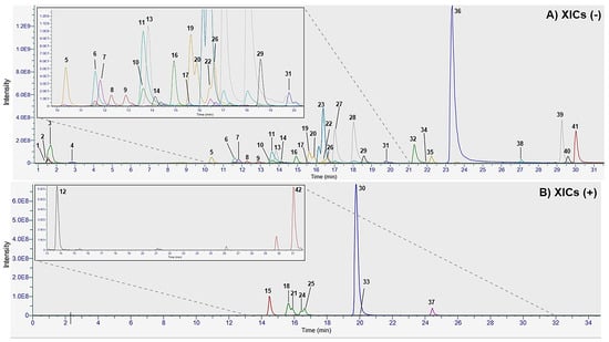
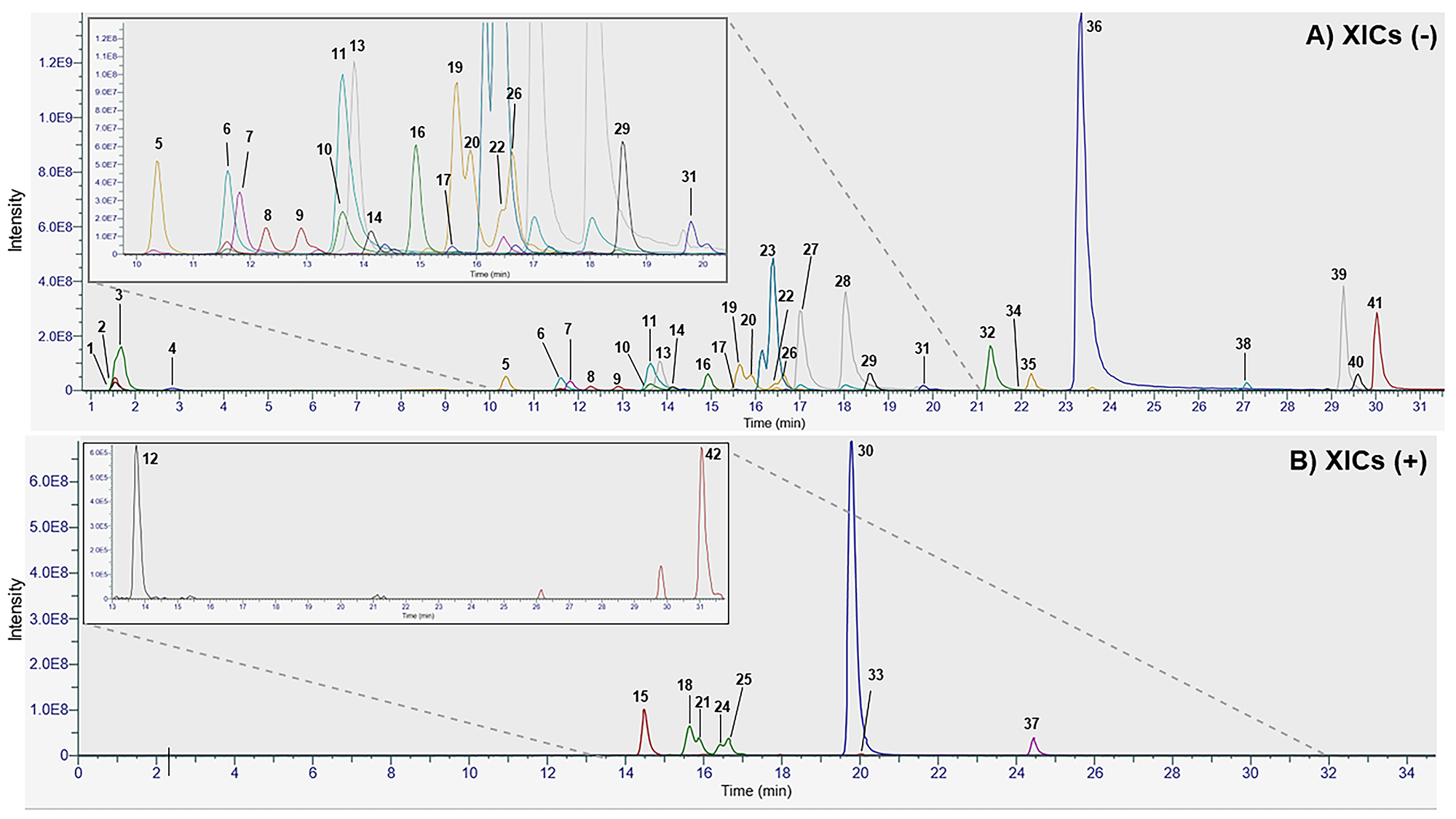
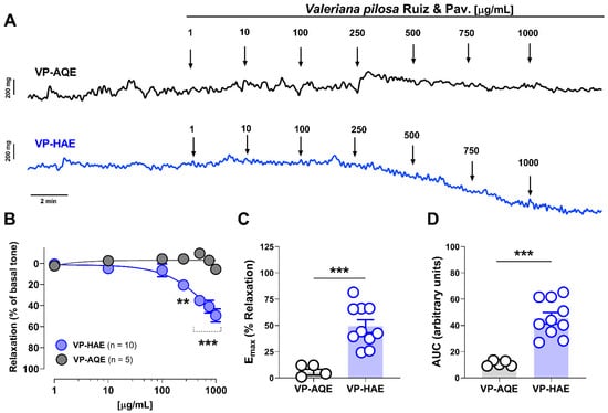
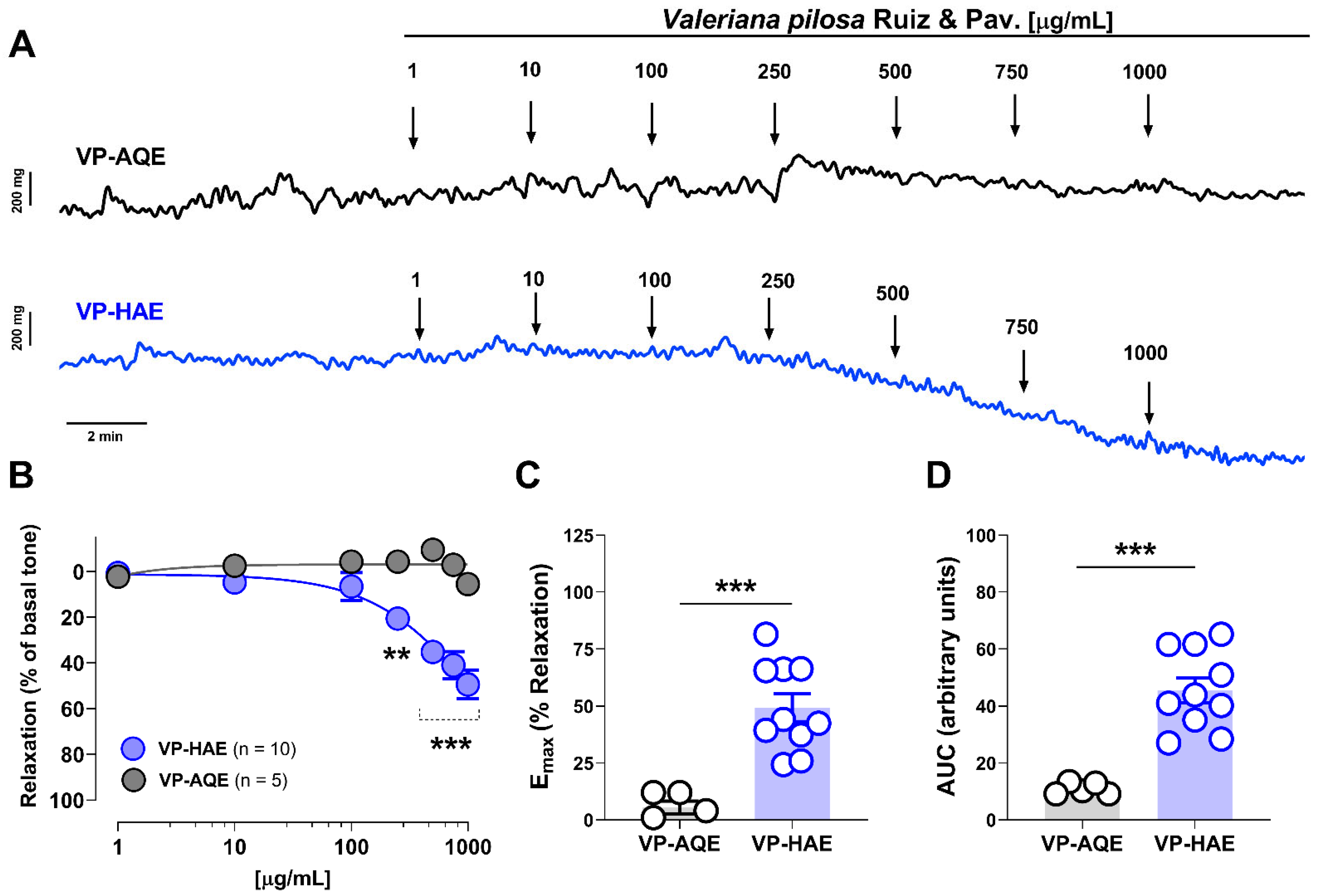
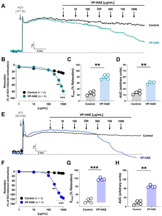
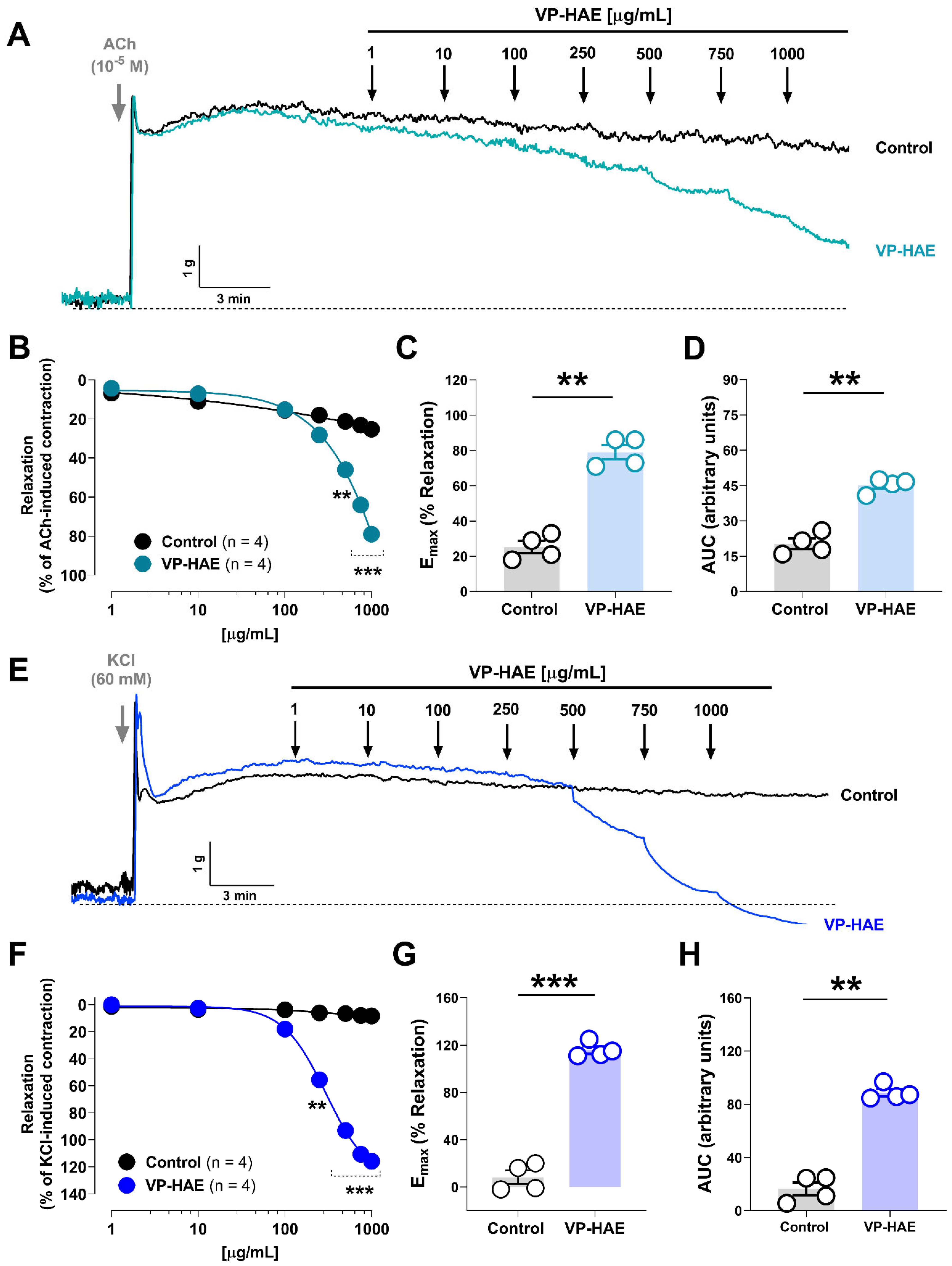
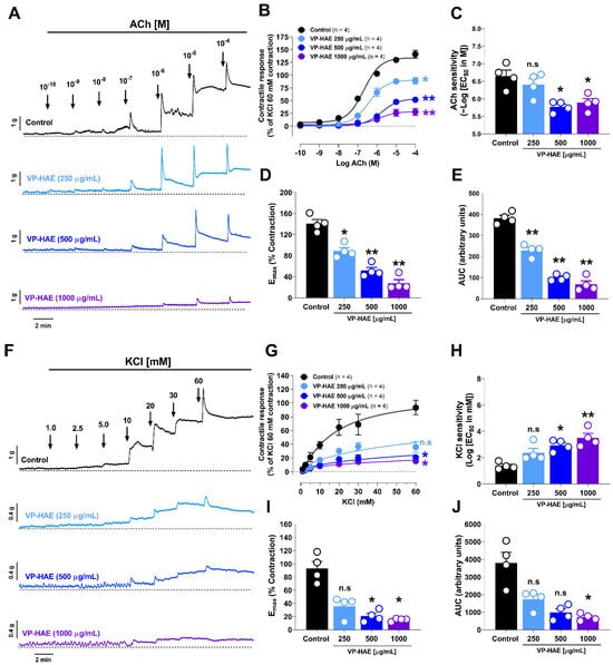
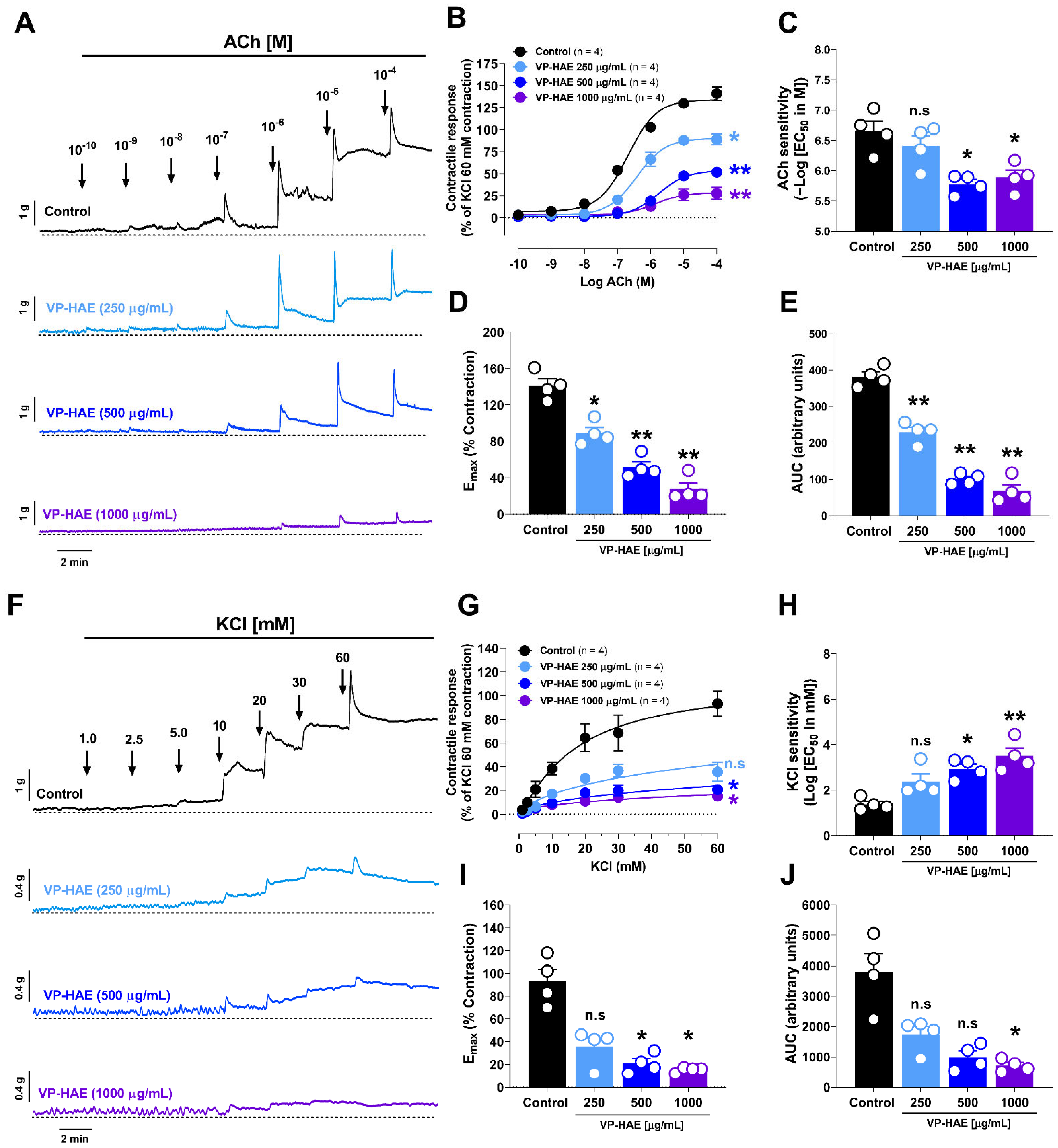
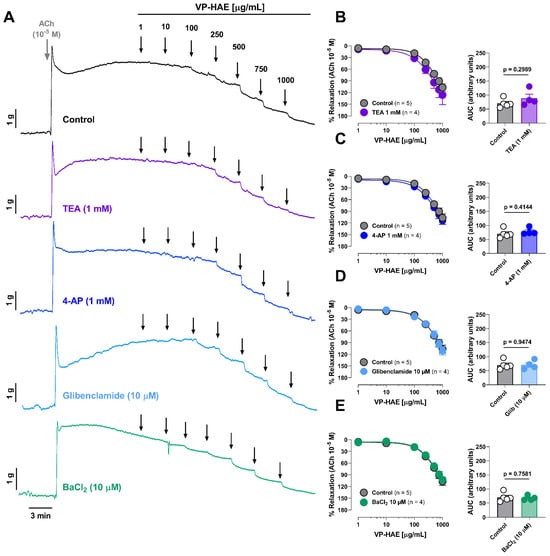
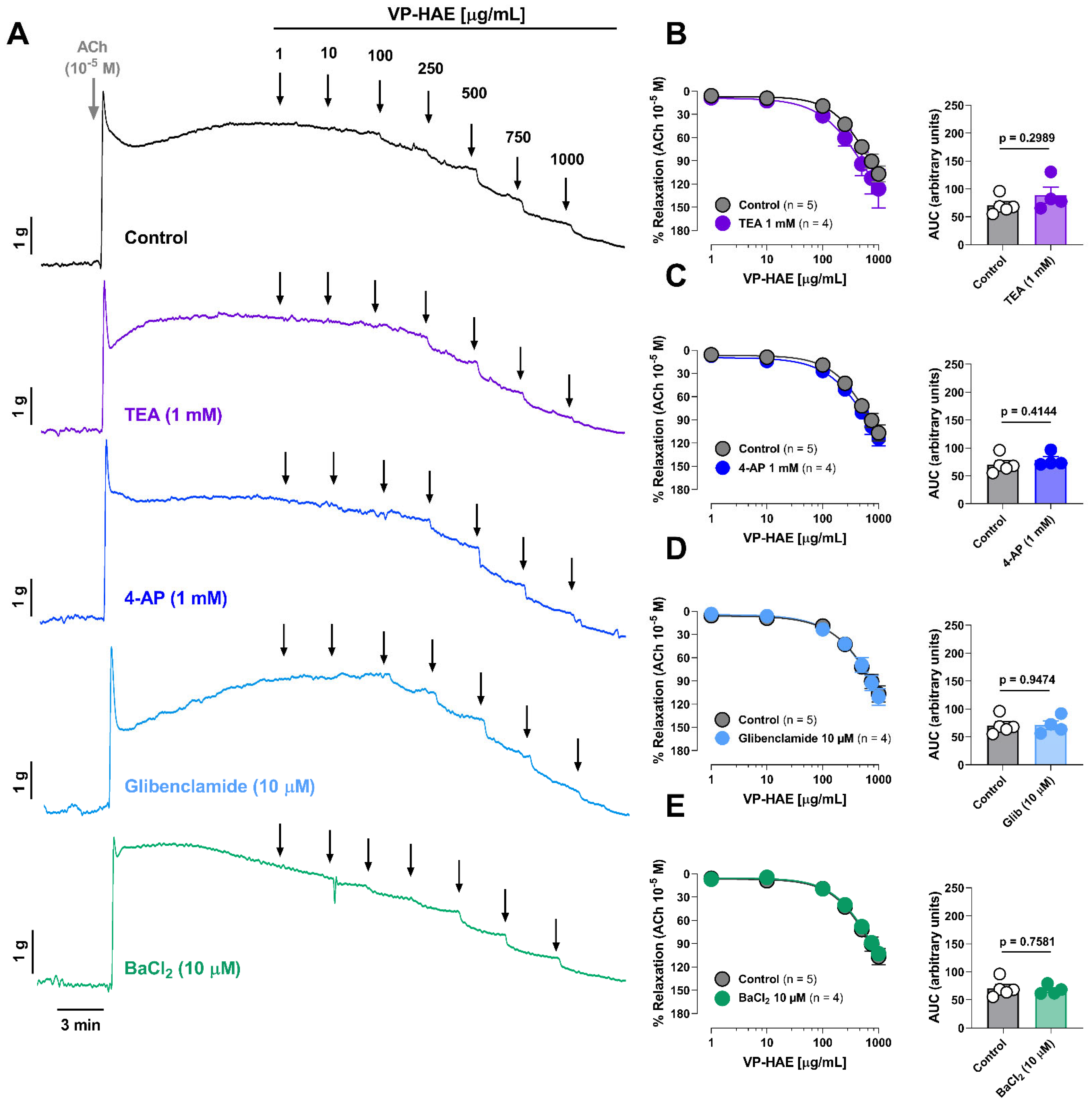
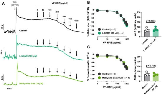
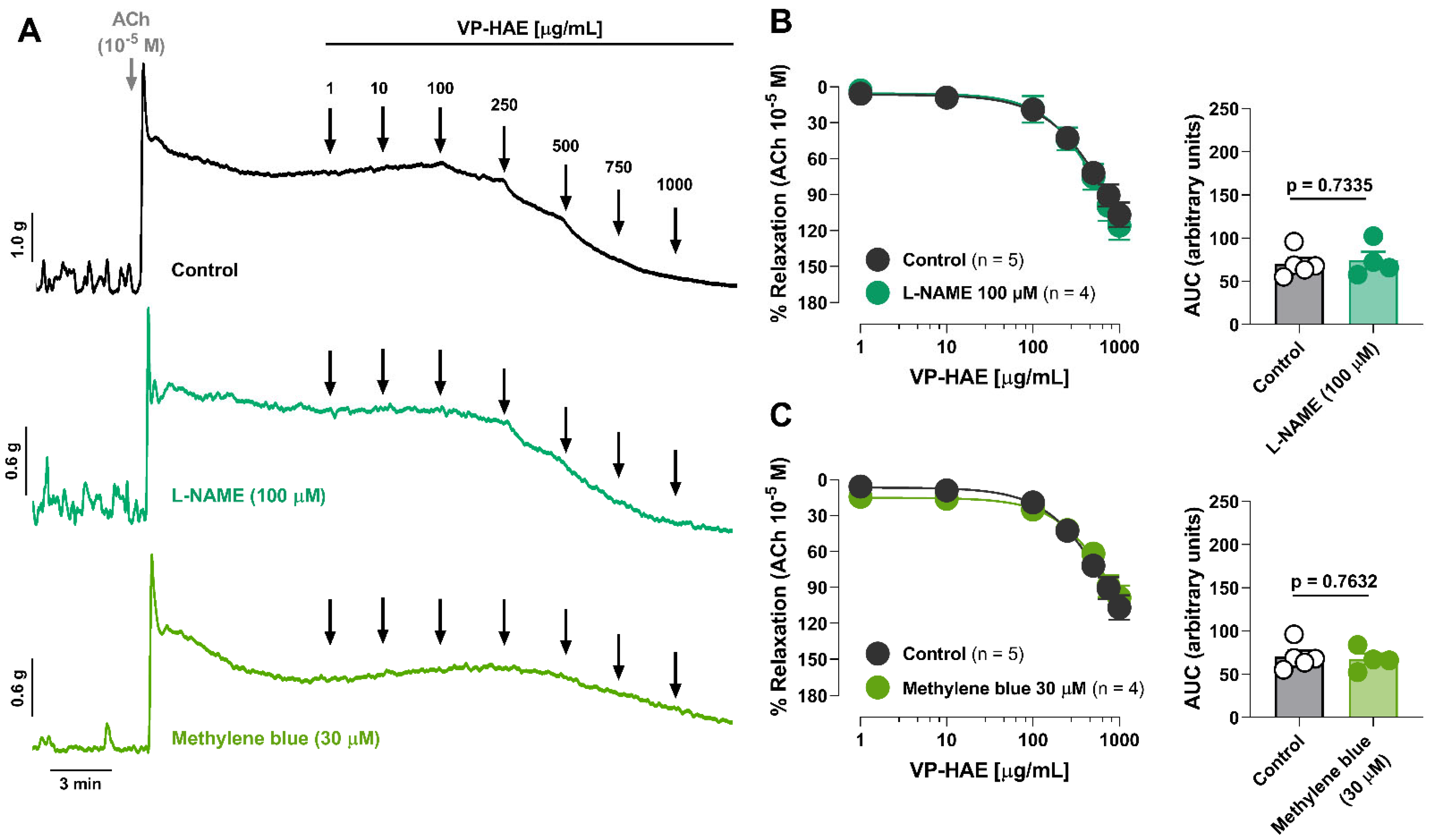
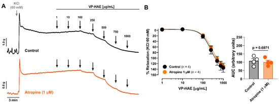
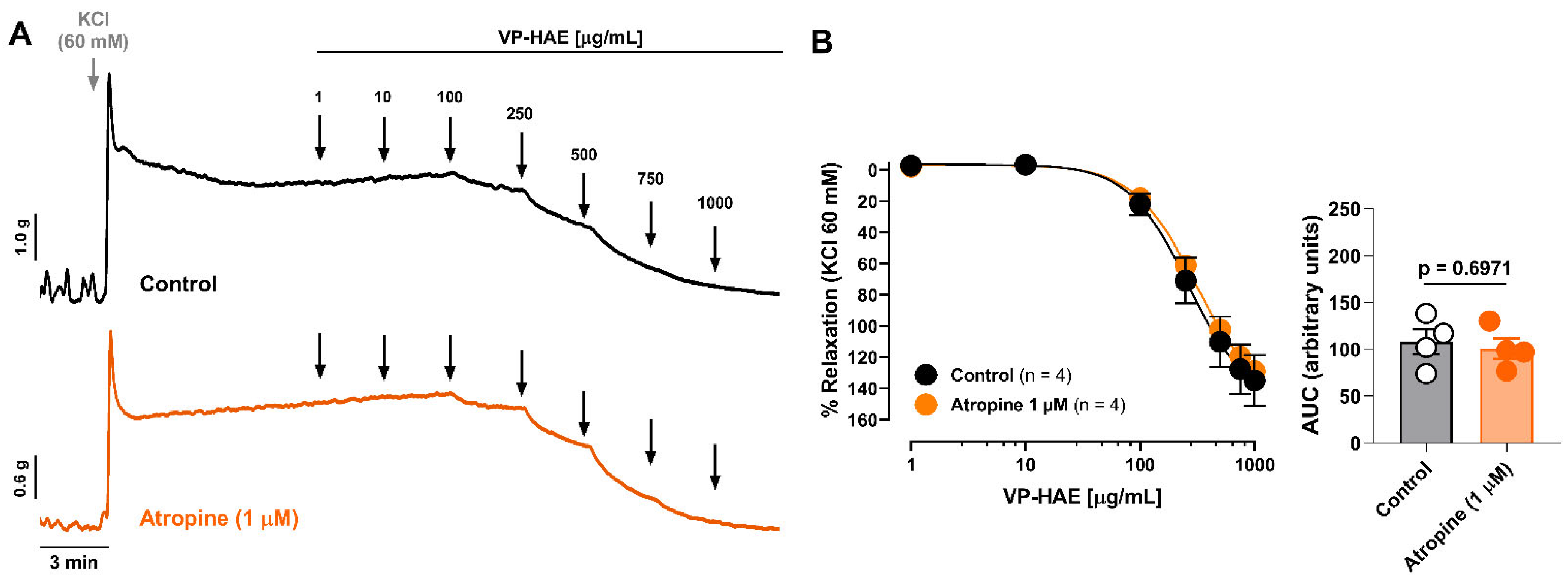
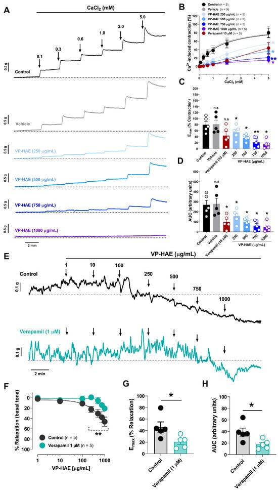
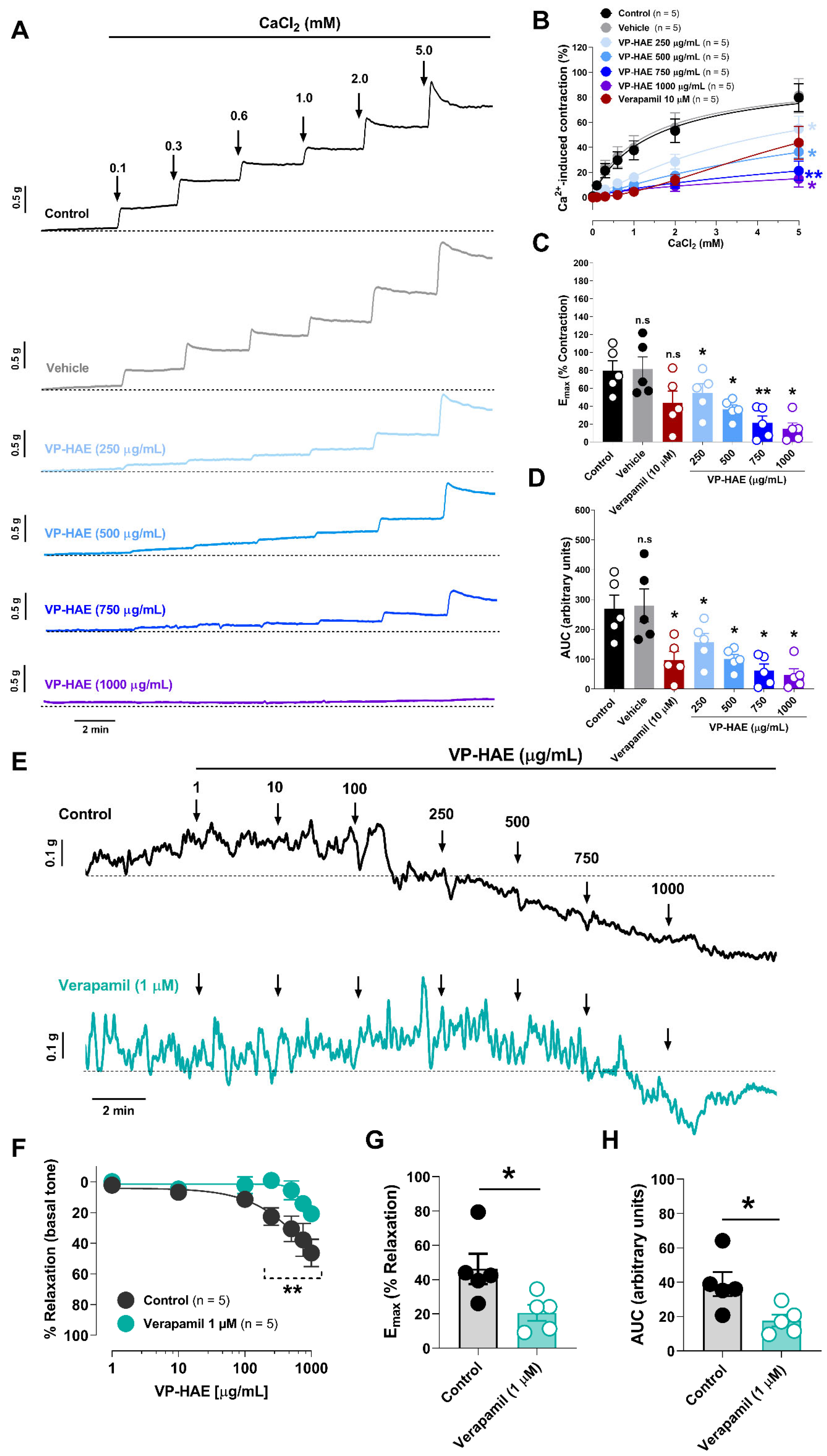

Disclaimer/Publisher’s Note: The statements, opinions and data contained in all publications are solely those of the individual author(s) and contributor(s) and not of MDPI and/or the editor(s). MDPI and/or the editor(s) disclaim responsibility for any injury to people or property resulting from any ideas, methods, instructions or products referred to in the content. |
© 2026 by the authors. Licensee MDPI, Basel, Switzerland. This article is an open access article distributed under the terms and conditions of the Creative Commons Attribution (CC BY) license.