


Abstract
Genotype-dependent regeneration limits genetic transformation in citrus. We adapted a localized Agrobacterium-mediated in planta transformation procedure to recover green fluorescent protein (GFP)-positive and MYC2-edited shoots from juvenile calamansi without conventional in vitro tissue culture. Seedlings were inoculated 18–20 d after germination, and GFP-positive shoots were identifiable approximately 30 d later. Four independent experiments using a GFP-marked empty editing vector yielded 49 GFP-positive seedlings among 384 inoculated seedlings. GFP-positive seedling frequencies ranged from 5.2% to 18.8% (mean ± standard deviation, 12.8 ± 5.7%); 33 seedlings produced uniformly fluorescent shoots. In a separate experiment using a clustered regularly interspaced short palindromic repeats (CRISPR)/CRISPR-associated protein 9 (Cas9) construct targeting MYC2, 13 GFP-positive shoots were recovered from 192 inoculated seedlings (6.8%). All 13 shoots yielded a GFP-specific polymerase chain reaction (PCR) product, and target-site amplicon sequencing detected mutations in every sample. Edited reads accounted for 82.8–99.5% of quality-filtered reads in individual samples. Under our production conditions, the year-round availability of polyembryonic calamansi seeds provided a regular supply of juvenile seedlings for inoculation. The procedure provides edited calamansi material for subsequent genetic and functional characterization. Our protocol was successfully applied to Shatian pummelo, as demonstrated by fluorescence screening and GFP-specific PCR results from a single preliminary trial.
1. Introduction
Citrus genome sequences have expanded opportunities to investigate reproductive biology, domestication, and agronomic traits [1,2], but functional validation remains constrained by long juvenility, high heterozygosity, and limited transformation capacity. Conventional Agrobacterium-mediated transformation relies on in vitro regeneration from epicotyl segments or other juvenile explants [3,4,5]. Genotype-dependent regeneration and the need for repeated subculture can limit the recovery of plant material for gene-function studies [6]. Methods that reduce reliance on tissue culture would therefore complement existing citrus genomic resources.
Alternative approaches include in planta transformation of Shatian pummelo (Citrus maxima cv. Shatian Yu) [7], Lisbon lemon and Pineapple sweet orange [8], and rough lemon [9], as well as Rhizobium rhizogenes-mediated hairy-root transformation followed by plant regeneration [10]. Cut–dip–budding delivery [11] and developmental regulator-mediated meristem induction [12] have further broadened the options for plant transformation. In citrus, co-delivery of editing components, developmental regulators, and transfer DNA (T-DNA) delivery enhancers has also enabled in planta editing [13]. These approaches provide a basis for evaluating additional recipient genotypes and delivery procedures.
Clustered regularly interspaced short palindromic repeats (CRISPR)/CRISPR-associated (Cas) systems support targeted mutagenesis for plant functional analysis and breeding [14,15,16]. Citrus applications include CRISPR-associated protein 9 (Cas9)/single-guide RNA (sgRNA) editing in sweet orange [17], improved editing with YAO promoter-driven Cas9 [18], and canker resistance following editing of Citrus sinensis LATERAL ORGAN BOUNDARIES 1 (CsLOB1) or its promoter [19,20]. Improved Cas9 expression systems and LbCas12a, a Cas12a nuclease from Lachnospiraceae bacterium ND2006, have extended these capabilities [21,22]. Their application nevertheless depends on recovering edited plant tissues after delivery.
Calamansi (Citrus microcarpa), also known as calamondin, is a kumquat–mandarin hybrid [2]. It produces polyembryonic seeds and has documented germplasm diversity in Hainan, China [23]. Under our production conditions, mature fruits were available throughout the year, providing a regular supply of juvenile seedlings. We therefore evaluated localized inoculation of decapitated calamansi seedlings, using a green fluorescent protein (GFP)-marked vector to quantify the frequency of seedlings with GFP-positive tissues and a MYC2-targeting derivative to assess editing. MYC2, which encodes a basic helix–loop–helix transcription factor, was selected as an editing target because stabilization of the MYC2 protein can enhance resistance to Huanglongbing (HLB) [24]. The present work assesses material recovery and target-site editing, rather than disease resistance. In a separate preliminary trial, we successfully applied the localized procedure to Shatian pummelo and assessed the regenerated tissues by GFP fluorescence screening and GFP-specific PCR.
2. Results
2.1. Identification of GFP-Positive Tissues in Calamansi
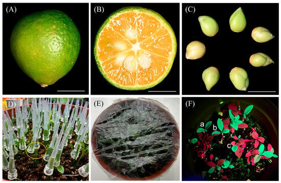
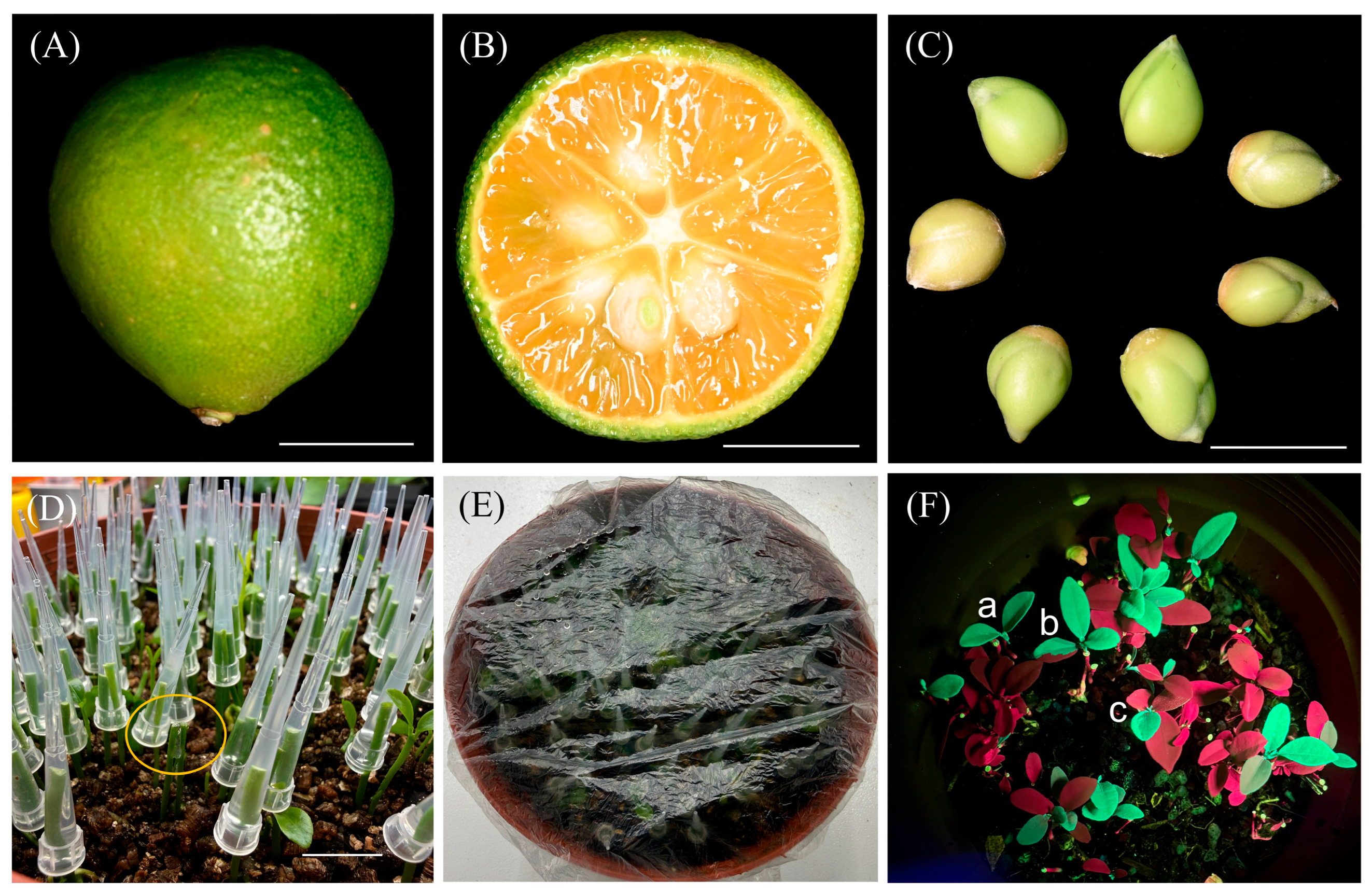
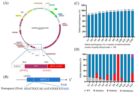
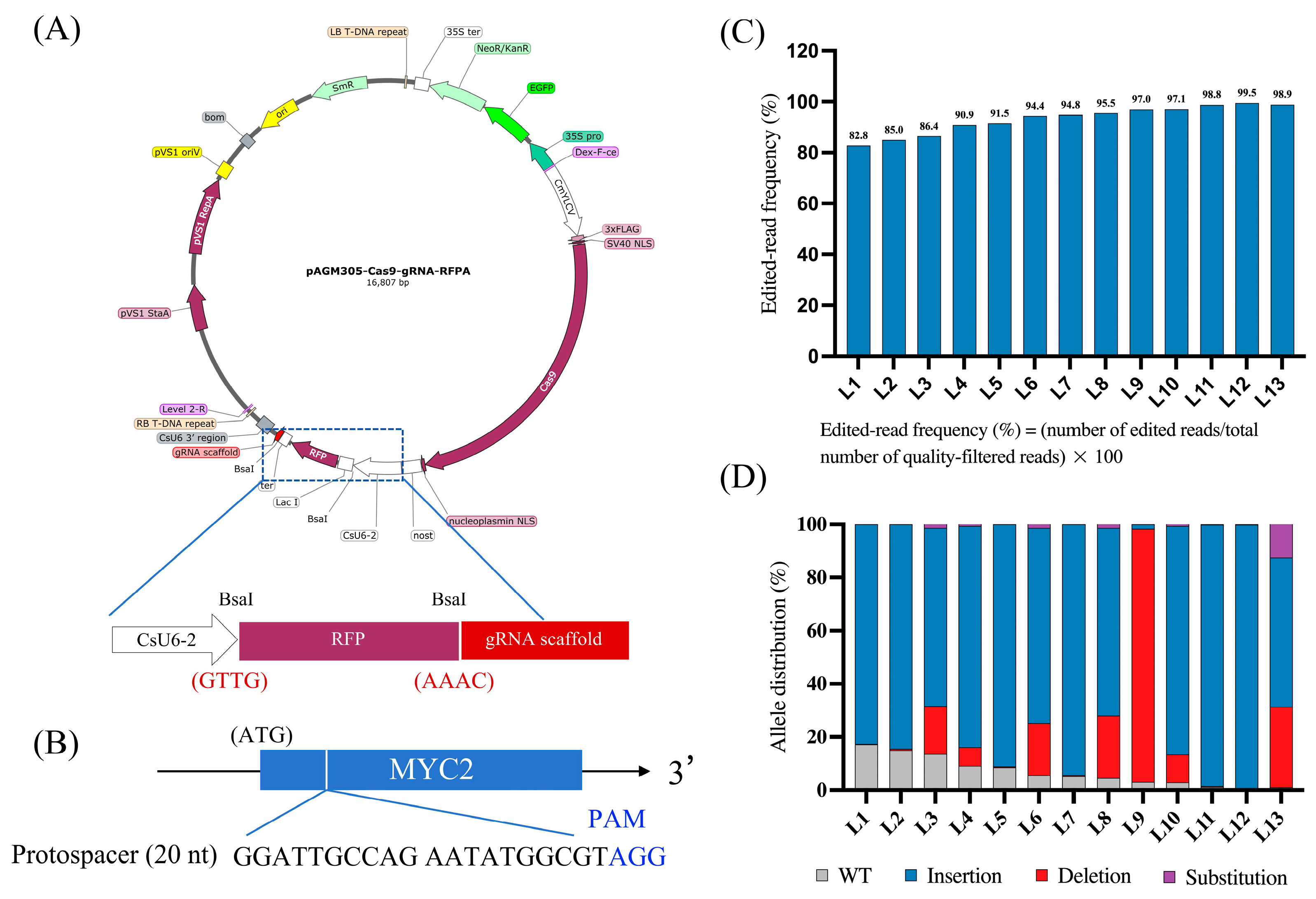
Calamansi fruits contained 7–13 seeds each (mean ± standard deviation, 9.1 ± 2.0; n = 10), and multiple embryos were visible after seed-coat removal (Figure 1A–C). Mean transverse and longitudinal diameters were 31.91 ± 1.14 and 32.99 ± 1.31 mm, respectively, with a fruit shape index of 1.03 ± 0.05. Seedlings reached the two- to four-leaf stage 18–20 d after germination and were used for inoculation. In four independent experiments, seedlings were inoculated with EHA105 carrying the GFP-marked empty editing vector pAGM305-Cas9-gRNA-RFPA, which contains Cas9 and GFP but lacks the inserted MYC2-targeting protospacer (Figure 2A).
Figure 1.
Localized inoculation and GFP-positive tissue identification in calamansi. (A) Mature fruit. (B) Transverse fruit section. (C) Seeds after seed-coat removal. Scale bars (A–C), 1 cm. (D) Decapitated seedlings fitted with medium-filled pipette tips for co-cultivation; the yellow circle indicates a cut site. (E) Plastic-film covering to maintain humidity. (F) GFP-distribution classes: a, uniformly GFP-positive shoot; b, chimeric shoot; c, single fluorescent leaf. Red fluorescence is consistent with chlorophyll autofluorescence.
Figure 2.
CRISPR/Cas9-mediated MYC2 editing in calamansi. (A) Empty editing vector pAGM305-Cas9-gRNA-RFPA. (B) Target site and protospacer-adjacent motif (PAM) within the MYC2 coding sequence. The protospacer is shown in its 5′–3′ orientation on the opposite strand relative to the reference CDS; its reverse complement and the complementary PAM occupy CDS positions 257–276 and 254–256, respectively. (C) Edited-read frequencies in L1–L13, each derived from a different inoculated seedling. (D) Proportions of reads classified as WT, insertion, deletion, or substitution. WT, reference-like reads; RFP, red fluorescent protein; EGFP, enhanced green fluorescent protein.
GFP-positive regenerated tissues were visible approximately 30 d after localized inoculation. Three fluorescence patterns were observed: uniformly GFP-positive shoots, chimeric shoots with fluorescent leaves or sectors, and fluorescence restricted to a single leaf (Figure 1F). Across four batches of 96 seedlings, GFP-positive seedling frequencies ranged from 5.2% to 18.8%, with a mean ± standard deviation (SD) of 12.8 ± 5.7%. Overall, 49 of the 384 inoculated seedlings produced GFP-positive tissues (Table 1). Of these, 33 produced uniformly fluorescent shoots, eight produced chimeric shoots, and eight had single-leaf fluorescence (Table 1). Uniformly GFP-positive shoots were observed in 33 of the 384 inoculated seedlings (8.6%; Table 1).
Table 1.
Recovery of GFP-positive seedlings in four independent calamansi experiments using the GFP-marked empty editing vector.
2.2. CRISPR/Cas9-Mediated Editing of MYC2
A DNA sequence encoding the MYC2-targeting spacer was inserted into the sgRNA expression cassette of the GFP-marked CRISPR/Cas9 binary vector pAGM305-Cas9-gRNA-RFPA, which contains a Cas9 expression cassette, an sgRNA expression cassette, and a GFP reporter for screening regenerated tissues (Figure 2A,B). Of 192 inoculated seedlings, 13 yielded GFP-positive shoots, designated L1–L13, with one shoot analyzed per positive seedling. GFP-positive seedling frequency was 6.8% (13/192) (Table 2A).
Table 2.
Recovery of GFP-positive shoots and target-site editing in the MYC2 experiment.
PCR analysis of all 13 shoots yielded the expected GFP-specific product, whereas the GFP-negative regenerated control showed no corresponding band (Figure S1). Representative regenerated tissue also retained GFP fluorescence approximately one year after inoculation (Figure S2).
Target-site amplicon sequencing detected MYC2 mutations in all 13 GFP-positive shoots (13/13, 100%). Edited reads accounted for 82.8–99.5% of quality-filtered reads in individual samples (Figure 2C; Table 2B). Insertions predominated in most samples, whereas L9 was dominated by deletions (Figure 2D). Representative allele sequences and their read frequencies are provided in Dataset S1.
2.3. Preliminary Application to Shatian Pummelo
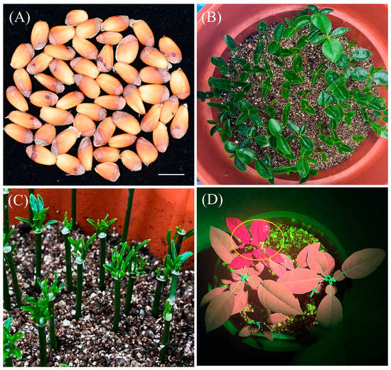
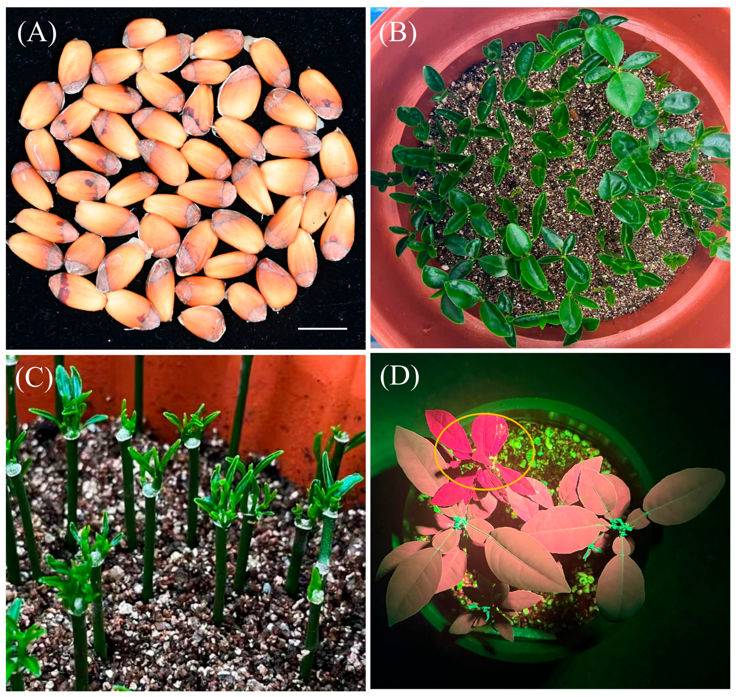
In a single preliminary experiment, 96 juvenile Shatian pummelo seedlings were inoculated with EHA105 carrying the GFP-marked plant overexpression vector Kg2E-35SA-FLAG (Figure S3). Seeds and juvenile seedlings are shown in Figure 3A,B. Approximately 30 d after the selection-medium tips were removed and the seedlings were transferred to light under plastic film, two to four shoots were observed at the cut sites (Figure 3C). Six seedlings produced GFP-positive tissues (6.25%; Table S2). Figure 3D shows representative GFP-positive tissues and a GFP-negative seedling (yellow circle) subjected to the same inoculation and selection procedures.
Figure 3.
Representative observations from the single Shatian pummelo trial. (A) Seeds. (B) Juvenile seed-derived seedlings. (C) Shoot development at cut sites approximately 30 d after removal of the selection-medium tips, during growth in light under plastic film. (D) Fluorescence screening of regenerated tissues. The yellow circle marks a GFP-negative seedling subjected to the same inoculation and selection procedures; GFP-positive regenerated tissues show green fluorescence. Scale bar in (A), 1 cm.
PCR with proID-F/GFP-R detected the expected 957-bp product in all six GFP-positive samples but not in the GFP-negative regenerated control (Figure S4). The control showed red chlorophyll-associated autofluorescence without detectable green fluorescence.
3. Discussion
Recovery of GFP-positive tissues in all four empty-vector batches and MYC2-edited shoots in a separate experiment supports the use of juvenile calamansi seedlings for in planta transformation and editing. Polyembryonic seeds and year-round fruit availability under our production conditions provided a continuing supply of recipients. Inoculation at 18–20 d after germination, followed by GFP screening approximately 30 d later, allowed an initial fluorescence assessment approximately seven weeks after germination. Together, these features support repeated transformation experiments without conventional explant culture or repeated in vitro subculture.
Previous citrus in planta methods have targeted apical buds and epicotyls in Lisbon lemon and Pineapple sweet orange [8], or wounded embryo axes in rough lemon [9]. Ramasamy et al. used Rhizobium rhizogenes-mediated hairy-root transformation followed by in vitro root-to-shoot regeneration [10]. In the present study, localized inoculation was evaluated in decapitated juvenile calamansi seedlings, with shoots regenerating on the inoculated plants. Shoots regenerated without co-delivery of developmental regulator genes used in other approaches [12,13], although 6-benzyladenine and naphthaleneacetic acid were supplied locally. Differences in recipient tissues and assessment criteria limit direct comparisons of recovery frequencies.
In the MYC2 experiment, 13 GFP-positive shoots were recovered from 192 inoculated seedlings (6.8%) (Table 2A), and all carried target-site edits, with edited-read frequencies of 82.8–99.5% (Table 2B). The recovered MYC2-edited shoots may support studies of MYC2 function during vegetative growth, including its potential roles in seedling development, leaf physiology, and pathogen responses. Following genotype characterization, comparative molecular and phenotypic analyses across independently derived MYC2-edited shoots could help evaluate the consequences of the observed target-site mutations.
Fluorescence screening and GFP-specific PCR provide complementary evidence for the preliminary application of the localized procedure to Shatian pummelo. All six GFP-positive materials yielded the approximately 957-bp GFP amplicon, whereas the GFP-negative regenerated comparator did not (Figure S4). The 6.25% value therefore represents a seedling-level GFP-positive frequency with PCR support from one batch. Because both the recipient material and the vector differed between the Shatian pummelo and calamansi experiments, differences in recovery frequency cannot be attributed solely to genotype. Independent trials and further genetic characterization are needed to assess reproducibility, chromosomal integration, and inheritance.
The modular vector design [25,26] facilitates sgRNA replacement for evaluation of additional loci using the same inoculation and screening workflow. Given that editing was detected in all recovered GFP-positive shoots, further optimization could focus on increasing the proportion of inoculated seedlings that yield GFP-positive shoots by evaluating wound treatments, local culture conditions, and selection regimes. Testing additional guides and loci would also determine whether the observed editing performance extends beyond the single MYC2 target.
4. Materials and Methods
4.1. Plant Material and Growth Conditions
Mature calamansi (Citrus microcarpa) fruits were collected from commercial orchards in Hainan Province, China. Transverse and longitudinal diameters of 10 fruits were measured using a digital caliper at the widest equatorial position and along the pedicel-to-stylar axis, respectively. The fruit shape index was calculated as the ratio of longitudinal diameter to transverse diameter. Seeds were counted per fruit, and seed coats were removed to examine embryos.
Seeds were treated with 1 M sodium hydroxide for 15 min to remove surface mucilage, rinsed under running water, and sown in peat moss, vermiculite, and perlite (3:2:1, v/v/v). Seedlings were grown at 25 ± 2 °C under a 16 h light/8 h dark photoperiod and a light intensity of approximately 100 μmol m−2 s−1. Seedlings with two to four fully expanded leaves were selected 18–20 d after germination. Shatian pummelo seedlings were grown under the same conditions and selected at a comparable leaf stage.
4.2. Vector Construction and MYC2 Target Selection
The pAGM305 backbone was derived from pCAMBIA1300 by site-directed mutagenesis of internal BsaI and BpiI sites and conversion to a Level 2 vector for Golden Gate assembly [25,26]. A coding sequence for an engineered variant of replication initiation protein A (RepA) was incorporated into the pVS1 replication module following the strategy of Szarzanowicz et al. [27]. The GFP-marked vector pAGM305-Cas9-gRNA-RFPA contains a Cas9 cassette driven by the Cestrum yellow leaf curling virus (CmYLCV) promoter, an sgRNA cassette driven by the Citrus sinensis U6-2 small nuclear RNA (CsU6-2) promoter, and a red fluorescent protein (RFP) dropout cassette for sgRNA cloning. The reporter is annotated as enhanced GFP (EGFP) in Figure 2A. The 16,807-bp empty-vector sequence is provided in Dataset S2; “empty” denotes the absence of the inserted MYC2 protospacer, not of Cas9 or GFP.
The MYC2 sequence used for target design was retrieved from the Citrus clementina v1.0 reference genome (Ciclev10011214m). A site in the 5′ coding region was selected using CRISPR-P 2.0 [28]. The selected 20-nt protospacer was 5′-GGATTGCCAGAATATGGCGT-3′, adjacent to a 5′-AGG-3′ protospacer-adjacent motif (PAM). In the reference coding sequence (CDS), the reverse complement of the protospacer occupies nucleotides 257–276, and the complementary PAM sequence, 5′-CCT-3′, occupies nucleotides 254–256. Coordinates are numbered with the A of the ATG start codon designated as nucleotide 1 (Dataset S3). Complementary oligonucleotides carrying GTTG and AAAC cloning overhangs (Table S1) were annealed and cloned in place of the RFP dropout cassette.
The GFP-marked plant overexpression vector Kg2E-35SA-FLAG was used in the Shatian pummelo trial. Its map and complete sequence are provided in Figure S3 and Dataset S4, respectively. EHA105 carrying either vector system was selected with spectinomycin, whereas regenerated plant tissues were selected with kanamycin (Table S3).
4.3. Agrobacterium Preparation and Localized Inoculation
The localized inoculation, co-cultivation, and selection procedure was adapted from the citrus in planta transformation method described by Ren et al. [29], with modifications to bacterial preparation, inoculation, and medium composition, as detailed below.
Agrobacterium tumefaciens EHA105 carrying pAGM305-Cas9-gRNA-RFPA, its MYC2-targeting derivative, or Kg2E-35SA-FLAG was cultured on LB agar containing 50 mg/L spectinomycin at 28 °C for 2 d. Colonies were used to inoculate liquid LB medium containing 50 mg/L spectinomycin, and the resulting cultures were incubated at 28 °C for 14 h. Bacterial cells were harvested and resuspended in liquid Murashige and Skoog (MS) medium containing 3% (w/v) sucrose. The MS basal salts with vitamins were obtained from Coolaber (catalog no. PM1011-50L, Beijing, China). The suspension was adjusted to an optical density at 600 nm (OD600) of approximately 0.6, as measured using an Ultrospec 10 cell density meter (Biochrom Ltd., Cambridge, UK), and incubated with 25 mg/L acetosyringone for 1–2 h before inoculation.
The co-cultivation and selection media were prepared with 0.35% (w/v) agarose (high-purity, low-electroendosmosis grade; TSINGKE, catalog no. TSJ001, Beijing, China) in water, without MS basal salts or added sucrose. The agarose was dissolved by heating and allowed to cool before the supplements were added. Both media contained 0.5 mg/L 6-benzyladenine (6-BA) and 0.1 mg/L naphthaleneacetic acid (NAA). The co-cultivation medium additionally contained 25 mg/L acetosyringone, whereas the selection medium contained 25 mg/L kanamycin. The detailed compositions of the two media are provided in Tables S4 and S5.
For co-cultivation, 15–20 μL of the supplemented medium was dispensed into white pipette tips and allowed to solidify before use. The pipette tips were used without prior sterilization. The shoot apex of each seedling was removed using sterile scissors or a blade, and the bacterial suspension was sprayed onto the fresh cut surface using a sterile 10-mL polyethylene terephthalate (PET) spray bottle. The inoculated seedlings were covered with plastic film to maintain humidity for 45 min. A pipette tip containing the solidified co-cultivation medium was then fitted over each cut stem, with the wide opening facing downward and the medium in contact with the cut surface. Seedlings were maintained under plastic film in darkness for 3 d.
After co-cultivation, the tips were replaced with tips preloaded with solidified selection medium. After 5 d of selection in darkness, the selection-medium tips were removed, and seedlings were returned to the growth conditions described in Section 4.1 under plastic film. Kanamycin was used at 25 mg/L for plant-tissue selection because preliminary observations indicated that 50 mg/L markedly inhibited shoot regeneration.
4.4. GFP Screening and PCR Verification
Regenerated calamansi tissues were screened approximately 30 d after inoculation and monitored during subsequent growth. A LUYOR-3415RG handheld lamp was used with Royal Blue excitation (nominally 440–460 nm) and yellow barrier glasses with a nominal 500-nm long-pass cutoff. Images were acquired with an iPhone 16 Pro Max camera behind the same filter. Tissues showing reproducible green fluorescence were scored as GFP-positive. Red fluorescence also observed in GFP-negative regenerated controls was interpreted as chlorophyll-associated autofluorescence, not RFP expression.
For the calamansi MYC2 experiment, leaf DNA was extracted approximately six months after inoculation using the cetyltrimethylammonium bromide (CTAB) method. All 13 GFP-positive shoots were analyzed alongside a GFP-negative regenerated control from the same batch that had undergone the same inoculation, regeneration, and selection procedures. The GFP-F/GFP-R primer pair (Table S1) was predicted to amplify a 717-bp fragment from the vector sequence provided in Dataset S2. PCR cycling consisted of initial denaturation at 95 °C for 3 min; 32 cycles of 95 °C, 60 °C, and 72 °C for 30 s each; and a final extension at 72 °C for 5 min. Products were separated by agarose gel electrophoresis (Figure S1).
All six GFP-positive Shatian pummelo samples were analyzed by PCR using the proID-F/GFP-R primer pair (Table S1). DNA from a GFP-negative shoot regenerated after the same inoculation and selection procedures was included as a negative comparator. This shoot showed red chlorophyll-associated autofluorescence without detectable green fluorescence. DNA extraction and PCR cycling were performed as described above. PCR products were assessed by agarose gel electrophoresis for the expected 957-bp amplicon.
4.5. Experimental Design and Quantification of GFP Positivity
The empty editing vector was evaluated in four independent batches (B1–B4) of 96 seedlings. GFP-positive seedling frequency (%) was calculated as the number of inoculated seedlings producing newly formed GFP-positive tissue divided by the total number of inoculated seedlings, multiplied by 100. Each seedling was counted once, irrespective of the number of shoots or GFP-positive tissues produced. This metric quantified GFP positivity among inoculated seedlings, not shoot regeneration frequency. In the separate MYC2 experiment, 192 seedlings were inoculated, and all 13 recovered GFP-positive shoots, each from a different seedling, underwent PCR and target-site sequencing. The percentage of GFP-positive shoots with detectable editing was calculated as (number of shoots with target-site edits/number of GFP-positive shoots analyzed) × 100.
Fruit traits are reported as mean ± SD (n = 10 fruits). Empty-vector results are summarized by batch counts, pooled GFP-positive seedling frequency, and the mean ± sample SD of four unrounded batch percentages. Recovery in the MYC2 experiment is reported as an overall proportion; no inference is made about variation among batches or differences between constructs. Sequencing reads were not treated as biological replicates.
4.6. Target-Site Amplicon Sequencing and Analysis
Target-site amplicon sequencing was performed by Hubei Hongshan Laboratory (Wuhan, China). Paired-end reads were analyzed using the CRISPRone platform with Cas9 selected as the nuclease. The edited-read frequency in Figure 2C was calculated as edited reads/total quality-filtered reads × 100. Figure 2D summarizes the reported reference-like (WT), insertion, deletion, and substitution classes. CRISPResso2 allele-frequency plots [30] in Dataset S1 show individual sequences and their abundance among aligned reads. These bulk-tissue read frequencies were not used to assign homozygous, heterozygous, or biallelic plant genotypes.
4.7. Preliminary Trial in Shatian Pummelo
A single batch of 96 seed-derived Shatian pummelo seedlings was inoculated with EHA105 carrying Kg2E-35SA-FLAG. Bacterial preparation and the local co-cultivation and kanamycin-selection steps followed Section 4.3. After the 5-d dark selection period, the selection-medium tips were removed, and seedlings were transferred to the light conditions described in Section 4.1 while remaining covered with plastic film to maintain humidity. Each positive seedling was counted once, irrespective of shoot number, and the GFP-positive seedling frequency was calculated as 6/96 × 100 = 6.25%. All six GFP-positive Shatian pummelo samples were subsequently analyzed by PCR using the proID-F/GFP-R primer pair (Table S1), with DNA from a GFP-negative regenerated shoot as the negative comparator (Section 4.4; Figure S4).
5. Conclusions
Localized Agrobacterium inoculation enabled identification of GFP-positive tissues and recovery of MYC2-edited calamansi shoots without conventional in vitro tissue culture. Thirteen edited GFP-positive shoots were obtained from 192 inoculated calamansi seedlings. In a single Shatian pummelo trial, six of 96 seedlings yielded GFP-positive regenerated tissues that were also positive by GFP-specific PCR. The recovered calamansi shoots provide material for citrus gene-function studies, while the preliminary Shatian pummelo results warrant further evaluation of the procedure in this cultivar. Genetic uniformity, integration, and inheritance require further characterization.
Supplementary Materials
The following supporting information can be downloaded at: https://www.mdpi.com/article/10.3390/plants15223544/s1, Table S1. sgRNA assembly oligonucleotides and GFP-specific PCR primers; Table S2. Fluorescence and GFP-specific PCR outcomes of the single Shatian pummelo trial; Table S3. Bacterial and plant-tissue selection for the vectors used; Table S4. Composition of the co-cultivation medium; Table S5. Composition of the selection medium; Figure S1. GFP-specific PCR analysis of calamansi shoots; Figure S2. Representative calamansi GFP fluorescence approximately one year after inoculation; Figure S3. Kg2E-35SA-FLAG vector map; Figure S4. GFP-specific PCR analysis of six Shatian pummelo materials and a GFP-negative regenerated comparator; Dataset S1. MYC2 allele-frequency plots; Dataset S2. pAGM305-Cas9-gRNA-RFPA sequence; Dataset S3. MYC2 reference CDS and target region; Dataset S4. Kg2E-35SA-FLAG sequence.
Author Contributions
Conceptualization, J.Z. and P.W.; methodology, J.Z. and P.W.; investigation, Z.F., P.Y., Z.L., and Z.Y.; formal analysis, Z.F.; writing—original draft preparation, Z.F.; writing—review and editing, P.W. and J.Z.; supervision, P.W. and J.Z., Z.F. and P.Y. contributed equally to this work. All authors have read and agreed to the published version of the manuscript.
Funding
This research was supported by the Guangdong S&T Program (2025B0202070005); the National Modern Agricultural (Citrus) Technology System of China (CARS-24); the Guangdong Provincial Special Fund for Modern Agricultural Industry Technology Innovation Teams (2024CXTD10); the Special Fund for the Rural Revitalization Strategy of Guangdong (Agricultural Science and Technology Capacity Improvement) (TS-1); the Seed Industry Revitalization Program of Guangdong Province (2024-NYP-00-046); and the Young Scientists Fund of the National Natural Science Foundation of China (32302472).
Data Availability Statement
GFP-positive seedling counts, sample-level edited-read frequencies, GFP-specific PCR gel images and fluorescence images for calamansi and Shatian pummelo, representative MYC2 allele-frequency plots, the MYC2 reference CDS, and the sequences of pAGM305-Cas9-gRNA-RFPA and Kg2E-35SA-FLAG are provided in the article and Supplementary Materials. Additional plasmid and construct information is available from the corresponding authors upon reasonable request.
Acknowledgments
We thank Jie Ren (j.ren@yale.edu), Xin Song (songxin@hbaas.com), and Shuting Zhang (zhangshuting@fafu.edu.cn) for providing detailed information on the in planta transformation procedure, which informed the optimization of the localized inoculation, co-cultivation, and selection steps used in this study. The authors used Codex (OpenAI; version 26.831.21537; accessed September 2026) for English-language revision and manuscript organization. The authors reviewed and edited the output and take full responsibility for the content of the manuscript.
Conflicts of Interest
The authors declare no conflicts of interest.
References
- Wang, X.; Xu, Y.; Zhang, S.; Cao, L.; Huang, Y.; Cheng, J.; Wu, G.; Tian, S.; Chen, C.; Liu, Y.; et al. Genomic analyses of primitive, wild and cultivated citrus provide insights into asexual reproduction. Nat. Genet. 2017, 49, 765–772. [Google Scholar] [CrossRef] [Scilit] [PubMed]
- Wu, G.A.; Terol, J.; Ibanez, V.; López-García, A.; Pérez-Román, E.; Borredá, C.; Domingo, C.; Tadeo, F.R.; Carbonell-Caballero, J.; Alonso, R.; et al. Genomics of the origin and evolution of Citrus. Nature 2018, 554, 311–316. [Google Scholar] [CrossRef] [Scilit] [PubMed]
- Peña, L.; Cervera, M.; Juárez, J.; Ortega, C.; Pina, J.; Durán-Vila, N.; Navarro, L. High efficiency Agrobacterium-mediated transformation and regeneration of citrus. Plant Sci. 1995, 104, 183–191. [Google Scholar] [CrossRef] [Scilit]
- Cervera, M.; Pina, J.A.; Juárez, J.; Navarro, L.; Peña, L. Agrobacterium-mediated transformation of citrange: Factors affecting transformation and regeneration. Plant Cell Rep. 1998, 18, 271–278. [Google Scholar] [CrossRef] [Scilit] [PubMed]
- Orbović, V.; Grosser, J.W. Citrus transformation using juvenile tissue explants. Methods Mol. Biol. 2015, 1224, 245–257. [Google Scholar] [CrossRef] [Scilit] [PubMed]
- Poles, L.; Licciardello, C.; Distefano, G.; Nicolosi, E.; Gentile, A.; La Malfa, S. Recent advances of in vitro culture for the application of new breeding techniques in citrus. Plants 2020, 9, 938. [Google Scholar] [CrossRef] [Scilit] [PubMed]
- Zhang, Y.-Y.; Zhang, D.-M.; Zhong, Y.; Chang, X.-J.; Hu, M.-L.; Cheng, C.-Z. A simple and efficient in planta transformation method for pommelo (Citrus maxima) using Agrobacterium tumefaciens. Sci. Hortic. 2017, 214, 174–179. [Google Scholar] [CrossRef] [Scilit]
- Khadgi, A.; Sagawa, C.H.D.; Vernon, C.; Mermaz, B.; Irish, V.F. Optimization of in planta methodology for genome editing and transformation in Citrus. Front. Plant Sci. 2024, 15, 1438031. [Google Scholar] [CrossRef] [Scilit] [PubMed]
- Chhabra, G.; Sharma, M.; Kalia, A.; Kaur, A.; Sandhu, J.S. Monitoring dissociation of chimerism through real-time PCR and scanning electron microscopy following in planta transformation of rough lemon (Citrus jambhiri Lush.). Plant Biotechnol. Rep. 2024, 18, 497–506. [Google Scholar] [CrossRef] [Scilit]
- Ramasamy, M.; Dominguez, M.M.; Irigoyen, S.; Padilla, C.S.; Mandadi, K.K. Rhizobium rhizogenes-mediated hairy root induction and plant regeneration for bioengineering citrus. Plant Biotechnol. J. 2023, 21, 1728–1730. [Google Scholar] [CrossRef] [Scilit] [PubMed]
- Cao, X.; Xie, H.; Song, M.; Lu, J.; Ma, P.; Huang, B.; Wang, M.; Tian, Y.; Chen, F.; Peng, J.; et al. Cut-dip-budding delivery system enables genetic modifications in plants without tissue culture. Innovation 2023, 4, 100345. [Google Scholar] [CrossRef] [Scilit] [PubMed]
- Maher, M.F.; Nasti, R.A.; Vollbrecht, M.; Starker, C.G.; Clark, M.D.; Voytas, D.F. Plant gene editing through de novo induction of meristems. Nat. Biotechnol. 2020, 38, 84–89. [Google Scholar] [CrossRef] [Scilit] [PubMed]
- Kelly, G.; Plesser, E.; Bdolach, E.; Arroyave, M.; Belausov, E.; Doron-Faigenboim, A.; Rozen, A.; Zemach, H.; Zach, Y.Y.; Goldenberg, L.; et al. In planta genome editing in citrus facilitated by co-expression of CRISPR/Cas and developmental regulators. Plant J. 2025, 122, e70155. [Google Scholar] [CrossRef] [Scilit] [PubMed]
- Chen, K.; Wang, Y.; Zhang, R.; Zhang, H.; Gao, C. CRISPR/Cas genome editing and precision plant breeding in agriculture. Annu. Rev. Plant Biol. 2019, 70, 667–697. [Google Scholar] [CrossRef] [Scilit] [PubMed]
- Zhang, Y.; Malzahn, A.A.; Sretenovic, S.; Qi, Y. The emerging and uncultivated potential of CRISPR technology in plant science. Nat. Plants 2019, 5, 778–794. [Google Scholar] [CrossRef] [Scilit] [PubMed]
- Gao, C. Genome engineering for crop improvement and future agriculture. Cell 2021, 184, 1621–1635. [Google Scholar] [CrossRef] [Scilit] [PubMed]
- Jia, H.; Wang, N. Targeted genome editing of sweet orange using Cas9/sgRNA. PLoS ONE 2014, 9, e93806. [Google Scholar] [CrossRef] [Scilit] [PubMed]
- Zhang, F.; LeBlanc, C.; Irish, V.F.; Jacob, Y. Rapid and efficient CRISPR/Cas9 gene editing in Citrus using the YAO promoter. Plant Cell Rep. 2017, 36, 1883–1887. [Google Scholar] [CrossRef] [Scilit] [PubMed]
- Jia, H.; Zhang, Y.; Orbović, V.; Xu, J.; White, F.F.; Jones, J.B.; Wang, N. Genome editing of the disease susceptibility gene CsLOB1 in citrus confers resistance to citrus canker. Plant Biotechnol. J. 2017, 15, 817–823. [Google Scholar] [CrossRef] [Scilit] [PubMed]
- Peng, A.; Chen, S.; Lei, T.; Xu, L.; He, Y.; Wu, L.; Yao, L.; Zou, X. Engineering canker-resistant plants through CRISPR/Cas9-targeted editing of the susceptibility gene CsLOB1 promoter in citrus. Plant Biotechnol. J. 2017, 15, 1509–1519. [Google Scholar] [CrossRef] [Scilit] [PubMed]
- Huang, X.; Wang, Y.; Wang, N. Highly efficient generation of canker-resistant sweet orange enabled by an improved CRISPR/Cas9 system. Front. Plant Sci. 2022, 12, 769907. [Google Scholar] [CrossRef] [Scilit] [PubMed]
- Jia, H.; Orbović, V.; Wang, N. CRISPR-LbCas12a-mediated modification of citrus. Plant Biotechnol. J. 2019, 17, 1928–1937. [Google Scholar] [CrossRef] [Scilit] [PubMed]
- Xin, Y.-H.; Wu, Y.-X.; Qiao, B.; Su, L.; Xie, S.-Q.; Ling, P. Evaluation on the phenotypic diversity of calamansi (Citrus microcarpa) germplasm in Hainan Island. Sci. Rep. 2022, 12, 371. [Google Scholar] [CrossRef] [Scilit] [PubMed]
- Zhao, P.; Yang, H.; Sun, Y.; Zhang, J.; Gao, K.; Wu, J.; Zhu, C.; Yin, C.; Chen, X.; Liu, Q.; et al. Targeted MYC2 stabilization confers citrus Huanglongbing resistance. Science 2025, 388, 191–198. [Google Scholar] [CrossRef] [Scilit] [PubMed]
- Čermák, T.; Curtin, S.J.; Gil-Humanes, J.; Čegan, R.; Kono, T.J.Y.; Konečná, E.; Belanto, J.J.; Starker, C.G.; Mathre, J.W.; Greenstein, R.L.; et al. A multipurpose toolkit to enable advanced genome engineering in plants. Plant Cell 2017, 29, 1196–1217. [Google Scholar] [CrossRef] [Scilit] [PubMed]
- Engler, C.; Youles, M.; Gruetzner, R.; Ehnert, T.-M.; Werner, S.; Jones, J.D.G.; Patron, N.J.; Marillonnet, S. A Golden Gate modular cloning toolbox for plants. ACS Synth. Biol. 2014, 3, 839–843. [Google Scholar] [CrossRef] [Scilit] [PubMed]
- Szarzanowicz, M.J.; Waldburger, L.M.; Busche, M.; Geiselman, G.M.; Kirkpatrick, L.D.; Kehl, A.J.; Tahmin, C.; Kuo, R.C.; McCauley, J.; Pannu, H.; et al. Binary vector copy number engineering improves Agrobacterium-mediated transformation. Nat. Biotechnol. 2025, 43, 1708–1716. [Google Scholar] [CrossRef] [Scilit] [PubMed]
- Liu, H.; Ding, Y.; Zhou, Y.; Jin, W.; Xie, K.; Chen, L.-L. CRISPR-P 2.0: An improved CRISPR-Cas9 tool for genome editing in plants. Mol. Plant 2017, 10, 530–532. [Google Scholar] [CrossRef] [Scilit] [PubMed]
- Ren, J.; Yin, Q.; Song, X.; Duan, Y.Y.; Zhang, X.; Li, R.Q.; Xie, K.D.; Zhang, F.; Irish, V.; Wu, X.M.; et al. A rapid and broadly applicable in planta transformation and genome editing system involving wounded epicotyls in citrus. Hortic. Adv. 2026, 4, 37. [Google Scholar] [CrossRef] [Scilit]
- Clement, K.; Rees, H.; Canver, M.C.; Gehrke, J.M.; Farouni, R.; Hsu, J.Y.; Cole, M.A.; Liu, D.R.; Joung, J.K.; Bauer, D.E.; et al. CRISPResso2 provides accurate and rapid genome editing sequence analysis. Nat. Biotechnol. 2019, 37, 224–226. [Google Scholar] [CrossRef] [Scilit] [PubMed]

Disclaimer/Publisher’s Note: The statements, opinions and data contained in all publications are solely those of the individual author(s) and contributor(s) and not of MDPI and/or the editor(s). MDPI and/or the editor(s) disclaim responsibility for any injury to people or property resulting from any ideas, methods, instructions or products referred to in the content. |
© 2026 by the authors. Licensee MDPI, Basel, Switzerland. This article is an open access article distributed under the terms and conditions of the Creative Commons Attribution (CC BY) license.