


Abstract
Ubiquitination dynamically regulates critical cellular processes, including cell cycle progression, apoptosis, DNA repair, and chromatin remodeling. Deubiquitinating enzymes (DUBs) negatively regulate protein ubiquitination and are equally important for protein regulation in diverse biological processes. Spermatogenesis is a highly regulated process involving spermatogonia self-renewal and differentiation, ensuring continuous sperm production. Using a loss-of-function mouse model, several DUBs have been shown to be involved in spermatogenesis. In addition, specific genetic variants in the DUB genes have been associated with spermatogenic failure and male infertility. These studies provide strong evidence that DUBs are necessary for normal spermatogenesis and may influence male fertility. However, the exact mechanism by which these DUBs regulate spermatogenesis is still under investigation. The aim of this review is to highlight recent advances in the regulatory roles of DUBs in mammalian spermatogenesis and provide insight into the molecular mechanisms underlying their potential actions. An in-depth understanding of DUB-mediated regulation of spermatogenesis will provide a scientific rationale for the discovery and development of novel DUB-targeted therapeutic strategies for male infertility.
1. Introduction
1.1. Spermatogenesis
Mammalian spermatogenesis has three key phases of germ cell development, namely, the proliferation of spermatogonia, meiosis in spermatocytes, and post-meiotic differentiation in spermatids [1,2,3]. Spermatogonia are diploid germline stem cells located in the seminiferous tubules of the testes. They undergo mitosis to produce two types of daughter cells. Type A spermatogonia (spermatogonial stem cells, SSCs) remain near the basement membrane of the tubule to maintain the supply of germline stem cells [2,4,5]. After a series of mitotic divisions, type B spermatogonia grow and differentiate into primary spermatocytes [2,6]. Diploid primary spermatocytes subsequently undergo two meiotic divisions and result in the production of haploid round spermatids. They then enter the process of spermiogenesis during which profound morphological and biochemical restructuring occurs, such as the formation of the acrosome and flagellum, giving rise to mature spermatozoa [7] (Figure 1).
Figure 1.
Mammalian spermatogenesis. Proliferative type A spermatogonia (SSCs) undergo a series of mitotic divisions. One of the daughter cells renews the stock of type A spermatogonia (undifferentiated spermatogonia), while the other becomes type B spermatogonia (differentiating spermatogonia). Type B spermatogonia give rise to primary spermatocytes, which undergo two meiotic divisions to produce haploid spermatids that undergo spermiogenesis to produce terminally differentiated spermatozoa.
1.2. The Ubiquitin–Proteasome System (UPS)
Ubiquitination is an important protein post-translational modification regulating many cellular processes in eukaryotes. This modification can mark proteins for degradation by the proteasome, or it can act as a signal for non-proteolytic functions of proteins [8,9,10]. Ubiquitination occurs through covalent attachment of a ubiquitin molecule to substrate lysine residue mediated by E1-activating, E2-conjugating, and E3 ubiquitin ligase enzymes [11] (Figure 2).
Figure 2.
An overview of the ubiquitin–proteasome system (UPS). Ubiquitin is activated with E1 (ubiquitin-activating enzyme) in an ATP-dependent manner, transferred to E2 (ubiquitin-conjugating enzyme), and then transferred to the substrate through E3 (ubiquitin ligase) recognition, forming a mono- or polyubiquitinated protein. K48 or K11 polyubiquitin chains lead to 26S proteasome-mediated degradation. Monoubiquitination or K63 polyubiquitin chains are non-proteolytic ubiquitination signals and participate in many biological processes. Deubiquitination is mediated by deubiquitinating enzymes (DUBs) that remove ubiquitin from substrates, allowing for ubiquitin recycling. The DUB family consists of six subgroups, USPs, UCHs, MJDs, OTUs, JAMMs, and MINDYs, classified based on the characteristics of their conserved domains.
The proteins can be conjugated with a single ubiquitin molecule (monoubiquitin) or chains of ubiquitin (polyubiquitination). Ubiquitin contains seven lysine residues (K6, K11, K27, K29, K33, K48, and K63) that can undergo distinct polyubiquitination chains. The polyubiquitinated chain using K48 is typically used as means of protein expression control and directs the protein to the 26S proteasome for degradation [12]. It has recently been demonstrated that linkages at other lysine residues such as K11, K29, and K33 also serve as signals for proteasomal targeting but are less common [13,14,15]. Compared with this, monoubiquitination or K63-linked polyubiquitination exerts non-proteolytic functions, such as protein trafficking, endocytosis, DNA repair, transcriptional regulation, and signal transduction.
Deubiquitinating enzymes (DUBs) reverse the process of ubiquitination and play critical roles in various cellular processes [8,16,17,18]. To date, more than one hundred human DUBs have been identified with varying substrate specificity, subcellular localization, and protein–protein interactions [19]. DUBs are classified into six families according to their domain structure: ubiquitin-specific proteases (USPs), which comprise the largest and most diverse subfamily, ubiquitin carboxy-terminal hydrolases (UCHs), ovarian tumor proteases (OTUs), Machado–Joseph disease protein domain proteases (MJDs), JAMM/MPN domain-associated metallopeptidases (JAMMs), and monocyte chemotactic protein-induced proteases (MINDYs) [16,20]. With the exception of JAMMs, which belong to the Zn2+-dependent metalloproteases, the others use the classical cysteine protease catalytic triad (Cysteine–Histidine–Asparagine/Aspartate) in the active site. Similar to E3 ubiquitin ligases, DUBs have specificity toward different ubiquitin linkage types and the substrate protein. Since DUBs function to antagonize the ubiquitination of substrates carried out by E3 ligase, the levels of ubiquitination are dynamically regulated by the actions of these two opposing enzymes, which are regulated at expression, subcellular location, or activity levels through mechanisms by protein-protein interactions and post-translational modifications.
2. Maintenance of Genome Integrity During Spermatogenesis
Spermatogenesis is divided into three phases: the mitotic phase of spermatogonia, the meiotic phase of spermatocytes, and the post-meiotic phase of spermatids (spermiogenesis) (Figure 3). The rapid mitotic division of spermatogonia has an increased risk of spontaneous DNA damage, primarily through DNA replication errors [21,22,23,24,25,26]. While these cells have various DNA repair mechanisms to maintain genome stability, unrepaired DNA damage can lead to the accumulation of mutations. Meiotic DNA double-stand breaks (DSBs) are generated early in meiotic prophase I and initiate homologous recombination (HR), producing genetic diversity and ensuring accurate transmission of genetic information to the next generation [27,28,29,30]. Defects in DSB repair can cause abnormal recombination and meiotic arrest in primary spermatocytes, leading to massive germ cell death [30,31,32]. During spermiogenesis, round spermatids undergo dramatic chromatin remodeling with histone replacement by protamine and subsequently differentiate into mature spermatozoa [33]. Defective histone-to-protamine replacement can lead to incomplete chromatin condensation and DNA damage. As sperm mature, DNA damage can be accumulated by various risk factors including oxidative stress, chromatin remodeling errors, and unrepaired DNA damage carried over from earlier stages, leading to increased sperm DNA fragmentation and consequently defective sperm production and infertility.
Figure 3.
A schematic representation of the stages of progression, the events, and the potential reasons of DNA damage occurring during mammalian spermatogenesis. The main events in each cellular phase (spermatogonia, preleptotene, leptotene, zygotene, pachytene, diplotene, spermatids) are marked with arrows.
The five main DNA repair mechanisms involved in the maintenance of genome integrity include base excision repair (BER), nucleotide excision repair (NER), HR repair, non-homologous end-joining (NHEJ) repair, and mismatch repair (MMR) [21,22,23,24,34]. An increasing number of studies have demonstrated that genetic variants in DNA repair-related genes are associated with male infertility [30,35,36,37,38]. In particular, MMR and HR repair have been most closely linked to male infertility [32,39]. For example, genetic variants in MSH4/MSH5 (MMR genes) and BRCA1/BRCA2/NBS1 (HR repair genes) have been shown to be associated with spermatogenetic failure in men and mice [32,39,40,41,42,43,44]. Many DUBs play a crucial role in regulating DNA damage repair and ensuring genomic stability [45,46]. By directly deubiquitinating DNA repair proteins, DUBs regulate their stability and repair activity, thereby promoting efficient DNA damage repair. In addition, DUBs can also regulate the accessibility of DNA repair proteins to sites of DNA damage by affecting the ubiquitination status of histones [45,47,48,49]. Recently, genetic variants in certain DUBs involved in DNA repair were found in men with non-obstructive azoospermia (complete absence of sperm in semen) [50]; however, it is currently not clear whether these mutations are directly associated with spermatogenic failure or whether they affect the DNA repair activity of DUBs. Further investigation is needed to elucidate the causal relationships between these genetic variants and male infertility.
3. Specific E3 Ubiquitin Ligases and Their Influences on Spermatogenesis
Spermatogenesis is tightly regulated by ubiquitination, which ensures that specific proteins are active only during the correct stages of germ cell development [51,52]. An increasing number of studies have revealed that the ubiquitin–proteasome system (UPS) is required for normal spermatogenesis, and impairment of the UPS causes severe defects in germ cell development, increased DNA damage, and male infertility [53,54,55,56]. During spermatogenesis in mammals, more than 70 distinct E3 ubiquitin ligases are expressed and execute diverse functions at different stages of spermatogenesis, including spermatogonia differentiation, meiosis, spermiogenesis, and spermiation (sperm release) [52,57,58,59].
HUWE1 (HECT, UBA, and WWE domain-containing protein 1) is a large HECT domain E3 ligase that controls the balance of SSC self-renewal and differentiation by regulating DNA damage response [53,60]. Huwe1-deficient mice show depletion of spermatogonia, spermatocytes, and extensive loss of spermatids, resulting in male infertility [53]. Additionally, HUWE1 is involved in Sertoli cell development by mediating the ubiquitination and degradation of WT1 (Wilms Tumor 1), a critical transcription factor specifically expressed in Sertoli cells [61].
CUL4A (Cullin-4A) and CUL4B (Cullin-4B) are ubiquitin ligase components of multimeric complexes involved in cell cycle progression, DNA repair, and other processes. CUL4A and CUL4B are essential, non-redundant E3 ubiquitin ligases that regulate distinct stages of spermatogenesis [62,63,64]. CUL4A is highly expressed in primary spermatocytes, while CUL4B is expressed in spermatogonia, Sertoli cells, and post-meiotic spermatids. CUL4A is critical for meiotic progression by maintaining genome stability. Cul4a-deficient mice exhibit extensive germ cell apoptosis due to the persistent DNA damage in pachytene spermatocytes, suggesting a deficiency in homologous recombination repair [62,63]. Unlike CUL4A, CUL4B is required for spermatid development (spermiogenesis), with its loss causing impaired spermatid maturation, reduced motility, and a low sperm count [64].
RNF126 (Ring finger protein 126) and UBR2 (Ubiquitin protein ligase E3 Component N-Recognin 2) are involved in homologous recombination repair and the maintenance of genomic integrity during meiosis I [65,66]. Rnf126-deficient mice exhibit reduced fertility characterized by spermatogenic arrest in meiotic prophase I [65]. Moreover, RNF126 gene variants (SNPs) are associated with non-obstructive azoospermia and oligozoospermia [65], highlighting the potential of RNF126 as a diagnostic marker for male infertility. Ubr2-deficient mice are infertile due to the deficiency in homologous chromosome pairing (synapsis), resulting in increased apoptosis in pachytene spermatocytes [66]. In particular, UBR2 mediates histone H2A ubiquitination (H2AK119ub), which induces transcriptional silencing of the sex chromosomes (X, Y) during meiosis I [67].
RNF8 (Ring finger protein 8) plays a critical role in spermatogenesis by ubiquitinating histones, primarily H2A and H2AX. RNF8-dependent histone ubiquitination is essential for chromatin remodeling, driven by histone-to-protamine exchange during spermatid maturation, and its deficiency leads to abnormal spermatozoa, resulting in male infertility [68,69].
PHF7 is critical for histone-to-protamine replacement during spermiogenesis by regulating histone ubiquitination (H2A and H3) and BRDT (Bromodomian testis associated) stability [70,71]. A loss of Phf7 leads to spermatogenic failure, characterized by impaired chromatin condensation in spermatids, resulting in male infertility.
RNF133 is an endoplasmic reticulum (ER)-anchored E3 ligase that plays a critical role in sperm development and fertility [72,73]. Rnf133-deficient mice display severe subfertility, characterized by abnormal morphology and reduced motility of sperm. RNF133 may play a role in ER-associated degradation (ERAD) by promoting the ubiquitination of target proteins to ensure male fertility.
ASB17 (Ankyrin repeat and SOCS box-containing 17) is a testis-specific E3 ligase that regulates the final stage of sperm release (spermiation) from the seminiferous tubules by degrading actin-binding protein Espin (ESPN) at the ectoplasmic specialization (ES) junction [74]. Asb17-deficient mice are fertile with normal testicular development, but they exhibit oligospermia due to failed sperm release and reduced germ cell apoptosis.
4. The Role of DUBs in Spermatogenesis
DUBs provide a critical regulatory layer by counteracting E3 ubiquitin ligase, potentially ensuring the precise control of protein modifications necessary for spermatogenesis. DUBs can play a critical role in different stages of spermatogenesis through diverse mechanisms (Figure 4): (i) SSC self-renewal: Spermatogonial stem cells (SSCs) undergo either self-renewal or differentiation to support spermatogenesis [75]. Recent studies have shown that certain DUBs, such as USP3, USP7, USP21, and USP22, are involved in stem cell maintenance by controlling the stability of core pluripotency factors such as Oct4, Sox2, and Nanog [76,77,78,79,80]. Although their specific roles in SSC maintenance require further investigation, DUBs may play a role in determining the fate of SSCs by regulating the stability of stemness-related proteins [81,82]. (ii) Cell cycle regulation: DUBs can regulate the cell cycle by deubiquitinating cell cycle regulators, which influences the proliferation and differentiation of undifferentiated spermatogonia [2,75]. (iii) Genome maintenance: DNA damage can occur during spermatogenesis. DUBs involved in DNA repair pathways play an important role in maintaining the genome integrity of germ cells [45,46]. (iv) Apoptosis regulation: During the first round of spermatogenesis, excess pre-meiotic spermatogonia are intentionally removed by an early wave of apoptosis [83]. Additionally, germ cell apoptosis is also required for the removal of damaged germ cells during both development and adult life, thereby maintaining genomic integrity. DUBs involved in the control of apoptosis play a role in maintaining testicular homeostasis. (v) Meiotic chromosome silencing: meiotic sex chromosome inactivation (MSCI) is essential for transcriptional silencing of sex chromosomes (XY body) during meiosis [84]. DUBs can influence MSCI by regulating H2A monoubiquitination, thus ensuring proper meiotic progression [85,86,87,88]. (vi) Transcriptional regulation: DUBs can influence the activity of transcription factors either directly or indirectly via deubiquitination, and are thus involved in transcriptional regulation [47,88,89]. (vii) Chromatin remodeling: During spermiogenesis, extensive chromatin remodeling occurs in spermatids, which involves histone-to-protamine exchange for chromatin condensation [33]. By regulating histone ubiquitination (H2A and H2B), DUBs may contribute to proper chromatin remodeling, which is crucial for sperm maturation and function [33,89,90]. (viii) Acrosome development: The acrosome is a specialized organelle that covers the anterior part of the sperm nucleus and is essential for successful fertilization. Since monoubiquitination serves as a signal for protein sorting, trafficking, and localization during acrosome assembly, DUBs are therefore involved in this process by regulating the balance of ubiquitination and deubiquitination of acrosomal proteins. The failure of these ubiquitination-related trafficking pathways can lead to defects in acrosome formation and result in defective sperm function.
Figure 4.
Schematic representation of critical role of DUBs involved in mammalian spermatogenesis (for details, see Section 4).
5. DUBs Potentially Associated with Male Infertility
This section will highlight the functional roles and molecular mechanisms of DUBs that are (potentially) involved in mammalian spermatogenesis (Table 1).
5.1. USP1
USP1 (Ubiquitin-specific protease 1) is a well-characterized DUB that plays an important role in the cellular response to DNA damage [48,91,92]. Usp1-deficient mice are infertile, characterized by the absence of germ cells, with only Sertoli cells remaining in the seminiferous tubules [93]. UAF1 is a cofactor that stimulates the activity of several DUBs, including USP1, USP12, and USP46 [94,95]. Similarly, germ cell-specific Uaf1-deficient mice are infertile, characterized by a reduced number of epididymal spermatozoa and defects in spermiogenesis [96], suggesting that the USP1/UAF1 complex is essential for normal spermatogenesis. Although the exact mechanism by which USP1 deficiency leads to complete infertility is not fully understood, the most likely mechanism may be the role of USP1 in DNA damage repair. USP1 deubiquitinates the FANCD2/FANCI complex, a key step in the Fanconi Anemia (FA) pathway that repairs DNA interstrand crosslinks (ICLs) [48,92]. As expected, USP1 deficiency impairs the FA pathway, leading to defective HR repair and increased genomic instability in mice [93]. Similarly, FA mouse models consistently exhibit male infertility [97,98], a characteristic of FA patients. Moreover, recent studies have shown that genetic variants in FA genes have been linked to male infertility including non-obstructive azoospermia [99,100]. Defects in DNA repair by mutations in FA genes may cause spermatogenic arrest, ultimately resulting in male infertility. USP1 is also involved in Proliferating Cell Nuclear Antigen (PCNA)-mediated translesion synthesis (TLS) [91]. By deubiquitinating PCNA, USP1 negatively regulates error-prone TLS activity and therefore can reduce the risk of genome instability in germ cells [91,101]. However, DNA repair defects cannot solely account for the complete loss of germ cells in Usp1-deficient mice; these phenotypes might be attributed to a combination of multiple mechanisms, including cell cycle regulation, transcription regulation, and chromosome remodeling. USP1 deubiquitinates and stabilizes inhibitor of differentiation (ID) proteins, including ID1, ID2, and ID3, which belong to the basic helix–loop–helix family of transcription factors [102]. By preventing degradation of ID proteins, USP1 may contribute to self-renewal of SSCs. Importantly, a recent study has identified rare genetic variants in the USP1 gene in male patients with non-obstructive azoospermia [50], but how these mutations affect USP1 function remains largely unknown. Moreover, a reduced level of USP1 expression was found in an azoospermic male with a reciprocal translocation of 46,X,t(Y;1)(p11.3;p31) [103]. Collectively, these studies suggest that USP1 is crucial for normal spermatogenesis, and further investigation is needed to fully understand the mechanisms by which USP1 influences spermatogenesis.
5.2. USP2
USP2 is highly expressed in elongated spermatids. In Usp2-deficient mice, the overall process of spermatogenesis is morphologically normal, but there are some abnormalities during the final stages of sperm maturation [104]. Sperm from Usp2-deficient mice have significantly reduced motility, which is particularly sensitive to environmental changes, suggesting that the sperm’s ability to sustain motility outside of the body is compromised. Moreover, the fertilization capacity of sperm from Usp2-deficient mice is severely impaired, particularly in the process of penetrating the outer layer of the egg (zona pellucida), suggesting that USP2 is essential for the stabilization of proteins within the spermatid, ensuring proper sperm structure and function during late development. Although the molecular mechanism or target protein responsible for poor sperm quality induced by USP2 deficiency remains unclear, the potential of USP2 as a diagnostic biomarker for male infertility has been highlighted. The level of USP2 expression may be used as a sperm selection marker to detect poor-quality sperm for improving IVF (In Vitro Fertilization) and ICSI (Intracytoplasmic Sperm Injection) outcomes.
5.3. USP7
USP7 has been shown to play a crucial role in the regulation of gene expression, DNA repair, and apoptosis [105,106]. In pachytene spermatocytes, SCML2 (a testis-specific polycomb protein) localizes to the XY body, a transcriptionally silenced nuclear region, where it recruits the USP7 [87]. SCML2-dependent recruitment of USP7 to the XY body is critical for regulating H2A ubiquitination [87], a modification linked to transcriptional repression in meiotic cells [88,89]. Mice lacking Scml2 have increased levels of H2A ubiquitination on the XY body and exhibit spermatogenic failure characterized by increased apoptotic spermatocytes, leading to male infertility. USP7 has been known to regulate cell proliferation and apoptosis by stabilizing p53 [107]. In response to DNA damage, USP7 stabilizes p53 by deubiquitinaton, triggering apoptosis to remove damaged cells [108]. This suggests that dysregulation of USP7 leads to enhanced apoptosis, resulting in a loss of germ cells. Indeed, a significantly increased level of USP7 expression has been found in infertile men with oligospermia (low sperm count) [109]. This suggests that USP7 may be developed as a potential biomarker for the prognosis, detection, and screening of oligospermia. Furthermore, a recent study has shown that USP7 may play a role in chromatin remodeling during spermiogenesis in mice [110]. USP7 negatively regulates histone ubiquitination (H2A and H2B) through interaction with Fam170a (a stress-inducible actin-binding protein) in spermatids, promoting histone-to-protamine exchange, which is essential for chromatin remodeling for sperm maturation.
5.4. USP8/UBPY
USP8 is highly increased in round and elongating spermatids and contributes to the formation of the mouse acrosome, which is indispensable for fertilization [111,112]. During spermiogenesis, monoubiquitination plays a role in the formation of the acrosome (cap-like structure on sperm head) by regulating vesicular transport and protein sorting [113]. Proper acrosome biogenesis is essential for male fertility because the acrosome contains the enzymes necessary for the sperm to penetrate the egg’s protective layers (zona pellucida) during fertilization [114,115]. USP8 interacts with MSJ-1 (male germ cell-specific J-domain protein 1), a sperm-specific molecular chaperone, suggesting that USP8 can regulate the stability and localization of essential proteins required for sperm maturation as a chaperone [111,116]. In addition, USP8, through its MIT (microtubule interacting and trafficking/transport) domain, directly associates with spermatid microtubules, likely linking vesical trafficking and cytoskeleton machinery [112]. Furthermore, USP8 is responsible for sorting and recruiting the MET, a receptor tyrosine kinase, to the developing acrosome, ultimately influencing sperm function for fertilization [117]. Notably, the SNP re7174015 located in the USP8 gene has been identified to be associated with severe impairments in human spermatogenesis, particularly in cases of non-obstructive azoospermia [118,119]. The relationship between this SNP and the function of USP8 is unknown; however, it is known that USP8-rs7174015 is located in the intron of USP8 and can modify the binding sites for transcription factors involved in spermatogenesis, which may affect USP8 expression and lead to male infertility, highlighting the potential of USP8 as a genetic marker for male infertility.
5.5. USP9X
USP9X (Ubiquitin-Specific Peptidase 9 X-Linked) is highly expressed in spermatogonia and primary spermatocytes, suggesting its critical role in the early stage of spermatogenesis [120]. Usp9x conditional knock-out mice exhibit spermatogenic failure, characterized by increased apoptosis in the early spermatocytes, leading to male infertility [120]; this suggests its crucial role in the transition from mitotic proliferation of spermatogonia to meiotic initiation. Moreover, USP9X variants have been identified in infertile men with oligospermia [121,122]. USP9X has previously been reported to deubiquitinate and stabilize pro-survival MCL1 (Myeloid Cell leukemia 1), thereby promoting cell survival [123]. In addition, USP9X is involved in the regulation of HR repair by stabilizing BRCA1 [124,125], suggesting that USP9X deficiency may increase genome instability, leading to massive germ cell apoptosis and consequently impaired spermatogenesis.
5.6. USP9Y
The Y-linked gene USP9Y was historically linked to male infertility. USP9Y is located on the azoospermia factor region a (AZFa) of the Y chromosome, which contains genes critical for spermatogenesis [126,127]. Deletions affecting USP9Y have been associated with azoospermia or severe oligospermia [128,129]. However, the view of USP9Y as an essential gene for male fertility has changed after finding men with complete USP9Y deletions who still have normal fertility [130], suggesting that USP9Y is not strictly required for spermatogenesis, or that compensatory mechanisms may exist.
5.7. USP11
USP11, which encodes X-linked USP11, is predominantly expressed in human SSCs [131]. Silencing USP11 leads to decreased proliferation and increased apoptosis in human SSCs [131]. The pro-survival effects of USP11 are mediated by its interaction with HOXC5 (Homeobox C5). USP11 stabilizes HOXC5 by deubiquitination, which then promotes the canonical WNT/β-catenin signaling pathway to maintain SSC proliferation. In addition, lower levels of USP11 expression were found in infertile men with non-obstructive azoospermia [131], suggesting that USP11 dysfunction may contribute to male infertility. Additionally, USP11 has been shown to be involved in DNA damage repair by stabilizing histone H2AX and BRCA2 through deubiquitination [132]. Moreover, USP11 also plays a critical role in regulating cell cycle progression and DNA damage responses by stabilizing p21, a cell cycle inhibitor [133]. However, despite its potential roles in spermatogenesis, Usp11-deficient mice are fertile [134]. A possible explanation for this observation could be significant genetic compensation through the upregulation of related USPs in mice.
5.8. USP12
USP12 requires interaction with two cofactors, UAF1 (WDR48) and WDR20, for its enzymatic activity [94]. A recent study has shown that USP12 interacts with UAF1 in mouse testes [96], suggesting that it may play a role in spermatogenesis as a complex with UAF1. However, the biological function of USP12 during spermatogenesis has not been studied yet. Given its known functions [135], the possible mechanisms involved in spermatogenesis include the following: (i) Genome maintenance: USP12 has been reported to be associated with DNA damage repair [136,137], suggesting that USP12 can play a role in the genome stability of germ cells. (ii) Regulation of apoptosis: USP12 has been shown to deubiquitinate BAX (BCL2-associated X protein), a proapoptotic protein [138], suggesting that USP12 can affect BAX activity, thus regulating germ cell apoptosis. (iii) Cell cycle regulation: USP12 influences cell proliferation by regulating the transcription of genes like c-Myc [139,140]. The role of USP12 in cell cycle regulation could affect SSC proliferation and maintenance. (iv) Androgen receptor (AR) signaling pathway: USP12, in complex with UAF1 and WDR20, deubiquitinates AR to enhance receptor stability and transcriptional activity [141], which may contribute to maintaining spermatogenesis (v) Deubiquitination of histones: Histone ubiquitination is involved in chromatin remodeling during sperm maturation [88,89]. USP12 is known to deubiquitinate histones H2A and H2B [142], suggesting that USP12 may participate in the regulation of gene expression and chromatin remodeling in germ cells.
5.9. USP14
USP14 is important for post-meiotic differentiation. Genetic studies using both Drosophila and mice have shown that a loss of USP14 function leads to male infertility characterized by defects in sperm maturation [143,144]. The absence of Drosophila Usp14 disrupts normal spermatid differentiation and reduces free ubiquitin levels in testes [143,144]. Usp14-deficient mice (also known as ataxia mice) display reduced sperm counts and produce malformed spermatozoa, including two-tailed spermatozoa and decapitated sperm bodies [143], highlighting its role in regulating spermiogenesis. USP14 is associated with the 26S proteasome and exerts a dual role to both activate and inhibit protein degradation [145], suggesting that USP14 possibly coordinates the highly organized process of spermiogenesis through its interaction with the proteasome that is specifically required for sperm maturation.
5.10. USP26
USP26 is an X-linked deubiquitinase that plays a critical role in spermatogenesis [146,147], including chromatin remodeling, protein stability, and androgen receptor signaling. USP26 is specifically expressed in the testis throughout all stages and it has been proposed to be involved in histone removal and in the regulation of protein turnover during spermatogenesis [148,149,150]. USP26 has been linked to the regulation of androgen receptor (AR). USP26 binds to AR and removes ubiquitin chains, preventing its degradation and increasing its transcriptional activity, which is essential for the maintenance of normal spermatogenesis [151,152]. Recently, it has been demonstrated that USP26 is localized on the XY body during pachytene, essential for the proper pairing and synapsis of the sex chromosomes [153]. Disruption of USP26 causes incomplete sex chromosome pairing, resulting in XY aneuploid spermatozoa and male infertility in both humans and mice. Beyond spermatogenesis, it has been shown that USP26 (along with USP37) is recruited to DSBs and regulates HR repair by removing RNF168-induced ubiquitin conjugates on histones (H2A, H2AX), specifically counteracting the BRCA1-RAP80 complex [154], suggesting its potential role in maintaining genome stability in germ cells. USP26 also regulates pluripotency in embryonic stem cells by stabilizing components of polycomb-repressive complex1 (PRC1) which are responsible for histone H2A ubiquitination, thereby repressing transcription of pluripotency genes such as Sox2 and Nanog [155]. This suggests that USP26 might be implicated in the regulation of differentiation and proliferation of SSCs. Mutations or lower expression of USP26 is associated with severely impaired spermatogenesis in men [150,156,157,158,159]. The genetic variants identified in USP26 are associated with various forms of male infertility, including Sertoli cell-only syndrome, azoospermia, and asthenoteratozoospermia (abnormal sperm motility and morphology). However, most of the identified USP26 mutations have no significant change in catalytic activity [160,161]. Therefore, further investigation is needed to clarify the direct association between these USP26 mutations and male infertility.
5.11. USP42
Usp42 expression in testis fluctuates throughout the different stages of spermatogenesis [162]. Usp42 is expressed from 2 weeks after birth and reaches its peak levels at round spermatid stages, then gradually decreases, suggesting that USP42 may have a role during the early and mid-stages of spermatogenesis. USP42 has been shown to deubiquitinate and stabilize p53 in response to cellular stress, inducing p53-dependent gene expression [163]. Given p53’s role in regulating the cell cycle and apoptosis, this interaction could be relevant for controlling the proliferation and survival of germ cells during spermatogenesis. Additionally, USP42 can deubiquitinate histone H2B, regulating transcriptional activity [164], suggesting its potential role in transcriptional regulation during spermatogenesis. USP42 is also involved in homologous recombination repair by promoting BRCA1 loading to DNA double-strand break (DSB) sites [165]. Despite its potential role in spermatogenesis, specific knock-out phenotypes (e.g., infertility or specific spermatogenic arrest) for Usp42 have not been identified.
5.12. USP44
USP44 (Ubiquitin-specific protease 44) is a critical DUB that plays a role in regulating embryonic stem cell differentiation and maintaining pluripotency by controlling the level of histone H2B monoubiquitination. USP44 is a direct target of the pluripotency factor Oct4 [166]. In embryonic stem cells, USP44 is highly expressed but is downregulated during differentiation [167]. USP44 acts as an antagonist to the E3 ligase RNF20, which is responsible for the monoubiquitination of histone H2B [167]. By counteracting RNF20, USP44 maintains low levels of H2B ubiquitination required for stem cell pluripotency. USP44 is highly expressed in testis compared to other tissues [168], suggesting that it plays a significant regulatory role during spermatogenesis. While many studies have confirmed the role of USP44 in embryonic stem cells, specific details about its exact function in SSCs are less defined. However, as a critical regulator of the stem cell state, USP44 may be implicated in the maintenance of undifferentiated states of SSCs. Besides stem cell maintenance, USP44 is known to be involved in regulating cell cycle progression and genomic stability, which may also contribute to normal spermatogenesis. USP44 plays a key role in mitosis and cell proliferation by regulating the mitotic spindle assembly checkpoint, thereby preventing premature anaphase onset and ensuring that chromosomes segregate properly during mitosis [169]. USP44 also contributes to the DNA damage response by promoting efficient DNA repair at DSBs, where it deubiquitinates histone H2A, restricting the chromatin recruitment of DNA repair proteins [170,171]. Therefore, a similar role of USP44 in regulating DSB repair in meiosis as in mitosis could be hypothesized and needs to be further investigated.
5.13. CYLD
CYLD (cylindromatosis) [172] has been known to play a critical role in spermatogenesis, primarily by regulating an early wave of germ cell apoptosis to eliminate excess cells [173]. CYLD negatively regulates the NF-κB signaling pathway, a key regulator of cell survival, by removing K63-linked ubiquitin chains from RIP1 (Receptor-interacting Protein 1) which are central to activating NF-κB [174,175]. The loss of Cyld leads to the activation of NF-κB and the subsequent expression of anti-apoptotic genes like Bcl2. This constitutive activation of NF-κB results in aberrant accumulation of spermatogonia and early spermatocytes due to decreased apoptosis [173]. While early-stage germ cells accumulate, later stages of spermatogenesis are severely impaired, resulting in a lack of spermatids and spermatozoa. CYLD has recently been revealed to interact with microtubules and modulate microtubule dynamics [176,177]. Moreover, the microtubule regulatory activity of CYLD is essential for its functions in cell cycle progression, especially in the regulation of mitotic entry, spindle formation and orientation, and cytokinesis [178,179]. CYLD interacts with PLK1, an important mitotic kinase, and regulates its activity by deubiquitinating K63-linked ubiquitin chains on PLK1 [180]. Mitotic spindle formation is regulated by direct interaction with centrosomal protein CEP192 [181]. Collectively, CYLD dysregulation may have multilevel cellular impacts in spermatogenesis where CYLD is mutated or its expression is lost. Although studies using mouse models demonstrate a critical role of CYLD in spermatogenesis, the direct link between CYLD mutations and human male infertility remains less clear.
5.14. UCHL1 and UCHL3
Ubiquitin C-terminal hydrolase L1 (UCHL1) is a pivotal member of the UCH family of deubiquitinating enzymes. UCHL1 and UCHL3 are the two most predominant isozymes in UCHs, sharing more than 50% sequence identity; however, their expression pattern and function in germ cells are distinct [182]. UCHL1 is highly expressed in undifferentiated spermatogonia and Sertoli cells [183], while UCHL3 is primarily expressed in meiotic pachytene spermatocytes and post-meiotic spermatids [184,185]. During mitotic division of undifferentiated spermatogonia, the asymmetric segregation of the UCHL1 protein between two daughter cells follows asymmetric division [186]. UCH-L1 expression levels in spermatogonia are associated with SSC self-renewal and differentiation as well as cell proliferation, making it an ideal candidate biomarker for asymmetric distribution during SSCs self-renewal and differentiation. A recent study using Uchl1-deficientmice demonstrated that UCHL1 plays a critical role in differentiation competence and metabolic regulation in SSCs [183]. While initial spermatogonia in Uchl-deficient mice present comparable to those of wild-type mice, they show a loss of long-term differentiation capacity, leading to progressive degeneration of seminiferous tubules. Furthermore, Uchl1-deficient mice-derived SSCs have a reduced capacity to regenerate full spermatogenesis after transplantation in vivo. Interestingly, both UCHL1 deficiency and UCHL1 overexpression impair normal spermatogenesis. Uchl1-deficient gad mice exhibit decreased apoptosis during the first round of spermatogenesis, resulting in an increased number of spermatogonia [187]. However, with age, Uchl1-deficient mice show progressive degeneration of the seminiferous tubules with a significant reduction in spermatogonia and primary spermatocytes. UCHL1 is also associated with later stages of spermatogenesis [187]. Uchl1-deficient gad mice display reduced sperm motility and an increased number of abnormal spermatozoa. Conversely, overexpression of UCHL1 also disrupts spermatogenesis [188]. Transgenic mice overexpressing UCHL1 display meiotic arrest of pachytene spermatocytes and increased apoptosis, resulting in a complete loss of post-meiotic germ cells.
UCHL3 is primarily expressed in pachytene spermatocytes and post-meiotic spermatids in mice [184], particularly in the acrosome and flagella [189]. A recent study has shown that decreased UCHL3 levels and activity are observed in men with asthenozoospermia (reduced sperm motility) and oligoasthenozoospermia (low sperm count and reduced sperm motility) [189]. Moreover, UCHL3 levels and activity are positively correlated with sperm count, motility, and fertilization rate, further supporting the importance of UCHL3 in sperm maturation and suggesting its potential as a biomarker for male infertility. Additionally, UCHL3 has been shown to be involved in DNA damage repair. Phosphorylated UCHL3 deubiquitinates RAD51 following DNA damage, promoting the interaction between RAD51 and BRCA2, which is required for HR repair [190]. UCHL3 also plays a role in facilitating cell viability upon DNA damage by inhibiting KU80 ubiquitination and enhancing KU80 retention at sites of DNA damage [191], suggesting its potential role in the maintenance of genome stability in germ cells, which may contribute to male fertility.
Compared to UCHL1 and UCHL3, the involvement of UCHL4 and UCHL5 in spermatogenesis remains unclear. Although UCHL4 is primarily expressed in spermatogonia and UCHL5 is expressed in spermatocytes and spermatids [184], their specific protein interactions within these cells are not fully defined. Notably, UCHL5 constitutes a component of proteasomes and INO80 chromatin remodeling complexes, where the DUB activity of UCLH5 is activated and inhibited, respectively [192,193,194,195,196]. In addition, a recent report has implicated UCH-L5 and INO80G (INO80 complex subunit G) as key factors of DNA damage repair [197,198], suggesting the potential role of UCHL5 in the maintenance of genome integrity during spermatogenesis. Further research is needed to explore the implication of this interaction in spermatogenesis and male fertility.
5.15. OTUD6A
The Ovarian Tumor (OTU) family of deubiquitinases (DUBs) consists of 16 enzymes in humans that exhibit polyubiquitin linkage-specific activity and regulate diverse cellular processes, including immune responses, DNA damage repair, cell cycle regulation, and signaling pathways [199,200]. OTUDs are a key subfamily of the OTU family, with important DUB activities, and have been linked to various human diseases [201]. OTUD6A is highly expressed in the mouse testis and its expression begins in type B spermatogonia during spermatogenesis and continues through the pachytene stage of spermatocytes [202]. Otud6a-deficient mice exhibit subfertility characterized by elevated germ cell death and a low sperm count, highlighting its essential role in spermatogenesis [202]. OTUD6A has been shown to interact with and stabilize various oncogenic proteins including c-MYC, BRG1 (chromatin remodeler), and AR (androgen receptor), promoting tumorigenesis [203]. By stabilizing these oncogenic proteins, OTUD6A may influence cell cycle progression, critical for stem cell division during spermatogenesis. OTUD6A may also be required for proper meiotic progression, though its specific targets in the testis have not been identified.
Table 1.
List of DUBs mentioned in this review and their proposed testicular substrates, function, and phenotype.
6. Clinical Significance of DUBs as a Biomarker for Male Infertility
In the mammalian reproductive system, sperm ubiquitination in the epididymis acts as a quality control mechanism, tagging defective, immature, or abnormal spermatozoa for elimination via phagocytosis in the epididymis during maturation [204,205]. This process marks surface proteins on defective sperm for degradation and is associated with poor sperm quality [206]. Studies have demonstrated that elevated levels of ubiquitination on the sperm surface strongly correlate with morphological abnormalities, low sperm concentration, and poor motility [206], making it a potential diagnostic marker. Measuring sperm ubiquitination may be an effective diagnostic tool for male infertility which allows for the separation of high-quality sperm for assisted reproductive technologies (ARTs). However, conflicting evidence regarding a positive correlation between sperm ubiquitination and good sperm quality exists [207], inspiring the development of more efficient diagnostic strategies [208].
Deubiquitinating enzymes (DUBs) are critical regulators of the ubiquitin–proteasome system (UPS) that play a significant role in spermatogenesis, sperm maturation, and fertilization. The assessment of DUB levels and activity in semen may provide insight into male infertility, particularly by identifying dysfunctional sperm. Several DUBs are emerging as key molecular biomarkers for diagnosing male infertility. UCHL3 is one of the most prominent DUB biomarkers. UCHL3 is localized in the acrosome and tail of spermatozoa, and its expression levels and enzymatic activity are positively correlated with sperm count, concentration, and motility [189]. The expression and activity of UCHL3 may serve as a positive indicator of fertilization rates in In Vitro Fertilization (IVF). USP2 plays a role in spermiogenesis and the transition of sperm through the epididymis [104], which is essential for the formation of high-quality sperm, suggesting that USP2 expression may serve as a diagnostic marker for male infertility. USP8 is involved in the regulation of the acrosome reaction [111,112], which enables the penetration of the oocytes and subsequent fertilization. This suggests that USP8 is a promising biomarker for predicting fertilization rates. USP26 is expressed specifically in the testes and is considered a significant marker for male infertility. Polymorphisms in the USP26 gene have been associated with human male infertility and may be used as indicators of defective spermatogenesis [150,156,157,158,159]. Lastly, the expression of USP7 is significantly elevated in men with oligospermia [109], highlighting the potential of USP7 as a molecular marker for the development of oligospermia.
7. Male Infertility and Cancer
A recent study found that male infertility is associated with a higher risk of developing cancer [209,210,211]. This association is likely due to shared biological pathways, including cell cycle regulation and DNA damage repair. Defects in genes involved in these processes might induce both spermatogenic failure and cancer development. Therefore, dysregulation of DUBs involved in DNA repair may be associated with both infertility and cancer. This connection suggests that DUB mutations associated with male infertility can be considered as a biomarker to assess future risk of developing cancer in infertility patients.
DUBs have emerged as promising anticancer targets in recent years [17,27,212,213,214]. The aberrant expression and dysregulation of DUBs are frequently found in many cancers, making them promising therapeutic targets for cancer therapy [215,216]. Small-molecule inhibitors targeting DUBs have been developed and are in preclinical development for various cancers [217,218]. However, the essential role of DUBs in spermatogenesis suggests that DUB inhibitors used for cancer treatment could potentially disrupt normal testicular function and cause infertility. Therefore, targeting DUBs as anticancer therapy requires careful consideration of its potential impact on fertility.
8. Conclusions
Spermatogenesis is a complex process crucial for male fertility involving the differentiation of spermatogonial stem cells into mature spermatozoa. Disruption of this highly ordered process is directly linked to male infertility. Therefore, understanding the molecular pathways involved in spermatogenesis is essential for addressing the challenge of male fertility. In this review, we summarized the significant progress in understanding the key roles of DUBs implicated in spermatogenesis. There is accumulating evidence that DUBs play a crucial role during spermatogenesis, and defects in specific DUBs have been associated with male infertility in both mice and humans. The target proteins that DUBs impact during spermatogenesis are still being studied, but identifying these proteins is crucial for understanding their exact molecular function.
In conclusion, deubiquitinating enzymes are essential for normal spermatogenesis, and their dysfunction has been associated with male infertility. Further investigation to identify the molecular mechanism of their regulatory roles in spermatogenesis will pave the way for potential future diagnostic tools and targeted treatments.
Funding
This work was supported by the Basic science Research Program through the National Research Foundation of Korea funded by the Ministry of Education (NRF-2020R1F1A1-068668).
Institutional Review Board Statement
Not applicable.
Informed Consent Statement
Not applicable.
Data Availability Statement
No new data were created or analyzed in this study.
Acknowledgments
The authors thank all of the lab members for assisting in the critical reading of the manuscript and for their helpful discussions.
Conflicts of Interest
The author declared that they have no competing financial interests.
References
- Cohen, P.E.; Pollard, J.W. Regulation of meiotic recombination and prophase I progression in mammals. Bioessays 2001, 23, 996–1009. [Google Scholar] [CrossRef] [Scilit]
- de Rooij, D.G. Proliferation and differentiation of spermatogonial stem cells. Reproduction 2001, 121, 347–354. [Google Scholar] [CrossRef]
- Hess, R.A.; Renato de Franca, L. Spermatogenesis and cycle of the seminiferous epithelium. Adv. Exp. Med. Biol. 2008, 636, 1–15. [Google Scholar] [CrossRef] [Scilit]
- de Rooij, D.G.; Russell, L.D. All you wanted to know about spermatogonia but were afraid to ask. J. Androl. 2000, 21, 776–798. [Google Scholar] [CrossRef] [Scilit] [PubMed]
- Waheeb, R.; Hofmann, M.C. Human spermatogonial stem cells: A possible origin for spermatocytic seminoma. Int. J. Androl. 2011, 34, e296–e305; discussion e305. [Google Scholar] [CrossRef] [Scilit]
- Phillips, B.T.; Gassei, K.; Orwig, K.E. Spermatogonial stem cell regulation and spermatogenesis. Philos. Trans. R. Soc. Lond. B Biol. Sci. 2010, 365, 1663–1678. [Google Scholar] [CrossRef] [Scilit] [PubMed]
- Jan, S.Z.; Hamer, G.; Repping, S.; de Rooij, D.G.; van Pelt, A.M.; Vormer, T.L. Molecular control of rodent spermatogenesis. Biochim. Biophys. Acta 2012, 1822, 1838–1850. [Google Scholar] [CrossRef] [Scilit]
- Amerik, A.Y.; Hochstrasser, M. Mechanism and function of deubiquitinating enzymes. Biochim. Biophys. Acta 2004, 1695, 189–207. [Google Scholar] [CrossRef] [Scilit] [PubMed]
- Clague, M.J.; Coulson, J.M.; Urbe, S. Cellular functions of the DUBs. J. Cell Sci. 2012, 125, 277–286. [Google Scholar] [CrossRef] [Scilit]
- Damgaard, R.B. The ubiquitin system: From cell signalling to disease biology and new therapeutic opportunities. Cell Death Differ. 2021, 28, 423–426. [Google Scholar] [CrossRef] [Scilit]
- Komander, D.; Rape, M. The ubiquitin code. Annu. Rev. Biochem. 2012, 81, 203–229. [Google Scholar] [CrossRef] [Scilit] [PubMed]
- Sadowski, M.; Suryadinata, R.; Tan, A.R.; Roesley, S.N.; Sarcevic, B. Protein monoubiquitination and polyubiquitination generate structural diversity to control distinct biological processes. IUBMB Life 2012, 64, 136–142. [Google Scholar] [CrossRef] [Scilit] [PubMed]
- Jin, L.; Williamson, A.; Banerjee, S.; Philipp, I.; Rape, M. Mechanism of ubiquitin-chain formation by the human anaphase-promoting complex. Cell 2008, 133, 653–665. [Google Scholar] [CrossRef] [Scilit]
- Thomas, J.J.; Abed, M.; Heuberger, J.; Novak, R.; Zohar, Y.; Beltran Lopez, A.P.; Trausch-Azar, J.S.; Ilagan, M.X.G.; Benhamou, D.; Dittmar, G.; et al. RNF4-Dependent Oncogene Activation by Protein Stabilization. Cell Rep. 2016, 16, 3388–3400. [Google Scholar] [CrossRef] [Scilit]
- Yu, Y.; Zheng, Q.; Erramilli, S.K.; Pan, M.; Park, S.; Xie, Y.; Li, J.; Fei, J.; Kossiakoff, A.A.; Liu, L.; et al. K29-linked ubiquitin signaling regulates proteotoxic stress response and cell cycle. Nat. Chem. Biol. 2021, 17, 896–905. [Google Scholar] [CrossRef] [Scilit]
- D’Andrea, A.; Pellman, D. Deubiquitinating enzymes: A new class of biological regulators. Crit. Rev. Biochem. Mol. Biol. 1998, 33, 337–352. [Google Scholar] [CrossRef] [Scilit] [PubMed]
- Harrigan, J.A.; Jacq, X.; Martin, N.M.; Jackson, S.P. Deubiquitylating enzymes and drug discovery: Emerging opportunities. Nat. Rev. Drug Discov. 2018, 17, 57–78. [Google Scholar] [CrossRef] [Scilit]
- Nijman, S.M.; Luna-Vargas, M.P.; Velds, A.; Brummelkamp, T.R.; Dirac, A.M.; Sixma, T.K.; Bernards, R. A genomic and functional inventory of deubiquitinating enzymes. Cell 2005, 123, 773–786. [Google Scholar] [CrossRef] [Scilit]
- Mevissen, T.E.T.; Komander, D. Mechanisms of Deubiquitinase Specificity and Regulation. Annu. Rev. Biochem. 2017, 86, 159–192. [Google Scholar] [CrossRef] [Scilit]
- Wilkinson, K.D. Regulation of ubiquitin-dependent processes by deubiquitinating enzymes. FASEB J. 1997, 11, 1245–1256. [Google Scholar] [CrossRef] [Scilit]
- Baarends, W.M.; van der Laan, R.; Grootegoed, J.A. DNA repair mechanisms and gametogenesis. Reproduction 2001, 121, 31–39. [Google Scholar] [CrossRef]
- Jaroudi, S.; SenGupta, S. DNA repair in mammalian embryos. Mutat. Res. 2007, 635, 53–77. [Google Scholar] [CrossRef] [Scilit]
- Ozturk, S.; Demir, N. DNA repair mechanisms in mammalian germ cells. Histol. Histopathol. 2011, 26, 505–517. [Google Scholar] [CrossRef] [Scilit]
- Wood, R.D. DNA repair in eukaryotes. Annu. Rev. Biochem. 1996, 65, 135–167. [Google Scholar] [CrossRef] [PubMed]
- Garcia-Rodriguez, A.; Gosalvez, J.; Agarwal, A.; Roy, R.; Johnston, S. DNA Damage and Repair in Human Reproductive Cells. Int. J. Mol. Sci. 2018, 20, 31. [Google Scholar] [CrossRef] [Scilit] [PubMed]
- Li, P.; Song, L.; Ma, L.; Han, C.; Li, L.; Lu, L.Y.; Liu, Y. BRCA1 preserves genome integrity during the formation of undifferentiated spermatogonia. EMBO Rep. 2025, 26, 3747–3772. [Google Scholar] [CrossRef] [Scilit]
- Chen, J.; Dexheimer, T.S.; Ai, Y.; Liang, Q.; Villamil, M.A.; Inglese, J.; Maloney, D.J.; Jadhav, A.; Simeonov, A.; Zhuang, Z. Selective and cell-active inhibitors of the USP1/ UAF1 deubiquitinase complex reverse cisplatin resistance in non-small cell lung cancer cells. Chem. Biol. 2011, 18, 1390–1400. [Google Scholar] [CrossRef] [Scilit]
- Inagaki, A.; Schoenmakers, S.; Baarends, W.M. DNA double strand break repair, chromosome synapsis and transcriptional silencing in meiosis. Epigenetics 2010, 5, 255–266. [Google Scholar] [CrossRef] [Scilit] [PubMed]
- Joshi, N.; Brown, M.S.; Bishop, D.K.; Borner, G.V. Gradual implementation of the meiotic recombination program via checkpoint pathways controlled by global DSB levels. Mol. Cell 2015, 57, 797–811. [Google Scholar] [CrossRef] [Scilit]
- Talibova, G.; Bilmez, Y.; Ozturk, S. DNA double-strand break repair in male germ cells during spermatogenesis and its association with male infertility development. DNA Repair 2022, 118, 103386. [Google Scholar] [CrossRef] [Scilit]
- Qin, J.; Huang, T.; Wang, J.; Xu, L.; Dang, Q.; Xu, X.; Liu, H.; Liu, Z.; Shao, C.; Zhang, X. RAD51 is essential for spermatogenesis and male fertility in mice. Cell Death Discov. 2022, 8, 118. [Google Scholar] [CrossRef] [Scilit]
- Sharan, S.K.; Pyle, A.; Coppola, V.; Babus, J.; Swaminathan, S.; Benedict, J.; Swing, D.; Martin, B.K.; Tessarollo, L.; Evans, J.P.; et al. BRCA2 deficiency in mice leads to meiotic impairment and infertility. Development 2004, 131, 131–142. [Google Scholar] [CrossRef] [Scilit]
- Hao, S.L.; Ni, F.D.; Yang, W.X. The dynamics and regulation of chromatin remodeling during spermiogenesis. Gene 2019, 706, 201–210. [Google Scholar] [CrossRef] [Scilit]
- Stojic, L.; Brun, R.; Jiricny, J. Mismatch repair and DNA damage signalling. DNA Repair 2004, 3, 1091–1101. [Google Scholar] [CrossRef] [Scilit] [PubMed]
- Dohle, G.R.; Halley, D.J.; Van Hemel, J.O.; van den Ouwel, A.M.; Pieters, M.H.; Weber, R.F.; Govaerts, L.C. Genetic risk factors in infertile men with severe oligozoospermia and azoospermia. Hum. Reprod. 2002, 17, 13–16. [Google Scholar] [CrossRef] [Scilit] [PubMed]
- Gunes, S.; Al-Sadaan, M.; Agarwal, A. Spermatogenesis, DNA damage and DNA repair mechanisms in male infertility. Reprod. Biomed. Online 2015, 31, 309–319. [Google Scholar] [CrossRef] [Scilit]
- Krausz, C.; Cioppi, F. Genetic Factors of Non-Obstructive Azoospermia: Consequences on Patients’ and Offspring Health. J. Clin. Med. 2021, 10, 4009. [Google Scholar] [CrossRef] [Scilit] [PubMed]
- Gu, A.; Ji, G.; Zhou, Y.; Long, Y.; Shi, X.; Fu, G.; Wang, S.; Song, L.; Wang, X. Polymorphisms of nucleotide-excision repair genes may contribute to sperm DNA fragmentation and male infertility. Reprod. Biomed. Online 2010, 21, 602–609. [Google Scholar] [CrossRef] [Scilit]
- Ji, G.; Long, Y.; Zhou, Y.; Huang, C.; Gu, A.; Wang, X. Common variants in mismatch repair genes associated with increased risk of sperm DNA damage and male infertility. BMC Med. 2012, 10, 49. [Google Scholar] [CrossRef] [Scilit]
- Fievet, A.; Bellanger, D.; Zahed, L.; Burglen, L.; Derrien, A.C.; Dubois d’Enghien, C.; Lespinasse, J.; Parfait, B.; Pedespan, J.M.; Rieunier, G.; et al. DNA repair functional analyses of NBN hypomorphic variants associated with NBN-related infertility. Hum. Mutat. 2020, 41, 608–618. [Google Scholar] [CrossRef] [Scilit]
- Ji, G.; Yan, L.; Liu, W.; Huang, C.; Gu, A.; Wang, X. Polymorphisms in double-strand breaks repair genes are associated with impaired fertility in Chinese population. Reproduction 2013, 145, 463–470. [Google Scholar] [CrossRef] [Scilit]
- Motooka, Y.; Tanaka, H.; Maeda, Y.; Katabuchi, M.; Mashimo, T.; Toyokuni, S. Heterozygous mutation in BRCA2 induces accelerated age-dependent decline in sperm quality with male subfertility in rats. Sci. Rep. 2025, 15, 447. [Google Scholar] [CrossRef] [Scilit]
- Zhang, X.; Ding, M.; Ding, X.; Li, T.; Chen, H. Six polymorphisms in genes involved in DNA double-strand break repair and chromosome synapsis: Association with male infertility. Syst. Biol. Reprod. Med. 2015, 61, 187–193. [Google Scholar] [CrossRef] [Scilit]
- Zhoucun, A.; Zhang, S.; Yang, Y.; Ma, Y.; Zhang, W.; Lin, L. The common variant N372H in BRCA2 gene may be associated with idiopathic male infertility with azoospermia or severe oligozoospermia. Eur. J. Obstet. Gynecol. Reprod. Biol. 2006, 124, 61–64. [Google Scholar] [CrossRef] [Scilit]
- Jacq, X.; Kemp, M.; Martin, N.M.; Jackson, S.P. Deubiquitylating enzymes and DNA damage response pathways. Cell Biochem. Biophys. 2013, 67, 25–43. [Google Scholar] [CrossRef] [Scilit] [PubMed]
- Kee, Y.; Huang, T.T. Role of Deubiquitinating Enzymes in DNA Repair. Mol. Cell. Biol. 2016, 36, 524–544. [Google Scholar] [CrossRef] [Scilit]
- Fetian, T.; Grover, A.; Arndt, K.M. Histone H2B ubiquitylation: Connections to transcription and effects on chromatin structure. Biochim. Biophys. Acta Gene Regul. Mech. 2024, 1867, 195018. [Google Scholar] [CrossRef] [Scilit] [PubMed]
- Kee, Y.; D’Andrea, A.D. Expanded roles of the Fanconi anemia pathway in preserving genomic stability. Genes Dev. 2010, 24, 1680–1694. [Google Scholar] [CrossRef] [Scilit] [PubMed]
- Sekiguchi, M.; Matsushita, N. DNA Damage Response Regulation by Histone Ubiquitination. Int. J. Mol. Sci. 2022, 23, 8187. [Google Scholar] [CrossRef] [Scilit]
- Li, Z.; Huang, Y.; Li, H.; Hu, J.; Liu, X.; Jiang, T.; Sun, G.; Tang, A.; Sun, X.; Qian, W.; et al. Excess of rare variants in genes that are key epigenetic regulators of spermatogenesis in the patients with non-obstructive azoospermia. Sci. Rep. 2015, 5, 8785. [Google Scholar] [CrossRef] [Scilit]
- Bose, R.; Manku, G.; Culty, M.; Wing, S.S. Ubiquitin-proteasome system in spermatogenesis. Adv. Exp. Med. Biol. 2014, 759, 181–213. [Google Scholar] [CrossRef] [Scilit] [PubMed]
- Richburg, J.H.; Myers, J.L.; Bratton, S.B. The role of E3 ligases in the ubiquitin-dependent regulation of spermatogenesis. Semin. Cell Dev. Biol. 2014, 30, 27–35. [Google Scholar] [CrossRef] [Scilit] [PubMed]
- Bose, R.; Sheng, K.; Moawad, A.R.; Manku, G.; O’Flaherty, C.; Taketo, T.; Culty, M.; Fok, K.L.; Wing, S.S. Ubiquitin Ligase Huwe1 Modulates Spermatogenesis by Regulating Spermatogonial Differentiation and Entry into Meiosis. Sci. Rep. 2017, 7, 17759. [Google Scholar] [CrossRef] [Scilit]
- Wang, J.; Zhou, Q.; Ding, J.; Yin, T.; Ye, P.; Zhang, Y. The Conceivable Functions of Protein Ubiquitination and Deubiquitination in Reproduction. Front. Physiol. 2022, 13, 886261. [Google Scholar] [CrossRef] [Scilit]
- Xiong, Y.; Yu, C.; Zhang, Q. Ubiquitin-Proteasome System-Regulated Protein Degradation in Spermatogenesis. Cells 2022, 11, 1058. [Google Scholar] [CrossRef] [Scilit]
- Zapata-Carmona, H.; Baron, L.; Kong, M.; Morales, P. Role of the Ubiquitin-Proteasome System in Mammalian Male Germ Cell Differentiation and Reproductive Fitness. Mol. Reprod. Dev. 2025, 92, e70050. [Google Scholar] [CrossRef] [Scilit]
- Hou, X.; Zhang, W.; Xiao, Z.; Gan, H.; Lin, X.; Liao, S.; Han, C. Mining and characterization of ubiquitin E3 ligases expressed in the mouse testis. BMC Genom. 2012, 13, 495. [Google Scholar] [CrossRef] [Scilit]
- Lv, P.; Liu, J.; Liu, X. The role of ubiquitin-conjugating enzyme in the process of spermatogenesis. Reprod. Biol. Endocrinol. 2024, 22, 110. [Google Scholar] [CrossRef] [Scilit]
- Nakamura, N. Ubiquitination regulates the morphogenesis and function of sperm organelles. Cells 2013, 2, 732–750. [Google Scholar] [CrossRef] [Scilit]
- Mandemaker, I.K.; van Cuijk, L.; Janssens, R.C.; Lans, H.; Bezstarosti, K.; Hoeijmakers, J.H.; Demmers, J.A.; Vermeulen, W.; Marteijn, J.A. DNA damage-induced histone H1 ubiquitylation is mediated by HUWE1 and stimulates the RNF8-RNF168 pathway. Sci. Rep. 2017, 7, 15353. [Google Scholar] [CrossRef] [Scilit] [PubMed]
- Liu, B.; Wang, M.; Cen, C.; Shen, Z.; Li, J.; Cui, X.; Zhao, X.; Chen, M.; Gao, F. HUWE1 is involved in Sertoli cell polarity establishment by ubiquitination mediated degradation of WT1. Cell. Mol. Life Sci. 2025, 82, 271. [Google Scholar] [CrossRef] [Scilit]
- Kopanja, D.; Roy, N.; Stoyanova, T.; Hess, R.A.; Bagchi, S.; Raychaudhuri, P. Cul4A is essential for spermatogenesis and male fertility. Dev. Biol. 2011, 352, 278–287. [Google Scholar] [CrossRef] [Scilit]
- Yin, Y.; Lin, C.; Kim, S.T.; Roig, I.; Chen, H.; Liu, L.; Veith, G.M.; Jin, R.U.; Keeney, S.; Jasin, M.; et al. The E3 ubiquitin ligase Cullin 4A regulates meiotic progression in mouse spermatogenesis. Dev. Biol. 2011, 356, 51–62. [Google Scholar] [CrossRef] [Scilit] [PubMed]
- Lin, C.Y.; Chen, C.Y.; Yu, C.H.; Yu, I.S.; Lin, S.R.; Wu, J.T.; Lin, Y.H.; Kuo, P.L.; Wu, J.C.; Lin, S.W. Human X-linked Intellectual Disability Factor CUL4B Is Required for Post-meiotic Sperm Development and Male Fertility. Sci. Rep. 2016, 6, 20227. [Google Scholar] [CrossRef] [Scilit]
- Liu, W.; Sun, X.; Li, F.; Jiang, Q.; An, J.; Wu, Y.; Yang, J.; Qin, M.; Zhao, Y.; Tang, Y.; et al. An essential role of the E3 ubiquitin ligase RNF126 in ensuring meiosis I completion during spermatogenesis. J. Adv. Res. 2025, 73, 231–245. [Google Scholar] [CrossRef] [Scilit] [PubMed]
- Kwon, Y.T.; Xia, Z.; An, J.Y.; Tasaki, T.; Davydov, I.V.; Seo, J.W.; Sheng, J.; Xie, Y.; Varshavsky, A. Female lethality and apoptosis of spermatocytes in mice lacking the UBR2 ubiquitin ligase of the N-end rule pathway. Mol. Cell. Biol. 2003, 23, 8255–8271. [Google Scholar] [CrossRef] [Scilit] [PubMed]
- An, J.Y.; Kim, E.A.; Jiang, Y.; Zakrzewska, A.; Kim, D.E.; Lee, M.J.; Mook-Jung, I.; Zhang, Y.; Kwon, Y.T. UBR2 mediates transcriptional silencing during spermatogenesis via histone ubiquitination. Proc. Natl. Acad. Sci. USA 2010, 107, 1912–1917. [Google Scholar] [CrossRef] [Scilit]
- Guo, Y.; Song, Y.; Guo, Z.; Hu, M.; Liu, B.; Duan, H.; Wang, L.; Yuan, T.; Wang, D. Function of RAD6B and RNF8 in spermatogenesis. Cell Cycle 2018, 17, 162–173. [Google Scholar] [CrossRef] [Scilit]
- Lu, L.Y.; Wu, J.; Ye, L.; Gavrilina, G.B.; Saunders, T.L.; Yu, X. RNF8-dependent histone modifications regulate nucleosome removal during spermatogenesis. Dev. Cell 2010, 18, 371–384. [Google Scholar] [CrossRef] [Scilit]
- Kim, C.R.; Noda, T.; Kim, H.; Kim, G.; Park, S.; Na, Y.; Oura, S.; Shimada, K.; Bang, I.; Ahn, J.Y.; et al. PHF7 Modulates BRDT Stability and Histone-to-Protamine Exchange during Spermiogenesis. Cell Rep. 2020, 32, 107950. [Google Scholar] [CrossRef] [Scilit]
- Wang, X.; Kang, J.Y.; Wei, L.; Yang, X.; Sun, H.; Yang, S.; Lu, L.; Yan, M.; Bai, M.; Chen, Y.; et al. PHF7 is a novel histone H2A E3 ligase prior to histone-to-protamine exchange during spermiogenesis. Development 2019, 146, dev191445. [Google Scholar] [CrossRef] [Scilit]
- Nian, H.; Zhang, W.; Shi, H.; Zhao, Q.; Xie, Q.; Liao, S.; Zhang, Y.; Zhang, Z.; Wang, C.; Han, C. Mouse RING finger protein Rnf133 is a testis-specific endoplasmic reticulum-associated E3 ubiquitin ligase. Cell Res. 2008, 18, 800–802. [Google Scholar] [CrossRef] [Scilit]
- Nozawa, K.; Fujihara, Y.; Devlin, D.J.; Deras, R.E.; Kent, K.; Larina, I.V.; Umezu, K.; Yu, Z.; Sutton, C.M.; Ye, Q.; et al. The testis-specific E3 ubiquitin ligase RNF133 is required for fecundity in mice. BMC Biol. 2022, 20, 161. [Google Scholar] [CrossRef] [Scilit]
- Shen, C.; Xu, J.; Zhou, Q.; Lin, M.; Lv, J.; Zhang, X.; Wu, Y.; Chen, X.; Yu, J.; Huang, X.; et al. E3 ubiquitin ligase ASB17 is required for spermiation in mice. Transl. Androl. Urol. 2021, 10, 4320–4332. [Google Scholar] [CrossRef] [Scilit]
- Oatley, J.M.; Brinster, R.L. Regulation of spermatogonial stem cell self-renewal in mammals. Annu. Rev. Cell Dev. Biol. 2008, 24, 263–286. [Google Scholar] [CrossRef] [Scilit] [PubMed]
- Kim, Y.J.; Park, K.H.; Lee, K.M.; Chun, Y.M.; Lee, J.W. Deubiquitinating Enzyme USP7 Is Required for Self-Renewal and Multipotency of Human Bone Marrow-Derived Mesenchymal Stromal Cells. Int. J. Mol. Sci. 2022, 23, 8674. [Google Scholar] [CrossRef] [Scilit] [PubMed]
- Kwon, S.K.; Lee, D.H.; Kim, S.Y.; Park, J.H.; Choi, J.; Baek, K.H. Ubiquitin-specific protease 21 regulating the K48-linked polyubiquitination of NANOG. Biochem. Biophys. Res. Commun. 2017, 482, 1443–1448. [Google Scholar] [CrossRef] [Scilit]
- Liu, C.; Sun, L.; Tan, Y.; Wang, Q.; Luo, T.; Li, C.; Yao, N.; Xie, Y.; Yi, X.; Zhu, Y.; et al. USP7 represses lineage differentiation genes in mouse embryonic stem cells by both catalytic and noncatalytic activities. Sci. Adv. 2023, 9, eade3888. [Google Scholar] [CrossRef] [Scilit] [PubMed]
- Rhie, B.H.; Antao, A.M.; Karapurkar, J.K.; Kim, M.S.; Jo, W.J.; Ramakrishna, S.; Kim, K.S. Ubiquitin-Specific Protease 3 Deubiquitinates and Stabilizes Oct4 Protein in Human Embryonic Stem Cells. Int. J. Mol. Sci. 2021, 22, 5584. [Google Scholar] [CrossRef] [Scilit]
- Sussman, R.T.; Stanek, T.J.; Esteso, P.; Gearhart, J.D.; Knudsen, K.E.; McMahon, S.B. The epigenetic modifier ubiquitin-specific protease 22 (USP22) regulates embryonic stem cell differentiation via transcriptional repression of sex-determining region Y-box 2 (SOX2). J. Biol. Chem. 2013, 288, 24234–24246. [Google Scholar] [CrossRef] [Scilit]
- Suresh, B.; Lee, J.; Kim, H.; Ramakrishna, S. Regulation of pluripotency and differentiation by deubiquitinating enzymes. Cell Death Differ. 2016, 23, 1257–1264. [Google Scholar] [CrossRef] [Scilit]
- Chandrasekaran, A.P.; Suresh, B.; Kim, H.H.; Kim, K.S.; Ramakrishna, S. Concise Review: Fate Determination of Stem Cells by Deubiquitinating Enzymes. Stem Cells 2017, 35, 9–16. [Google Scholar] [CrossRef] [Scilit]
- Rodriguez, I.; Ody, C.; Araki, K.; Garcia, I.; Vassalli, P. An early and massive wave of germinal cell apoptosis is required for the development of functional spermatogenesis. EMBO J. 1997, 16, 2262–2270. [Google Scholar] [CrossRef] [Scilit] [PubMed]
- Turner, J.M. Meiotic sex chromosome inactivation. Development 2007, 134, 1823–1831. [Google Scholar] [CrossRef] [Scilit]
- Baarends, W.M.; Wassenaar, E.; van der Laan, R.; Hoogerbrugge, J.; Sleddens-Linkels, E.; Hoeijmakers, J.H.; de Boer, P.; Grootegoed, J.A. Silencing of unpaired chromatin and histone H2A ubiquitination in mammalian meiosis. Mol. Cell. Biol. 2005, 25, 1041–1053. [Google Scholar] [CrossRef] [Scilit] [PubMed]
- Fernandez-Capetillo, O.; Mahadevaiah, S.K.; Celeste, A.; Romanienko, P.J.; Camerini-Otero, R.D.; Bonner, W.M.; Manova, K.; Burgoyne, P.; Nussenzweig, A. H2AX is required for chromatin remodeling and inactivation of sex chromosomes in male mouse meiosis. Dev. Cell 2003, 4, 497–508. [Google Scholar] [CrossRef] [Scilit]
- Luo, M.; Zhou, J.; Leu, N.A.; Abreu, C.M.; Wang, J.; Anguera, M.C.; de Rooij, D.G.; Jasin, M.; Wang, P.J. Polycomb protein SCML2 associates with USP7 and counteracts histone H2A ubiquitination in the XY chromatin during male meiosis. PLoS Genet. 2015, 11, e1004954. [Google Scholar] [CrossRef] [Scilit] [PubMed]
- Vissers, J.H.; Nicassio, F.; van Lohuizen, M.; Di Fiore, P.P.; Citterio, E. The many faces of ubiquitinated histone H2A: Insights from the DUBs. Cell Div. 2008, 3, 8. [Google Scholar] [CrossRef] [Scilit]
- Sheng, K.; Liang, X.; Huang, S.; Xu, W. The role of histone ubiquitination during spermatogenesis. Biomed. Res. Int. 2014, 2014, 870695. [Google Scholar] [CrossRef] [Scilit]
- Tanaka, H.; Baba, T. Gene expression in spermiogenesis. Cell. Mol. Life Sci. 2005, 62, 344–354. [Google Scholar] [CrossRef] [Scilit]
- Huang, T.T.; Nijman, S.M.; Mirchandani, K.D.; Galardy, P.J.; Cohn, M.A.; Haas, W.; Gygi, S.P.; Ploegh, H.L.; Bernards, R.; D’Andrea, A.D. Regulation of monoubiquitinated PCNA by DUB autocleavage. Nat. Cell Biol. 2006, 8, 339–347. [Google Scholar] [CrossRef] [Scilit] [PubMed]
- Nijman, S.M.; Huang, T.T.; Dirac, A.M.; Brummelkamp, T.R.; Kerkhoven, R.M.; D’Andrea, A.D.; Bernards, R. The deubiquitinating enzyme USP1 regulates the Fanconi anemia pathway. Mol. Cell 2005, 17, 331–339. [Google Scholar] [CrossRef] [Scilit]
- Kim, J.M.; Parmar, K.; Huang, M.; Weinstock, D.M.; Ruit, C.A.; Kutok, J.L.; D’Andrea, A.D. Inactivation of murine Usp1 results in genomic instability and a Fanconi anemia phenotype. Dev. Cell 2009, 16, 314–320. [Google Scholar] [CrossRef] [Scilit]
- Cohn, M.A.; Kee, Y.; Haas, W.; Gygi, S.P.; D’Andrea, A.D. UAF1 is a subunit of multiple deubiquitinating enzyme complexes. J. Biol. Chem. 2009, 284, 5343–5351. [Google Scholar] [CrossRef] [Scilit]
- Cohn, M.A.; Kowal, P.; Yang, K.; Haas, W.; Huang, T.T.; Gygi, S.P.; D’Andrea, A.D. A UAF1-containing multisubunit protein complex regulates the Fanconi anemia pathway. Mol. Cell 2007, 28, 786–797. [Google Scholar] [CrossRef] [Scilit] [PubMed]
- Wang, Z.; Li, T.; Liu, D.; Li, M.; Liu, S.; Yu, X.; Li, H.; Song, H.; Zhao, W.; Liu, Z.; et al. The deubiquitinase cofactor UAF1 interacts with USP1 and plays an essential role in spermiogenesis. iScience 2024, 27, 109456. [Google Scholar] [CrossRef] [Scilit]
- Tsui, V.; Crismani, W. The Fanconi Anemia Pathway and Fertility. Trends Genet. 2019, 35, 199–214. [Google Scholar] [CrossRef] [Scilit]
- Xu, H.; Zhang, Y.; Wang, C.; Fu, Z.; Lv, J.; Yang, Y.; Zhang, Z.; Qi, Y.; Meng, K.; Yuan, J.; et al. Research progress on the fanconi anemia signaling pathway in non-obstructive azoospermia. Front. Endocrinol. 2024, 15, 1393111. [Google Scholar] [CrossRef] [Scilit] [PubMed]
- Ibrahim, R.; Malcher, A.; Kurpisz, M. Summarizing the human genes and their variants causative of non-obstructive azoospermia uncovered using whole genome/exome sequencing. Reprod. Biol. 2025, 25, 101048. [Google Scholar] [CrossRef] [Scilit]
- Krausz, C.; Riera-Escamilla, A.; Chianese, C.; Moreno-Mendoza, D.; Ars, E.; Rajmil, O.; Pujol, R.; Bogliolo, M.; Blanco, I.; Rodriguez, I.; et al. From exome analysis in idiopathic azoospermia to the identification of a high-risk subgroup for occult Fanconi anemia. Genet. Med. 2019, 21, 189–194. [Google Scholar] [CrossRef] [Scilit]
- Laan, R.; Baarends, W.M.; Wassenaar, E.; Roest, H.P.; Hoeijmakers, J.H.; Grootegoed, J.A. Expression and possible functions of DNA lesion bypass proteins in spermatogenesis. Int. J. Androl. 2005, 28, 1–15. [Google Scholar] [CrossRef] [Scilit]
- Williams, S.A.; Maecker, H.L.; French, D.M.; Liu, J.; Gregg, A.; Silverstein, L.B.; Cao, T.C.; Carano, R.A.; Dixit, V.M. USP1 deubiquitinates ID proteins to preserve a mesenchymal stem cell program in osteosarcoma. Cell 2011, 146, 918–930. [Google Scholar] [CrossRef] [Scilit]
- Li, G.; Iqbal, F.; Wang, L.; Xu, Z.; Che, X.; Yu, W.; Shi, L.; Guo, T.; Zhou, G.; Jiang, X.; et al. Meiotic defects and decreased expression of genes located around the chromosomal breakpoint in the testis of a patient with a novel 46,X,t(Y;1)(p11.3;p31) translocation. Int. J. Mol. Med. 2017, 40, 367–377. [Google Scholar] [CrossRef] [Scilit] [PubMed]
- Bedard, N.; Yang, Y.; Gregory, M.; Cyr, D.G.; Suzuki, J.; Yu, X.; Chian, R.C.; Hermo, L.; O’Flaherty, C.; Smith, C.E.; et al. Mice lacking the USP2 deubiquitinating enzyme have severe male subfertility associated with defects in fertilization and sperm motility. Biol. Reprod. 2011, 85, 594–604. [Google Scholar] [CrossRef] [Scilit] [PubMed]
- Nicholson, B.; Suresh Kumar, K.G. The multifaceted roles of USP7: New therapeutic opportunities. Cell Biochem. Biophys. 2011, 60, 61–68. [Google Scholar] [CrossRef] [Scilit]
- Pozhidaeva, A.; Bezsonova, I. USP7: Structure, substrate specificity, and inhibition. DNA Repair 2019, 76, 30–39. [Google Scholar] [CrossRef] [Scilit] [PubMed]
- Li, M.; Chen, D.; Shiloh, A.; Luo, J.; Nikolaev, A.Y.; Qin, J.; Gu, W. Deubiquitination of p53 by HAUSP is an important pathway for p53 stabilization. Nature 2002, 416, 648–653. [Google Scholar] [CrossRef] [Scilit]
- Kruse, J.P.; Gu, W. Modes of p53 regulation. Cell 2009, 137, 609–622. [Google Scholar] [CrossRef] [Scilit]
- Farahani, M.; Yaghobi, Z.; Ramezani, M.; Piravar, Z. USP7 and SET9 Expression in The Oligospermic Human Semen: A Case-Control Study. Int. J. Fertil. Steril. 2022, 16, 306–309. [Google Scholar] [CrossRef] [Scilit]
- Cheng, J.; Gu, Y.; Wang, Y.; Xun, J.; Wang, G.; Wang, Y.; Wang, J.; Li, Y.; Sun, F. Fam170a deficiency causes male infertility by impairing histone-to-protamine exchange during mouse spermiogenesis. Nucleic Acids Res. 2025, 53, gkaf023. [Google Scholar] [CrossRef] [Scilit]
- Berruti, G.; Martegani, E. mUBPy and MSJ-1, a deubiquitinating enzyme and a molecular chaperone specifically expressed in testis, associate with the acrosome and centrosome in mouse germ cells. Ann. N. Y. Acad. Sci. 2002, 973, 5–7. [Google Scholar] [CrossRef] [Scilit]
- Berruti, G.; Ripolone, M.; Ceriani, M. USP8, a regulator of endosomal sorting, is involved in mouse acrosome biogenesis through interaction with the spermatid ESCRT-0 complex and microtubules. Biol. Reprod. 2010, 82, 930–939. [Google Scholar] [CrossRef] [Scilit]
- Haraguchi, C.M.; Mabuchi, T.; Hirata, S.; Shoda, T.; Hoshi, K.; Yokota, S. Ubiquitin signals in the developing acrosome during spermatogenesis of rat testis: An immunoelectron microscopic study. J. Histochem. Cytochem. 2004, 52, 1393–1403. [Google Scholar] [CrossRef] [Scilit]
- Berruti, G.; Paiardi, C. Acrosome biogenesis: Revisiting old questions to yield new insights. Spermatogenesis 2011, 1, 95–98. [Google Scholar] [CrossRef] [Scilit] [PubMed]
- Moreno, R.D. Human globozoospermia-related genes and their role in acrosome biogenesis. WIREs Mech. Dis. 2023, 15, e1589. [Google Scholar] [CrossRef] [Scilit] [PubMed]
- Berruti, G.; Martegani, E. The deubiquitinating enzyme mUBPy interacts with the sperm-specific molecular chaperone MSJ-1: The relation with the proteasome, acrosome, and centrosome in mouse male germ cells. Biol. Reprod. 2005, 72, 14–21. [Google Scholar] [CrossRef] [Scilit]
- Berruti, G.; Paiardi, C. USP8/UBPy-regulated sorting and the development of sperm acrosome: The recruitment of MET. Reproduction 2015, 149, 633–644. [Google Scholar] [CrossRef] [Scilit]
- Cervan-Martin, M.; Bossini-Castillo, L.; Rivera-Egea, R.; Garrido, N.; Lujan, S.; Romeu, G.; Santos-Ribeiro, S.; Ivirma, G.; Lisbon Clinical, G.; Castilla, J.A.; et al. Evaluation of Male Fertility-Associated Loci in a European Population of Patients with Severe Spermatogenic Impairment. J. Pers. Med. 2020, 11, 22. [Google Scholar] [CrossRef] [Scilit]
- Kosova, G.; Scott, N.M.; Niederberger, C.; Prins, G.S.; Ober, C. Genome-wide association study identifies candidate genes for male fertility traits in humans. Am. J. Hum. Genet. 2012, 90, 950–961. [Google Scholar] [CrossRef] [Scilit] [PubMed]
- Kishi, K.; Uchida, A.; Takase, H.M.; Suzuki, H.; Kurohmaru, M.; Tsunekawa, N.; Kanai-Azuma, M.; Wood, S.A.; Kanai, Y. Spermatogonial deubiquitinase USP9X is essential for proper spermatogenesis in mice. Reproduction 2017, 154, 135–143. [Google Scholar] [CrossRef] [Scilit]
- Liu, W.; Wang, Y.W.; Zhang, H.; Xie, X.F.; Ma, A.; Zaman, Q.; Javed, A.R.; Abbas, T.; Shah, W.; Ahmad, R.; et al. Computationally predicted pathogenic USP9X mutation identified in infertile men does not affect spermatogenesis in mice. Zool. Res. 2022, 43, 225–228. [Google Scholar] [CrossRef] [Scilit]
- Riera-Escamilla, A.; Vockel, M.; Nagirnaja, L.; Xavier, M.J.; Carbonell, A.; Moreno-Mendoza, D.; Pybus, M.; Farnetani, G.; Rosta, V.; Cioppi, F.; et al. Large-scale analyses of the X chromosome in 2354 infertile men discover recurrently affected genes associated with spermatogenic failure. Am. J. Hum. Genet. 2022, 109, 1458–1471. [Google Scholar] [CrossRef] [Scilit]
- Schwickart, M.; Huang, X.; Lill, J.R.; Liu, J.; Ferrando, R.; French, D.M.; Maecker, H.; O’Rourke, K.; Bazan, F.; Eastham-Anderson, J.; et al. Deubiquitinase USP9X stabilizes MCL1 and promotes tumour cell survival. Nature 2010, 463, 103–107. [Google Scholar] [CrossRef] [Scilit] [PubMed]
- Lu, Q.; Zhang, F.L.; Lu, D.Y.; Shao, Z.M.; Li, D.Q. USP9X stabilizes BRCA1 and confers resistance to DNA-damaging agents in human cancer cells. Cancer Med. 2019, 8, 6730–6740. [Google Scholar] [CrossRef] [Scilit] [PubMed]
- O’Dea, R.; Santocanale, C. Non-canonical regulation of homologous recombination DNA repair by the USP9X deubiquitylase. J. Cell Sci. 2020, 133, jcs233437. [Google Scholar] [CrossRef] [Scilit] [PubMed]
- Lee, K.H.; Song, G.J.; Kang, I.S.; Kim, S.W.; Paick, J.S.; Chung, C.H.; Rhee, K. Ubiquitin-specific protease activity of USP9Y, a male infertility gene on the Y chromosome. Reprod. Fertil. Dev. 2003, 15, 129–133. [Google Scholar] [CrossRef] [Scilit]
- Sun, C.; Skaletsky, H.; Birren, B.; Devon, K.; Tang, Z.; Silber, S.; Oates, R.; Page, D.C. An azoospermic man with a de novo point mutation in the Y-chromosomal gene USP9Y. Nat. Genet. 1999, 23, 429–432. [Google Scholar] [CrossRef] [Scilit]
- Hopps, C.V.; Mielnik, A.; Goldstein, M.; Palermo, G.D.; Rosenwaks, Z.; Schlegel, P.N. Detection of sperm in men with Y chromosome microdeletions of the AZFa, AZFb and AZFc regions. Hum. Reprod. 2003, 18, 1660–1665. [Google Scholar] [CrossRef] [Scilit]
- Kamp, C.; Huellen, K.; Fernandes, S.; Sousa, M.; Schlegel, P.N.; Mielnik, A.; Kleiman, S.; Yavetz, H.; Krause, W.; Kupker, W.; et al. High deletion frequency of the complete AZFa sequence in men with Sertoli-cell-only syndrome. Mol. Hum. Reprod. 2001, 7, 987–994. [Google Scholar] [CrossRef] [Scilit]
- Luddi, A.; Margollicci, M.; Gambera, L.; Serafini, F.; Cioni, M.; De Leo, V.; Balestri, P.; Piomboni, P. Spermatogenesis in a man with complete deletion of USP9Y. N. Engl. J. Med. 2009, 360, 881–885. [Google Scholar] [CrossRef] [Scilit]
- Gao, J.; Xu, Z.; Song, W.; Huang, J.; Liu, W.; He, Z.; He, L. USP11 regulates proliferation and apoptosis of human spermatogonial stem cells via HOXC5-mediated canonical WNT/beta-catenin signaling pathway. Cell. Mol. Life Sci. 2024, 81, 211. [Google Scholar] [CrossRef] [Scilit]
- Ting, X.; Xia, L.; Yang, J.; He, L.; Si, W.; Shang, Y.; Sun, L. USP11 acts as a histone deubiquitinase functioning in chromatin reorganization during DNA repair. Nucleic Acids Res. 2019, 47, 9721–9740. [Google Scholar] [CrossRef] [Scilit]
- Deng, T.; Yan, G.; Song, X.; Xie, L.; Zhou, Y.; Li, J.; Hu, X.; Li, Z.; Hu, J.; Zhang, Y.; et al. Deubiquitylation and stabilization of p21 by USP11 is critical for cell-cycle progression and DNA damage responses. Proc. Natl. Acad. Sci. USA 2018, 115, 4678–4683. [Google Scholar] [CrossRef] [Scilit]
- Yan, Y.; Wang, X.; Chaput, D.; Shin, M.K.; Koh, Y.; Gan, L.; Pieper, A.A.; Woo, J.A.; Kang, D.E. X-linked ubiquitin-specific peptidase 11 increases tauopathy vulnerability in women. Cell 2022, 185, 3913–3930.e19. [Google Scholar] [CrossRef] [Scilit]
- Niu, K.; Shi, Y.; Lv, Q.; Wang, Y.; Chen, J.; Zhang, W.; Feng, K.; Zhang, Y. Spotlights on ubiquitin-specific protease 12 (USP12) in diseases: From multifaceted roles to pathophysiological mechanisms. J. Transl. Med. 2023, 21, 665. [Google Scholar] [CrossRef] [Scilit]
- Cukras, S.; Lee, E.; Palumbo, E.; Benavidez, P.; Moldovan, G.L.; Kee, Y. The USP1-UAF1 complex interacts with RAD51AP1 to promote homologous recombination repair. Cell Cycle 2016, 15, 2636–2646. [Google Scholar] [CrossRef] [Scilit] [PubMed]
- Murai, J.; Yang, K.; Dejsuphong, D.; Hirota, K.; Takeda, S.; D’Andrea, A.D. The USP1/UAF1 complex promotes double-strand break repair through homologous recombination. Mol. Cell. Biol. 2011, 31, 2462–2469. [Google Scholar] [CrossRef] [Scilit] [PubMed]
- Choi, H.S.; Lim, E.S.; Baek, K.H. Deubiquitinating Enzyme USP12 Regulates the Pro-Apoptosis Protein Bax. Int. J. Mol. Sci. 2022, 23, 13107. [Google Scholar] [CrossRef] [Scilit]
- Jiang, L.; Qi, X.; Lai, M.; Zhou, J.; Yuan, M.; You, J.; Liu, Q.; Pan, J.; Zhao, L.; Ying, M.; et al. WDR20 prevents hepatocellular carcinoma senescence by orchestrating the simultaneous USP12/46-mediated deubiquitination of c-Myc. Proc. Natl. Acad. Sci. USA 2024, 121, e2407904121. [Google Scholar] [CrossRef] [Scilit] [PubMed]
- Tang, L.J.; Li, Y.; Liu, Y.L.; Wang, J.M.; Liu, D.W.; Tian, Q.B. USP12 regulates cell cycle progression by involving c-Myc, cyclin D2 and BMI-1. Gene 2016, 578, 92–99. [Google Scholar] [CrossRef] [Scilit]
- Burska, U.L.; Harle, V.J.; Coffey, K.; Darby, S.; Ramsey, H.; O’Neill, D.; Logan, I.R.; Gaughan, L.; Robson, C.N. Deubiquitinating enzyme Usp12 is a novel co-activator of the androgen receptor. J. Biol. Chem. 2013, 288, 32641–32650. [Google Scholar] [CrossRef] [Scilit]
- Joo, H.Y.; Jones, A.; Yang, C.; Zhai, L.; Smith, A.D.t.; Zhang, Z.; Chandrasekharan, M.B.; Sun, Z.W.; Renfrow, M.B.; Wang, Y.; et al. Regulation of histone H2A and H2B deubiquitination and Xenopus development by USP12 and USP46. J. Biol. Chem. 2011, 286, 7190–7201. [Google Scholar] [CrossRef] [Scilit]
- Crimmins, S.; Sutovsky, M.; Chen, P.C.; Huffman, A.; Wheeler, C.; Swing, D.A.; Roth, K.; Wilson, J.; Sutovsky, P.; Wilson, S. Transgenic rescue of ataxia mice reveals a male-specific sterility defect. Dev. Biol. 2009, 325, 33–42. [Google Scholar] [CrossRef] [Scilit]
- Kovacs, L.; Nagy, A.; Pal, M.; Deak, P. Usp14 is required for spermatogenesis and ubiquitin stress responses in Drosophila melanogaster. J. Cell Sci. 2020, 133, jcs237511. [Google Scholar] [CrossRef] [Scilit] [PubMed]
- Hu, M.; Li, P.; Song, L.; Jeffrey, P.D.; Chenova, T.A.; Wilkinson, K.D.; Cohen, R.E.; Shi, Y. Structure and mechanisms of the proteasome-associated deubiquitinating enzyme USP14. EMBO J. 2005, 24, 3747–3756. [Google Scholar] [CrossRef] [Scilit]
- Wang, P.J.; McCarrey, J.R.; Yang, F.; Page, D.C. An abundance of X-linked genes expressed in spermatogonia. Nat. Genet. 2001, 27, 422–426. [Google Scholar] [CrossRef] [Scilit]
- O’Flynn O’Brien, K.L.; Varghese, A.C.; Agarwal, A. The genetic causes of male factor infertility: A review. Fertil. Steril. 2010, 93, 1–12. [Google Scholar] [CrossRef] [Scilit]
- Lin, Y.W.; Hsu, T.H.; Yen, P.H. Localization of ubiquitin specific protease 26 at blood-testis barrier and near Sertoli cell-germ cell interface in mouse testes. Int. J. Androl. 2011, 34, e368–e377. [Google Scholar] [CrossRef] [Scilit] [PubMed]
- Nuti, F.; Krausz, C. Gene polymorphisms/mutations relevant to abnormal spermatogenesis. Reprod. Biomed. Online 2008, 16, 504–513. [Google Scholar] [CrossRef] [Scilit]
- Stouffs, K.; Lissens, W.; Tournaye, H.; Van Steirteghem, A.; Liebaers, I. Possible role of USP26 in patients with severely impaired spermatogenesis. Eur. J. Hum. Genet. 2005, 13, 336–340. [Google Scholar] [CrossRef] [Scilit] [PubMed]
- Dirac, A.M.; Bernards, R. The deubiquitinating enzyme USP26 is a regulator of androgen receptor signaling. Mol. Cancer Res. 2010, 8, 844–854. [Google Scholar] [CrossRef] [Scilit] [PubMed]
- Wang, J.; Zhao, X.; Hong, R.; Wang, J. USP26 deubiquitinates androgen receptor (AR) in the maintenance of sperm maturation and spermatogenesis through the androgen receptor signaling pathway. Adv. Clin. Exp. Med. 2020, 29, 1153–1160. [Google Scholar] [CrossRef] [Scilit]
- Liu, C.; Liu, H.; Zhang, H.; Wang, L.; Li, M.; Cai, F.; Wang, X.; Wang, L.; Zhang, R.; Yang, S.; et al. Paternal USP26 mutations raise Klinefelter syndrome risk in the offspring of mice and humans. EMBO J. 2021, 40, e106864. [Google Scholar] [CrossRef] [Scilit]
- Typas, D.; Luijsterburg, M.S.; Wiegant, W.W.; Diakatou, M.; Helfricht, A.; Thijssen, P.E.; van den Broek, B.; Mullenders, L.H.; van Attikum, H. The de-ubiquitylating enzymes USP26 and USP37 regulate homologous recombination by counteracting RAP80. Nucleic Acids Res. 2015, 43, 6919–6933. [Google Scholar] [CrossRef] [Scilit]
- Ning, B.; Zhao, W.; Qian, C.; Liu, P.; Li, Q.; Li, W.; Wang, R.F. USP26 functions as a negative regulator of cellular reprogramming by stabilising PRC1 complex components. Nat. Commun. 2017, 8, 349. [Google Scholar] [CrossRef] [Scilit] [PubMed]
- Liu, C.; Shen, Y.; Shen, Q.; Zhang, W.; Wang, J.; Tang, S.; Wu, H.; Tian, S.; Cong, J.; He, X.; et al. Novel Mutations in X-Linked, USP26-Induced Asthenoteratozoospermia and Male Infertility. Cells 2021, 10, 1594. [Google Scholar] [CrossRef] [Scilit]
- Luddi, A.; Crifasi, L.; Quagliarello, A.; Governini, L.; De Leo, V.; Piomboni, P. Single nucleotide polymorphisms of USP26 in azoospermic men. Syst. Biol. Reprod. Med. 2016, 62, 372–378. [Google Scholar] [CrossRef] [Scilit]
- Ma, Q.; Li, Y.; Guo, H.; Li, C.; Chen, J.; Luo, M.; Jiang, Z.; Li, H.; Gui, Y. A Novel Missense Mutation in USP26 Gene Is Associated With Nonobstructive Azoospermia. Reprod. Sci. 2016, 23, 1434–1441. [Google Scholar] [CrossRef] [Scilit]
- Xia, J.D.; Chen, J.; Han, Y.F.; Chen, H.; Yu, W.; Chen, Y.; Dai, Y.T. Association of 370-371insACA, 494T>C, and 1423C>T haplotype in ubiquitin-specific protease 26 gene and male infertility: A meta-analysis. Asian J. Androl. 2014, 16, 720–724. [Google Scholar] [CrossRef] [Scilit]
- Liu, Y.L.; Zheng, J.; Mi, Y.J.; Zhao, J.; Tian, Q.B. The impacts of nineteen mutations on the enzymatic activity of USP26. Gene 2018, 641, 292–296. [Google Scholar] [CrossRef] [Scilit] [PubMed]
- Mousavi, S.Z.; Askari, M.; McElreavey, K.; Bashamboo, A.; Salehi, N.; Rastari, M.; Rokhsattalab, Z.; Soltani, B.M.; Totonchi, M. A novel candidate missense variant in the catalytic domain of USP26 associated with asthenoteratozoospermia. Gene 2026, 982, 149945. [Google Scholar] [CrossRef] [Scilit] [PubMed]
- Kim, Y.K.; Kim, Y.S.; Yoo, K.J.; Lee, H.J.; Lee, D.R.; Yeo, C.Y.; Baek, K.H. The expression of Usp42 during embryogenesis and spermatogenesis in mouse. Gene Expr. Patterns 2007, 7, 143–148. [Google Scholar] [CrossRef] [Scilit]
- Hock, A.K.; Vigneron, A.M.; Carter, S.; Ludwig, R.L.; Vousden, K.H. Regulation of p53 stability and function by the deubiquitinating enzyme USP42. EMBO J. 2011, 30, 4921–4930. [Google Scholar] [CrossRef] [Scilit]
- Hock, A.K.; Vigneron, A.M.; Vousden, K.H. Ubiquitin-specific peptidase 42 (USP42) functions to deubiquitylate histones and regulate transcriptional activity. J. Biol. Chem. 2014, 289, 34862–34870. [Google Scholar] [CrossRef] [Scilit]
- Matsui, M.; Sakasai, R.; Abe, M.; Kimura, Y.; Kajita, S.; Torii, W.; Katsuki, Y.; Ishiai, M.; Iwabuchi, K.; Takata, M.; et al. USP42 enhances homologous recombination repair by promoting R-loop resolution with a DNA-RNA helicase DHX9. Oncogenesis 2020, 9, 60. [Google Scholar] [CrossRef] [Scilit]
- Boyer, L.A.; Lee, T.I.; Cole, M.F.; Johnstone, S.E.; Levine, S.S.; Zucker, J.P.; Guenther, M.G.; Kumar, R.M.; Murray, H.L.; Jenner, R.G.; et al. Core transcriptional regulatory circuitry in human embryonic stem cells. Cell 2005, 122, 947–956. [Google Scholar] [CrossRef] [Scilit] [PubMed]
- Fuchs, G.; Shema, E.; Vesterman, R.; Kotler, E.; Wolchinsky, Z.; Wilder, S.; Golomb, L.; Pribluda, A.; Zhang, F.; Haj-Yahya, M.; et al. RNF20 and USP44 regulate stem cell differentiation by modulating H2B monoubiquitylation. Mol. Cell 2012, 46, 662–673. [Google Scholar] [CrossRef] [Scilit] [PubMed]
- Quesada, V.; Diaz-Perales, A.; Gutierrez-Fernandez, A.; Garabaya, C.; Cal, S.; Lopez-Otin, C. Cloning and enzymatic analysis of 22 novel human ubiquitin-specific proteases. Biochem. Biophys. Res. Commun. 2004, 314, 54–62. [Google Scholar] [CrossRef] [Scilit]
- Stegmeier, F.; Rape, M.; Draviam, V.M.; Nalepa, G.; Sowa, M.E.; Ang, X.L.; McDonald, E.R., 3rd; Li, M.Z.; Hannon, G.J.; Sorger, P.K.; et al. Anaphase initiation is regulated by antagonistic ubiquitination and deubiquitination activities. Nature 2007, 446, 876–881. [Google Scholar] [CrossRef] [Scilit]
- Mosbech, A.; Lukas, C.; Bekker-Jensen, S.; Mailand, N. The deubiquitylating enzyme USP44 counteracts the DNA double-strand break response mediated by the RNF8 and RNF168 ubiquitin ligases. J. Biol. Chem. 2013, 288, 16579–16587. [Google Scholar] [CrossRef] [Scilit]
- Nakada, S. Opposing roles of RNF8/RNF168 and deubiquitinating enzymes in ubiquitination-dependent DNA double-strand break response signaling and DNA-repair pathway choice. J. Radiat. Res. 2016, 57, i33–i40. [Google Scholar] [CrossRef] [Scilit]
- Bignell, G.R.; Warren, W.; Seal, S.; Takahashi, M.; Rapley, E.; Barfoot, R.; Green, H.; Brown, C.; Biggs, P.J.; Lakhani, S.R.; et al. Identification of the familial cylindromatosis tumour-suppressor gene. Nat. Genet. 2000, 25, 160–165. [Google Scholar] [CrossRef] [Scilit]
- Wright, A.; Reiley, W.W.; Chang, M.; Jin, W.; Lee, A.J.; Zhang, M.; Sun, S.C. Regulation of early wave of germ cell apoptosis and spermatogenesis by deubiquitinating enzyme CYLD. Dev. Cell 2007, 13, 705–716. [Google Scholar] [CrossRef] [Scilit]
- Chen, Z.J. Ubiquitin signalling in the NF-kappaB pathway. Nat. Cell Biol. 2005, 7, 758–765. [Google Scholar] [CrossRef] [Scilit]
- Sun, S.C. CYLD: A tumor suppressor deubiquitinase regulating NF-kappaB activation and diverse biological processes. Cell Death Differ. 2010, 17, 25–34. [Google Scholar] [CrossRef] [Scilit] [PubMed]
- Gao, J.; Huo, L.; Sun, X.; Liu, M.; Li, D.; Dong, J.T.; Zhou, J. The tumor suppressor CYLD regulates microtubule dynamics and plays a role in cell migration. J. Biol. Chem. 2008, 283, 8802–8809. [Google Scholar] [CrossRef] [Scilit]
- Wickstrom, S.A.; Masoumi, K.C.; Khochbin, S.; Fassler, R.; Massoumi, R. CYLD negatively regulates cell-cycle progression by inactivating HDAC6 and increasing the levels of acetylated tubulin. EMBO J. 2010, 29, 131–144. [Google Scholar] [CrossRef] [Scilit] [PubMed]
- Mathis, B.J.; Lai, Y.; Qu, C.; Janicki, J.S.; Cui, T. CYLD-mediated signaling and diseases. Curr. Drug Targets 2015, 16, 284–294. [Google Scholar] [CrossRef] [Scilit]
- Yang, Y.; Zhou, J. CYLD—A deubiquitylase that acts to fine-tune microtubule properties and functions. J. Cell Sci. 2016, 129, 2289–2295. [Google Scholar] [CrossRef] [Scilit] [PubMed]
- Stegmeier, F.; Sowa, M.E.; Nalepa, G.; Gygi, S.P.; Harper, J.W.; Elledge, S.J. The tumor suppressor CYLD regulates entry into mitosis. Proc. Natl. Acad. Sci. USA 2007, 104, 8869–8874. [Google Scholar] [CrossRef] [Scilit]
- Gomez-Ferreria, M.A.; Bashkurov, M.; Mullin, M.; Gingras, A.C.; Pelletier, L. CEP192 interacts physically and functionally with the K63-deubiquitinase CYLD to promote mitotic spindle assembly. Cell Cycle 2012, 11, 3555–3558. [Google Scholar] [CrossRef] [Scilit]
- Kwon, J. The new function of two ubiquitin C-terminal hydrolase isozymes as reciprocal modulators of germ cell apoptosis. Exp. Anim. 2007, 56, 71–77. [Google Scholar] [CrossRef] [Scilit]
- Alpaugh, W.F.; Voigt, A.L.; Dardari, R.; Su, L.; Al Khatib, I.; Shin, W.; Goldsmith, T.M.; Coyle, K.M.; Tang, L.A.; Shutt, T.E.; et al. Loss of Ubiquitin Carboxy-Terminal Hydrolase L1 Impairs Long-Term Differentiation Competence and Metabolic Regulation in Murine Spermatogonial Stem Cells. Cells 2021, 10, 2265. [Google Scholar] [CrossRef] [Scilit] [PubMed]
- Kwon, J.; Wang, Y.L.; Setsuie, R.; Sekiguchi, S.; Sakurai, M.; Sato, Y.; Lee, W.W.; Ishii, Y.; Kyuwa, S.; Noda, M.; et al. Developmental regulation of ubiquitin C-terminal hydrolase isozyme expression during spermatogenesis in mice. Biol. Reprod. 2004, 71, 515–521. [Google Scholar] [CrossRef] [Scilit] [PubMed]
- Kurihara, L.J.; Semenova, E.; Levorse, J.M.; Tilghman, S.M. Expression and functional analysis of Uch-L3 during mouse development. Mol. Cell. Biol. 2000, 20, 2498–2504. [Google Scholar] [CrossRef] [Scilit] [PubMed]
- Luo, J.; Megee, S.; Dobrinski, I. Asymmetric distribution of UCH-L1 in spermatogonia is associated with maintenance and differentiation of spermatogonial stem cells. J. Cell. Physiol. 2009, 220, 460–468. [Google Scholar] [CrossRef] [Scilit]
- Kwon, J.; Mochida, K.; Wang, Y.L.; Sekiguchi, S.; Sankai, T.; Aoki, S.; Ogura, A.; Yoshikawa, Y.; Wada, K. Ubiquitin C-terminal hydrolase L-1 is essential for the early apoptotic wave of germinal cells and for sperm quality control during spermatogenesis. Biol. Reprod. 2005, 73, 29–35. [Google Scholar] [CrossRef] [Scilit]
- Wang, Y.L.; Liu, W.; Sun, Y.J.; Kwon, J.; Setsuie, R.; Osaka, H.; Noda, M.; Aoki, S.; Yoshikawa, Y.; Wada, K. Overexpression of ubiquitin carboxyl-terminal hydrolase L1 arrests spermatogenesis in transgenic mice. Mol. Reprod. Dev. 2006, 73, 40–49. [Google Scholar] [CrossRef] [Scilit]
- Wang, M.; Yu, T.; Hu, L.; Cheng, Z.; Li, M. Ubiquitin Carboxy-Terminal HydrolaseL3 Correlates with Human Sperm Count, Motility and Fertilization. PLoS ONE 2016, 11, e0165198. [Google Scholar] [CrossRef] [Scilit]
- Luo, K.; Li, L.; Li, Y.; Wu, C.; Yin, Y.; Chen, Y.; Deng, M.; Nowsheen, S.; Yuan, J.; Lou, Z. A phosphorylation-deubiquitination cascade regulates the BRCA2-RAD51 axis in homologous recombination. Genes Dev. 2016, 30, 2581–2595. [Google Scholar] [CrossRef] [Scilit]
- Nishi, R.; Wijnhoven, P.W.G.; Kimura, Y.; Matsui, M.; Konietzny, R.; Wu, Q.; Nakamura, K.; Blundell, T.L.; Kessler, B.M. The deubiquitylating enzyme UCHL3 regulates Ku80 retention at sites of DNA damage. Sci. Rep. 2018, 8, 17891. [Google Scholar] [CrossRef] [Scilit] [PubMed]
- Hamazaki, J.; Iemura, S.; Natsume, T.; Yashiroda, H.; Tanaka, K.; Murata, S. A novel proteasome interacting protein recruits the deubiquitinating enzyme UCH37 to 26S proteasomes. EMBO J. 2006, 25, 4524–4536. [Google Scholar] [CrossRef] [Scilit]
- Qiu, X.B.; Ouyang, S.Y.; Li, C.J.; Miao, S.; Wang, L.; Goldberg, A.L. hRpn13/ADRM1/GP110 is a novel proteasome subunit that binds the deubiquitinating enzyme, UCH37. EMBO J. 2006, 25, 5742–5753. [Google Scholar] [CrossRef] [Scilit]
- Sahtoe, D.D.; van Dijk, W.J.; El Oualid, F.; Ekkebus, R.; Ovaa, H.; Sixma, T.K. Mechanism of UCH-L5 activation and inhibition by DEUBAD domains in RPN13 and INO80G. Mol. Cell 2015, 57, 887–900. [Google Scholar] [CrossRef] [Scilit]
- Yao, T.; Song, L.; Jin, J.; Cai, Y.; Takahashi, H.; Swanson, S.K.; Washburn, M.P.; Florens, L.; Conaway, R.C.; Cohen, R.E.; et al. Distinct modes of regulation of the Uch37 deubiquitinating enzyme in the proteasome and in the Ino80 chromatin-remodeling complex. Mol. Cell 2008, 31, 909–917. [Google Scholar] [CrossRef] [Scilit]
- Yao, T.; Song, L.; Xu, W.; DeMartino, G.N.; Florens, L.; Swanson, S.K.; Washburn, M.P.; Conaway, R.C.; Conaway, J.W.; Cohen, R.E. Proteasome recruitment and activation of the Uch37 deubiquitinating enzyme by Adrm1. Nat. Cell Biol. 2006, 8, 994–1002. [Google Scholar] [CrossRef] [Scilit]
- Nishi, R.; Wijnhoven, P.; le Sage, C.; Tjeertes, J.; Galanty, Y.; Forment, J.V.; Clague, M.J.; Urbe, S.; Jackson, S.P. Systematic characterization of deubiquitylating enzymes for roles in maintaining genome integrity. Nat. Cell Biol. 2014, 16, 1016–1026. [Google Scholar] [CrossRef] [Scilit]
- Poli, J.; Gasser, S.M.; Papamichos-Chronakis, M. The INO80 remodeller in transcription, replication and repair. Philos. Trans. R. Soc. Lond. B Biol. Sci. 2017, 372, 20160290. [Google Scholar] [CrossRef] [Scilit]
- Deng, Y.; Liu, P. OTU deubiquitinases in disease: Roles and targeting. Trends Mol. Med. 2026, 32, 84–97. [Google Scholar] [CrossRef] [Scilit] [PubMed]
- Mevissen, T.E.; Hospenthal, M.K.; Geurink, P.P.; Elliott, P.R.; Akutsu, M.; Arnaudo, N.; Ekkebus, R.; Kulathu, Y.; Wauer, T.; El Oualid, F.; et al. OTU deubiquitinases reveal mechanisms of linkage specificity and enable ubiquitin chain restriction analysis. Cell 2013, 154, 169–184. [Google Scholar] [CrossRef] [Scilit] [PubMed]
- Chen, L.; Dong, R.; Huan, X. Deubiquitinating Enzyme OTUDs: Focus on Cancers and Antiviral Response. Oncol. Res. 2025, 33, 2833–2856. [Google Scholar] [CrossRef] [Scilit]
- Yang, F.; Wang, Z.; Li, D.; Gao, F.; Hou, X. Otud6a Knockout Leads to Male Subfertility in Mice. Mol. Reprod. Dev. 2025, 92, e70022. [Google Scholar] [CrossRef] [Scilit]
- Fu, X.; Zhao, J.; Yu, G.; Zhang, X.; Sun, J.; Li, L.; Yin, J.; Niu, Y.; Ren, S.; Zhu, Y.; et al. OTUD6A promotes prostate tumorigenesis via deubiquitinating Brg1 and AR. Commun. Biol. 2022, 5, 182. [Google Scholar] [CrossRef] [Scilit]
- Sutovsky, P.; Moreno, R.; Ramalho-Santos, J.; Dominko, T.; Thompson, W.E.; Schatten, G. A putative, ubiquitin-dependent mechanism for the recognition and elimination of defective spermatozoa in the mammalian epididymis. J. Cell Sci. 2001, 114, 1665–1675. [Google Scholar] [CrossRef] [Scilit]
- Sutovsky, P.; Neuber, E.; Schatten, G. Ubiquitin-dependent sperm quality control mechanism recognizes spermatozoa with DNA defects as revealed by dual ubiquitin-TUNEL assay. Mol. Reprod. Dev. 2002, 61, 406–413. [Google Scholar] [CrossRef] [Scilit]
- Sutovsky, P.; Hauser, R.; Sutovsky, M. Increased levels of sperm ubiquitin correlate with semen quality in men from an andrology laboratory clinic population. Hum. Reprod. 2004, 19, 628–638. [Google Scholar] [CrossRef] [Scilit]
- Muratori, M.; Marchiani, S.; Forti, G.; Baldi, E. Sperm ubiquitination positively correlates to normal morphology in human semen. Hum. Reprod. 2005, 20, 1035–1043. [Google Scholar] [CrossRef] [Scilit] [PubMed]
- Kovac, J.R.; Pastuszak, A.W.; Lamb, D.J. The use of genomics, proteomics, and metabolomics in identifying biomarkers of male infertility. Fertil. Steril. 2013, 99, 998–1007. [Google Scholar] [CrossRef] [Scilit]
- Eisenberg, M.L.; Betts, P.; Herder, D.; Lamb, D.J.; Lipshultz, L.I. Increased risk of cancer among azoospermic men. Fertil. Steril. 2013, 100, 681–685. [Google Scholar] [CrossRef] [Scilit] [PubMed]
- Hanson, B.M.; Eisenberg, M.L.; Hotaling, J.M. Male infertility: A biomarker of individual and familial cancer risk. Fertil. Steril. 2018, 109, 6–19. [Google Scholar] [CrossRef] [Scilit] [PubMed]
- Nagirnaja, L.; Aston, K.I.; Conrad, D.F. Genetic intersection of male infertility and cancer. Fertil. Steril. 2018, 109, 20–26. [Google Scholar] [CrossRef] [Scilit]
- Chan, W.C.; Liu, X.; Magin, R.S.; Girardi, N.M.; Ficarro, S.B.; Hu, W.; Tarazona Guzman, M.I.; Starnbach, C.A.; Felix, A.; Adelmant, G.; et al. Accelerating inhibitor discovery for deubiquitinating enzymes. Nat. Commun. 2023, 14, 686. [Google Scholar] [CrossRef] [Scilit]
- Liang, Q.; Dexheimer, T.S.; Zhang, P.; Rosenthal, A.S.; Villamil, M.A.; You, C.; Zhang, Q.; Chen, J.; Ott, C.A.; Sun, H.; et al. A selective USP1-UAF1 inhibitor links deubiquitination to DNA damage responses. Nat. Chem. Biol. 2014, 10, 298–304. [Google Scholar] [CrossRef] [Scilit] [PubMed]
- Mistry, H.; Hsieh, G.; Buhrlage, S.J.; Huang, M.; Park, E.; Cuny, G.D.; Galinsky, I.; Stone, R.M.; Gray, N.S.; D’Andrea, A.D.; et al. Small-molecule inhibitors of USP1 target ID1 degradation in leukemic cells. Mol. Cancer Ther. 2013, 12, 2651–2662. [Google Scholar] [CrossRef] [Scilit]
- Luise, C.; Capra, M.; Donzelli, M.; Mazzarol, G.; Jodice, M.G.; Nuciforo, P.; Viale, G.; Di Fiore, P.P.; Confalonieri, S. An atlas of altered expression of deubiquitinating enzymes in human cancer. PLoS ONE 2011, 6, e15891. [Google Scholar] [CrossRef] [Scilit] [PubMed]
- Yuan, T.; Yan, F.; Ying, M.; Cao, J.; He, Q.; Zhu, H.; Yang, B. Inhibition of Ubiquitin-Specific Proteases as a Novel Anticancer Therapeutic Strategy. Front. Pharmacol. 2018, 9, 1080. [Google Scholar] [CrossRef] [Scilit] [PubMed]
- Liu, P.; Chen, Z.; Guo, Y.; He, Q.; Pan, C. Recent advances in small molecule inhibitors of deubiquitinating enzymes. Eur. J. Med. Chem. 2025, 287, 117324. [Google Scholar] [CrossRef] [Scilit]
- Zheng, L.L.; Wang, L.T.; Pang, Y.W.; Sun, L.P.; Shi, L. Recent advances in the development of deubiquitinases inhibitors as antitumor agents. Eur. J. Med. Chem. 2024, 266, 116161. [Google Scholar] [CrossRef] [Scilit]
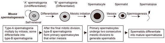
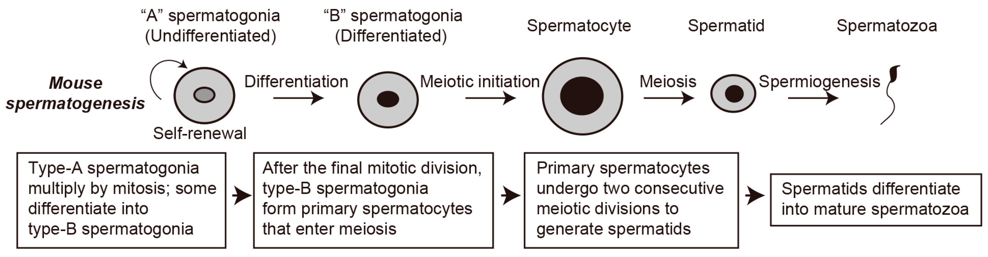
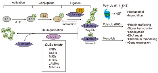
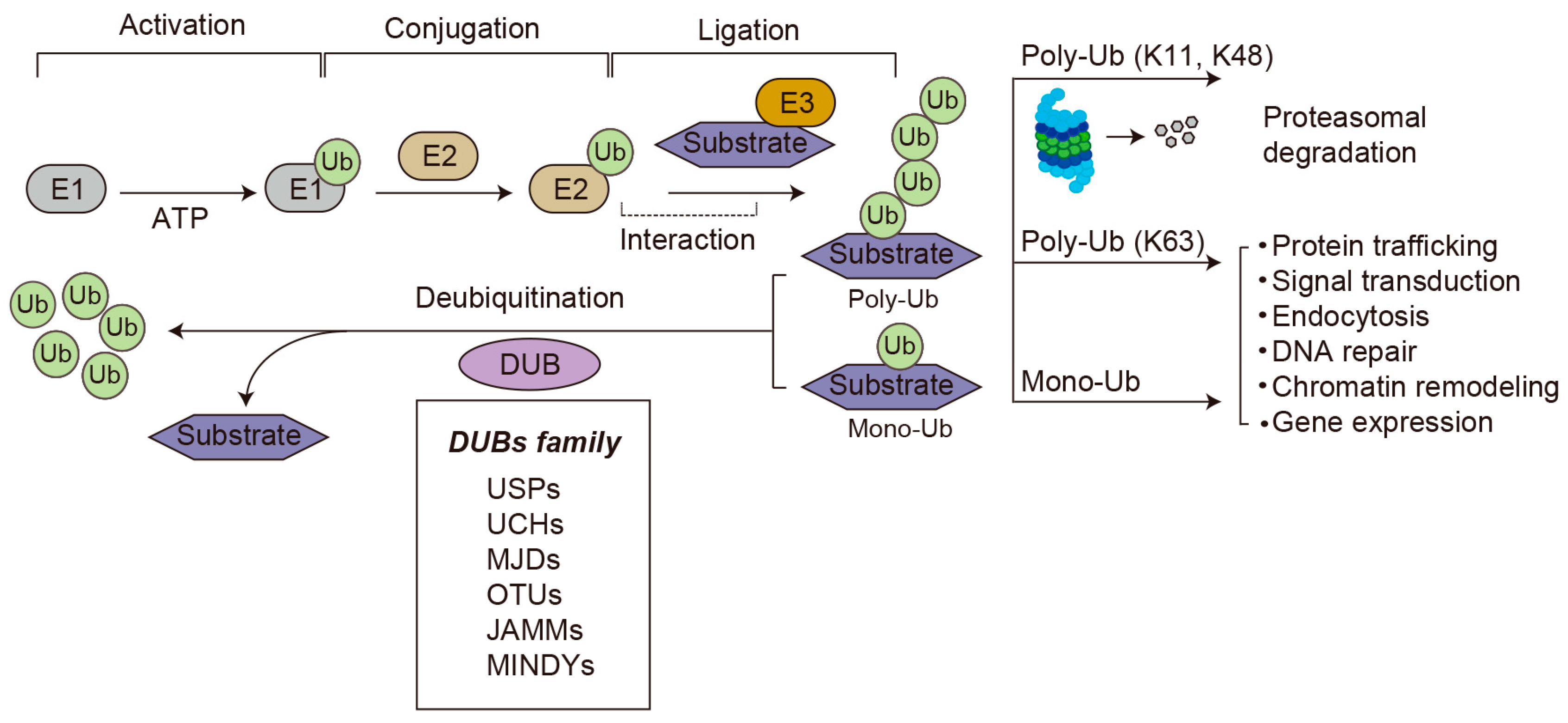
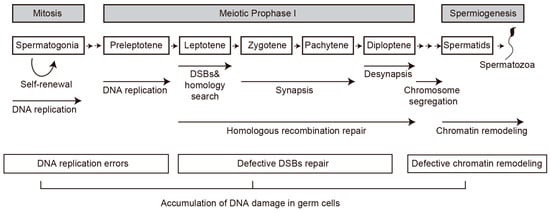
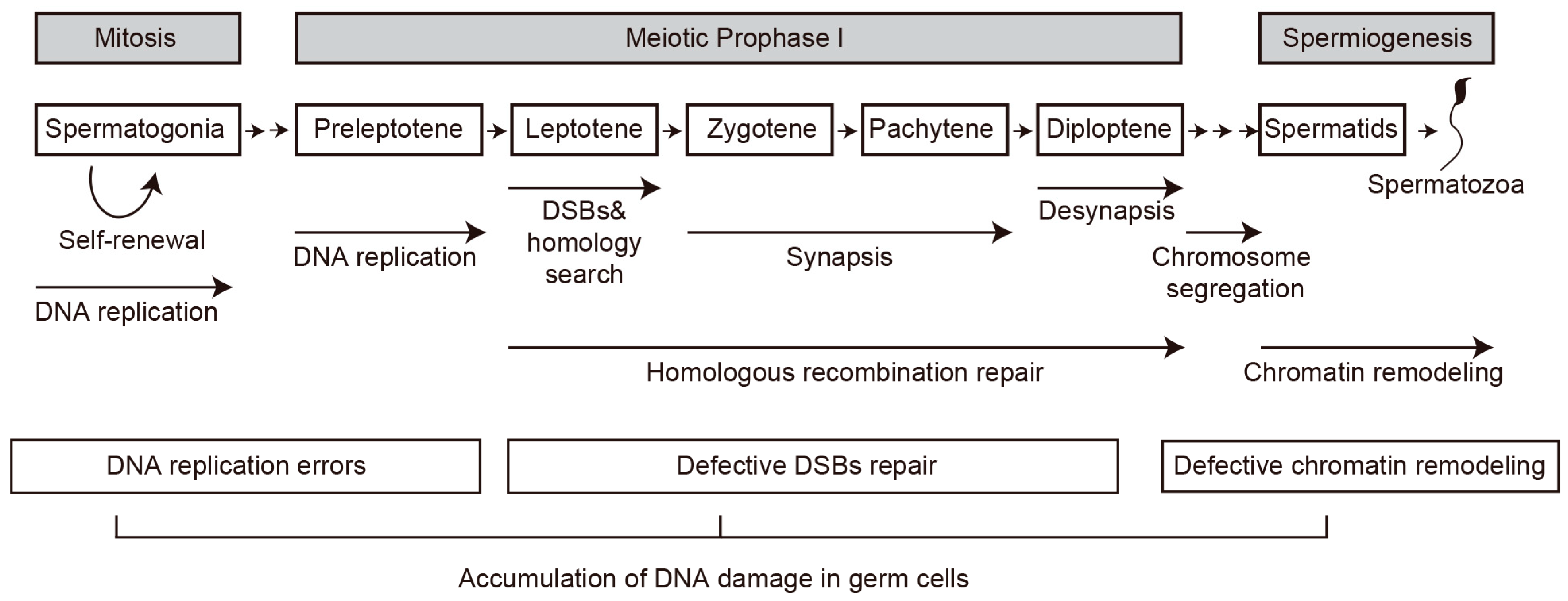
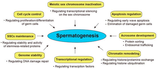
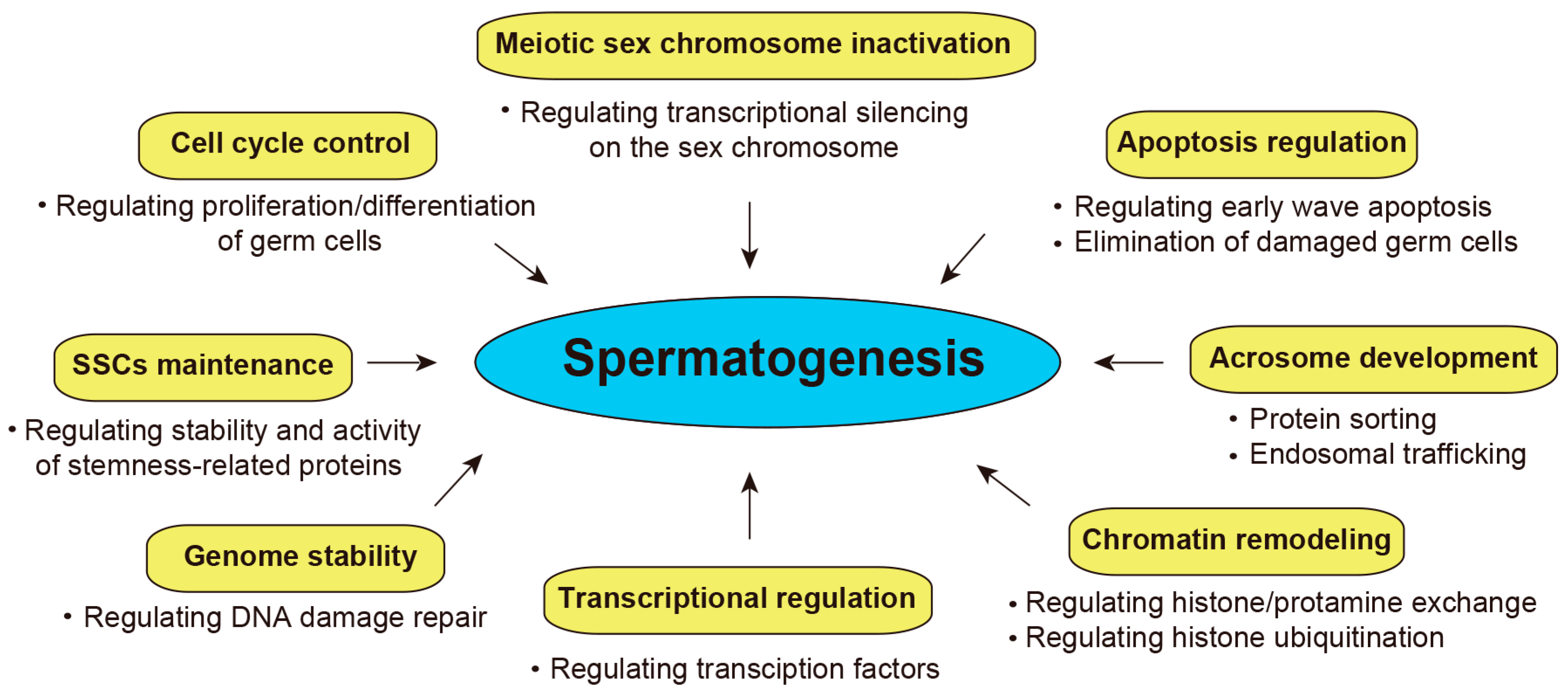
Disclaimer/Publisher’s Note: The statements, opinions and data contained in all publications are solely those of the individual author(s) and contributor(s) and not of MDPI and/or the editor(s). MDPI and/or the editor(s) disclaim responsibility for any injury to people or property resulting from any ideas, methods, instructions or products referred to in the content. |
© 2026 by the author. Licensee MDPI, Basel, Switzerland. This article is an open access article distributed under the terms and conditions of the Creative Commons Attribution (CC BY) license.