


Abstract
Introduction: Peripheral blood mononuclear cells (PBMCs) constitute a diverse population of cells involved in adaptive and innate immunity, playing an essential role in pathogen recognition, immune signaling, and immune response modulation. Vitamin D deficiency through the regulation of vitamin D receptor (VDR) and calcium-sensing receptor (CaSR) gene expression could influence the apoptotic functioning of PBMCs, which, despite its importance in the immune response, has not been sufficiently explored. Objectives: This research aimed to detect differences in the mRNA expression of CaSR, VDR, and apoptosis of PBMcs between elderly women with hip fractures and vitamin D deficiency and healthy young women, as well as in older women both at baseline and after administration of calcitriol. Methods: A case–control study involving 44 women (22 and 20, respectively) was conducted. The case group (hip fracture) was administered 2 µg/day of calcitriol for two weeks and a before-and-after comparison was made. The baseline gene expression of VDR and CaSR in PBMCs, as well as the effects of calcitriol on both the VDR/CaSR regulation and PBMC apoptosis, were studied in both groups. Serum bone biomarkers were also assessed. Results: No differences were observed in creatinine and calcium serum levels between the young and elderly osteoporotic women studied. Serum phosphorus and 25-hydroxyvitamin D (25(OH)D) were low in osteoporotic fractured women with vitamin D deficiency. In contrast, intact parathyroid hormone (PTH1–84) and alkaline phosphatase were high, while no significant difference in calcitriol [l,25(OH)2D3] serum levels was observed. In elderly women, serum calcium, phosphorus, alkaline phosphatase, 25(OH)D, and calcitriol remained unchanged after intravenous calcitriol therapy; however, PTH1–84 decreased after the treatment. In comparison to the young women, the elderly women showed decreased VDR and increased CaSR mRNA expression in PBMCs, as well as higher monocyte apoptosis. Conclusions: Calcitriol administration increased both VDR and CaSR mRNA expression in PBMCs and decreased PBMC apoptosis. Conclusions: The results obtained support the role of the vitamin D endocrine system as a regulator of the immune response and thus may contribute to explaining certain aspects of the immune dysfunction reported in individuals with vitamin D insufficiency.
1. Introduction
The vitamin D endocrine system (VDES), traditionally recognized for its role in calcium homeostasis and bone metabolism, has emerged as a key regulator of the immune system. Its immunomodulatory effects are mainly exerted through its active metabolite, 1,25-dihydroxyvitamin D (1,25(OH)2D or calcitriol), which acts on the vitamin D receptor (VDR), a member of the nuclear receptor superfamily which is widely expressed in immune cells such as monocytes, macrophages, dendritic cells, and activated T and B lymphocytes, where it exerts important immunomodulatory and antiproliferative properties [1,2,3,4,5,6,7].
Vitamin D3 is produced in the skin from 7-dehydrocholesterol by UV irradiation or is nutritionally provided through diet. The liver and other tissues metabolize vitamin D into 25-hydroxyvitamin D (25(OH)D), the main circulating form of vitamin D and the cornerstone of the VDES. 25-Hydroxyvitamin D is subsequently metabolized to 1,25(OH)2D by the CYP27B enzyme; this mainly occurs in the kidney, although other tissues, including the parathyroid gland, various epithelial cells, and cells of the immune system, also contain this enzyme [1]. The expression of the CYP27B1 enzyme in immune cells allows for the local production of calcitriol, which enables autocrine and paracrine regulation of the immune response [1,2].
Vitamin D (25(OH)D) deficiency is perhaps the most common nutritional deficiency in the world, which particularly affects many at-risk groups [8]. Organizations and scientific societies worldwide have published guidelines for recommended desirable 25(OH)D serum levels and vitamin D intake [9], but the minimal thresholds of 25(OH)D across age groups and populations are still debated [10]. Nevertheless, a consensus on the definition of sufficiency, insufficiency, or deficiency of vitamin D, as well as desirable serum 25(OH)D levels, has not yet been reached [11,12]. Severe deficiency is commonly defined as <10 ng/mL, deficiency as 10 to 20 ng/mL, and insufficiency is defined as 20–29.99 ng/mL, with levels between 30 and 50 ng/mL could be considered adequate and safe [13].
Elderly female patients who have suffered fractures and undergone surgery typically present severe vitamin D deficiency [14,15,16,17,18,19]. Aging causes a decrease in serum levels of 25-hydroxyvitamin D through various mechanisms (e.g., decreased vitamin D synthesis, decreased intestinal absorption) [20]. Immobility indoors and inflammation resulting from surgery, which worsens the inflammatory status of the elderly, are factors that contribute significantly to the decrease in 25-hydroxyvitamin D levels observed in older women undergoing surgical treatment for hip fractures, such as those who participated in this study [20,21]. As prolonged vitamin D deficiency is a determining factor in the development of osteoporosis, an adequate level of 25(OH)D must be maintained regardless of age, in order to prevent increased bone turnover and loss of bone quantity and quality [14,15,16,17,18,19].
The relative impact that vitamin D deficiency could have on both calcitriol autocrine and paracrine actions may be a factor that has not yet been sufficiently considered, which can mediate the alterations in immune response, cellular growth and differentiation, and various organ and tissue functions. Vitamin D deficiency associated with aging is one of the causes contributing to the negative regulation of VDR, leading to a less effective immune response [15,16,17,22]. The regulation of VDR expression is an important mechanism modulating the responsiveness of target tissues to calcitriol; in fact, the biological activity of calcitriol in cells is directly proportional to the tissue VDR concentration [23,24,25]. VDR expression is modulated by 1,25(OH)2D3 and several other hormones (including retinoic acid glucocorticoids and estrogen). The gene expression of VDR is also influenced by various physiological states such as age, pregnancy, lactation, and dietary calcium restriction. It has been shown that vitamin D deficiency decreased VDR mRNA levels in all the tissues studied [24,25,26]. Furthermore, the expression of VDR mRNA was decreased by vitamin D deficiency in PBMCs from elderly women but increased and returned to control values after intravenous calcitriol treatment [27,28]. Vitamin D signaling through the VDR involves genomic (modulation of immunoregulatory gene transcription) and non-genomic (rapid signaling through membrane and cytoplasmic VDR) mechanisms [5]. Interaction with the calcium-sensing receptor (CaSR) and regulation of mineral homeostasis influence the activation of intracellular signaling pathways, such as mTOR and autophagy, which are relevant to the function of monocytes and lymphocytes [6].
Peripheral blood mononuclear cells (PBMCs) are a diverse population of immune cells readily accessible, including both innate immune cells responsible for rapid pathogen responses and adaptive immune cells involved in specific immune responses. PBMCs play an essential role in pathogen recognition, immune signaling, and modulation of the immune response and express VDR and are thus targets for the immunomodulatory actions of calcitriol [29,30]. PBMCs have been shown to present CaSR, initially cloned from bovine parathyroid glands [31]. The main function of CaSR is to regulate bone and mineral metabolism by influencing parathyroid hormone secretion, urinary calcium excretion, and bone remodeling; however, there are expressed ubiquitously in the body, exerting pleiotropic actions in cells, including modification of proliferation, differentiation, and programmed cell death (apoptosis) [29,32,33]. CaSR may act as an immunomodulator in PBMCs depending on vitamin D, as well as in the ‘reversal’ phase of bone remodeling [31,33].
Vitamin D deficiency through the regulation of vitamin D receptor (VDR) and calcium-sensing receptor (CaSR) expression, in addition to influencing intestinal calcium absorption [15,34,35] and immune responses [25,36,37], could influence the apoptotic functioning of PBMCs, which, despite its importance in the immune response, has not been sufficiently studied [38,39].
This research aimed to detect differences in the mRNA expression of CaSR, VDR, and apoptosis of PBMCs between elderly women with hip fractures and a vitamin D-deficient status and healthy young women, as well as in elderly women both at baseline and after administration of calcitriol.
2. Materials and Methods
2.1. Subjects and Procedures
Blood samples were obtained at baseline from 20 young women as a control group (mean age: 33 years) and from 22 elderly women with a history of hip fractures (mean age: 75 years) in the previous three months (considered as a strict inclusion criterion), both before and after calcitriol treatment (intravenous administration of 2 µg calcitriol (Calcijex®, Roche, Basel, Switzerland) daily for two weeks). Exclusion criteria were that at the time of the study, the participants were not suffering from diabetes mellitus or intercurrent infections, malabsorption, nephrolithiasis, primary hyperparathyroidism, hyperthyroidism, hypercalcemia, creatinine clearance < 30 mL/min, neoplastic disease within the last 5 years and were not undergoing treatment with vitamin D metabolites or analogs or drugs that can modify vitamin D levels, calcium channel blockers, calmodulin antagonists, steroids, or immunosuppressive therapy.
Authorization for the study was obtained from the Biomedical Research Ethics Committee of the province of Córdoba. All participating women signed the informed consent form. The harmonized tripartite standards of the Declaration of Helsinki, the Organic Law on Biomedical Research of 15/2007 of 3 July, the Organic Law on Personal Data Protection (LOPD) of 13 December 2018, the code of ethics of the “Organización Médica Colegial” (OMC), the basic regulatory law 41/2002 on patient autonomy and rights and obligations regarding clinical information and documentation, of 14 November, and the standards of good clinical practice were respected.
2.2. Serum Parameters Analysis
Total calcium, serum phosphorus and alkaline phosphatase were measured using an autoanalyzer ADVIA Centaur XP (Siemens Healthineers, Erlangen, Germany). Serum PTH1–84 was determined via the allegro immunoradiometric assay from the Nichols Institute (San Juan Capistrano, CA, USA). Vitamin D status was assessed by measuring the level of [25(OH)D] with a chemiluminescence autoanalyzer Architect c-16000 (Abbott, Chicago, IL, USA), and 1,25(OH)2D3 (calcitriol) was determined via radioimmunoassay with an RIA kit (Immunodiagnostic systems Ltd., Boldon, UK). According to the serum levels, a 25(OH)D deficiency was defined as <20 ng/mL, insufficiency was defined as 20–29.99 ng/mL, and a normal level was defined as >30 ng/mL [22,23].
2.3. Apoptosis Measurement
PBMCs were separated by centrifugation on a Ficoll-Hypaque density gradient. The percentage of apoptotic cells was quantified as those positive for propidium iodide, as it can enter the nucleus and bind to the DNA of cells with a broken membrane. Apoptosis was assessed via flow cytometry with fluorescence-activated cell sorting (FACScan) (Beckton Dickinson, San Jose, CA, USA). Briefly, cells were washed and fixed in 70% ethanol for 3 h at 4 °C. Then, the cells were resuspended in 0.5 mL PBS to which 0.5 mL of RNase solution (l mg/mL) and propidium iodine (50 mg/mL) were added. Apoptotic cells were characterized by the ‘hypodiploid’ peak on flow cytometric analysis [40,41].
2.4. Gene Expression Quantification
Total RNA was isolated with Trizol Reagent from Invitrogen (Carlsbad, CA, USA). and the amounts of VDR and CaSR mRNA were measured via quantitative RT-PCR (qRT-PCR). The quantification of different amplicons was accomplished via laser-induced fluorescence with the ABI 373 A Stretch Sequencer from Applied Biosystems (Foster City, CA, USA) [42,43].
The primers for VDR were 5′ TGAAGGCTGCAAAGGCITCTTCAGGC 3′ (forward) and 5′ GGATGAACTCCITCATCATGCCGATG 3′ (reverse); those for CaSR were 5′ ATTGAGGGGGAGCCCACCTGCTGCT 3′ (forward) and 5′ AAAGAGGGTGAGTGCGATCCCAAAGG 3′ (reverse); and those for actin (housekeeping gene used for normalization) were 5′ CGTCACCAACTGGGACGACATGGAG 3′ (forward) and 5′ GGCGTACAGTAGCACAGCCTGGA 3′ (reverse).
2.5. Statistical Study
Data were analyzed using the Sigma statistical package for microcomputers from Horus (Madrid, Spain). The Shapiro–Wilk test was performed to check whether the values of each variable followed a normal distribution. As this was not the case, non-parametric tests were performed. To compare means in the case–control study, a Mann–Whitney U test was performed, and in the before-and-after study, a Wilcoxon signed rank test was performed (i.e., to compare VDR and CaSR gene expression values) within each group before and after calcitriol administration). All values are expressed as mean ± standard deviation. Statistical significance was accepted for p < 0.05.
3. Results
The 22 elderly women admitted for hip fractures did not differ from the young women in the control group in terms of ethnicity or anthropometric measurements (weight, abdominal circumference), etc. Before treatment with calcitriol, various parameters were compared between the group of young women and the group of older women with fractures.
No significant differences were observed in creatinine, calcium, and calcitriol (1,25(OH)2D) serum levels between the two groups. However, serum phosphorus (p < 0.05) and 25(OH)D (p < 0.001) were significantly decreased, whereas PTH1–84 (p < 0.0001) and alkaline phosphatase (p < 0.001) were significantly higher in older women (Table 1). As shown in the table below, none of the groups of women studied had sufficient levels of vitamin D (25(OH)D > 30 ng/mL). Thus, the mean serum 25(OH)D level in the group of elderly women with fractures was 8.0 ± 4.5 ng/mL, indicating a more severe vitamin D deficiency. In the group of young women, the mean value was between 20 and 30 ng/mL, which is considered insufficient.
Table 1.
Serum biochemical parameters in young women (as controls) vs. those in elderly women with osteoporosis and hip fractures who had severe 25(OH)D deficiency and mild secondary hyperparathyroidism.
In elderly osteoporotic women, total serum calcium (9.7 ± 0.4 mg/dL), phosphorus (2.5 ± 0.7 mg/dL), alkaline phosphatase (67 ± 18 UI/L), 25(OH)D3 (7 ± 4.8 ng/mL), and calcitriol (41 ± 12.0 pg/mL) remained stable after intravenous calcitriol therapy; in contrast, PTH1–84 decreased significantly after calcitriol treatment (46 ± 6 ng/mL; p < 0.001).
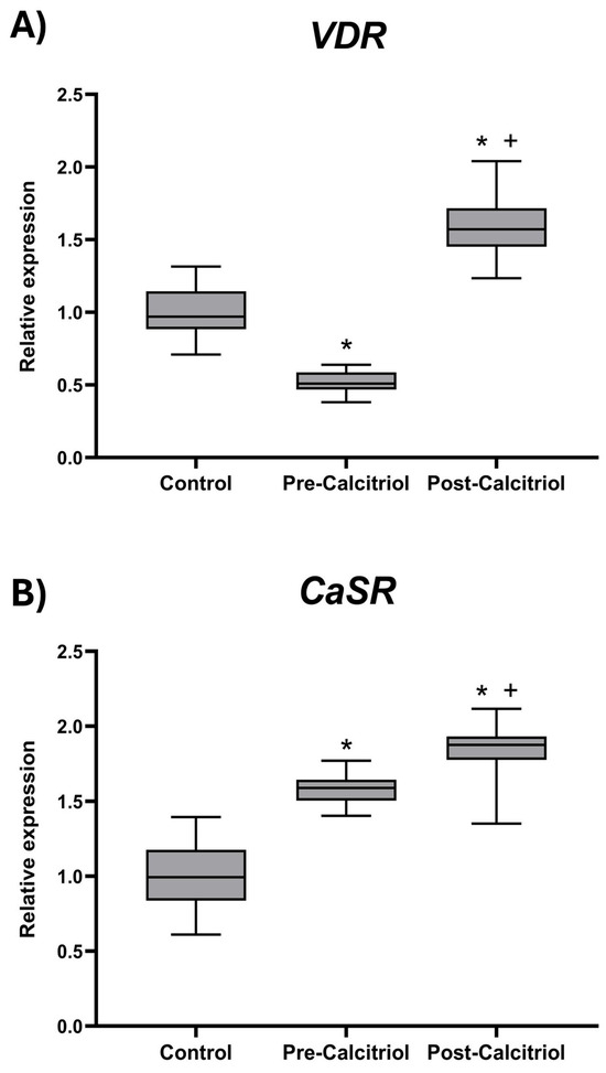
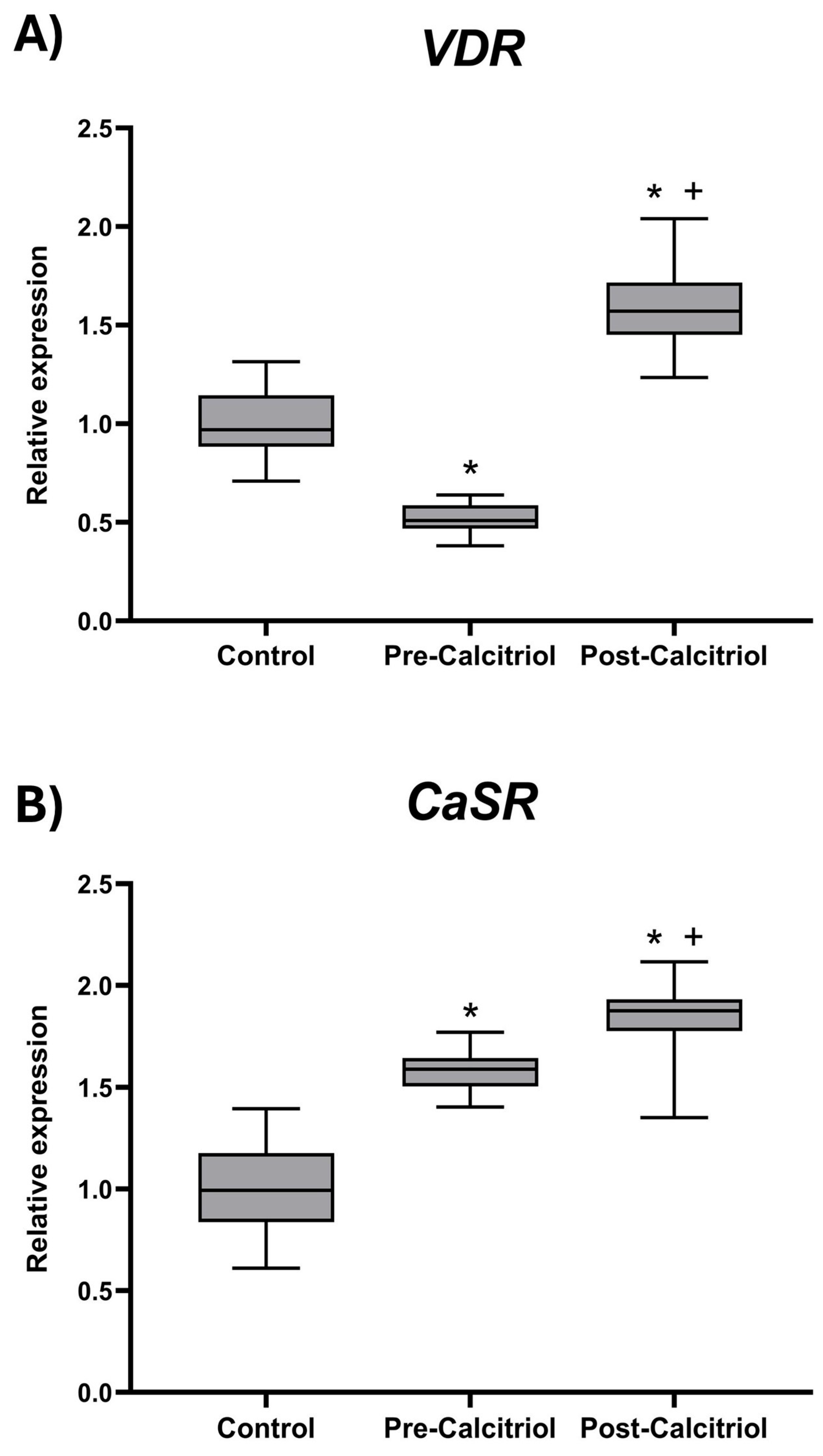
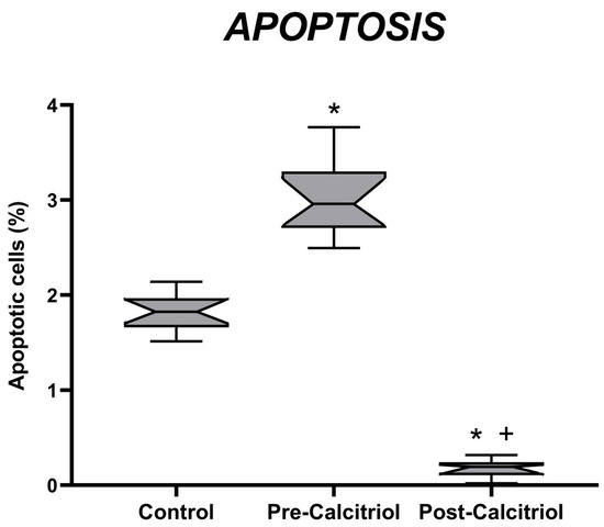
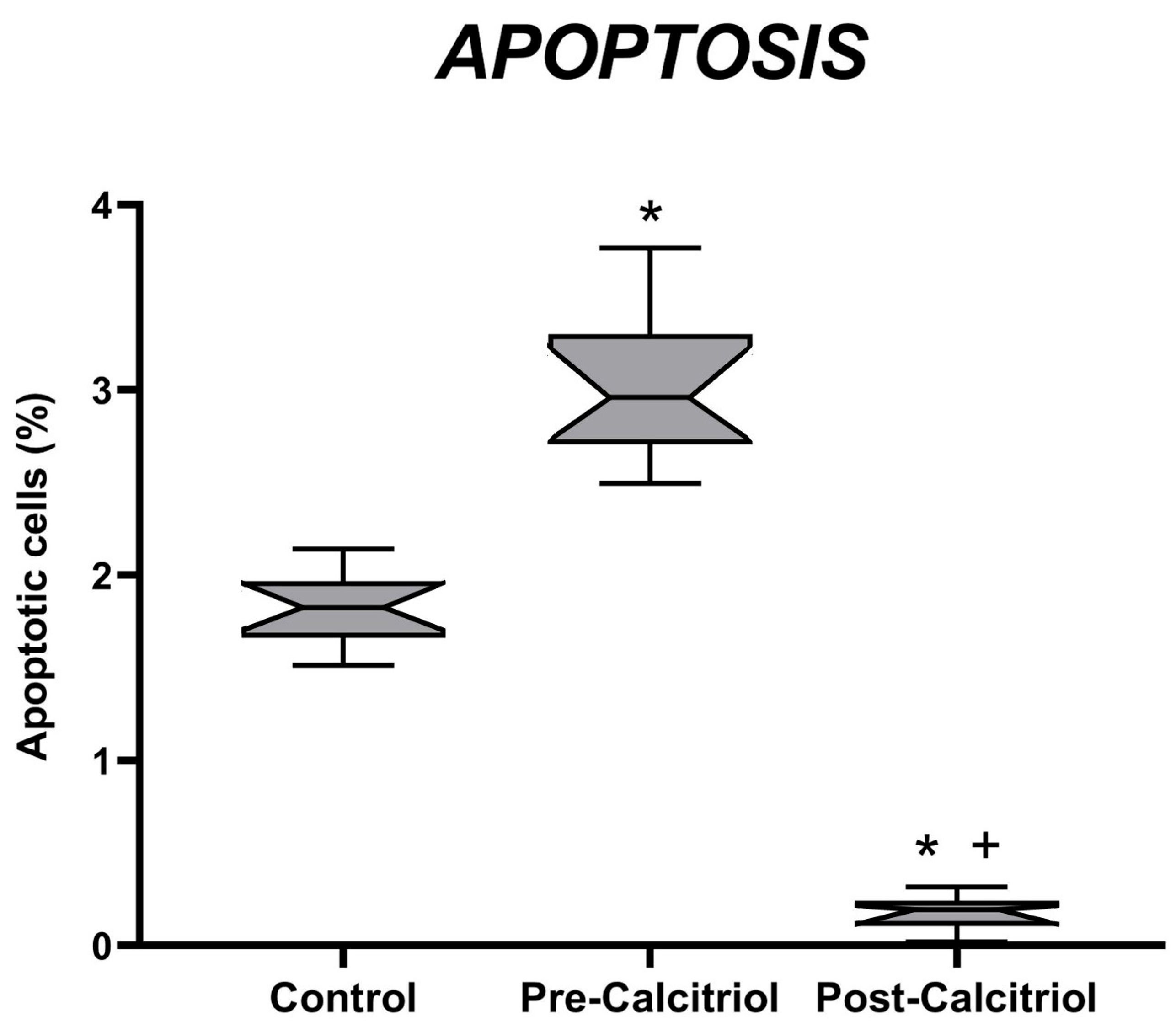
As shown in Figure 1, elderly osteoporotic women with vitamin D deficiency presented decreased VDR (p < 0.05) and increased CaSR (p < 0.05) mRNA expression levels in PBMCs, in comparison with the control group of women, as well as a higher PBMC apoptosis level (Figure 2). After treatment with calcitriol, in the group of elderly women, an increase in VDR (p < 0.05) and CaSR (p < 0.01) mRNA expression in PBMCs was observed, compared to pre-treatment values and compared to those of the control group (Figure 1).
Figure 1.
Expression levels of VDR (A) and CaSR (B) coding genes. Total RNA was isolated from PBMCs derived from the women included in the study. The mRNA levels of the VDR and CaSR genes were quantified in each of the RNA samples obtained from the control group (n = 20, young women) and the group of elderly women (n = 22) before (Pre-calcitriol) and after (Post-calcitriol) treatment with calcitriol. The graphs show the relative expression of each gene for each of the study groups. * p < 0.05 vs. Control; + p < 0.05 vs. Pre-calcitriol. The notches in each group indicate a 95% confidence interval (CI).
Figure 2.
Quantification of monocyte apoptosis in PBMCs derived from young women (n = 20) and elderly women (n = 22), before (Pre-calcitriol) and after (Post-calcitriol) treatment with calcitriol. The percentage of apoptotic monocytes was determined in each sample. * p < 0.05 vs. Control; + p < 0.05 vs. Pre-calcitriol. The notches in each group indicate a 95% confidence interval (CI).
The percentage of apoptotic cells was also studied in PBMCs. Before treatment with calcitriol, the group of elderly women had a higher percentage of apoptotic cells than the control group. However, after treatment with calcitriol, the percentage of apoptosis in elderly women decreased significantly. It was more than 14 times lower than the values obtained before treatment and more than 8 times lower than the values in the control group (Figure 2).
4. Discussion
The results of this study demonstrated that all elderly women with hip fractures who underwent surgery had serum 25(OH)D levels in the deficiency range, lower than those of young women in the control group, but higher levels of PTH1–84 in the secondary hyperparathyroidism range. In addition, they had lower expressions of VDR and CaSR and increased apoptosis in PBMCs. A short course of intravenous calcitriol treatment every 24 h in these patients induced an upregulation of VDR and CaSR, as well as a marked decrease in apoptosis in PBMCs.
The results of the present study also confirm the previously reported higher expression of CaSR mRNA in monocytes from osteoporotic women with vitamin D insufficiency, when compared to that in the controls [31,33]. These patients also showed an increase in monocyte CaSR mRNA expression after intravenous calcitriol treatment, which induced a significant decrease in serum PTH1–84 without an increase in serum calcium [44,45].
The current results also reveal higher expression of CaSR mRNA in PBMCs from osteoporotic elderly women with hip fractures and severe 25(OH)D deficiency, compared to those from young women. These patients also showed an increase in CaSR mRNA expression in monocytes after intravenous treatment with calcitriol, which induced a significant decrease in serum PTH1–84 without a concomitant increase in serum calcium [44,45].
Calcium is the main regulator of CaSR; therefore, extracellular calcium participates in the modulation of the immune response, possibly acting through transmembrane CaSR in mature monocytes/macrophages [46,47]. This upregulation of CaSR expression has previously been described in vitro in the parathyroid glands (PTG) and kidney [48]; as well as in HL-60 cells, which occurred during their differentiation into cells with a monocyte/macrophage phenotype in response to calcitriol treatment.
The increases in VDR and CaSR induced by calcitriol might contribute to the decrease in monocyte apoptosis either directly, through changes in cell monocyte cycle progression [30], or indirectly, through the inhibition of HLA-DR expression [49,50] or a change in the pattern of cytokines secreted by the immune system in response to calcitriol [51,52]. This occurs because calcitriol diminishes the expression of type II HLA antigens (DR, DP and DQ) on monocytes [53,54], as well as inhibiting and/or exerting modulating actions on cytokines [55,56].
Increased expression of VDR and CaSR interacting together may decrease PBMCs apoptosis, allowing them to fight pathogens for a longer period, while modifying the secretion of proinflammatory cytokines through the nuclear factor κB (NF-κB) signaling pathway to modify the inflammatory response [30,51,52]. These findings suggest that stimulation of CaSR via VDR could be exploited to regulate the immune system through monocyte and macrophage activities and reduce inflammatory damage; as such, it shows potential as a target for the prevention and treatment of inflammatory diseases [55,56].
Furthermore, the effects of VDR upregulation on the regulation of the innate and adaptive immune systems are extensive. Stimulation of VDR in monocytes (as well as macrophages, and neutrophils) induces the secretion of the antibacterial peptides cathelicidin and defensin, which play important roles in innate immune defenses due to their ability to lyse bacteria [1,57]. Serum levels of 25(OH)D are associated with the expression and functionality of Toll-like receptors (TLRs), especially those involved in viral responses [1]. The activation of innate immunity receptors, such as TLR2, increases the expression of VDR, 1α-hydroxylase, and cathelicidin, suggesting that the VDES plays a role in the innate immune response against bacterial pathogens [57]. VDR stimulation has been shown to inhibit T cell expansion and modulate cytokine expression with Th2 polarization [58], as well as to inhibit B lymphocyte differentiation and proliferation and immunoglobulin secretion. Other immunological effects attributed to VDR stimulation include dendritic cell maturation, decreased HLA class II expression, and enhanced antigen processing and presentation, which induce the production of more tolerogenic cytokines [57,59,60]. This evidence collectively supports the idea that vitamin D deficiency plays a role in immune system dysfunction [61,62,63,64].
The strength of this study lies in its simplicity, using an in vivo model of women with age-related osteoporosis and hip fracture using clinical blood samples. These women constitute a homogeneous cohort with vitamin D deficiency (measured as 25-hydroxyvitamin D). The administration of calcitriol, the hormone of the vitamin D endocrine system, in a short course, which does not produce an increase in blood calcium levels, allows us to clearly assess the biological response, showing that VDR stimulation, in addition to producing auto-upregulation, induces a clear increase in CaSR expression, producing a decrease in PBMCs apoptosis, which may contribute to improving the altered innate immune response and reducing the high rate of infections as a complication of the natural history observed in women with hip fractures and vitamin D deficiency [65].
The main weakness of this study lies in the absence of additional molecular and mechanical analyses to further clarify the relationship between clear VDR/CaSR signaling and PBMC apoptosis. Another weakness we could consider is that, as this is a single-center study, we have less external validity. If we had a larger sample size and greater ethnic diversity, the patients could have been stratified by race and then compared. Another aspect that should be considered in future studies is to carry out longer follow-up, in order to find out whether the changes observed are maintained over time.
5. Conclusions
Our present results support the increasing evidence that the vitamin D endocrine system plays a role not only as a regulator of calcium homeostasis and bone health, but also as a regulator of the immune response. Therefore, this study may contribute to explaining the immune dysfunction associated with vitamin D deficiency.
Furthermore, stimulation of the VDR by vitamin D (or its structural analogs) can be considered as a therapeutic target for the prevention and treatment of infectious, inflammatory, or autoimmune diseases.
Author Contributions
J.C.-V., J.M.Q.-G. and A.C.-D. contributed to the conception and design of the study. A.N.-P., J.L.G.-C. and J.M.Q.-G. collected information from the women’s medical records. A.C. and L.V.-A. carried out the molecular studies. J.C.-V. and L.V.-A. carried out the biochemical serum analyses. A.N.-P., A.C. and L.V.-A. organized the database. J.L.G.-C. performed the statistical study. J.M.Q.-G. acquired funding. A.N.-P., A.C. and J.M.Q.-G. wrote the first draft of the manuscript. J.C.-V. and A.C.-D. carried out revision and proofreading of the manuscript. All authors have read and agreed to the published version of the manuscript.
Funding
Supported in part by the project from FIS (Fondo de Investigación Sanitaria, Instituto de Salud Carlos III, Spain) project code 96/1662.
Institutional Review Board Statement
This study was conducted in accordance with the Declaration of Helsinki and approved by the Research Ethics Committee of Córdoba on 27 November 2021 (code VDR-FRACT; reference number 4527).
Informed Consent Statement
All participating women gave written informed consent before the blood test and data collection began.
Data Availability Statement
The original contributions presented in this study are included in the article. Further inquiries can be directed to the corresponding authors.
Acknowledgments
The authors are indebted to all the women who participated for their selfless collaboration. The authors would also like to thank Maimónides Biomedical Research Institute of Córdoba (IMIBIC) and Reina Sofía University Hospital for their technical support and all the assistance they have provided. Thanks to Simon Craig Exelby for the language correction.
Conflicts of Interest
All authors have no conflicts of interest to declare. This research has no sponsors or incentives. Neither the participating investigators nor the hospital center received any financial compensation.
Abbreviations
The following abbreviations are used in this manuscript:
| VDES | Vitamin D endocrine system |
| VDR | Vitamin D receptor |
| CaSR | Calcium-sensing receptor |
| PBMCs | Peripheral blood mononuclear cells |
References
- Artusa, P.; White, J.H. Vitamin D and its analogs in immune system regulation. Pharmacol. Rev. 2025, 77, 100032. [Google Scholar] [CrossRef] [Scilit]
- Daryabor, G.; Gholijani, N.; Kahmini, F.R. A review of the critical role of vitamin D axis on the immune system. Exp. Mol. Pathol. 2023, 132–133, 104866. [Google Scholar] [CrossRef] [Scilit]
- Sîrbe, C.; Rednic, S.; Grama, A.; Pop, T.L. An Update on the Effects of Vitamin D on the Immune System and Autoimmune Diseases. Int. J. Mol. Sci. 2022, 17, 9784. [Google Scholar] [CrossRef] [Scilit] [PubMed]
- Argano, C.; Torres, A.; Orlando, V.; Cangialosi, V.; Maggio, D.; Pollicino, C.; Corrao, S. Molecular Insight into the Role of Vitamin D in Immune-Mediated Inflammatory Diseases. Int. J. Mol. Sci. 2025, 26, 4798. [Google Scholar] [CrossRef] [Scilit] [PubMed]
- Trochoutsou, A.I.; Kloukina, V.; Samitas, K.; Xanthou, G. Vitamin-D in the Immune System: Genomic and Non-Genomic Actions. Mini Rev. Med. Chem. 2015, 15, 953–963. [Google Scholar] [CrossRef] [Scilit]
- Dimitrov, V.; Barbier, C.; Ismailova, A.; Wang, Y.; Dmowski, K.; Salehi-Tabar, R.; Memari, B.; Groulx-Boivin, E.; White, J.H. Vitamin D-regulated Gene Expression Profiles: Species-specificity and Cell-specific Effects on Metabolism and Immunity. Endocrinology 2021, 162, bqaa218. [Google Scholar] [CrossRef] [Scilit] [PubMed]
- Umar, M.; Sastry, K.S.; Chouchane, A.I. Role of Vitamin D Beyond the Skeletal Function: A Review of the Molecular and Clinical Studies. Int. J. Mol. Sci. 2018, 19, 1618. [Google Scholar] [CrossRef] [Scilit]
- Hilger, J.; Friedel, A.; Herr, R.; Rausch, T.; Roos, F.; Wahl, D.A.; Pierroz, D.D.; Weber, P.; Hoffmann, K. A systematic review of vitamin D status in populations worldwide. Br. J. Nutr. 2014, 111, 23–45. [Google Scholar] [CrossRef] [Scilit]
- Bouillon, R. Comparative analysis of nutritional guidelines for vitamin D. Nat. Rev. Endocrinol. 2017, 13, 466–479. [Google Scholar] [CrossRef] [Scilit]
- Demay, M.B.; Pittas, A.G.; Bikle, D.D.; Diab, D.L.; Kiely, M.E.; Lazaretti-Castro, M.; Lips, P.; Mitchell, D.M.; Murad, M.H.; Powers, S.; et al. Vitamin D for the Prevention of Disease: An Endocrine Society Clinical Practice Guideline. J. Clin. Endocrinol. Metab. 2024, 109, 1907–1947. [Google Scholar] [CrossRef] [Scilit]
- Holick, M.F. The Debatable Clinical Utility of the 2024 Vitamin D Guideline: Bridging the Gap Between Current Guidelines, Practical Clinical Recommendations, and Utilization of Emerging Evidence in Vitamin D Disease Prevention. Endocr. Pract. 2025, 31, 399–402. [Google Scholar] [CrossRef] [Scilit]
- Shah, V.P.; Nayfeh, T.; Alsawaf, Y.; Saadi, S.; Farah, M.; Zhu, Y.; Firwana, M.; Seisa, M.; Wang, Z.; Scragg, R.; et al. A Systematic Review Supporting the Endocrine Society Clinical Practice Guidelines on Vitamin D. J. Clin. Endocrinol. Metab. 2024, 109, 1961–1974. [Google Scholar] [CrossRef] [Scilit]
- Bethesda, M. Fact Sheet for Health Professionals: Vitamin D. National Institutes of Health, Division of Dietary Supplements. Available online: https://ods.od.nih.gov/factsheets/VitaminD-HealthProfessional (accessed on 19 November 2025).
- Bouillon, R.; Antonio, L.; Olarte, O.R. Calcifediol (25OH Vitamin D3) Deficiency: A Risk Factor from Early to Old Age. Nutrients 2022, 14, 1168. [Google Scholar] [CrossRef] [Scilit]
- Quesada-Gómez, J.M.; Alonso, J.; Bouillon, R. Vitamin D insufficiency as a determinant of hip fractures. Osteoporos. Int. 1996, 6, 42–47. [Google Scholar] [CrossRef] [Scilit]
- Meunier, P.J.; Chapuy, M.C.; Arlot, M.E.; Delmas, P.D.; Duboeuf, F. Can we stop bone loss and prevent hip fractures in the elderly? Osteoporos. Int. 1994, 4, S71–S76. [Google Scholar] [CrossRef] [Scilit]
- Boonen, S.; Rizzoli, R.; Meunier, P.J.; Stone, M.; Nuki, G.; Syversen, U.; Lehtonen-Veromaa, M.; Lips, P.; Johnell, O.; Reginster, J.-Y. The need for clinical guidance in the use of calcium and vitamin D in the management of osteoporosis: A consensus report. Osteoporos. Int. 2004, 15, 511–519. [Google Scholar] [CrossRef] [Scilit] [PubMed]
- Chiriac, C.; Ciurea, O.A.; Lipan, M.; Capusa, C.S.; Mircescu, G. Vitamin D Deficiency, Bone Turnover Markers and Arterial Calcifications in Non-Dialysis Chronic Kidney Disease Patients. Acta Endocrinol. 2024, 20, 12–20. [Google Scholar] [CrossRef] [Scilit] [PubMed]
- Bhattoa, H.P.; Vasikaran, S.; Trifonidi, I.; Kapoula, G.; Lombardi, G.; Jørgensen, N.R.; Pikner, R.; Miura, M.; Chapurlat, R.; Hiligsmann, M.; et al. Update on the role of bone turnover markers in the diagnosis and management of osteoporosis: A consensus paper from The European Society for Clinical and Economic Aspects of Osteoporosis, Osteoarthritis and Musculoskeletal Diseases (ESCEO), International Osteoporosis Foundation (IOF), and International Federation of Clinical Chemistry and Laboratory Medicine (IFCC). Osteoporos. Int. 2025, 36, 579–608. [Google Scholar] [CrossRef] [Scilit]
- Quesada, J.M.; Coopmans, W.; Ruiz, B.; Aljama, P.; Jans, I.; Bouillon, R. Influence of vitamin D on parathyroid function in the elderly. J. Clin. Endocrinol. Metab. 1992, 75, 494–501. [Google Scholar] [CrossRef] [Scilit] [PubMed]
- Quesada, J.M.; Jans, I.; Benito, P.; Jimenez, J.A.; Bouillon, R. Vitamin D Status of Elderly People in Spain. Age Ageing 1989, 18, 392–397. [Google Scholar] [CrossRef] [Scilit]
- Oliveri, C.; Xourafa, A.; Agostino, R.M.; Corigliano, V.; Botindari, A.; Gaudio, A.; Morabito, N.; Allegra, A.; Catalano, A. Exploring the Association Between Platelet Count, the Systemic Immune Inflammation Index, and Fracture Risk in Postmenopausal Women with Osteoporosis: A Cross-Sectional Study. J. Clin. Med. 2025, 14, 5453. [Google Scholar] [CrossRef] [Scilit] [PubMed]
- Hernández, D.Á.; Díaz, M.L.N.; Alonso, C.G.; García, E.C.; Andía, J.B.C. Tissue-Specific Effect of VDR Gene Polymorphisms on the Response to Calcitriol. WOS. 2008. Available online: https://digibuo.uniovi.es/dspace/handle/10651/23741 (accessed on 27 August 2025).
- Mocharla, H.; Butch, A.W.; Pappas, A.A.; Flick, J.T.; Weinstein, R.S.; De Togni, P.; Jilka, R.L.; Roberson, P.K.; Parfitt, A.M.; Manolagas, S.C. Quantification of vitamin D receptor mRNA by competitive polymerase chain reaction in PBMC: Lack of correspondence with common allelic variants. J. Bone Miner. Res. 1997, 12, 726–733. [Google Scholar] [CrossRef] [Scilit]
- Chao, G.; Lin, A.; Bao, Y. A study of the association of vitamin D receptor (VDR) as a predictive biomarker for immune checkpoint inhibitor therapy with immune invasion in colon adenocarcinoma. J. Pharm. Biomed. Anal. 2025, 252, 116510. [Google Scholar] [CrossRef] [Scilit]
- Unger, T.; Borghi, C.; Charchar, F.; Khan, N.A.; Poulter, N.R.; Prabhakaran, D.; Ramirez, A.; Schlaich, M.; Stergiou, G.S.; Tomaszewski, M.; et al. 2020 International Society of Hypertension Global Hypertension Practice Guidelines. Hypertension 2020, 75, 1334–1357. [Google Scholar] [CrossRef] [Scilit]
- Zineb, R.; Zhor, B.; Odile, W.; Marthe, R.-R. Distinct, tissue-specific regulation of vitamin D receptor in the intestine, kidney, and skin by dietary calcium and vitamin D. Endocrinology 1998, 139, 1844–1852. [Google Scholar] [CrossRef]
- Healy, K.D.; Frahm, M.A.; DeLuca, H.F. 1,25-Dihydroxyvitamin D3 up-regulates the renal vitamin D receptor through indirect gene activation and receptor stabilization. Arch. Biochem. Biophys. 2005, 433, 466–473. [Google Scholar] [CrossRef] [Scilit]
- Poon, A.H.; Gong, L.; Brasch-Andersen, C.; Litonjua, A.A.; Raby, B.A.; Hamid, Q.; Laprise, C.; Weiss, S.T.; Altman, R.B.; Klein, T.E. Very important pharmacogene summary for VDR. Pharmacogenetics Genom. 2012, 22, 758–763. [Google Scholar] [CrossRef] [Scilit] [PubMed]
- Farhana, A.; Khan, Y.S.; Alsrhani, A. Vitamin D at the intersection of health and disease: The immunomodulatory perspective. Int. J. Health Sci. 2024, 18, 1–4. [Google Scholar]
- Peterlik, M.; Kállay, E.; Cross, H.S. Calcium Nutrition and Extracellular Calcium Sensing: Relevance for the Pathogenesis of Osteoporosis, Cancer and Cardiovascular Diseases. Nutrients 2013, 5, 302–327. [Google Scholar] [CrossRef] [Scilit] [PubMed]
- Norman, A.W.; Mena, F.R.; Silva, B. Structure Function Studies: Identification of Vitamin D Analogs for the Ligand-Binding Domains of Important Proteins in the Vitamin D-Endocrine System. Rev. Endocr. Metab. Disord. 2001, 2, 229–238. [Google Scholar] [CrossRef] [Scilit]
- Chattopadhyay, N.; Mithal, A.; Brown, E.M. The Calcium-Sensing Receptor: A Window into the Physiology and Pathophysiology of Mineral Ion Metabolism. Endocr. Rev. 1996, 17, 289–307. [Google Scholar] [CrossRef] [Scilit]
- Chen, J.; Gan, X.; Song, J.; Gao, L.; Shao, M.; Wang, Y.; Gao, Y. A Systematic Review and Meta-Analysis of Barriers Affecting Early Ambulation in Older Patients After Hip Fracture Surgery. Nurs. Health Sci. 2025, 27, e70177. [Google Scholar] [CrossRef] [Scilit]
- Hoong, C.W.S.; Saul, D.; Khosla, S.; Sfeir, J.G. Advances in the management of osteoporosis. BMJ 2025, 390, e081250. [Google Scholar] [CrossRef] [Scilit]
- Ensrud, K.E.; Duong, T.; A Cauley, J.; Heaney, R.P.; Wolf, R.L.; Harris, E.; Cummings, S.R. Low fractional calcium absorption increases the risk for hip fracture in women with low calcium intake. Ann. Intern. Med. 2000, 132, 345–353. [Google Scholar] [CrossRef] [Scilit]
- Xia, X.; Xu, F.; Dai, D.; Xiong, A.; Sun, R.; Ling, Y.; Qiu, L.; Wang, R.; Ding, Y.; Lin, M.; et al. VDR is a potential prognostic biomarker and positively correlated with immune infiltration: A comprehensive pan-cancer analysis with experimental verification. Biosci. Rep. 2024, 44, BSR20231845. [Google Scholar] [CrossRef] [Scilit]
- Abiri, E.; Hemmatian, N. Investigating apoptosis in peripheral blood mononuclear cells among the elderly in the post-COVID-19 era. BMC Immunol. 2025, 26, 86. [Google Scholar] [CrossRef] [Scilit]
- Holick, M.F.; Binkley, N.C.; Bischoff-Ferrari, H.A.; Gordon, C.M.; Hanley, D.A.; Heaney, R.P.; Murad, M.H.; Weaver, C.M. Guidelines for preventing and treating vitamin D deficiency and insufficiency revisited. J. Clin. Endocrinol. Metab. 2012, 97, 1153–1158. [Google Scholar] [CrossRef] [Scilit] [PubMed]
- Ichiyoshi, H.; Kiyozuka, Y.; Kishimoto, Y.; Fukuhara, S.; Tsubura, A. Massive telomere loss and telomerase RNA expression in dexamethasone-induced apoptosis in mouse thymocytes. Exp. Mol. Pathol. 2003, 75, 178–186. [Google Scholar] [CrossRef] [Scilit] [PubMed]
- Nicoletti, I.; Migliorati, G.; Pagliacci, M.C.; Grignani, F.; Riccardi, C. A rapid and simple method for measuring thymocyte apoptosis by propidium iodide staining and flow cytometry. J. Immunol. Methods 1991, 139, 271–279. [Google Scholar] [CrossRef] [Scilit]
- Fernandez, P.M.; Pluta, L.J.; Fransson-Steen, R.; Goldsworthy, T.L.; Fox, T.R. Reverse transcription–polymerase chain reaction–based methodology to quantify differential gene expression directly from microdissected regions of frozen tissue sections. Mol. Carcinog. 1997, 20, 317–326. [Google Scholar] [CrossRef] [Scilit]
- Blüher, M.; Krohn, K.; Wallaschofski, H.; Braverman, L.E.; Paschke, R. Cytokine gene expression in autoimmune thyroiditis in BioBreeding/Worcester rats. Thyroid 1999, 9, 1049–1055. [Google Scholar] [CrossRef] [Scilit] [PubMed]
- Lv, J.; Zhang, Y.; Tian, S.; Sun, K. Serum of 25-Hydroxyvitamin D and Intact Parathyroid Hormone Levels in Postmenopausal Women with Hip and Upper Limb Fractures. J. Am. Geriatr. Soc. 2016, 64, 1068–1072. [Google Scholar] [CrossRef] [Scilit]
- Ha, J.; Jo, K.; Lim, D.-J.; Lee, J.-M.; Chang, S.-A.; Kang, M.I.L.; Cha, B.Y.; Kim, M.-H. Parathyroid hormone and Vitamin D are associated with the risk of metabolic obesity in a middle-aged and older Korean population with preserved renal function: A cross-sectional study. PLoS ONE 2017, 12, e0175132. [Google Scholar] [CrossRef] [Scilit]
- Olszak, I.T.; Poznansky, M.C.; Evans, R.H.; Olson, D.; Kos, C.; Pollak, M.R.; Brown, E.M.; Scadden, D.T. Extracellular calcium elicits a chemokinetic response from monocytes in vitro and in vivo. J. Clin. Investig. 2000, 105, 1299–1305. [Google Scholar] [CrossRef] [Scilit]
- Canton, J. Macropinocytosis: New Insights Into Its Underappreciated Role in Innate Immune Cell Surveillance. Front. Immunol. 2018, 9, 2286. [Google Scholar] [CrossRef] [Scilit] [PubMed]
- Bas, S.; Aguilera-Tejero, E.; Bas, A.; Estepa, J.C.; Lopez, I.; Madueño, J.A.; Rodriguez, M. The influence of the progression of secondary hyperparathyroidism on the set point of the parathyroid hormone-calcium curve. J. Endocrinol. 2005, 184, 241–247. [Google Scholar] [CrossRef] [Scilit]
- Bertho, N.; DrénOu, B.; Laupeze, B.; Le Berre, C.; Amiot, L.; Grosset, J.-M.; Fardel, O.; Charron, D.; Mooney, N.; Fauchet, R. HLA-DR-Mediated Apoptosis Susceptibility Discriminates Differentiation Stages of Dendritic/Monocytic APC. J. Immunol. 2000, 164, 2379–2385. [Google Scholar] [CrossRef] [Scilit]
- Zhang, S.-Y.; Xu, Q.-P.; Shi, L.-N.; Li, S.-W.; Wang, W.-H.; Wang, Q.-Q.; Lu, L.-X.; Xiao, H.; Wang, J.-H.; Li, F.-Y.; et al. Soluble CD4 effectively prevents excessive TLR activation of resident macrophages in the onset of sepsis. Signal Transduct. Target. Ther. 2023, 8, 236. [Google Scholar] [CrossRef] [Scilit]
- Estaquier, J.; Idziorek, T.; Zou, W.; Emilie, D.; Farber, C.M.; Bourez, J.M.; Ameisen, J.C. T helper type 1/T helper type 2 cytokines and T cell death: Preventive effect of interleukin 12 on activation-induced and CD95 (FAS/APO-1)-mediated apoptosis of CD4+ T cells from human immunodeficiency virus-infected persons. J. Exp. Med. 1995, 182, 1759–1767. [Google Scholar] [CrossRef] [Scilit]
- Eslick, J.; Scatizzi, J.C.; Albee, L.; Bickel, E.; Bradley, K.; Perlman, H. IL-4 and IL-10 Inhibition of spontaneous monocyte apoptosis is associated with flip upregulation. Inflammation 2004, 28, 139–145. [Google Scholar] [CrossRef] [Scilit] [PubMed]
- Tokuda, N.; Levy, R.B. 1,25-Dihydroxyvitamin D3 stimulates phagocytosis but suppresses HLA-DR and CD13 antigen expression in human mononuclear phagocytes. Exp. Biol. Med. 1996, 211, 244–250. [Google Scholar] [CrossRef] [Scilit]
- Wherry, T.L.T.; Dassanayake, R.P.; Bannantine, J.P.; Mooyottu, S.; Stabel, J.R. Vitamin D3 alters macrophage phenotype and endosomal trafficking markers in dairy cattle naturally infected with Mycobacterium avium subsp. paratuberculosis. Front. Cell. Infect. Microbiol. 2022, 12, 1021657. [Google Scholar] [CrossRef] [Scilit]
- Bakis, H.; Bouthemy, C.; Corcuff, J.; Lauro, C.; Guidicelli, G.; Cargou, M.; Guibet, C.; Taton, B.; Merville, P.; Couzi, L.; et al. 25-hydroxyvitamin D sufficiency is associated with lower de novo anti-HLA donor specific antibody and better kidney transplant outcomes. HLA 2024, 103, e15187. [Google Scholar] [CrossRef] [Scilit] [PubMed]
- Riazati, N.; Engle-Stone, R.; Stephensen, C.B. Association of Vitamin D Status with Immune Markers in a Cohort of Healthy Adults. J. Nutr. 2025, 155, 621–633. [Google Scholar] [CrossRef] [Scilit] [PubMed]
- Wimalawansa, S.J. Infections and Autoimmunity—The Immune System and Vitamin D: A Systematic Review. Nutrients 2023, 15, 3842. [Google Scholar] [CrossRef] [Scilit]
- Eelen, G.; Verlinden, L.; Van Camp, M.; Van Hummelen, P.; Marchal, K.; De Moor, B.; Mathieu, C.; Carmeliet, G.; Bouillon, R.; Verstuyf, A. The Effects of 1α,25-dihydroxyvitamin D3 on the expression of DNA replication genes. J. Bone Miner. Res. 2004, 19, 133–146. [Google Scholar] [CrossRef] [Scilit]
- Maboshe, W.; Macdonald, H.M.; Wassall, H.; Fraser, W.D.; Tang, J.C.Y.; Fielding, S.; Barker, R.N.; Vickers, M.A.; Ormerod, A.; Thies, F. Low-Dose Vitamin D3 Supplementation Does Not Affect Natural Regulatory T Cell Population but Attenuates Seasonal Changes in T Cell-Produced IFN-γ: Results From the D-SIRe2 Randomized Controlled Trial. Front. Immunol. 2021, 12, 623087. [Google Scholar] [CrossRef] [Scilit]
- Zhou, Q.; Qin, S.; Zhang, J.; Zhon, L.; Pen, Z.; Xing, T. 1,25(OH) 2 D 3 induces regulatory T cell differentiation by influencing the VDR/PLC-γ1/TGF-β1/pathway. Mol. Immunol. 2017, 91, 156–164. [Google Scholar] [CrossRef] [Scilit]
- Tokuda, N.; Kano, M.; Meiri, H.; Nomoto, K.; Naito, S. Calcitriol therapy modulates the cellular immune responses in hemodialysis patients. Am. J. Nephrol. 2000, 20, 129–137. [Google Scholar] [CrossRef] [Scilit] [PubMed]
- Brandi, L.; Egfjord, M.; Olgaard, K. Comparison between 1α(OH)D3 and 1,25(OH)2D 3 on the suppression of plasma PTH levels in uremic patients, evaluated by the ‘whole’ and ‘intact’ PTH assays. Nephron Clin. Pract. 2005, 99, c128–c137. [Google Scholar] [CrossRef] [Scilit]
- Fernandez, G.J.; Ramírez-Mejía, J.M.; Urcuqui-Inchima, S. Vitamin D boosts immune response of macrophages through a regulatory network of microRNAs and mRNAs. J. Nutr. Biochem. 2022, 109, 109105. [Google Scholar] [CrossRef] [Scilit] [PubMed]
- Peruzzu, D.; Dupuis, M.L.; Pierdominici, M.; Fecchi, K.; Gagliardi, M.C.; Ortona, E.; Pagano, M.T. Anti-Inflammatory Effects of 1,25(OH)2D/Calcitriol in T Cell Immunity: Does Sex Make a Difference? Int. J. Mol. Sci. 2022, 23, 9164. [Google Scholar] [CrossRef] [Scilit] [PubMed]
- Lim, C.; Roh, Y.H.; Kim, S.; Nam, K.W. Preoperative Vitamin D Deficiency is Associated with Postoperative Functional Recovery and Complications after Hip Fracture Surgery. J. Bone Metab. 2021, 28, 333–338. [Google Scholar] [CrossRef] [Scilit] [PubMed]

Disclaimer/Publisher’s Note: The statements, opinions and data contained in all publications are solely those of the individual author(s) and contributor(s) and not of MDPI and/or the editor(s). MDPI and/or the editor(s) disclaim responsibility for any injury to people or property resulting from any ideas, methods, instructions or products referred to in the content. |
© 2026 by the authors. Licensee MDPI, Basel, Switzerland. This article is an open access article distributed under the terms and conditions of the Creative Commons Attribution (CC BY) license.