


Abstract
Trichomonas vaginalis can adhere to the host surface and damage host cells during the parasitic process. Our previous research found that T. vaginalis infection caused host cell death and apoptosis. However, there have been no reports on the new pathogenic mechanisms of host cell damage caused by T. vaginalis, such as ferroptosis. In this study, we found that T. vaginalis reduced the viability of host cells by inducing ferroptosis. During infection with host cells, the mRNA transcription and protein expression levels of TvCP39 increased in T. vaginalis. The results of animal experiments indicated that TvCP39 positively affected the pathogenicity and parasitism of T. vaginalis. After knocking down the expression of TvCP39, the ability of T. vaginalis to induce HaCaT cell ferroptosis significantly decreased, indicating that TvCP39 played a role in inducing host cell ferroptosis in T. vaginalis infection. The overexpression of TvCP39 in HaCaT cells showed that TvCP39 could induce host cell ferroptosis. Immunofluorescence and Western blotting revealed TvCP39 could be transported into host cells from the cytoplasm of trophozoites, and we identified 17 molecules interacting with TvCP39 in host cells, including FTH1 and HMOX2 that are involved in regulating cell ferroptosis. The changes in ferroptosis levels in HMOX2-KO or FTH1-KO HaCaT cells infected with TvCP39 knockdown T. vaginalis showed that HMOX2 and FTH1 were involved in host cell ferroptosis induced by TvCP39. Moreover, we found that the interaction between Arginine at the 16th position in TvCP39 and HMOX2, as well as the interaction between Glycine at the 24th position and FTH1, were both required for TvCP39 to induce host cell ferroptosis. In conclusion, we found a new way for T. vaginalis to damage host cells, and our study provided a basis for exploring new drug targets against T. vaginalis infection and new ideas for the treatment and prevention of trichomoniasis.
1. Introduction
Trichomonas vaginalis (T. vaginalis) is a flagellated protozoan parasite found in the human urogenital tract that is responsible for human trichomoniasis, which is one of the most common, non-viral sexually transmitted infections (STIs) worldwide. According to the World Health Organization (WHO), each year, T. vaginalis causes an estimated 110 million new cases in the world, and clinical reports indicate that the cases of T. vaginalis infection can exceed the total instances of all STIs caused by bacteria infection [1]. T. vaginalis infection generally causes vaginitis and cervicitis in women, and urethritis and prostatitis in men. T. vaginalis infection in pregnant women may cause abortion, premature birth or a low birth weight fetus. In recent years, an increasing number of studies have shown that T. vaginalis infection can increase the risk of human immunodeficiency virus (HIV), human papillomavirus (HPV) and mycoplasma infection [2,3] and is associated with infertility [4,5] and cervical cancer in women and prostate cancer in men [6]. At present, metronidazole and derivatives are the preferred drug for treating the infection of T. vaginalis in clinics. Unfortunately, more and more reports indicate that strains of T. vaginalis resistant to these drugs have been discovered. Therefore, it is necessary to explore new drugs or vaccines for the treatment or prevention of T. vaginalis infection.
Revealing the pathogenic mechanism of T. vaginalis and finding new drug targets are the premise of exploring drugs for the treatment of trichomoniasis. In recent years, ferroptosis, a programmed cell death driven by iron-dependent phospholipid peroxidation, is appreciated as likely one of the most widespread and ancient forms of cell death and has been demonstrated to play an important role in infections caused by various pathogens [7,8]. The occurrence of ferroptosis in cells is regulated by multiple cellular metabolic events, including redox homeostasis, iron handling, mitochondrial activity, and the metabolism of amino acids, lipids and sugars, in addition to numerous signaling pathways relevant to disease [9,10,11]. Some studies have shown that infections with Plasmodium [12], Toxoplasma gondii [13], Trypanosoma cruzi [14], and Leishmania major can induce host cell ferroptosis, and the mechanisms by which these parasites cause ferroptosis in host cells are diverse. However, whether T. vaginalis induces host cell ferroptosis for pathogenicity has not been reported yet. Therefore, elucidating the relationship between T. vaginalis infection and host cell ferroptosis will provide new insights into its pathogenic mechanisms.
In the process of infection and colonization, T. vaginalis can secrete a variety of virulence factors to parasitize and damage host cells. According to the genome sequence, this parasite approximately contains 310 proteinases, of which about half (156) are cysteine proteinases (CPs). Although most of these CPs appear to function primarily as lysosomal enzymes, they also play additional roles that differ from those of the host enzymes [15]. T. vaginalis cysteine proteinases (TvCPs) participate in trichomonad pathogeneses such as cytoadherence, complement resistance, immune evasion, the induction of host cell apoptosis, haemolysis, and cytotoxicity [16]. Currently, nine TvCPs have been partially characterized, and TvCP39 is a 39 kDa CP involved in cytotoxicity [17], which degrades several extracellular matrix proteins (including fibronectin, distinct types of collagen, IgG, and IgA) and hemoglobin [18]. TvCP39 can be delivered to host cells and mediates cytotoxicity through damage of the plasma membrane, and the ability of T. vaginalis to destroy the HeLa cell is reduced when trophozoites are pretreated with the TvCP39 specific antibody in a concentration-dependent manner [19]. We discovered that TvCP39 was essential for the adhesion of T. vaginalis and served as a key virulence factor responsible for inhibiting host cell proliferation, inducing host cell apoptosis and death, and promoting HPV infection [20]. In addition, TvCP39 has been found in vaginal secretions and is suggested to be a potential biomarker for T. vaginalis infection [21]. However, it is unclear whether TvCP39 can induce host cell ferroptosis.
In this study, we investigated the effect of T. vaginalis infection on host cell ferroptosis and analyzed the mechanism by which T. vaginalis produced TvCP39 to promote host cell ferroptosis. Our study found a new way for T. vaginalis to damage host cells and elucidated the potential pathogenicity mechanism of T. vaginalis infection that promoted host cell ferroptosis, which provided the foundation for discovering new drug targets against T. vaginalis infection.
2. Materials and Methods
2.1. Ethics Statement
The study was reviewed and approved by the Ethics Review Committee of Xinxiang Medical University (Reference No. XYLL-20210184). We made all efforts to alleviate the distress and pain of the experimental animals, as well as reduce their numbers, as much as possible, and euthanasia was performed as follows: the experimental animal was placed in a confined space and exposed to 60–70% CO2 for 5 min.
2.2. Parasite Maintenance and Cell Culture
The strain of T. vaginalis was identified as actin genotype E by PCR restriction fragment length polymorphism [22] and preserved in the laboratory of Pathogenic Biology, Xinxiang Medical University. The trophozoites were cultured in complete TYM medium in a humidified chamber containing 5% CO2 with a temperature of 37 °C and harvested from the stationary phase by centrifugation for subsequent experiments [23].
The HaCaT cells and 293T cells were cultured on DMEM with 1% penicillin–streptomycin solution and 10% fetal calf serum in a humidified chamber containing 5% CO2 at 37 °C.
2.3. Transmission Electron Microscopy (TEM)
The HaCaT cells infected with T. vaginalis trophozoites at a 2:1 ratio (trophozoite:cell) for 24 h were washed three times with PBS (pH 7.4) and harvested, and then fixed in 2.5% glutaraldehyde in 0.1 M cacodylate buffer (pH 7.2). After fixation, the cells were post-fixed for 40 min in a solution containing 1% OsO4 and 0.8% potassium ferrocyanide in 0.1 M cacodylate buffer. Then, the cells were washed three times with PBS, dehydrated in acetone and embedded in Epon. Ultra-thin sections (60–70 nm thick) were stained with uranyl acetate and lead citrate and were observed using a Tecnai Spirit TEM (FEI company, Eindhoven the Netherlands).
2.4. Lactic Dehydrogenase (LDH) Cytotoxicity Assay
A cytotoxicity assay was performed using the Cytotoxicity LDH Assay Kit-WST (DOJINDO, Beijing, China). According to the manufacturer’s instructions, the culture supernatant was measured for absorbance (OD490) after the HaCaT cells were infected with T. vaginalis trophozoites at a 2:1 ratio (trophozoite:cell) for 0 h, 12 h, 24 h, 36 h, and 48 h. The culture supernatant of normal HaCaT cells was used as the control. The variation curves of LDH were drawn based on the absorbance (OD490).
2.5. Intracellular Fe2+ Assay
HaCaT cells were seeded in a 96-well plate with a density of 4 × 10⁴ cells/well and cultured in a humidified chamber containing 5% CO2 at 37 °C. After cell adhesion, the culture medium was removed and the cells were washed twice with HBSS. The HaCaT cells were infected with T. vaginalis trophozoites at a 2:1 ratio (trophozoite:cell) for 24 h. For quantitation on the Fe2+ levels in HaCaT cells, FerroOrange (DOJINDO, Beijing, China) was applied according to the manufacturer’s protocols. Normal HaCaT cells were used as the control.
2.6. Intracellular Reactive Oxygen Species (ROS) Assay
The HaCaT cells were prepared and infected with T. vaginalis, as described in the “Intracellular Fe2+ assay”. For quantitation on the ROS levels in HaCaT cells, ROS Assay Kit -Photo-oxidation Resistant DCFH-DA (DOJINDO, Beijing, China) was applied according to the manufacturer’s protocols. Normal HaCaT cells were used as the control.
2.7. Intracellular Lipid Peroxidation (LPO) Assay
The HaCaT cells were prepared and infected with T. vaginalis, as described in the “Intracellular Fe2+ assay”. For quantitation on the LPO levels in HaCaT cells, Lipid Peroxidation Probe-BDP 581/591 C11 (DOJINDO, Beijing, China) was applied according to the manufacturer’s protocols. Normal HaCaT cells were used as the control.
2.8. Cell Activity Assay
The HaCaT cells were cultured with Fer-1 at a final concentration of 1 μM or 10 μM for 1 h before infection with T. vaginalis. As described in the “Intracellular Fe2+ assay”, the HaCaT cells were infected with T. vaginalis. The cell culture medium and T. vaginalis were removed and the culture plates were washed 2 times with PBS. PBS and 0.4% trypan blue were then added at a ratio of 9:1, and the cells were examined by optical microscopy (Nikon Corp, Tokyo, Japan). Five visual fields were randomly selected for each sample, and the number of cells was counted. In each field of vision, the percentage cell viability was calculated as follows: (the number of unstained cells/the number of total cells) × 100.
2.9. Real-Time PCR (qPCR)
The primers used for qPCR detection were designed and synthesized according to the target gene sequences (Table S1). The primers’ amplification efficiency and specificity were tested by qPCR with gradient diluted cDNA as a template. The qPCR reaction was run on a Quant StudioTM 5 system with SYBR qPCR Master Mix (Vazyme, Nanjing, China). The reaction mixture contained 2 × ChamQ Universal SYBR qPCR Master Mix, forward primer, reverse primer, cDNA and ddH2O. The amplification protocol consisted of a pre-denaturation step at 95 °C for 30 s, 40 cycles of a circular reaction at 95 °C for 10 s and 60 °C for 30 s, and a melting curve at 95 °C for 15 s, 60 °C for 30 s and 95 °C for 15 s. The mRNA transcription levels of the target genes were normalized to the internal reference gene (GAPDH for HaCaT cells genes and TvActin for T. vaginalis genes). Relative transcription levels were calculated by the 2−ΔΔCt method.
2.10. Transcriptome Analysis
T. vaginalis trophozoites were infected with HaCaT cells at a 2:1 ratio (trophozoite:cell) for 24 h; then, 1 × 10⁷ of the trophozoites were harvested and washed twice with PBS. The trophozoites were sent to Personalbio (Shanghai Personal Biotechnology Co., Ltd., Shanghai, China) for RNA-seq, and the same number of trophozoites uninfected with HaCaT cells were used as controls. ClusterProfiler (v4.6.0) software was used to carry out Gene Ontology (GO) enrichment and Kyoto Encyclopedia of Genes and Genomes (KEGG) pathway analysis on the differential genes, focusing on the significant enrichment pathway with p-value < 0.05.
2.11. Western Blotting
Soluble proteins were extracted from T. vaginalis trophozoites, HaCaT and 293T cells, respectively, and the concentration of the proteins was determined by the Bradford method. The proteins were separated by SDS–PAGE, and the separated proteins were transferred onto nitrocellulose membranes (Merck Chemicals Shanghai Co., Ltd., Shanghai, China). The membranes were blocked with 5% (w/v) skim milk/PBS-0.5% Tween 20 (PBST) and then incubated with specific anti-TvCP39, anti-TvGAPDH, anti-HA, anti-Flag, anti-HMOX2, anti-FTH1 or anti-GAPDH mouse antibodies (1:100 dilution) for 1 h at 37 °C. Then, the membranes were washed 3 times with PBST for 5 min, and goat anti-mouse IgG–horseradish peroxidase (Sigma Shanghai, Shanghai, China) was applied as secondary antibodies for 1 h at 37 °C. Finally, an enhanced chemiluminescence (ECL) kit (Vazyme Biotech, Nanjing, China) was used to visualize the bands on the basis of the operating manuals.
2.12. Infection of Mice with T. Vaginalis
As previously reported [24,25], we established an animal model of T. vaginalis infection in mice. In brief, forty female BALB/c mice aged six weeks were purchased from Henan Skobes Biotechnology Co., Ltd. (Henan, China) and randomly divided into four groups (n = 10). On the 9th and 1st day before infection with T. vaginalis, each mouse was subcutaneously injected with 500 μg of estradiol valerate (dissolved in 100 μL sesame oil). Starting from the 5th to 2nd day before infection with T. vaginalis, each mouse was injected intraperitoneally with dexamethasone every day, and the injection amount was calculated based on body weight (10 mg/kg, dissolved in 100 μL PBS). The vaginas of mice in each group were injured by sterile cell brushes, and the mice in three groups were vaginally inoculated with 1 × 10⁶ T. vaginalis. Within 4–6 h before T. vaginalis infection, these three groups of mice were vaginally inoculated with 100 μL of PBS, normal rat IgG (400 μg/mL) or anti-rTvCP39 polyclonal antibodies (PcAbs, 400 μg/mL), respectively. The treatments were repeated every 1 day for a total of 3 times. Non-infected mice were used as the control group. After infection, the successful infection of T. vaginalis was determined by observing the trophozoites in the vaginal washing solution of the mice under a microscope. Starting from the 1st to 5th day after infection, the number of trophozoites in the washing solution for mice vagina were counted and a 450 nm absorption of the vaginal flush was detected after 24 h of culture every day.
2.13. Histopathological Section Analysis
As described above, on day 5 post-infection, all mice were euthanized by CO2, which was performed as follows: the mice were placed in a confined space and exposed to 60–70% CO2 for 5 min. Fresh vaginal tissues were fixed in 10% formalin solution, processed for ultra-thin vaginal tissue sections, and stained with hematoxylin and eosin (H&E dye).
2.14. RNA Interference
According to the gene sequence of TvCP39 (NCBI Gene ID: 4762040), the small interfering RNA (siRNAs, sense: 5′-GUCUCCCAAUCUUCAAGAACATT-3’, antisense: 5′-UGUUCUUGAAGAUUGGGAGACTT-3′) was designed to interfere with the expression of TvCP39. TvCP39-siRNA and negative control siRNA (Nc-siRNA, sense: 5′-CGUCCCGUAGCCCACUAAAdTdT-3′, antisense: 5′-UUUAGUGGGCUACGGGACGdTdT-3′) were synthesized by GENEWIZ® (Suzhou, China). A total of 20 μmol of siRNA and 1.2 μL of Lipofectamine 2000 transfection medium (Invitrogen, Thermo Fisher Scientific, Waltham, MA, USA) were diluted into 50 μL of OptiMEM medium (Thermo Fisher Scientific). The diluted siRNA and Lipofectamine 2000 were mixed evenly to form the transfection complex, and the transfection complex was allowed to stand for 20 min. A total of 400 μL of incomplete TYM medium containing 5 × 10⁵ T. vaginalis trophozoites and 100 μL of transfection complex were added into a 24-well plate, and then the cell culture plate was placed in a humidified chamber maintained at 37 °C and 5% CO2. After 12 h, the fresh complete TYM medium was used to replace the original medium, and the trophozoites were cultured for a further 24—72 h. qPCR and Western blotting were used to detect the interference efficiency of TvCP39-siRNA.
As described above, T. vaginalis trophozoites with TvCP39 knocked down were used to infect the HaCaT cells, and the assays of LDH cytotoxicity, intracellular Fe2+, intracellular ROS and intracellular LPO were used to analyze whether TvCP39 was involved in the induction of HaCaT cell ferroptosis by T. vaginalis.
2.15. The Transfection of pCMV-3HA-TvCP39 into HaCaT Cells
The eukaryotic expression vector of pCMV-3HA-TvCP39 was constructed and transfected into HaCaT cells as follows: 1 × 10⁶ HaCaT cells were resuspended into 1.5 mL OptiMEM medium (Thermo Fisher Scientific, Waltham, United States) and the cell suspension was added into a 6-well cell culture plate; pCMV-3HA-TvCP39 with a mass of 2500 ng and 10 μL of Lipofectamine 2000 (Thermo Fisher Scientific) were added separately into 250 μL OptiMEM medium (Thermo Fisher Scientific), and two mixtures were formed, gently pipetted, and incubated at room temperature for 5 min in the dark. The two mixtures were then combined and incubated at room temperature for 20 min in the dark to prepare the transfection reagent complex. The transfection reagent complex was added into 1 × 10⁶ HaCaT cells and cultured in a humidified chamber containing 5% CO2 at 37 °C for 24 h. Simultaneously, a blank control group and a pCMV-3HA group were set. TvCP39 gene overexpression in HaCaT cells was detected by Western blotting.
As described above, under the condition of overexpression of TvCP39 in HaCaT cells, the assays of LDH cytotoxicity, intracellular Fe2+, intracellular ROS and intracellular LPO were used to analyze whether TvCP39 could induce HaCaT cell ferroptosis.
2.16. Immunofluorescence Analysis of the Location of TvCP39 in Trophozoites and HaCaT Cells Infected with T. vaginalis
As described above, T. vaginalis trophozoites were harvested and smeared on a poly-L-lysine treated glass slide for 15 min. After the glass slides were washed three times with PBS, the trophozoites were fixed with 4% paraformaldehyde in PBS for 10 min at room temperature and permeabilized with 1% TritonX-100 in Tris-buffered saline (TBS) for 10 min. After three washes in PBS containing 0.05% Tween 20 (PBST), the trophozoites or HaCaT cells were blocked with PBST containing 5% (w/v) bovine serum albumin (BSA) for 1 h at 37 °C. After three washes in PBST, TvActin (rabbit-derived) and TvCP39 (rat-derived) PcAbs (1:100 dilution) were added to slides overnight at 4 °C. After three washes in PBST, the trophozoites were maintained in darkness for 40 min in goat anti-rabbit IgG antibody labeled with Cy3 and goat anti-rat IgG antibody labeled with FITC diluted at 1:1000. After three washes in PBST, the fluorescent stain Hoechst was used to stain the nuclei of trophozoite. After washing with TBST, fluorescent mounting medium was added, and the trophozoites were examined by a confocal microscope (Nikon Corp, Tokyo, Japan).
HaCaT cells were inoculated into a 24-well plate containing cell climbing slices. When the growth convergence of cells reached 90%, the HaCaT cells were infected with T. vaginalis trophozoites at a 2:1 ratio (trophozoite:cell) for 24 h. Perform fluorescence staining for TvCP39 in HaCaT cells was conducted according to the above steps. The goat anti-rat IgG antibody labeled with Cy3 was used as a secondary antibody for recognizing TvCP39, and phalloidin-FITC labeling was used to stain the cytoskeleton of HaCaT cells.
2.17. Yeast Two-Hybrid
In previous studies, we constructed a cDNA library of host cells [26,27]. The DUALhunter starter kit (Dualsystems Biotech AG, Schlieren, Switzerland) was used for conducting yeast two-hybrid experiments. According to the manufacturer’s protocols, the bait plasmid pDHB1-TvCP39 was constructed, identified and transformed into yeast NMY51. After confirmation of the expression of pDHB1-TvCP39, functional assay and optimization of the screening stringency, the proteins interacting with TvCP39 were screened from the host cell cDNA library. The plasmids in positive colonies were extracted using the Yeast Plasmid Extraction Kit (Omega Bio-Tek, Norcross, GA, USA) and then transformed into E. coli DH5α for amplification. The universal primers of pPR3-N prey plasmid were used to detect the fragments inserted in the selected prey vectors by PCR. The isolated positive prey plasmids were co-transformed separately with the bait plasmid pDHB1-TvCP39 into yeast NMY51 for verifying the interaction between the two molecules. The plasmids of pTSU2-APP, pPR3-N and pNubG-Fe65 were used as the bait control, negative prey control and positive prey control, respectively. The fragments inserted in these identified prey plasmids were sequenced, and the DNA sequences were matched in GenBank to obtain genetic information.
2.18. Co-Immunoprecipitation (Co-IP)
The eukaryotic expression vectors pDsRed-N1-Flag-TvCP39, pCMV-3HA-HMOX2 and pCMV-3HA-FTH1 were successfully constructed, and pCMV-3HA-HMOX2 and pCMV-3HA-FTH1 were co-transferred separately with pDsRed-N1-Flag-TvCP39 into 293 T cells (60 mm well, 12 μL Lipofectamine 2000). After 24 h, the 293 T cells were harvested and washed with PBS. According to the manufacturer’s instructions, Co-IP was performed using the Pierce™ Co-Immunoprecipitation Kit (Thermo Fisher Scientific). In brief, the AminoLink Plus Coupling Resin was immobilized with 3.5 μg of affinity-purified anti-HA antibody or mouse IgG as the control (Thermo Fisher Scientific) at 37 °C for 2 h. Then, 350 μL of ice-cold IP Lysis Buffer and protein samples from one 60 mm plate of the 239 transfected T cells were added to the immobilized resin and gently mixed for 2 h at room temperature. After washing, the bound samples were eluted in 60 μL of elution buffer via centrifugation at 2000 g for 2 min. Western blotting was used to analyze the protein samples collected by Co-IP.
2.19. Knockout of Target Genes in HaCaT Cells
Based on the gene sequences of hFTH1 (NCBI Gene ID: 2495) and hHMOX2 (NCBI Gene ID: 3163), the sgRNAs were designed using CRISPOR (http://crispor.tefor.net, accessed on 9 March 2023), and the sequences of sgRNAs were hFTH1-gRNA-A1: 5′-TTTCCCGAACGGGCACCCTGAGG-3′, hFTH1-gRNA-A2: 5′-GTAGGACATGTTCTTCAGTGTGG-3′, hHMOX2-gRNA-A1: 5′- GCTGTGCGTCCACTTACCAAGGG-3′, and hHMOX2-gRNA-A2: 5′- TCGGGAGGAGGGTACCGGTGTGG-3′. The gene editing technology of CRISPR/Cas9 was used to knock out FTH1 and HMOX2 genes in HaCaT cells, respectively. Selected monoclonal cells and identified target genes were knocked out through PCR and sequencing. Western blotting was used to analyze the protein levels of FTH1 and HMOX2 in the knocked out HaCaT cells.
As described above, T. vaginalis trophozoites with knocked down TvCP39 were used to infect the HaCaT cells with knocked out HMOX2 or FTH1 (HMOX2/FTH1), and the assays of intracellular Fe2+, intracellular ROS and intracellular LPO were used to analyze whether TvCP39 and HMOX2/FTH1 were simultaneously involved in the induction of HaCaT cell ferroptosis by T. vaginalis.
2.20. Protein Molecular Docking
The amino acid sequences of the proteins HMOX2 (UniProt ID: P30519) and FTH1 (UniProt ID: P02794) were obtained from the UniProt database (https://www.uniprot.org/, accessed on 13 September 2024), and the amino acid sequence of the TvCP39 protein (TrichDB ID: TVAGG3_1033760) was searched for in the TrichDB database (https://trichdb.org/, accessed on 13 September 2024). The protein sequence combinations of TvCP39 and HMOX2, as well as TvCP39 and FTH1, were submitted separately to AlphaFold Multimer v3 (https://alphafoldserver.com/, accessed on 16 September 2024) for the prediction of the protein interaction model. pLDDT and PAE were used to analyze the confidence levels of the protein interaction models, and 5 protein interaction models were generated for each protein combination. The protein interaction model with the highest interface-predicted TM score (iPtm) and predicted TM score (Ptm) was selected from the 5 protein interaction models for subsequent research. The interaction sites between two proteins were analyzed using the software Visual Studio Code (v1.85), Python (v3.10), and BioPython (v1.84).
2.21. Site-Specific Mutations in the TvCP39 Gene
Based on the predicted interaction sites between TvCP39 and HMOX2, as well as TvCP39 and FTH1, the nucleotides encoding these amino acids of the interaction sites were specifically mutated in pDsRed-Express N1-Flag-TvCP39 by using the Mut Express II Fast Mutagenesis Kit V2 (Vazyme, Nanjing, China). The site-directed mutated pDsRed-Express N1-Flag-TvCP39 and pCMV-3HA-HMOX2 or pCMV-3HA-FTH1 were co-transferred into 293 T cells, respectively, and the interactions between these mutated TvCP39 proteins and HMOX2/FTH1 were analyzed by Co-IP and immunocolocalization.
As described above, under the condition of the overexpression of site-directed mutated TvCP39 in HaCaT cells, the assays of intracellular Fe2+, intracellular ROS and intracellular LPO were used to analyze whether the interaction between TvCP39 and HMOX2/FTH1 played a role in the induction of HaCaT cell ferroptosis.
2.22. Statistical Analyses
The analyses were conducted based on data obtained from three individual experiments. SPSS for Windows version 16 software (IBMCorp., Armonk, NY, USA) was used for the statistical analyses. One-way analysis of variance (ANOVA) followed by Duncan’s multiple range test was performed to analyze the differences among different experimental groups, and p < 0.05 indicated statistical significance.
3. Results
3.1. T. vaginalis Induces Host Cell Ferroptosis Through the Pathways of Glutathione Metabolism, Iron Metabolism and Oxidative Damage
Cell ferroptosis is commonly characterized by the destruction of the cell membrane and changes in mitochondrial morphology, including the reduction in mitochondrial size, the increase in bilayer membrane density, the reduction in or disappearance of mitochondrial cristae, and the rupture of the mitochondrial outer membrane. In addition, the occurrence of ferroptosis in cells can also cause the accumulation of intracellular Fe2+, ROS, and LPO. At present, there have been no reported studies on whether T. vaginalis infection induces host cell ferroptosis. In this study, we observed the mitochondria of cells under an electron microscope, and the results showed that compared with the control group cells, in HaCaT cells infected with T. vaginalis, the size and cristae of mitochondria was significantly reduced and some mitochondrial cristae completely disappeared, the density of the mitochondrial bilayer membrane was significantly increased, and the outer membrane of the mitochondria had ruptured (Figure 1A). The level of LDH in the cell culture supernatant is commonly used to evaluate the integrity of cell membrane, and high levels of LDH indicate more severe cell damage. We found that after 24 h, 36 h, and 48 h of infection with T. vaginalis on HaCaT cells, the LDH levels in the T. vaginalis infection group were significantly higher than those in the control group (Figure 1B). Moreover, after the HaCaT cells were infected with T. vaginalis for 24 h, we measured the levels of intracellular Fe2+, ROS, and LPO, and our results indicated that the fluorescence intensity of probes labeled with intracellular Fe2+, ROS, and LPO in the T. vaginalis infection group was significantly higher than that in the control group (Figure 1C–E), revealing that T. vaginalis infection promoted the increase in Fe2+, ROS, and LPO in host cells. The T. vaginalis infection causing these changes in mitochondrial morphology elevated LDH in the cell culture supernatant and intracellular Fe2+, ROS and LPO showed that T. vaginalis infection could induce host cell ferroptosis. Moreover, after HaCaT cell treatment with ferroptosis inhibitor Fer-1, the decrease in cell activity caused by T. vaginalis infection was significantly reversed (Figure 1F), which indicated that ferroptosis induced by T. vaginalis infection was one of the main factors causing host cell damage.
Figure 1.
T. vaginalis infection causes host cells to exhibit the ferroptosis phenotype and express changes in the key molecules regulating ferroptosis. (A) Observation of the mitochondria of HaCaT cells infected with T. vaginalis under an electron microscope. (B) Detection of the level of lactic dehydrogenase (LDH) in the culture supernatant of HaCaT cells infected with T. vaginalis. (C) Analysis of the level of intracellular Fe2+ after the HaCaT cells infected with T. vaginalis. Hoechst (blue) was used to stain the nuclei, and FerroOrange (red) was used to label intracellular Fe2+. (D) Analysis of the level of intracellular ROS after the HaCaT cells were infected with T. vaginalis. DCFH-DA (green) was used to reflect intracellular ROS levels. (E) Analysis of the level of intracellular LPO after the HaCaT cells were infected with T. vaginalis. Hoechst (blue) was used to stain the nuclei. C11-BODIPY exhibited red fluorescence in normal cells but transitions to green fluorescence in cells undergoing lipid peroxidation. (F) Analysis of cell viability after HaCaT cells were infected with T. vaginalis. The dead cells were stained with blue by trypan blue. (G) Detection of the mRNA transcription of key molecules regulating ferroptosis in host cells infected with T. vaginalis, including SLC3A2, SLC7A11, GPX4, NCOA4, TFR1, PCBP1, ACSL4, TF, FTH1, KEAP1, NRF2 and HMOX1. Histograms showing the mean ± SD of three independent experimental results. p ≥ 0.05, p < 0.05, p < 0.01 and p < 0.001 represent statistical significances and are labeled as “ns”, “*”, “**” and “***”, respectively.
Cell ferroptosis, driven by iron-dependent phospholipid peroxidation, is regulated by multiple cellular metabolic events, including redox homeostasis, iron handling, mitochondrial activity, and the metabolism of amino acids, lipids and sugars, in addition to numerous signaling pathways relevant to glutathione metabolism, iron metabolism, lipid peroxidation and oxidative damage. In order to explore the possible signaling pathways through which T. vaginalis infection could induce host cell ferroptosis, we measured the mRNA transcription of key molecules regulating ferroptosis in host cells infected with T. vaginalis. Our test results showed that the mRNA levels of HMOX1, PCBP1 and TFR1 were significantly increased in HaCaT cells infected with T. vaginalis, while the mRNA levels of SLC3A2, SLC7A11, GPX4, FTH1 and NRF2 were significantly decreased (Figure 1G).
SLC3A2, SLC7A11, and GPX4 are key molecules in the glutamate metabolism signaling pathway, and a decrease in their expression levels indicates the promotion of cell ferroptosis. TFR1, PCBP1, HMOX1 and FTH1 play an important role in intracellular iron metabolism. The increased expression of TFR1, PCBP1 and HMOX1 and decreased expression of FTH1 promote the accumulation of Fe2+ and Fenton reaction in cells, which leads to the occurrence of cell ferroptosis. NRF2 has the function of antioxidant damage in cell ferroptosis, and the reduced expression of this molecule can promote cell ferroptosis. By analyzing the expression of these key molecules, we speculate that T. vaginalis infection can induce host cell ferroptosis through the pathways of glutathione metabolism, iron metabolism and oxidative damage.
3.2. Significant Changes in Gene Expression Profile of T. vaginalis After Infection with Host Cells
The morphology and gene expression of T. vaginalis undergo significant changes after contact with host cells. In this study, we conducted transcriptomic analysis on the gene transcription changes in T. vaginalis after 24 h of infection with HaCaT cells. The results showed that 3723 genes were significantly upregulated and 2783 genes were downregulated in T. vaginalis infected with HaCaT cells (Figure 2A). A KEGG pathway analysis indicated that the differentially expressed genes were mainly focused on aminoacyl-tRNA biosynthesis in genetic information processing, starch and sucrose metabolism, fatty acid biosynthesis, galactose metabolism, fatty acid degradation, glycolysis/gluconeogenesis, pyrimidine metabolism, cysteine and methionine metabolism in metabolism, ABC transporters in environmental information processing, and peroxisome in cellular processes (Figure 2B). The result of GO enrichment showed that these differentially expressed genes were mainly enriched in terms such as nucleobase-containing small molecule metabolic process, cellular anatomical entity and chromatin (Figure 2C). Results from the KEGG and GO analyses suggest that significant changes occur in the metabolism, molecular transport, and cytoskeleton of T. vaginalis after infection with host cells, most of which involve the catalysis of proteases. We analyzed the types of proteins expressed by these upregulated genes and found that 273 of them were proteases, while 22% of these proteases were cysteine proteases (Figure 2D). We plotted a volcano plot of differentially expressed genes of T. vaginalis after the infection of HaCaT cells, and the volcano plot showed that TvCP39 appeared in these upregulated genes (Figure 2E). In addition, we detected the TvCP39 levels of mRNA transcription and protein expression in T. vaginalis by qPCR and Western blotting, and the results showed that the mRNA transcription and protein expression levels of TvCP39 had significantly increased in T. vaginalis after the infection of HaCaT cells (Figure 2F).
Figure 2.
Transcriptomic analysis of the gene expression changes in T. vaginalis after infection with HaCaT cells. (A) Differential expression genes of T. vaginalis after infection of HaCaT cells. (B) KEGG pathway analysis of the differential expression genes of T. vaginalis after infection of HaCaT cells. (C) GO enrichment of the differential expression genes of T. vaginalis after infection of HaCaT cells. (D) Proportion of cysteine-type proteases in peptidase protease in upregulated genes of T. vaginalis after infection of HaCaT cells. (E) Volcano plot of differential expression genes of T. vaginalis after infection of HaCaT cells. (F) mRNA transcription and protein expression of TvCP39 in T. vaginalis after infection of HaCaT cells. Histograms showing the mean ± SD of three independent experimental results. p ≥ 0.05, p < 0.05, p < 0.01 and p < 0.001 represent statistical significances and was labeled as “**”. The original WB image can be found in the supplementary materials.
3.3. Pathogenicity of T. vaginalis Infection in Mice Significantly Decreases After TvCP39 Is Blocked by Antibodies
There have been reports indicating that TvCP39 plays a role in the infection and damage of host cells by T. vaginalis. As shown in Figure 3A, after mice were vaginally inoculated with.
Figure 3.
Animal experiment analysis of the effect of TvCP39 on the parasitism and pathogenesis of T. vaginalis. (A) Schematic diagram of the process of mice infected with T. vaginalis and blocked therapy. (B) Histopathological section analysis of vaginal tissue lesions. Black arrow: Pyogenic vaginitis; hemorrhage; and mucosal epithelial cells degeneration, necrosis and layered peeling. Yellow arrow: Inflammation affecting the muscular layer; a large amount of inflammatory exudate is visible in the vaginal cavity. Red arrow: Mucosal submucosal edema and mild inflammatory cell infiltration. (C) The number of T. vaginalis in the washing solution for mice vagina, and the OD450 value of the vaginal flush after 24 h of culture. Histograms and curves show the mean ± SD of three independent experimental results. p ≥ 0.05, p < 0.05, p < 0.01 and p < 0.001 represent statistical significances and are labeled as “ns”, “*”, “**” and “***”, respectively.
T. vaginalis blocking therapy was performed using anti-rTvCP39 PcAbs. The analysis of the histopathological section showed that the vaginal tissues in the control group mice presented a complete vaginal wall structure, a clear mucosal epithelial layer, a regular arrangement of the basal cell layer without dysplasia, dense intrinsic layer stroma, and normal blood vessels without inflammatory cell infiltration, while the vaginal tissues in the PBS or IgG group mice had extensive degeneration and necrosis, layered peeling, and the infiltration of neutrophil and monocyte macrophages into the lamina propria. Compared with the PBS or IgG group, the vaginal tissue lesions of mice in the anti-rTvCP39 PcAbs group were significantly improved (Figure 3B), which indicated that TvCP39 played a positive role in the damage of host tissue cells by T. vaginalis.
By counting the number of trophozoites in the washing solution for mice vagina and measuring the OD450 value of the vaginal flush after 24 h of culture, we found that the number of T. vaginalis in the anti-rTvCP39 PcAbs group was significantly lower than that in the PBS or IgG group on the first, second, third, fourth, and fifth day after infection (Figure 3C). These results demonstrate that TvCP39 not only positively affects the pathogenicity of trophozoites during T. vaginalis infection but also contributes to the parasitism of trophozoites.
3.4. TvCP39 Plays a Role in T. vaginalis Inducing Host Cell Ferroptosis
TvCP39, as a virulence factor, actively participates in the process of T. vaginalis damaging host cells. However, it is currently unclear whether TvCP39 is involved in inducing host cell ferroptosis by T. vaginalis. To clarify this issue, siRNA interference was employed to knock down the expression of TvCP39 in T. vaginalis trophozoites and the low expression of TvCP39 in T. vaginalis was maintained for at least 72 h (Figure S1).
After 24 h, 36 h, and 48 h of infection with T. vaginalis in HaCaT cells, we detected that the LDH levels in the supernatant of the Nc-siRNA and T. vaginalis groups were significantly higher than those in the control group, while this phenomenon was partially reversed in the TvCP39-siRNA group (Figure 4A), which indicated that the damage effect of T. vaginalis in host cells was significantly weakened after the expression level of TvCP39 was inhibited. Similarly, after the HaCaT cells were infected with T. vaginalis for 24 h, we observed that the levels of intracellular Fe2+, ROS, and LPO were significantly reduced in the TvCP39-siRNA group, compared with the Nc-siRNA and T. vaginalis groups (Figure 4B–D), which showed that knocking down the expression of TvCP39 reduced the effect of T. vaginalis infection on promoting the increase in intracellular Fe2+, ROS, and LPO. All these results demonstrate the role of TvCP39 in the induction host cell ferroptosis by T. vaginalis infection.
Figure 4.
TvCP39 participates in inducing host cells ferroptosis during T. vaginalis infection. (A) Detection of the level of LDH in the culture supernatant of HaCaT cells infected with T. vaginalis with TvCP39 knocked down. (B) Analysis of the level of intracellular Fe2+ after HaCaT cells were infected with T. vaginalis with TvCP39 knocked down. (C) Analysis of the level of intracellular ROS after HaCaT cells were infected with T. vaginalis with TvCP39 knocked down. (D) Analysis of the level of intracellular LPO after HaCaT cells were infected with T. vaginalis with TvCP39 knocked down. (E) Detection of the mRNA transcription of key molecules regulating ferroptosis in host cells infected with T. vaginalis with TvCP39 knocked down. Histograms showing the mean ± SD of three independent experimental results. p ≥ 0.05, p < 0.05, p < 0.01 and p < 0.001 represent statistical significances and are labeled as “ns”, “*”, “**” and “***”, respectively.
In this study, we found that the expression levels of HMOX1 and PCBP1 significantly increased, and the expression levels of SLC3A2, SLC7A11, GPX4, FTH1, and NRF2 significantly decreased in HaCaT cells infected with T. vaginalis. However, when the expression of TvCP39 was knocked down in T. vaginalis, the expression changes in HMOX1, PCBP1, GPX4, and NRF2 in HaCaT cells caused by the infection of T. vaginalis were significantly reversed (Figure 4E), which indicated that TvCP39 may have had the ability to inhibit host cell antioxidant damage and promote intracellular Fe2+ release in the induction of host cell ferroptosis by T. vaginalis infection.
3.5. TvCP39 Can Induce Host Cell Ferroptosis
We revealed that TvCP39 played an important role in host cell ferroptosis induced by the infection of T. vaginalis. So during the process of T. vaginalis infecting host cells, could TvCP39 produced by trophozoites directly induce host cell ferroptosis? To address this scientific question, we overexpressed TvCP39 in HaCaT cells and analyzed the ferroptosis-related phenotype of the cells overexpressing TvCP39. As shown in Figure 5A, after the transfection of pCMV-3HA-TvCP39 into HaCaT cells for 24 h, we successfully detected the TvCP39 recombinant protein in HaCaT cells by Western blotting. We also found that compared with the control and pCMV-3HA groups, the LDH level in the supernatant of the pCMV-3HA-TvCP39 group was significantly increased after the 12 h, 24 h, 36 h, and 48 h transfection of pCMV-3HA-TvCP39 (Figure 5B), displaying that TvCP39 could directly damage host cells. Moreover, after the 24 h transfection of pCMV-3HA-TvCP39, we observed that the levels of intracellular Fe2+, ROS, and LPO were significantly higher than those of the control and pCMV-3HA groups (Figure 5C–E), which demonstrated that TvCP39 could promote the increase in intracellular Fe2+, ROS, and LPO. All these results indicate that the TvCP39 produced by T. vaginalis can directly induce host cell ferroptosis.
Figure 5.
TvCP39 induces host cells ferroptosis and expression changes in key molecules regulating ferroptosis. (A) Western blotting analysis of TvCP39 overexpression in host cells (Lane M: molecular weight standard in kDa; Lane 1: transfection of empty vector pCMV-3HA into HaCaT cells; and Lane 2: transfection of the vector pCMV-3HA-TvCP39 into HaCaT cells). (B) Detection of the level of LDH in the culture supernatant of HaCaT cells transfected with pCMV-3HA-TvCP39. (C) Analysis of the level of intracellular Fe2+ after HaCaT cells transfected with pCMV-3HA-TvCP39. (D) Analysis of the level of intracellular ROS after the HaCaT cells were transfected with pCMV-3HA-TvCP39. (E) Analysis of the level of intracellular LPO after HaCaT cells were transfected with pCMV-3HA-TvCP39. (F) Detection of the mRNA transcription of key molecules regulating ferroptosis in host cells transfected with pCMV-3HA-TvCP39. Histograms showing the mean ± SD of three independent experimental results. p ≥ 0.05, p < 0.05, p < 0.01 and p < 0.001 represent statistical significances and are labeled as “ns”, “*”, “**” and “***”, respectively.
In addition, in the HaCaT cells overexpressing TvCP39, we detected a significant increase in the expression level of HMOX1 and a significant decrease in the expression levels of GPX4, FTH1, and NRF2 through qPCR (Figure 5F). Therefore, TvCP39 in host cells may reduce the antioxidant damage ability of cells by inhibiting the expression of NRF2 and GPX4 and increase the release of Fe2+ by promoting the expression of HMOX1 and inhibiting the expression of FTH1, ultimately playing a positive role in facilitating cell ferroptosis.
3.6. TvCP39 Is Transported into Host Cells by T. vaginalis and Can Interact with Host Cell Molecules
To determine whether TvCP39 can induce ferroptosis in host cells, we overexpressed TvCP39 in HaCaT cells and found that TvCP39 promoted the occurrence of the ferroptosis phenotype. However, as an extracellular parasite, does T. vaginalis produce TvCP39 in host cells to perform this function? In this study, TvActin on the membrane of trophozoites was recognized by the antibody labeled with red fluorescence, presenting the profile of T. vaginalis, and TvCP39 was recognized by the antibody labeled with green fluorescence and localized in the cytoplasm and extracellular of trophozoites by immunofluorescence (Figure 6A), which indicated that TvCP39 could be transported outside the trophozoites. Moreover, after T. vaginalis infected HaCaT cells for 24 h, trophozoites were removed by repeatedly washing the HaCaT cells with PBS, and the cells were examined under an inverted microscope to ensure the complete removal of trophozoites. The total protein of the HaCaT cells infected with T. vaginalis and cell culture supernatants were harvested, and we detected human GAPDH protein (hGAPDH) and TvCP39 protein in the total protein of the HaCaT cells after the removal of trophozoites and TvCP39 in the cell culture supernatants by Western blotting, while TvTubulin, the cytoskeleton protein of T. vaginalis, was not detected (Figure 6B). The results of this Western blotting indicate that the TvCP39 protein we detected was transported out of the trophozoites and into the host cells during the infection process of T. vaginalis, rather than from the contamination of HaCaT cells by uncleaned trophozoites. We localized the cell membrane and nucleus of the HaCaT cells infected with T. vaginalis using Phalloidin and Hoechst, respectively, and TvCP39 was detected in the cytoplasm by the antibody labeled with red fluorescence (Figure 6C), which confirmed that TvCP39 could be transported into host cells during T. vaginalis infection.
Figure 6.
Analysis of TvCP39 transported into host cells and interacting with host cell molecules. (A) Immunofluorescence assay of the localization of TvCP39 in T. vaginalis trophozoites. TvActin was used as a marker molecule to present the cell membrane of trophozoites. The primary antibodies of anti-TvCP39 and anti-TvActin were used to confirm TvCP39 and TvActin in T. vaginalis trophozoites. Hoechst (blue) was used to stain the nuclei, and FITC (green) and Cy3 (red) were used to label TvCP39 and TvActin, respectively. The primary antibody of serum from normal rats without immunization was used as the control to recognize TvCP39. (B) Western blotting assay of T. vaginalis producing TvCP39 in HaCaT cells. After infection with T. vaginalis for 24 h, the HaCaT cells and cell culture supernatant were harvested, and TvCP39, TvTubulin and huGAPDH were detected by Western blotting. (C) Immunofluorescence assay of TvCP39 in HaCaT cells. After infection with T. vaginalis for 24 h, TvCP39 in HaCaT cells were detected by immunofluorescence. (D) The interactions between TvCP39 and HMOX2, as well as TvCP39 and FTH1, were analyzed by co-immunoprecipitation. After the co-transfection of 293 T cells with pDsRed-N1-TvCP39 and pCMV-3HA-HMOX2/FTH1, the immune precipitates were obtained from cellular lysis by AminoLink Plus Coupling Resin immobilized with affinity-purified anti-HA antibody or mouse IgG as the control. The primary antibodies of anti-Flag and anti-HA were used to explore TvCP39 and HMOX2/FTH1 in the precipitates and cellular lysis, respectively. (E) Immunocolocalization analyzing the localization of rTvCP39 and rHMOX2/rFTH1 in 293T cells. After the co-transfection of 293 T cells with pDsRed-N1-TvCP39 and pCMV-3HA-HMOX2/FTH1, the primary antibodies of anti-Flag and anti-HA were used to study TvCP39 and HMOX2/FTH1 in the 293 T cells, respectively. The Cy3 (red) and FITC (green) was used to label TvCP39 and HMOX2/FTH1. The signal intensities on the white lines of the representative micrographs measured by ImageJ (https://imagej.net/ij/, accessed on 10 September 2026) were shown in the corresponding plots. The original WB image can be found in the supplementary materials.
To further investigate the mechanism of TvCP39 inducing ferroptosis in host cells, we screened for the molecules that interacted with TvCP39 in host cells using yeast two-hybrid. We successfully constructed the bait vector pDHB1-TvCP39 and tested the functionality and self-activation of the yeast two-hybrid system (Figure S2). As shown in Figure S3, in the yeast two-hybrid screening, 34 positive clones encoding proteins that showed a potential interaction with TvCP39 in yeast cells were obtained from the cDNA library of host cells. The extracted plasmids from the positive clones were analyzed by DNA sequencing and from searching within GenBank, and 17 molecules interacting with TvCP39 were identified after removing the proteins screened repeatedly (Table S2). Then, the captured plasmids for these 17 molecules were co-transformed with pDHB1-TvCP39 into yeast cells to verify the interaction, and the results indicated that TvCP39 interacted with any of these 17 proteins (Figure S4). In the molecules interacting with TvCP39, HMOX2 and FTH1 (HMOX2/FTH1), as the key molecules regulating ferroptosis, were selected for subsequent studies.
We successfully constructed the eukaryotic expression vectors pDsRed-N1-TvCP39, pCMV-3HA-HMOX2 and pCMV-3HA-FTH1, and the vectors were transfected into 293 T cells and could express rTvCP39, rHMOX2 and rFTH1 fused with Flag and HA tag proteins, respectively (Figure S5). The interactions between TvCP39 and HMOX2, as well as TvCP39 and FTH1, were further identified through co-immunoprecipitation, and the results demonstrated that there was an interaction between TvCP39 and HMOX2/FTH1 (Figure 6D). Moreover, the immunocolocalization of rTvCP39 and rHMOX2/rFTH1 also showed that the localization of rTvCP39 and rHMOX2/rFTH1 in 293T cells was proximate (Figure 6E).
3.7. HMOX2 and FTH1 Participate in TvCP39 Inducing Host Cell Ferroptosis During T. vaginalis Infection
We found that TvCP39 could be transported into host cells to induce ferroptosis during T. vaginalis infection, and TvCP39 interacted with HMOX2/FTH1 in host cells. So, was HMOX2/FTH1 involved in TvCP39 inducing host cell ferroptosis in T. vaginalis infection? To find the answer to this question, we separately constructed HaCaT cell lines with HMOX2 or FTH1 gene deletion using the gene editing technology of CRISPR/Cas9 (Figure 7A), and identified the successful knockout of HMOX2 or FTH1 genes in HaCaT cells by PCR and Western blotting (Figure S6). After the HaCaT cells were infected with T. vaginalis for 24 h, as shown in Figure 7B–D, we detected that the levels of intracellular Fe2+, ROS, and LPO were significantly reduced in the group of T. vaginalis with Nc-siRNA infecting HaCaT cells with HMOX2 knocked out (NC+HMOX2-KO), compared with the group of T. vaginalis with Nc-siRNA infecting wild-type HaCaT cells (NC+WT), which indicated HMOX2 played a promoting role in T. vaginalis infection inducing host cell ferroptosis. Similarly, the levels of intracellular Fe2+, ROS, and LPO in the group of T. vaginalis with TvCP39-siRNA infecting HaCaT cells with HMOX2 knocked out (TvCP39-siRNA+HMOX2-KO) were significantly lower than those of the groups of T. vaginalis with TvCP39-siRNA infecting wild-type HaCaT cells (TvCP39-siRNA +WT) and NC+HMOX2-KO. The results determine that HMOX2 is involved in TvCP39 inducing host cell ferroptosis during T. vaginalis infection.
Figure 7.
Analysis of HMOX2 and FTH1 participating in TvCP39 inducing host cell ferroptosis during T. vaginalis infection. (A) Schematic diagram of knocking out the genes of HMOX2 and FTH1 in HaCaT cells. (B) Analysis of the level of intracellular Fe2+ after HaCaT cells with HMOX2/FTH1-KO are infected with T. vaginalis with TvCP39 knocked down. (C) Analysis of the level of intracellular ROS after HaCaT cells with HMOX2/FTH1-KO are infected with T. vaginalis with TvCP39 knocked down. (D) Analysis of the level of intracellular LPO after HaCaT cells with HMOX2/FTH1-KO are infected with T. vaginalis with TvCP39 knocked down. Histograms showing the mean ± SD of three independent experimental results. p ≥ 0.05, p < 0.05, p < 0.01 and p < 0.001 represent statistical significances and are labeled as “ns”, “*”, “**” and “***”, respectively.
Moreover, the levels of intracellular Fe2+, ROS, and LPO were significantly elevated in the group of T. vaginalis with Nc-siRNA infecting HaCaT cells with FTH1 knocked out (NC+FTH1-KO), compared with the NC+WT group, which indicated FTH1 had an inhibitory effect on host cell ferroptosis induced by T. vaginalis infection, consistent with previous reports that FTH1 prevented cell ferroptosis by storing Fe2+. However, the levels of intracellular Fe2+, ROS, and LPO in the group of T. vaginalis with TvCP39-siRNA infecting HaCaT cells with FTH1 knocked out (TvCP39-siRNA+ FTH1-KO) were significantly lower than those of the NC+FTH1-KO group and did not show significant changes compared to the TvCP39-siRNA +WT group (Figure 7B–D). The results show that FTH1 does not play a role in host cell ferroptosis induced by T. vaginalis with a low expression of TvCP39 but has an effect on host cell ferroptosis induced by in T. vaginalis with a normal expression of TvCP39. Thus, when the expression of TvCP39 is restricted in T. vaginalis, the role of FTH1 is deprived in host cell ferroptosis induced by T. vaginalis. In summary, FTH1 participates in TvCP39 inducing host cell ferroptosis in the infection of T. vaginalis.
3.8. The Key Sites Interacting with HMOX2/FTH1 in TvCP39 Mediate Host Cell Ferroptosis
We confirmed the interaction between TvCP39 and HMOX2/FTH1, and HMOX2 and FTH1 were involved in TvCP39 inducing host cell ferroptosis in the infection of T. vaginalis.
However, does the interaction between TvCP39 and HMOX2/FTH1 play a role in inducing cell ferroptosis? If so, what are the key interaction sites that enable this function? In order to clarify these issues, we predicted the interaction sites between TvCP39 and HMOX2/FTH1 using protein molecular docking technology. As shown in Figure 8A, in the amino acid sequence of TvCP39, Serine (S) at position 13, Arginine (R) at position 16, and Glycine (G) at position 24 formed hydrogen bonds with HMOX2, which caused TvCP39 to bind with HMOX2. Similarly, there were hydrogen bonds between the TvCP39 of Arginine at position 16, Glycine at position 24, and Glutamine (Q) at position 28 and FTH1. In the interaction sites between TvCP39 and HMOX2/FTH1, the amino acids of “S”, “R”, “G” and “Q” were replaced separately by the “A” amino acid in the amino acid sequence sites 13, 16, 24 and 28 of pFlag-TvCP39 vectors (Figure 8B). Through co-immunoprecipitation, we found that after replacing “R” at position 16 and “G” at position 24 with “A”, the ability of the TvCP39 mutant to bind to HMOX2 and FTH1 was significantly reduced, respectively (Figure 8C). These results show that “R” at position 16 and “G” at position 24 in TvCP39 are key binding sites for HMOX2 and FTH1, respectively.
Figure 8.
Analysis of HaCaT cell ferroptosis mediated by the key sites interacting with HMOX2/FTH1 in TvCP39. (A) Analysis of the interaction sites between TvCP39 and HMOX2/FTH1 by protein molecular docking. (B) The sequences of amino acids, from the 11th to 29th site, for TvCP39 wild-type (WT) and site-specific mutations (M1, M2, M3 and M4). In M1, M2, M3 and M4, the amino acids of “S”, “R”, “G” and “Q” were replaced separately by the “A” amino acid in the amino acid sequence sites 13, 16, 24 and 28. (C) Immunoprecipitation assay of the interaction between TvCP39 mutations and HMOX2/FTH1. HMOX2 and FTH1 bound to immobilization resin with affinity-purified anti-HA antibody were used as the references, respectively. We analyzed the ability of wild-type and mutations of TvCP39 to bind to HMOX2/FTH1. The red round bubbles were used to visually present the semi quantitative binding of TvCP39 to HMOX2/FTH1. (D) Analysis of the level of Fe2+ in HaCaT cells overexpressing TvCP39 wild-type or mutations of the site of “R” at position 16 or “G” at position 24 replaced by “A”. (E) Analysis of the level of ROS in HaCaT cells overexpressing wild-type or mutations of TvCP39. (F) Analysis of the level of LPO in HaCaT cells overexpressing wild-type or mutations of TvCP39. Histograms showing the mean ± SD of three independent experimental results. p ≥ 0.05, p < 0.05, p < 0.01 and p < 0.001 represent statistical significances and are labeled as “ns”, “*” and “***”, respectively. The original WB images can be found in the supplementary materials.
In addition, when the key site of “R” at position 16 or “G” at position 24 was mutated, the levels of intracellular Fe2+, ROS, and LPO induced by TvCP39 significantly decreased (Figure 8D–F), which indicated that these key sites interacting with HMOX2/FTH1 played an important role in TvCP39 inducing cell ferroptosis under the tested overexpression conditions.
4. Discussion
Pathogens commandeer regulated cell death for their own propagation and survival. Ferroptosis, as a research hotspot in recent years, has been confirmed to occur during the infection process of various pathogens [28]. For instance, HBV causes ferroptosis in hepatocytes by suppressing SLC7A11 expression through H3K27Me3 modification, while the overexpression of SLC7A11 or treatment with ferrostatin-1 rescues hepatocytes from ferroptosis and prevents acute liver failure [29]. SARS-CoV-2 infection results in ferroptosis and pyroptosis gene signatures in COVID-19 patients [30]. Mycobacterium tuberculosis reduces the expression of GPX4 and increases free iron, mitochondrial superoxide and lipid peroxidation in infected host cells, causing ferroptosis [31]. The enhanced mortality of the mice infected with Plasmodium is associated with an increased production of mitochondrial superoxide, lipid peroxidation and increased free iron within KCs—hallmarks of cell death by ferroptosis [32]. Ferroptosis and inflammation appeared in the hippocampus after Toxoplasma gondii infection, and the deferiprone inhibits ferroptosis and attenuates inflammation, improving the overall health and hippocampal pathological manifestations in infected mice [13]. So far, the pathogenic mechanism of T. vaginalis, a common extracellular parasite, has not been fully elucidated, and it is also unclear whether the infection of T. vaginalis will induce host cells ferroptosis. In the current research, we found that T. vaginalis infection induced ferroptosis in host cells by observing changes in mitochondrial morphology, detecting the increase in the level of LDH in cell culture supernatants and accumulating intracellular Fe2+, ROS, and LPO. Moreover, we analyzed the mRNA transcription of key molecules regulating ferroptosis in host cells infected with T. vaginalis and found that T. vaginalis infection could alter the pathways of glutathione metabolism, iron metabolism and oxidative damage. Our study is the first to clarify that infections with T. vaginalis can lead to host cell ferroptosis, which may be achieved by altering the glutathione metabolism, iron metabolism and oxidative damage pathways in host cells. These results provide a basis and idea for exploring the novel pathogenesis of T. vaginalis infection.
The relationship between T. vaginalis and host cells is very complex during the parasitic process of this parasite [33,34]. The successful infection of T. vaginalis requires the existence of several virulence mechanisms such as the transformation of the trophozoite into an ameboid form, trophozoite-to-cell adhesions [35,36], hemolytic activity [37], trichomonad proteinase activity [38], contact-independent mechanisms and cell-detaching factors [39,40], phagocytosis [39,41], and host immune system evasion [37,39,42,43]. One of the important determinants of the pathogenicity of T. vaginalis are the hydrolases and cytotoxic molecules produced by the trophozoites. T. vaginalis produces some cytotoxic molecules and the molecules damage the host cell plasma membrane for T. vaginalis, obtaining essential fatty acids [39]. The 60 kDa cysteine protease has been identified as having the ability to degrade hemoglobin into heme and globin [44], which supports that T. vaginalis may use hemoglobin as a source of iron. Several molecules of T. vaginalis have perforin-like activity and create pores in erythrocyte membranes [39]. Moreover, T. vaginalis also secretes various lytic factors with phospholipase A2 activity, which can destroy nucleated cells and erythrocytes and specifically degrade phosphatidylcholine [45]. The acidic metabolites and other factors produced by T. vaginalis can induce host cells apoptosis without contact-dependent mechanisms, and Pindak et al. showed that one of these factors was known as the cell-detaching factor and had trypsin-like activity [40]. Many types of hydrolases have been isolated from T. vaginalis, including cysteine proteinases strongly associated with the virulence of the trophozoites [38]. Currently, approximately 156 cysteine proteases have been found in T. vaginalis [46], and most of these molecules are necessary for the efficient protein-mediated adhesion of T. vaginalis to host cells. The cysteine proteinases have trypsin-like activity and function as cell-detaching factors by degrading the laminin, fibronectin, and other components of the host cell. In this study, we analyzed the gene expression level of T. vaginalis by transcriptome and found that the gene expression profile of T. vaginalis changed significantly after the infection of the host cells. The results of the bioinformatics analysis indicated that these differentially expressed genes were mainly involved in T. vaginalis metabolism, molecular transport, and cytoskeleton and that most of these differentially expressed genes had the catalysis of proteases.. In addition, we also found that the mRNA transcription and protein expression levels of TvCP39 significantly increased in T. vaginalis after the infection of the host cells, which suggested that TvCP39 may have played an important role in T. vaginalis infecting and damaging host cells.
CPs are vital for various stages of parasite life cycles, including development, host tissue invasion, and migration. T. vaginalis contains multiple cysteine proteinases [47], and TvCP39 produced by T. vaginalis is a 39 kDa cysteine proteinase that has been found in vaginal washes from patients with trichomoniasis. Our previous research has shown that TvCP39 can be used as a target antigen for the detection of T. vaginalis infection [21]. TvCP39 is localized in the parasite surface, can specifically binds to host cell surfaces and is involved in T. vaginalis cytotoxicity in HeLa, DU145 and vaginal epithelial cells [16]. All the results of these in vitro studies support the cytotoxicity of TvCP39 as a virulence factor in T. vaginalis infecting host cells. However, the expression and proteolytic activity of TvCP39 are regulated by several environmental host factors, such as iron, trichocystatin-2 and polyamine putrescine [17,48,49]. Our results showed the vaginal tissue lesions of mice in the anti-rTvCP39 PcAbs group were significantly milder than those in the PBS and IgG group. Moreover, the number of T. vaginalis in the washing solution for mice vagina in the anti-rTvCP39 PcAbs group significantly decreased, compared with the PBS and IgG group. Our study showed TvCP39 positively affected the parasitism and pathogenesis of T. vaginalis through animal experiments. These results imply that TvCP39 can be used as a candidate antigen or target for the development of vaccines or drugs against T. vaginalis infection.
In the infection of Plasmodium chabaudi, the accumulation of hemozoin in Kupffer cells triggers cell death by necrosis and ferroptosis [32]. Activated CD8+ T cells were shown to infiltrate the cerebrum of experimental cerebral malaria mice and upregulate TFR1 expression in primary neurons, which may be an important event for inducing ferroptosis in experimental cerebral malaria [50]. Infection with Plasmodium berghei ANKA causes hemolysis, releasing large amounts of heme that lead to the upregulation of heme oxygenase-1 at implantation sites in early pregnancy, which in turn leads to an increase in iron deposition in this tissue, causing oxidative stress via lipid peroxidation, and a decrease in GPX4 expression, inducing ferroptosis [51]. Toxoplasma gondii infection can promote ferroptosis in host cells and GPX4 plays a pivotal role in regulating infection and proliferation [52]. The excretory/secretory products of Clonorchis sinensis downregulate the expression of GPX4, SLC7A11 and NRF2 in host cells, cause GSH depletion, malondialdehyde accumulation, mitochondrial structure damage and iron overload, and ultimately lead to host cell ferroptosis [53]. csi-miR-96–5p in extracellular vesicles secreted by Clonorchis sinensis can reduce Fe2+ and MDA, increase GSH, SLC7A11 and GPX4, and significantly inhibit ferroptosis through suppressing the expression of PTEN [54]. The infective protoscolex form of Cystic echinococcosis induces liver cell ferroptosis though enrichment Fe2+ and cellular oxidation [55]. In this study, our results showed that after the HaCaT cells were infected with T. vaginalis with TvCP39 knocked down, the LDH levels in the supernatant and the levels of intracellular Fe2+, ROS, and LPO were significantly reduced and the expression changes in HMOX1, PCBP1, GPX4, and NRF2 in HaCaT cells caused by the infection of T. vaginalis were significantly reversed. These results indicate that TvCP39 participates in host cell ferroptosis induced by T. vaginalis and that this participation process may involve inhibiting host cell antioxidant damage and promoting intracellular Fe2+ release.
In addition, we overexpressed TvCP39 in HaCaT cells and found that the LDH level in the supernatant and the levels of intracellular Fe2+, ROS, and LPO were significantly increased in the HaCaT cells overexpressing TvCP39. We also found a significant increase in the expression level of HMOX1 and a significant decrease in the expression levels of GPX4, FTH1, and NRF2 in the HaCaT cells overexpressing TvCP39. These results show that TvCP39 can reduce the antioxidant damage ability of cells by inhibiting the expression of NRF2 and GPX4, and increase the release of Fe2+ by promoting the expression of HMOX1 and inhibiting the expression of FTH1, ultimately inducing host cell ferroptosis.
A previous study has shown that extracellular vesicles (EVs) containing signaling proteins, metabolic enzymes, and cytoskeletal proteins are released by T. vaginalis and internalized by ectocervical cells and induce the secretion of inflammatory cytokines IL-6 and IL-8 [56]. The T. vaginalis virus could be encapsulated within T. vaginalis EVs and then transmitted to host cells, modulating pro-inflammatory responses [57]. In our study, we found that TvCP39 was localized in the cytoplasm of trophozoites and transported to the extracellular vesicles of trophozoites. After infection with T. vaginalis, TvCP39 could be detected in the cytoplasm of the host cells. However, it is still unclear how TvCP39 is transported from T. vaginalis to the host cells, which needs further research. Moreover, we screened 17 molecules interacting with TvCP39 from the HaCaT cells through yeast two-hybrid, and HMOX2 and FTH1 were selected for subsequent studies. Further research is needed to determine whether other molecules that interact with TvCP39 also play a role in TvCP39-induced host cell ferroptosis during T. vaginalis infection.
The heme oxygenase (HO) enzyme system catabolizes heme to carbon monoxide, ferrous iron, and biliverdin-IXα, which is reduced to bilirubin-IXα by biliverdin reductase. The activity of HO is represented by two distinct isozymes, the inducible form (HO-1) and a constitutive form (HO-2), which are encoded by the genes of HMOX1 and HMOX2, respectively [58]. Many studies have shown that HMOX1 contributes to the degradation of heme, potentially promoting ferroptosis by elevating intracellular labile iron levels [59,60]. In this study, TvCP39 induced ferroptosis in HaCaT cells and significantly increased the intracellular Fe2+ level and expression level of HMOX1, which meant that TvCP39 could upregulate the expression of HMOX1 to promote the production of Fe2+, leading to host cell ferroptosis. In addition to the active site, HMOX2 contains additional heme-binding motifs that are not found in HMOX1, designated heme-regulatory motifs [61,62], and domains containing “C-P” motifs whose binding affinity for heme is subject to redox regulation. Ferric heme bound to heme-regulatory motifs may be reversibly transferred to the HMOX2 catalytic site for degradation or released, depending on its redox status [63,64]. We discovered the interaction between TvCP39 and HMOX2 through yeast two-hybrid and co-immunoprecipitation in the current study. Further research on the infection of HMOX2-KO HaCaT cells by T. vaginalis with TvCP39 knocked down has revealed that HMOX2 participated in TvCP39 inducing host cell ferroptosis in the infection of T. vaginalis. Moreover, we used protein molecular docking to determine that the binding of TvCP39 to HMOX2 was achieved through the formation of hydrogen between HMOX2 and “S” at position 13, “R” at position 16, and “G” at position 24 in the amino acid sequence of TvCP39. “R” at position 16 in TvCP39 was a key binding site for HMOX2, which was confirmed by a site-specific mutation and co-immunoprecipitation. We also found that the key binding site, “R” at position 16 in TvCP39, mediating the interaction between TvCP39 and HMOX2, played an important role in TvCP39 inducing cell ferroptosis.
Ferritin, composed of FTH1 and ferritin light chain subunits, plays a crucial role in maintaining iron homeostasis by sequestering excess cellular iron, thus preventing oxidative damage from the Fenton reaction [65]. FTH1 is essential for maintaining cellular iron homeostasis in ferroptosis, which is intricately regulated by iron metabolism [66]. FTH1 plays a pivotal role in mitigating ferroptosis by oxidizing Fe2+ to inert Fe3+ for safe storage [67], and the downregulation of FTH1 exacerbates iron overload and lipid peroxidation. FTH1-mediated ferroptosis is triggered by the release of free Fe2+ through NCOA4-dependent ferritinophagy, leading to increased ROS production and lipid peroxidation [68,69]. In our study, the interaction between TvCP39 and FTH1 was confirmed, and we found that FTH1 was involved in TvCP39 inducing host cell ferroptosis during T. vaginalis infection through the infection of FTH1-KO HaCaT cells by T. vaginalis with TvCP39 knocked down. In addition, the result of protein molecular docking displayed that TvCP39 binding to FTH1 was achieved through the formation of hydrogen between FTH1 and “R” at position 16, “G” at position 24, and “Q” at position 28 in the amino acid sequence of TvCP39. “G” at position 24 in TvCP39 was a key binding site for FTH1, which mediated the interaction between TvCP39 and FTH1 and played an important role in TvCP39 inducing cell ferroptosis.
However, TvCP39 has protease activity. In addition to interacting with HMOX2/FTH1 to mediate host cell ferroptosis, further research is needed to determine whether the protease activity of TvCP39 plays a role in mediating host cell ferroptosis. Moreover, further research is needed to find out whether “R” at position 16 and “G” at position 24 in TvCP39 affect the localization and enzyme activity of TvCP39 in host cells.
5. Conclusions
In conclusion, we found that T. vaginalis infection induced host cell ferroptosis and altered the pathways of glutathione metabolism, iron metabolism and oxidative damage in host cells. After the infection of the host cells, the gene expression profile of T. vaginalis underwent significant changes and the expression of TvCP39 significantly increased in T. vaginalis. TvCP39 contributed to the pathogenicity and parasitism of trophozoites during T. vaginalis infection and played a role in inducing host cell ferroptosis in T. vaginalis infection. Moreover, TvCP39 could be transported into host cells and interact with the molecules of the host cell. The interactions between TvCP39 and HMOX2/FTH1 were achieved through forming hydrogen between HMOX2 and “R” at position 16 and FTH1 and “G” at position 24 in TvCP39, respectively. In TvCP39, the key binding sites for HMOX2 and FTH1 mediated the interaction between TvCP39 and HMOX2/FTH1 and played an important role in TvCP39 inducing cell ferroptosis. Our study revealed a new way for T. vaginalis to damage host cells, and the finding provided a basis for exploring new drug targets against T. vaginalis infection and new ideas for the treatment and prevention of trichomoniasis.
Supplementary Materials
The following supporting information can be downloaded at https://www.mdpi.com/article/10.3390/biom16121697/s1, Figure S1: The expression of TvCP39 was knocked down by siRNA in T. vaginalis trophozoites for 72 h; Figure S2: Examination of the functionality and self-activation of yeast two-hybrid system; Figure S3: The molecules interacting with TvCP39 in host cells were screened by yeast two-hybrid; Figure S4: The screened molecules were co-transformed into yeast cells with bait TvCP39 to verify the interaction, respectively; Figure S5: The expression of TvCP39, HMOX2 and FTH1 in 293T cells; Figure S6: Identification of the successful knockout of HMOX2 or FTH1 genes in HaCaT cells by PCR and Western blotting; Table S1: The sequences of oligonucleotide primers for qPCR; Table S2: Gene information of TvCP39 interacting molecules screened by yeast two-hybrid assay. File S1: Original Western blot images.
Author Contributions
Conceptualization, Z.Z. and X.M.; data curation, W.T., Y.Z. and R.L.; formal analysis, S.W., Z.Y. and X.T.; funding acquisition, Z.Z. and X.M.; investigation, W.T., Y.Z., R.L., Y.H., Y.Q., Y.L. and W.S.; methodology, X.M.; project administration, Z.Z.; supervision, Z.Z.; validation, W.T., Y.Z. and X.M.; visualization, Z.Z. and W.T.; writing—original draft, Z.Z. and W.T.; writing—review and editing, Z.Z. All authors have read and agreed to the published version of the manuscript.
Funding
This study was financially supported by the Training Plan for Young Backbone Teachers in Colleges and Universities of Henan Province (No. 2023GGJS104), the Program for Science and Technology Innovation Talents in Higher Education of Henan Province (No. 25HASTIT051), the Natural Science Foundation of Henan Province (No. 252300423183), the Henan Provincial University-Enterprise Collaborative Innovation Project (No. 26AXQXT057), the Doctoral Scientific Research Activation Foundation of Xinxiang Medical University (No. XYBSKYZZ202140 and XYBSKYZZ201631) and the Opening Foundation of Shangqiu Medical College (No. KFKT23007). The funders had no role in study design, data collection and analysis, decision to publish or preparation of the manuscript.
Institutional Review Board Statement
The study was reviewed and approved by the Ethics Review Committee of Xinxiang Medical University (Reference No. XYLL-20210184). We made all efforts to alleviate the distress and pain of the experimental animals, as well as reduce their numbers, as much as possible, and euthanasia was performed as follows: the experimental animal was placed in a confined space and exposed to 60–70% CO2 for 5 min.
Informed Consent Statement
Not applicable.
Data Availability Statement
The original contributions presented in this study are included in the article/supplementary material. Further inquiries can be directed to the corresponding author(s).
Conflicts of Interest
The authors declare no conflicts of interest.
Abbreviations
The following abbreviations are used in this manuscript:
| PBS | Phosphate-Buffered Saline |
| HaCaT | Human immortalized keratinocyte |
| TvESPs | The excretory–secretory proteins of T. vaginalis |
| DMEM | Dulbecco’s Modified Eagle Medium |
| TvCP39 | T. vaginalis cysteine proteinase 39 |
| TYM | Diamond’s Trypticase Yeast Extract Maltose Complete |
| NC | Negative control |
| ANOVA | Analysis of variance |
References
- Rowley, J.; Vander Hoorn, S.; Korenromp, E.; Low, N.; Unemo, M.; Abu-Raddad, L.J.; Chico, R.M.; Smolak, A.; Newman, L.; Gottlieb, S.; et al. Chlamydia, gonorrhoea, trichomoniasis and syphilis: Global prevalence and incidence estimates, 2016. Bull. World Health Organ. 2019, 97, 548–562p. [Google Scholar] [CrossRef] [Scilit] [PubMed]
- Makarova, N.; Henning, T.; Taylor, A.; Dinh, C.; Lipscomb, J.; Aubert, R.; Hanson, D.; Phillips, C.; Papp, J.; Mitchell, J.; et al. Topical tenofovir protects against vaginal simian HIV infection in macaques coinfected with Chlamydia trachomatis and Trichomonas vaginalis. Aids 2017, 31, 745–752. [Google Scholar] [CrossRef] [Scilit] [PubMed]
- Mei, X.; Zhang, R.; Li, D.; Xie, X.; Yao, Y.; Gao, M.; Zhao, L.; Zhu, S.; Tian, X.; Yang, Z.; et al. Association between the infections of Trichomonas vaginalis and uterine cervical human papillomavirus: A meta-analysis. J. Obstet. Gynaecol. 2023, 43, 2194986. [Google Scholar] [CrossRef] [Scilit] [PubMed]
- Zhang, Z.; Li, Y.; Lu, H.; Li, D.; Zhang, R.; Xie, X.; Guo, L.; Hao, L.; Tian, X.; Yang, Z.; et al. A systematic review of the correlation between Trichomonas vaginalis infection and infertility. Acta Trop. 2022, 236, 106693. [Google Scholar] [CrossRef] [Scilit] [PubMed]
- Zhang, Z.; Li, F.; Deng, Y.; Li, Y.; Sheng, W.; Tian, X.; Yang, Z.; Wang, S.; Guo, L.; Hao, L.; et al. Trichomonas vaginalis excretory secretory proteins reduce semen quality and male fertility. Acta Trop. 2023, 238, 106794. [Google Scholar] [CrossRef] [Scilit] [PubMed]
- Zhang, Z.; Li, D.; Li, Y.; Zhang, R.; Xie, X.; Yao, Y.; Zhao, L.; Tian, X.; Yang, Z.; Wang, S.; et al. The correlation between Trichomonas vaginalis infection and reproductive system cancer: A systematic review and meta-analysis. Infect. Agent. Cancer 2023, 18, 15. [Google Scholar] [CrossRef] [Scilit] [PubMed]
- Bedoui, S.; Herold, M.J.; Strasser, A. Emerging connectivity of programmed cell death pathways and its physiological implications. Nat. Rev. Mol. Cell Biol. 2020, 21, 678–695. [Google Scholar] [CrossRef] [Scilit] [PubMed]
- Bogacz, M.; Krauth-Siegel, R.L. Tryparedoxin peroxidase-deficiency commits trypanosomes to ferroptosis-type cell death. Elife 2018, 7, e37503. [Google Scholar] [CrossRef] [Scilit] [PubMed]
- Bi, G.; Liang, J.; Bian, Y.; Shan, G.; Huang, Y.; Lu, T.; Zhang, H.; Jin, X.; Chen, Z.; Zhao, M.; et al. Polyamine-mediated ferroptosis amplification acts as a targetable vulnerability in cancer. Nat. Commun. 2024, 15, 2461. [Google Scholar] [CrossRef] [Scilit] [PubMed]
- Jiang, X.; Stockwell, B.R.; Conrad, M. Ferroptosis: Mechanisms, biology and role in disease. Nat. Rev. Mol. Cell Biol. 2021, 22, 266–282. [Google Scholar] [CrossRef] [Scilit] [PubMed]
- Liang, D.; Minikes, A.M.; Jiang, X. Ferroptosis at the intersection of lipid metabolism and cellular signaling. Mol. Cell 2022, 82, 2215–2227. [Google Scholar] [CrossRef] [Scilit] [PubMed]
- Kain, H.S.; Glennon, E.K.K.; Vijayan, K.; Arang, N.; Douglass, A.N.; Fortin, C.L.; Zuck, M.; Lewis, A.J.; Whiteside, S.L.; Dudgeon, D.R.; et al. Liver stage malaria infection is controlled by host regulators of lipid peroxidation. Cell Death Differ. 2020, 27, 44–54. [Google Scholar] [CrossRef] [Scilit] [PubMed]
- Wang, C.; Xie, L.; Xing, Y.; Liu, M.; Yang, J.; Gao, N.; Cai, Y. Iron-overload-induced ferroptosis in mouse cerebral toxoplasmosis promotes brain injury and could be inhibited by Deferiprone. PLoS Negl. Trop. Dis. 2023, 17, e0011607. [Google Scholar] [CrossRef] [Scilit] [PubMed]
- Loo, V.G.; Lalonde, R.G. Role of iron in intracellular growth of Trypanosoma cruzi. Infect. Immun. 1984, 45, 726–730. [Google Scholar] [CrossRef] [Scilit] [PubMed]
- Sajid, M.; McKerrow, J.H. Cysteine proteases of parasitic organisms. Mol. Biochem. Parasitol. 2002, 120, 1–21. [Google Scholar] [CrossRef] [Scilit] [PubMed]
- Carvajal-Gamez, B.I.; Quintas-Granados, L.I.; Arroyo, R.; Vázquez-Carrillo, L.I.; Ramón-Luing Lde, L.; Carrillo-Tapia, E.; Alvarez-Sánchez, M.E. Putrescine-dependent re-localization of TvCP39, a cysteine proteinase involved in Trichomonas vaginalis cytotoxicity. PLoS ONE 2014, 9, e107293. [Google Scholar] [CrossRef] [Scilit] [PubMed]
- Puente-Rivera, J.; Ramón-Luing Lde, L.; Figueroa-Angulo, E.E.; Ortega-López, J.; Arroyo, R. Trichocystatin-2 (TC-2): An endogenous inhibitor of cysteine proteinases in Trichomonas vaginalis is associated with TvCP39. Int. J. Biochem. Cell Biol. 2014, 54, 255–265. [Google Scholar] [CrossRef] [Scilit] [PubMed]
- Hernández-Gutiérrez, R.; Avila-González, L.; Ortega-López, J.; Cruz-Talonia, F.; Gómez-Gutierrez, G.; Arroyo, R. Trichomonas vaginalis: Characterization of a 39-kDa cysteine proteinase found in patient vaginal secretions. Exp. Parasitol. 2004, 107, 125–135. [Google Scholar] [CrossRef] [Scilit] [PubMed]
- Ramón-Luing Lde, L.; Rendón-Gandarilla, F.J.; Puente-Rivera, J.; Ávila-González, L.; Arroyo, R. Identification and characterization of the immunogenic cytotoxic TvCP39 proteinase gene of Trichomonas vaginalis. Int. J. Biochem. Cell Biol. 2011, 43, 1500–1511. [Google Scholar] [CrossRef] [Scilit] [PubMed]
- Mei, X.; Sheng, W.; Wang, H.; Tian, W.; Zhang, Y.; Zhou, J.; Zhang, H.; Song, Z.; Liu, P.; Han, Y.; et al. TvCP39 is a key molecule in the pathogenicity and the synergy with human papillomavirus infection of Trichomonas vaginalis. Int. J. Biol. Macromol. 2025, 319, 145523. [Google Scholar] [CrossRef] [Scilit] [PubMed]
- Li, Y.; Li, F.; Tian, W.; Zhang, Y.; Wang, W.; Yang, Z.; Tian, X.; Wang, S.; Mei, X.; Zhang, Z. Establishment of a programmatic detection method for Trichomonas vaginalis based on double antibody sandwich ELISA targeting TvCP39 antigen. Acta Trop. 2024, 260, 107489. [Google Scholar] [CrossRef] [Scilit] [PubMed]
- Zhang, Z.; Kang, L.; Wang, W.; Zhao, X.; Li, Y.; Xie, Q.; Wang, S.; He, T.; Li, H.; Xiao, T.; et al. Prevalence and genetic diversity of Trichomonas vaginalis clinical isolates in a targeted population in Xinxiang City, Henan Province, China. Parasit. Vectors 2018, 11, 124. [Google Scholar] [CrossRef] [Scilit] [PubMed]
- Zhang, Z.; Song, X.; Zhang, Z.; Li, H.; Duan, Y.; Zhang, H.; Lu, H.; Luo, C.; Wang, M. The molecular characterization and immune protection of adhesion protein 65 (AP65) of Trichomonas vaginalis. Microb. Pathog. 2021, 152, 104750. [Google Scholar] [CrossRef] [Scilit] [PubMed]
- Barajas-Mendiola, M.A.; Castellano, L.E.; Barrios-Rodiles, M.; Deveze-Alvarez, M.A.; Avila, E.E.; Cuéllar-Mata, P. Reduced Trichomonas vaginalis viability in mice pretreated with parasite DNA. Parasitology 2019, 146, 1636–1645. [Google Scholar] [CrossRef] [Scilit] [PubMed]
- Mei, X.; Sheng, W.; Zhang, Y.; Tian, W.; Tian, X.; Yang, Z.; Wang, S.; Zhang, Z. Trichomonas vaginalis adhesion protein 65 facilitates human papillomavirus entry via SPCS1-mediated upregulation of CD151 and HSPG2 in keratinocyte lineage. Infect. Dis. Poverty 2025, 14, 114. [Google Scholar] [CrossRef] [Scilit] [PubMed]
- Zhang, Z.; Deng, Y.; Sheng, W.; Song, X.; Li, Y.; Li, F.; Pan, Y.; Tian, X.; Yang, Z.; Wang, S.; et al. The interaction between adhesion protein 33 (TvAP33) and BNIP3 mediates the adhesion and pathogenicity of Trichomonas vaginalis to host cells. Parasit. Vectors 2023, 16, 210. [Google Scholar] [CrossRef] [Scilit] [PubMed]
- Zhang, Z.; Song, X.; Deng, Y.; Li, Y.; Li, F.; Sheng, W.; Tian, X.; Yang, Z.; Mei, X.; Wang, S. Trichomonas vaginalis adhesion protein 65 (TvAP65) modulates parasite pathogenicity by interacting with host cell proteins. Acta Trop. 2023, 246, 106996. [Google Scholar] [CrossRef] [Scilit] [PubMed]
- Bell, H.N.; Stockwell, B.R.; Zou, W. Ironing out the role of ferroptosis in immunity. Immunity 2024, 57, 941–956. [Google Scholar] [CrossRef] [Scilit] [PubMed]
- Liu, G.Z.; Xu, X.W.; Tao, S.H.; Gao, M.J.; Hou, Z.H. HBx facilitates ferroptosis in acute liver failure via EZH2 mediated SLC7A11 suppression. J. Biomed. Sci. 2021, 28, 67. [Google Scholar] [CrossRef] [Scilit] [PubMed]
- Peleman, C.; Van Coillie, S.; Ligthart, S.; Choi, S.M.; De Waele, J.; Depuydt, P.; Benoit, D.; Schaubroeck, H.; Francque, S.M.; Dams, K.; et al. Ferroptosis and pyroptosis signatures in critical COVID-19 patients. Cell Death Differ. 2023, 30, 2066–2077. [Google Scholar] [CrossRef] [Scilit] [PubMed]
- Amaral, E.P.; Costa, D.L.; Namasivayam, S.; Riteau, N.; Kamenyeva, O.; Mittereder, L.; Mayer-Barber, K.D.; Andrade, B.B.; Sher, A. A major role for ferroptosis in Mycobacterium tuberculosis-induced cell death and tissue necrosis. J. Exp. Med. 2019, 216, 556–570. [Google Scholar] [CrossRef] [Scilit] [PubMed]
- Hirako, I.C.; Antunes, M.M.; Rezende, R.M.; Hojo-Souza, N.S.; Figueiredo, M.M.; Dias, T.; Nakaya, H.; Menezes, G.B.; Gazzinelli, R.T. Uptake of Plasmodium chabaudi hemozoin drives Kupffer cell death and fuels superinfections. Sci. Rep. 2022, 12, 19805. [Google Scholar] [CrossRef] [Scilit] [PubMed]
- Ryan, C.M.; de Miguel, N.; Johnson, P.J. Trichomonas vaginalis: Current understanding of host-parasite interactions. Essays Biochem. 2011, 51, 161–175. [Google Scholar] [CrossRef] [Scilit] [PubMed]
- Lin, W.C.; Chang, W.T.; Chang, T.Y.; Shin, J.W. The Pathogenesis of Human Cervical Epithelium Cells Induced by Interacting with Trichomonas vaginalis. PLoS ONE 2015, 10, e0124087. [Google Scholar] [CrossRef] [Scilit] [PubMed]
- Lustig, G.; Ryan, C.M.; Secor, W.E.; Johnson, P.J. Trichomonas vaginalis contact-dependent cytolysis of epithelial cells. Infect. Immun. 2013, 81, 1411–1419. [Google Scholar] [CrossRef] [Scilit] [PubMed]
- Kusdian, G.; Woehle, C.; Martin, W.F.; Gould, S.B. The actin-based machinery of Trichomonas vaginalis mediates flagellate-amoeboid transition and migration across host tissue. Cell Microbiol. 2013, 15, 1707–1721. [Google Scholar] [CrossRef] [Scilit] [PubMed]
- Petrin, D.; Delgaty, K.; Bhatt, R.; Garber, G. Clinical and microbiological aspects of Trichomonas vaginalis. Clin. Microbiol. Rev. 1998, 11, 300–317. [Google Scholar] [CrossRef] [Scilit] [PubMed]
- Hernández, H.M.; Marcet, R.; Sarracent, J. Biological roles of cysteine proteinases in the pathogenesis of Trichomonas vaginalis. Parasite 2014, 21, 54. [Google Scholar] [CrossRef] [Scilit] [PubMed]
- Figueroa-Angulo, E.E.; Rendón-Gandarilla, F.J.; Puente-Rivera, J.; Calla-Choque, J.S.; Cárdenas-Guerra, R.E.; Ortega-López, J.; Quintas-Granados, L.I.; Alvarez-Sánchez, M.E.; Arroyo, R. The effects of environmental factors on the virulence of Trichomonas vaginalis. Microbes Infect. 2012, 14, 1411–1427. [Google Scholar] [CrossRef] [Scilit] [PubMed]
- Pindak, F.F.; Mora de Pindak, M.; Gardner, W.A., Jr. Contact-independent cytotoxicity of Trichomonas vaginalis. Genitourin. Med. 1993, 69, 35–40. [Google Scholar] [CrossRef] [Scilit] [PubMed]
- Harp, D.F.; Chowdhury, I. Trichomoniasis: Evaluation to execution. Eur. J. Obstet. Gynecol. Reprod. Biol. 2011, 157, 3–9. [Google Scholar] [CrossRef] [Scilit] [PubMed]
- Mielczarek, E.; Blaszkowska, J. Trichomonas vaginalis: Pathogenicity and potential role in human reproductive failure. Infection 2016, 44, 447–458. [Google Scholar] [CrossRef] [Scilit] [PubMed]
- Fichorova, R.N. Impact of T. vaginalis infection on innate immune responses and reproductive outcome. J. Reprod. Immunol. 2009, 83, 185–189. [Google Scholar] [CrossRef] [Scilit] [PubMed]
- Min, D.Y.; Hyun, K.H.; Ryu, J.S.; Ahn, M.H.; Cho, M.H. Degradations of human immunoglobulins and hemoglobin by a 60 kDa cysteine proteinase of Trichomonas vaginalis. Korean J. Parasitol. 1998, 36, 261–268. [Google Scholar] [CrossRef] [Scilit] [PubMed]
- Lubick, K.J.; Burgess, D.E. Purification and analysis of a phospholipase A2-like lytic factor of Trichomonas vaginalis. Infect. Immun. 2004, 72, 1284–1290. [Google Scholar] [CrossRef] [Scilit] [PubMed]
- Hirt, R.P.; de Miguel, N.; Nakjang, S.; Dessi, D.; Liu, Y.C.; Diaz, N.; Rappelli, P.; Acosta-Serrano, A.; Fiori, P.L.; Mottram, J.C. Trichomonas vaginalis pathobiology new insights from the genome sequence. Adv. Parasitol. 2011, 77, 87–140. [Google Scholar] [CrossRef] [Scilit] [PubMed]
- Carlton, J.M.; Hirt, R.P.; Silva, J.C.; Delcher, A.L.; Schatz, M.; Zhao, Q.; Wortman, J.R.; Bidwell, S.L.; Alsmark, U.C.; Besteiro, S.; et al. Draft genome sequence of the sexually transmitted pathogen Trichomonas vaginalis. Science 2007, 315, 207–212. [Google Scholar] [CrossRef] [Scilit] [PubMed]
- Hernandez-Gutierrez, R.; Ortega-López, J.; Arroyo, R. A 39-kDa cysteine proteinase CP39 from Trichomonas vaginalis, which is negatively affected by iron may be involved in trichomonal cytotoxicity. J. Eukaryot. Microbiol. 2003, 50, 696–698. [Google Scholar] [CrossRef] [Scilit] [PubMed]
- Carvajal-Gamez, B.I.; Carrillo, L.V.; Torres-Romero, J.C.; Camacho-Nuez, M.; Ponce-Regalado, M.D.; Camarillo, C.L.; Alvarez-Sánchez, M.E. Recombinant Trichomonas vaginalis eIF-5A protein expressed from a eukaryotic system binds specifically to mammalian and putative trichomonal eIF-5A response elements (EREs). Parasitol. Int. 2016, 65, 625–631. [Google Scholar] [CrossRef] [Scilit] [PubMed]
- Liang, J.; Shen, Y.; Wang, Y.; Huang, Y.; Wang, J.; Zhu, Q.; Tong, G.; Yu, K.; Cao, W.; Wang, Q.; et al. Ferroptosis participates in neuron damage in experimental cerebral malaria and is partially induced by activated CD8(+) T cells. Mol. Brain 2022, 15, 57. [Google Scholar] [CrossRef] [Scilit] [PubMed]
- Cariaco, Y.; Almeida, M.P.O.; Araujo, E.C.B.; Briceño, M.P.P.; Durán-Rodriguez, A.T.; Franco, R.R.; Espindola, F.S.; Silva, N.M. Inhibition of Heme Oxygenase-1 by Zinc Protoporphyrin IX Improves Adverse Pregnancy Outcomes in Malaria During Early Gestation. Front. Immunol. 2022, 13, 879158. [Google Scholar] [CrossRef] [Scilit] [PubMed]
- Gu, Y.; Niu, Z.; Lu, H.H.; Li, S.A.; Zhou, D.H. Inhibition of GPX4 by Toxoplasma gondii Promotes Ferroptosis and Enhances Its Proliferation in Acute and Chronic Infection. Cells 2025, 14, 756. [Google Scholar] [CrossRef] [Scilit] [PubMed]
- Zhang, H.; Wang, X.; Zhang, X.; Ma, Y.; Bao, P.; Yu, Y.; Wang, Y.; Gong, P.; Zhang, N.; Lee, S.O.; et al. Hepatic ferroptosis induced by Clonorchis sinensis exacerbates liver fibrosis. PLoS Negl. Trop. Dis. 2025, 19, e0013164. [Google Scholar] [CrossRef] [Scilit] [PubMed]
- Wen, L.; Li, M.; Yin, J. PTEN Deficiency Induced by Extracellular Vesicle miRNAs from Clonorchis sinensis Potentiates Cholangiocarcinoma Development by Inhibiting Ferroptosis. Int. J. Mol. Sci. 2024, 25, 10350. [Google Scholar] [CrossRef] [Scilit] [PubMed]
- Zhai, S.; Yang, Y.; Zhou, Y.; Lai, Q.; Li, K.; Liu, S.; Li, W.; Gao, F.; Guan, J. Echinococcus granulosus-Induced Liver Damage Through Ferroptosis in Rat Model. Cells 2025, 14, 328. [Google Scholar] [CrossRef] [Scilit] [PubMed]
- Twu, O.; de Miguel, N.; Lustig, G.; Stevens, G.C.; Vashisht, A.A.; Wohlschlegel, J.A.; Johnson, P.J. Trichomonas vaginalis exosomes deliver cargo to host cells and mediate host∶parasite interactions. PLoS Pathog. 2013, 9, e1003482. [Google Scholar] [CrossRef] [Scilit] [PubMed]
- Rada, P.; Hrdý, I.; Zdrha, A.; Narayanasamy, R.K.; Smutná, T.; Horáčková, J.; Harant, K.; Beneš, V.; Ong, S.C.; Tsai, C.Y.; et al. Double-Stranded RNA Viruses Are Released From Trichomonas vaginalis Inside Small Extracellular Vesicles and Modulate the Exosomal Cargo. Front. Microbiol. 2022, 13, 893692. [Google Scholar] [CrossRef] [Scilit] [PubMed]
- Ryter, S.W. Heme Oxygenase-1: An Anti-Inflammatory Effector in Cardiovascular, Lung, and Related Metabolic Disorders. Antioxidants 2022, 11, 555. [Google Scholar] [CrossRef] [Scilit] [PubMed]
- Wei, R.; Zhao, Y.; Wang, J.; Yang, X.; Li, S.; Wang, Y.; Yang, X.; Fei, J.; Hao, X.; Zhao, Y.; et al. Tagitinin C induces ferroptosis through PERK-Nrf2-HO-1 signaling pathway in colorectal cancer cells. Int. J. Biol. Sci. 2021, 17, 2703–2717. [Google Scholar] [CrossRef] [Scilit] [PubMed]
- Yang, C.; Wang, T.; Zhao, Y.; Meng, X.; Ding, W.; Wang, Q.; Liu, C.; Deng, H. Flavonoid 4,4′-dimethoxychalcone induced ferroptosis in cancer cells by synergistically activating Keap1/Nrf2/HMOX1 pathway and inhibiting FECH. Free Radic. Biol. Med. 2022, 188, 14–23. [Google Scholar] [CrossRef] [Scilit] [PubMed]
- McCoubrey, W.K., Jr.; Huang, T.J.; Maines, M.D. Heme oxygenase-2 is a hemoprotein and binds heme through heme regulatory motifs that are not involved in heme catalysis. J. Biol. Chem. 1997, 272, 12568–12574. [Google Scholar] [CrossRef] [Scilit] [PubMed]
- Fleischhacker, A.S.; Sharma, A.; Choi, M.; Spencer, A.M.; Bagai, I.; Hoffman, B.M.; Ragsdale, S.W. The C-terminal heme regulatory motifs of heme oxygenase-2 are redox-regulated heme binding sites. Biochemistry 2015, 54, 2709–2718. [Google Scholar] [CrossRef] [Scilit] [PubMed]
- Fleischhacker, A.S.; Carter, E.L.; Ragsdale, S.W. Redox Regulation of Heme Oxygenase-2 and the Transcription Factor, Rev-Erb, Through Heme Regulatory Motifs. Antioxid. Redox Signal 2018, 29, 1841–1857. [Google Scholar] [CrossRef] [Scilit] [PubMed]
- Fleischhacker, A.S.; Gunawan, A.L.; Kochert, B.A.; Liu, L.; Wales, T.E.; Borowy, M.C.; Engen, J.R.; Ragsdale, S.W. The heme-regulatory motifs of heme oxygenase-2 contribute to the transfer of heme to the catalytic site for degradation. J. Biol. Chem. 2020, 295, 5177–5191. [Google Scholar] [CrossRef] [Scilit] [PubMed]
- Harrison, P.M.; Arosio, P. The ferritins: Molecular properties, iron storage function and cellular regulation. Biochim. Biophys. Acta 1996, 1275, 161–203. [Google Scholar] [CrossRef] [Scilit] [PubMed]
- Kong, N.; Chen, X.; Feng, J.; Duan, T.; Liu, S.; Sun, X.; Chen, P.; Pan, T.; Yan, L.; Jin, T.; et al. Baicalin induces ferroptosis in bladder cancer cells by downregulating FTH1. Acta Pharm. Sin. B 2021, 11, 4045–4054. [Google Scholar] [CrossRef] [Scilit] [PubMed]
- Di Sanzo, M.; Quaresima, B.; Biamonte, F.; Palmieri, C.; Faniello, M.C. FTH1 Pseudogenes in Cancer and Cell Metabolism. Cells 2020, 9, 2554. [Google Scholar] [CrossRef] [Scilit] [PubMed]
- Mancias, J.D.; Wang, X.; Gygi, S.P.; Harper, J.W.; Kimmelman, A.C. Quantitative proteomics identifies NCOA4 as the cargo receptor mediating ferritinophagy. Nature 2014, 509, 105–109. [Google Scholar] [CrossRef] [Scilit] [PubMed]
- Liu, Y.C.; Gong, Y.T.; Sun, Q.Y.; Wang, B.; Yan, Y.; Chen, Y.X.; Zhang, L.J.; Zhang, W.D.; Luan, X. Ferritinophagy induced ferroptosis in the management of cancer. Cell Oncol. 2024, 47, 19–35. [Google Scholar] [CrossRef] [Scilit] [PubMed]
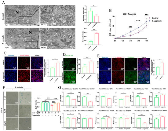
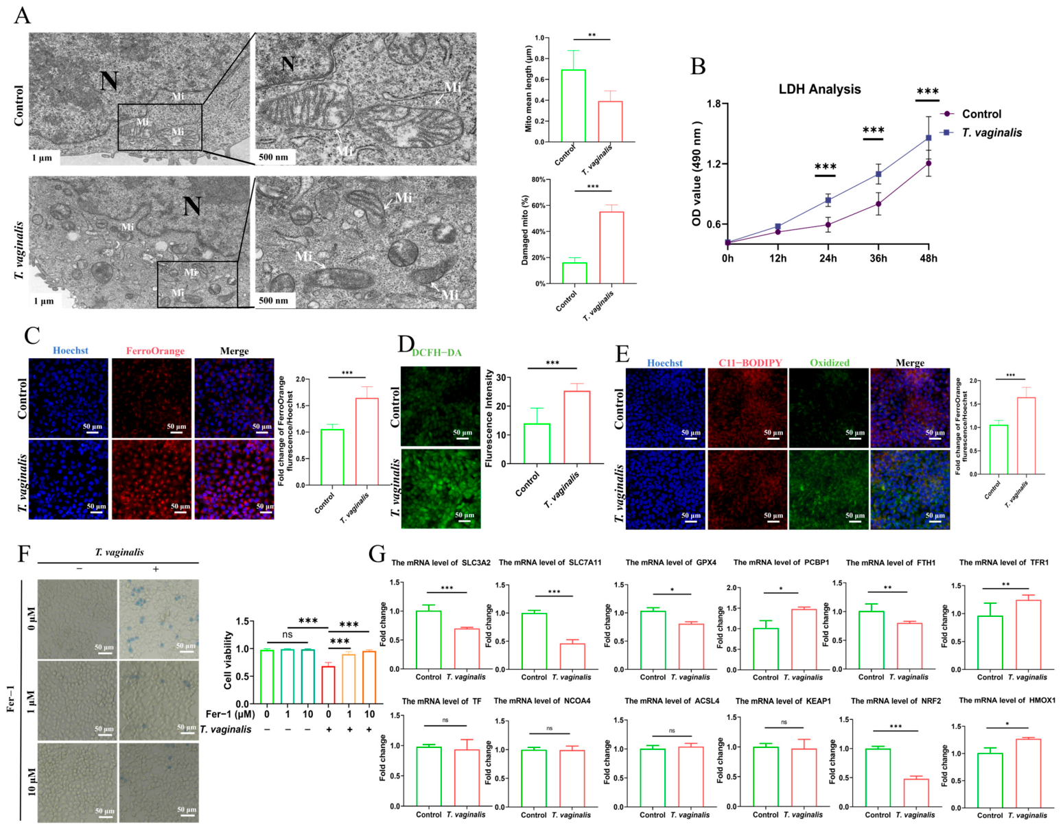
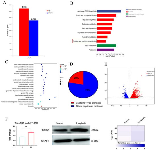
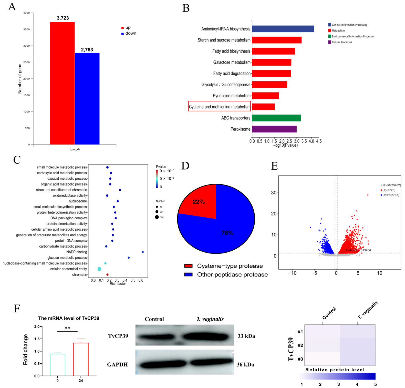
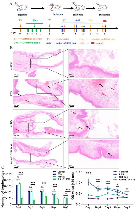
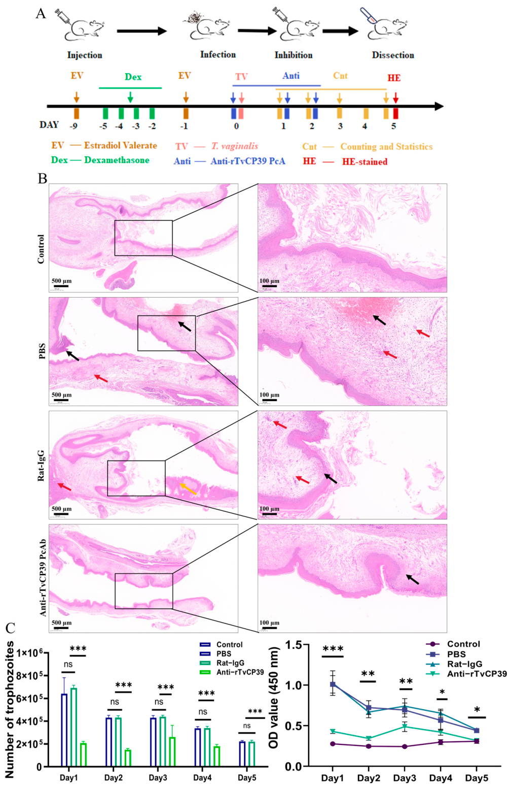
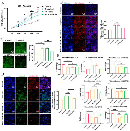
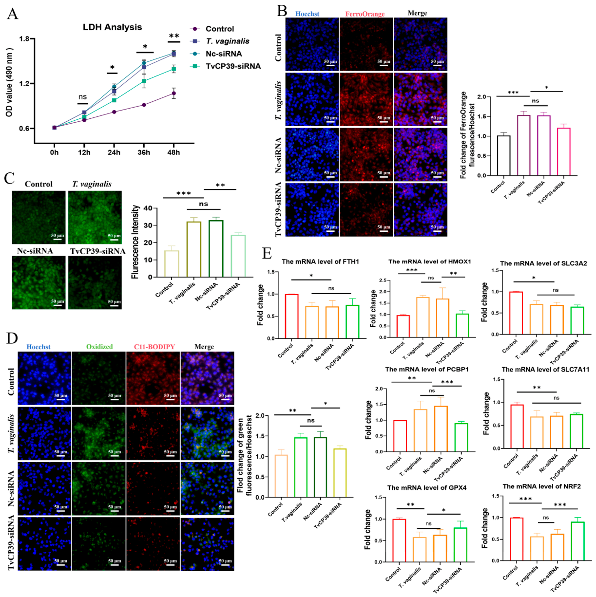
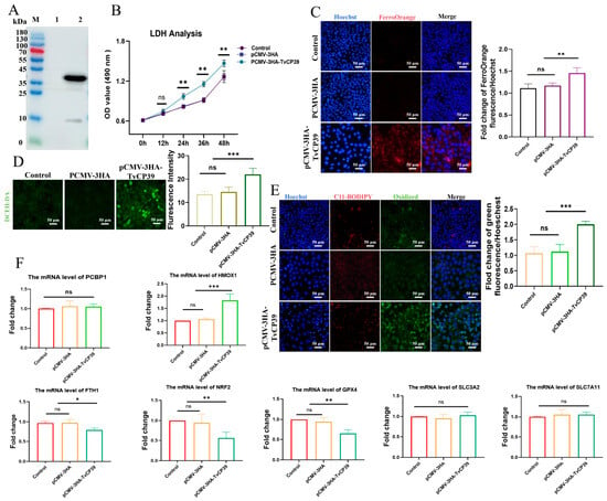
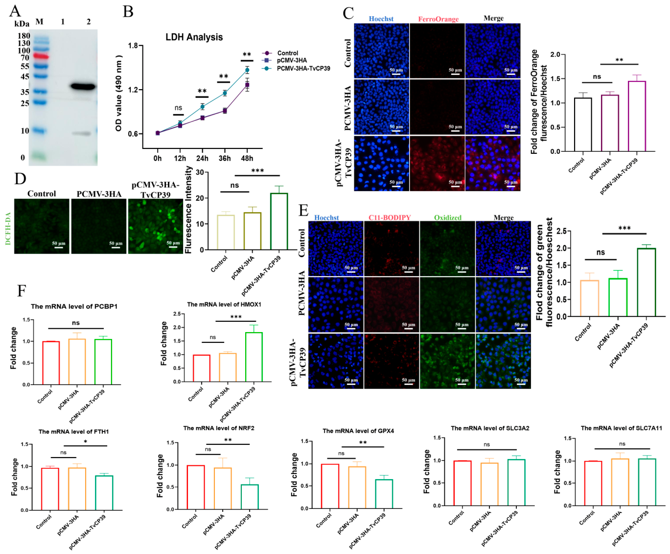
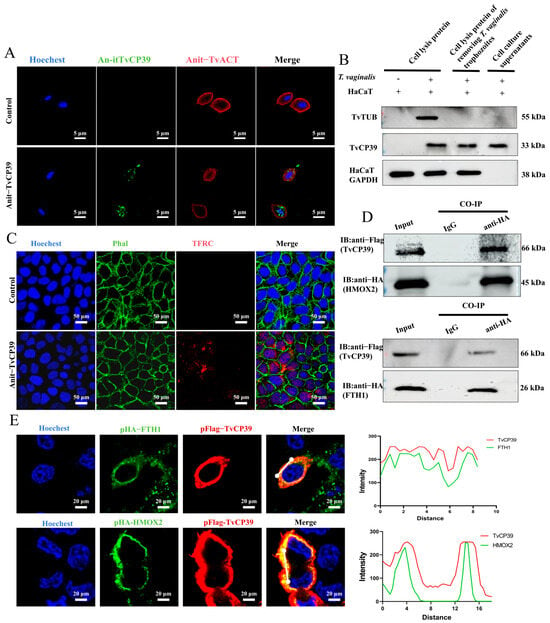
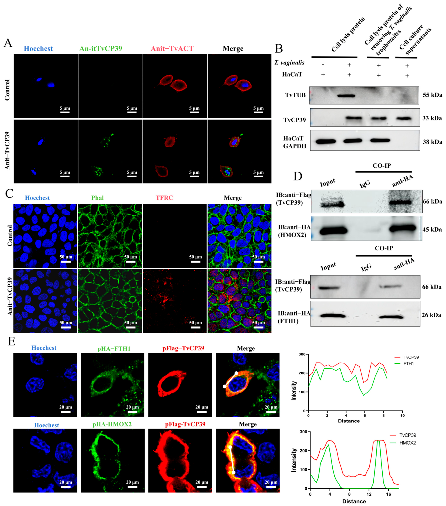
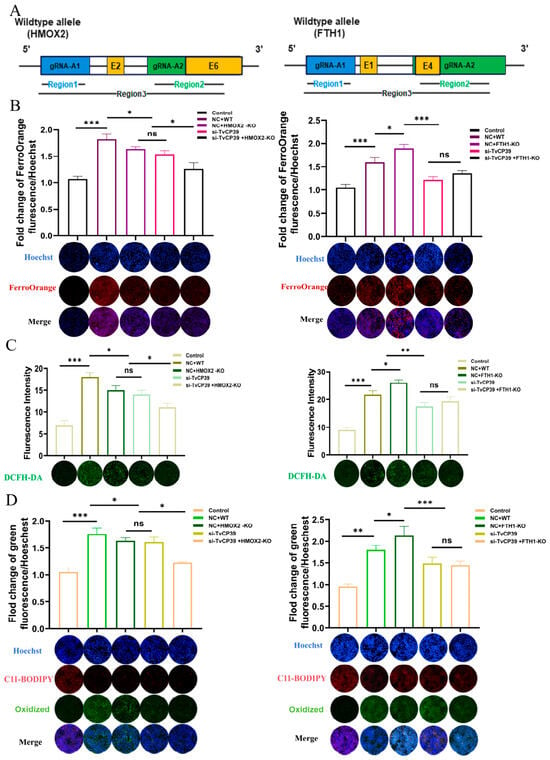
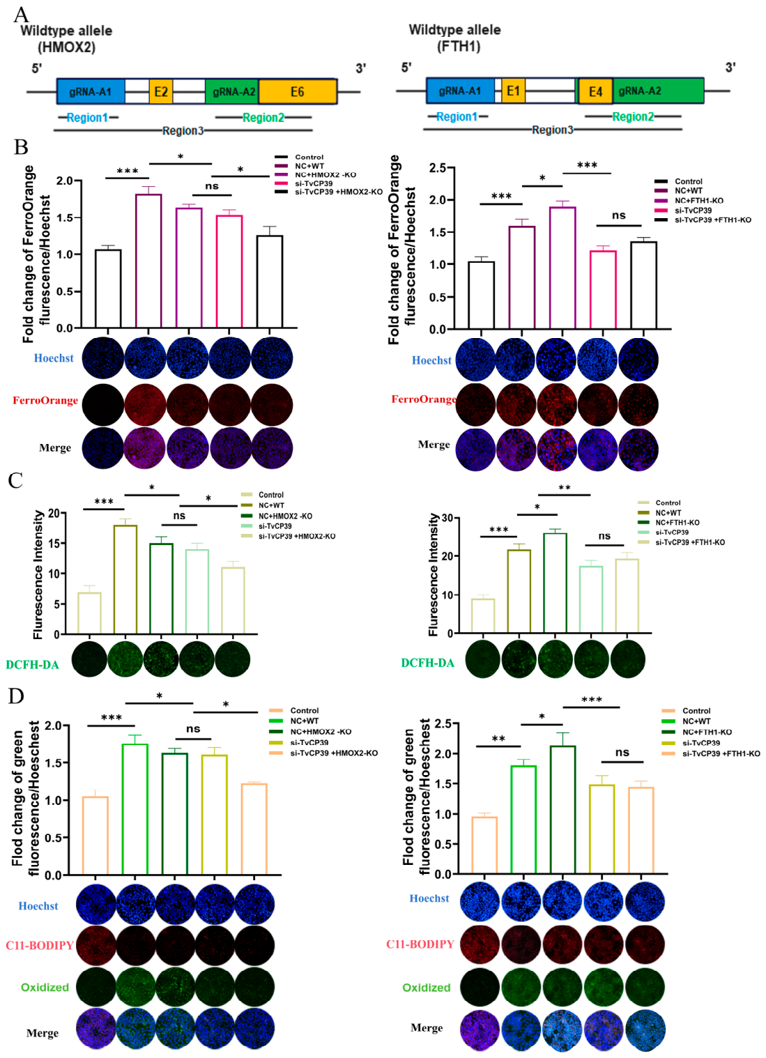
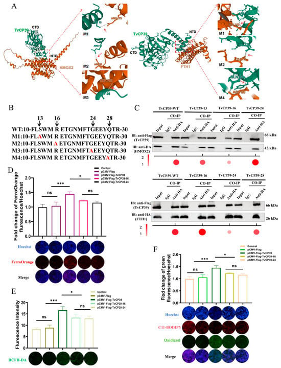
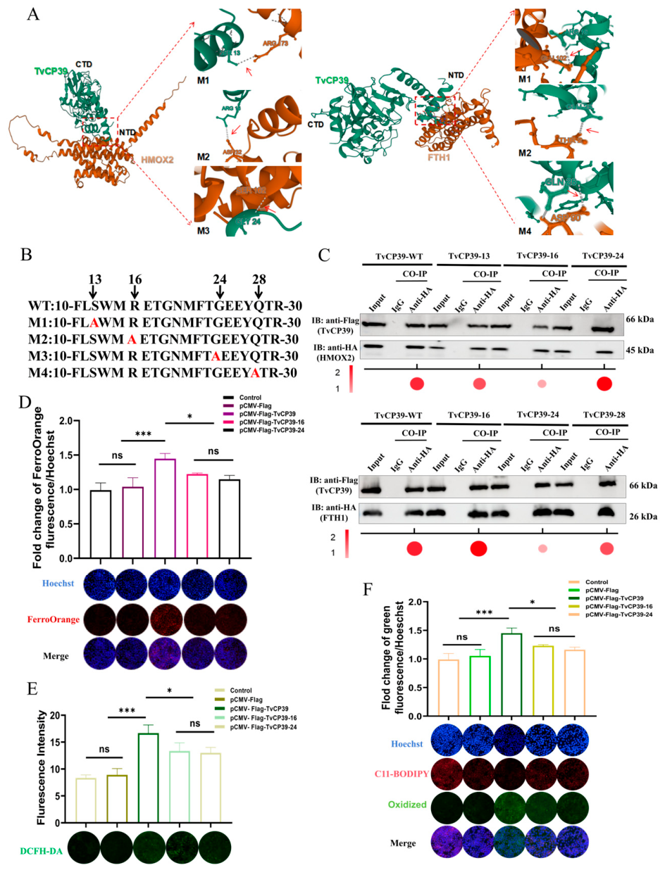
Disclaimer/Publisher’s Note: The statements, opinions and data contained in all publications are solely those of the individual author(s) and contributor(s) and not of MDPI and/or the editor(s). MDPI and/or the editor(s) disclaim responsibility for any injury to people or property resulting from any ideas, methods, instructions or products referred to in the content. |
© 2026 by the authors. Licensee MDPI, Basel, Switzerland. This article is an open access article distributed under the terms and conditions of the Creative Commons Attribution (CC BY) license.