


Abstract
RNAs are essential for gene transcription and protein translation. Yet they also play a crucial role in mediating DNA repair, including RNA-guided DNA base damage repair. However, it is unknown whether RNA integrity can alter DNA repair, modulating genome stability. To address this, we studied the effects of oxidative RNA damage 8-oxoG and an abasic site (AP site) on RNA-guided DNA synthesis and its fidelity by human DNA polymerases on an RNA-DNA hybrid and revealed the underlying molecular mechanisms. We showed that 8-oxoG on an RNA template inhibited pol β DNA synthesis and reduced the fidelity of pol β and pol η by promoting incorrect nucleotide and ribonucleotide incorporation in DNA. We found that an RNA AP site eliminated pol β DNA synthesis and promoted pol η incorporation of dA opposite the AP site. Using AlphaFold3-mediated molecular dynamics simulation, we found that the RNA 8-oxoG and AP site induced unique conformational changes in the RNA-DNA hybrid substrate to disrupt pol β and pol η substrate interaction, leading to RNA-guided nucleotide misincorporation. Our results provide the first insight into the new role of RNA integrity in modulating genome integrity through RNA-guided DNA repair on R-loops.
1. Introduction
RNAs are essential intermediates that transcribe genetic information and guide protein synthesis. RNAs can also serve as a template to guide DNA synthesis during retrotransposon replication, telomere elongation, and viral genome amplification [1,2,3,4]. However, a growing number of studies over the past decades have shown that RNA plays a more active role in maintaining genome stability and integrity than initially thought. An earlier study by Storici et al. has provided mechanistic insights into double-strand DNA break repair (DSBR) in yeast. The study has revealed that RNA can serve as a template for yeast DNA polymerases to mediate RNA-templated DSBR [5]. A later study has shown that nascent RNA can also mediate error-free DSBR on transcribed genes via classical non-homologous end-joining (C-NHEJ) in human cells [6]. Moreover, it has been found that human translesion DNA polymerase pol θ can perform RNA-templated DNA synthesis to mediate microhomology-mediated end-joining (MMEJ)-mediated DSBR in mouse induced pluripotent stem cells [7]. Chakraborty et al. have also demonstrated that human pol η can promote RNA-templated error-free DSBR through the transcription-coupled non-homologous end-joining (TC-NHEJ) pathway [8]. A recent study from our group has further identified a pathway by which RNA guides pol β and pol η to repair DNA base damage through a unique nick-translation mechanism [9]. All these findings support that RNA can play an active role in maintaining genome stability through various RNA-guided DNA repair pathways on RNA-DNA hybrids of R-loops.
However, similar to DNA, RNA molecules are frequently damaged by a variety of endogenous and exogenous genotoxicants. These include reactive oxygen species (ROS) [10], alkylating agent methyl methanesulfonate (MMS) [11], chemotherapeutic agents temozolomide and oxaliplatin, among others [12], and environmental genotoxicants, including UV [13,14], nitrosoureas [15,16], formaldehyde [17], and ethanol [18]. RNA damage includes UV-induced RNA pyrimidine dimers, 1N-methylA and 3N-methylC [16], 8-oxoG [17], 8-oxoA on RNA, and RNA cross-links, among others. RNA bases are more susceptible to a variety of damage than DNA bases due to the partially or fully single-stranded nature of RNA [19,20,21]. This is supported by the fact that 8-oxoG levels on RNA are about 14–25-fold higher than those on DNA [21], indicating that RNA can form nuclear and cytoplasmic hotspots of genotoxicity.
RNA damage can cause a variety of cellular dysfunctions that are associated with diseases such as Alzheimer’s and Parkinson’s diseases [22,23,24,25]. It has been shown that 8-oxoG on RNA can significantly reduce the production of proteins [26,27]. 8-oxoG on mRNA decreases the efficiency of peptide synthesis by four orders of magnitude [28]. However, 8-oxoG on the mRNA of amyloid precursor protein (APP) can also significantly increase the production of hydrophobic amyloid β (Aβ) peptides in mammalian cells [29], suggesting that RNA 8-oxoG can directly cause protein mutations by inducing the misincorporation of incorrect amino acids into the APP protein, promoting β-amyloid aggregation and progression of Alzheimer’s disease. Similar to oxidative RNA damage, alkylating RNA damage can reduce protein peptide bond formation [30]. Moreover, RNA damage can disrupt ribosome homeostasis by sequestering ribosomes in cells, causing translational stalling and stress and reducing protein translational efficiency [31]. In contrast, RNA damage is also applied as a strategy for anti-cancer and anti-viral therapy [12]. For example, the anti-cancer drug doxorubicin can intercalate into RNA strands, disrupting RNA structures and functions [32]. On the other hand, cisplatin can induce cross-links between ribosomes and mRNAs to inhibit protein synthesis [33].
It is generally accepted that damaged RNAs in the cytoplasm and their resulting functionally defective peptides can be processed by several cellular pathways. These include no-go decay (NGD) of mRNA degradation mediated by RNA endonucleases [34,35], degradation of nascent and truncated peptides by the proteasome, and ribosome rescue [36,37]. While these studies have provided insights into the cellular effects of RNA damage, they are limited to revealing only the effects of RNA damage in modulating the functions of cytoplasmic RNA and the mechanisms for processing damaged RNA in the cytoplasm. However, in the nucleus, RNAs are also susceptible to endogenous and environmental genotoxicants that can simultaneously damage DNA during gene transcription, where R-loops with RNA-DNA hybrids are constantly generated. This allows RNA to serve as the template for DNA repair polymerases during RNA-guided DNA repair [5,9]. Thus, it is conceivable that RNA damage may modulate RNA-guided/templated DNA repair and its fidelity, thereby altering genome stability and integrity. Revealing the effects of RNA damage on genome stability and their underlying mechanisms will not only allow us to understand the impact of RNA damage and other types of RNA modifications on genome stability, but more importantly, open a new avenue to elucidate the interactions between the transcriptome and genome and their roles in regulating cellular functions, disease progression, and therapy.
Because the most frequently occurring damage on RNA is oxidative RNA base damage, which mainly includes 8-oxoG [31] and RNA abasic site (AP site), we asked if oxidative RNA damage can modulate the efficiency and fidelity of RNA-guided DNA synthesis and repair on RNA-DNA hybrids of R-loops that are generated in the nucleus in cells. We hypothesize that 8-oxoGs and AP sites on RNA can alter the efficiency and fidelity of DNA repair polymerases during RNA-guided DNA base damage repair, thereby compromising genome integrity. We found that 8-oxoG or two adjacent 8-oxoGs on an RNA template opposite a DNA gap exhibited no effect or an inhibitory effect on pol β RNA-guided DNA synthesis, while they did not alter pol η DNA synthesis either. However, 8-oxoGs on RNA stimulated nucleotide misincorporation by both DNA polymerases. On the other hand, an AP site on RNA completely eliminated pol β DNA gap-filling synthesis activity, while it significantly reduced the DNA gap-filling synthesis activity of pol η. Using the AI tool AlphaFold3-based computational modeling and molecular dynamics simulations (MD), we found that 8-oxoG and an AP site on RNA resulted in the distortion of the RNA template and downstream DNA primer in the pol β and pol η-RNA-DNA hybrid complex, thereby disrupting the interaction of pol β and pol η with the RNA template and the enzyme catalysis. Our results demonstrate the crucial roles of RNA integrity in regulating RNA-guided DNA base damage repair on R-loops.
2. Materials and Methods
2.1. Materials
The oligonucleotides were synthesized by Integrated DNA Technology (IDT) Inc. (Coralville, IA, USA) and Eurofins Genomics (Louisville, KY, USA). RNA oligonucleotides containing 8-oxoG or AP sites were synthesized using the procedures shown in Section 2.2. Radionucleotide, γ-32P-ATP (3000 μCi/mmol), was purchased from Revvity (Boston, MA, USA). Micro Bio-Spin 6 chromatography columns were purchased from Bio-Rad Laboratories (Hercules, CA, USA). Pol β was expressed and purified as previously described [38]. Human pol η, pol ν, and pol θ were a generous gift provided by Wei Yang from the National Institute of Diabetes and Digestive and Kidney Diseases (NIDDK)/National Institutes of Health (NIH) as described previously [39]. Human pol λ, pol ι, and pol κ were purchased from ENZYMAX (Lexington, KY, USA). All other standard chemical reagents were purchased from ThermoFisher Scientific (Waltham, MA, USA) and Sigma-Aldrich (St. Louis, MO, USA).
2.2. Solid-Phase Synthesis and Characterization of Modified Oligonucleotides
Oligonucleotides were synthesized on a 394 ABI DNA synthesizer using CPG supports and 2′-O-TBDMS-phosphoramidites of U, A, C, and G, which were purchased from Glen Research (Sterling, VA, USA) or ChemGenes Corporation (Wilmington, MA, USA), and those for 8-oxoG and 1,2-dideoxyribofuranose, an RNA AP site analog, were prepared in-house [40,41]. Universal supports (Glen Unysupport, Glen Research, Sterling, VA, USA) were also used in some instances. 5-ethylthio-1H-tetrazole (0.25 M) in acetonitrile was used as the coupling reagent; 3% trichloroacetic acid in dichloromethane was used for detritylation; a 2,6-lutidine/acetic anhydride solution was used for capping; and an iodine (0.02 M) in tetrahydrofuran (THF)/pyridine/water solution was used in the oxidation step (purchased from Glen Research). Coupling times of 10 min or 45 s were used in the syntheses of RNA or DNA, respectively. Oligonucleotides were deacetylated/debenzoylated/deformylated and cleaved from the CPG support in the presence of 1:1 aq. methylamine (40%) and aq. ammonia (28–30%) with applied heat (60 °C, 1.5 h). A mixture of N-methylpyrrolidinone/triethylamine/trihydrofluoride•triethylamine (3:1:2) was used for deprotection of the silyl groups (60 °C, 1.5 h), followed by purification via gel electrophoresis (20% denaturing PAGE). C18-Sep-Pak cartridges were used to desalt the oligonucleotide solutions, with 5 mM NH4OAc as the ion exchange buffer, followed by concentration under reduced pressure. Oligonucleotides were dissolved in H2O. All oligonucleotide solutions were quantified via UV-vis and used without further purification.
2.3. Characterization and Quantification of RNA Oligonucleotides Using MALDI-TOF and UV-Vis Spectroscopy
Samples for mass spectrometry of all RNA oligonucleotides with or without RNA damage were prepared using 3-hydroxypicolinic acid as the matrix. The concentrations of all RNA oligonucleotides were obtained by measuring their UV-vis spectra at 60–90 °C on a Perkin Elmer Lambda 365 UV-Vis spectrophotometer (PerkinElmer, Shelton, CO, USA) using quartz cuvettes with a 1 cm path length. All synthesized RNA oligonucleotides were further characterized and validated using MALDI-TOF (See the Supplementary Figure S1).
2.4. Construction of RNA-DNA Hybrid Substrates
To test RNA-templated DNA gap-filling synthesis and fidelity by different DNA polymerases, RNA-templated DNA substrates containing 1 nucleotide (nt) and 2 nt gaps were constructed by annealing varying lengths of the 32P-labeled upstream DNA primer and downstream DNA primer containing a 5′-phosphate with the 26 nt RNA template containing either one 8-oxoG or two 8-oxoGs [(8-oxoG)2] opposite the DNA gaps and their DNA-templated control substrates (Supplementary Table S1). Substrates with 1 nt gaps at various locations relative to 8-oxoG on RNA were constructed and annealed, making substrates with 1 nt or 2 nt gaps directly opposite, 1 nt, and 4 nt away from the RNA damage. All substrates were assembled by annealing the upstream and downstream primers with the template strands at a molar ratio of 1:3:3. All the upstream DNA primers were radiolabeled at the 5′-end using 32P-ATP (3000 µCi/mmol) radionucleotide purchased from PerkinElmer Inc. (Boston, MA, USA). The downstream DNA primer was phosphorylated at the 5′-end using 1 mM cold ATP.
2.5. Determination of RNA-Guided DNA Synthesis Activity and Fidelity of Pol β and Pol η and Ribonucleotide Incorporation by Pol η
RNA-guided DNA synthesis activities by pol β and pol η were measured by incubating the substrates (50 nM) containing DNA-RNA, DNA-RNA-8-oxoG, or DNA-RNA-AP-site, respectively, with increasing enzyme concentrations in the presence of 50 μM dNTPs or NTPs at 37 °C for 60 min in a 10 µL reaction mixture in 1× base excision repair (BER) reaction buffer with 50 mM Tris-HCl, pH 7.5, 50 mM KCl, 5 mM Mg2+, 0.1 mM EDTA, 0.1 mg/ml bovine serum albumin, and 0.01% Nonidet NP-40. The fidelity of pol β and pol η was determined by incubating 50 nM substrates with increased enzyme concentrations in the presence of 50 μM dCTP, dGTP, dATP, and dTTP or 50 μM CTP, GTP, ATP, and UTP, respectively, at 37 °C for 30 min or 60 min in a 10 µL reaction mixture in 1× BER reaction buffer. The reactions were stopped using 2× stopping buffer containing 95% deionized formamide, 10 mM EDTA, 0.05% (w/v) bromophenol blue (Sigma-Aldrich, St. Louis, MO, USA), and 0.05% (w/v) xylene cyanol (Sigma-Aldrich, St. Louis, MO, USA) and subjected to denaturation at 95 °C for 5 min. Substrates and products were separated by 15% urea-denaturing polyacrylamide gel and detected using the Pharos FX Plus PhosphorImager (Bio-Rad Laboratories, Hercules, CA, USA). All experiments were independently repeated at least three times.
2.6. Steady-State Kinetics of RNA-Guided DNA Synthesis by Pol β and Pol η
The steady-state kinetics experiments of pol β and pol η DNA synthesis were performed using a fixed concentration of the DNA polymerases and substrates, with various concentrations of individual dNTPs at 5–500 μM for the 1 nt gap substrates (50 nM or 100 nM). The enzymes were incubated with the substrates at either 25 °C or 37 °C at different time intervals (0 to 30 min) in a reaction mixture (10 μL) containing 1× BER buffer. The reactions were stopped with 2× stopping buffer, followed by incubation at 95 °C for 5 min. Substrates and products were separated by a 15% urea-denaturing polyacrylamide gel and detected using the Pharos FX Plus PhosphorImager (Bio-Rad Laboratories, Hercules, CA, USA). The apparent Michaelis–Menten constants, Vmax, Km, and kcat were calculated using the Enzyme Kinetics Module of Prism-GraphPad software, version 9.0.2.
2.7. AlphaFold3-Based Molecular Dynamics Simulations for Pol β and Pol η Interactions with RNA-Templated DNA Gap Substrates
The molecular models of the complexes of the RNA-templated 1 nt DNA gap substrate with human pol β and pol η in the presence of different incoming dNTPs were constructed using the AI tool, AlphaFold3 version 3.0.1 and 3.0.4 with the full-length amino acid sequence of pol β and amino acid residues 1–432 of pol η. The molecular models included pol β-RNA-DNA hybrid substrate complexes containing G or 8-oxoG or an AP site on the RNA template opposite the 1 nt DNA gap in the presence of dCTP or dATP. The models for pol η-RNA-templated DNA gap substrate complexes included those with a G or 8-oxoG or AP site opposite the 1 nt DNA gap in the presence of dCTP and dATP. The substrates for the molecular modeling and simulations were listed in Supplemental Table S1 (Supplemental Table S1 red). The 8-oxoG and AP site on guanine 15 of the RNA template were created by adding an oxygen atom at C’8 or deleting the base of the nucleotide using Chimera X version 1.11. The AlphaFold3-generated models were then aligned to the original crystal structures of pol β and pol η, refined, and finalized as the models for conducting the molecular dynamics simulation experiments. CHARMM-GUI solution builder version 3.8 was used to create a solvated box for the pol β and pol η-RNA-DNA hybrid substrate complexes, with water molecules and various ions surrounding the protein substrate complexes at 310.15 K, 0.15 M KCl, and 0.05 M MgCl2. A rectangular water box was created with an edge distance of 10.0 Å. The parameters and rtf files for the altered atoms and residues were also generated with CGenEF. The 8-oxoG and AP site on the RNA template were generated from the 8-oxodG and AP site in the DNA substrates by editing the corresponding DNA parameters and rtf files. The molecular dynamics simulations were conducted with NAMD version 3.0 [42] using the CHARMM36m force field [43], with covalent bonds from hydrogen atoms constrained by the SHAKEH algorithm [44], and long-range electrostatic interactions calculated with the Particle Mesh Ewald (PME) method [45]. The Langevin thermostat was used for maintaining the temperature of the system, whereas the Nose–Hoover Langevin piston with a piston decay time of 25 fs and a damping coefficient of 1 ps−1 was used [46] for handling the pressure. After the initial 10,000-step energy minimization and 125 ps equilibration, production runs of 500 ns were performed with a 2fs time step at a constant temperature of 303.15 K. The results of the simulation experiments were analyzed using the software VMD version 2.0. The occupancy percentages of different amino acid residues in pol β and pol η were determined to indicate their hydrogen-bonding interactions with the incoming nucleotide, the RNA template, and the upstream and downstream DNA primers. All molecular dynamics simulation experiments were repeated 2–3 times.
3. Results
3.1. RNA Base Damage Modulates RNA-Guided DNA Gap-Filling Synthesis by Pol β and Pol η
Studies from our and other groups have shown that DNA polymerases can use RNA as a template to synthesize DNA and repair DNA damage on an RNA-DNA hybrid [5,8,9]. It has also been shown that oxidative base damage on a DNA template alters DNA synthesis activity during DNA replication and repair, causing GC-AT transversion mutations [47,48,49,50]. Thus, it is possible that oxidative RNA damage, such as 8-oxoGs and AP sites, can alter RNA-guided DNA synthesis and repair. To test this, we initially examined the effects of 8-oxoG and an AP site on an RNA template on RNA-guided DNA synthesis by repair and translesion DNA polymerases, including pol β, pol λ, pol ι, pol κ, pol η, pol ν, and pol θ (Figure 1). The results showed that in the presence of RNA 8-oxoG on the RNA-templated 1 nt DNA gap substrate, 25 nM pol β performed comparable 1 nt gap-filling synthesis activity (Figure 1, lane 2) with its activity on the RNA-templated substrate without RNA damage (Figure 1, lane 18), whereas 25 nM pol λ only exhibited weak 1 nt gap-filling synthesis activity (Figure 1, lane 3) that was similar to the undamaged substrate (Figure 1, lane 19). The translesion DNA polymerases pol ι, pol κ, and pol ν failed to insert any nucleotides to fill in the gap opposite to 8-oxoG on the RNA template (Figure 1, lanes 4, 5, 7) as they did on the undamaged substrate (Figure 1, lanes 20–21), except that pol ν exhibited efficient 1 nt gap-filling synthesis on the substrate with the undamaged RNA template (Figure 1, lane 23). On the other hand, pol η showed robust RNA-guided DNA gap-filling and strand-displacement synthesis activities (Figure 1, lane 6) as it did on the undamaged substrate (Figure 1, lane 22). Pol θ only exhibited weak 1 nt gap-filling synthesis on the substrate containing 8-oxoG on the RNA template (Figure 1, lane 8), as compared to its efficient gap-filling and strand-displacement synthesis activities on the undamaged substrate (Figure 1, lane 24). On the 1 nt gap substrate containing an RNA AP site, pol β, pol λ, pol ι, pol κ, and pol ν all failed to insert any nucleotides opposite the 1 nt DNA gap (Figure 1, lanes 10–13 and 15). Pol η and pol θ only inserted 1 to 3 nt (Figure 1, lanes 14 and 16). The results indicated that a single 8-oxoG on RNA did not alter the RNA-guided DNA synthesis activity of pol β, pol λ, pol η, and pol θ. In contrast, an AP site on the RNA template significantly inhibited RNA-guided DNA synthesis by DNA polymerases, resulting in the accumulation of DNA gaps and strand breaks on an RNA-DNA hybrid.
Figure 1.
RNA-guided DNA synthesis on a 1 nt gap substrate with 8-oxoG or an AP site by DNA polymerases. RNA-guided DNA synthesis on a 1 nt gap substrate (50 nM) without or with 8-oxoG or an AP site on the RNA template was determined in the presence of 25 nM pol β, pol λ, pol ι, pol κ, pol η, pol ν, and pol θ, 50 μM dNTPs, and 5 mM Mg2+. The reactions were incubated at 37 °C for 30 min. Lanes 2–8 represent DNA synthesis on the substrate with RNA 8-oxoG, and lanes 10–16 represent DNA synthesis on the substrate with an RNA AP site. Lanes 18–24 represent DNA synthesis on an undamaged RNA substrate. Lanes 1, 9, and 17 represent substrate only. The synthesized DNA products are indicated by red circles with their sizes. All experiments were performed at least in triplicate. Uncropped original gel images can be found in the Supplementary Materials.
We further determined the frequency and location effects of 8-oxoG on RNA on the RNA-guided DNA synthesis activity by pol β and pol η with the RNA-templated 1 nt or 2 nt DNA gap substrates containing single or two RNA 8-oxoGs opposite to the DNA gap (Figure 2A,D), or 1 nt or 4 nt away from the DNA gap (Figure 2B–E). The results showed that 0.5–50 nM pol β efficiently incorporated 1 nt opposite the gap with one 8-oxoG (Figure 2A–C), while its DNA synthesis was slightly decreased when the damage was located 1 nt or 4 nt away from the DNA gap (Figure 2B,C and lanes 2–7) compared with the activity on an undamaged substrate (Figure 2B,C, lanes 9–14). However, on the substrate containing two RNA 8-oxoGs opposite a 2 nt DNA gap, pol β at 0.5–50 nM predominantly inserted 1 nt (Figure 2D, lanes 2–7) with inefficient 2 nt insertion activity at 5–20 nM (Figure 2D, lanes 5–7) as compared with the undamaged substrate (Figure 2D, lanes 9–14). Interestingly, pol β 1 nt gap-filling synthesis activity opposite 8-oxoG was also decreased in the presence of an adjacent 8-oxoG on the RNA template (Figure 2E, lanes 2–7). Pol β 1 nt insertion activity with two 8-oxoGs located at 1 nt or 4 nt away from the DNA gap was restored to its activity on the undamaged substrate (Figure 2E, compare lanes 9–14, lanes 16–21 with lanes 9–14 of Figure 2D). The results indicated that multiple adjacent 8-oxoGs on the RNA template significantly inhibited pol β gap-filling synthesis activity, constraining pol β to predominantly insert 1 nt opposite 8-oxoGs (Figure 2D, lanes 2–7). Consequently, this can cause the accumulation of unrepaired 1 nt gaps and single-strand breaks on the RNA-templated DNA substrate. Our results further indicated that the locations of 8-oxoGs on the RNA template also modulated pol β gap-filling DNA synthesis activity (Figure 2E).
Figure 2.
Pol β RNA-guided DNA synthesis with gap substrates with 8-oxoG at different locations. Pol β RNA-guided DNA synthesis was determined using the RNA-templated 1 nt or 2 nt DNA gap substrates (50 nM) with one or two 8-oxoGs or no damage on the RNA template. The substrates were incubated with 0.5–50 nM pol β, 50 μM dNTPs, and 5 mM Mg2+ at 37 °C for 60 min. (A) represents the reactions with the substrate containing RNA 8-oxoG opposite a 1 nt gap and an undamaged substrate. (B) represents the reactions with the substrate containing RNA 8-oxoG 1 nt away from the gap and the corresponding undamaged substrate. (C) indicates the reactions with the substrate containing RNA 8-oxoG 4 nt away from the gap. (D) indicates the reactions from the substrate containing two adjacent 8-oxoGs opposite to a 2 nt gap and a non-RNA damage substrate. For (A,D), lanes 2–7 represent pol β DNA synthesis on damaged substrate. Lanes 9–14 represent pol β DNA synthesis on a non-RNA damage substrate. (E) represents the reactions from the substrate with two adjacent 8-oxoGs opposite the 1 nt gap (lanes 2–7), the reactions with two adjacent 8-oxoGs 1 nt away from the 1 nt gap (lanes 9–14), and two adjacent 8-oxoGs 4 nt away from the 1 nt gap (lanes 16–21). Lanes 1, 8, and 15 represent the substrate only. The products are indicated by red circles with their sizes. All experiments were performed at least in triplicate as represented by the error bars in the graphs. Uncropped original gel images can be found in the Supplementary Materials.
Similar to pol β, 0.1–50 nM pol η performed efficient gap-filling and strand-displacement DNA synthesis on the 1 nt DNA gap substrate with 8-oxoG at different locations on the RNA template relative to the DNA gap (Figure 3A–C, lanes 2–8) as it did on the undamaged substrate (Figure 3A–C, lanes 10–16). For the substrate containing two 8-oxoGs at different locations on the RNA template, 0.5–50 nM pol η exhibited efficient gap-filling and strand-displacement synthesis activity (Figure 3D,E, lanes 2–8). The DNA polymerase efficiently inserted 1–16 nt (the full-length DNA synthesis product), 1–14 nt, and 1–11 nt across the substrate (Figure 3D,E, lanes 5–8). On the substrate with the undamaged RNA template, pol η at 1–50 nM exhibited efficient DNA synthesis activity (Figure 3D, lanes 12–16). The same concentrations of pol η exhibited distributive DNA synthesis on the substrate containing 8-oxoGs (Figure 3D,E, lanes 4–8, 3E, lanes 12–16 and lanes 18–24). The results indicated that 8-oxoG on the RNA template had little effect on pol η DNA gap-filling and strand-displacement synthesis activities.
Figure 3.
RNA-guided DNA synthesis with gap substrates without or with RNA 8-oxoG at different locations by pol η. RNA-guided DNA synthesis by pol η (0.1–50 nM) was determined using the substrates (50 nM) without or with RNA 8-oxoG, 50 μM dNTPs, and 5 mM Mg2+ at 37 °C for 60 min. (A) represents the reactions with the substrate without or with RNA 8-oxoG opposite a 1 nt gap. (B,C) indicate the reactions with the substrate without or with RNA 8-oxoG 1 nt or 4 nt away from the 1 nt gap. (D) represents the reactions with the substrates without or with two adjacent RNA 8-oxoGs opposite the 2 nt and 1 nt gap. For (A–D), lanes 2–8 represent pol η DNA synthesis on damaged substrate. Lanes 10–16 represent pol η DNA synthesis on the non-RNA damage substrate. (E) represents the reactions from the substrate with two adjacent 8-oxoGs opposite the 1 nt gap (lanes 2–8), the reactions with two adjacent 8-oxoGs 1 nt away from the 1 nt gap (lanes 10–16), and two adjacent 8-oxoGs 4 nt away from the 1 nt gap (lanes 18–24). Lanes 1, 9, and 17 represent the substrate only. The synthesized DNA products are indicated by red circles with their sizes. All experiments were performed at least in triplicate as represented by the error bars in the graphs. Uncropped original gel images can be found in the Supplementary Materials.
3.2. RNA Damage Facilitates RNA-Guided Nucleotide Misincorporation by Pol β and Pol η
We then asked if RNA damage can alter the fidelity of RNA-guided DNA synthesis by pol β and pol η. We tested this by determining pol β correct and incorrect nucleotide insertion on RNA-templated 1 nt or 2 nt gap substrate containing one or two 8-oxoGs (Figure 4). We found that 5–25 nM pol β incorporated dC and dA (Figure 4A, lanes 2–4 and 6–8) opposite 8-oxoG with reduced activity compared with the undamaged substrate (Figure 4A, lanes 10–12). The quantitative analysis further demonstrated that with the presence of 8-oxoG on the RNA template, 10–25 nM pol β generated a significantly higher percentage of 8-oxoG:dA misincorporation product than that for correct nucleotide insertion (Figure 4A, bottom left). In contrast, pol β exhibited a much lower percentage of the correct nucleotide G:dC insertion product in the presence of RNA 8-oxoG compared with the substrate without RNA damage (Figure 4A, bottom right), indicating that RNA 8-oxoG promoted pol β nucleotide misincorporation and inhibited its gap-filling synthesis.
Figure 4.
The fidelity of pol β RNA-guided DNA synthesis with gap substrates without or with RNA 8-oxoG. (A) indicates the correct and incorrect nucleotide incorporation of pol β RNA-guided DNA synthesis on the 1 nt gap substrate (50 nM) with or without RNA 8-oxoG. The bottom left and right panels represent the comparison between pol β correct and incorrect nucleotide insertion on the 1 nt gap substrate as well as the comparison of pol β correct nucleotide insertion with or without RNA 8-oxoG on the substrate. (B) represents pol β correct and incorrect nucleotide insertion on the 2 nt gap substrate with or without two RNA 8-oxoGs. The bottom left and right panels indicate the comparison between pol β correct and incorrect nucleotide insertion on the 2 nt gap substrate as well as the comparison of pol β correct nucleotide insertion with or without RNA 8-oxoG on the substrate. The reactions were performed in the presence of 5–25 nM pol β, 50 μM dNTPs, and 5 mM Mg2+ at 37 °C for 60 min. Lanes 2–4 represent pol β dCTP incorporation opposite RNA 8-oxoG. Lanes 6–8 represent pol β dATP opposite RNA 8-oxoG. Lanes 10–12 represent pol β dCTP incorporation opposite a DNA gap. Lanes 1, 5, and 9 represent the substrate only. The synthesized DNA products are indicated by red circles with their sizes. All experiments were performed at least three times, as represented by the error bars in the graphs. Uncropped original gel images can be found in the Supplementary Materials.
For the substrate containing two 8-oxoGs and a 2 nt gap, 10–25 nM pol β only incorporated one dC or dA (Figure 4B, lanes 2–4 and 6–8), leaving a 1 nt DNA gap. On the other hand, 10–25 nM pol β predominantly performed 2 nt gap-filling synthesis activity on the undamaged substrate (Figure 4B, lanes 10–12). Similar to the substrate with a single RNA 8-oxoG (Figure 4A), pol β generated a higher percentage of 8-oxoG:dA mismatch product than that of correct nucleotide insertion product on the damaged RNA template (Figure 4B, bottom left), leading to a much lower percentage of correct nucleotide insertion product than the substrate without RNA damage (Figure 4B, bottom right). The results indicated that 8-oxoG on the RNA template compromised the efficiency and fidelity of pol β RNA-guided DNA synthesis. This was further supported by the results of the steady-state enzyme kinetics of pol β (Table 1), showing that pol β generated 8-oxoG:dA mismatch product at a catalytic efficiency of 0.13 × 10⁵ M−1 s−1, which was ~4.3-fold of that for correct basepair of dC:8-oxoG at 0.03 × 10⁵ M−1 s−1 (Table 1). This indicated that 8-oxoG on the RNA template reduced the fidelity of pol β gap-filling DNA synthesis by 80% (Table 1). Although pol β generated dA:8-oxoG mismatch product with a 34-fold lower efficiency than the correct incorporation dC:G (0.13 × 10⁵ M−1 s−1 vs. 4.4 × 10⁵ M−1 s−1) (Table 1), 8-oxoG on the RNA template still induced nucleotide misincorporation with significant efficiency. These results indicated that 8-oxoG on RNA inhibited pol β gap-filling synthesis and promoted nucleotide mismatches, thereby reducing the efficiency and fidelity during RNA-guided DNA base damage repair.
Table 1.
Steady-state kinetics of pol β RNA-guided DNA synthesis with 8-oxoG on the RNA template.
In contrast, pol η at 5–25 nM generated the 8-oxoG:dG, 8-oxoG:dA, and 8-oxoG:dT mismatch products independent of 8-oxoG on the RNA template (Figure 5A,B, lanes 6–8, 10–12, and 14–16). However, 8-oxoG on the RNA template led to more multi-nucleotide mismatch products than a normal G on the template (compare lanes 10–12 in Figure 5A with lanes 10–12 in Figure 5B). For the substrate containing two 8-oxoGs on the RNA template with a 2 nt DNA gap, pol η also predominantly performed 1 nt gap-filling synthesis in the presence of all other dNTPs (Figure 5C,D, lanes 2–4, 6–8, and 14–16), except that in the presence of dATP, it extended 8-oxoG:dA to perform misbasepair-mediated strand-displacement synthesis activity (Figure 5C, lanes 10–12). The results demonstrated that 8-oxoGs on RNA stimulated pol η multi-nucleotide misincorporation, reducing the fidelity of pol η RNA-guided DNA synthesis. The steady-state kinetics results further demonstrated that pol η predominantly incorporated dCTP to basepair with 8-oxoG on the RNA template at a catalytic efficiency of 241 × 105 M−1 s−1 (Table 2), which was comparable with that for dC:G basepair at 439 × 105 M−1 s−1 (Table 2). This further increased the frequency of pol η nucleotide misincorporation and decreased pol η fidelity by 3.6-fold as compared with pol η DNA synthesis fidelity (Table 2). The results indicated that 8-oxoG on RNA significantly promoted pol η nucleotide misincorporation and infidelity of its DNA synthesis. Since pol η performed RNA-guided DNA gap-filling and strand-displacement synthesis activities, it is possible that 8-oxoG on RNA can promote pol η-mediated multiple nucleotide mismatches, leading to transition and transversion DNA mutations.
Figure 5.
The fidelity of pol η RNA-guided DNA synthesis on DNA gap substrates with or without RNA 8-oxoG. (A,B) indicate pol η RNA-guided DNA synthesis on the 1 nt gap substrates (50 nM) with or without RNA 8-oxoG. (C,D) represent pol η RNA-guided DNA synthesis on the 2 nt gap substrates (50 nM) with two RNA 8-oxoGs or without 8-oxoG. Pol η DNA synthesis was determined in the presence of 5–25 nM of the enzyme, 50 μM dNTPs, and 5 mM Mg2+ at 37 °C for 60 min. For all the substrates, lanes 2–4, 6–8, 10–12, and 14–16 represent pol η incorporation of dC, dA, dG, and dT, respectively, and lanes 1, 5, 9, and 13 represent substrate only. The synthesized DNA products and their sizes are indicated. All experiments were performed at least three times, as represented by the error bars in the graphs. Uncropped original gel images can be found in the Supplementary Materials.
Table 2.
Steady-state kinetics of pol η RNA-guided DNA synthesis with 8-oxoG on the RNA template.
3.3. RNA Damage Facilitates RNA-Guided Ribonucleotide Incorporation by Pol η
Since genome-embedded ribonucleotides are also one of the frequently occurring non-canonical nucleotides in genomic DNA [51,52], and several DNA polymerases can incorporate ribonucleotides into the genome during DNA replication [53,54,55,56], and cellular concentrations of rNTPs are much higher than dNTPs [53,57], we then asked if RNA base damage can also facilitate the incorporation of ribonucleotides through RNA-guided DNA synthesis. To test this, we examined the effects of 8-oxoG on the RNA template on ribonucleotide incorporation into DNA during RNA-guided DNA synthesis by pol β and pol η. We found that pol β failed to incorporate ribonucleotides into DNA. Pol η at 0.5–50 nM exhibited comparable DNA rNTP incorporation activity with or without 8-oxoG on the RNA template (Figure 6A–C, lanes 2–7). Also, 10–50 nM pol η generated comparable 8-oxoG:rC product with G:rC (compare lanes 9–11 in Figure 6B with lanes 9–11 in Figure 6D). Moreover, the same concentrations of pol η only generated a small amount of 8-oxoG:rA mismatch product (Figure 6B, lanes 13–15). The results indicated that 8-oxoG on the RNA template led to efficient rC incorporation opposite RNA 8-oxoG into DNA with a small amount of 8-oxoG:rA mismatch, causing ribonucleotide accumulation in DNA via RNA-guided DNA repair.
Figure 6.
RNA-guided ribonucleotide incorporation into DNA by pol η and its fidelity on a 1 nt gap substrate with or without 8-oxoG. RNA-guided DNA ribonucleotide incorporation by pol η on a 1 nt gap substrate (50 nM) and its fidelity were determined in the presence of 0.5–50 nM pol η, 50 μM NTPs, and 5 mM Mg2+ at 37 °C for 60 min. (A,B) indicate pol η ribonucleotide incorporation into DNA with RNA 8-oxoG. (C,D) indicate pol η ribonucleotide incorporation into DNA on the substrate without RNA damage. Lanes 2–7 indicate pol η incorporation of all ribonucleotides into DNA. Lanes 9–11 and 13–15 represent pol η incorporation of CTP or ATP opposite RNA 8-oxoG or an undamaged G. Lanes 1, 8, and 12 represent the substrate only. All experiments were repeated at least three times, as represented by the error bars in the graphs. Uncropped original gel images can be found in the Supplementary Materials.
3.4. An AP Lesion on an RNA Template Inhibits RNA-Guided Nucleotide Incorporation by Pol η and Modulates Its Fidelity
Since RNA AP sites frequently accumulate on RNA-DNA hybrids, we then determined whether an AP site on the RNA template can alter the efficiency and fidelity of RNA-guided DNA synthesis (Figure 7). The results indicated that pol η at 0.5–50 nM performed effective gap-filling and strand-displacement DNA synthesis activities on the 1 nt DNA gap substrate with an AP site on the RNA template (Figure 7A, lanes 2–7). Moreover, we found that 5–25 nM pol η incorporated all four different nucleotides opposite the AP site on the RNA template (Figure 7B, lanes 2–4, 6–8, 10–12, and 14–16), with the highest activity in incorporating dA (Figure 7B, lanes 10–12) and the lowest activity in inserting dT (Figure 7B, lanes 14–16). However, at the same concentrations, pol β showed no nucleotide incorporation opposite the AP site on the RNA template (Supplementary Figure S2A). The results indicated that an AP site on RNA eliminated pol β DNA synthesis and induced pol η incorrect nucleotide incorporation, leading to the accumulation of DNA strand breaks on RNA-DNA hybrids and potentially mutations through R-loops.
Figure 7.
Pol η RNA-guided DNA synthesis on a 1 nt gap opposite an RNA AP site. Pol η RNA-guided DNA synthesis on the substrate (50 nM) containing a 1 nt gap opposite an RNA AP site was examined in the presence of 0.5–50 nM pol η, 50 μM dNTPs, and 5 mM Mg2+ at 37 °C for 30 min. (A) and (B) indicate pol η nucleotide incorporation opposite an AP site in the presence of all four dNTPs and individual dNTPs. For panel (A), lanes 2–7 represent pol η DNA synthesis with an RNA AP site in the presence of all four dNTPs. Lane 1 indicates substrate only. For panel (B), lanes 2–4, 6–8, 10–12, and 14–16 indicate pol η DNA synthesis with the RNA AP site in the presence of dCTP, dGTP, dATP, or dTTP. Lanes 1, 5, 9, and 13 indicate the substrate only. All experiments were repeated at least three times, as represented by the error bars in the graphs. Uncropped original gel images can be found in the Supplementary Materials.
3.5. RNA Damage Alters the Structures of the RNA-DNA Hybrid Substrate and Pol β and Pol η Conformation to Modulate RNA-Guided DNA Synthesis
To further reveal the molecular basis underlying the effects of RNA damage on pol β and pol η DNA synthesis and its fidelity, we used molecular dynamics simulation (MD) to elucidate the interaction of pol β and pol η with the 1 nt DNA gap substrates containing an 8-oxoG and abasic site (AP site) on the RNA template (Figure 8 and Figure 9 and Supplementary Videos S1 and S2). The results indicated that in the pol β-substrate ternary complex, 8-oxoG on the RNA template induced a large-angle bending of the downstream primer in the presence of an incoming dCTP (Video S1A, the left panel) as compared with the little change in the downstream primer with a G on the RNA template (Video S1A, the right panel). The bending of the downstream primer appeared to be caused by the repulsion between 8-oxoG and dCTP (Video S1A, left panel). In contrast, 8-oxoG on the RNA template formed a stable interaction with the incoming dATP (Video S1B, compare the structure in the left panel with that in the right panel). Similarly, 8-oxoG on the RNA template led to the bending of the RNA-DNA hybrid substrate in the pol η-1 nt DNA gap complex in the presence of dCTP and dATP (Videos S2A,B, compare the structure in the left panel with that in the right panel). This resulted in the downstream primer folding towards the upstream primer, which was pushed by the finger domain of pol η (Videos S2A,B, left panel). In contrast, the AP site on the RNA template disrupted the helical structure of the upstream DNA-RNA hybrid of the substrate, shifting the upstream primer away from the downstream primer and resulting in basestacking between dATP and the last nucleotide of the upstream primer (Video S2C, compare the left panel with the right panel). The significant conformational change was caused by the insertion of the thumb domain into the minor groove of the upstream of the DNA-RNA hybrid substrate, along with the palm and little finger domains moving towards dATP and the RNA template (Video S2C, compare the left panel with the right panel).
Figure 8.
Molecular dynamics simulations of the ternary complexes of pol β with a 1 nt DNA gap substrate containing RNA 8-oxoG. The MD experiments for pol β-1 nt DNA gap RNA 8-oxoG-incoming dCTP or dATP were performed for 500 ns under the conditions described in Section 2. (A) shows the MD results of the ternary complex of pol β-1 nt DNA gap RNA 8-oxoG-dCTP. (B) demonstrates the hydrogen bond occupancy profiles of pol β amino acid residues that mediate the enzyme interaction with dCTP, 8-oxoG, or G on the RNA template, and the upstream primer. (C) illustrates the dynamic changes in the distance (Å) between the 3′-OH of the terminus nucleotide of the upstream primer and the α-phosphate of the incoming dCTP over time. (D) indicates the MD results of the ternary complex of pol β-1 nt gap DNA-RNA 8-oxoG-incoming dATP. (E) indicates the hydrogen bond occupancy profiles of pol β amino acid residues that mediate the enzyme interaction with dATP, 8-oxoG, or G on the RNA template, and the upstream primer. (F) illustrates the dynamic changes in the distance between the 3′-OH of the terminus nucleotide of the upstream primer and the α-phosphate of the incoming dATP over time. The MD experiments were performed two to three times.
Figure 9.
Molecular dynamics simulations of the ternary complexes of pol η with a 1 nt DNA gap substrate containing an RNA 8-oxoG. The MD experiments for pol η-1 nt DNA gap RNA 8-oxoG-incoming dCTP or dATP were performed for 500 ns under the conditions described in Section 2. (A) shows the MD results of the ternary complex of pol-1 nt DNA gap RNA 8-oxoG-dCTP. (B) demonstrates the hydrogen bond occupancy profiles of pol η amino acid residues that mediate the enzyme interaction with dCTP, 8-oxoG, or G on the RNA template, and the upstream primer. (C) illustrates the dynamic changes in the distance (Å) between the 3′-OH of the terminus nucleotide of the upstream primer and the α-phosphate of the incoming dCTP. (D,G) indicate the MD results of the ternary complex of pol η-1 nt gap DNA-RNA 8-oxoG or AP site-incoming dATP. (E,H) indicate the hydrogen bond occupancy profiles of pol η amino acid residues that mediate the enzyme interaction with dATP, 8-oxoG/G, or AP site/G on the RNA template, and the upstream primer. (F,I) illustrate the dynamic changes in the distance between the 3′-OH of the terminus nucleotide of the upstream primer and the α-phosphate of the incoming dATP. The MD experiments were performed two or three times.
The MD results further demonstrated that, at 111 ns, in the pol β substrate dCTP ternary complex (Figure 8A), 8-oxoG on the RNA template induced distortion of the RNA-DNA hybrid, disrupting 8-oxoG and dCTP basepairing by “wedging out” the base of dCTP, presumably through the hydrophobic interaction between the ring structures of 8-oxoG and dCTP, pushing away the dCTP pyrimidine ring towards the 3′-terminal nucleotide at the upstream primer (Figure 8A). This also resulted in the interactions of Ser275 with the 3′-OH group of dCTP and Arg149 and Arg40 with the β- and γ-phosphates, orientating the 3′-OH group of the upstream terminal nucleotide and the α-phosphate of dCTP (Figure 8A). The amino acid occupancy bonding analysis further revealed that 8-oxoG induced the unique interaction of Arg40, Arg149, Gly189, Tyr36, and Asp276 with dCTP, but with a significant loss of the interaction with Ser180, Arg183, and Asn279 (Figure 8B(a)). 8-oxoG on the RNA template completely “knocked off” the interaction of Arg283, His34, and Lys280 with the RNA template (Figure 8B(b)). On the other hand, 8-oxoG induced the unique interaction between Arg258 and Lys234 with the upstream DNA primer of the substrate (Figure 8B(c)), while it increased the interaction of Arg254 with the upstream primer but caused the loss of the interaction of Asp256 and Ser104 with the primer (Figure 8B(c)). The MD results further showed that the dynamic interaction between the 3′-OH of the upstream primer and the α-phosphate of dCTP fell into a large range of distances at 5–9 Å (Figure 8C).
In contrast, pol β formed stable Hoogsteen basepairing between 8-oxoG and dATP and a hydrogen bond between Ser180 and the γ-phosphate of dATP at 226 ns (Figure 8D). Moreover, pol β strengthened its interaction with dATP through Arg149 and Ser180 but induced new interactions with dATP through Arg183, Asn279, Asp190, Gly189, and Gly274 (Figure 8E(a)). Also, 8-oxoG induced new interactions between Arg283, Lys280, and His34 of pol β with the RNA template (Figure 8E(b)), strengthening pol β grip on the RNA strand. RNA 8-oxoG diminished pol β interaction with the upstream primer through Asp256, Lys27, and Arg254 (Figure 8E(c)). However, the effect was offset by the new interaction of Ser104 with the upstream primer (Figure 8E(c)). The unique interaction profiles between pol β and dATP, the RNA template, and the upstream DNA primer led to the distance between the 3′-OH of the upstream nucleotide and the α-phosphate of dATP predominantly sustained at 4 Å with a dynamic change between 4 and 6 Å (Figure 8F).
For the pol η-substrate ternary complex, pol η generated a perfect 8-oxoG:dCTP basepair (Figure 9A). 8-oxoG on the RNA template caused little change in the major amino acids Arg 55, Arg 61, Cys16, Ile48, Lys231, Phe17, and Tyr52 that interacted with dCTP (Figure 9B(a)). However, 8-oxoG disrupted the interactions between Ser41 and Lys323 with the RNA template, and those between Arg 61 and Lys224 and the upstream primer. But it promoted the interaction of Gln38 and Tyr39 with the RNA template and Arg 256 interaction with the upstream primer (Figure 9B(b,c)). These unique enzyme–substrate interaction profiles determined the distance between the 3′-OH of the terminal nucleotide of the upstream primer and the α-phosphate of dCTP at 3.5–6 Å (Figure 9C). On the other hand, 8-oxoG was turned away from the incoming dATP, pushing the nucleotide to form the basestacking interaction with the terminal nucleotide of the upstream primer (Figure 9D). However, 8-oxoG on the RNA only increased the interaction of Phe17 and knocked off the interaction of Tyr 52 with the nucleotide (Figure 9E(a)). 8-oxoG disrupted the interaction of pol η Arg371 and Tyr39 with the RNA template, but it induced pol η Asn324 interaction with the template (Figure 9E(b)). Furthermore, it reduced and eliminated the interaction between Arg256, Ser113, and Glu116 of pol η with the upstream primer (Figure 9E(c)). Consequently, this sustained the distance between the 3′-OH group and the α-phosphate of dATP at 3.5 Å within 225 ns and 5–6 Å at 225–500 ns (Figure 9F).
Similar to 8-oxoG, the AP site on the RNA template in the pol η-substrate ternary complex resulted in the base stacking interaction between dATP and the 3′-terminus nucleotide of the upstream primer (Figure 9G) but without altering pol η interaction with dATP (Figure 9H(a)). However, the AP site eliminated pol η interaction with the RNA template at Arg371 and Tyr39 and induced the RNA interaction at Asn324 and Gln38 (Figure 9H(b)), whereas it also knocked off the pol η interaction with the upstream primer at Ser113 and Glu116 (Figure 9H(c)). The substrate interaction profiles increased the distance between the 3′-OH group and the α-phosphate of dATP to 5.5–6 Å at the first 400 ns, 8–13 Å at 400–475 ns, and 5 Å at 475–500 ns (Figure 9I).
Interestingly, our results showed that pol β failed to perform any DNA synthesis opposite an RNA AP site (Supplementary Figure S2A). This is further supported by the MD results from the ternary complex of pol β-1 nt DNA gap with the RNA AP site (Supplementary Figure S2B, panel a), showing that the basestacking formed between the incoming dATP and the terminal nucleotide at the upstream primer (dT) was lost at 192 ns of the simulation, and the distance between the 3′-hydroxyl group and the α-phosphate of dATP was increased to 5.94 Å (Supplementary Figure S2B, panel b). At 330 ns and 433 ns, the dynamic interaction between pol β and the substrate increased the distance between the 3′-hydroxyl group and α-phosphate to 10.42 Å and 13.10 Å, respectively (Supplementary Figure S2B, panels c and d). This further resulted in breakage of the basepairing between the terminal nucleotide of the upstream primer and the RNA template, causing the formation of a 3′-1 nt flap (Supplementary Figure S2B, panels c and d, as indicated by red arrows). The results indicated that pol β interaction with the RNA-templated 1 nt DNA gap with an RNA AP site induced a 1 nt 3′-DNA flap, thereby disrupting the catalysis of pol β RNA-guided gap-filling DNA synthesis. In contrast, pol η managed to mainly use stable basestacking between the terminal nucleotide of the upstream DNA primer and the incoming dATP to sustain the distance between the 3′-hydroxyl group of the terminal nucleotide of the DNA primer and the α-phosphate of dATP into 3.5–6 Å (Figure 9I), thereby reducing the activity of pol η gap-filling DNA synthesis (Figure 9).
4. Discussion
In this study, for the first time, we discovered the crucial roles of RNA damage in modulating RNA-guided DNA synthesis activity by altering the activities and fidelity of repairing DNA polymerases pol β and pol η on an RNA-DNA hybrid of R-loops. Our results support a hypothetical model in which ROS generated by cellular energy metabolism and environmental genotoxicants simultaneously induce RNA 8-oxoG (Figure 10, top left) or an AP site (Figure 10, top right) and a DNA gap on RNA-DNA hybrids of R-loops. 8-oxoG on RNA can inhibit pol β gap-filling synthesis (Figure 10, left pathway) or promote pol β and pol η nucleotide misincorporation (Figure 10, right pathway). On the other hand, an AP site on RNA of R-loops promotes pol η to insert different types of dNTPs to fill in the DNA gap (Figure 10, right pathway), while it completely eliminates pol β gap-filling synthesis (Figure 10, left pathway). All these effects lead to the accumulation of DNA gaps and nucleotide mismatches that can potentially lead to transversion and transition mutations. Thus, our results suggest that RNA damage on R-loops can result in genome instability by reducing the efficiency and fidelity of pol β and pol η RNA-guided gap-filling synthesis activities (Figure 10).
Figure 10.
RNA damage modulates genome stability via RNA-guided DNA base damage repair. Reactive oxygen species (ROS) generated from cellular energy metabolism or environmental genotoxicants simultaneously induce RNA 8-oxoG (top left) or an AP site (top right) and a DNA gap in the RNA-DNA hybrids of R-loops. Subsequently, 8-oxoG on RNA can inhibit pol β RNA-guided gap-filling synthesis (left pathway) or promote pol β and pol η to perform nucleotide misincorporation (right pathway). On the other hand, an AP site on RNA of R-loops allows pol η to insert any of the four dNTPs to fill in the gap (right pathway), while it completely eliminates pol β gap-filling synthesis (left pathway). All of these lead to the accumulation of DNA gaps and strand breaks that can potentially cause mutations and genome instability.
RNA damage has been identified for several decades [10,58]. However, our understanding of the cellular effects of RNA damage is limited to those on protein translation in the cytoplasm. Since RNA damage can also frequently occur in the nucleus, especially on RNA-DNA hybrids of R-loops, and RNA can guide DNA base damage repair [5,7,8,9], it is conceivable that RNA damage can modulate the efficiency and fidelity of DNA damage repair on R-loops, potentially leading to mutations and genome instability. This notion is supported by our findings showing that oxidative RNA damage, 8-oxoG, and AP sites significantly altered the activities and fidelity of RNA-guided DNA synthesis by the human repair and translesion DNA polymerases, pol β, pol λ, pol η, pol ν, and pol θ (Figure 1, Figure 2C,D, Figure 4, Figure 5A,C, Figure 6A,B and Figure 7, and Table 1 and Table 2). It is found that clustered RNA 8-oxoG lesions can accumulate in cells under oxidative stress [59,60]. Our results further demonstrated that clustered 8-oxoGs on the RNA template caused the accumulation of DNA gaps and strand breaks on RNA-DNA hybrids of R-loops (Figure 2D). Moreover, we showed that 8-oxoG and an AP site on an RNA template caused nucleotide mismatches by pol η via multi-nucleotide DNA synthesis (Figure 1, Figure 3, Figure 5 and Figure 7, and Table 2).
It should be noted that although we observed that pol β and pol η incorporated different dNTPs opposite 8-oxoG on the RNA template (Figure 4 and Figure 5), pol β and pol η only generated a single well-defined DNA gap-filling synthesis product (Figure 1) in the presence of all four dNTPs. The results indicated that the human polymerases preferentially chose and incorporated only a single specific deoxyribonucleotide opposite 8-oxoG on the RNA template in the presence of dNTPs. It should also be noted that our steady-state enzyme kinetics data showed that pol β efficiently and preferentially generated 8-oxoG:dA mismatch product with a catalytic efficiency of 0.13 × 10⁵ M−1 s−1, which was ~4.3-fold that of the correct basepair of dC:8-oxoG at 0.03 × 10⁵ M−1 s−1 (Table 1). Since the pol β incorporation of dATP exhibited high catalytic efficiency (Table 1), the results indicated that the single band product detected in Figure 1 in the presence of all four dNTPs was generated from pol β dATP incorporation.
Furthermore, our findings that the dA:8-oxoG misbasepair from pol β and pol η suggest that 8-oxoG can potentially lead to GC=AT transversion mutations through RNA-guided DNA base damage repair on R-loops. This is consistent with previous reports showing that 8-oxodG on a DNA template also induces GC=AT transversion mutations [47,48,49,50]. Thus, our study suggests that nuclear RNA damage can disrupt genome integrity by causing DNA strand breaks, mutations, and genetic instability via R-loops in the nucleus. Since recently developed CRISPR/Cas9-mediated gene therapies are mediated by the formation of R-loops and the hybrids between guide RNAs and their DNA templates, our findings further suggest that it is possible that RNA damage on guide RNAs can compromise the efficacy of CRISPR/Cas9-mediated gene therapy. Thus, our study has provided novel insights into the impact of RNA damage on both genome stability and gene therapy.
Our study also indicates that oxidative RNA damage on R-loops may disrupt human genome integrity by facilitating ribonucleotide incorporation into DNA (Figure 6). Our findings are supported by the fact that RNase H deficiency in cells stimulates the incorporation of rNTPs into DNA by human DNA polymerases [53,54,55,56,61]. Recent findings also show that ribonucleotides frequently accumulate on R-loops, G4 structures, TSSs, and CpG islands in promoter regions [62]. Since pol η bears a large active site, it is possible that it can accommodate rNTPs and incorporate ribonucleotides into non-B-form DNA structures more efficiently than other DNA polymerases [49,63] via R-loops. Since genome-embedded ribonucleotides are also a major cause of DNA mutations, and our results showed that 8-oxoG and AP sites on RNA caused the accumulation of nucleotide mismatches and single-strand breaks on R-loops by inhibiting DNA polymerases (Figure 2D, lanes 2–7, Figure 7), we suggest that there are multiple pathways for RNA damage on R-loops to induce genome instability (Figure 10).
Our results further suggest that RNA damage-induced adverse effects on genome integrity via R-loops can be sustained in cells. This is because although it has been shown that in E. coli, 8-oxodG on DNA can be removed by Mut T and Mut M to prevent mutations [64,65], it remains unknown if Mut T or M can remove an 8-oxoG on RNA of a DNA/RNA hybrid. Also, it is unknown if 8-oxoG on RNA can be removed by MTH1, MTH2, OGG1, NUDT5, and MUTYH in human cells. Moreover, it has been shown that RNA 8-oxoG on RNA-DNA hybrids can inhibit the activity of RNA processing enzymes, including Xrn1 [66], RNase T1, and RNase H [67]. This can prevent RNA 8-oxoG clearance and increase its potential to cause genome instability through RNA-guided DNA repair on R-loops. Furthermore, it is possible that RNA damage can confer a long-term effect on genome stability through long-lived RNAs (LL-RNAs), which have recently been identified in mouse neural cells that can be stably retained in the adult mouse brain for 2 years [68]. Since the LL-RNAs are also located in genes involved in multiple DNA repair pathways, including G2/M DNA damage checkpoint regulation, double-stranded DNA (dsDNA) break repair, and homology-directed and homologous recombination DNA repair (HRR) [68], it is conceivable that RNA damage on LL-RNAs can play a significant role in altering genome stability in human cells by modulating the capacity of various DNA repair pathways in a long-lasting manner.
Using AlphaFold3-based computational modeling and MD, our study further revealed the structural basis of RNA damage-mediated nucleotide misincorporation by pol β and pol η. Our results indicated that pol β strictly followed the DNA basepairing rule and employed either unstable correct 8-oxoG (anti):dCTP basepair mediated by the large distortion of the downstream primer (Supplementary Video S1A and Figure 8A) or stabilized incorrect 8-oxoG (syn):dATP Hoogsteen basepairing (Supplementary Video S1B and Figure 8D) to achieve the appropriate distance between the 3′-OH group of the terminus nucleotide of the upstream primer and α-phosphate of the incoming nucleotide for catalysis (Figure 8C,F). On the other hand, pol η managed to maintain the perfect 8-oxoG:dCTP basepair by bending the downstream RNA-DNA hybrid (Supplementary Video S2A and Figure 9A), maintaining the distance between the 3′-OH of the upstream nucleotide and the α-phosphate of dCTP at 3.5–6.5Å (Figure 9C). For the 8-oxoG:dATP mismatch basepairing and AP site:dATP, pol η developed a unique base stacking between dATP and the 3′-end nucleotide to sustain the distance between 3′-OH and the α-phosphate of dATP at 3.5–7Å (Figure 9D–I). The results indicated that pol β and pol η adopted distinct mechanisms to accomplish their RNA-guided DNA synthesis activity on R-loops. It appears that this is due to the unique plasticity of the DNA polymerases that is governed by their structural–functional differences.
Our results in Figure 8 indicated that pol β only performed gap-filling synthesis opposite 8-oxoG on the RNA template but failed to perform any DNA synthesis opposite an AP site (Supplementary Figure S2A). This is further supported by our MD results showing that the distance between the 3′-hydroxyl group of the terminal nucleotide of the upstream DNA primer and the α-phosphate of the incoming nucleotide triphosphate dATP was over 10 Å with a distorted conformation of pol β (Supplementary Figure S2B), which completely disrupted pol β enzymatic activity. In contrast, our results showed that RNA 8-oxoG and the AP site induced a distorted conformation in the Pol η-RNA-DNA hybrid substrate complexes. However, pol η managed to mainly use basestacking between the terminal nucleotide of the upstream DNA primer and the incoming dATP to sustain the 3′-hydroxyl group of the terminal nucleotide of the DNA primer and the α-phosphate of dATP into 3.5–6 Å (Figure 9I), thereby reducing the activity of pol η gap-filling DNA synthesis (Figure 9).
It should be noted that in this study, we focused on addressing the important role of RNA base damage 8-oxoG and the AP site in modulating RNA-guided DNA base damage repair using RNA-templated DNA gap substrates. It is possible that RNA base damage can affect DNA base damage repair through a different mechanism than DNA base damage. This possibility needs to be elucidated in the future.
5. Conclusions
Our study has revealed the crucial roles of RNA integrity in modulating RNA-guided DNA synthesis activity by altering the activities and fidelity of repairing DNA polymerases pol β and pol η on an RNA-DNA hybrid of R-loops. Our study also indicates that oxidative RNA damage on R-loops disrupts human genome integrity by facilitating ribonucleotide incorporation into DNA. Our results further suggest that RNA damage-induced adverse effects on genome integrity via R-loops can be sustained in cells and potentially modulate disease etiology, progression, and therapy.
Supplementary Materials
The following supporting information can be downloaded at: https://www.mdpi.com/article/10.3390/biom16111602/s1, Figure S1: MALDI TOF corresponding to ONs T1 (top), T2 (middle), and T3 (bottom), using HPA as matrix; Figure S2: Pol β RNA-guided DNA synthesis on a 1 nt gap opposite an RNA AP site and the molecular dynamics simulations of the ternary complexes of pol β with a 1 nt DNA gap substrate containing an RNA AP site; Table S1: Supplementary Table S1; Uncropped original gel images can be found in the Supplementary Materials; simulation Videos S1,S2 deposited in Zenodo (https://doi.org/10.5281/zenodo.21303003, accessed on 29 October 2026).
Author Contributions
Conceptualization, M.O. and Y.L.; methodology, M.O., D.G.-F., M.J.E.R., P.C. and Y.L.; software, L.B., J.F. and P.C.; validation, M.O., L.B., J.F., P.C. and Y.L.; formal analysis, M.O., L.B., J.F. and Y.L.; investigation, M.O. and Y.L.; resources, M.J.E.R. and Y.L.; data curation, M.O., L.B., J.F. and Y.L.; writing—original draft, M.O. and Y.L.; writing—review and editing, M.O., M.J.E.R., P.C. and Y.L.; visualization, M.O., L.B., J.F., P.C. and Y.L.; supervision, Y.L., P.C. and M.J.E.R.; project administration, Y.L.; funding acquisition, Y.L., P.C. and M.J.E.R. All authors have read and agreed to the published version of the manuscript.
Funding
This work is supported by the NIH grants R03ES035200, R03NS132097, R01ES038094, and RM1NS143850 to Y.L., P.C., and M.J.E.R. M.J.E.R. also acknowledges support from NIGMS, via 2R15GM132816-02, as well as from a Teacher–Scholar Award, and TH-21-028, from the Henry Dreyfus Foundation. Bruker ultrafleXtreme MALDI-TOF used in this publication was funded by the National Science Foundation (NSF) under grant MRI-2117934 and supported by the Analytical Resources Core (RRID: SCR_021758) under the Office of the Vice President for Research at Colorado State University. M.O. was supported by the Dissertation Fellowship from the University Graduate School of Florida International University.
Institutional Review Board Statement
Not applicable.
Informed Consent Statement
Not applicable.
Data Availability Statement
The original contributions presented in this study are included in the article/Supplementary Material. Further inquiries can be directed to the corresponding author. The videos of the molecular dynamics simulation of the DNA polymerases were deposited into Zenodo and linked with ORCID to enhance the public accessibility of the results. A Zenodo personal access token for Yuan Liu was created for the deposit (10.5281/zenodo.21303003).
Acknowledgments
We thank Wei Yang, NIDDK/NIH, for generously providing us with purified human pol η. We also thank the members of the Liu Laboratory at Florida International University for their helpful discussions and insightful comments and suggestions. The hypothetical model was created using BioRender.
Conflicts of Interest
The authors declare no conflicts of interest.
References
- Gebhard, L.G.; Filomatori, C.V.; Gamarnik, A.V. Functional RNA elements in the dengue virus genome. Viruses 2011, 3, 1739–1756. [Google Scholar] [CrossRef] [Scilit] [PubMed]
- Havecker, E.R.; Gao, X.; Voytas, D.F. The diversity of LTR retrotransposons. Genome Biol. 2004, 5, 225. [Google Scholar] [CrossRef] [Scilit] [PubMed][Green Version]
- Lai, C.K.; Miller, M.C.; Collins, K. Roles for RNA in telomerase nucleotide and repeat addition processivity. Mol. Cell 2003, 11, 1673–1683. [Google Scholar] [CrossRef] [Scilit] [PubMed]
- Greider, C.W.; Blackburn, E.H. A telomeric sequence in the RNA of Tetrahymena telomerase required for telomere repeat synthesis. Nature 1989, 337, 331–337. [Google Scholar] [CrossRef] [Scilit] [PubMed]
- Storici, F.; Bebenek, K.; Kunkel, T.A.; Gordenin, D.A.; Resnick, M.A. RNA-templated DNA repair. Nature 2007, 447, 338–341. [Google Scholar] [CrossRef] [Scilit] [PubMed]
- Chakraborty, A.; Tapryal, N.; Venkova, T.; Horikoshi, N.; Pandita, R.K.; Sarker, A.H.; Sarkar, P.S.; Pandita, T.K.; Hazra, T.K. Classical non-homologous end-joining pathway utilizes nascent RNA for error-free double-strand break repair of transcribed genes. Nat. Commun. 2016, 7, 13049. [Google Scholar] [CrossRef] [Scilit] [PubMed]
- Chandramouly, G.; Zhao, J.; McDevitt, S.; Rusanov, T.; Hoang, T.; Borisonnik, N.; Treddinick, T.; Lopezcolorado, F.W.; Kent, T.; Siddique, L.A.; et al. Poltheta reverse transcribes RNA and promotes RNA-templated DNA repair. Sci. Adv. 2021, 7, eabf1771. [Google Scholar] [CrossRef] [Scilit] [PubMed]
- Chakraborty, A.; Tapryal, N.; Islam, A.; Sarker, A.H.; Manohar, K.; Mitra, J.; Hegde, M.L.; Hazra, T. Human DNA polymerase eta promotes RNA-templated error-free repair of DNA double-strand breaks. J. Biol. Chem. 2023, 299, 102991. [Google Scholar] [CrossRef] [Scilit] [PubMed]
- Tsegay, P.S.; Hernandez, D.; Qu, F.; Olatunji, M.; Mamun, Y.; Chapagain, P.; Liu, Y. RNA-guided DNA base damage repair via DNA polymerase-mediated nick translation. Nucleic Acids Res. 2023, 51, 166–181. [Google Scholar] [CrossRef] [Scilit] [PubMed]
- Olatunji, M.; Liu, Y. RNA damage and its implications in genome stability. DNA Repair 2025, 147, 103821. [Google Scholar] [CrossRef] [Scilit] [PubMed]
- Thomas, E.N.; Kim, K.Q.; McHugh, E.P.; Marcinkiewicz, T.; Zaher, H.S. Alkylative damage of mRNA leads to ribosome stalling and rescue by trans translation in bacteria. eLife 2020, 9, e61984. [Google Scholar] [CrossRef] [Scilit] [PubMed]
- Simms, N.; Knight, J.R.P. RNA damage: The forgotten target of clinical compounds. Front. RNA Res. 2023, 1, 1248236. [Google Scholar] [CrossRef] [Scilit]
- Zhou, Y.; Panhale, A.; Shvedunova, M.; Balan, M.; Gomez-Auli, A.; Holz, H.; Seyfferth, J.; Helmstadter, M.; Kayser, S.; Zhao, Y.; et al. RNA damage compartmentalization by DHX9 stress granules. Cell 2024, 187, 1701–1718 e1728. [Google Scholar] [CrossRef] [Scilit] [PubMed]
- Sinha, N.K.; McKenney, C.; Yeow, Z.Y.; Li, J.J.; Nam, K.H.; Yaron-Barir, T.M.; Johnson, J.L.; Huntsman, E.M.; Cantley, L.C.; Ordureau, A.; et al. The ribotoxic stress response drives UV-mediated cell death. Cell 2024, 187, 3652–3670 e3640. [Google Scholar] [CrossRef] [Scilit] [PubMed]
- Shooter, K.V.; Howse, R.; Merrifield, R.K. The reaction of alkylating agents with bacteriophage R17. Biological effects of phosphotriester formation. Biochem. J. 1974, 137, 313–317. [Google Scholar] [CrossRef] [Scilit] [PubMed]
- Shooter, K.V.; Howse, R.; Shah, S.A.; Lawley, P.D. The molecular basis for biological inactivation of nucleic acids. The action of methylating agents on the ribonucleic acid-containing bacteriophage R17. Biochem. J. 1974, 137, 303–312. [Google Scholar] [CrossRef] [Scilit] [PubMed]
- Gonzalez-Rivera, J.C.; Sherman, M.W.; Wang, D.S.; Chuvalo-Abraham, J.C.L.; Hildebrandt Ruiz, L.; Contreras, L.M. RNA oxidation in chromatin modification and DNA-damage response following exposure to formaldehyde. Sci. Rep. 2020, 10, 16545. [Google Scholar] [CrossRef] [Scilit] [PubMed]
- Ren, J.C.; Banan, A.; Keshavarzian, A.; Zhu, Q.; Lapaglia, N.; McNulty, J.; Emanuele, N.V.; Emanuele, M.A. Exposure to ethanol induces oxidative damage in the pituitary gland. Alcohol 2005, 35, 91–101. [Google Scholar] [CrossRef] [Scilit] [PubMed]
- Vagbo, C.B.; Svaasand, E.K.; Aas, P.A.; Krokan, H.E. Methylation damage to RNA induced in vivo in Escherichia coli is repaired by endogenous AlkB as part of the adaptive response. DNA Repair 2013, 12, 188–195. [Google Scholar] [CrossRef] [Scilit] [PubMed]
- Hofer, T.; Seo, A.Y.; Prudencio, M.; Leeuwenburgh, C. A method to determine RNA and DNA oxidation simultaneously by HPLC-ECD: Greater RNA than DNA oxidation in rat liver after doxorubicin administration. Biol. Chem. 2006, 387, 103–111. [Google Scholar] [CrossRef] [Scilit] [PubMed]
- Hofer, T.; Badouard, C.; Bajak, E.; Ravanat, J.L.; Mattsson, A.; Cotgreave, I.A. Hydrogen peroxide causes greater oxidation in cellular RNA than in DNA. Biol. Chem. 2005, 386, 333–337. [Google Scholar] [CrossRef] [Scilit] [PubMed]
- Nunomura, A.; Perry, G.; Hirai, K.; Aliev, G.; Takeda, A.; Chiba, S.; Smith, M.A. Neuronal RNA oxidation in Alzheimer’s disease and Down’s syndrome. Ann. N. Y Acad. Sci. 1999, 893, 362–364. [Google Scholar] [CrossRef] [Scilit] [PubMed]
- Shan, X.; Lin, C.L. Quantification of oxidized RNAs in Alzheimer’s disease. Neurobiol. Aging 2006, 27, 657–662. [Google Scholar] [CrossRef] [Scilit] [PubMed]
- Zhang, J.; Perry, G.; Smith, M.A.; Robertson, D.; Olson, S.J.; Graham, D.G.; Montine, T.J. Parkinson’s disease is associated with oxidative damage to cytoplasmic DNA and RNA in substantia nigra neurons. Am. J. Pathol. 1999, 154, 1423–1429. [Google Scholar] [CrossRef] [Scilit] [PubMed]
- Kikuchi, A.; Takeda, A.; Onodera, H.; Kimpara, T.; Hisanaga, K.; Sato, N.; Nunomura, A.; Castellani, R.J.; Perry, G.; Smith, M.A.; et al. Systemic increase of oxidative nucleic acid damage in Parkinson’s disease and multiple system atrophy. Neurobiol. Dis. 2002, 9, 244–248. [Google Scholar] [CrossRef] [Scilit] [PubMed]
- Shan, X.; Tashiro, H.; Lin, C.L. The identification and characterization of oxidized RNAs in Alzheimer’s disease. J. Neurosci. 2003, 23, 4913–4921. [Google Scholar] [CrossRef] [Scilit] [PubMed]
- Shan, X.; Chang, Y.; Lin, C.L. Messenger RNA oxidation is an early event preceding cell death and causes reduced protein expression. FASEB J. 2007, 21, 2753–2764. [Google Scholar] [CrossRef] [Scilit] [PubMed]
- Simms, C.L.; Hudson, B.H.; Mosior, J.W.; Rangwala, A.S.; Zaher, H.S. An active role for the ribosome in determining the fate of oxidized mRNA. Cell Rep. 2014, 9, 1256–1264. [Google Scholar] [CrossRef] [Scilit] [PubMed]
- Dai, D.P.; Gan, W.; Hayakawa, H.; Zhu, J.L.; Zhang, X.Q.; Hu, G.X.; Xu, T.; Jiang, Z.L.; Zhang, L.Q.; Hu, X.D.; et al. Transcriptional mutagenesis mediated by 8-oxoG induces translational errors in mammalian cells. Proc. Natl. Acad. Sci. USA 2018, 115, 4218–4222. [Google Scholar] [CrossRef] [Scilit] [PubMed]
- Hudson, B.H.; Zaher, H.S. O6-Methylguanosine leads to position-dependent effects on ribosome speed and fidelity. RNA 2015, 21, 1648–1659. [Google Scholar] [CrossRef] [Scilit] [PubMed]
- Yan, L.L.; Zaher, H.S. How do cells cope with RNA damage and its consequences? J. Biol. Chem. 2019, 294, 15158–15171. [Google Scholar] [CrossRef] [Scilit] [PubMed]
- Zhu, K.; Henning, D.; Iwakuma, T.; Valdez, B.C.; Busch, H. Adriamycin inhibits human RH II/Gu RNA helicase activity by binding to its substrate. Biochem. Biophys. Res. Commun. 1999, 266, 361–365. [Google Scholar] [CrossRef] [Scilit] [PubMed]
- Heminger, K.A.; Hartson, S.D.; Rogers, J.; Matts, R.L. Cisplatin inhibits protein synthesis in rabbit reticulocyte lysate by causing an arrest in elongation. Arch. Biochem. Biophys. 1997, 344, 200–207. [Google Scholar] [CrossRef] [Scilit] [PubMed]
- Doma, M.K.; Parker, R. Endonucleolytic cleavage of eukaryotic mRNAs with stalls in translation elongation. Nature 2006, 440, 561–564. [Google Scholar] [CrossRef] [Scilit] [PubMed]
- Tsuboi, T.; Kuroha, K.; Kudo, K.; Makino, S.; Inoue, E.; Kashima, I.; Inada, T. Dom34:Hbs1 plays a general role in quality-control systems by dissociation of a stalled ribosome at the 3′ end of aberrant mRNA. Mol. Cell 2012, 46, 518–529. [Google Scholar] [CrossRef] [Scilit] [PubMed]
- Shoemaker, C.J.; Eyler, D.E.; Green, R. Dom34:Hbs1 promotes subunit dissociation and peptidyl-tRNA drop-off to initiate no-go decay. Science 2010, 330, 369–372. [Google Scholar] [CrossRef] [Scilit] [PubMed]
- Shoemaker, C.J.; Green, R. Kinetic analysis reveals the ordered coupling of translation termination and ribosome recycling in yeast. Proc. Natl. Acad. Sci. USA 2011, 108, E1392–E1398. [Google Scholar] [CrossRef] [Scilit] [PubMed]
- Beaver, J.M.; Lai, Y.; Xu, M.; Casin, A.H.; Laverde, E.E.; Liu, Y. AP endonuclease 1 prevents trinucleotide repeat expansion via a novel mechanism during base excision repair. Nucleic Acids Res. 2015, 43, 5948–5960. [Google Scholar] [CrossRef] [Scilit] [PubMed]
- Weng, P.J.; Gao, Y.; Gregory, M.T.; Wang, P.; Wang, Y.; Yang, W. Bypassing a 8,5′-cyclo-2′-deoxyadenosine lesion by human DNA polymerase η at atomic resolution. Proc. Natl. Acad. Sci. USA 2018, 115, 10660–10665. [Google Scholar] [CrossRef] [Scilit] [PubMed]
- Nagaya, Y.; Kitamura, Y.; Nakashima, R.; Shibata, A.; Ikeda, M.; Kitade, Y. Practical and Reliable Synthesis of 1,2-Dideoxy-d-ribofuranose and its Application in RNAi Studies. Nucleosides Nucleotides Nucleic Acids 2016, 35, 64–75. [Google Scholar] [CrossRef] [Scilit] [PubMed]
- Choi, Y.J.; Gibala, K.S.; Ayele, T.; Deventer, K.V.; Resendiz, M.J.E. Biophysical properties, thermal stability and functional impact of 8-oxo-7,8-dihydroguanine on oligonucleotides of RNA-a study of duplex, hairpins and the aptamer for preQ1 as models. Nucleic Acids Res. 2017, 45, 2099–2111. [Google Scholar] [CrossRef] [Scilit] [PubMed]
- Phillips, J.C.; Hardy, D.J.; Maia, J.D.C.; Stone, J.E.; Ribeiro, J.V.; Bernardi, R.C.; Buch, R.; Fiorin, G.; Hénin, J.; Jiang, W.; et al. Scalable molecular dynamics on CPU and GPU architectures with NAMD. J. Chem. Phys. 2020, 153, 044130. [Google Scholar] [CrossRef] [Scilit] [PubMed]
- Huang, J.; Rauscher, S.; Nawrocki, G.; Ran, T.; Feig, M.; de Groot, B.L.; Grubmuller, H.; MacKerell, A.D., Jr. CHARMM36m: An improved force field for folded and intrinsically disordered proteins. Nat. Methods 2017, 14, 71–73. [Google Scholar] [CrossRef] [Scilit] [PubMed]
- Ryckaert, J.-P.; Ciccotti, G.; Herman, J.C.B. Numerical integration of the cartesian equations of motion of a system with constraints: Molecular dynamics of n-alkanes. J. Comput. Phys. 1977, 23, 327–341. [Google Scholar] [CrossRef] [Scilit]
- Essmann, U.; Perera, L.; Berkowitz, M.L.; Darden, T.; Lee, H.; Pedersen, L.G. A smooth particle mesh Ewald method. J. Chem. Phys. 1995, 103, 8577–8593. [Google Scholar] [CrossRef] [Scilit]
- Nosé, S.; Klein, M.L. Constant pressure molecular dynamics for molecular systems. Mol. Phys. 1983, 50, 1055––1076. [Google Scholar] [CrossRef] [Scilit]
- Shibutani, S.; Takeshita, M.; Grollman, A.P. Insertion of specific bases during DNA synthesis past the oxidation-damaged base 8-oxodG. Nature 1991, 349, 431–434. [Google Scholar] [CrossRef] [Scilit] [PubMed]
- Cheng, K.C.; Cahill, D.S.; Kasai, H.; Nishimura, S.; Loeb, L.A. 8-Hydroxyguanine, an abundant form of oxidative DNA damage, causes G → T and A → C substitutions. J. Biol. Chem. 1992, 267, 166–172. [Google Scholar] [CrossRef] [Scilit]
- Su, Y.; Egli, M.; Guengerich, F.P. Human DNA polymerase eta accommodates RNA for strand extension. J. Biol. Chem. 2017, 292, 18044–18051. [Google Scholar] [CrossRef] [Scilit] [PubMed]
- Hahm, J.Y.; Park, J.; Jang, E.-S.; Chi, S.W. 8-Oxoguanine: From oxidative damage to epigenetic and epitranscriptional modification. Exp. Mol. Med. 2022, 54, 1626–1642. [Google Scholar] [CrossRef] [Scilit] [PubMed]
- Caldecott, K.W. Molecular biology. Ribose--an internal threat to DNA. Science 2014, 343, 260–261. [Google Scholar] [PubMed]
- Williams, J.S.; Kunkel, T.A. Ribonucleotides in DNA: Origins, repair and consequences. DNA Repair 2014, 19, 27–37. [Google Scholar] [CrossRef] [Scilit] [PubMed]
- Nick McElhinny, S.A.; Watts, B.E.; Kumar, D.; Watt, D.L.; Lundstrom, E.B.; Burgers, P.M.; Johansson, E.; Chabes, A.; Kunkel, T.A. Abundant ribonucleotide incorporation into DNA by yeast replicative polymerases. Proc. Natl. Acad. Sci. USA 2010, 107, 4949–4954. [Google Scholar] [CrossRef] [Scilit] [PubMed]
- Yao, N.Y.; Schroeder, J.W.; Yurieva, O.; Simmons, L.A.; O’Donnell, M.E. Cost of rNTP/dNTP pool imbalance at the replication fork. Proc. Natl. Acad. Sci. USA 2013, 110, 12942–12947. [Google Scholar] [CrossRef] [Scilit] [PubMed]
- Nick McElhinny, S.A.; Kumar, D.; Clark, A.B.; Watt, D.L.; Watts, B.E.; Lundstrom, E.B.; Johansson, E.; Chabes, A.; Kunkel, T.A. Genome instability due to ribonucleotide incorporation into DNA. Nat. Chem. Biol. 2010, 6, 774–781. [Google Scholar] [CrossRef] [Scilit] [PubMed]
- Clausen, A.R.; Zhang, S.; Burgers, P.M.; Lee, M.Y.; Kunkel, T.A. Ribonucleotide incorporation, proofreading and bypass by human DNA polymerase delta. DNA Repair 2013, 12, 121–127. [Google Scholar] [CrossRef] [Scilit] [PubMed]
- Traut, T.W. Physiological concentrations of purines and pyrimidines. Mol. Cell. Biochem. 1994, 140, 1–22. [Google Scholar] [CrossRef] [Scilit] [PubMed]
- Cordes, J.; Zhao, S.; Engel, C.M.; Stingele, J. Cellular responses to RNA damage. Cell 2025, 188, 885–900. [Google Scholar] [CrossRef] [Scilit] [PubMed]
- Burroughs, M.R.; Sweet, P.J.; Contreras, L.M. Optimized chemical labeling method for isolation of 8-oxoG-modified RNA, ChLoRox-Seq, identifies mRNAs enriched in oxidation and transcriptome-wide distribution biases of oxidation events post environmental stress. RNA Biol. 2024, 21, 132–148. [Google Scholar] [CrossRef] [Scilit] [PubMed]
- Eom, S.; Peak, J.; Park, J.; Ahn, S.H.; Cho, Y.K.; Jeong, Y.; Lee, H.S.; Lee, J.; Ignatova, E.; Lee, S.E.; et al. Widespread 8-oxoguanine modifications of miRNA seeds differentially regulate redox-dependent cancer development. Nat. Cell Biol. 2023, 25, 1369–1383. [Google Scholar] [CrossRef] [Scilit] [PubMed]
- Ruiz, J.F.; Juarez, R.; Garcia-Diaz, M.; Terrados, G.; Picher, A.J.; Gonzalez-Barrera, S.; Fernandez de Henestrosa, A.R.; Blanco, L. Lack of sugar discrimination by human Pol mu requires a single glycine residue. Nucleic Acids Res. 2003, 31, 4441–4449. [Google Scholar] [CrossRef] [Scilit] [PubMed]
- Kundnani, D.L.; Lee, Y.; Yang, T.; Warner, T.P.; Martinez-Figueroa, F.; Channagiri, T.; Xu, P.; Eckerty, R.A.; Sun, M.; Randhawa, S.; et al. Human nuclear ribomes reveal DNA-embedded ribonucleotides as epigenetic modulators of transcription-associated DNA supercoiling. Cell 2026, 189, 6648–6665.e11. [Google Scholar] [CrossRef] [Scilit] [PubMed]
- Su, Y.; Egli, M.; Guengerich, F.P. Mechanism of Ribonucleotide Incorporation by Human DNA Polymerase η. J. Biol. Chem. 2016, 291, 3747–3756. [Google Scholar] [CrossRef] [Scilit] [PubMed]
- Tajiri, T.; Maki, H.; Sekiguchi, M. Functional cooperation of MutT, MutM and MutY proteins in preventing mutations caused by spontaneous oxidation of guanine nucleotide in Escherichia coli. Mutat. Res. 1995, 336, 257–267. [Google Scholar] [CrossRef] [Scilit] [PubMed]
- Michaels, M.L.; Cruz, C.; Grollman, A.P.; Miller, J.H. Evidence that MutY and MutM combine to prevent mutations by an oxidatively damaged form of guanine in DNA. Proc. Natl. Acad. Sci. USA 1992, 89, 7022–7025. [Google Scholar] [CrossRef] [Scilit] [PubMed]
- Phillips, C.N.; Schowe, S.; Langeberg, C.J.; Siddique, N.; Chapman, E.G.; Resendiz, M.J.E. Processing of RNA Containing 8-Oxo-7,8-Dihydroguanosine (8-oxoG) by the Exoribonuclease Xrn-1. Front. Mol. Biosci. 2021, 8, 780315. [Google Scholar] [CrossRef] [Scilit] [PubMed]
- Herbert, C.; Dzowo, Y.K.; Urban, A.; Kiggins, C.N.; Resendiz, M.J.E. Reactivity and Specificity of RNase T1, RNase A, and RNase H toward Oligonucleotides of RNA Containing 8-Oxo-7,8-dihydroguanosine. Biochemistry 2018, 57, 2971–2983. [Google Scholar] [CrossRef] [Scilit] [PubMed]
- Zocher, S.; McCloskey, A.; Karasinsky, A.; Schulte, R.; Friedrich, U.; Lesche, M.; Rund, N.; Gage, F.H.; Hetzer, M.W.; Toda, T. Lifelong persistence of nuclear RNAs in the mouse brain. Science 2024, 384, 53–59. [Google Scholar] [CrossRef] [Scilit] [PubMed]
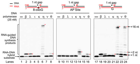
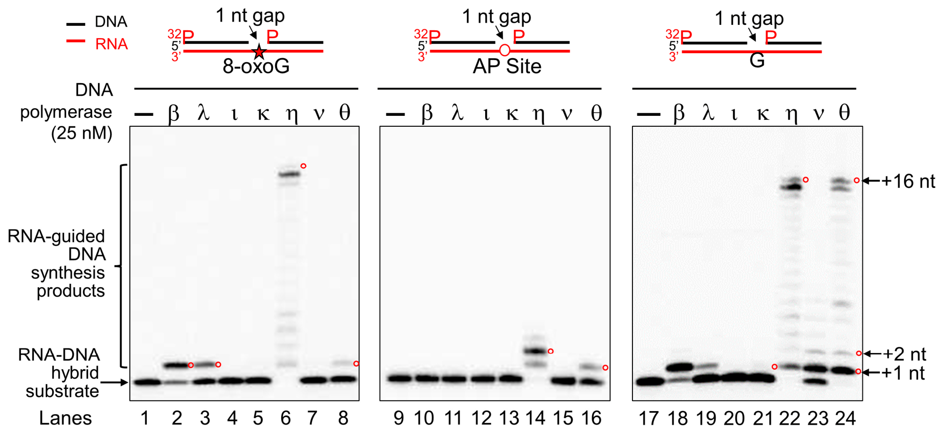
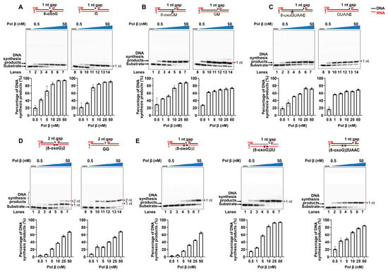
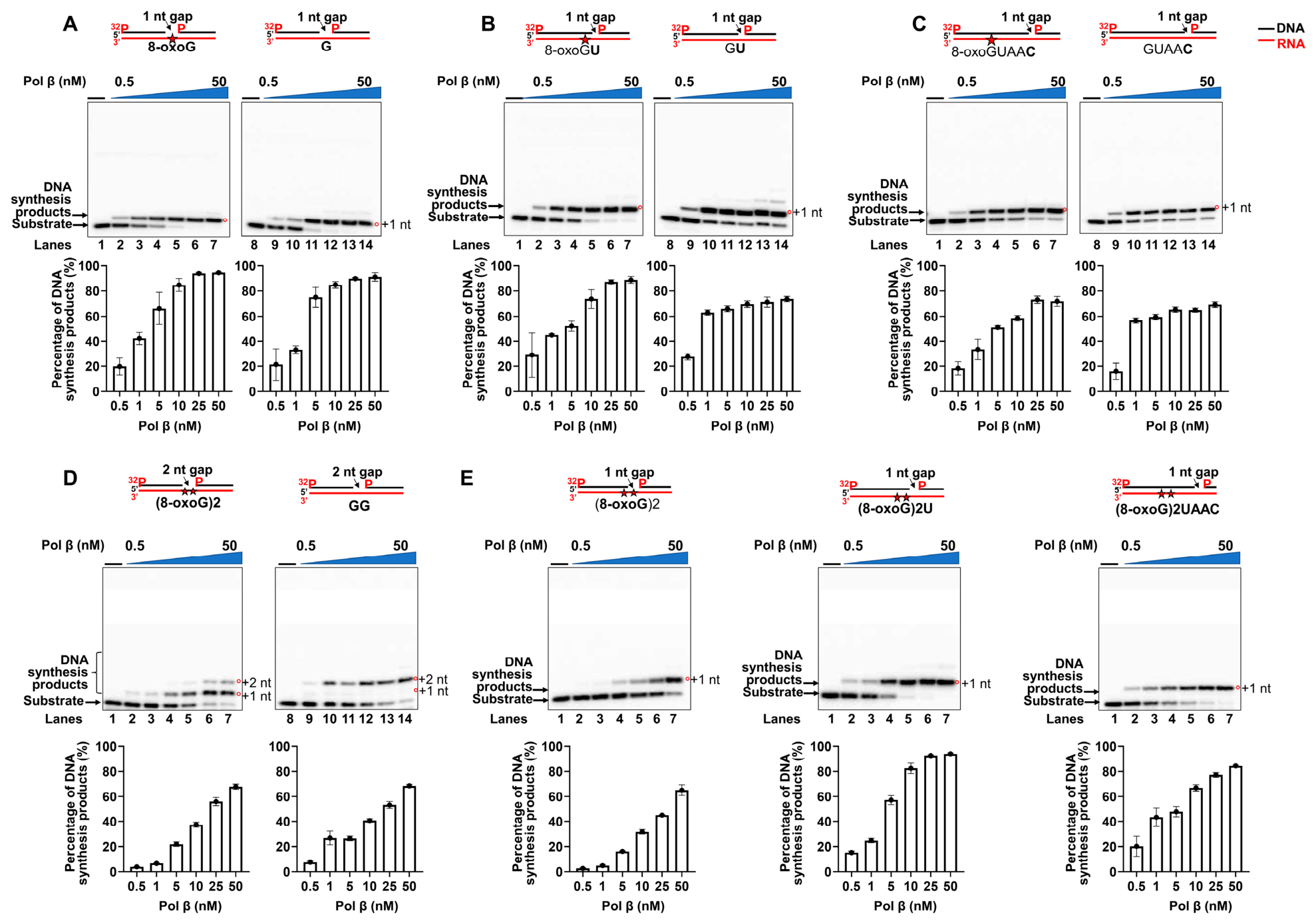
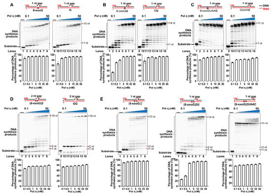
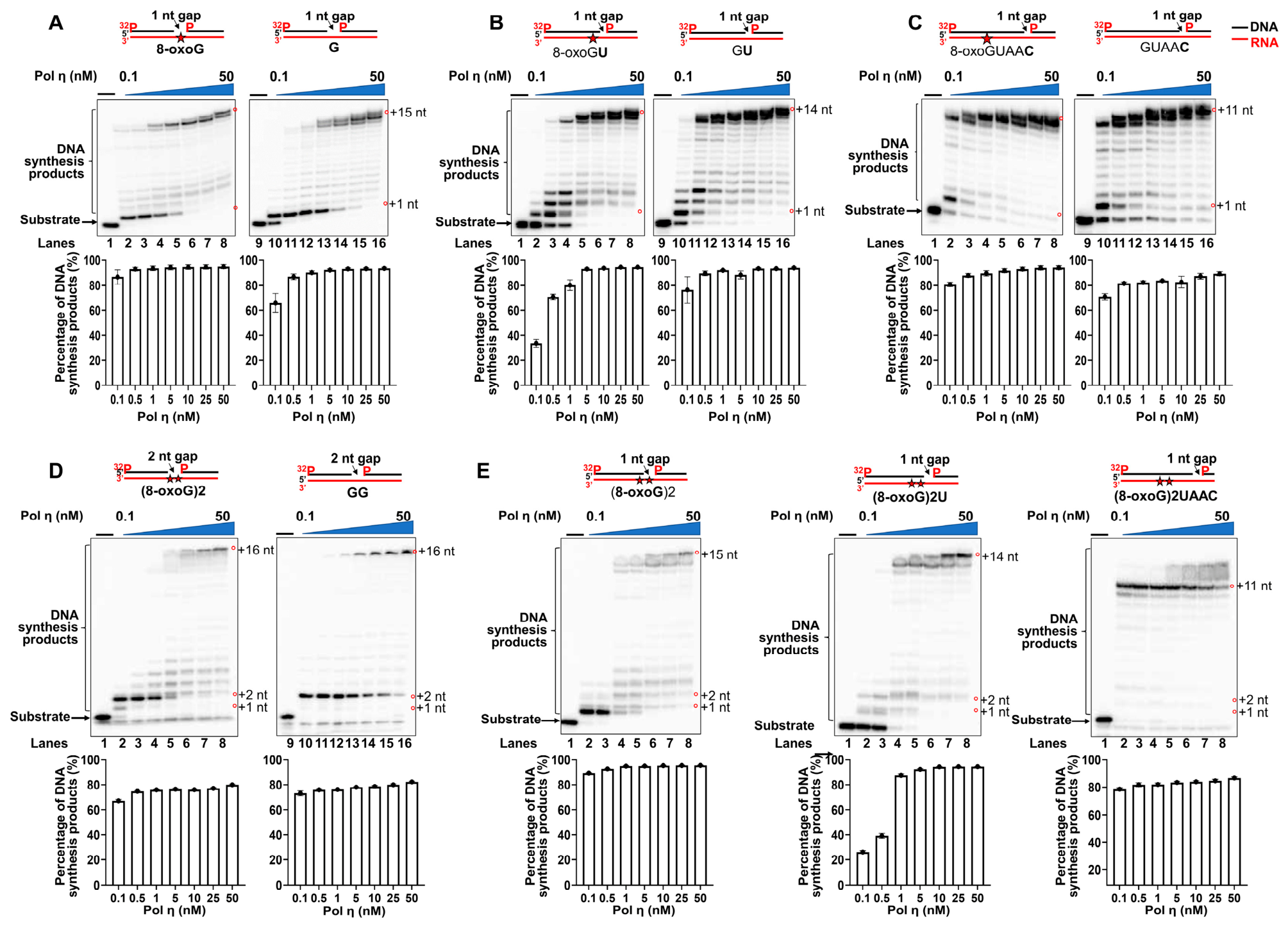
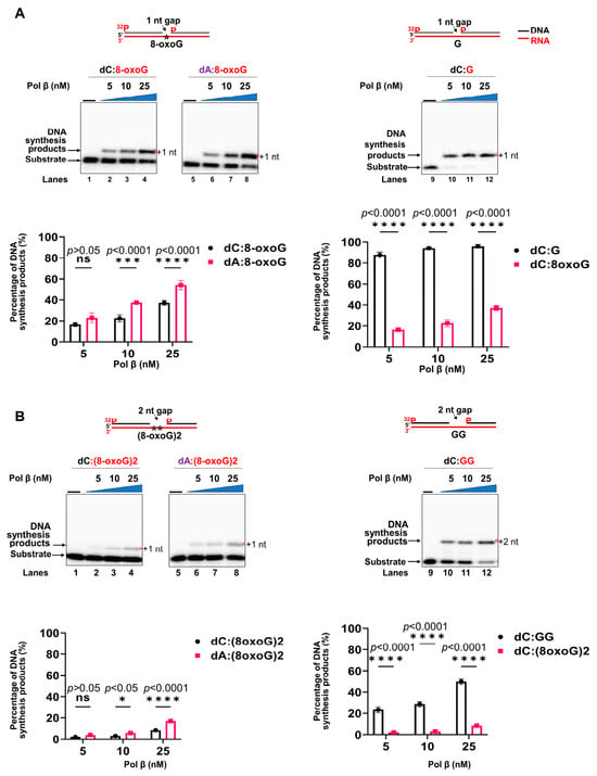
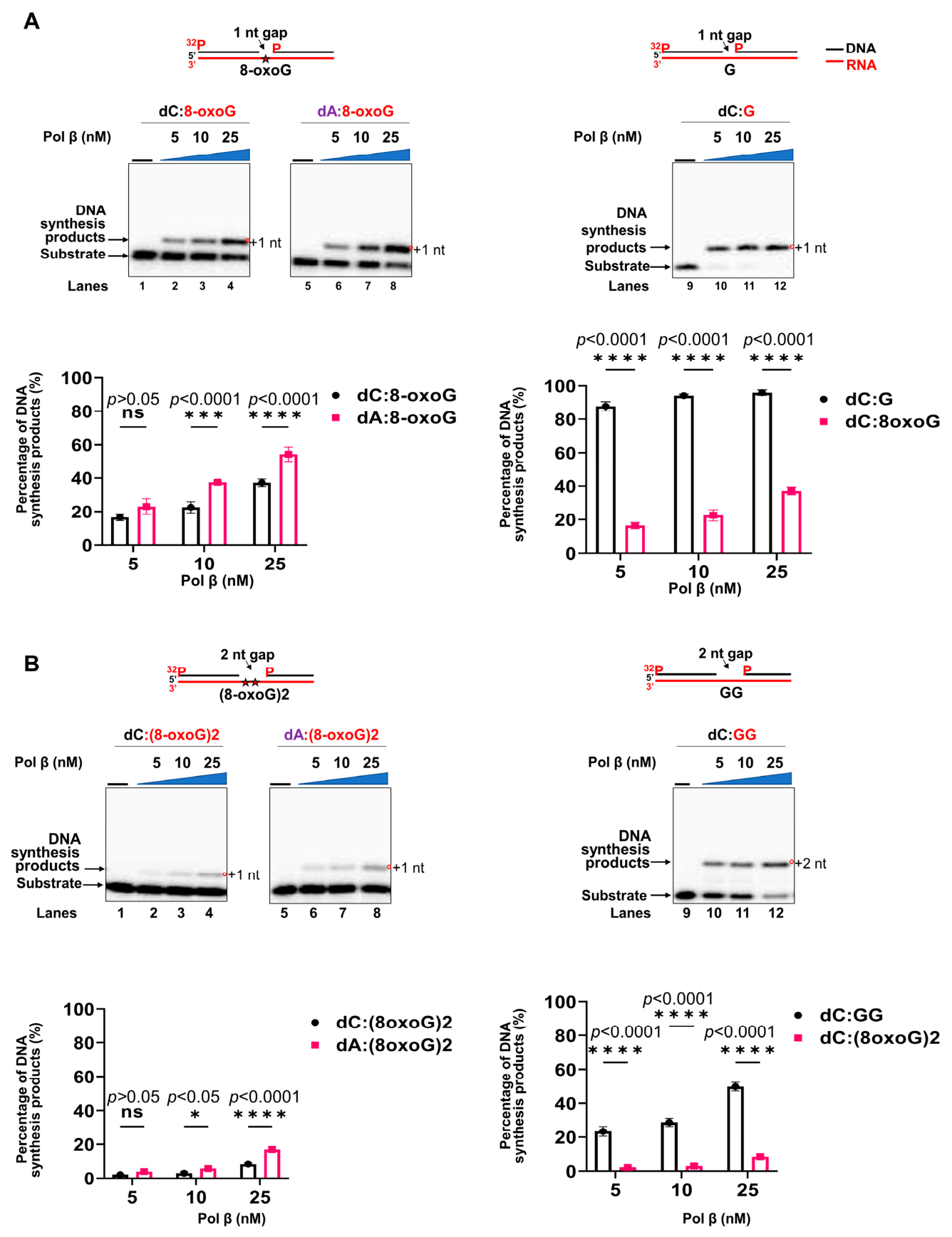
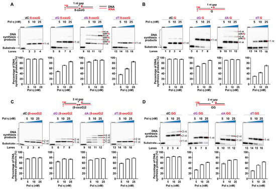
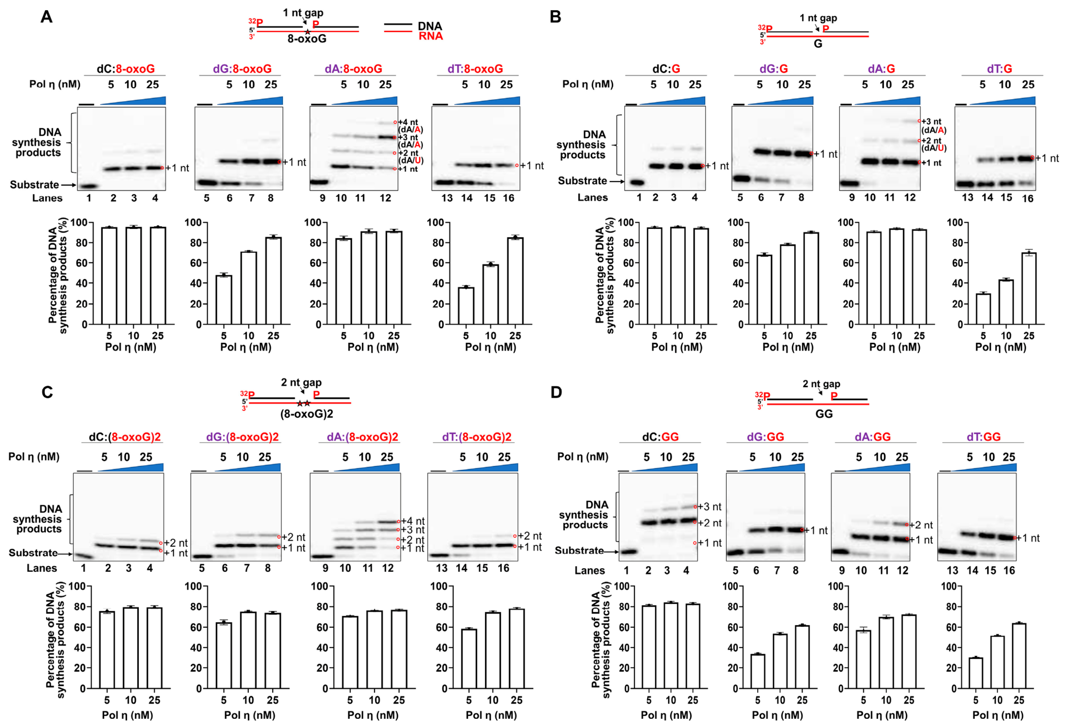
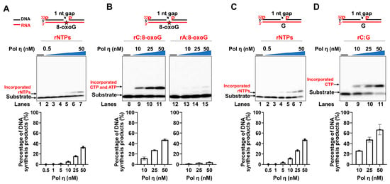
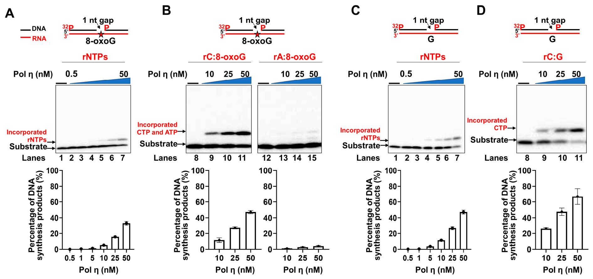
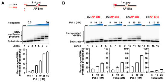
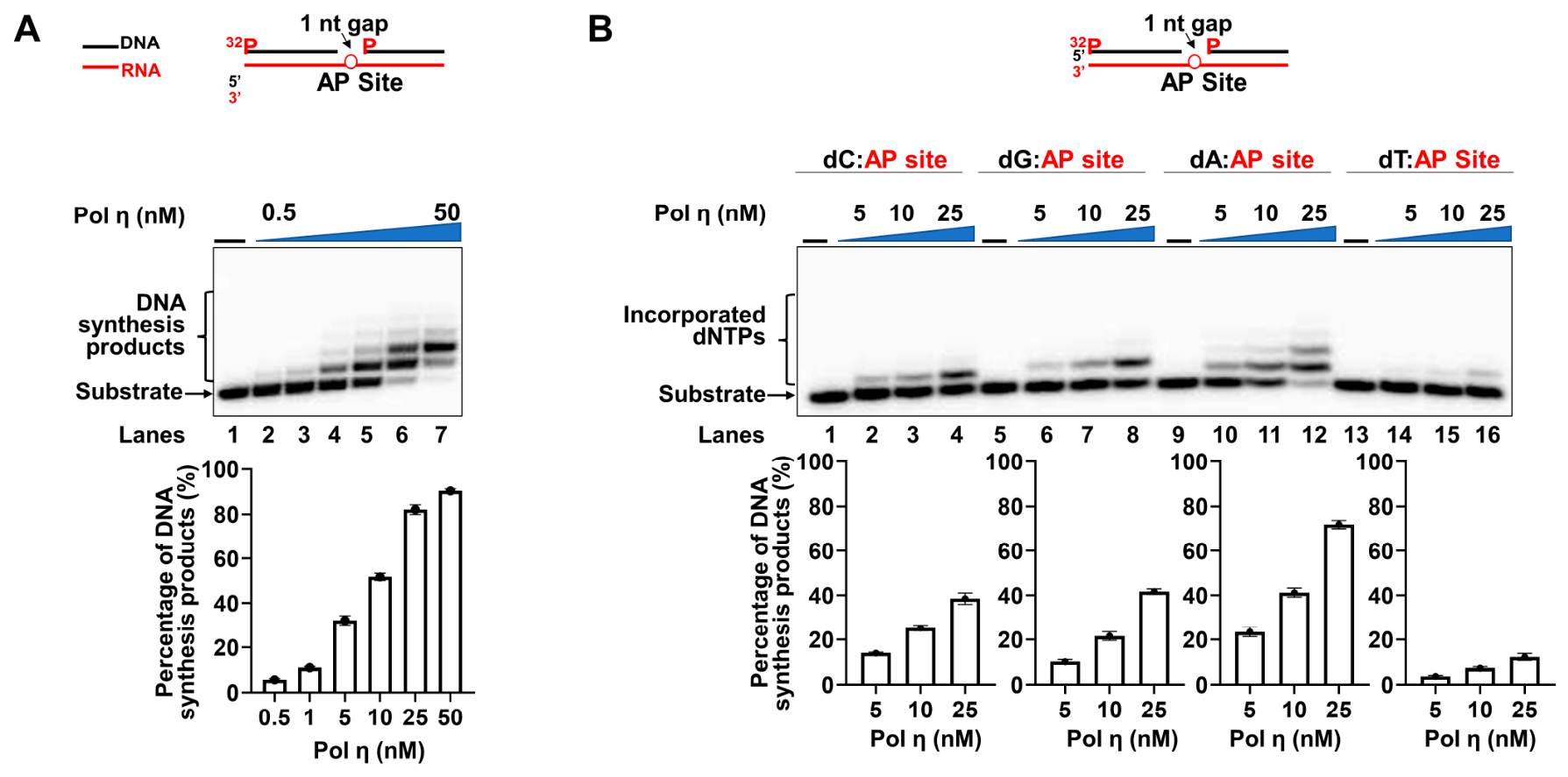
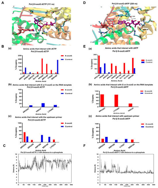
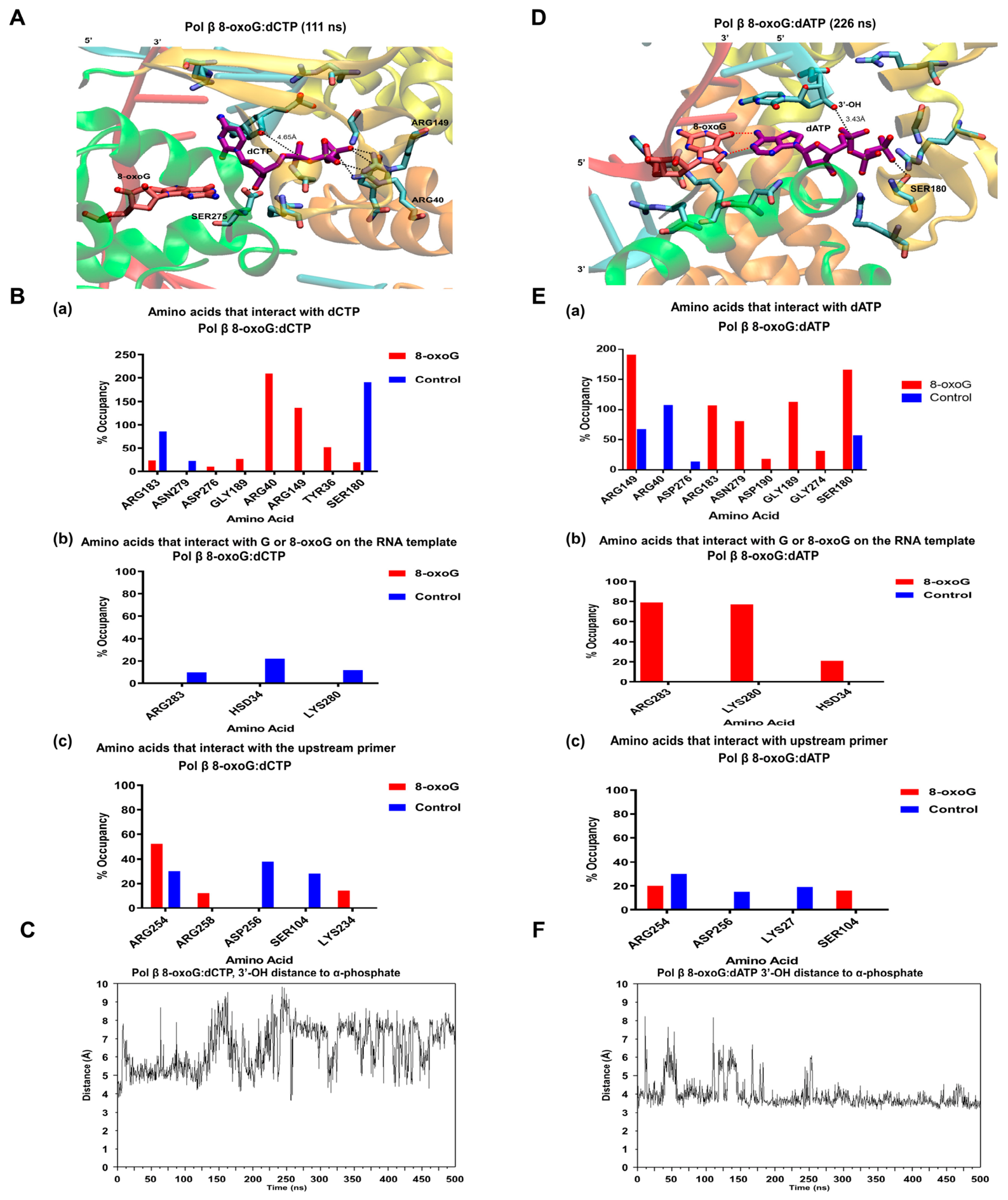
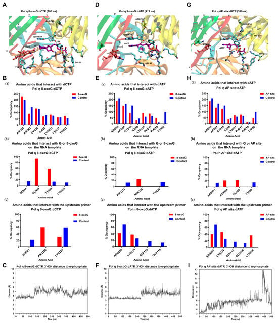
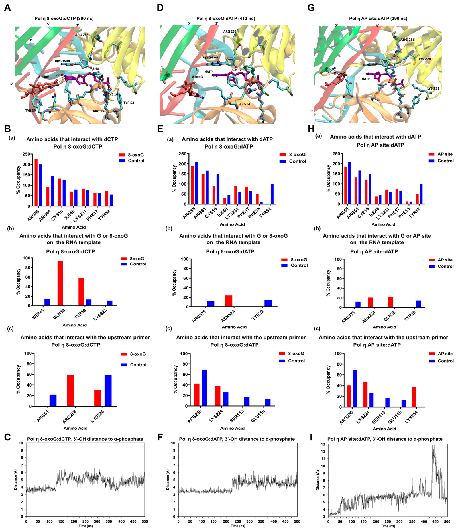
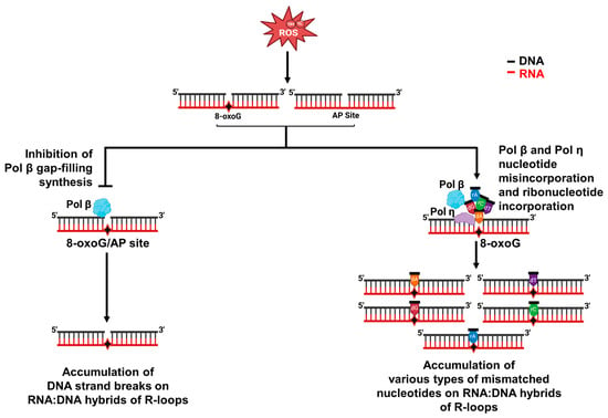
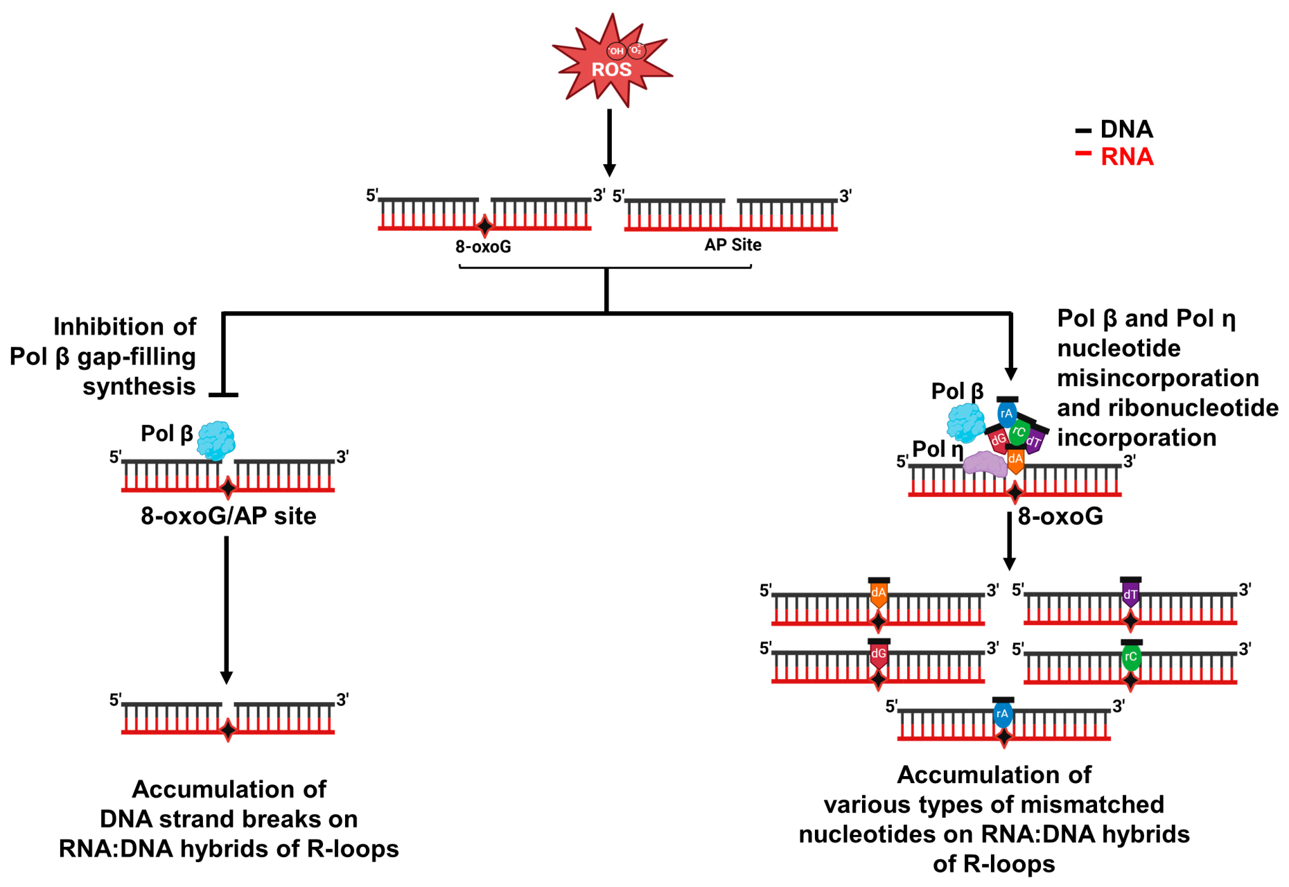

Disclaimer/Publisher’s Note: The statements, opinions and data contained in all publications are solely those of the individual author(s) and contributor(s) and not of MDPI and/or the editor(s). MDPI and/or the editor(s) disclaim responsibility for any injury to people or property resulting from any ideas, methods, instructions or products referred to in the content. |
© 2026 by the authors. Licensee MDPI, Basel, Switzerland. This article is an open access article distributed under the terms and conditions of the Creative Commons Attribution (CC BY) license.