


Abstract
mRNA–Lipid nanoparticles (LNPs) for cancer vaccines are often considered a linear progression, in which formulation, biodistribution, innate stimulation, antigen expression, and tumor control form a continuum. They are not. This review connects the manufacturing inputs and lipid composition to particle attributes, innate sensing, organ and cell exposure, cytosolic translation, antigen presentation, therapeutic response, and hepatic safety. This review aims to provide a mechanism-aligned framework from critical quality attributes (CQAs) and critical process parameters (CPPs) to innate sensing, multimodal imaging, and preclinical efficacy and safety that can be used to design and interpret preclinical studies of mRNA-LNP cancer vaccines, with explicit attention to orthotopic pancreatic ductal adenocarcinoma (PDAC) models. Principal findings: (i) total organ signal is an inadequate surrogate for productive delivery; the decisive variable is translation of antigen in a permissive antigen-presenting cell (APC) during a defined inflammatory window; (ii) formulation and process variables including size, PDI, encapsulation, RNA integrity and capping, lipid composition, PEG-lipid architecture, mixing conditions, and storage history alter this conversion; and (iii) multimodal imaging is most informative when carrier or payload tracking is paired with reporter or antigen expression, immune profiling, tumor response, and liver-safety assays. We propose a release-to-response architecture for preclinical studies and identify the evidence required for clinically interpretable pharmacology. We reviewed peer-reviewed studies on mRNA cancer vaccination, quantitative imaging, PDAC models, LNP quality-by-design, and formulation-dependent innate immunology. The total organ signal is an inadequate surrogate for productive delivery; the decisive variable is translating antigen into an antigen-presenting cell during a permissive inflammatory window. Particle size, polydispersity, encapsulation, RNA integrity and capping, lipid composition, PEG-lipid architecture, mixing conditions, and storage history can alter this conversion. Multimodal imaging is most informative when carrier or payload tracking is paired with reporter or antigen expression, immune profiling, tumor response, and liver safety assays. PDAC studies have shown that hepatic or splenic exposure can be either a liability or intentional immune reprogramming. We propose a release-to-response architecture for preclinical studies and identify the evidence required for clinically interpretable pharmacology.
1. Introduction: The Therapeutic Object Is a Formulated Message
An mRNA cancer vaccine is not simply an antigen sequence administered in a dose. It is a transient information system in which RNA identity, chemical quality, particle architecture, tissue exposure, intracellular trafficking, innate signaling, and antigen presentation are coupled. The LNP protects a labile, anionic transcript, promotes cellular entry, and can assist endosomal escape; the RNA then uses host translation machinery to generate antigenic protein [1,2,3]. The same formula can also affect complement system activation, myeloid cell recruitment, cytokine profile, and lymphatic drainage. Therefore, “the vaccine” includes at least two pharmacologically active ingredients: RNA and lipid nanocarrier.
One common interpretative mistake in the field is equating particle uptake, RNA delivery, transgene expression, inflammation, and cancer regression as interchangeable indicators. Flares in the liver do not imply RNA integrity; RNA integrity does not imply cytoplasmic delivery; protein expression does not imply cross-priming; and cancer regression does not imply antigen-specific immunity. This is particularly important for solid tumors, where the limiting step is usually not getting the RNA lipid nanoparticle to the tumor but whether a proper antigen-presenting cell recognizes the translated RNA and whether costimulatory conditions are present to prime it [4,5].
The immune pathway can be considered from both anatomical and temporal perspectives: site of administration or tumor; draining lymph node (dLN); antigen-presenting cell (APC); MHC-peptide complex formation; T cell activation; access to tumor cells; and memory. In systemic PDAC, the spleen, liver metastasis, and tumor-associated myeloid compartments may be additional intended or unintended sites of action for immunotherapy. Multimodal imaging can connect these layers only if each modality is assigned a biological quantity and paired with an assay that measures a different layer [6,7].
This review integrates four areas often discussed separately: LNP quality attributes and manufacturing control; lipid- and payload-dependent innate sensing; multimodal imaging of carrier, RNA, translation, immune activation, and response; and preclinical efficacy and safety, with particular attention to orthotopic pancreatic models. No formulation or destination is universally optimal; interpret biodistribution in the context of quality, immune mechanisms, target compartment, and therapeutic intent.
This review aims to construct a mechanism-aligned release-to-response framework that connects (i) LNP quality attributes and manufacturing control; (ii) lipid- and payload-dependent innate sensing; (iii) multimodal imaging of carrier, RNA, translation, immune activation, and response; and (iv) preclinical efficacy and safety, with particular attention to orthotopic PDAC models. This review is a targeted scoping review aligned with the PRISMA-ScR principles [8], not a systematic review or meta-analysis. The scope is restricted to mRNA-LNP cancer vaccines and mRNA-LNP platforms with direct mechanistic or translational relevance to oncology; non-mRNA vaccines, viral-vector vaccines, and non-LNP nucleic acid carriers are out of scope. Where inferences are drawn from non-oncology platforms (e.g., prophylactic VLP or lipoplex studies), they are explicitly qualified as mechanism-anchored boundary conditions rather than as direct quantitative evidence for mRNA-LNP cancer vaccines (see Section “Scope and Evidence Boundaries” and Section 3.1).
Scope and Evidence Boundaries
This was a targeted scoping review and not a systematic review or meta-analysis.
To make the evidence-selection process transparent, Figure 1 provides a visual overview of the literature search strategy. In brief, we searched PubMed/MEDLINE, Scopus, Web of Science, Embase, and the Cochrane Library for records published between [January 2010] and [March 2026]. The search combined terms for mRNA cancer vaccines, lipid nanoparticles, quality by design, critical quality attributes, critical process parameters, innate immunity, Toll-like receptors, RIG-I-like receptors, STING, biodistribution, positron emission tomography, magnetic resonance imaging, pancreatic ductal adenocarcinoma, hepatotoxicity, and tumor microenvironment. We also screened the reference lists of relevant primary studies and reviews. Records were included if they reported primary mammalian data or directly relevant process-development, imaging, or mechanistic evidence on mRNA–LNP cancer vaccines, LNP biodistribution and imaging, PDAC models, CQA/CPP relationships, formulation-dependent innate sensing, or hepatic safety. Reviews and context papers were retained only for framing concepts, identifying assay requirements, and defining translational gaps. Records were excluded if they were not in English, did not address mRNA–LNP or cancer-vaccine biology, or reported only in vitro data without a translational or mechanistic link. Titles and abstracts were screened, full texts were assessed, and the final evidence set was organized into the four thematic domains of this review: formulation quality and manufacturing control; lipid- and payload-dependent innate sensing; multimodal imaging of carrier, RNA, translation, immune activation, and response; and preclinical efficacy and safety in orthotopic pancreatic models [9,10,11,12,13,14,15,16,17,18,19]. Figure 1 schematically illustrates the literature-search and evidence-selection strategy and annotates the number of records at each stage.
Figure 1.
Visual overview of the literature search and evidence-selection strategy. We identified records from electronic databases (PubMed/MEDLINE, Scopus, Web of Science, Embase, and Cochrane Library) and through citation searching of relevant primary studies and reviews. After removing duplicates, we screened titles and abstracts against the prespecified inclusion criteria. We then assessed the full texts for eligibility.
This review aims to provide a mechanism-aligned framework from critical quality attributes (CQAs) and critical process parameters (CPPs) to innate sensing, multimodal imaging, and preclinical efficacy and safety that can be used to design and interpret preclinical studies of mRNA-LNP cancer vaccines, with explicit attention to orthotopic pancreatic ductal adenocarcinoma (PDAC) models. Principal findings: (i) total organ signal is an inadequate surrogate for productive delivery; the decisive variable is translation of antigen in a permissive antigen-presenting cell (APC) during a defined inflammatory window; (ii) formulation and process variables including size, PDI, encapsulation, RNA integrity and capping, lipid composition, PEG-lipid architecture, mixing conditions, and storage history alter this conversion; and (iii) multimodal imaging is most informative when carrier or payload tracking is paired with reporter or antigen expression, immune profiling, tumor response, and liver-safety assays. We propose a release-to-response architecture for preclinical studies and identify the evidence required for clinically interpretable pharmacology.
The evidence base has three important limitations. First, few studies have quantified carrier exposure, full-length RNA, translated antigens, immune activation, tumor response, and organ toxicity in the same animal. Second, imaging labels and reporter fusions can alter the pharmacology of the intended measurement. Third, numerical ranges for critical quality attributes (CQAs) depend on formulations and process parameters; specifications such as 70–120 nm, PDI < 0.2–0.3, or >80% encapsulation efficiency should not be treated as absolute requirements. The review clearly states these as design criteria, not technical details [20,21]. Fourth, several mechanistic inferences in Section 3 are drawn from non-oncology platforms (e.g., protein, VLP adjuvanticity; prophylactic lipoplex vaccination). These serve as boundary conditions rather than direct quantitative evidence, and the matrix in Table 1 should be interpreted as a conceptual scaffold requiring empirical calibration for each mRNA-LNP cancer-vaccine formulation. No formulation or destination is universally optimal; interpret biodistribution in the context of quality, immune mechanisms, the target compartment, and therapeutic intent. To bridge this gap, the field must shift from linear assumptions to a mechanism-aligned, release-to-response framework (Figure 2).
Table 1.
Formulation-induced decoupling of critical quality attributes (CQAs) from pharmacological outcomes in mRNA–lipid nanoparticle cancer vaccines. Alterations in lipid composition, RNA integrity, or process parameters can yield acceptable physicochemical release profiles while fundamentally disrupting the exposure-to-translation-to-immunity cascade. This framework identifies the specific biological layer compromised by common formulation perturbations, the false clinical inferences they risk generating, and the orthogonal assays required to distinguish true biological potency from nominal physicochemical quality. Abbreviations: CPP, critical process parameter; CQA, critical quality attribute; EE, encapsulation efficiency; FRR, flow rate ratio; LNP, lipid nanoparticle; N/P, nitrogen-to-phosphate ratio; PDI, polydispersity index; PEG, polyethylene glycol; RNA, ribonucleic acid; TFF, tangential-flow filtration.
Figure 2.
From linear assumptions to a mechanism-aligned release-to-response architecture for mRNA–LNP cancer vaccines. (A) The conventional fallacy: assuming that organ-level biodistribution (e.g., hepatic or splenic accumulation) linearly translates to antigen expression and tumor control. (B) The mechanistic reality: the delivery cascade consists of at least five distinct biological layers. Formulation perturbations (such as RNA degradation or detrimental innate signaling) can decouple specific steps, meaning carrier exposure does not guarantee productive translation or adaptive priming. (C) The proposed solution is a release-to-response framework in which formulations must pass through a functional potency gate before being evaluated using cell-resolved multimodal imaging, such as calculating the Delivery Function Index (DFI) in distinct antigen-presenting cell compartments (e.g., dLN cDC1s versus hepatic APCs).
2. Formulation Quality of the Biological Mechanism
2.1. LNP Composition, Critical Quality Attributes, and Critical Process Parameters
The classic LNP formulation comprises an ionizable lipid, a phospholipid helper, cholesterol or a cholesterol analog [5,23], and PEG-lipid. Ionizable lipid controls RNA binding during formation and controls pH-sensitive interactions with the membrane following internalization via endocytosis. Phospholipids and cholesterol affect the structure and endosomal transport. PEG-lipids affect aggregation and protein adsorption. Although these are formulation components, their effects are reflected in critical quality attributes of the LNP, such as particle size, polydispersity index, encapsulation efficiency, surface charge, RNA identity and integrity, capping, lipid profile, cargo loading, morphology, and stability [39].
Additional CQAs required for reproducibility and clinical translation and increasingly reflected in regulatory expectations include the empty (lipid-only) particle fraction and the distribution of RNA across filled particles (measurable by cryo-EM, analytical ultracentrifugation [AUC], or asymmetric-flow field-flow fractionation [AF4]); residual solvents (per ICH Q3C, e.g., ethanol, isopropanol, tert-butanol); lipid-related substances and degradation products (oxidized ionizable lipid, lysolipids, aldehyde adducts generated by tertiary-amine oxidation, measurable by LC-MS); and a quantitative profile of double-stranded RNA (dsRNA) impurities (RP-UHPLC, J2 immunoblot, or cellulose-based cleanup with confirmatory LC-MS). These CQAs are aligned with ICH Q6B specification principles, ICH Q3C residual-solvent limits, the WHO Guidelines for the production and quality control of mRNA vaccines for human use, the European Pharmacopeia general chapter 5.36 (adopted January 2025), the EMA draft guideline on the quality aspects of mRNA vaccines (2025), and the USP draft general chapter on mRNA vaccines.
CQAs matter not for their ability to predict effectiveness on their own, but for their role as a coupled state vector. Variations in the PEG-lipid could improve aggregation while reducing internalization; variations in the N/P ratio would increase encapsulation while altering surface charge and activation; variations in the phospholipid would maintain particle shape while altering the cytokine profile and/or tropism. In particular, a recent study on variations in bioactive phospholipids is enlightening: despite unchanged particle structure under cryo-EM, variations in bioactive phospholipids altered physicochemical properties, protein expression, cytokine profiles in human peripheral blood mononuclear cells, and cell tropism [39]. This observation does not prove that any phospholipid is a better choice as a component in cancer vaccines. Instead, it shows that the “helper” lipid can serve as a variable in immunological experiments.
Operational definitions and methodological gaps. The principal CQAs and their standard analytical methods, with limitations, are as follows. (1) Particle size and polydispersity index (PDI): hydrodynamic diameter and dispersity are typically measured by dynamic light scattering (DLS); DLS is intensity-weighted and biased toward larger particles in polydisperse samples; orthogonal nanoparticle tracking analysis (NTA), cryo-TEM, or analytical ultracentrifugation (AUC) are required for heterogeneous formulations. (2) Encapsulation efficiency (EE): the fraction of RNA protected from RNase, quantified as (total − free)/total by the RiboGreen fluorescence assay; RiboGreen can underestimate EE when RNA adsorbs to the LNP surface, and orthogonal size-exclusion chromatography (SEC) or AUC is recommended. (3) RNA identity and integrity: typically by capillary gel electrophoresis (Fragment Analyzer, Bioanalyzer); integrity alone does not capture 5′ cap structure (capped vs. uncapped) or 5′-phosphate status, which require orthogonal LC-MS or enzymatic assays. (4) Empty/filled particle fraction: a critical yet frequently unreported attribute, measurable by cryo-EM, AUC, or asymmetric-flow field-flow fractionation (AF4). (5) Purity (lipid-related substances and degradation products): requires LC-MS quantification of oxidized ionizable lipid, lysolipids, and aldehyde adducts formed by tertiary-amine oxidation [37,40]. (6) Residual solvents: headspace GC per ICH Q3C [41]. (7) dsRNA impurities: RP-UHPLC [27], J2 immunoblot, or cellulose-based cleanup with confirmatory LC-MS. (8) Surface charge (zeta potential): electrophoretic light scattering; sensitive to ionic strength and dilution, and should be reported alongside the buffer composition. Methodological gaps remain in (a) standardized reference materials for LNP CQAs; (b) validated in vitro potency assays that predict in vivo translation; (c) cell-resolved assays of APC translation in lymphoid tissue; and (d) integration of CQA metadata into imaging and efficacy datasets as a pre-specified covariate or blocking factor.
Critical process parameters (CPPs) determine whether a formulation produces the same biological product at different scales. Various variables, including FRR, TFR, mixer geometry, temperature, aqueous-phase pH, buffers, N/P ratio, lipid concentration, and tangential-flow filtration downstream, can affect particle size, polydispersity index (PDI), encapsulation efficiency, RNA integrity, and protein expression. Using definitive screening and Box–Behnken designs, Ly et al. showed that process and formulation parameters affect CQA profiles, protein expression, and cytokine activation, and that saRNA is more difficult to encapsulate and preserve than messenger RNA (mRNA) [33]. The QbD literature provides the same answer: a process is controlled not only by achieving a reproducible mean particle size [30,31,42,43,44].
Process changes, including mixture transfer, material replacement, PEG-lipid source, buffer changes, freeze/thawing, or lyophilization, may perturb the exposure-to-translation cascade. However, in regulatory practice, the consequences of such changes are evaluated through risk-based comparability exercises [41] rather than presumed automatically to constitute a biological modification. Comparability is established when physicochemical CQAs, biological potency, and (where applicable) safety and efficacy endpoints remain within pre-defined acceptance criteria following the change. A risk-tiered approach that distinguishes major from minor process changes, with orthogonal potency and innate-signature testing reserved for higher-risk changes, preserves the principle that biological activity must be verified without categorically treating every process change as a de novo biological product. A restricted release panel is acceptable only when supported by formulation-specific justification, pre-defined acceptance criteria aligned with ICH Q6B and WHO TRS 1039 Annex 3, and a documented comparability protocol [45]. RNA integrity and cap formation are critical, as encapsulated but chemically damaged RNA may still participate in sensing without being translated. However, high expression may be accompanied by a poor cytokine signature or liver exposure, as shown in Table 1. Consequently, a cancer vaccine LNP release process requires a functional potency test in addition to physicochemical characterization [46,47].
2.2. Quality by Design Should Include Biological Response Surfaces
A sound QbD strategy links the target product profile (TPP) to the CPPs, CQAs, critical material attributes, and design space [30,31,42,43,44]. Specifically, for cancer vaccines, the design space cannot be limited to traditional features such as particle size, polydispersity index (PDI), and encapsulation efficiency. Rather, response surfaces related to the translated antigen, APC phenotype, inflammatory kinetics, and hepatic exposure must be included. This is especially true if the vaccine formulation is immunostimulatory. The end product aims not for maximum immune stimulation but for a balanced combination of delivery and appropriate innate immune signaling.
Release testing must verify identity, size/PDI, encapsulation, RNA integrity and capping, residuals, stability, translational capability, and a specific innate signature for each formulation. After process scale-up or modification, evaluate CQAs and biological activity; a batch that meets size and EE criteria but shows different cytokine release or APC transfection is not biologically equivalent.
The literature on microfluidic manufacturing also warns against viewing technology as a gold standard independent of the design space. The merit of microfluidic mixing lies in establishing the reproducibility of the CPP-CQA relationship rather than relying on the device name. Continuous manufacturing, tangential flow filtration (TFF), freeze-drying, and other mixing technologies can all be viable options, so long as equivalence is established [32,34,38]. This manufacturing discipline is directly relevant to imaging: a biodistribution study that does not report batch CQAs cannot distinguish biological variation from product variation.
2.3. Regulatory Context and Analytical Scalability
The CQA/CPP framework described above is not a self-defined scientific construct; it is anchored in established regulatory guidance. ICH Q6B specifies test procedures and acceptance criteria for biotechnological/biological products; ICH Q5E defines comparability principles for manufacturing-process changes; and ICH Q3C sets limits for residual solvents (e.g., ethanol, isopropanol, tert-butanol used in LNP processing). For mRNA vaccines specifically, the WHO Guidelines for the production and quality control of mRNA vaccines for human use [48] provide the global benchmark, complemented by the European Pharmacopeia general chapter 5.36 (adopted January 2025), the EMA draft guideline on the quality aspects of mRNA vaccines [28], the USP draft general chapter on mRNA vaccines, and FDA considerations for the quality, safety, and efficacy of mRNA vaccines (2023). Cancer-vaccine developers should align their pre-specified CQA panel with these documents and structure comparability exercises per ICH Q5E. The scalability of analytical methods is an underappreciated translational bottleneck: methods developed at bench scale (e.g., dynamic light scattering, RiboGreen encapsulation, Fragment Analyzer integrity) must be transferable to QC laboratories with validated acceptance criteria, while higher-resolution orthogonal methods cryo-EM and analytical ultracentrifugation for empty/filled quantification [25], RP-UHPLC for dsRNA [27], LC-MS for lipid-related substances [37,40], and micro-flow imaging for sub-visible particles [49] require method qualification and, where applicable, transfer to contract analytical laboratories. Recent orthogonal analytical pipelines [24,50] and multi-attribute CQA mapping studies [26] provide the operational template for a GMP-compatible, scalable mRNA-LNP analytical strategy. A complete release package should specify, for each CQA, the analytical method, the qualification/validation status, the site of analysis, and the orthogonal method used to resolve ambiguities (e.g., AUC as orthogonal to DLS for polydisperse samples).
3. Innate Immunology Is Formulation- and Payload-Dependent
3.1. Separate RNA Sensing from Carrier and Adjuvant Effects
The intrinsic response to mRNA-LNP constructs is complex. RNA molecules can bind to endosomal and cytoplasmic nucleic acid sensors depending on the sequence, chemical modifications, purification, cellular location, and RNA dose. Lipid composition affects lipid uptake, endosomal residence time, endosomal escape, and binding to nucleic acid sensors. Co-delivered agonists can add a defined pathway, such as STING or TLR7/8, whereas phospholipid and PEG-lipid changes can modify the tissue and cellular context in which signaling occurs.
Within antigen-presenting cells (APCs), sensor-specific biochemistry governs mRNA fate and should be specified rather than presumed. Endosomal TLR3, TLR7, and TLR8 sense single- and double-stranded RNA in a sequence- and modification-dependent manner; cytoplasmic sensors include the RIG-I/MDA5 helicases, protein kinase R (PKR; phosphorylating eIF2α to halt cap-dependent translation), and the OAS-RNase L axis (degrading single-stranded RNA and amplifying innate signaling) [29,51]. ADAR1-mediated A-to-I editing of endogenous and exogenous RNAs dampens MDA5/TLR3 hyperactivation, providing a constitutive brake on cytoplasmic RNA sensing [52]. The chemical identity of the 5′ cap, the presence of N1-methylpseudouridine (m1Ψ) and 5-methylcytidine (m5C), and the extent of dsRNA removal by HPLC or cellulose-based purification are pivotal: nucleoside modifications suppress TLR3/7/8 recognition and reduce PKR-mediated translational arrest, while residual dsRNA can reactivate these pathways even in the presence of m1Ψ [53,54]. Consequently, the same lipid composition can produce a productive or detrimental innate state depending on RNA chemistry, a feature that must be specified, not assumed, for each mRNA-LNP formulation. Lipid-side variables (ionizable-lipid pKa, helper-lipid identity, PEG-lipid desorption, and co-delivered agonists) further shift the balance between endosomal escape (productive translation) and prolonged endosomal residence (detrimental TLR activation).
The simplified statement that “LNPs activate TLRs” should therefore be replaced by a testable formulation-specific model. Endosomal TLR3, TLR7/8, and TLR9; RIG-I-like receptors; inflammasome pathways; complement; and STING may contribute to different combinations. The exact contribution depends on RNA chemistry, lipid structure, adjuvant, route, and cell type. In a primary mouse study of an LNP-adjuvanted norovirus VLP vaccine, the adjuvant effect depended on the ionizable lipid and type I interferon signaling and was partially TLR9-dependent [55]. This was not a cancer vaccine efficacy study, but it is a useful mechanistic warning: LNP adjuvanticity can be biologically active even when the LNP carries protein rather than mRNA, and pathway dependence should be demonstrated rather than presumed.
Although this norovirus VLP platform [55] is protein-based and mechanistically distinct from an mRNA-LNP cancer vaccine, it is invoked here solely as a mechanistic indicator of pathway dependence (i.e., that LNP adjuvanticity can engage TLR9 and type I IFN), not as quantitative evidence for cancer-vaccine adjuvant activity. We do not extrapolate the magnitude, kinetics, or anatomical compartment of TLR9/IFN-I activation from a VLP context to an mRNA-LNP cancer vaccine without independent validation in oncology models. The same caveat applies to other lipid-platform studies discussed below (e.g., [56], which used cationic lipoplexes in a non-tumor context).
Miao et al. provide a contrasting example in cancer models. Heterocyclic ionizable lipids increased anti-tumor activity through STING-mediated immune-cell activation with limited systemic cytokine expression [57]. Pollard et al. showed that type I interferon can also counteract antigen-specific immunity in another lipid-based mRNA-vaccine context [56]. These findings are not inconsistent, but they derive from different lipid platforms (heterocyclic ionizable lipids [57] vs. cationic lipoplexes in [55]) and different biological contexts (subcutaneous tumor models vs. prophylactic protein vaccination). They are therefore invoked here as qualitative, mechanism-anchored boundary conditions rather than as a validated quantitative “response surface” for mRNA-LNP cancer vaccines. The matrix in Table 2 is proposed as a conceptual scaffold whose threshold values (magnitude, duration, compartment) must be empirically defined for each mRNA-LNP chemistry in future oncology studies; it should not be read as a validated predictor. An inflammatory surge can activate APCs, halt protein synthesis, inflict tissue harm, or expand systemic exposure.
Table 2.
Formulation-dependent innate immune signaling in mRNA–LNP cancer vaccines: distinguishing productive priming from detrimental reactogenicity. The adjuvant effect of an mRNA-LNP is not a single axis but a multidimensional response surface dictated by the formulation components. This matrix maps specific formulation drivers to their primary innate sensing pathways, highlighting how temporal kinetics and anatomical compartmentalization determine whether the resulting inflammation augments antigen-specific cross-priming or actively suppresses the therapeutic objectives. Note on quantitative thresholds: The designations “productive”, “detrimental”, “context-dependent”, “redirective”, and “suppressive” refer to qualitative, mechanism-anchored categories rather than numerical thresholds. Defining the magnitude (e.g., cytokine AUC in pg·h/mL), duration (hours vs. days), and cellular compartment (endosomal vs. cytoplasmic; cDC1 vs. pDC vs. Kupffer cell) at which a signal transitions between categories is a research priority and cannot be inferred from the cited studies alone. The matrix is proposed as a conceptual scaffold for hypothesis-driven empirical calibration, not as a validated predictor. Abbreviations: APC, antigen-presenting cell; CARPA, complement activation-related pseudo-allergy; cDC, conventional dendritic cell; IFN-I, type I interferon; IFNAR, interferon-α/β receptor; N/P, nitrogen-to-phosphate; PEG, polyethylene glycol; STING, stimulator of interferon genes; TLR, Toll-like receptor.
3.2. Phospholipids and PEG-Lipids Are Not Inert Excipients
Phospholipids shape membrane structure, affect charge, and influence opsonization, complement interactions, and endosomal trafficking. They may also be involved in signaling and metabolism through lipid mediation [60]. Direct evidence for the cancer vaccine benefit from a particular bioactive phospholipid remains incomplete; however, formulation-screening data show that structural substitutions can produce distinct cytokine signatures and modest changes in cellular tropism [39]. This observation underscores a key experimental distinction: a phospholipid can be assessed for its structural, biodistribution-modifying, and intrinsic stimulus effects, but all aspects must be measured independently, as shown in Table 3.
Table 3.
Formulation-dependent innate immune signaling in mRNA–LNP cancer vaccines: distinguishing productive priming from detrimental reactogenicity. The adjuvant effect of an mRNA-LNP is not a single axis but a multi-dimensional response surface dictated by the formulation components. This matrix maps specific formulation drivers to their primary innate sensing pathways, highlighting how temporal kinetics and anatomical compartmentalization determine whether the resulting inflammation augments antigen-specific cross-priming or actively suppresses the therapeutic objectives. Abbreviations: APC, antigen-presenting cell; BLI, bioluminescence imaging; dLN, draining lymph node; FDG, 2-deoxy-2-[18F]fluoro-D-glucose; LC-MS, liquid chromatography–mass spectrometry; MRI, magnetic resonance imaging; NIR, near-infrared; PET, positron emission tomography; RT-qPCR, reverse-transcription quantitative polymerase chain reaction; SPECT, single-photon emission computed tomography.
PEGylation is also a trade-off and not a clear win. PEG-lipid conjugates can reduce aggregation and opsonization; however, PEG density, anchor structure, and PEG desorption kinetics affect particle size, interactions with serum proteins, tissue distribution, lymph node drainage, cell uptake, and dosing regimens. The “PEG shield” may protect the drug during storage and circulation but prevent cellular or endosomal uptake. Alternatively, inadequate PEGylation may increase aggregation, protein adsorption, and nonspecific cell uptake. PEG-specific antibodies and modified corona formation can also occur after multiple administrations [58,61].
This list of mechanistic features is important for cancer vaccination, as lymph node delivery efficiency and APC function cannot be assessed solely by particle size. For example, an efficient-draining formulation that encapsulates RNA into endosomes can trigger inflammation but does not promote translation. APC uptake, combined with strong type I interferon signaling, can downregulate antigen production. In addition, spleen- or liver-targeting formulations can be useful for treating metastatic PDAC but are unwanted for localized vaccines. Therefore, an appropriate comparison should include all these formulation parameters.
3.3. Co-Delivered Agonists Can Redirect the Cancer-Immunity Cycle
STING, TLR7/8, and TLR9 agonists and other immunostimulants can supply costimulation or remodel suppressive myeloid niches. However, adjuvants cannot be treated as universal boosters, as their intracellular localization, release profile, dose, and relationship with antigen expression determine whether they enhance antigen presentation or cause deleterious reactogenicity. New adjuvant formulations based on Toll-like receptor (TLR) agonists show that different TLR agonists can have different immune effects, even in structurally similar lipid nanoparticle (LNP) designs [65].
This point is particularly important for PDAC.” with “This point is particularly important for PDAC. Xu et al. used MC3 LNPs to deliver mutant KRAS G12D mRNA, along with cGAMP, to enhance hepatocellular delivery for immune cell reprogramming and to prevent liver metastasis formation [69]. In an orthotopic PDAC model, Luo et al. used spleen-targeting LNPs to deliver KRAS mRNA, along with 3M-052 and irinotecan-loaded silicasomes, to induce immunogenic cell death during chemotherapy [68]. This work does not imply that each hepatocyte/spleen signal is useful for therapy; rather, it emphasizes the importance of organ delivery for the overall therapeutic strategy. Figure 3 corresponds to innate immune signaling.
Figure 3.
Formulation-dependent innate immune signaling pathways in mRNA–LNP cancer vaccines. The figure depicts the intracellular fate of an mRNA–LNP (input, top) and the four cellular sensing zones that determine whether the encoded antigen is productively translated or triggers detrimental inflammation. (A) Endosomal sensing: TLR3, TLR7, TLR8, and TLR9 in the endosomal lumen recognize dsRNA, ssRNA, and CpG DNA motifs; the ionizable-lipid pKa, helper-lipid identity, and PEG-lipid desorption kinetics shift the balance between endosomal escape (productive) and prolonged endosomal residence (detrimental TLR activation). (B) Cytoplasmic sensing and translation: RIG-I and MDA5 helicases recognize cytoplasmic dsRNA; PKR phosphorylates eIF2α to arrest cap-dependent translation; the OAS–RNase L axis degrades ssRNA and amplifies innate signaling; productive translation feeds antigen peptides into MHC class I presentation. (C) Organellar sensing: RIG-I/MDA5 signal through MAVS on the mitochondrial outer membrane; cGAS senses cytosolic DNA (including mitochondrial DNA released by LNP-induced stress) and produces cGAMP, which activates STING on the endoplasmic reticulum; ionizable-lipid destabilization can contribute to mitochondrial stress. (D) Regulation: ADAR1 (p110 nuclear, p150 cytoplasmic) performs A-to-I editing of endogenous and exogenous dsRNA, dampening MDA5/TLR3 hyperactivation and preventing autoimmune-like inflammation. Right panel—formulation-dependent outcome: a balance scale summarizes how mRNA modifications (m1Ψ, m5C), 5′ cap structure, and dsRNA purity (depleted duplexes) shift the outcome toward productive translation (antigen presentation, green) or detrimental sensing (hyperinflammation, red). Image was created with BioRender.com.
4. Multimodal Imaging Should Follow the Causal Chain
4.1. Five Biological Layers
A well-defined imaging strategy describes five layers in succession: carrier binding, intact RNA, protein synthesis, immune response, and therapy effectiveness. Each step has distinct failure points. A fluorescent lipid probe may indicate label detachment; RNA targeting may reflect RNA fragmentation; reporter proteins may not mimic antigen processing; FDG uptake may reflect nonspecific inflammation; and tumor regression may reflect toxic or vascular effects. A single imaging method cannot answer all questions of this chain, as shown in Table 4.
Table 4.
Multimodal readout. Abbreviations: CARPA, complement activation-related pseudo-allergy; CPP, critical process parameter; CQA, critical quality attribute; dLN, draining lymph node; EE, encapsulation efficiency; FRR, flow rate ratio; IFN-I, type I interferon; LC-MS, liquid chromatography–mass spectrometry; LNP, lipid nanoparticle; N/P, nitrogen-to-phosphate; PEG, polyethylene glycol; RT-qPCR, reverse-transcription quantitative polymerase chain reaction; SORT, selective organ targeting; STING, stimulator of interferon genes; TFF, tangential-flow filtration; TLR, Toll-like receptor.
4.2. Carrier and Payload Tracking
Fluorescence enables high-throughput comparative studies in small animals; however, tissue attenuation, label leakage, and quantitative mass balance considerations limit this technique. Di et al. used DiR, liquid chromatography-mass spectrometry, IVIS, and luciferase experiments to show that organ exposure and functional expression do not scale linearly [76]. The key insight was not the fluorescence itself but the deliberate decoupling of carrier exposure from expression.
Expanding on this idea, PET/CT can be used to study deeper tissues and larger animals. Dual radionuclide/NIR showed initial mRNA vaccine uptake in cynomolgus macaques and correlated the signal with antigen expression in cell-resolved professional antigen-presenting cells [70]. In addition, DOTA-lipid/64Cu showed that lymph node access was more stochastic in nonhuman primates than in mice [77]. Together, these data suggest a general principle: whole-body PET can measure formulation distribution, but ex vivo flow cytometry and histology are necessary to identify the cells that have taken up the formulation.
Radiolabeled lipids or chelators can separate from the active formulation, whereas the fluorophore bound to RNA can remain attached even to degraded fragments. Therefore, the protocol should include label-free or orthogonal measurements of full-length RNA, and, when possible, a mass-balance calculation that accounts for residual syringe activity, decay, tissue recovery, and dose normalization.
4.3. Translation and Immune-Activation Imaging
Bioluminescence remains an efficient comparative readout in mice, as it reflects protein production. However, its limitations include substrate delivery, tissue attenuation, reporter half-life, and poor translation to humans. An mRNA-encoded PET reporter offers deeper and more clinically compatible imaging; however, reporter fusion can alter antigen processing and surface expression. Blizard et al. demonstrated transient translated-antigen imaging in mice and longer lymph node signals in nonhuman primates and showed why an untagged-antigen control is essential [72].
MRI and 18F-FDG PET can resolve local and dLN inflammatory responses, but they do not establish antigen specificity, as shown in Table 5. In a rat study, MRI/PET signals correlated with cytokine kinetics and the spatial organization of immune cells, demonstrating that imaging can report the organization and timing of inflammation rather than directly assess tumor immunity [73]. Raman or related label-free chemical imaging may add spatial localization in excised tissue, but it should be treated as an exploratory localization layer rather than proof of intact mRNA, translation, or presentation [71,78]. The correct question is not “How inflamed is the tissue?” but “Which inflammatory state occurs at the same place and time as productive antigen presentation?”
Table 5.
Formulation-induced decoupling of critical quality attributes (CQAs) from biological and pharmacological outcomes in RNA nanoparticle therapeutics. Changes in RNA integrity, lipid composition, PEG-lipid density, N/P ratio, ionizable lipid chemistry, co-delivered agonists, or surface-targeting chemistry may produce acceptable physicochemical CQAs while substantially altering RNA translation, innate immune activation, cellular/tissue tropism, and ultimately therapeutic efficacy. The table highlights potential false pharmacological inferences and identifies the orthogonal assays required to distinguish physicochemical quality from true biological potency. Abbreviations: ALP, alkaline phosphatase; ALT, alanine aminotransferase; AST, aspartate aminotransferase; APC, antigen-presenting cell; LNP, lipid nanoparticle; MRI, magnetic resonance imaging; PDAC, pancreatic ductal adenocarcinoma; PET, positron emission tomography.
5. Preclinical Efficacy: The Tumor Model Determines What the Signal Means
The most persuasive efficacy studies combine immunocompetent hosts, established tumors, defined antigens, and orthogonal response assays. Oberli et al. used LNP-assisted mRNA encoding melanoma antigens in B16F10 models and observed CD8 responses, tumor reduction, and survival benefits, but did not assign the priming contribution of each transfected cell type [36]. Kranz et al. used RNA-lipoplexes for systemic delivery to DCs and macrophages, coupling interferon-alpha induction to effector and memory responses [74]. These platforms should not be conflated, as lipoplex charge and RNA sensing are part of their mechanism of action.
Personalized mutanome vaccination and mutant MHC-II epitope studies have shown that antigen sequence, processing route, and epitope breadth are as important as total protein expression [81,82]. The HPV16 RNA-LPX model demonstrated complete regression and protective memory; however, viral antigens generally provide a stronger immunological starting point than neoantigens [83]. Intratumoral mRNAs encoding IL-21, IL-7, and 4-1BB ligand further show that local immunomodulatory expression can remodel systemic immunity, although this is mechanistically distinct from vaccination with tumor antigens [84].
In addition to the B16F10 melanoma, HPV16 RNA-LPX, STING/TLR knockout, and KRAS-driven PDAC studies already cited, additional immunocompetent preclinical models are relevant to mRNA-LNP cancer-vaccine evaluation. Among syngeneic models commonly used in immuno-oncology: MC38 (murine colorectal adenocarcinoma; moderately immunogenic, microsatellite-stable); CT26 (murine colon carcinoma; high mutational load, T-cell inflamed); 4T1 (mammary carcinoma; metastatic model with spontaneous lung and bone dissemination); A20 (B-cell lymphoma; hematological context for systemic delivery); TC-1 (HPV16 E6/E7-driven; viral-antigen comparator to antigen-specific mRNA vaccines); and Renca (renal cell carcinoma; poorly immunogenic, useful for adjuvant co-formulation studies). Side-by-side TME comparisons of B16F10, MC38, and CT26 have demonstrated differential immune infiltration and differential response to checkpoint blockade [79,85], and rational model-selection criteria have been proposed based on mutational load, T-cell-inflamed status, and TME composition. For pancreatic cancer, the KPC genetically engineered mouse model (KrasG12D; Trp53R172H; Pdx1-Cre) recapitulates the human disease’s desmoplastic stroma, immune suppression, and metastatic pattern [80,86] and is particularly relevant for evaluating hepatic/splenic reprogramming strategies in mRNA-LNP cancer vaccines. We recommend that future mRNA-LNP cancer-vaccine studies pre-specify the model(s) and the rationale based on the intended mechanism: dLN cDC1 cross-priming (B16F10, TC-1); systemic myeloid reprogramming (MC38, 4T1, A20); intratumoral cytokine expression (any accessible flank model); or orthotopic PDAC reprogramming (KPC, KPC-derived syngeneic lines such as KPC-Y, Panc02-OVA).
PDAC presents a more challenging setting because pancreatic tumors are fibrotic, myeloid-rich, spatially heterogeneous, and often resistant to checkpoint blockade. The KRAS G12D/cGAMP study matters because it does not automatically treat liver exposure as pathological. Hepatic APC reprogramming, CD8 effector function, metastatic suppression, and survival are linked in a KPC setting [69]. The KRAS/3M-052 plus irinotecan-silicasome study similarly couples vaccine priming with immunogenic cell death and splenic targeting in an orthotopic model [68]. The experimental inference is conditional, and the relevant question is whether the organ receiving the formulation is a therapeutic compartment for the selected antigen–adjuvant pair.
Human PDAC data provide a translational anchor, but not a substitute for mechanistic imaging. In a phase I adjuvant study of autogene cevumeran (BNT122), neoantigen-specific responses were detected in eight of 16 treated patients and were associated with a longer recurrence-free survival [75]. The non-randomized design does not establish that a particular LNP or lipoplex biodistribution pattern causes a clinical outcome. However, it supports the measurement of clonotype persistence, neoantigen breadth, and functional T-cell state in preclinical studies.
6. Hepatotoxicity, Off-Target Accumulation, and Misleading Safety Proxies
If the vaccine is administered intramuscularly or intranodally, liver exposure is considered off-target; however, in metastatic PDAC, this exposure may be desirable. Safety concerns include whether exposure is necessary, which liver cells are exposed, and whether RNA translation and burden occur in the liver.
Beyond hepatocellular injury, four additional risk classes are directly relevant to mRNA-LNP cancer vaccines and should be evaluated in parallel with hepatic readouts. (i) Systemic cytokine release, notably IL-6, TNF-α, and type I interferon, can occur in a formulation- and dose-dependent manner and is mechanistically linked to innate sensing of the ionizable lipid and residual dsRNA [59,87,88]. (ii) Complement activation-related pseudo-allergy (CARPA) is an acute, non-IgE-mediated reaction triggered by lipid surfaces;. However, originally characterized for liposomes and Doxil, the same framework has been mapped to rare mRNA-LNP vaccine hypersensitivity reactions and warrants prospective monitoring, particularly in patients with high cumulative exposure or pre-existing complement activation [62,63,64]. (iii) Anti-PEG IgG and IgM antibodies can develop after a first dose of PEGylated LNPs and accelerate blood clearance or trigger hypersensitivity on repeat dosing, a particularly important consideration for cancer vaccines, which often require booster schedules and prolonged administration intervals [88,89]. (iv) The theoretical possibility of mRNA reverse-transcription and genomic integration has been examined in vitro [90,91]; current regulatory and mechanistic syntheses consider in vivo integration biologically implausible [92], yet this question is especially pertinent for cancer vaccines administered to patients with long life expectancies after curative-intent therapy, and we recommend explicit discussion and dedicated long-term follow-up in translational programs. The minimum safety package for repeat-dose cancer-vaccine studies should therefore include systemic cytokine time-courses, complement endpoints (C3a/C5a, Bb, CH50), anti-PEG antibody titers before each dose, and (where feasible) tissue-residence and clearance assays of the formulated mRNA.
The liver is a predictable sink because of its sinusoidal architecture, lipoprotein interactions, fenestrated endothelium, and abundant phagocytes. ApoE-mediated uptake contributes to hepatocyte delivery for conventional formulations, but hepatic signals are distributed among hepatocytes, Kupffer cells, and liver endothelial cells. Sago et al. showed that clinically relevant LNPs interact with multiple liver cell populations, and that functional mRNA delivery does not equal nucleic acid distribution [93]. Therefore, a whole-liver fluorescence image cannot be assigned to hepatocyte transfection or interpreted as evidence of injury.
Several mechanisms may coexist. Ionizable lipids or degradation products may perturb membranes or accumulate after repeated dosing. PEG-lipid shedding and the protein corona may alter uptake. Complement, myeloid, inflammasome, and nucleic acid sensing may produce transient cytokine release or more sustained tissue injury. Unintended protein translation may add compartment-specific toxicity. Therefore, the term “LNP hepatotoxicity” is too broad unless the study distinguishes between the carrier, RNA, translation, inflammation, and pathology.
The minimum biochemical analysis must include sequential determinations of ALT, AST, alkaline phosphatase, bilirubin, albumin, and possibly also coagulation indices. Such measurements must be combined with blinded histopathological assessment, correction for liver mass, cellular-level localization, cytokine dynamics, and analysis of recovery after the last dose. A transient cytokine increase or a slight transaminase rise may indicate reversible reactogenicity, whereas sustained increases, bilirubin disturbances, necrosis, cholestasis, and impaired synthesis suggest more serious injury. Studies with repeated doses are critical, since one-time administration experiments might overlook lipid accumulation, adaptation, delayed toxicity, or inflammation.
Off-target accumulation can reduce vaccine efficiency without overt injury by diverting the dose away from the injection site or dLN pathways. Hepatic translation can also alter antigen processing or tolerance; however, the antigen-sink hypothesis remains unproven and should not be used as a post hoc explanation for tumor-vaccine failure. The relevant metric is the ratio of translated antigen in the desired APC compartment to that in liver cell subsets, along with recovery kinetics and tumor response as shown in Figure 4.
Figure 4.
Preclinical safety package for mRNA–LNP cancer vaccines. The figure maps the four classes of risk discussed in Section 6 to the recommended monitoring panel for each. (1) Systemic cytokine release IL-6, TNF-α, and type I interferon can occur in a formulation- and dose-dependent manner, mechanistically linked to innate sensing of the ionizable lipid and residual dsRNA; monitoring: longitudinal systemic cytokine time-courses. (2) Complement activation-related pseudo-allergy (CARPA) is an acute, non-IgE-mediated reaction triggered by lipid surfaces; monitoring: C3a, C5a, Bb, and CH50 within 30 min of infusion, IgE/IgG work-up, high-risk patient pre-screening. (3) Anti-PEG IgG/IgM antibody responses on repeat dosing can accelerate blood clearance and reduce tissue exposure on booster doses; monitoring: anti-PEG IgG/IgM titers before each dose, PK comparison versus first dose, formulation desensitization or PEG-lipid substitution. (4) Theoretical mRNA persistence/integration examined in vitro but considered biologically implausible in vivo; monitoring: tissue-residence and clearance assays, dedicated long-term follow-up for cancer-vaccine patients with long life expectancies. The figure highlights the long-life-expectancy context of cancer vaccines as a rationale for extended follow-up of categories (3) and (4). The image was created with BioRender.com.
The field now has evidence that this ratio can be manipulated. Muscle-selective and low-liver-accumulation LNPs can reduce liver and spleen expression while preserving cellular immunity or tumor suppression in selected animal models [94,95]. SORT chemistry and endogenous targeting have shown that charge, PEG-lipid desorption, and serum protein adsorption can redirect tropism [66,67,95]. These results support engineering control rather than a universal “liver-free” requirement. Conversely, the PDAC cGAMP/KRAS study showed that hepatic exposure can be exploited [69]. Therefore, safety and efficacy must be analyzed using the same exposure map.
Batch comparability is an underappreciated safety variable. Suppose that scale-up changes the PDI, RNA integrity, PEG-lipid distribution, or innate activation; a different hepatic exposure may be misattributed to animal variation. Report CQA metadata for every biological batch and include the formulation batch as a prespecified covariate or blocking factor in the imaging analysis. Do not declare a formulation translationally ready if the same nominal recipe produces different functional delivery indices across multiple lots, as shown in Table 6.
Table 6.
Hepatic and off-target interpretation in preclinical cancer vaccine studies. Abbreviations: BLI, bioluminescence imaging; cDC, conventional dendritic cell; dLN, draining lymph node; DFI, delivery function index; FDG, 2-deoxy-2-[18F]fluoro-D-glucose; ICD, immunogenic cell death; MRI, magnetic resonance imaging; PDAC, pancreatic ductal adenocarcinoma; PET, positron emission tomography; SPECT, single-photon emission computed tomography; TME, tumor microenvironment.
7. A Unified Validation Architecture for Orthotopic PDAC Models
The proposed experiment should use an immunocompetent orthotopic PDAC model with a defined tumor antigen or KRAS-mutant mRNA construct. Prespecified implantation, randomization, treatment, imaging, allocation concealment, and blinded pathology. Separate the efficacy and terminal biodistribution cohorts [96].
The panel should include therapeutic mRNA–LNP, empty LNP, naked mRNA (where interpretable), reporter mRNA, and labeled payload cohorts. Targeting studies should include exposure-matched, low-liver or muscle-selective formulations. Intentional hepatic or splenic reprogramming should use cGAMP or 3M-052 as exploratory comparators, and not as pure LNP controls. Testing quality-attribute release and functional potency is essential for each batch.
Coordinate multimodal imaging through multiple layers of tissues, such as PET/CT or SPECT, to assess the distribution of carriers; optical reporters to track the translation process in mice; MRI or FDG-PET to evaluate tumors and inflammation processes; and histology, flow cytometry, spatial profiling, or Raman imaging to identify the cell types as shown in Figure 3. We collected tumor tissue, pancreas, draining and non-draining lymph nodes, spleen, liver, kidneys, lungs, heart, blood, and dissemination sites.
The mechanistic endpoint of choice is the delivery function index, which is defined as the ratio of the area under the curve of the translated antigen signal in the prespecified antigen-presenting cell (APC) population to the carrier or intact RNA signal in the same population. Calculate separately for cDC1, cDC2, macrophages, monocytes, tumor cells, and hepatic cell populations (if relevant). We did not normalize the index across different labels, doses, or tumor models. The hypothesis states that translation within dLN APCs, but not the whole-organ carrier signal, will better predict antigen-specific day 7 CD8+ T cell expansion and consequently tumor control see Figure 5.
Immune readouts should cover antigen-specific CD8+ and CD4+ T cells, intracellular cytokine production, TCR expansion and persistence, APC maturation, myeloid cell composition, NK cell status, tumor cell antigen presentation, and the co-localization of APCs and T cells. The therapeutic readouts include tumor volume or tumor burden by MRI, survival, metastasis, antigen loss escape, protection upon rechallenge, and durability of response. Depletion or adoptive transfer studies are needed when claiming antigen specificity of tumor control [35].
Assess safety endpoints concurrently with efficacy endpoints. Align serum chemistry, cytokine levels, body weight, behavior, organ weights, and blinded liver histology with cell-specific hepatic localization and recovery. The study must detail the mRNA dose administered, particle concentration, RNA molecules per particle (if available), CQA profile, imaging dose, label chemistry, and analysis thresholds [96]. A complete preclinical study of an mRNA-LNP cancer vaccine should also document the regulatory and analytical-scalability pathway. We recommend explicit alignment of the pre-specified CQA panel with ICH Q6B and WHO TRS 1039 Annex 3 expectations, a prospective comparability protocol per ICH Q5E for any planned process change, residual-solvent reporting per ICH Q3C, and pre-specified orthogonal analytical methods (RP-UHPLC for dsRNA; cryo-EM or AUC for empty/filled fraction; LC-MS for lipid-related substances; micro-flow imaging for sub-visible particles). The analytical methods should be qualified (or at minimum method-validated) at the site that will perform release testing, and comparability protocols should specify both pre-defined acceptance criteria and the decision tree when those criteria are exceeded [38,50]. These elements are essential for a preclinical package to be interpretable by regulatory reviewers and translatable to clinical-grade manufacturing.
Figure 5.
Release-to-response validation architecture for orthotopic PDAC. The figure consolidates the proposed study design into a single integrated schematic organized across three stages with two decision gates. Stage 1 (Batch Qualification & Comparability): batch-qualified product with a pre-specified CQA panel aligned to ICH Q6B and WHO TRS 1039 Annex 3 (size, PDI, encapsulation, RNA integrity, capping, lipid profile, empty/filled fraction, residual solvents, lipid-related substances, dsRNA impurities); a prospective comparability protocol per ICH Q5E for any planned process change, with risk-tiered orthogonal potency and innate-signature testing; and orthogonal analytical methods (cryo-EM/AUC for empty/filled fraction; RP-UHPLC for dsRNA; LC-MS for lipid-related substances; micro-flow imaging for sub-visible particles). Decision Gate 1: release and comparability thresholds must be met before proceeding to in vivo imaging. Stage 2 (Multimodal Imaging & DFI): multimodal imaging across the five biological layers (carrier, intact RNA, translation, immune activation, therapeutic response) per Figure 4; cell-resolved delivery function index (DFI) calculation in cDC1, cDC2, macrophage, monocyte, tumor, and hepatic compartments (DFI = AUC of translated antigen signal in the prespecified APC population ÷ carrier or intact RNA signal in the same population). Decision Gate 2: mechanism-aligned DFI thresholds must be met before efficacy claims. Stage 3 (Integrated Endpoints): integrated safety and efficacy endpoints with statistical batch blocking: antigen-specific CD8+ T-cell expansion, TCR clonotype persistence, tumor volume by MRI, survival, metastasis, antigen-loss escape, protection upon rechallenge, and parallel hepatic safety (ALT, AST, ALP, bilirubin, albumin, complement, anti-PEG, cytokines). The flow demonstrates how each stage feeds into the next, with decision gates that prevent premature efficacy claims when comparability or orthogonal-assay thresholds are not met. Refer to Section 7 and Table 7 for the mechanism-aligned DFI compartment matrix. The image was created with BioRender.com.
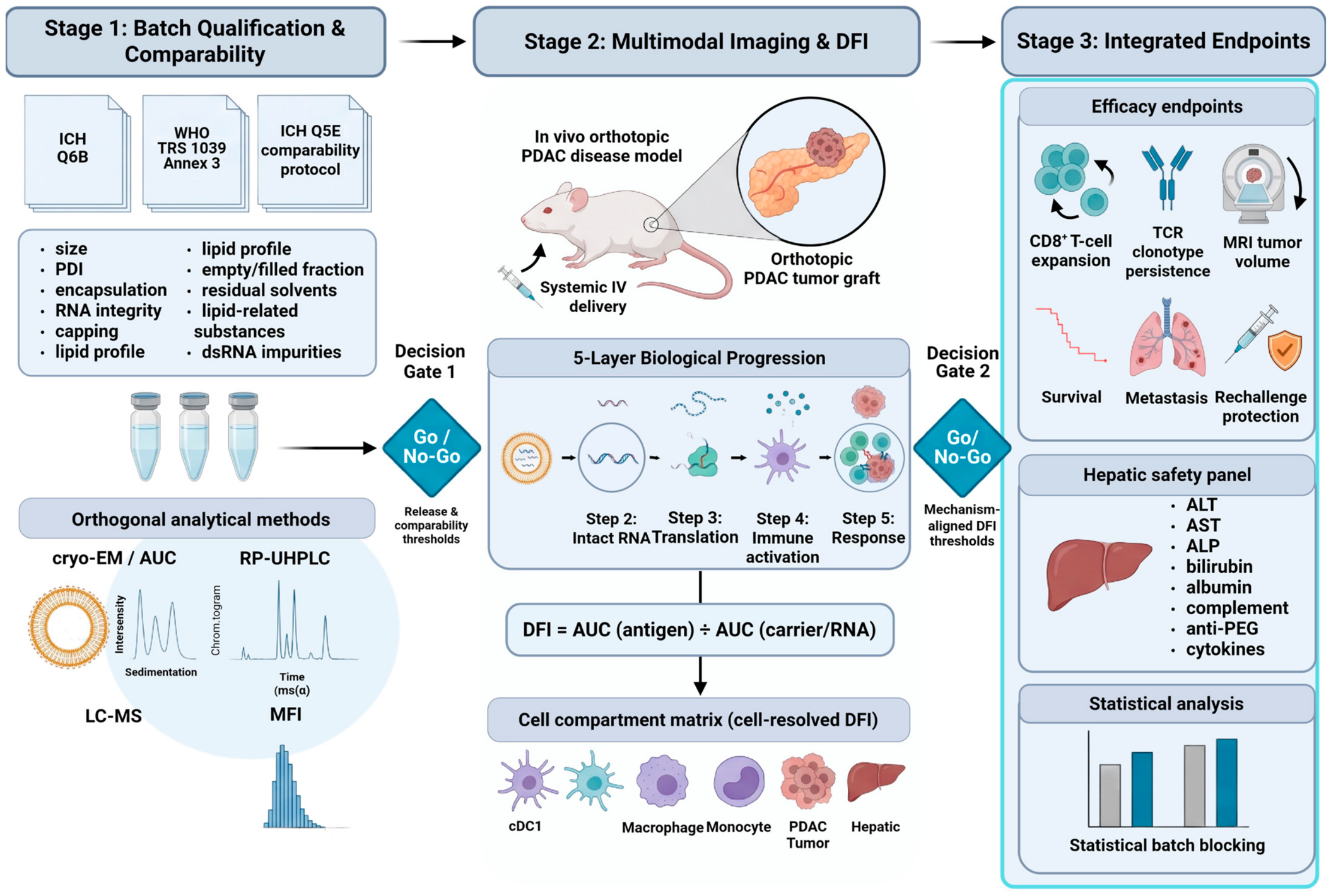
Table 7.
Mechanism-aligned assessment of translated antigen delivery across preclinical tumor contexts. The table summarizes how the optimal cellular and tissue compartment for evaluating productive mRNA delivery varies according to tumor type and therapeutic mechanism. It emphasizes that DFI should be interpreted in context, distinguishing therapeutically relevant antigen translation from off-target distribution. Orthogonal functional readouts, including CD8+ T-cell responses, tumor regression, metastatic burden, TME remodeling, and survival, are required to establish whether translated antigens contribute to the intended therapeutic outcome.
8. Future Directions and Conclusions
The field should shift from a “biodistribution map” to a “release-to-response” map. The first step is a batch-qualified product with a defined CQA and a CPP history. The second step is an imaging design in which the carrier, intact RNA, translated antigen, immune activation, and tumor response are temporally linked. The third step is a cell-resolved analysis that identifies the APC or tumor compartment responsible for functional neoantigen translation. The fourth step is a safety analysis that distinguishes intentional hepatic pharmacology from unintentional accumulation and injury.
Practical priorities include functional translation and innate potency in release specifications; treat phospholipid and PEG-lipid changes as mechanistic perturbations; evaluate adjuvants by pathway, dose, compartment, and kinetics; use untagged-antigen and label-retention controls; and compare local, lymphoid, hepatic, and splenic exposure in orthotopic PDAC.
A falsifiable prediction is that, in a matched head-to-head study, the area under the translated-antigen signal in dLN APCs will better predict antigen-specific T-cell expansion and tumor control than the whole-organ carrier signal does. The second prediction is that CQA drift will change this relationship before it produces an obvious change in particle size or encapsulation. Longitudinal imaging, cell-resolved terminal assays, and batch-blocked statistical models can be used to test both predictions.
mRNA–LNP cancer vaccines are not limited by a lack of imaging modalities or by an absence of impressive mouse responses. They are limited by incomplete causal linkage. The most informative study will show what was manufactured, where it went, which cells received an intact and translatable message, what innate state followed, whether the response was antigen-specific, whether the tumor was controlled, and whether the liver recovered. This evidentiary standard is required for a formulation to move from an attractive nanoparticle phenotype to a credible translational vaccine.
Author Contributions
M.A.O. and A.A.A.A. conceptualized the review, supervised the project, and contributed to writing, review, and editing. A.A. performed the literature search, prepared the original draft, and contributed to data curation and validation. Contributed to investigation, data curation, and review. O.G. contributed to methodology, validation, and review. E.Q. provided resources, supported data curation, and contributed to the review. A.A. worked on visualization and review, review, and editing. All authors have read and agreed to the published version of the manuscript.
Funding
This research received no external funding.
Institutional Review Board Statement
Not applicable.
Informed Consent Statement
Not applicable.
Data Availability Statement
No new data were created or analyzed in this study. Data sharing is not applicable to this article.
Acknowledgments
We used Paperpal (version 2.0.21) solely as a language-editing and proofreading tool to improve sentence structure, grammar, clarity, and the manuscript’s overall readability. The tool was not used to generate the scientific content, research ideas, data, analysis, interpretation, or conclusions of the manuscript. All content, including the selection and synthesis of the literature and the final interpretation presented in this review, was developed and critically reviewed by the authors.
Conflicts of Interest
The authors declare no conflicts of interest.
Abbreviations
| AF4 | asymmetric-flow field-flow fractionation |
| ALP | alkaline phosphatase |
| ALT | alanine aminotransferase |
| APC | antigen-presenting cell |
| AST | aspartate aminotransferase |
| AUC | area under the curve (pharmacokinetic) OR analytical ultracentrifugation (analytical, with context) |
| BLI | bioluminescence imaging |
| CARPA | complement activation-related pseudo-allergy |
| cDC | conventional dendritic cell |
| CPP | critical process parameter |
| CQA | critical quality attribute |
| DFI | delivery function index |
| dLN | draining lymph node |
| DLS | dynamic light scattering |
| dsRNA | double-stranded ribonucleic acid |
| EE | encapsulation efficiency |
| FDG | 2-deoxy-2-[18F]fluoro-D-glucose |
| FRR | flow rate ratio |
| GEM | genetically engineered mouse (model) |
| HPLC | high-performance liquid chromatography |
| ICH | International Council for Harmonization of Technical Requirements for Pharmaceuticals for Human Use |
| IFN-I | type I interferon |
| IFNAR | interferon-α/β receptor |
| IVT | in vitro transcription |
| KRAS | Kirsten rat sarcoma viral oncogene |
| LC-MS | liquid chromatography–mass spectrometry |
| LNP | lipid nanoparticle |
| m1Ψ | N1-methylpseudouridine |
| m5C | 5-methylcytidine |
| MFI | micro-flow imaging |
| MHC | major histocompatibility complex |
| MRI | magnetic resonance imaging |
| mRNA | messenger ribonucleic acid |
| N/P | nitrogen-to-phosphate (ratio) |
| NTA | nanoparticle tracking analysis |
| PDAC | pancreatic ductal adenocarcinoma |
| pDC | plasmacytoid dendritic cell |
| PDI | polydispersity index |
| PEG | polyethylene glycol |
| PET | positron emission tomography |
| PKR | protein kinase R (eukaryotic translation initiation factor 2-alpha kinase 2) |
| QbD | quality by design |
| RLR | RIG-I-like receptor |
| RNase L | ribonuclease L |
| RP-UHPLC | reversed-phase ultra-high-performance liquid chromatography |
| RT-qPCR | reverse-transcription quantitative polymerase chain reaction |
| saRNA | self-amplifying ribonucleic acid |
| SEC | size-exclusion chromatography |
| SPECT | single-photon emission computed tomography |
| STING | stimulator of interferon genes |
| TFF | tangential-flow filtration |
| TFR | total flow rate |
| TLR | Toll-like receptor |
| TME | tumor microenvironment |
| TPP | target product profile |
| VLP | virus-like particles |
References
- Sahin, U.; Karikó, K.; Türeci, Ö. mRNA-based therapeutics—Developing a new class of drugs. Nat. Rev. Drug Discov. 2014, 13, 759–780. [Google Scholar] [CrossRef] [Scilit]
- Pardi, N.; Hogan, M.J.; Porter, F.W.; Weissman, D. mRNA vaccines—A new era in vaccinology. Nat. Rev. Drug Discov. 2018, 17, 261–279. [Google Scholar] [CrossRef] [Scilit] [PubMed]
- Hou, X.; Zaks, T.; Langer, R.; Dong, Y. Lipid nanoparticles for mRNA delivery. Nat. Rev. Mater. 2021, 6, 1078–1094. [Google Scholar] [CrossRef] [Scilit] [PubMed]
- Di, J.; Du, Z.; Wu, K.; Jin, S.; Wang, X.; Li, T.; Xu, Y. Biodistribution and Non-linear Gene Expression of mRNA LNPs Affected by Delivery Route and Particle Size. Pharm. Res. 2022, 39, 105–114. [Google Scholar] [CrossRef] [Scilit]
- Hao, J.; Li, Y.; Huang, L.; Qi, N.; Sun, Y.; He, C.; Yang, S.; Niu, Z.; Qi, X.; Xiang, B. Smart Nanoarchitectures for Precision RNA Delivery: Harnessing Endogenous and Exogenous Stimuli in Cancer Treatment. Theranostics 2025, 15, 7747–7778. [Google Scholar] [CrossRef] [Scilit]
- du Bois, H.; Heim, T.A.; Lund, A.W. Tumor-draining lymph nodes: At the crossroads of metastasis and immunity. Sci. Immunol. 2021, 6, eabg3551. [Google Scholar] [CrossRef] [Scilit]
- Zhu, M. Immunological perspectives on spatial and temporal vaccine delivery. Adv. Drug Deliv. Rev. 2021, 178, 113966. [Google Scholar] [CrossRef] [Scilit]
- Tricco, A.C.; Lillie, E.; Zarin, W.; O’Brien, K.K.; Colquhoun, H.; Levac, D.; Moher, D.; Peters, M.D.J.; Horsley, T.; Weeks, L. PRISMA extension for scoping reviews (PRISMA-ScR): Checklist and explanation. Ann. Intern. Med. 2018, 169, 467–473. [Google Scholar] [CrossRef] [Scilit] [PubMed]
- Dalabehera, M.; Ghosh, A.; Mohanty, S.; Chellapan, D.K.; Chaudhari, S.; Ale, Y.; Poonia, N.; Subudhi, R.N.; Khanna, M.K.; Lim, H.G. mRNA delivery systems 2.0: Engineering extrahepatic delivery for non-vaccine therapeutics. Mater. Today Bio 2025, 35, 102584. [Google Scholar] [CrossRef] [Scilit]
- Han, J.; Lim, J.; Wang, C.-P.J.; Han, J.-H.; Shin, H.E.; Kim, S.-N.; Jeong, D.; Lee, S.H.; Chun, B.-H.; Park, C.G. Lipid nanoparticle-based mRNA delivery systems for cancer immunotherapy. Nano Converg. 2023, 10, 36. [Google Scholar] [CrossRef] [Scilit]
- Hasan, N.; Aftab, M.; Ikram, S.; Mustofa, A.Z.; Sriwidodo, S.; Darusman, H.S.; Amir, M.N.; Tockary, T.A.; Uchida, S. Recent advances in lipid nanoparticles for cancer vaccine delivery: Challenges and future perspectives. Int. J. Pharm. X 2026, 11, 100484. [Google Scholar] [CrossRef] [Scilit]
- Ramadan, E.; Ahmed, A.; Naguib, Y.W. Advances in mRNA LNP-based cancer vaccines: Mechanisms, formulation aspects, challenges, and future directions. J. Pers. Med. 2024, 14, 1092. [Google Scholar] [CrossRef] [Scilit]
- Hussain, M.S.; Khan, G. Current clinical applications of RNA-LNPs in cancer: A promising horizon for targeted therapies. EXCLI J. 2025, 24, 321. [Google Scholar] [CrossRef] [Scilit]
- Yuan, Y.; Sun, W.; Xie, J.; Zhang, Z.; Luo, J.; Han, X.; Xiong, Y.; Yang, Y.; Zhang, Y. RNA nanotherapeutics for hepatocellular carcinoma treatment. Theranostics 2025, 15, 965. [Google Scholar] [CrossRef] [Scilit]
- Berikkhanova, K.; Inuwa, I.; Jibo, A.G.; Berikkhanov, N.; Bikhanov, N.; Sultan, Y.; Omarbekov, A. Hybrid nanocarriers for cancer therapy: Advancements in co-delivery of gene therapy and immunotherapy. Int. J. Mol. Sci. 2025, 27, 248. [Google Scholar] [CrossRef] [Scilit]
- Khawar, M.B.; Afzal, A.; Si, Y.; Sun, H. Steering the course of CAR T cell therapy with lipid nanoparticles. J. Nanobiotechnol. 2024, 22, 380. [Google Scholar] [CrossRef] [Scilit]
- Fernandes, S.; Cassani, M.; Cavalieri, F.; Forte, G.; Caruso, F. Emerging strategies for immunotherapy of solid tumors using lipid-based nanoparticles. Adv. Sci. 2024, 11, 2305769. [Google Scholar] [CrossRef] [Scilit]
- Grippin, A.J.; Lee, D.; Parkes, E.E.; Jiang, W.; Kim, B.Y.S. Nanotechnology for immuno-oncology. Nat. Cancer 2025, 6, 1311–1325. [Google Scholar] [CrossRef] [Scilit]
- Ben-Akiva, E.; Chapman, A.; Mao, T.; Irvine, D.J. Linking vaccine adjuvant mechanisms of action to function. Sci. Immunol. 2025, 10, eado5937. [Google Scholar] [CrossRef] [Scilit]
- Ott, P.A. The promises and challenges of neoantigen cancer vaccines. Nat. Biotechnol. 2026, 44, 740–751. [Google Scholar] [CrossRef] [Scilit]
- Colonna, G. Overcoming Barriers in Cancer Biology Research: Current Limitations and Solutions. Cancers 2025, 17, 2102. [Google Scholar] [CrossRef] [Scilit]
- Kowalski, P.S.; Rudra, A.; Miao, L.; Anderson, D.G. Delivering the messenger: Advances in technologies for therapeutic mRNA delivery. Mol. Ther. 2019, 27, 710–728. [Google Scholar] [CrossRef] [Scilit] [PubMed]
- Sahay, G.; Eygeris, Y.; Gupta, M.; Kim, J. Chemistry of Lipid Nanoparticles for RNA Delivery Published as part of the Accounts of Chemical Research special issue “mRNA Therapeutics”. Acc. Chem. Res. 2022, 55, 2–12. [Google Scholar]
- Parot, J.; Mehn, D.; Jankevics, H.; Markova, N.; Carboni, M.; Olaisen, C.; Hoel, A.D.; Sigfusdottir, M.S.; Meier, F.; Drexel, R. Quality assessment of LNP-RNA therapeutics with orthogonal analytical techniques. J. Control. Release 2024, 367, 385–401. [Google Scholar] [CrossRef] [Scilit]
- Thaller, A.; Schmauder, L.; Frieß, W.; Winter, G.; Menzen, T.; Hawe, A.; Richter, K. SV-AUC as a stability-indicating method for the characterization of mRNA-LNPs. Eur. J. Pharm. Biopharm. 2023, 182, 152–156. [Google Scholar] [CrossRef] [Scilit]
- Tynan, K.J.; Singh, K.; Mouchahoir, T.; Lowenthal, M.S.; Phinney, K.W. A review of analytical techniques for characterization of mRNA drug substance in therapeutics and their suitability as a multi-attribute method. Discov. Pharm. Sci. 2026, 2, 19. [Google Scholar] [CrossRef] [Scilit]
- Rosa, S.S.; Zhang, S.; Sari, Y.; Marques, M.P.C. A (RP) UHPLC/UV analytical method to quantify dsRNA during the mRNA vaccine manufacturing process. Anal. Methods 2024, 16, 5146–5153. [Google Scholar] [CrossRef] [Scilit]
- Huang, D.; Li, N.; Dong, X. Advances in mRNA vaccine research in the field of quality control. Biologicals 2024, 88, 101799. [Google Scholar] [CrossRef] [Scilit]
- Verbeke, R.; Cheng, M.H.Y.; Cullis, P.R. A historical overview on mRNA vaccine development. In Trends in mRNA Vaccine Research; Wiley-VCH GmbH: Hoboken, NJ, USA, 2025; pp. 1–27. [Google Scholar]
- Daniel, S.; Kis, Z.; Kontoravdi, C.; Shah, N. Quality by Design for enabling RNA platform production processes. Trends Biotechnol. 2022, 40, 1213–1228. [Google Scholar] [CrossRef] [Scilit]
- Hong Hoang, J.; Ott, M.; Samaridou, E.; Beck-Broichsitter, M.; Simon, J. Industrial Perspective on the Manufacturing of Lipid Nanoparticles for Nucleic Acid Delivery. Pharmaceutics 2026, 18, 489. [Google Scholar] [CrossRef] [Scilit]
- Khan, M.D.F.H.; Baudin, F.; Sudalaiyadum Perumal, A.; Kamen, A.A. Freeze-drying of mRNA-LNPs vaccines: A review. Vaccines 2025, 13, 853. [Google Scholar] [CrossRef] [Scilit]
- Ly, H.H.; Daniel, S.; Soriano, S.K.V.; Kis, Z.; Blakney, A.K. Optimization of lipid nanoparticles for saRNA expression and cellular activation using a design-of-experiment approach. Mol. Pharm. 2022, 19, 1892. [Google Scholar] [CrossRef] [Scilit]
- Hengelbrock, A.; Schmidt, A.; Strube, J. Formulation of nucleic acids by encapsulation in lipid nanoparticles for continuous production of mRNA. Processes 2023, 11, 1718. [Google Scholar] [CrossRef] [Scilit]
- Altosole, T.; Rotta, G.; Uras, C.R.M.; Bornheimer, S.J.; Fenoglio, D. An optimized flow cytometry protocol for simultaneous detection of T cell activation induced markers and intracellular cytokines: Application to SARS-CoV-2 immune individuals. J. Immunol. Methods 2023, 515, 113443. [Google Scholar] [CrossRef] [Scilit]
- Oberli, M.A.; Reichmuth, A.M.; Dorkin, J.R.; Mitchell, M.J.; Fenton, O.S.; Jaklenec, A.; Anderson, D.G.; Langer, R.; Blankschtein, D. Lipid nanoparticle assisted mRNA delivery for potent cancer immunotherapy. Nano Lett. 2017, 17, 1326–1335. [Google Scholar] [CrossRef] [Scilit] [PubMed]
- Hashiba, K.; Taguchi, M.; Sakamoto, S.; Otsu, A.; Maeda, Y.; Ebe, H.; Okazaki, A.; Harashima, H.; Sato, Y. Overcoming thermostability challenges in mRNA–lipid nanoparticle systems with piperidine-based ionizable lipids. Commun. Biol. 2024, 7, 556. [Google Scholar] [CrossRef] [Scilit]
- Sanyal, G.; Särnefält, A.; Kumar, A. Considerations for bioanalytical characterization and batch release of COVID-19 vaccines. npj Vaccines 2021, 6, 53. [Google Scholar] [CrossRef] [Scilit]
- Gilleron, J.; Querbes, W.; Zeigerer, A.; Borodovsky, A.; Marsico, G.; Schubert, U.; Manygoats, K.; Seifert, S.; Andree, C.; Stöter, M. Image-based analysis of lipid nanoparticle–mediated siRNA delivery, intracellular trafficking and endosomal escape. Nat. Biotechnol. 2013, 31, 638–646. [Google Scholar] [CrossRef] [Scilit] [PubMed]
- Girgis, M.; Saed, A.; Alishetty, S.; Carrasco, M.; O’Neill, D.; Baby, N.; Petruncio, G.; Hoemann, C.; Albano, M.; Peng, X. Gauging lipid vulnerabilities in mRNA-LNPs under various environmental stressors through TIMS-TOF analysis. RSC Adv. 2026, 16, 18984–18994. [Google Scholar] [CrossRef] [Scilit]
- Branch, S.K. Guidelines from the international conference on harmonisation (ICH). J. Pharm. Biomed. Anal. 2005, 38, 798–805. [Google Scholar] [CrossRef] [Scilit]
- Gurba-Bryśkiewicz, L.; Maruszak, W.; Smuga, D.A.; Dubiel, K.; Wieczorek, M. Quality by design (QbD) and design of experiments (DOE) as a strategy for tuning lipid nanoparticle formulations for RNA delivery. Biomedicines 2023, 11, 2752. [Google Scholar] [CrossRef] [Scilit]
- Mehta, M.; Bui, T.A.; Yang, X.; Aksoy, Y.; Goldys, E.M.; Deng, W. Lipid-based nanoparticles for drug/gene delivery: An overview of the production techniques and difficulties encountered in their industrial development. ACS Mater. Au 2023, 3, 600. [Google Scholar] [CrossRef] [Scilit]
- Sunoqrot, S.; Abdel Gaber, S.A.; Abujaber, R.; Al-Majawleh, M.; Talhouni, S. Lipid-and polymer-based nanocarrier platforms for cancer vaccine delivery. ACS Appl. Bio Mater. 2024, 7, 4998–5019. [Google Scholar] [CrossRef] [Scilit]
- Geigert, J. Upstream Process Control in the Manufacture of the Biopharmaceutical Drug Substance. In The Challenge of CMC Regulatory Compliance for Biopharmaceuticals; Springer: Cham, Switzerland, 2026; pp. 225–271. [Google Scholar]
- Raina, N.; Singh, A.K.; Islam, A. Biological Implications of Polyethylene Glycol and PEGylation: Therapeutic Approaches Based on Biophysical Studies and Protein Structure-Based Drug Design Tools. In Innovations and Implementations of Computer Aided Drug Discovery Strategies in Rational Drug Design; Singh, S.K., Ed.; Springer Singapore: Singapore, 2021; pp. 273–294. [Google Scholar] [CrossRef] [Scilit]
- Bergs, T.; Schwaneberg, U.; Barth, S.; Hermann, L.; Grunwald, T.; Mayer, S.; Biermann, F.; Sözer, N. Application cases of biological transformation in manufacturing technology. CIRP J. Manuf. Sci. Technol. 2020, 31, 68–77. [Google Scholar] [CrossRef] [Scilit]
- Buckley, L.A.; Sutherland, J.E.; Borude, P.; Broudic, K.; Collin, P.; Hillegas, A.; Maclauchlin, C.; Saleh, A.F.; Sharma, A.; Thomas, J. An Industry Perspective on the Use of Novel Excipients in Lipid Nanoparticles—Nonclinical Considerations. Int. J. Toxicol. 2025, 44, 196–210. [Google Scholar] [CrossRef] [Scilit]
- Fawaz, I.; Schaz, S.; Boehrer, A.; Garidel, P.; Blech, M. Micro-flow imaging multi-instrument evaluation for sub-visible particle detection. Eur. J. Pharm. Biopharm. 2023, 185, 55–70. [Google Scholar] [CrossRef] [Scilit]
- Davidson, C.G.; Kapsali, E.; Ioannou, S.; Kopilovic, B.; Hussain, M.; Perrie, Y.; Rattray, Z. The Use of Enhanced Analytical Pipelines for the Characterization of Poly (A) and Poly (A)-LNP Formulation Critical Quality Attributes. Mol. Pharm. 2025, 22, 7383. [Google Scholar] [CrossRef] [Scilit]
- Verbeke, R.; Hogan, M.J.; Loré, K.; Pardi, N. Innate immune mechanisms of mRNA vaccines. Immunity 2022, 55, 1993–2005. [Google Scholar] [CrossRef] [Scilit]
- Samuel, C.E. Adenosine deaminase acting on RNA (ADAR1), a suppressor of double-stranded RNA–triggered innate immune responses. J. Biol. Chem. 2019, 294, 1710–1720. [Google Scholar] [CrossRef] [Scilit]
- Pardi, N.; Krammer, F. mRNA vaccines for infectious diseases—Advances, challenges and opportunities. Nat. Rev. Drug Discov. 2024, 23, 838–861. [Google Scholar] [CrossRef] [Scilit] [PubMed]
- Huang, X.; Kong, N.; Zhang, X.; Cao, Y.; Langer, R.; Tao, W. The landscape of mRNA nanomedicine. Nat. Med. 2022, 28, 2273–2287. [Google Scholar] [CrossRef] [Scilit]
- Dai, W.; Xing, M.; Sun, L.; Lv, L.; Wang, X.; Wang, Y.; Pang, X.; Guo, Y.; Ren, J.; Zhou, D. Lipid nanoparticles as adjuvant of norovirus VLP vaccine augment cellular and humoral immune responses in a TLR9-and type I IFN-dependent pathway. J. Virol. 2024, 98, e01699-01624. [Google Scholar] [CrossRef] [Scilit]
- Pollard, C.; Rejman, J.; De Haes, W.; Verrier, B.; Van Gulck, E.; Naessens, T.; De Smedt, S.; Bogaert, P.; Grooten, J.; Vanham, G. Type I IFN counteracts the induction of antigen-specific immune responses by lipid-based delivery of mRNA vaccines. Mol. Ther. 2013, 21, 251–259. [Google Scholar] [CrossRef] [Scilit]
- Miao, L.; Li, L.; Huang, Y.; Delcassian, D.; Chahal, J.; Han, J.; Shi, Y.; Sadtler, K.; Gao, W.; Lin, J. Delivery of mRNA vaccines with heterocyclic lipids increases anti-tumor efficacy by STING-mediated immune cell activation. Nat. Biotechnol. 2019, 37, 1174–1185. [Google Scholar] [CrossRef] [Scilit]
- Chatzikleanthous, D.; O’Hagan, D.T.; Adamo, R. Lipid-based nanoparticles for delivery of vaccine adjuvants and antigens: Toward multicomponent vaccines. Mol. Pharm. 2021, 18, 2867–2888. [Google Scholar] [CrossRef] [Scilit]
- Takano, T.; Kumagai, K.; Iuchi, H.; Terahara, K.; Mizuike, A.; Sasaki, E.; Adachi, Y.; Kotaki, R.; Moriyama, S.; Ota, S. A poorly reactogenic lipid nanoparticle-mRNA vaccine unveils an innate immune pathway for adverse reactions. npj Vaccines 2026, 11, 131. [Google Scholar] [CrossRef] [Scilit] [PubMed]
- Azzam, K.M.; Fessler, M.B. Crosstalk between reverse cholesterol transport and innate immunity. Trends Endocrinol. Metab. 2012, 23, 169–178. [Google Scholar] [CrossRef] [Scilit]
- Lee, Y.; Jeong, M.; Park, J.; Jung, H.; Lee, H. Immunogenicity of lipid nanoparticles and its impact on the efficacy of mRNA vaccines and therapeutics. Exp. Mol. Med. 2023, 55, 2085–2096. [Google Scholar] [CrossRef] [Scilit] [PubMed]
- Szebeni, J. Unique features and collateral immune effects of mRNA-LNP COVID-19 vaccines: Plausible mechanisms of adverse events and complications. Pharmaceutics 2025, 17, 1327. [Google Scholar] [CrossRef] [Scilit]
- Szebeni, J.; Koller, A. Multisystem endothelial inflammation: A key driver of adverse events following mRNA-containing COVID-19 vaccines. Vaccines 2025, 13, 855. [Google Scholar] [CrossRef] [Scilit]
- Szebeni, J.; Storm, G.; Ljubimova, J.Y.; Castells, M.; Phillips, E.J.; Turjeman, K.; Barenholz, Y.; Crommelin, D.J.A.; Dobrovolskaia, M.A. Applying lessons learned from nanomedicines to understand rare hypersensitivity reactions to mRNA-based SARS-CoV-2 vaccines. Nat. Nanotechnol. 2022, 17, 337–346. [Google Scholar] [CrossRef] [Scilit]
- Tomeh, M.A.; Hoover, G.; Horst, S.; De Leeuw, E.; Chengalvala, M.; Shattock, R.; Watkinson, A. Marburg self-amplifying mRNA-lipid nanoparticle vaccine: Differential effects when co-formulated with Toll-like Receptor agonists. RSC Pharm. 2026, 3, 1086–1101. [Google Scholar] [CrossRef] [Scilit]
- Cheng, Q.; Wei, T.; Farbiak, L.; Johnson, L.T.; Dilliard, S.A.; Siegwart, D.J. Selective organ targeting (SORT) nanoparticles for tissue-specific mRNA delivery and CRISPR–Cas gene editing. Nat. Nanotechnol. 2020, 15, 313–320. [Google Scholar] [CrossRef] [Scilit]
- Dilliard, S.A.; Cheng, Q.; Siegwart, D.J. On the mechanism of tissue-specific mRNA delivery by selective organ targeting nanoparticles. Proc. Natl. Acad. Sci. USA 2021, 118, e2109256118. [Google Scholar] [CrossRef] [Scilit]
- Luo, L.; Wang, X.; Liao, Y.P.; Nel, A.E. KRAS mRNA Spleen-Targeting Lipid Nanoparticles Synergize with Irinotecan Silicasomes to Robustly Augment the Cancer Immunity Cycle in Pancreatic Cancer. Adv. Sci. 2025, 12, e04886. [Google Scholar] [CrossRef] [Scilit]
- Xu, X.; Wang, X.; Liao, Y.-P.; Luo, L.; Nel, A.E. Reprogramming the tolerogenic immune response against pancreatic cancer metastases by lipid nanoparticles delivering a STING agonist plus mutant KRAS mRNA. ACS Nano 2025, 19, 8579. [Google Scholar] [CrossRef] [Scilit]
- Lindsay, K.E.; Bhosle, S.M.; Zurla, C.; Beyersdorf, J.; Rogers, K.A.; Vanover, D.; Xiao, P.; Araínga, M.; Shirreff, L.M.; Pitard, B. Visualization of early events in mRNA vaccine delivery in non-human primates via PET–CT and near-infrared imaging. Nat. Biomed. Eng. 2019, 3, 371–380. [Google Scholar] [CrossRef] [Scilit]
- Rachamala, H.K. Translational advances in lipid nanoparticle drug delivery systems for cancer therapy: Current status and future horizons. Pharmaceutics 2025, 17, 1315. [Google Scholar] [CrossRef] [Scilit]
- Blizard, G.S.; Dwivedi, G.; Pan, Y.-G.; Hou, C.; Etersque, J.M.; Said, H.; Chevrier, A.; Lavertu, M.; Ni, H.; Davis, B. Monitoring mRNA vaccine antigen expression in vivo using PET/CT. Nat. Commun. 2025, 16, 2234. [Google Scholar] [CrossRef] [Scilit]
- Madi, S.; Xie, F.; Farhangi, K.; Hsu, C.-Y.; Cheng, S.-H.; Aweda, T.; Radaram, B.; Slania, S.; Lambert, T.; Rambo, M. MRI/PET multimodal imaging of the innate immune response in skeletal muscle and draining lymph node post vaccination in rats. Front. Immunol. 2023, 13, 1081156. [Google Scholar] [CrossRef] [Scilit]
- Kranz, L.M.; Diken, M.; Haas, H.; Kreiter, S.; Loquai, C.; Reuter, K.C.; Meng, M.; Fritz, D.; Vascotto, F.; Hefesha, H. Systemic RNA delivery to dendritic cells exploits antiviral defence for cancer immunotherapy. Nature 2016, 534, 396–401. [Google Scholar] [CrossRef] [Scilit]
- Rojas, L.A.; Sethna, Z.; Soares, K.C.; Olcese, C.; Pang, N.; Patterson, E.; Lihm, J.; Ceglia, N.; Guasp, P.; Chu, A. Personalized RNA neoantigen vaccines stimulate T cells in pancreatic cancer. Nature 2023, 618, 144–150. [Google Scholar] [CrossRef] [Scilit] [PubMed]
- Buckley, M.; Araínga, M.; Maiorino, L.; Pires, I.S.; Kim, B.J.; Michaels, K.K.; Dye, J.; Qureshi, K.; Zhang, Y.J.; Mak, H. Visualizing lipid nanoparticle trafficking for mRNA vaccine delivery in non-human primates. Mol. Ther. 2025, 33, 1105–1117. [Google Scholar] [CrossRef] [Scilit]
- Saccu, G.; Fagoonee, S. Lipid nanoparticles and extracellular vesicles: Emerging cell free therapeutic platforms for hepatobiliary cancers. Front. Cell Dev. Biol. 2026, 14, 1812373. [Google Scholar] [CrossRef] [Scilit]
- Sahin, U.; Derhovanessian, E.; Miller, M.; Kloke, B.-P.; Simon, P.; Löwer, M.; Bukur, V.; Tadmor, A.D.; Luxemburger, U.; Schrörs, B. Personalized RNA mutanome vaccines mobilize poly-specific therapeutic immunity against cancer. Nature 2017, 547, 222–226. [Google Scholar] [CrossRef] [Scilit] [PubMed]
- Dreyer, S.B.; Beer, P.; Hingorani, S.R.; Biankin, A.V. Improving outcomes of patients with pancreatic cancer. Nat. Rev. Clin. Oncol. 2025, 22, 439–456. [Google Scholar] [CrossRef] [Scilit]
- Ahmady-Nield, F.; Luwor, R.B.; Kannourakis, G. Mechanisms of Immune Cell Dysregulation in Pancreatic Ductal Adenocarcinoma. Biology 2026, 15, 1599. [Google Scholar] [CrossRef] [Scilit]
- Alspach, E.; Lussier, D.M.; Miceli, A.P.; Kizhvatov, I.; DuPage, M.; Luoma, A.M.; Meng, W.; Lichti, C.F.; Esaulova, E.; Vomund, A.N. MHC-II neoantigens shape tumour immunity and response to immunotherapy. Nature 2019, 574, 696–701. [Google Scholar] [CrossRef] [Scilit]
- Grunwitz, C.; Salomon, N.; Vascotto, F.; Selmi, A.; Bukur, T.; Diken, M.; Kreiter, S.; Türeci, Ö.; Sahin, U. HPV16 RNA-LPX vaccine mediates complete regression of aggressively growing HPV-positive mouse tumors and establishes protective T cell memory. Oncoimmunology 2019, 8, e1629259. [Google Scholar] [CrossRef] [Scilit]
- Hamouda, A.E.I.; Filtjens, J.; Brabants, E.; Kancheva, D.; Debraekeleer, A.; Brughmans, J.; Jacobs, L.; Bardet, P.M.R.; Knetemann, E.; Lefesvre, P. Intratumoral delivery of lipid nanoparticle-formulated mRNA encoding IL-21, IL-7, and 4-1BBL induces systemic anti-tumor immunity. Nat. Commun. 2024, 15, 10635. [Google Scholar] [CrossRef] [Scilit]
- Johnson, D.B.; Frampton, G.M.; Rioth, M.J.; Yusko, E.; Xu, Y.; Guo, X.; Ennis, R.C.; Fabrizio, D.; Chalmers, Z.R.; Greenbowe, J. Targeted next generation sequencing identifies markers of response to PD-1 blockade. Cancer Immunol. Res. 2016, 4, 959–967. [Google Scholar] [CrossRef] [Scilit]
- Emens, L.A.; Bruno, R.; Rubin, E.H.; Jaffee, E.M.; McKee, A.E. Report on the third FDA–AACR oncology dose-finding workshop. Cancer Immunol. Res. 2017, 5, 1058–1061. [Google Scholar] [CrossRef] [Scilit]
- Pantazopoulou, V.; Kubota, C.S.; Ogawa, S.; Gulay, K.C.M.; Lin, X.; Song, H.; Weitz, J.R.; Tiriac, H.; Lowy, A.M.; Engle, D.D. Experimental models of pancreas cancer: What has been the impact for precision medicine? J. Clin. Investig. 2025, 135, e191945. [Google Scholar] [CrossRef] [Scilit]
- Sánchez-León, E.; Huang-Lin, E.; Amils, R.; Abrusci, C. Production and characterisation of an exopolysaccharide by Bacillus amyloliquefaciens: Biotechnological applications. Polymers 2023, 15, 1550. [Google Scholar] [CrossRef] [Scilit]
- Goell, J.H.; Hilton, I.B. CRISPR/Cas-based epigenome editing: Advances, applications, and clinical utility. Trends Biotechnol. 2021, 39, 678–691. [Google Scholar] [CrossRef] [Scilit]
- Chen, J.; Ye, Z.; Huang, C.; Qiu, M.; Song, D.; Li, Y.; Xu, Q. Lipid nanoparticle-mediated lymph node–targeting delivery of mRNA cancer vaccine elicits robust CD8+ T cell response. Proc. Natl. Acad. Sci. USA 2022, 119, e2207841119. [Google Scholar] [CrossRef] [Scilit] [PubMed]
- Angues, R.V.; Bustos, Y.P. SARS-CoV-2 vaccination and the multi-hit hypothesis of oncogenesis. Cureus 2023, 15, e50703. [Google Scholar] [CrossRef] [Scilit]
- Acevedo-Whitehouse, K.; Bruno, R. Potential health risks of mRNA-based vaccine therapy: A hypothesis. Med. Hypotheses 2023, 171, 111015. [Google Scholar] [CrossRef] [Scilit]
- Sago, C.D.; Krupczak, B.R.; Lokugamage, M.P.; Gan, Z.; Dahlman, J.E. Cell Subtypes Within the Liver Microenvironment Differentially Interact with Lipid Nanoparticles. Cell. Mol. Bioeng. 2019, 12, 389–397. [Google Scholar] [CrossRef] [Scilit]
- Chen, J.; Xu, Y.; Zhou, M.; Xu, S.; Varley, A.J.; Golubovic, A.; Lu, R.X.Z.; Wang, K.C.; Yeganeh, M.; Vosoughi, D. Combinatorial design of ionizable lipid nanoparticles for muscle-selective mRNA delivery with minimized off-target effects. Proc. Natl. Acad. Sci. USA 2023, 120, e2309472120. [Google Scholar] [CrossRef] [Scilit]
- Wang, X.; Tian, Z.; Wang, M.; Cai, X.; Yang, S.; Zeng, J.; Fang, Y.; Bai, X.; Wang, P.; Sun, Y. Low-liver-accumulation lipid nanoparticles enhance the efficacy and safety of HPV therapeutic tumor vaccines. J. Transl. Med. 2025, 23, 893. [Google Scholar] [CrossRef] [Scilit]
- Agolia, J.P.; Xie, P.Y.; Korah, M.; Fallah, M.; Reveron-Thornton, R.F.; Guo, C.; Reddy, B.; Sivasubramanian, R.; Longaker, M.T.; Chaudhuri, O.; et al. Protocol for orthotopic implantation of a collagen hydrogel to model pancreatic ductal adenocarcinoma in mice. STAR Protoc. 2026, 7, 104337. [Google Scholar] [CrossRef] [Scilit]
- Su, X.; Fricke, J.; Kavanagh, D.G.; Irvine, D.J. In vitro and in vivo mRNA delivery using lipid-enveloped pH-responsive polymer nanoparticles. Mol. Pharm. 2011, 8, 774–787. [Google Scholar] [CrossRef] [Scilit]
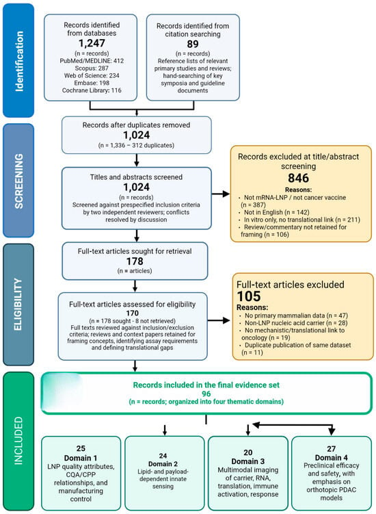
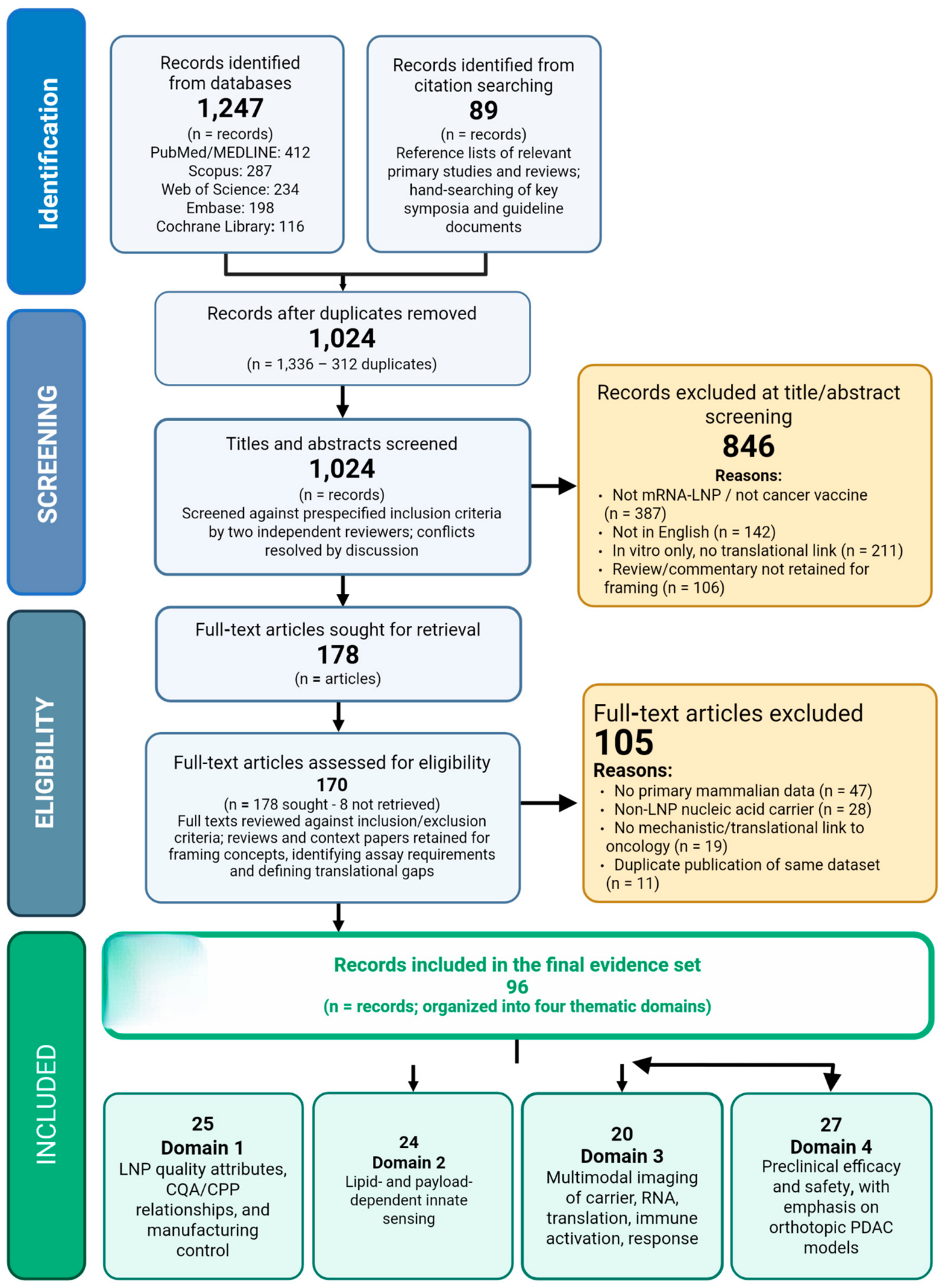
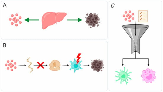
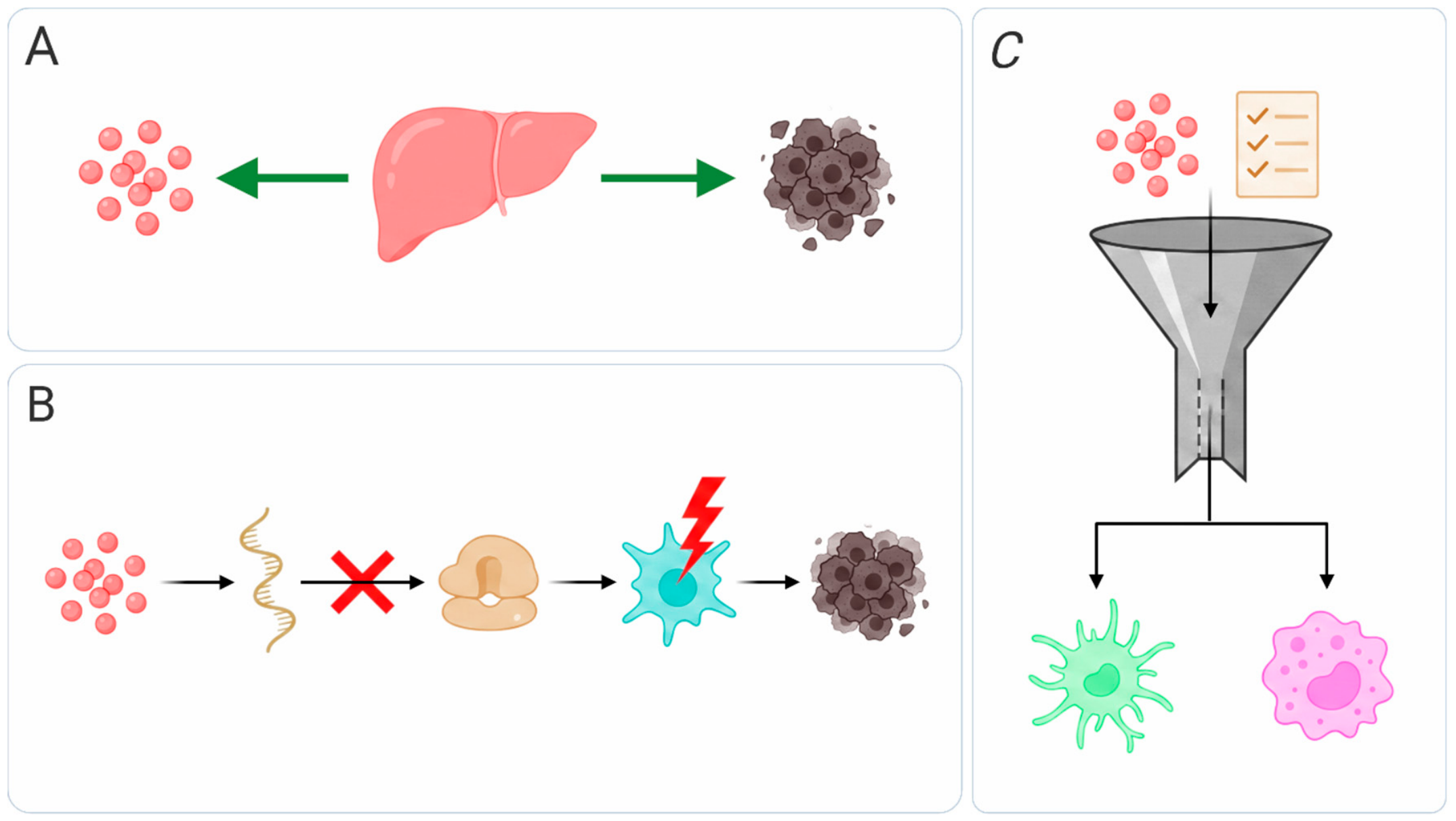
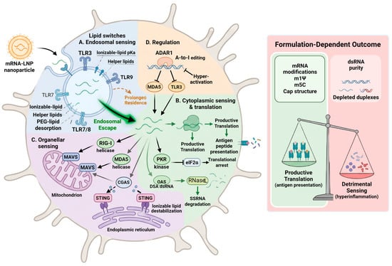
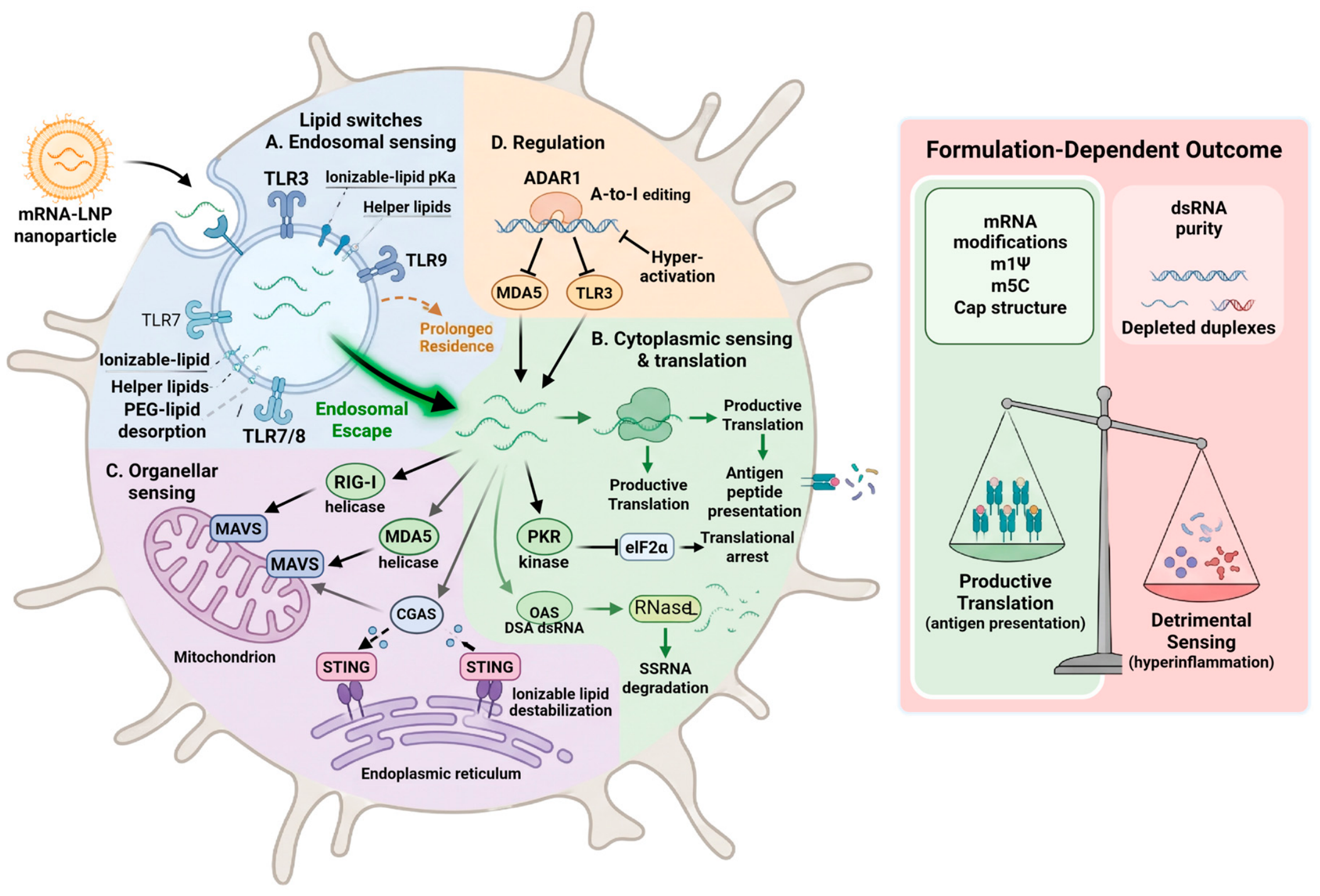
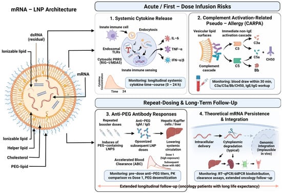
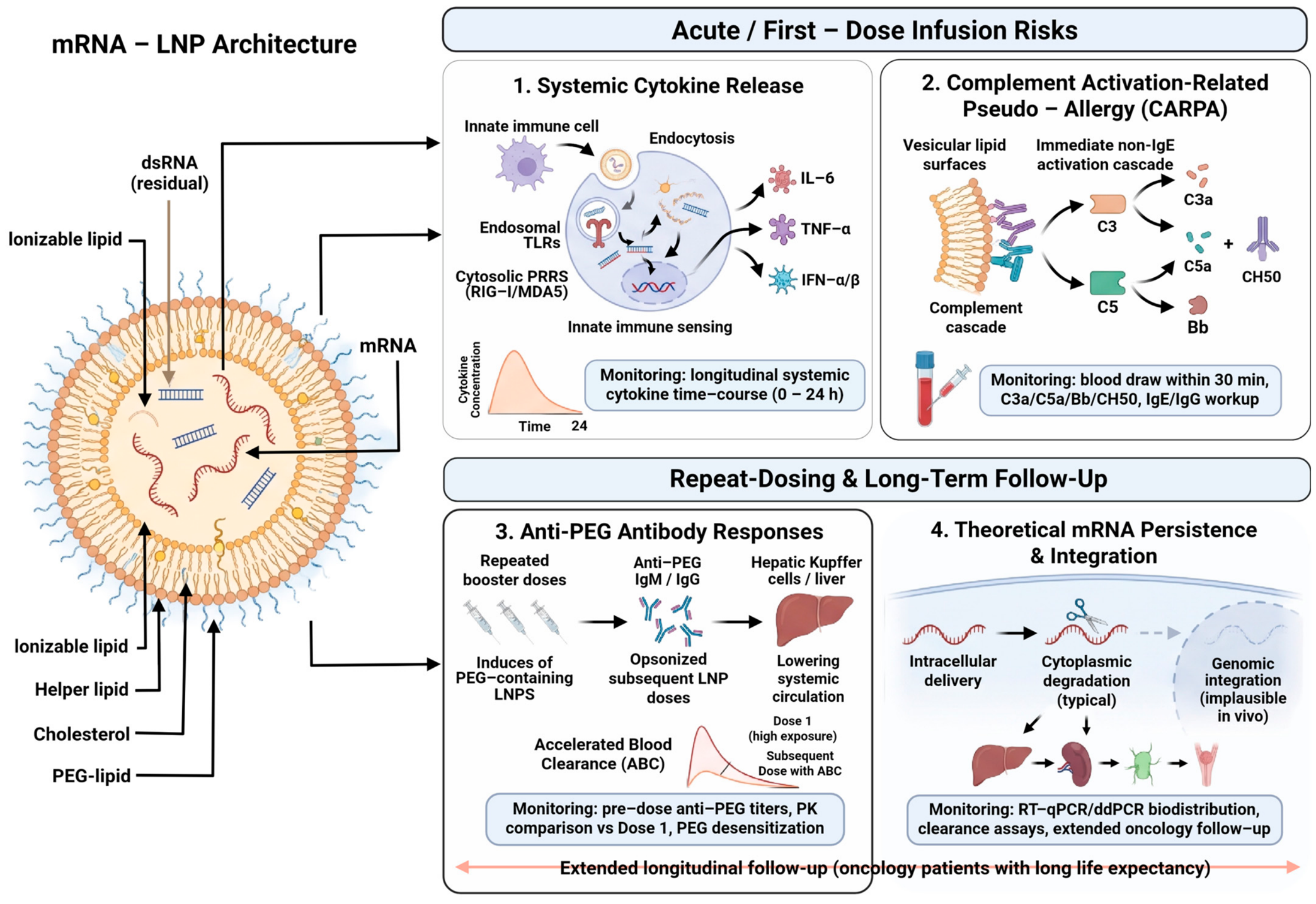

Disclaimer/Publisher’s Note: The statements, opinions and data contained in all publications are solely those of the individual author(s) and contributor(s) and not of MDPI and/or the editor(s). MDPI and/or the editor(s) disclaim responsibility for any injury to people or property resulting from any ideas, methods, instructions or products referred to in the content. |
© 2026 by the authors. Licensee MDPI, Basel, Switzerland. This article is an open access article distributed under the terms and conditions of the Creative Commons Attribution (CC BY) license.