


Abstract
Animal bile has been used for millennia in traditional Chinese medicine as a cough suppressant and bronchodilator. As the primary functional constituents of bile, bile acids (BAs) are believed to mediate these physiological effects. Although the role of BAs in enterohepatic circulation is well-characterized, and recent advances have highlighted their immunomodulatory functions, their pathophysiological significance in the respiratory system has been largely overlooked. This review synthesizes emerging evidence on the multidimensional roles of BAs and their receptor-mediated signaling pathways, with a focus on their contribution to the pathogenesis of major respiratory diseases such as asthma, pulmonary fibrosis, and lung injury.
1. Introduction
Bile, a yellow–green viscous fluid synthesized by hepatocytes, exhibits diverse physiological functions and therapeutic potential [1]. Traditional Chinese medicine (TCM) has long harnessed animal bile for medicinal purposes, with sources ranging from mammals (bovine, porcine, bear) to fish, reptiles, and birds, encompassing up to 44 distinct species [2]. Historically, animal bile was used in ancient China to relieve cough and promote choleresis.
Bile acids (BAs) and their salts are the primary functional constituents of bile, mediating most of its physiological effects [3,4]. Beyond their canonical role in emulsifying and absorbing dietary lipids, BAs function as pleiotropic signaling molecules with anti-inflammatory and immunomodulatory properties [5]. For instance, two lithocholic acid (LCA) derivatives, 3-oxoLCA and isoalloLCA, have been demonstrated to regulate T cell differentiation by suppressing Th17 and promoting regulatory T (Treg) cells [6]. Similarly, 3β-hydroxy-deoxycholic acid attenuates immunostimulatory responses in dendritic cells via modulation of farnesoid X receptor (FXR) activity [7]. Collectively, these findings underscore the pivotal role of BAs and their receptors in shaping immune homeostasis, a process intimately linked to respiratory function. This review provides a comprehensive update on the roles and mechanisms of BAs and their receptors in respiratory diseases, aiming to inform future therapeutic strategies and translational applications.
2. Synthesis, Metabolism, and Enterohepatic Circulation of BAs
2.1. Synthesis and Metabolism of BAs
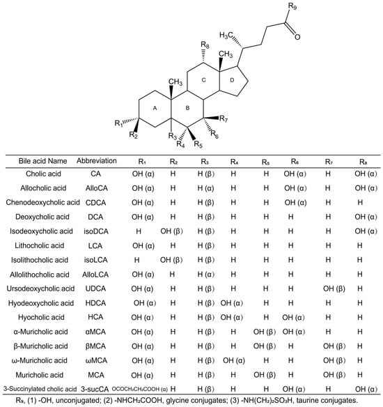
Synthesized from cholesterol in the liver and further modified by gut microbiota, BAs are amphipathic molecules containing both hydrophilic and hydrophobic moieties, conferring detergent-like properties [8]. This structural amphipathicity underpins their classical role in digestion and absorption, as well as their emerging functions as signaling molecules that regulate systemic metabolism and immunity [9]. BA structure varies in side-chain length, carboxylation, hydroxylation, conjugation status, and A/B ring stereochemistry (cis vs. trans), rendering them among the most structurally diverse biomolecules in nature [10,11] (Figure 1).
Figure 1.
Chemical structures of BAs. Chemical structure and group position of each common BA.
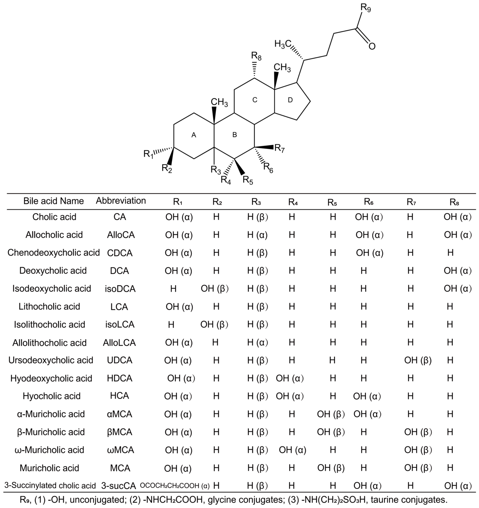
BAs are broadly classified into primary and secondary BAs. As illustrated in Figure 2, primary BAs, cholic acid (CA) and chenodeoxycholic acid (CDCA), are synthesized from cholesterol in the liver via the classical pathway and alternative pathways [12]. The classical pathway generates roughly equal amounts of CA and CDCA, accounting for over 90% of total BA production under physiological conditions [13,14]. In rodents, most CDCA is rapidly converted into muricholic acids (MCAs) [5]. Following synthesis, BAs are conjugated with taurine (T) or glycine (G) via bile acid-CoA synthetase (BACS) and bile acid coenzyme A: amino acid N-acetyltransferase (BAAT) in the liver [15], a process that enhances their solubility and reduces toxicity.
Figure 2.
Synthesis and metabolism of BAs in vivo. (a) Liver: Cholesterol is converted to primary BAs (e.g., CA and CDCA) via the classical and alternative pathways. (b) Intestine: Gut microbiota convert primary BAs to secondary BAs .
In the intestine, Bile salt hydrolase (BSH) deconjugates primary BAs by hydrolyzing glycine and taurine conjugates [16]. BSH enzymes are predominantly expressed in Gram-positive Firmicutes (e.g., Lactobacillus, Enterococcus) and certain Gram-negative Bacteroidetes, facilitating BA deconjugation and reducing BA toxicity [17]. Subsequent 7α/β-dehydroxylation, catalyzed by enzymes mainly found in Clostridia and Eubacteria, generates secondary BAs [18]. Gut microbiota also mediate BA oxidation and epimerization via hydroxysteroid dehydrogenases, which reversibly oxidize and reduce the 3α-, 7α-, or 12α-hydroxyl groups [19]. Numerous bacterial species, including Eggerthella lenta, Clostridium scindens, Clostridium hiranonis, Clostridium hylemonae [20], Escherichia coli, and Bacteroides fragilis, participate in BA oxidoreduction [21]. Additionally, Bacteroides, Eubacterium, and Lactobacillus contribute to BA esterification, while Clostridium, Fusobacterium, Peptococcus, and Pseudomonas engage in desulfurization [13]. Thus, the gut microbiota profoundly enrich BA diversity and regulate BA synthesis and transport.
2.2. Enterohepatic Circulation of BAs
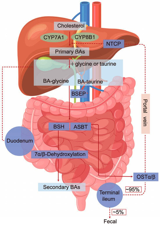
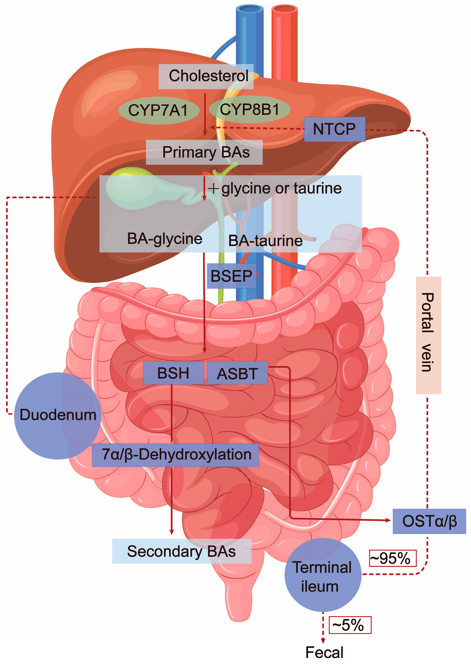
BAs synthesized in hepatocytes are actively secreted across the canalicular membrane into bile and stored in the gallbladder. Postprandial release of cholecystokinin triggers gallbladder contraction, releasing bile into the duodenum, where BAs form mixed micelles with dietary lipids to facilitate absorption [22]. The majority of BAs are then absorbed by the ileal epithelium via the apical sodium-dependent bile acid transporter (ASBT) and transported into the portal circulation via the heterodimeric organic solute transporter α/β (OSTα/β) [23]. Upon reaching the liver, BAs are taken up by hepatocytes primarily via the sodium taurocholate cotransporting polypeptide (NTCP) on the sinusoidal membrane, thereby completing the enterohepatic circulation (Figure 3). Approximately 95% of the BA pool is recycled through this pathway, with the remaining 5% (approximately 0.5 g/day) excreted in feces and replaced by de novo hepatic synthesis [22]. The entire BA pool recirculates 4 to 12 times daily, ensuring maintenance of a stable BA concentration and composition critical for normal physiological function [22,24].
Figure 3.
Enterohepatic circulation of BAs. BAs are predominantly secreted by BSEP and subsequently absorbed by the ileal epithelium via ASBT, then transported to the portal vein circulation via OSTα/β. Reabsorbed BAs enter hepatocytes through NTCP and are returned to the liver via enterohepatic circulation. Small amounts of primary BAs also reach the colon, where gut microbiota convert them to secondary BAs. Approximately 95% of the BAs are reabsorbed into the hepatic portal vein, completing the enterohepatic cycle.
3. Homeostatic Regulation of BA Metabolism
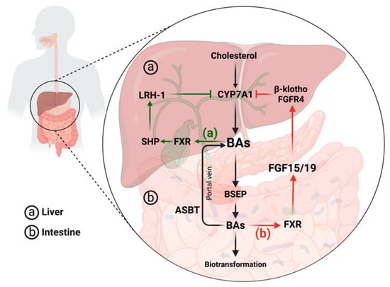
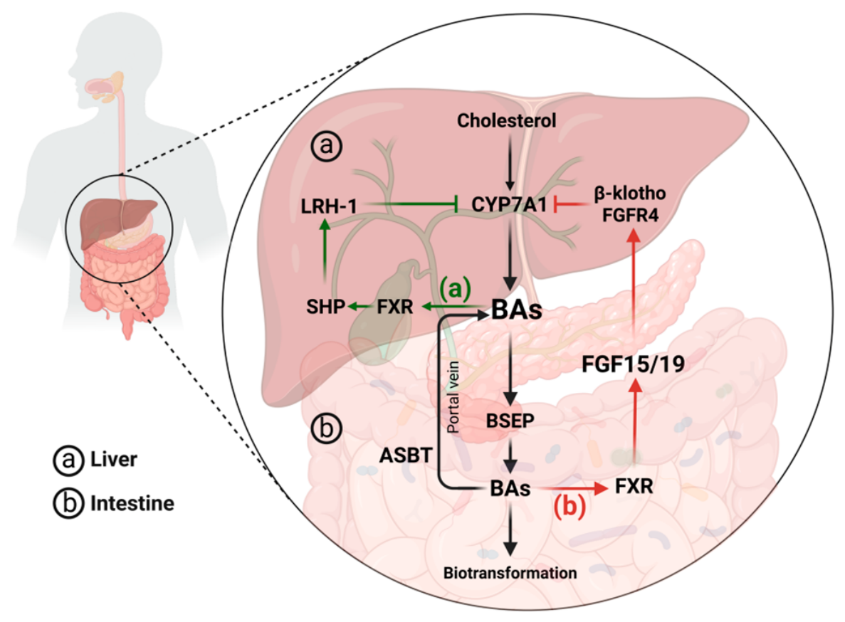
The enzymes and transporters involved in the synthesis, metabolism, and transport of BAs are chiefly regulated by BA receptors [25]. Of these, FXR is one of the most studied receptors. The biosynthesis of BAs is primarily regulated by two negative feedback pathways, both of which involve the activation of FXR [26]. The BAs synthesized in the liver are absorbed into the small intestine by ASBT. In the enterocyte, BAs activate the nuclear FXR, which leads to the subsequent release of fibroblast growth factor (FGF) 19 (or its rodent ortholog FGF15) [27]. FGF15/19 travels to the liver, binding to the fibroblast growth factor receptor 4 (FGFR4) β-Klotho complex and inhibiting CYP7A1 [28]. Meanwhile, increased BA production in the liver promoted hepatic FXR-small heterodimer partner (SHP) signaling [9]. SHP suppresses the expression of the gene CYP7A1 and reduces the production of BAs by binding to liver receptor homolog-1 (LRH-1) [29] (Figure 4). These two pathways are crucial for maintaining BA homeostasis and preventing excessive BA synthesis. However, emerging evidence suggests that local BA receptor signaling in extrahepatic tissues, including the lung, may operate through distinct, tissue-specific regulatory mechanisms that remain poorly defined [30].
Figure 4.
FXR-mediated BA negative feedback regulatory mechanism. (a) Liver: FXR-SHP pathway. Increased hepatic BA production enhances FXR-SHP signaling. SHP inhibits CYP7A1 expression by binding to LRH-1, reducing BA synthesis. (b) Intestine: FXR-FGF15/19 pathway. BAs are absorbed into the small intestine via ASBT. In enterocytes, they activate FXR, leading to FGF19/15 release. FGF15/19 inhibits CYP7A1 by binding to the FGFR4 β-Klotho complex. Arrowheads indicate promotion, and blunt-ended lines indicate inhibition.
4. The Gut–Lung Axis as a Target for Pulmonary Disease Treatment
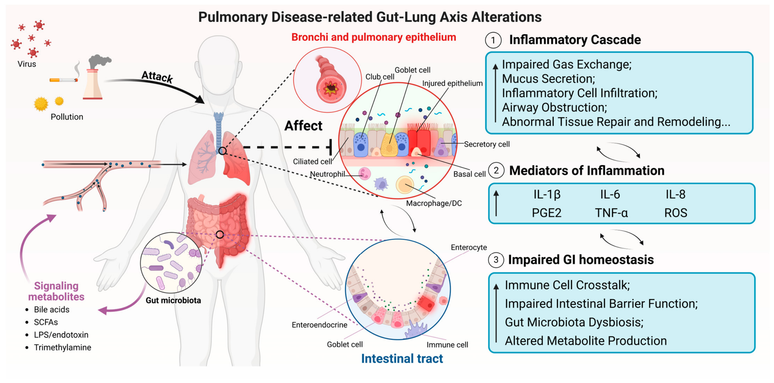
Despite the lung and the intestine having physiological distinctions, the gastrointestinal and respiratory tracts originate from a shared embryonic source and exhibit several anatomical similarities. Recent clinical research has substantiated that gut microbiota significantly influence the etiology and course of respiratory diseases [31,32]. Multiple pulmonary diseases frequently coincide with gut microbiota dysbiosis, potentially linked to disease progression [33]. A mutual regulatory process, or crosstalk, exists between the gut and the lungs, called the gut–lung axis [34].
The gut–lung axis suggests that gut microbiota and their metabolites may connect the intestine and pulmonary tissue via the bloodstream and lymphatic system [35], producing bidirectional effects. The gut microbiota may modify metabolic homeostasis, thereby affecting several pathways, including inflammation and immunology, and influencing the progression of pulmonary diseases [36]. Metabolites produced by the gut microbiota, such as BAs, short-chain fatty acids, and lipopolysaccharides (LPS), can influence the pulmonary immune microenvironment via systemic circulation [37,38,39] (Figure 5).
Figure 5.
The gut–lung axis: Regulation of pulmonary function via gut microbiota-metabolites. Impaired gut function increases the production and influx of pro-inflammatory mediators into systemic circulation and weakens immune responses, further exacerbating respiratory diseases. Microbiota-metabolites affect lung function through the gut–lung axis and reduce inflammatory responses in the respiratory tract (e.g., bronchial smooth muscle contraction, mucus secretion and inflammatory cell infiltration). Arrowheads indicate promotion, blunt-ended lines indicate inhibition, and dashed lines indicate the magnified regions.
Several gut species have now been linked to BA remodeling in the context of pulmonary pathology. Clostridiales and Eubacterium, which carry the BA-inducible (bai) operon for 7α-dehydroxylation (Section 2.1), generate deoxycholic acid (DCA) and LCA [18]. In a separate line of evidence, DCA and its receptor, Takeda G protein-coupled receptor 5 (TGR5), increase host resistance to influenza virus [40]. Bacteroides dorei enhances BSH activity to restore secondary BA production, and the BAs it produces alleviate influenza virus infection in mice via TGR5 signaling [41]. Gut bacteria can also conjugate BAs with amino acids other than glycine and taurine to produce microbial amino acid-conjugated BAs [42], which circulate systemically as host-microbial co-metabolites [43]. In a dietary model, inulin fibre restructured the microbiota-derived BA pool and elevated CA; CA then drove type 2 inflammation at both the intestinal and pulmonary barrier surfaces, dependent on interleukin (IL)-33 and ILC2s. Crucially, knocking out the BSH gene in a single colonizing species (Bacteroides ovatus) abolished both the elevation of cholic acid and the pulmonary inflammation [44].
Quantitative profiling studies further demonstrate that the BA pool undergoes measurable remodeling in respiratory diseases. At the systemic level, total BAs were significantly increased in a rat asthma model and negatively correlated with the IFN-γ/IL-4 ratio, suggesting a shift toward T-helper 2 cells (Th2); levels normalized after treatment, paralleling gut microbiota remodeling and improved pulmonary inflammation [45]. In severe COVID-19, primary BA synthesis is selectively depleted, with reduced CDCA and G-CDCA in elderly patients [46]. At the airway level, these changes can be anchored to absolute concentrations: tracheal aspirates from ventilated patients contained 6.2 ± 2.1 versus 1.1 ± 0.4 µmol/L/g of total BAs in those with and without ventilator-associated pneumonia [47], and bronchoalveolar lavage fluid (BALF) from lung transplant recipients reached a median of 1.6 µmol/L (range 0–32) in those who later developed bronchiolitis obliterans syndrome versus 0.3 µmol/L (range 0–16) in those who did not [48]. At the tissue level, data are sparse. Profiling of lung homogenates from obese mice with allergic airway disease revealed selective elevation of βMCA and tβMCA, although the values were presented as relative abundances rather than absolute concentrations [49]. Because circulating BAs are continuously delivered to the lung through the pulmonary circulation, their presence in the pulmonary compartment is expected. What remains to be established is which individual BA species are present, at what concentrations, and to what extent they derive from systemic circulation versus local reflux or aspiration.
Despite these open questions, BAs remain among the most attractive candidates for mediating gut–lung crosstalk, as they are uniquely positioned at the intersection of metabolism and immunity [50]. Their ability to activate specific nuclear and G protein-coupled receptors expressed in pulmonary immune and structural cells provides a direct mechanistic link between intestinal BA metabolism and respiratory health [51].
5. Role of BAs and Their Receptors in Respiratory Diseases
5.1. Role of BA Receptors in the Pathogenesis of Respiratory Diseases
5.1.1. FXR: A Master Regulator of Pulmonary Inflammation
In the respiratory system, activation of BA receptors can significantly modulate host metabolism and immune function [52]. Among these, FXR is the most extensively characterized target, with accumulating evidence underscoring its pivotal role in modulating respiratory pathologies through immunometabolic crosstalk. TGR5 plays a crucial role in inflammation and immune regulation [53], but more studies are needed to elucidate its mechanism of action and effects in respiratory diseases.
FXR, encoded by the NR1H4 gene and belonging to the nuclear receptor superfamily, is a BA ligand-activated transcription factor [54] expressed mainly in the liver and gastrointestinal tract but also in non-classical BA-targeted tissues such as the lungs and blood vessels [51].
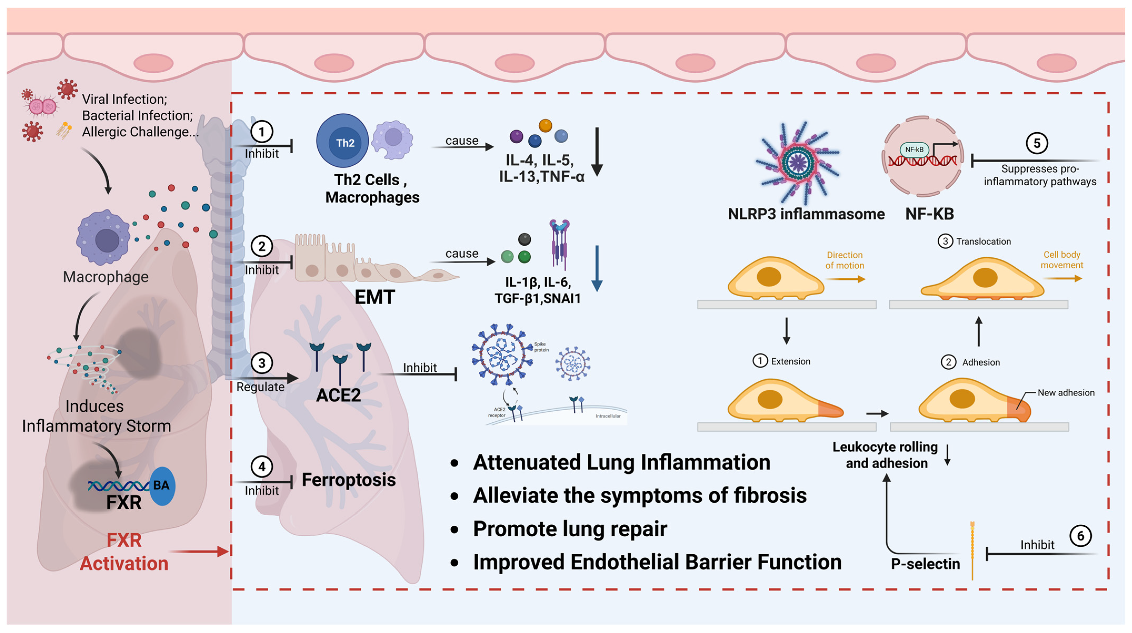
FXR regulates inflammatory responses: FXR activation in monocytes and macrophages suppresses the toll-like receptor 9 (TLR9)-dependent inflammatory responses, regulates dendritic cell differentiation and inhibits the production of pro-inflammatory factors by natural killer T cells [55,56]. In the LPS-induced mouse model of acute lung injury (ALI), FXR activation can inhibit pro-inflammatory gene expression [57]. In the ovalbumin (OVA)-induced acute rat asthma model, FXR may block the infiltration of inflammatory cells into healthy lung tissue by suppressing the secretion of IL-4, IL-5, and IL-13 from activated Th2 and tumor necrosis factor-α (TNF-α) from activated alveolar macrophages [58], thereby exerting anti-inflammatory effects.
FXR regulates cell fate: Various infections trigger a storm of pro-inflammatory cytokines, including IL-6, leading to diffuse alveolar damage in patients [59]. IL-6 promotes ferroptosis and impairs regeneration of alveolar epithelial type II cells during polyriboinosinic–polyribocytidylic acid-induced murine lung injury by regulating the FXR-PKM2-YAP axis. FXR agonist improves polyriboinosinic–polyribocytidylic acid-associated murine lung inflammation, ferroptosis, and regeneration in mice [60]. In the LPS-induced mouse model of ALI, FXR activation can promote the proliferation of lung microvascular endothelial cells and improve lung permeability and regeneration [57].
FXR regulates viral infection: In the context of coronavirus disease 2019 (COVID-19), the downstream signaling pathways of FXR can regulate the expression of angiotensin-converting enzyme 2 (ACE2), as well as modulate the activation of the immune system and the release of inflammatory factors, which may contribute to the prevention and treatment of COVID-19 [61].
Thus, through its diverse roles in inflammation, tissue regeneration, and viral infection, FXR can be a biomarker and therapeutic target for these respiratory diseases (Figure 6).
Figure 6.
Mechanism of FXR action in lung diseases and its signaling regulatory network. After pathogens invade and cause lung injury, BA activates FXR, which regulates the epithelial–mesenchymal transition (EMT) process and curbs lung fibrosis. Additionally, FXR suppresses pro-inflammatory pathways like NF-κB and NLRP3, activates FOXM1 to boost lung repair factor expression, and adjusts cytokine release. Through these actions, FXR plays a key role in repairing the lungs and fighting inflammation. Arrowheads indicate promotion, blunt-ended lines indicate inhibition. Created in BioRender. Wan, J. (2026) https://BioRender.com/wb217cg.
5.1.2. TGR5: Gatekeeper of Immune Tolerance
TGR5, a member of the G protein-coupled receptor family, is expressed in multiple tissues, including the lung [62]. Activation of TGR5 can specifically inhibit the TLR4-NF-κB signaling axis and suppress the expression levels of LPS-induced pro-inflammatory factors, such as TNF-α and IL-6 [63]. TGR5 can also increase the ratio of IL-10 to IL-12 expression by activating protein kinase A signaling, effectively inhibiting the polarization of macrophages towards the pro-inflammatory M1 phenotype [53]. In addition, TGR5 promotes nitric oxide production by phosphorylating endothelial nitric oxide synthase, leading to the downregulation of adhesion molecule expression and monocyte adhesion, thereby hindering leukocyte transport and contributing to the anti-inflammatory effects of TGR5 [64]. It also showed similar effects to FXR in COVID-19 [12]. Overall, activating TGR5 exerts anti-inflammatory and immunomodulatory effects through regulating macrophage M1/M2 polarization and promoting vascular protection, among other mechanisms, highlighting its potential in the treatment of respiratory diseases.
5.1.3. Beyond FXR and TGR5: An Expanding Spectrum of Receptors
FXR and TGR5 are the most extensively studied BA receptors. Other BA receptors can also directly or indirectly influence the physiological functions of the respiratory system [9].
RORγt: The microbial secondary BA isolithocholic acid (isoLCA) is a reversible inhibitor of RORγt [65]. Targeting RORγt may therefore offer a therapeutic approach for non-type 2 asthma: in a house dust mite (HDM) model, a RORγt inhibitor blocked both IL-17 and IL-22 as well as airway hyperresponsiveness and neutrophilia, an effect not reproduced by anti-IL-17 alone [66].
S1PR2: BAs signal through S1PR2 [67], which is expressed in various lung cell types and regulates inflammatory cytokine release, barrier integrity, and immune cell migration. In an ovalbumin-induced asthma model, pharmacological inhibition of S1PR2 reduced IL-4, IL-5, and IL-13 levels in BALF and attenuated lung inflammation [68,69]. More recently, isoLCA was shown to ameliorate lung inflammation through S1PR2-dependent metabolic rewiring of alveolar macrophages, identifying a microbiota-dependent BA that acts as a selective airway immunomodulator [70].
Bitter taste receptors (TAS2Rs): BAs are physiological ligands for human and mouse TAS2Rs [71]. TAS2Rs are expressed on airway smooth muscle cells, and their activation induces localized calcium signaling that relaxes airway smooth muscle and reverses obstruction in a mouse model of asthma [72].
PPARγ, PXR, and CAR can also bind BAs or modulate BA metabolism, but direct evidence for their involvement in pulmonary BA signaling remains limited. In summary, BA receptors beyond FXR and TGR5 can shape the pulmonary immune microenvironment, although studies in this area remain limited. Whether specific microbial BAs reach their targets and activate these receptors in the lung remains to be determined.
5.2. Pathophysiological Roles of BAs in Respiratory Diseases
Building upon the receptor-mediated mechanisms discussed above, this section systematically examines the roles of BAs in specific respiratory diseases. While BA receptor activation generally exerts protective effects, the net impact of BAs in each disease context depends on multiple factors including BA composition, concentration, duration of exposure, and the underlying pathological milieu. The following subsections synthesize current evidence across major respiratory conditions, highlighting both therapeutic potential and, where relevant, pathogenic contributions.
5.2.1. Asthma
Asthma is a common chronic inflammatory airway disease affecting an estimated 260 million individuals worldwide, a number projected to reach 275 million by 2050, driven largely by population growth and modifiable risk factors such as high body mass index, smoking, and air pollution [73]. The disease is characterized by variable respiratory symptoms and airflow limitation, often associated with airway inflammation and remodeling [74]. Emerging evidence suggests that BAs, including those produced via microbial modification, exert protective effects on allergic airway inflammation through multiple mechanisms.
Conjugated BAs, such as tauroursodeoxycholic acid (T-UDCA), significantly attenuate inflammatory responses, mucus metaplasia, and airway hyperresponsiveness in asthma models. These effects are partly mediated by alleviation of the unfolded protein response (UPR) [75]. Specifically, taurohyodeoxycholic acid (T-HDCA) improves allergic asthma by regulating the immune balance of CD4+ T cell subsets [76]. Mechanistically, conjugated BAs preferentially interact with activated transcription factor 6α (ATF6α), a key UPR transducer, thereby decreasing allergen-induced UPR activation and resolving allergic airway disease without significantly engaging BA receptors [77]. Clinical relevance is supported by quantitative mRNA analysis of airway epithelial brushings, which demonstrated significant upregulation of multiple ATF6α-related transcripts in asthma patients compared to healthy controls [77]. These findings suggest a novel therapeutic avenue for allergic asthma using select conjugated BAs.
Unconjugated BAs also show therapeutic promise. CDCA ameliorates ovalbumin-induced histological changes by attenuating inflammatory cell infiltration, mucus hypersecretion, and goblet cell hyperplasia [78]. UDCA reduces eosinophilic airway inflammation in sensitized mice via ligation of dendritic cell nuclear FXR [79]. Moreover, UDCA ameliorates airway remodeling, suppresses apoptosis of airway epithelial cells, and dampens Th2 immune responses, collectively contributing to its protective effects in allergic airway inflammation [80].
5.2.2. Pulmonary Fibrosis (PF)
PF is a chronic and progressive pulmonary disease characterized by progressive deposition of extracellular matrix proteins, ultimately leading to respiratory failure and death [81]. Several BAs have demonstrated anti-fibrotic properties in preclinical models.
T-CDCA significantly reduces TNF-α and tissue inhibitor of metalloproteinases (TIMP)-2 expression in pulmonary tissues of fibrotic mice, accompanied by decreased alveolar inflammatory exudate [82]. T-UDCA accelerates recovery from crystalline silica-induced lung injury, an effect linked to reduction of IFN-γ-producing CD103− TEM-like and IL-17A- producing CD103+ TRM-like Treg cells in the lung [83].
EMT is a process by which epithelial cells lose polarity and intercellular adhesion and transition to mesenchymal cells, implicated in bleomycin (BLM)-induced PF [84,85]. In BLM-challenged mouse lungs, both the ERS marker GRP78 and the UPR marker CCAAT/enhancer-binding protein homologous protein (CHOP) were upregulated. T-UDCA pretreatment suppresses the upregulation of GRP78 and CHOP, suggesting that inhibition of ERS contributes to its anti-fibrotic effects [86]. Additionally, T-UDCA attenuates BLM-induced TGF-β1 upregulation and Smad2/3 activation, inhibits α-SMA upregulation, and prevents E-cadherin downregulation, collectively indicating suppression of EMT [87].
Obeticholic acid (OCA), a semi-synthetic FXR agonist and CDCA analogue, protects against BLM-induced lung damage by suppressing EMT, inflammation, and collagen deposition [88]. Notably, OCA administration after PF establishment still significantly improves pulmonary function [89]. OCA also alleviates histopathological changes, oxidative stress, and inflammation induced by nitrogen mustard [90], highlighting its therapeutic potential across diverse fibrotic insults.
5.2.3. Acute Lung Injury (ALI)
ALI and its more severe form, ARDS, are critical clinical syndromes with high morbidity and mortality [91]. ALI is characterized by neutrophil infiltration, alveolar-capillary barrier disruption, diffuse alveolar damage, non-cardiogenic pulmonary edema, and robust local and systemic inflammation, culminating in progressive respiratory failure and hypoxemia [92,93].
Sepsis-induced ALI. Sepsis is the most common risk factor for ALI [94]. UDCA alleviates sepsis-induced lung injury by attenuating inflammation, oxidative stress, and pulmonary barrier dysfunction. Mechanistically, these effects are linked to inactivation of the STING pathway and suppression of PANoptosis-like cell death [95].
Fat embolism syndrome (FES)-induced ALI. Pre-treatment with UDCA attenuates pathological and biochemical changes in FES-induced ALI [96]. UDCA significantly inhibits inflammatory responses and suppresses TNF-α and IL-1β expression in vitro by inhibiting p38 MAPK/NF-κB signaling and activating FXR [97]. These findings provide new evidence for the therapeutic potential of UDCA in ALI.
LPS-induced ALI. CDCA significantly ameliorates lung tissue pathology in LPS-induced ALI mice, modulates cytokine profiles, and alters expression of relevant targets [98]. Both CDCA and UDCA mitigate endotoxin-induced lung injury in rats through modulation of AQP1 and AQP5 expression, reduction of oxidative stress, regulation of apoptotic pathways (BAX, caspase-3, BCL-2), and attenuation of NF-κB-driven pro-inflammatory activity [99]. OCA prevents LPS-induced ALI by enhancing lung FXR activity and blocking phosphorylation of ERK1/2, JNK, p38, and Akt [100].
Bacterial pneumonia-induced ALI. DCA exhibits rapid bactericidal activity against Streptococcus pneumoniae in vitro and reduces pneumococcal colonization [101]. In LPS-induced ALI, bear bile powder components (T-UDCA and T-CDCA) alleviate lung injury by inhibiting NF-κB activation and reducing CD14 expression [102]. Specifically, T-CDCA reduces pulmonary edema and neutrophil infiltration in Staphylococcus aureus-infected lung, downregulates HMGB1 expression, and attenuates inflammatory mediator production through modulation of TLR2 signaling in macrophages [103]. These findings position T-CDCA as a promising candidate for preventing and treating bacterial ALI.
5.2.4. Viral Pneumonia
Viral pneumonia, commonly community-acquired, is caused by influenza virus, parainfluenza virus, respiratory syncytial virus, human metapneumovirus, and adenovirus [104]. BAs have emerged as important modulators of antiviral host defense.
Influenza. Secondary BAs act on distal lung tissue by inhibiting NLRP3 inflammasome activation via the TGR5-cAMP-PKA axis, thereby reducing lung inflammation during later infection stages [41]. DCA, through TGR5 signaling, increases host resistance to influenza virus by suppressing virus replication and neutrophil-dependent tissue damage [40].
COVID-19. The COVID-19 pandemic, caused by the severe acute respiratory SARS-CoV-2, represents the most extensive global health crisis in a century. Recent evidence links BAs to COVID-19 pathogenesis and treatment [61]. UDCA reduces ACE2 levels in nasal epithelium by inhibiting FXR, thereby decreasing susceptibility to SARS-CoV-2 infection [105,106,107]. Beyond preventing spike-ACE2 binding, UDCA inhibits aberrant airway epithelial cell migration and enhances the restoration of the epithelial basal layer [108]. Derivatives of UDCA also enhance ACE2 exopeptidase activity, suggesting activation of the ACE2/Ang(1-7)/MasR axis to counteract COVID-19-related inflammation [109]. Additionally, CDCA significantly inhibits the activity of Nsp15, a coronavirus uridine-specific endoribonuclease essential for viral replication [110]. Thus, BAs protect against COVID-19 through multiple complementary mechanisms.
5.2.5. Lung Cancer
Lung cancer is the leading cause of cancer-related deaths worldwide, comprising non-small cell lung cancer (NSCLC) and small cell lung cancer (SCLC). Lung adenocarcinoma (LUAD), a major NSCLC subtype, accounts for approximately 40% of cases. Emerging evidence indicates that BAs are also implicated in lung cancer, where they modulate tumor biology and treatment responses through mechanisms that vary with the BA species and the receptors involved.
Modulation of chemotherapy. Autophagy may promote resistance to doxorubicin (DOX) in LUAD [111]. UDCA attenuates tumor cell proliferation and modulates MAPK pathway phosphorylation (ERK, JNK, p38), altering Beclin1, LC3B-II/LC3B-I, and P62 expression ratios to diminish DOX-induced autophagy [112]. This suggests UDCA as an adjunct to enhance chemotherapy efficacy.
Modulation of immunotherapy. BA metabolism shapes the efficacy of immune checkpoint inhibitors in NSCLC patients [113]. T-LCA enhances anti-tumor immunity by boosting T cell activation and cytotoxic function while modulating immune checkpoints [113]. UDCA dose-dependently suppresses herpesvirus entry mediator expression and slows tumor growth in mouse lung cancer models [114].
Direct anti-tumor effects. CDCA inhibits LUAD cell proliferation, migration, and invasion while promoting apoptosis. Mechanistically, CDCA suppresses integrin α5β1 signaling by inhibiting expression of α5 and β1 subunits and phosphorylated FAK, with concomitant increase in p53 levels, a downstream target of this pathway [115]. These findings highlight the therapeutic potential of BAs in LUAD.
Context-dependent cellular effects. DCA promotes NSCLC cell migration in a TGR5-dependent manner [116]. TGR5 signaling is itself ligand-biased: in NSCLC cells, the Gs-biased agonist INT-777 inactivates YAP and inhibits proliferation, whereas the β-arrestin 1-biased agonist R399 activates YAP and promotes growth [117]. These findings illustrate the ligand-biased and context-dependent nature of TGR5 signaling in NSCLC.
In summary, in lung cancer, BAs cannot be regarded as a single class of drugs. Whether BAs produce protective or harmful effects may depend on the specific type of BA, the receptors they act upon, and the cellular environment.
5.2.6. Protective Effects of BAs on Other Respiratory Diseases
Cystic fibrosis (CF). CF is an autosomal recessive disorder caused by mutations in the CF transmembrane conductance regulator (CFTR) gene, leading to defective Cl− channels in mucus- and sweat-producing cells [118]. The lungs are most severely affected, with alterations in airway surface liquid (ASL) increasing mucus viscosity and impairing epithelial function, ultimately compromising mucociliary clearance and bacterial elimination [119]. UDCA stimulates Cl− channels in normal airway epithelium [120], and inhibits transepithelial Na+ transport in both normal and CF bronchial epithelial cells without altering transporter expression. The resulting increase in ASL height suggests therapeutic potential for UDCA in CF [121].
Pulmonary arterial hypertension (PAH). PAH is a fatal disease characterized by progressive pulmonary vascular remodeling [122]. OCA exerts significant therapeutic effects in monocrotaline-induced PAH, improving exercise capacity and reducing right ventricular hypertrophy and pulmonary vascular remodeling [123,124].
Pulmonary edema. In LPS-induced pulmonary edema, UDCA decreases BALF TNF-α/IL-1β levels and lung wet/dry ratios. It stimulates alveolar fluid clearance by upregulating ENaC and Na/K-ATPase [125].
Rheumatoid arthritis-associated interstitial lung disease (RA-ILD). RA-ILD represents a severe extra-articular manifestation of rheumatoid arthritis, progressing from chronic inflammation to irreversible pulmonary fibrosis [126]. Through multiple receptor-mediated pathways, BAs modulate innate and adaptive immune responses, regulate fibrosis progression, and exert multifaceted therapeutic effects in RA-ILD [127].
Table 1 systematically summarizes BA-mediated pulmonary pathobiology mechanisms and therapeutic implications.
Table 1.
Systematic summary of BA-mediated pulmonary pathobiology mechanisms and therapeutic implications.
5.2.7. BAs and the Lungs: Therapeutic Potential Versus Risk of Injury
While BAs hold significant therapeutic potential for respiratory diseases, emerging evidence also implicates them in lung injury under certain conditions. A fundamental mechanism underlying this dual behavior is their direct, receptor-independent interaction with biological membranes. Unconjugated BAs are amphipathic weak acids that partition directly into phospholipid bilayers, where they function as protonophores and ionophores and thereby uncouple oxidative phosphorylation [128]. Mild uncoupling by BAs reduces mitochondrial H2O2 generation by 30–40% without inhibiting the electron transport chain, an antioxidant action [129]. Conversely, excessive membrane perturbation can induce Ca2+-dependent mPTP opening, mitochondrial destruction, loss of membrane potential, and ultimately apoptosis and necrosis [130,131,132]. Notably, individual BAs differ markedly: G-CDCA and T-CDCA permeabilize ANT-containing liposomes, whereas T-UDCA lacks such activity and protects mitochondria [130]. This dichotomy between membranolytic and membrane-protective BAs is a key determinant of their opposing pulmonary effects.
In addition to these direct membrane effects, other mechanisms contribute to BA-associated lung injury. BA aspiration, often secondary to gastroesophageal reflux disease, may also contribute to lung injury [133,134], through interactions with the secretory phospholipase A2 pathway, disruption of surfactant function and structure, modulation of inflammatory and local immune responses, and direct cellular toxicity [135]. Through these mechanisms, aspirated BAs disrupt immunity, promote fibrosis, impair cell viability, and trigger inflammatory cascades [136].
In summary, different BAs exhibit distinct mechanisms of action in different tissue microenvironments. This helps explain why BAs have both therapeutic potential and the risk of causing damage. Therefore, the dual role of BAs in lung health and disease necessitates rigorous investigation when developing BA-based therapeutics.
6. Conclusions and Perspectives
BAs, traditionally recognized for their role in promoting intestinal lipid digestion and absorption, have emerged as critical regulators of respiratory health and disease. Through binding to specific receptors (FXR, TGR5) and interacting with the gut microbiota, BAs modulate diverse physiological and pathological processes in the respiratory system, including antiviral and antibacterial defense, inflammation, and immune homeostasis.
Despite significant progress, several key questions remain. First, the complex crosstalk among BA receptor-triggered signaling pathways—such as MAPK, NF-κB, and TLR cascades—requires further elucidation and may reveal novel therapeutic targets. Second, understanding the cell type-specific effects of BAs on different immune and epithelial cell populations within the lung will be essential for developing targeted therapies. Third, the gut–lung axis represents an increasingly important framework for studying BAs; integrating microbiota analyses with BA profiling may uncover new mechanisms linking intestinal metabolism to pulmonary outcomes.
From a translational perspective, BA-based therapeutics face substantial challenges in moving from bench to bedside. Current evidence derives primarily from preclinical animal models, with a notable scarcity of reliable clinical data. Key questions regarding optimal dosing, formulation, patient selection, and long-term safety remain unanswered. The dual nature of BAs—protective in some contexts yet injurious in others—underscores the need for rigorous, context-dependent investigation. As new technologies (metabolomics, single-cell sequencing, organoid models) continue to advance, the association between BAs and respiratory diseases will become increasingly clear. With continued research effort, the clinical application of BA-based therapies for respiratory diseases may soon be realized.
Author Contributions
J.W.: Writing—Original Draft, Visualization. H.Z.: Writing—Review and Editing. D.H.: Visualization. D.L.: Writing—Review and Editing. J.Y.: Writing—Review and Editing. G.W.: Writing—Review and Editing. L.L.: Writing—Review and Editing. C.H.: Supervision. X.W.: Conceptualization, Funding acquisition. L.W.: Conceptualization, Writing—Review and Editing, Funding acquisition. All authors have read and agreed to the published version of the manuscript.
Funding
This research was funded by the Tianshan Talent Medical and Health Leading Talent Project of Xinjiang Uygur Autonomous Region, grant number TSYC202401A016, the Corps Science and Technology Plan Project, grant numbers 2023AB045 and 2024AB071, and the National Natural Science Foundation of China, grant numbers 82560831 and U25A20174.
Institutional Review Board Statement
Not applicable.
Informed Consent Statement
Not applicable.
Data Availability Statement
No new data were created or analyzed in this study.
Acknowledgments
The authors acknowledge the use of BioRender.com for creating Figure 6 and ChemDraw version 22.2.0 for drawing the chemical structures.
Conflicts of Interest
The authors declare no competing financial interest.
Abbreviations
BAs, bile acids; T, taurine; G, glycine; CA, Cholic acid; CDCA, Chenodeoxycholic acid; CYP7A1, cytochrome P450 family 7 subfamily A member 1; CYP27A1, cytochrome P450 family 27 subfamily A member 1; UDCA, Ursodeoxycholic acid; MCA, Muricholic acid; BACS, bile acid-CoA synthetase; BAAT, bile acid CoA: amino acid N-acyltransferase; BSEP, bile salt export pump; MRP2, multidrug resistance-associated protein 2; ASBT, apical sodium-dependent bile acid transporter; OSTα/β, organic solute transporter α/β; NTCP, sodium taurocholate cotransporting polypeptide; BSH, bile salt hydrolase; FXR, farnesoid X receptor; TGR5, takeda G protein-coupled receptor 5; FGF15/19, fibroblast growth factor 15/19; FGFR4, fibroblast growth factor receptor 4; SHP, small heterodimer partner signaling; LRH-1, liver receptor homolog-1; DCA, Deoxycholic acid; LCA, Lithocholic acid; HDCA, Hyodeoxycholic acid; SCFA, short-chain fatty acids; ALI, acute lung injury; OVA, ovalbumin; OCA, obeticholic acid; PF, pulmonary fibrosis; BLM, bleomycin; NSCLC, non-small cell lung cancer; COVID-19, coronavirus disease 2019; ACE2, angiotensin-converting enzyme 2; HDM, house dust mite; ARDS, acute respiratory distress syndrome; FES, fat embolism syndrome; SARS-CoV-2, severe acute respiratory syndrome coronavirus 2; Nsp15, nonstructural protein 15; DOX, doxorubicin; CF, cystic fibrosis.
References
- Chinese Pharmacopoeia Commission. Pharmacopoeia of the People’s Republic of China; China Medical Science Press: Beijing, China, 2025. [Google Scholar]
- Wang, D.Q.; Carey, M.C. Therapeutic uses of animal biles in traditional Chinese medicine: An ethnopharmacological, biophysical chemical and medicinal review. World J. Gastroenterol. 2014, 20, 9952–9975. [Google Scholar] [CrossRef] [Scilit]
- Hegyi, P.; Maléth, J.; Walters, J.R.; Hofmann, A.F.; Keely, S.J. Guts and Gall: Bile Acids in Regulation of Intestinal Epithelial Function in Health and Disease. Physiol. Rev. 2018, 98, 1983–2023. [Google Scholar] [CrossRef] [Scilit]
- Perino, A.; Demagny, H.; Velazquez-Villegas, L.; Schoonjans, K. Molecular Physiology of Bile Acid Signaling in Health, Disease, and Aging. Physiol. Rev. 2021, 101, 683–731. [Google Scholar] [CrossRef] [Scilit]
- Fiorucci, S.; Marchianò, S.; Urbani, G.; Di Giorgio, C.; Distrutti, E.; Zampella, A.; Biagioli, M. Immunology of bile acids regulated receptors. Prog. Lipid Res. 2024, 95, 101291. [Google Scholar] [CrossRef] [Scilit]
- Hang, S.; Paik, D.; Yao, L.; Kim, E.; Trinath, J.; Lu, J.; Ha, S.; Nelson, B.N.; Kelly, S.P.; Wu, L.; et al. Bile acid metabolites control T(H)17 and T(reg) cell differentiation. Nature 2019, 576, 143–148. [Google Scholar] [CrossRef] [Scilit]
- Campbell, C.; McKenney, P.T.; Konstantinovsky, D.; Isaeva, O.I.; Schizas, M.; Verter, J.; Mai, C.; Jin, W.B.; Guo, C.J.; Violante, S.; et al. Bacterial metabolism of bile acids promotes generation of peripheral regulatory T cells. Nature 2020, 581, 475–479. [Google Scholar] [CrossRef] [Scilit]
- Di Ciaula, A.; Bonfrate, L.; Baj, J.; Khalil, M.; Garruti, G.; Stellaard, F.; Wang, H.H.; Wang, D.Q.; Portincasa, P. Recent Advances in the Digestive, Metabolic and Therapeutic Effects of Farnesoid X Receptor and Fibroblast Growth Factor 19: From Cholesterol to Bile Acid Signaling. Nutrients 2022, 14, 4950. [Google Scholar] [CrossRef] [Scilit]
- Jia, W.; Li, Y.; Cheung, K.C.P.; Zheng, X. Bile acid signaling in the regulation of whole body metabolic and immunological homeostasis. Sci. China Life Sci. 2024, 67, 865–878. [Google Scholar] [CrossRef] [Scilit]
- Ridlon, J.M.; Gaskins, H.R. Another renaissance for bile acid gastrointestinal microbiology. Nat. Rev. Gastroenterol. Hepatol. 2024, 21, 348–364. [Google Scholar] [CrossRef] [Scilit]
- Hofmann, A.F.; Hagey, L.R.; Krasowski, M.D. Bile salts of vertebrates: Structural variation and possible evolutionary significance. J. Lipid Res. 2010, 51, 226–246. [Google Scholar] [CrossRef] [Scilit]
- Fiorucci, S.; Urbani, G.; Biagioli, M.; Sepe, V.; Distrutti, E.; Zampella, A. Bile acids and bile acid activated receptors in the treatment of COVID-19. Biochem. Pharmacol. 2024, 228, 115983. [Google Scholar] [CrossRef] [Scilit]
- Jia, W.; Xie, G.; Jia, W. Bile acid-microbiota crosstalk in gastrointestinal inflammation and carcinogenesis. Nat. Rev. Gastroenterol. Hepatol. 2018, 15, 111–128. [Google Scholar] [CrossRef] [Scilit]
- Jia, W.; Wei, M.; Rajani, C.; Zheng, X. Targeting the alternative bile acid synthetic pathway for metabolic diseases. Protein Cell 2021, 12, 411–425. [Google Scholar] [CrossRef] [Scilit]
- Fuchs, C.D.; Simbrunner, B.; Baumgartner, M.; Campbell, C.; Reiberger, T.; Trauner, M. Bile acid metabolism and signalling in liver disease. J. Hepatol. 2025, 82, 134–153. [Google Scholar] [CrossRef] [Scilit]
- Chu, H.; Song, X.; Liu, C.; Ma, T.; Cui, W. Gut microbial bile salt hydrolase as a metabolic gatekeeper in digestive homeostasis and disease. Front. Immunol. 2026, 17, 1870619. [Google Scholar] [CrossRef] [Scilit]
- Bustos, A.Y.; Font de Valdez, G.; Fadda, S.; Taranto, M.P. New insights into bacterial bile resistance mechanisms: The role of bile salt hydrolase and its impact on human health. Food Res. Int. 2018, 112, 250–262. [Google Scholar] [CrossRef] [Scilit]
- Režen, T.; Rozman, D.; Kovács, T.; Kovács, P.; Sipos, A.; Bai, P.; Mikó, E. The role of bile acids in carcinogenesis. Cell. Mol. Life Sci. 2022, 79, 243. [Google Scholar] [CrossRef] [Scilit]
- Guo, X.; Okpara, E.S.; Hu, W.; Yan, C.; Wang, Y.; Liang, Q.; Chiang, J.Y.L.; Han, S. Interactive Relationships between Intestinal Flora and Bile Acids. Int. J. Mol. Sci. 2022, 23, 8343. [Google Scholar] [CrossRef] [Scilit]
- Doden, H.; Sallam, L.A.; Devendran, S.; Ly, L.; Doden, G.; Daniel, S.L.; Alves, J.M.P.; Ridlon, J.M. Metabolism of Oxo-Bile Acids and Characterization of Recombinant 12α-Hydroxysteroid Dehydrogenases from Bile Acid 7α-Dehydroxylating Human Gut Bacteria. Appl. Environ. Microbiol. 2018, 84, e00235-18. [Google Scholar] [CrossRef] [Scilit]
- Doden, H.L.; Ridlon, J.M. Microbial Hydroxysteroid Dehydrogenases: From Alpha to Omega. Microorganisms 2021, 9, 469. [Google Scholar] [CrossRef] [Scilit]
- Li, T.; Chiang, J.Y.L. Bile Acid Signaling in Metabolic and Inflammatory Diseases and Drug Development. Pharmacol. Rev. 2024, 76, 1221–1253. [Google Scholar] [CrossRef] [Scilit]
- Jiao, T.Y.; Ma, Y.D.; Guo, X.Z.; Ye, Y.F.; Xie, C. Bile acid and receptors: Biology and drug discovery for nonalcoholic fatty liver disease. Acta Pharmacol. Sin. 2022, 43, 1103–1119. [Google Scholar] [CrossRef] [Scilit]
- Chiang, J.Y. Bile acids: Regulation of synthesis. J. Lipid Res. 2009, 50, 1955–1966. [Google Scholar] [CrossRef] [Scilit]
- Staels, B.; Fonseca, V.A. Bile acids and metabolic regulation: Mechanisms and clinical responses to bile acid sequestration. Diabetes Care 2009, 32, S237–S245. [Google Scholar] [CrossRef] [Scilit]
- Won, T.H.; Arifuzzaman, M.; Parkhurst, C.N.; Miranda, I.C.; Zhang, B.; Hu, E.; Kashyap, S.; Letourneau, J.; Jin, W.B.; Fu, Y.; et al. Host metabolism balances microbial regulation of bile acid signalling. Nature 2025, 638, 216–224. [Google Scholar] [CrossRef] [Scilit]
- Katafuchi, T.; Makishima, M. Molecular Basis of Bile Acid-FXR-FGF15/19 Signaling Axis. Int. J. Mol. Sci. 2022, 23, 6046. [Google Scholar] [CrossRef] [Scilit]
- van de Peppel, I.P.; Bodewes, F.; Verkade, H.J.; Jonker, J.W. Bile acid homeostasis in gastrointestinal and metabolic complications of cystic fibrosis. J. Cyst. Fibros. 2019, 18, 313–320. [Google Scholar] [CrossRef] [Scilit]
- Yu, H.; Nie, R.; Shen, C. The role of bile acids in regulating glucose and lipid metabolism. Endocr. J. 2023, 70, 359–374. [Google Scholar] [CrossRef] [Scilit]
- Myszor, I.T.; Lapka, K.; Hermannsson, K.; Rekha, R.S.; Bergman, P.; Gudmundsson, G.H. Bile acid metabolites enhance expression of cathelicidin antimicrobial peptide in airway epithelium through activation of the TGR5-ERK1/2 pathway. Sci. Rep. 2024, 14, 6750. [Google Scholar] [CrossRef] [Scilit]
- de Vos, W.M.; Tilg, H.; Van Hul, M.; Cani, P.D. Gut microbiome and health: Mechanistic insights. Gut 2022, 71, 1020–1032. [Google Scholar] [CrossRef] [Scilit]
- Burrows, K.; Ngai, L.; Chiaranunt, P.; Watt, J.; Popple, S.; Forde, B.; Denha, S.; Olyntho, V.M.; Tai, S.L.; Cao, E.Y.; et al. A gut commensal protozoan determines respiratory disease outcomes by shaping pulmonary immunity. Cell 2025, 188, 316–330.e12. [Google Scholar] [CrossRef] [Scilit]
- Ye, F.; Li, L.; Wang, J.; Yang, H. Advances in gut-lung axis research: Clinical perspectives on pneumonia prevention and treatment. Front. Immunol. 2025, 16, 1576141. [Google Scholar] [CrossRef] [Scilit]
- Budden, K.F.; Shukla, S.D.; Rehman, S.F.; Bowerman, K.L.; Keely, S.; Hugenholtz, P.; Armstrong-James, D.P.H.; Adcock, I.M.; Chotirmall, S.H.; Chung, K.F.; et al. Functional effects of the microbiota in chronic respiratory disease. Lancet Respir. Med. 2019, 7, 907–920. [Google Scholar] [CrossRef] [Scilit]
- Dong, Y.; He, L.; Zhu, Z.; Yang, F.; Ma, Q.; Zhang, Y.; Zhang, X.; Liu, X. The mechanism of gut-lung axis in pulmonary fibrosis. Front. Cell. Infect. Microbiol. 2024, 14, 1258246. [Google Scholar] [CrossRef] [Scilit]
- Harris, N.L.; Marsland, B.J. The gut-lung axis: Protozoa join the party. Cell 2025, 188, 275–277. [Google Scholar] [CrossRef] [Scilit]
- Qian, G.; Jiang, W.; Zou, B.; Feng, J.; Cheng, X.; Gu, J.; Chu, T.; Niu, C.; He, R.; Chu, Y.; et al. LPS inactivation by a host lipase allows lung epithelial cell sensitization for allergic asthma. J. Exp. Med. 2018, 215, 2397–2412. [Google Scholar] [CrossRef] [Scilit]
- Enaud, R.; Prevel, R.; Ciarlo, E.; Beaufils, F.; Wieërs, G.; Guery, B.; Delhaes, L. The Gut-Lung Axis in Health and Respiratory Diseases: A Place for Inter-Organ and Inter-Kingdom Crosstalks. Front. Cell. Infect. Microbiol. 2020, 10, 9. [Google Scholar] [CrossRef] [Scilit]
- Jiang, M.; Wang, J.; Li, Z.; Xu, D.; Jing, J.; Li, F.; Ding, J.; Li, Q. Dietary Fiber-Derived Microbial Butyrate Suppresses ILC2-Dependent Airway Inflammation in COPD. Mediat. Inflamm. 2024, 2024, 6263447. [Google Scholar] [CrossRef] [Scilit]
- Nagai, M.; Moriyama, M.; Ishii, C.; Mori, H.; Watanabe, H.; Nakahara, T.; Yamada, T.; Ishikawa, D.; Ishikawa, T.; Hirayama, A.; et al. High body temperature increases gut microbiota-dependent host resistance to influenza A virus and SARS-CoV-2 infection. Nat. Commun. 2023, 14, 3863. [Google Scholar] [CrossRef] [Scilit]
- He, S.; Lu, S.; Yang, T.; Ma, H.; He, Y.; Mi, J.; Yue, K.; Huang, Y.; Song, L.; Xiao, Y.; et al. Bacteroides dorei RX2020-derived bile acid alleviates influenza virus infection through TGR5 signaling. Cell Commun. Signal. 2025, 23, 382. [Google Scholar] [CrossRef] [Scilit]
- Zhao, Q.; Duan, Z.; Lai, R.; Ma, P. Novel microbially transformed bile acids: Biosynthesis, structure, and function. Pharmacol. Res. 2025, 216, 107775. [Google Scholar] [CrossRef] [Scilit]
- Reilly, E.R.; Charron-Lamoureux, V.; Mannochio-Russo, H.; Morgan, E.W.; Boyle, N.R.; Koo, I.; Coudriet, E.; Hao, F.; Mohanty, I.; Perdew, G.H.; et al. Systemic rhythmicity of host and bacterial bile acid amidates in the mouse. Cell Syst. 2026, 17, 101541. [Google Scholar] [CrossRef] [Scilit]
- Arifuzzaman, M.; Won, T.H.; Li, T.T.; Yano, H.; Digumarthi, S.; Heras, A.F.; Zhang, W.; Parkhurst, C.N.; Kashyap, S.; Jin, W.B.; et al. Inulin fibre promotes microbiota-derived bile acids and type 2 inflammation. Nature 2022, 611, 578–584. [Google Scholar] [CrossRef] [Scilit]
- Wan, J.; Zong, H.; Wang, C.; Yu, J.; Li, D.; He, D.; Liu, L.; Wang, G.; Wang, L. Multi-omics analysis reveals therapeutic mechanisms of sipeimine in ovalbumin-induced asthmatic rats. J. Ethnopharmacol. 2026, 368, 121812. [Google Scholar] [CrossRef] [Scilit]
- Liu, S.; Xu, W.; Tu, B.; Xiao, Z.; Li, X.; Huang, L.; Yuan, X.; Luo, S.; Zhou, J.; Yang, X.; et al. Significant Reduction of Chenodeoxycholic Acid and Glycochenodeoxycholic Acid in the Elderly with Severe COVID-19. Biomolecules 2025, 15, 943. [Google Scholar] [CrossRef] [Scilit]
- Wu, Y.C.; Hsu, P.K.; Su, K.C.; Liu, L.Y.; Tsai, C.C.; Tsai, S.H.; Hsu, W.H.; Lee, Y.C.; Perng, D.W. Bile acid aspiration in suspected ventilator-associated pneumonia. Chest 2009, 136, 118–124. [Google Scholar] [CrossRef] [Scilit]
- D’Ovidio, F.; Mura, M.; Tsang, M.; Waddell, T.K.; Hutcheon, M.A.; Singer, L.G.; Hadjiliadis, D.; Chaparro, C.; Gutierrez, C.; Pierre, A.; et al. Bile acid aspiration and the development of bronchiolitis obliterans after lung transplantation. J. Thorac. Cardiovasc. Surg. 2005, 129, 1144–1152. [Google Scholar] [CrossRef] [Scilit]
- Manni, M.L.; Heinrich, V.A.; Buchan, G.J.; O’Brien, J.P.; Uvalle, C.; Cechova, V.; Koudelka, A.; Ukani, D.; Rawas-Qalaji, M.; Oury, T.D.; et al. Nitroalkene fatty acids modulate bile acid metabolism and lung function in obese asthma. Sci. Rep. 2021, 11, 17788. [Google Scholar] [CrossRef] [Scilit]
- Zhu, W.; Han, L.; He, L.; Wei, S.; Li, J.; Xin, L.; Zhang, H.; Shen, J.; Song, Y.; Zhou, J.; et al. Parabacteroides goldsteinii-derived outer membrane vesicles alleviate acute lung injury via modulation of bile acid metabolism. J. Nanobiotechnol. 2026, 24, 390. [Google Scholar] [CrossRef] [Scilit]
- Comeglio, P.; Morelli, A.; Adorini, L.; Maggi, M.; Vignozzi, L. Beneficial effects of bile acid receptor agonists in pulmonary disease models. Expert Opin. Investig. Drugs 2017, 26, 1215–1228. [Google Scholar] [CrossRef] [Scilit]
- Cai, J.; Rimal, B.; Jiang, C.; Chiang, J.Y.L.; Patterson, A.D. Bile acid metabolism and signaling, the microbiota, and metabolic disease. Pharmacol. Ther. 2022, 237, 108238. [Google Scholar] [CrossRef] [Scilit]
- Haselow, K.; Bode, J.G.; Wammers, M.; Ehlting, C.; Keitel, V.; Kleinebrecht, L.; Schupp, A.K.; Häussinger, D.; Graf, D. Bile acids PKA-dependently induce a switch of the IL-10/IL-12 ratio and reduce proinflammatory capability of human macrophages. J. Leukoc. Biol. 2013, 94, 1253–1264. [Google Scholar] [CrossRef] [Scilit]
- Adorini, L.; Trauner, M. FXR agonists in NASH treatment. J. Hepatol. 2023, 79, 1317–1331. [Google Scholar] [CrossRef] [Scilit]
- Fiorucci, S.; Biagioli, M.; Zampella, A.; Distrutti, E. Bile Acids Activated Receptors Regulate Innate Immunity. Front. Immunol. 2018, 9, 1853. [Google Scholar] [CrossRef] [Scilit]
- Renga, B.; Mencarelli, A.; Cipriani, S.; D’Amore, C.; Carino, A.; Bruno, A.; Francisci, D.; Zampella, A.; Distrutti, E.; Fiorucci, S. The bile acid sensor FXR is required for immune-regulatory activities of TLR-9 in intestinal inflammation. PLoS ONE 2013, 8, e54472. [Google Scholar] [CrossRef] [Scilit]
- Zhang, L.; Li, T.; Yu, D.; Forman, B.M.; Huang, W. FXR protects lung from lipopolysaccharide-induced acute injury. Mol. Endocrinol. 2012, 26, 27–36. [Google Scholar] [CrossRef] [Scilit]
- Wu, J.N.; Chen, J.R.; Chen, J.L. Role of Farnesoid X Receptor in the Pathogenesis of Respiratory Diseases. Can. Respir. J. 2020, 2020, 9137251. [Google Scholar] [CrossRef] [Scilit]
- Herold, T.; Jurinovic, V.; Arnreich, C.; Lipworth, B.J.; Hellmuth, J.C.; von Bergwelt-Baildon, M.; Klein, M.; Weinberger, T. Elevated levels of IL-6 and CRP predict the need for mechanical ventilation in COVID-19. J. Allergy Clin. Immunol. 2020, 146, 128–136.e4. [Google Scholar] [CrossRef] [Scilit]
- Yang, D.; Liang, H.; Zhu, X.; Li, B.; Li, C.; Hu, G.; Du, X.; Dang, G.; Song, Y.; Ma, X.; et al. Farnesoid X Receptor Protects Murine Lung against IL-6-promoted Ferroptosis Induced by Polyriboinosinic-Polyribocytidylic Acid. Am. J. Respir. Cell Mol. Biol. 2024, 70, 364–378. [Google Scholar] [CrossRef] [Scilit]
- Huang, X.; Liu, X.; Li, Z. Bile acids and coronavirus disease 2019. Acta Pharm. Sin. B 2024, 14, 1939–1950. [Google Scholar] [CrossRef] [Scilit]
- Kawamata, Y.; Fujii, R.; Hosoya, M.; Harada, M.; Yoshida, H.; Miwa, M.; Fukusumi, S.; Habata, Y.; Itoh, T.; Shintani, Y.; et al. A G protein-coupled receptor responsive to bile acids. J. Biol. Chem. 2003, 278, 9435–9440. [Google Scholar] [CrossRef] [Scilit]
- Wang, Y.D.; Chen, W.D.; Yu, D.; Forman, B.M.; Huang, W. The G-protein-coupled bile acid receptor, Gpbar1 (TGR5), negatively regulates hepatic inflammatory response through antagonizing nuclear factor κ light-chain enhancer of activated B cells (NF-κB) in mice. Hepatology 2011, 54, 1421–1432. [Google Scholar] [CrossRef] [Scilit]
- Kida, T.; Tsubosaka, Y.; Hori, M.; Ozaki, H.; Murata, T. Bile acid receptor TGR5 agonism induces NO production and reduces monocyte adhesion in vascular endothelial cells. Arter. Thromb. Vasc. Biol. 2013, 33, 1663–1669. [Google Scholar] [CrossRef] [Scilit]
- Paik, D.; Yao, L.; Zhang, Y.; Bae, S.; D’Agostino, G.D.; Zhang, M.; Kim, E.; Franzosa, E.A.; Avila-Pacheco, J.; Bisanz, J.E.; et al. Human gut bacteria produce Τ(H)17-modulating bile acid metabolites. Nature 2022, 603, 907–912. [Google Scholar] [CrossRef] [Scilit]
- Lamb, D.; De Sousa, D.; Quast, K.; Fundel-Clemens, K.; Erjefält, J.S.; Sandén, C.; Hoffmann, H.J.; Kästle, M.; Schmid, R.; Menden, K.; et al. RORγt inhibitors block both IL-17 and IL-22 conferring a potential advantage over anti-IL-17 alone to treat severe asthma. Respir. Res. 2021, 22, 158. [Google Scholar] [CrossRef] [Scilit]
- Thibaut, M.M.; Bindels, L.B. Crosstalk between bile acid-activated receptors and microbiome in entero-hepatic inflammation. Trends Mol. Med. 2022, 28, 223–236. [Google Scholar] [CrossRef] [Scilit]
- Ha, A.W.; Garcia, J.G.N.; Dudek, S.M. S1PR2 Signaling in the Lung: Understanding Its Role in Health and Disease. Cells 2025, 15, 10. [Google Scholar] [CrossRef] [Scilit]
- Xu, S.; Guo, K.; Ding, N.; Wu, X.; Wang, S.; Qin, Z.; Su, C.; Wang, X.; Liu, C.; Li, X.; et al. JTE-013 ameliorates allergic asthma by targeting the S1PR2-HDAC3-p53 signaling axis to improve mitochondrial homeostasis. Int. Immunopharmacol. 2026, 187, 117228. [Google Scholar] [CrossRef] [Scilit]
- Wolska, M.; Rodríguez-Viso, P.; Świątkowska, A.; Bulanda, E.; Wypych, T.P. A microbiota-dependent bile acid reprograms alveolar macrophages to control lung inflammation. Signal Transduct. Target. Ther. 2026, 11, 35. [Google Scholar] [CrossRef] [Scilit]
- Ziegler, F.; Steuer, A.; Di Pizio, A.; Behrens, M. Physiological activation of human and mouse bitter taste receptors by bile acids. Commun. Biol. 2023, 6, 612. [Google Scholar] [CrossRef] [Scilit]
- Deshpande, D.A.; Wang, W.C.; McIlmoyle, E.L.; Robinett, K.S.; Schillinger, R.M.; An, S.S.; Sham, J.S.; Liggett, S.B. Bitter taste receptors on airway smooth muscle bronchodilate by localized calcium signaling and reverse obstruction. Nat. Med. 2010, 16, 1299–1304. [Google Scholar] [CrossRef] [Scilit]
- Oh, J.; Kim, S.; Kim, M.S.; Abate, Y.H.; Abd ElHafeez, S.; Abdelkader, A.; Abdi, P.; Abdulah, D.M.; Aboagye, R.G.; Abolhassani, H.; et al. Global, regional, and national burden of asthma and atopic dermatitis, 1990–2021, and projections to 2050: A systematic analysis of the Global Burden of Disease Study 2021. Lancet Respir. Med. 2025, 13, 425–446. [Google Scholar] [CrossRef] [Scilit]
- Papi, A.; Brightling, C.; Pedersen, S.E.; Reddel, H.K. Asthma. Lancet 2018, 391, 783–800. [Google Scholar] [CrossRef] [Scilit]
- Siddesha, J.M.; Nakada, E.M.; Mihavics, B.R.; Hoffman, S.M.; Rattu, G.K.; Chamberlain, N.; Cahoon, J.M.; Lahue, K.G.; Daphtary, N.; Aliyeva, M.; et al. Effect of a chemical chaperone, tauroursodeoxycholic acid, on HDM-induced allergic airway disease. Am. J. Physiol. Cell. Mol. Physiol. 2016, 310, L1243–L1259. [Google Scholar] [CrossRef] [Scilit]
- You, Y.; Li, J.; Wang, J.; Luo, X.; Liu, J.; Wang, X.; Yi, S.; He, R.; Shi, Y.; Xu, J.; et al. Taurohyodeoxycholic acid ameliorates inflammatory response in a murine model of ovalbumin (OVA)-induced allergic asthma. Immunol. Lett. 2025, 277, 107096. [Google Scholar] [CrossRef] [Scilit]
- Nakada, E.M.; Bhakta, N.R.; Korwin-Mihavics, B.R.; Kumar, A.; Chamberlain, N.; Bruno, S.R.; Chapman, D.G.; Hoffman, S.M.; Daphtary, N.; Aliyeva, M.; et al. Conjugated bile acids attenuate allergen-induced airway inflammation and hyperresponsiveness by inhibiting UPR transducers. JCI Insight 2019, 4, e98101. [Google Scholar] [CrossRef] [Scilit]
- Shaik, F.B.; Panati, K.; Narasimha, V.R.; Narala, V.R. Chenodeoxycholic acid attenuates ovalbumin-induced airway inflammation in murine model of asthma by inhibiting the T(H)2 cytokines. Biochem. Biophys. Res. Commun. 2015, 463, 600–605. [Google Scholar] [CrossRef] [Scilit]
- Willart, M.A.; van Nimwegen, M.; Grefhorst, A.; Hammad, H.; Moons, L.; Hoogsteden, H.C.; Lambrecht, B.N.; Kleinjan, A. Ursodeoxycholic acid suppresses eosinophilic airway inflammation by inhibiting the function of dendritic cells through the nuclear farnesoid X receptor. Allergy 2012, 67, 1501–1510. [Google Scholar] [CrossRef] [Scilit]
- Işık, S.; Karaman, M.; Çilaker Micili, S.; Çağlayan-Sözmen, Ş.; Bağrıyanık, H.A.; Arıkan-Ayyıldız, Z.; Uzuner, N.; Karaman, Ö. Beneficial effects of ursodeoxycholic acid via inhibition of airway remodelling, apoptosis of airway epithelial cells, and Th2 immune response in murine model of chronic asthma. Allergol. Immunopathol. 2017, 45, 339–349. [Google Scholar] [CrossRef] [Scilit]
- Koudstaal, T.; Funke-Chambour, M.; Kreuter, M.; Molyneaux, P.L.; Wijsenbeek, M.S. Pulmonary fibrosis: From pathogenesis to clinical decision-making. Trends Mol. Med. 2023, 29, 1076–1087. [Google Scholar] [CrossRef] [Scilit]
- Zhou, C.; Shi, Y.; Li, J.; Zhang, W.; Wang, Y.; Liu, Y.; Liu, J. The effects of taurochenodeoxycholic acid in preventing pulmonary fibrosis in mice. Pak. J. Pharm. Sci. 2013, 26, 761–765. [Google Scholar]
- Li, C.; He, Y.Y.; Zhang, Y.T.; You, Y.C.; Yuan, H.Y.; Wei, Y.G.; Chen, X.; Chen, J. Tauroursodeoxycholic acid (TUDCA) disparate pharmacological effects to lung tissue-resident memory T cells contribute to alleviated silicosis. Biomed. Pharmacother. 2022, 151, 113173. [Google Scholar] [CrossRef] [Scilit]
- Ishida, Y.; Kuninaka, Y.; Mukaida, N.; Kondo, T. Immune Mechanisms of Pulmonary Fibrosis with Bleomycin. Int. J. Mol. Sci. 2023, 24, 3149. [Google Scholar] [CrossRef] [Scilit]
- Liu, W.; Han, X.; Li, Q.; Sun, L.; Wang, J. Iguratimod ameliorates bleomycin-induced pulmonary fibrosis by inhibiting the EMT process and NLRP3 inflammasome activation. Biomed. Pharmacother. 2022, 153, 113460. [Google Scholar] [CrossRef] [Scilit]
- Tanaka, Y.; Ishitsuka, Y.; Hayasaka, M.; Yamada, Y.; Miyata, K.; Endo, M.; Kondo, Y.; Moriuchi, H.; Irikura, M.; Tanaka, K.; et al. The exacerbating roles of CCAAT/enhancer-binding protein homologous protein (CHOP) in the development of bleomycin-induced pulmonary fibrosis and the preventive effects of tauroursodeoxycholic acid (TUDCA) against pulmonary fibrosis in mice. Pharmacol. Res. 2015, 99, 52–62. [Google Scholar] [CrossRef] [Scilit]
- Tong, B.; Fu, L.; Hu, B.; Zhang, Z.C.; Tan, Z.X.; Li, S.R.; Chen, Y.H.; Zhang, C.; Wang, H.; Xu, D.X.; et al. Tauroursodeoxycholic acid alleviates pulmonary endoplasmic reticulum stress and epithelial-mesenchymal transition in bleomycin-induced lung fibrosis. BMC Pulm. Med. 2021, 21, 149. [Google Scholar] [CrossRef] [Scilit]
- Comeglio, P.; Filippi, S.; Sarchielli, E.; Morelli, A.; Cellai, I.; Corcetto, F.; Corno, C.; Maneschi, E.; Pini, A.; Adorini, L.; et al. Anti-fibrotic effects of chronic treatment with the selective FXR agonist obeticholic acid in the bleomycin-induced rat model of pulmonary fibrosis. J. Steroid Biochem. Mol. Biol. 2017, 168, 26–37. [Google Scholar] [CrossRef] [Scilit]
- Comeglio, P.; Filippi, S.; Sarchielli, E.; Morelli, A.; Cellai, I.; Corno, C.; Pini, A.; Adorini, L.; Vannelli, G.B.; Maggi, M.; et al. Therapeutic effects of obeticholic acid (OCA) treatment in a bleomycin-induced pulmonary fibrosis rat model. J. Endocrinol. Investig. 2019, 42, 283–294. [Google Scholar] [CrossRef] [Scilit]
- Meshanni, J.A.; Lee, J.M.; Vayas, K.N.; Sun, R.; Jiang, C.; Guo, G.L.; Gow, A.J.; Laskin, J.D.; Laskin, D.L. Suppression of Lung Oxidative Stress, Inflammation, and Fibrosis following Nitrogen Mustard Exposure by the Selective Farnesoid X Receptor Agonist Obeticholic Acid. J. Pharmacol. Exp. Ther. 2024, 388, 586–595. [Google Scholar] [CrossRef] [Scilit]
- Liu, C.; Xiao, K.; Xie, L. Advances in the use of exosomes for the treatment of ALI/ARDS. Front. Immunol. 2022, 13, 971189. [Google Scholar] [CrossRef] [Scilit]
- Long, M.E.; Mallampalli, R.K.; Horowitz, J.C. Pathogenesis of pneumonia and acute lung injury. Clin. Sci. 2022, 136, 747–769. [Google Scholar] [CrossRef] [Scilit]
- Zheng, J.; Li, Y.; Kong, X.; Guo, J. Exploring immune-related pathogenesis in lung injury: Providing new insights Into ALI/ARDS. Biomed. Pharmacother. 2024, 175, 116773. [Google Scholar] [CrossRef] [Scilit]
- Shen, Y.; He, Y.; Pan, Y.; Liu, L.; Liu, Y.; Jia, J. Role and mechanisms of autophagy, ferroptosis, and pyroptosis in sepsis-induced acute lung injury. Front. Pharmacol. 2024, 15, 1415145. [Google Scholar] [CrossRef] [Scilit]
- He, Y.Q.; Deng, J.L.; Zhou, C.C.; Jiang, S.G.; Zhang, F.; Tao, X.; Chen, W.S. Ursodeoxycholic acid alleviates sepsis-induced lung injury by blocking PANoptosis via STING pathway. Int. Immunopharmacol. 2023, 125, 111161. [Google Scholar] [CrossRef] [Scilit]
- Niu, F.; Li, H.; Xu, X.; Sun, L.; Gan, N.; Wang, A. Ursodeoxycholic acid protects against lung injury induced by fat embolism syndrome. J. Cell. Mol. Med. 2020, 24, 14626–14632. [Google Scholar] [CrossRef] [Scilit]
- He, X.; Shi, J.; Bu, L.; Zhou, S.; Wu, K.; Liang, G.; Xu, X.; Wang, A. Ursodeoxycholic acid alleviates fat embolism syndrome-induced acute lung injury by inhibiting the p38 MAPK/NF-κB signalling pathway through FXR. Biochem. Pharmacol. 2024, 230, 116574. [Google Scholar] [CrossRef] [Scilit]
- He, C.; Jiang, M.; Xiong, Q.; Huang, Z. Investigation of the mechanism of chenodeoxycholic acid in treating acute lung injury through network pharmacology and experimental validation. Sci. Rep. 2025, 15, 5814. [Google Scholar] [CrossRef] [Scilit]
- Milivojac, T.; Grabež, M.; Amidžić, L.; Prtina, A.; Krivokuća, A.; Malicevic, U.; Barudžija, M.; Matičić, M.; Uletilović, S.; Mandić-Kovačević, N.; et al. Ursodeoxycholic and chenodeoxycholic bile acids alleviate endotoxininduced acute lung injury in rats by modulating aquaporin expression and pathways associated with apoptosis and inflammation. Front. Pharmacol. 2025, 16, 1484292. [Google Scholar] [CrossRef] [Scilit]
- Fei, J.; Fu, L.; Hu, B.; Chen, Y.H.; Zhao, H.; Xu, D.X.; Li, J.B. Obeticholic acid alleviate lipopolysaccharide-induced acute lung injury via its anti-inflammatory effects in mice. Int. Immunopharmacol. 2019, 66, 177–184. [Google Scholar] [CrossRef] [Scilit]
- Vidal, J.E.; Wier, M.N.; Angulo-Zamudio, U.A.; McDevitt, E.; Jop Vidal, A.G.; Alibayov, B.; Scasny, A.; Wong, S.M.; Akerley, B.J.; McDaniel, L.S. Prophylactic Inhibition of Colonization by Streptococcus pneumoniae with the Secondary Bile Acid Metabolite Deoxycholic Acid. Infect. Immun. 2021, 89, e0046321. [Google Scholar] [CrossRef] [Scilit]
- Cheng, L.; Tian, H.L.; Lei, H.Y.; Wang, Y.Z.; Jiao, M.J.; Liang, Y.H.; Wu, Z.Z.; Deng, X.K.; Ren, Y.S. Bear Bile Powder Ameliorates LPS-Induced Acute Lung Injury by Inhibiting CD14 Pathway and Improving Intestinal Flora: Exploration of “Fei (Lung)-Dachang (Large Intestine) Interaction” Theory. Chin. J. Integr. Med. 2025, 31, 821–829. [Google Scholar] [CrossRef] [Scilit]
- Gong, Z.; Mao, W.; Ren, P.; Hao, Z.; Zhao, J.; Yu, Z.; Zhao, Y.; Feng, Y.; Liu, B.; Zhang, S. Taurochenodeoxycholic acid ameliorates the Staphylococcus aureus infection-induced acute lung injury through toll-like receptor 2 in mice. Int. Immunopharmacol. 2024, 142, 113228. [Google Scholar] [CrossRef] [Scilit]
- Febbo, J.; Revels, J.; Ketai, L. Viral Pneumonias. Infect. Dis. Clin. North Am. 2024, 38, 163–182. [Google Scholar] [CrossRef] [Scilit]
- Brevini, T.; Maes, M.; Webb, G.J.; John, B.V.; Fuchs, C.D.; Buescher, G.; Wang, L.; Griffiths, C.; Brown, M.L.; Scott, W.E., 3rd; et al. FXR inhibition may protect from SARS-CoV-2 infection by reducing ACE2. Nature 2023, 615, 134–142. [Google Scholar] [CrossRef] [Scilit]
- Lee, H.; Kim, M.G.; Yeom, S.W.; Noh, S.J.; Jeong, C.Y.; Kim, M.J.; Kang, M.G.; Ko, J.H.; Park, S.C.; Kweon, H.T.; et al. Association Between Ursodeoxycholic Acid and Clinical Outcomes in Patients with COVID-19 Infection: Population-Based Cohort Study. JMIR Public Health Surveill. 2024, 10, e59274. [Google Scholar] [CrossRef] [Scilit]
- John, B.V.; Bastaich, D.; Webb, G.; Brevini, T.; Moon, A.; Ferreira, R.D.; Chin, A.M.; Kaplan, D.E.; Taddei, T.H.; Serper, M.; et al. Ursodeoxycholic acid is associated with a reduction in SARS-CoV-2 infection and reduced severity of COVID-19 in patients with cirrhosis. J. Intern. Med. 2023, 293, 636–647. [Google Scholar] [CrossRef] [Scilit]
- Thuy, P.X.; Bao, T.D.D.; Moon, E.Y. Ursodeoxycholic acid ameliorates cell migration retarded by the SARS-CoV-2 spike protein in BEAS-2B human bronchial epithelial cells. Biomed. Pharmacother. 2022, 150, 113021. [Google Scholar] [CrossRef] [Scilit]
- Fiorillo, B.; Marchianò, S.; Moraca, F.; Sepe, V.; Carino, A.; Rapacciuolo, P.; Biagioli, M.; Limongelli, V.; Zampella, A.; Catalanotti, B.; et al. Discovery of Bile Acid Derivatives as Potent ACE2 Activators by Virtual Screening and Essential Dynamics. J. Chem. Inf. Model. 2022, 62, 196–209. [Google Scholar] [CrossRef] [Scilit]
- Ma, Y.; Luo, M.; Deng, Y.; Yang, X.; Wang, X.; Chen, G.; Qin, Z.; Deng, Y.; Nan, M.; Chen, Y.; et al. Antibiotic-Induced Primary Biles Inhibit SARS-CoV-2 Endoribonuclease Nsp15 Activity in Mouse Gut. Front. Cell. Infect. Microbiol. 2022, 12, 896504. [Google Scholar] [CrossRef] [Scilit]
- Toriyama, K.; Takano, N.; Kokuba, H.; Kazama, H.; Moriya, S.; Hiramoto, M.; Abe, S.; Miyazawa, K. Azithromycin enhances the cytotoxicity of DNA-damaging drugs via lysosomal membrane permeabilization in lung cancer cells. Cancer Sci. 2021, 112, 3324–3337. [Google Scholar] [CrossRef] [Scilit]
- Li, Y.; Zhao, H.; Shen, Z.; Zheng, Y.; Jiang, Y.; Song, Y.; Cai, Y. Enhancing DOX efficacy against NSCLC through UDCA-mediated modulation of the TGF-β/MAPK autophagy pathways. Sci. Rep. 2024, 14, 27169. [Google Scholar] [CrossRef] [Scilit]
- Lee, S.H.; Kim, S.; Lee, J.; Kim, Y.; Joo, Y.; Heo, J.Y.; Lee, H.; Lee, C.; Hwang, G.S.; Park, H. Comprehensive metabolomic analysis identifies key biomarkers and modulators of immunotherapy response in NSCLC patients. Drug Resist. Updat. 2024, 77, 101159. [Google Scholar] [CrossRef] [Scilit]
- Xu, X.; Shang, B.; Wu, H.; Jin, X.; Wang, J.; Li, J.; Li, D.; Liang, B.; Wang, X.; Su, L.; et al. FXR shapes an immunosuppressive microenvironment in PD-L1lo/-non-small cell lung cancer by upregulating HVEM. JCI Insight 2025, 10, e190716. [Google Scholar] [CrossRef] [Scilit]
- Shen, D.; Zeng, Y.; Zhang, W.; Li, Y.; Zhu, J.; Liu, Z.; Yan, Z.; Huang, J.A. Chenodeoxycholic acid inhibits lung adenocarcinoma progression via the integrin α5β1/FAK/p53 signaling pathway. Eur. J. Pharmacol. 2022, 923, 174925. [Google Scholar] [CrossRef] [Scilit]
- Liu, X.; Chen, B.; You, W.; Xue, S.; Qin, H.; Jiang, H. The membrane bile acid receptor TGR5 drives cell growth and migration via activation of the JAK2/STAT3 signaling pathway in non-small cell lung cancer. Cancer Lett. 2018, 412, 194–207. [Google Scholar] [CrossRef] [Scilit]
- Ma, L.; Yang, F.; Wu, X.; Mao, C.; Guo, L.; Miao, T.; Zang, S.K.; Jiang, X.; Shen, D.D.; Wei, T.; et al. Structural basis and molecular mechanism of biased GPBAR signaling in regulating NSCLC cell growth via YAP activity. Proc. Natl. Acad. Sci. USA 2022, 119, e2117054119. [Google Scholar] [CrossRef] [Scilit]
- Endres, T.M.; Konstan, M.W. What Is Cystic Fibrosis? JAMA 2022, 327, 191. [Google Scholar] [CrossRef] [Scilit]
- Mall, M.A.; Burgel, P.R.; Castellani, C.; Davies, J.C.; Salathe, M.; Taylor-Cousar, J.L. Cystic fibrosis. Nat. Rev. Dis. Primers 2024, 10, 53. [Google Scholar] [CrossRef] [Scilit]
- Hendrick, S.M.; Mroz, M.S.; Greene, C.M.; Keely, S.J.; Harvey, B.J. Bile acids stimulate chloride secretion through CFTR and calcium-activated Cl- channels in Calu-3 airway epithelial cells. Am. J. Physiol. Cell. Mol. Physiol. 2014, 307, L407–L418. [Google Scholar] [CrossRef] [Scilit]
- Mroz, M.S.; Harvey, B.J. Ursodeoxycholic acid inhibits ENaC and Na/K pump activity to restore airway surface liquid height in cystic fibrosis bronchial epithelial cells. Steroids 2019, 151, 108461. [Google Scholar] [CrossRef] [Scilit]
- Shen, Y.H.; Ding, D.; Lian, T.Y.; Qiu, B.C.; Yan, Y.; Wang, P.W.; Zhang, W.H.; Jing, Z.C. Panorama of artery endothelial cell dysfunction in pulmonary arterial hypertension. J. Mol. Cell. Cardiol. 2024, 197, 61–77. [Google Scholar] [CrossRef] [Scilit]
- Comeglio, P.; Filippi, S.; Sarchielli, E.; Morelli, A.; Cellai, I.; Corno, C.; Adorini, L.; Vannelli, G.B.; Maggi, M.; Vignozzi, L. Therapeutic effects of the selective farnesoid X receptor agonist obeticholic acid in a monocrotaline-induced pulmonary hypertension rat model. J. Endocrinol. Investig. 2019, 42, 951–965. [Google Scholar] [CrossRef] [Scilit]
- Vignozzi, L.; Morelli, A.; Cellai, I.; Filippi, S.; Comeglio, P.; Sarchielli, E.; Maneschi, E.; Vannelli, G.B.; Adorini, L.; Maggi, M. Cardiopulmonary protective effects of the selective FXR agonist obeticholic acid in the rat model of monocrotaline-induced pulmonary hypertension. J. Steroid Biochem. Mol. Biol. 2017, 165, 277–292. [Google Scholar] [CrossRef] [Scilit]
- Niu, F.; Xu, X.; Zhang, R.; Sun, L.; Gan, N.; Wang, A. Ursodeoxycholic acid stimulates alveolar fluid clearance in LPS-induced pulmonary edema via ALX/cAMP/PI3K pathway. J. Cell. Physiol. 2019, 234, 20057–20065. [Google Scholar] [CrossRef] [Scilit]
- Sullivan, D.I.; Ascherman, D.P. Rheumatoid Arthritis-Associated Interstitial Lung Disease (RA-ILD): Update on Prevalence, Risk Factors, Pathogenesis, and Therapy. Curr. Rheumatol. Rep. 2024, 26, 431–449. [Google Scholar] [CrossRef] [Scilit]
- Shi, Y.J.; Lin, S.; Shi, Y.C.; Xie, J. From Gut to Lung: The Role of Bile Acids in Rheumatoid Arthritis-Associated Interstitial Lung Disease (RA-ILD). J. Inflamm. Res. 2025, 18, 10331–10340. [Google Scholar] [CrossRef] [Scilit]
- Samartsev, V.N.; Khoroshavina, E.I.; Pavlova, E.K.; Dubinin, M.V.; Semenova, A.A. Bile Acids as Inducers of Protonophore and Ionophore Permeability of Biological and Artificial Membranes. Membranes 2023, 13, 472. [Google Scholar] [CrossRef] [Scilit]
- Pavlova, E.K.; Samartsev, V.N.; Pavlova, S.I.; Dubinin, M.V. Interaction of bile acids with functionally active liver mitochondria: Uncoupling activity, detergent effect, and antioxidant action. J. Bioenerg. Biomembr. 2025, 57, 379–390. [Google Scholar] [CrossRef] [Scilit]
- Schulz, S.; Schmitt, S.; Wimmer, R.; Aichler, M.; Eisenhofer, S.; Lichtmannegger, J.; Eberhagen, C.; Artmann, R.; Tookos, F.; Walch, A.; et al. Progressive stages of mitochondrial destruction caused by cell toxic bile salts. Biochim. Biophys. Acta (BBA) Biomembr. 2013, 1828, 2121–2133. [Google Scholar] [CrossRef] [Scilit]
- Zhao, Q.; Dai, M.Y.; Huang, R.Y.; Duan, J.Y.; Zhang, T.; Bao, W.M.; Zhang, J.Y.; Gui, S.Q.; Xia, S.M.; Dai, C.T.; et al. Parabacteroides distasonis ameliorates hepatic fibrosis potentially via modulating intestinal bile acid metabolism and hepatocyte pyroptosis in male mice. Nat. Commun. 2023, 14, 1829. [Google Scholar] [CrossRef] [Scilit]
- Li, X.; Ruan, T.; Wang, S.; Sun, X.; Liu, C.; Peng, Y.; Tao, Y. Mitochondria at the Crossroads of Cholestatic Liver Injury: Targeting Novel Therapeutic Avenues. J. Clin. Transl. Hepatol. 2024, 12, 792–801. [Google Scholar] [CrossRef] [Scilit]
- Chen, B.; You, W.J.; Liu, X.Q.; Xue, S.; Qin, H.; Jiang, H.D. Chronic microaspiration of bile acids induces lung fibrosis through multiple mechanisms in rats. Clin. Sci. 2017, 131, 951–963. [Google Scholar] [CrossRef] [Scilit]
- Aldhahrani, A.; Verdon, B.; Ward, C.; Pearson, J. Effects of bile acids on human airway epithelial cells: Implications for aerodigestive diseases. ERJ Open Res. 2017, 3, 00107-2016. [Google Scholar] [CrossRef] [Scilit]
- De Luca, D.; Alonso, A.; Autilio, C. Bile acid-induced lung injury: Update of reverse translational biology. Am. J. Physiol.-Lung Cell. Mol. Physiol. 2022, 323, L93–L106. [Google Scholar] [CrossRef] [Scilit]
- Liu, Y.; Wang, C.; Chen, H.; Zhang, M.; Zhu, J.; Zhang, X.; Ji, L.; Zhu, S.; Zhang, W.; Chen, B. The profibrotic effects of chronic microaspiration of bile acids on lungs of rats at different stages. Int. Immunopharmacol. 2020, 84, 106545. [Google Scholar] [CrossRef] [Scilit]

Disclaimer/Publisher’s Note: The statements, opinions and data contained in all publications are solely those of the individual author(s) and contributor(s) and not of MDPI and/or the editor(s). MDPI and/or the editor(s) disclaim responsibility for any injury to people or property resulting from any ideas, methods, instructions or products referred to in the content. |
© 2026 by the authors. Licensee MDPI, Basel, Switzerland. This article is an open access article distributed under the terms and conditions of the Creative Commons Attribution (CC BY) license.