


Abstract
Background/Objectives: β-Glucosidases from lactic acid bacteria are valuable biocatalysts for carbohydrate conversion, flavour enhancement, and bioactive glycoside biotransformation. Although GH3 family enzymes have been characterized from several Lactobacillus species, no systematic study exists for Lentilactobacillus buchneri—a GRAS strain with plant-polysaccharide-degrading potential. This work aimed to clone, heterologously express, and comprehensively characterize a novel GH3 β-glucosidase (LbBgl3) from L. buchneri, with a particular focus on its catalytic properties, substrate profile, stability, and the unexpected EDTA-mediated thermostabilization mechanism. Methods: A novel β-glucosidase gene (LbBgl3) from L. buchneri was successfully expressed in Escherichia coli. Results: Biochemical characterization revealed that LbBgl3 is a cold-adapted, moderately acidophilic enzyme, exhibiting optimal activity at 37 °C and pH 5.0, with a maximum specific activity of 799.67 U·mg−1. Under optimal conditions, the enzyme displayed kinetic parameters toward pNPG with a Km of 1.697 mM, Vmax of 442.5 μmol·mg−1·min−1, kcat of 634.32 s−1, and kcat/Km of 373.79 mM−1·s−1. LbBgl3 exhibited high salt tolerance, with activity peaking at ~140% at 0.5 M NaCl and retaining ~68% at 2.0 M NaCl, while showing sensitivity to glucose inhibition. Notably, EDTA significantly enhanced both the activity and stability of LbBgl3. At 50 mM, the relative activity increased to 161%, and stability at 25 °C was prolonged. Molecular docking simulations suggested that EDTA binds near the substrate-binding pocket, forming hydrogen bonds with Ala57, Gln766, Ser768, and Lys770, thereby stabilizing the local conformation and enhancing thermal resistance. Conclusions: This study presents the first characterization of a GH3 β-glucosidase from L. buchneri and reveals a non-classical stabilizing effect of EDTA, offering valuable insights for enzyme engineering and biocatalytic applications.
1. Introduction
Lactic acid bacteria (LAB) are generally recognized as safe (GRAS) probiotic starters cultures with diverse biological functions, including modulation of intestinal microbiota, enhancement of immune responses, and exertion of antioxidant and anti-inflammatory activities [1]. As dominant microorganisms in the intestinal tract, LAB inhibit pathogenic bacteria growth and promote the proliferation of probiotics such as Bifidobacterium spp., which together with other probiotics contribute to the formation of a protective barrier for intestinal health [2]. Notably, LAB possess unique metabolic capabilities and act as efficient biocatalysts in biotechnology, enabling the conversion of macromolecular carbohydrates and polysaccharides into value-added chemicals and functional foods [3]. The diverse metabolic capabilities of LAB are highly dependent on the versatility of their enzymatic systems, including glycoside hydrolases for carbohydrate metabolism [4], proteases for protein hydrolysis [5], as well as malic enzymes and lactate dehydrogenases involved in organic acid transformation [6].
Among these enzymes, glycoside hydrolases—particularly β-glucosidases (BGLs, EC 3.2.1.21) derived from Lactobacillus species—have garnered considerable attention due to their remarkable catalytic properties. These enzymes have been widely applied in multiple fields, such as the biotransformation of functional components [7], improvement of food flavor [8], and production of bioenergy [9]. For instance, the β-glucosidase Bgy1 from L. brevis catalyzes the conversion of ginsenosides Rb1, F2, and GypXVII into their low-molecular-weight derivatives (Rd, CK, and GypXXXV, respectively), which possess enhanced pharmaceutical value. In the food industry, LAB-derived β-glucosidases assume a crucial role in flavor modulation by hydrolyzing non-volatile, water-soluble glycosidic precursors naturally present in plant-based food matrices. This hydrolytic process releases a diverse array of aromatic compounds, including monoterpene alcohols (linalool, geraniol, nerol), C13-norisoprenoid derivatives, and aromatic alcohols (benzyl alcohol, phenethyl alcohol), collectively contributing to the distinctive aroma profiles of numerous food products. Furthermore, BGLs serve as rate-limiting enzymes in bioenergy production, catalyzing the key hydrolysis step of lignocellulosic biomass into fermentable sugars such as glucose and cellobiose—a depolymerization step essential for biofuel synthesis [10].
According to the Carbohydrate-Active Enzymes (CAZy) database, β-glucosidases are primarily categorized into two glycoside hydrolase (GH) families: GH1 and GH3. Most reported GH3 family β-glucosidases are of bacterial origin, including genera such as Bacteroides, Bifidobacterium, Cellulomonas, Flavobacterium, and Lactobacillus [11]. Research on probiotic-derived GH3 family β-glucosidases has focused primarily on Bacteroides, Bifidobacterium, and Lactobacillus [12]. Unlike the former two, GH3 family β-glucosidases from LAB typically exhibit optimal activity at 30–45 °C and pH 5.0–7.5, indicating psychrotolerant and acidophilic characteristics. Furthermore, structural domains variations confer distinct functionalities, enabling applications in the biotransformation of glycosidic bioactive components such as ginsenosides, soy isoflavones, prunasin, and geniposide. For example, LpBgla from L. paracasei TK1501, with optimal activity at 30 °C and pH 5.5, specifically hydrolyzes geniposide, salicin, polydatin, esculin, and arbutin to generate low-molecular-weight bioactive components [13]. Similarly, BaBgl3A from B. adolescentis ATCC 15703 shows optimal activity at 45 °C and pH 6.5, exhibits specific hydrolytic activity toward β-1,3 and β-1,4-glycosidic bonds, and has been used to convert ginsenoside Rb1 to Rd and GypXVII [14]. Additionally, a β-glucosidase from L. casei SC1, with optimal conditions of 30 °C and pH 7.5, effectively hydrolyzes daidzin [15]. These findings collectively establish LAB as a rich source of β-glucosidase biocatalysts, with considerable promise for future exploration of high-performance enzymes.
Despite the promising applications of Lactobacillus-derived β-glucosidases, structural and functional investigation of enzymes from the genus Lentilactobacillus remains limited. As the type strain of this genus, L. buchneri has been approved by the European Union as a silage additive due to its ability to reduce fiber content in silage [16]. Such carbohydrate degradation may be mediated by β-glucosidases produced by Lentilactobacillus species. However, the lack of comprehensive structural and functional data on L. buchneri-derived β-glucosidases has considerably hindered in-depth exploration of the molecular mechanisms underlying their biotransformation of bioactive components. In this study, a novel GH3 family β-glucosidase, designated LbBgl3, from L. buchneri was heterologously expressed in Escherichia coli, and its enzymatic characteristics and amino acid sequence sites affecting thermostability were investigated.
2. Materials and Methods
2.1. Bacterial Strains and Chemicals
E. coli BL21(DE3) (General, Hefei, Anhui, China) was used as the host for expressing the LbBgl3 gene. Gentiobiose, Sophorose and 4-Nitrophenyl-β-D-xylopyranoside (pNPX) were purchased from Megazyme (Bray, Ireland). p-Nitrophenyl-β-D-glucopyranoside (pNPG), p-Nitrophenyl-β-D-galactopyranoside (pNPGal), and o-Nitrophenyl-β-D-galactopyranoside (oNPGal) were also obtained from Megazyme. All reagents were analytically pure unless otherwise specified.
2.2. Bioinformatics Analysis
The target protein sequence was analyzed using the BLAST program on the NCBI database (https://blast.ncbi.nlm.nih.gov). Multiple sequence alignment was performed using Clustal W (https://www.genome.jp/tools-bin/clustalw, accessed on 2 September 2025). Amino acid composition analysis was performed via the Expasy ProtParam (https://web.expasy.org/protparam/). The domain architecture of LbBgl3 was analyzed with the SMART program (http://smart.embl-heidelberg.de, accessed on 22 December 2025). Structural visualization was conducted with PyMOL 2.6.0a0 (Open-Source Build) software.
2.3. Expression and Purification of LbBgl3
The recombinant strain E. coli BL21(DE3)-pET-30a(+)-LbBgl3 was activated by overnight shaking at 37 °C and 220 rpm. The culture was then incubated until the OD600 reached 0.4–0.8. Isopropyl-β-D-1-thiogalactopyranoside (IPTG) was added to the kanamycin-supplemented LB medium to a final concentration of 0.6 mM, and protein expression was induced by incubation overnight at 16 °C and 220 rpm. After induction, bacterial cells were harvested by centrifugation at 9028× g for 10 min. The collected cells were resuspended in lysis buffer (20 mM Tris-HCl, 500 mM NaCl, pH 7.0) and disrupted by ultrasonication (Power: 300 W, working time: 6 s, interval time: 10 s, total crushing time: 25 min). The cell lysate was centrifuged at 9028× g for 10 min at 4 °C to remove cell debris, and the supernatant was collected. The purified recombinant LbBgl3 solution was stored at 4 °C until further use. Recombinant LbBgl3 was purified from the supernatant using Ni-immobilized metal affinity chromatography (Ni-IDA) magnetic beads. Protein purity and molecular weight were assessed by 12% sodium dodecyl sulfate-polyacrylamide gel electrophoresis (SDS-PAGE), and protein concentration was quantified using a bicinchoninic acid (BCA) protein assay kit.
2.4. Enzyme Assay
The activity of recombinant β-glucosidase LbBgl3 was determined using pNPG as the substrate. The reaction system consisted of 250 μL of 4 mM pNPG and 250 μL of appropriate concentration purified enzyme solution, incubated at 35 °C for 10 min. The final concentration of purified LbBgl3 in the reaction system was 0.5 μg/mL, as determined by BCA protein assay. The reaction was terminated by adding 1.5 mL of 1 M Na2CO3. The absorbance of the released p-nitrophenol (pNP) was measured at 405 nm using a UV-visible spectrophotometer. Three biological replicates were performed for each experiment, and a control (with termination solution added before the enzyme) was included to account for non-enzymatic hydrolysis.
The hydrolytic activities of LbBgl3 towards disaccharide substrates (cellobiose, sophorose and gentiobiose) were determined using the glucose oxidase-peroxidase (GOD-POD) method. The absorbance was measured at 520 nm, and the amount of released glucose was quantified using a glucose standard curve.
One unit (U) of β-glucosidase activity was defined as the amount of enzyme required to hydrolyze 1 μmol of pNPG (or glycoside substrates) and release 1 μmol of pNP (or glucose) per minute under the aforementioned reaction conditions.
2.5. Optimal Catalytic Conditions and Thermal Stability
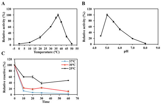
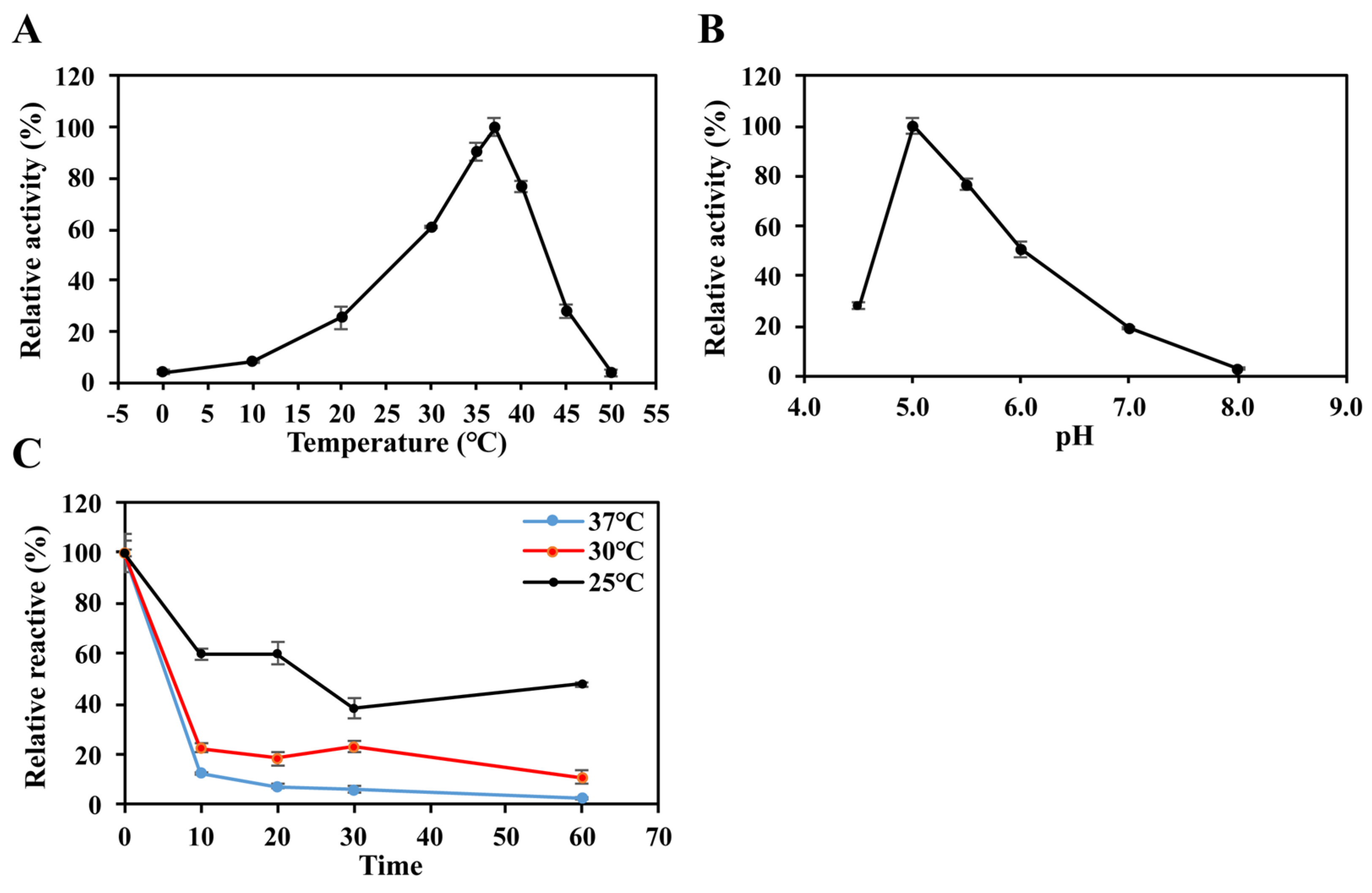
The optimal temperature for LbBgl3 activity was determined by assaying activity over a range of 0–50 °C under the standard reaction conditions. The optimal pH was determined using citrate-phosphate buffer (pH 4.5–8.0) at the optimal temperature. The highest relative activity was defined as 100%.
The thermostability was assessed by incubating purified enzyme solutions at 25, 30, and 37 °C for 0, 10, 20, 30, and 60 min, respectively. At each time point, samples were collected, and their residual enzyme activities were assayed under the pre-determined optimal reaction conditions. The non-incubated original enzyme solution was used as a control (with its activity set as 100%), and the relative enzyme activity at each time point was calculated to evaluate the thermostability of LbBgl3.
2.6. Assay of Substrate Specificity and Kinetic Constants
In order to comprehensively elucidate the substrate specificity of LbBgl3, the enzymatic activity was systematically evaluated under the pre-established optimal catalytic conditions. A series of substrates, including artificial substrates pNPG, pNPX, pNPGal and natural disaccharide substrates (cellobiose, gentiobiose, sophorose), were employed at a concentration of 4 mM, respectively. For artificial substrate hydrolytic activity assessment, the pNPG-based quantification assay was performed following established protocols. Regarding the natural disaccharide substrates, the GOD-POD method was utilized to determine the hydrolytic activity.
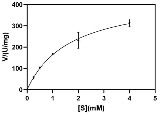
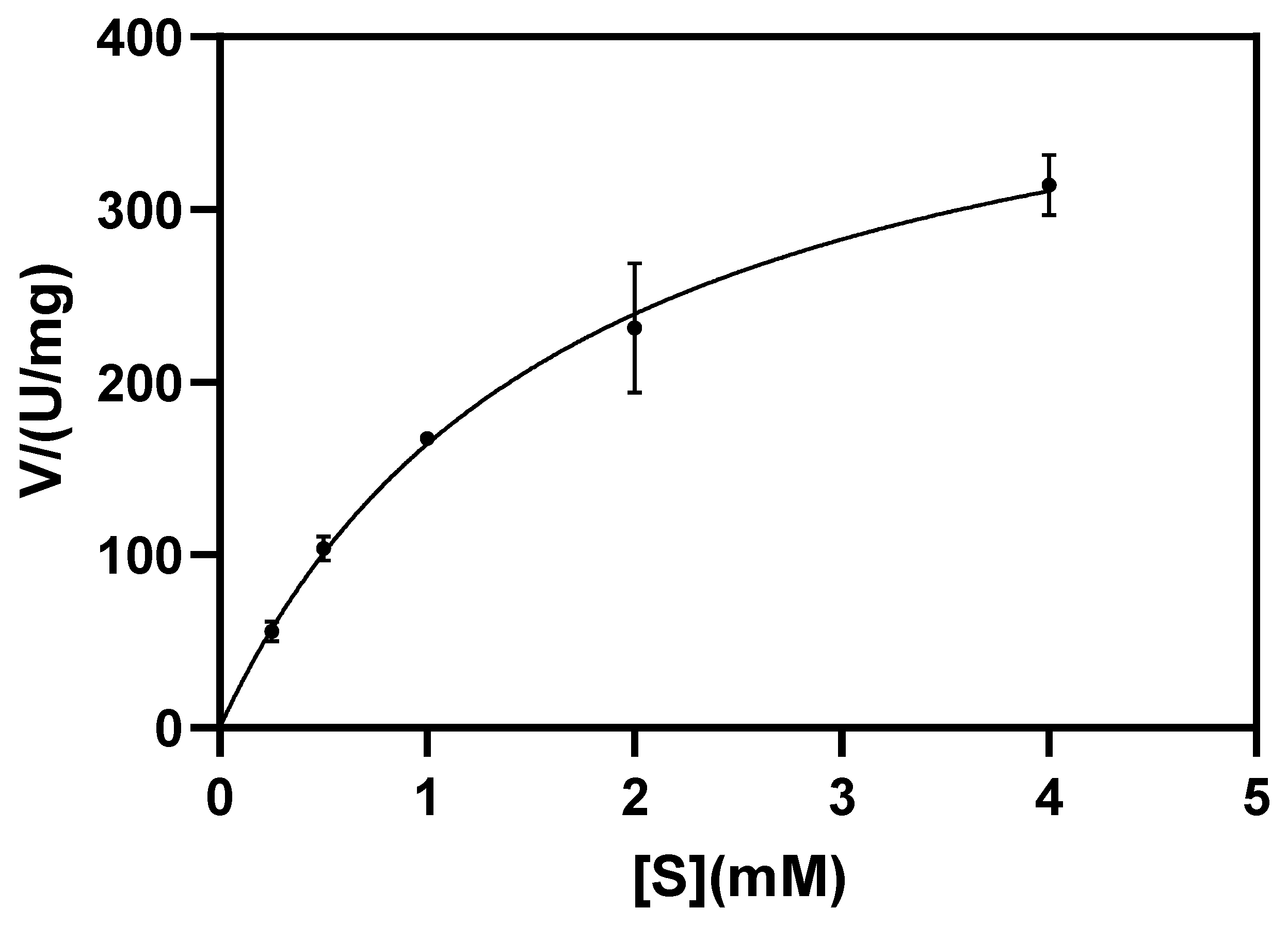
The kinetic parameters of LbBgl3 with respect to pNPG were meticulously determined under the optimal reaction conditions. Specifically, the enzymatic activity assays were conducted using pNPG at final concentrations of 0.25, 0.5, 1.0, 2.0, and 4.0 mM. Following a 10-min reaction period, the absorbance at 405 nm was precisely recorded. The Michaelis constant (Km), maximum reaction velocity (Vmax), turnover number (kcat), and catalytic efficiency (kcat/Km) were calculated by nonlinear regression fitting of the Michaelis–Menten equation. Given the high homogeneity of purified LbBgl3 verified by native electrophoresis, the active fraction was regarded as 100%.
where Vmax is the maximum reaction rate (μmol·mg−1·min−1), M is the molecular mass of LbBgl3 (kDa), and division by 60 converts units from min−1 to s−1. All assays were performed in triplicate. All assays were performed in triplicate.
2.7. The Influence of Metal Ions and Chemical Reagents on LbBgl3
Using pNPG as the substrate, the effects of metal ions (K+, Ca2+, Zn2+, Mg2+, Mn2+, Fe2+, Fe3+, Cu2+, Na+, Pb2+, Ni2+, Li+, Cd2+, Co2+), the inhibitor SDS, the chelator EDTA, and organic solvents (methanol, isopropanol, β-mercaptoethanol) on LbBgl3 activity were investigated. Metal ions, SDS, and EDTA were added to a final concentration of 5 mM. Organic solvents were added to a final concentration of 5% (v/v). Relative activity was determined as described above, with each condition tested in triplicate.
2.8. Influences of Additives on Enzyme Activity
The tolerance of LbBgl3 to methanol, ethanol, glucose, NaCl, and EDTA was evaluated by adding these compounds to the standard reaction system. The control group without additives was set to 100% relative activity. Methanol and ethanol were tested at final concentrations of 5%, 10%, 15%, 20%, and 25% (v/v). Glucose and NaCl were tested at final concentrations of 0.5, 1.0, 1.5, and 2.0 M. EDTA was tested at final concentrations of 5, 25, 50, 100, 200, and 250 mM. After incubation of each reaction system under the established optimal catalytic conditions, the relative enzymatic activity of LbBgl3 was determined using the pNPG-based assay method described previously, with each concentration tested in triplicate.
2.9. Three-Dimensional Modeling and Molecular Docking
The three-dimensional structure of LbBgl3 was predicted using AlphaFold3, and the quality of the predicted structure was evaluated using Venn diagrams. Molecular docking between LbBgl3 and the ligands (EDTA) was performed with AutoDockTools 1.5.7 (Scripps, La Jolla, CA, USA), where the docking box was set to 50 Å × 50 Å × 50 Å with a grid spacing of 0.375 Å. The center coordinates of the substrate binding pocket (−9.7, 19.14, 9.01) were predicted using Deepsite (https://open.playmolecule.org/tools, accessed on 21 January 2026) [17] and used as the center coordinates for the molecular docking box. This study used the H2Y2− form of EDTA (derived from Na2EDTA) for docking at pH 5.0. For molecular docking simulations, the optimal binding conformation was screened from all generated poses according to minimal binding energy, absence of steric clashes, and occupancy within the substrate-binding pocket. Hydrogen bonds were defined using a heavy-atom distance cutoff ≤ 3.6 Å and a marginal hydrogen bond angle threshold of 63.0°.
2.10. Isothermal Titration Calorimetry (ITC) Measurement
ITC experiments were performed at 25 °C (298 K) using a NANO ITC calorimeter (TA Instruments, New Castle, DE, USA), following the experimental design described in previous literature [18]. The experimental parameters were set as follows: Macromolecule Binding Active (MBA, the concentration of active macromolecules capable of binding) was 0.05 mM, Macromolecule in Cell (MB, the concentration of macromolecules in the reaction cell) was 0.175 mM, and Constant Dilution Parameter (CDP, the constant dilution heat parameter) was 0.01 mM; all reagents were pretreated before the experiments. For sample loading and titration parameters, 0.05 mM MBA and 0.175 mM MB were separately aspirated into the titration syringe with an initial volume of 50 μL for each, and the CDP-related system was added to the sample cell with an initial volume of 300 μL. The titration procedure consisted of 20 consecutive injections, with a single injection volume of 2 μL and an interval of 120 s between adjacent injections; the stirring speed of the syringe was set at 350 r/min during the titration. Two groups were set up in the experiment: the experimental group (MBA/MB titrated against the CDP-related system) and the control group (MBA/MB titrated against blank solvent). The operation procedures and experimental parameters were identical for both groups, and the data from the blank titration were used to obtain the CDP value. All experimental and control groups were conducted in duplicate to ensure the reliability and reproducibility of the results.
After the experiments, the obtained isotherms were integrated and the blank heat effects from the control group were subtracted. Global fitting was performed using the 1:1 heteroassociation model (A + B→AB), and the binding stoichiometry (n), dissociation constant (Kd), entropy change (ΔS), and enthalpy change (ΔH) were calculated by incorporating the MBA, MB, and CDP parameters. The fitted data were processed using Origin 2021 software to generate the experimental plots meeting the requirements of academic journal publication.
2.11. Statistical Analysis
All experiments were performed at least three times, and the data are presented as mean ± standard deviation (SD). Statistical analysis and graphing were performed using Microsoft Excel 2019 (Microsoft Corporation, Redmond, WA, USA) and Adobe Illustrator 2022 (Adobe Systems Incorporated, San Jose, CA, USA).
3. Results
3.1. Sequence and Structural Analysis
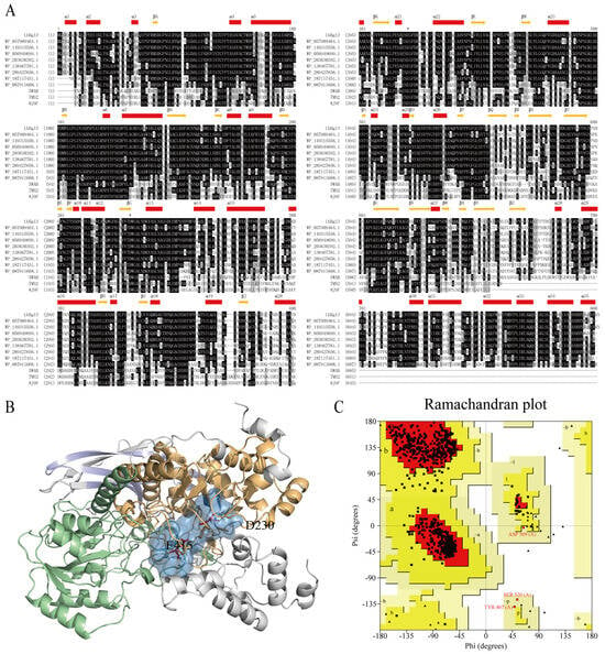
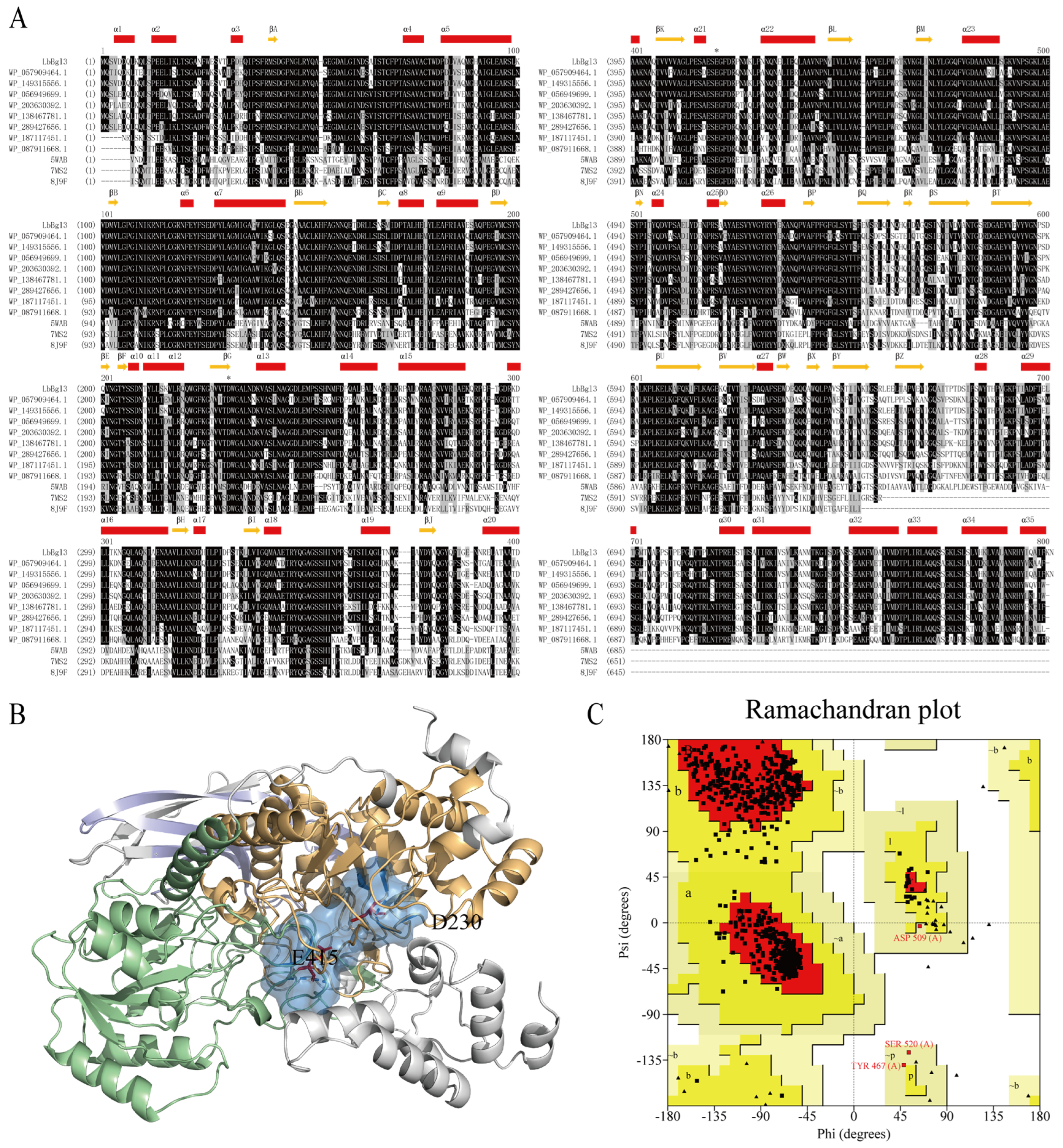
The gene encoding a GH3 family β-glucosidase from L. buchneri was retrieved from the NCBI database (GenBank No: CAM3048833.1). Sequence analysis revealed a 2385 bp open reading frame encoding a 794-amino acid polypeptide, designated LbBgl3. BLAST analysis indicated that LbBgl3 shares the highest sequence identity (99.87%) with a β-glucosidase from L. buri (NCBI Reference Sequence: WP_149315556.1), followed by 78.41% identity with an enzyme from L. kisonensis (GenBank: AZEB01000046.1) and 43.80% identity with a structurally characterized GH3 β-glucosidase (Protein Data Bank, PDB: 7MS2). Multiple sequence alignment identified highly conserved catalytic residues: aspartic acid (D230) in the TDWG motif and glutamic acid (E415) in the ESEGF motif (Figure 1A). Importantly, the distribution pattern of these conserved catalytic residues and their corresponding motif locations exhibits a remarkable degree of congruence with those of previously reported GH3 family β-glucosidases [19].
Figure 1.
Sequence and structural analysis of LbBgl3. (A) Multiple sequence alignment of LbBgl3 with homologs. The homologue set consisted of four β-glucosidases from Lentilactobacillus, including LbBgl3 and three homologous enzymes (NCBI accession numbers: WP_149315556.1, WP_056949699.1, and WP_203630392.1), along with three β-glucosidases from the GH3 family with resolved crystal structures (PDB IDs: 5WAB, 7MS2, and 8J9F). (B) Domain architecture and catalytic residues of LbBgl3. The (α/β)8 barrel, (α/β)6 sandwich, and FnIII domains are shown in light orange, pale green, and light blue, respectively. Catalytic residues D230 and E415 are highlighted as red sticks. (C) Ramachandran map of LbBgl3.
The three-dimensional structure of LbBgl3, predicted by AlphaFold3, was of high quality (Figure 1B). Structural analysis revealed that LbBgl3 comprise the typical GH3 family structural domains, including a (β/α)8 barrel domain (residues 18–284), an (α/β)6 sandwich domain (residues 315–545), and a fibronectin type III (FnIII) domain (residues 582–652). The catalytic residue D230 is localized within the (β/α)8 barrel domain, while E415 is positioned at the interface between the (β/α)8 barrel and (α/β)6 sandwich domains, with a distance of approximately 6.0 Å between them (Figure 1B). FnIII domains are typical domains of GH3 family enzymes, but their specific functions remain unclear. Additionally, DeepSite prediction identified three potential substrate-binding pockets, among which the pocket within the (β/α)8 barrel domain was most consistent with the expected catalytic conformation (Figure 1B).
The Ramachandran plot (Figure 1C) remains as a complementary validation metric, showing that 96.4% of residues are in favored regions, further confirming the structural reliability. In addition, the AlphaFold3-predicted structure achieved a mean pLDDT score of 88.6, indicating high overall confidence, with the catalytic core domains exhibiting the highest scores (>90).
3.2. Expression and Purification of Recombinant LbBgl3
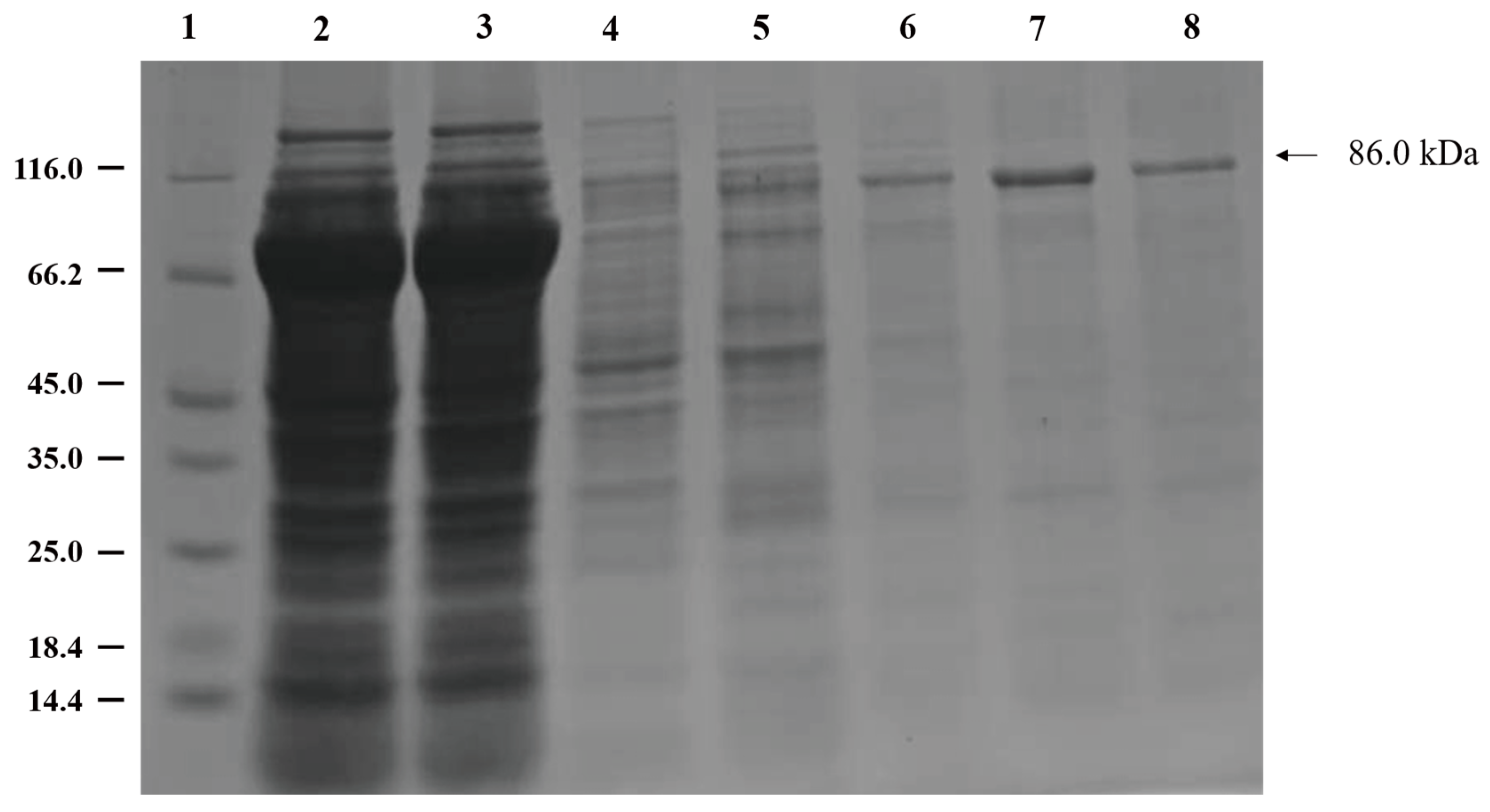
Recombinant LbBgl3, containing an N-terminal 6 × His tag, was successfully expressed in E. coli BL21(DE3) and purified using Ni-IDA magnetic beads. The theoretical molecular weight was calculated to be 86.0 kDa. SDS-PAGE analysis of the purified protein showed a single band with an apparent molecular weight of approximately 86.0 kDa (Figure 2), confirming successful expression and purification to homogeneity.
Figure 2.
SDS-PAGE of LbBgl3 expression and purification. Lane 1: Marker; Lane 2: Cell lysates supernatant. Lane 3: Waste liquid without combined magnetic beads; Lane 4: 0 mM imidazole elution buffer. Lane 5: 40 mM imidazole elution buffer. Lane 6: 100 mM imidazole elution buffer. Lane 7: 200 mM imidazole elution buffer. Lane 8: 300 mM imidazole elution buffer.
3.3. Enzymatic Properties of Recombinant LbBgl3
3.3.1. Optimal Temperature and pH
Recombinant LbBgl3 exhibited maximum activity at 37 °C and pH 5.0 (Figure 3A,B). This enzyme is an acidophilic cold-adapted [20] enzyme and shares enzymatic properties similar to those of typical β-glucosidases derived from Lactobacillus species (Table 1). LbBgl3 displayed pronounced temperature sensitivity, characterized by relatively high retention of catalytic activity at low temperatures and rapid inactivation at moderate to high temperatures. Specifically, it retained over 60% of its initial activity within the temperature range of 30–40 °C, while maintaining detectable activity at lower temperatures (≤25 °C). In contrast, a sharp decline in catalytic activity was observed when the temperature exceeded 45 °C, with nearly complete inactivation at 50 °C. This temperature-dependent pattern is consistent with the characteristics of cold-adapted enzymes. Furthermore, thermostability characterization of LbBgl3 demonstrated poor thermoresilience. Upon incubation at 37 °C for 10 min, the enzyme exhibited less than 13% of its initial activity (Figure 3C). Conversely, when incubated at 25 °C for the same duration, approximately 60% of its initial relative activity was retained (Figure 5B), further confirming its typical cold-adapted enzyme features [20]. Comparative analysis with β-glucosidases classified as cold-adapted, mesophilic, and thermophilic [21] revealed significant differences in the amino acid composition of LbBgl3 (Table 2). Specifically, marked compositional characteristics unique to cold-adapted enzymes, particularly LbBgl3, were identified by comparing amino acid profiles of cold-adapted LbBgl3 and M-GH3_A from Marinomonas sp. ef1 [22], mesophilic MtBgl85 derived from Microbulbifer thermotolerans DAU221 [23], and thermophilic Bgl3B originating from Talaromyces leycettanus [24]. As a typical cold-adapted β-glucosidase, LbBgl3 accumulates high levels of Leu, Lys and Gln but contains less rigid Arg and aromatic Trp relative to mesophilic and thermophilic counterparts; this composition weakens intramolecular salt bridges and hydrophobic stacking, endowing the active pocket with adequate conformational flexibility to sustain catalytic efficiency under low-temperature conditions. Compared with another cold-adapted ortholog M-GH3_A, LbBgl3 features elevated Ala and Pro contents, which moderately strengthens backbone rigidity and avoids excessive structural looseness induced by low temperature. Distinct from mesophilic MtBgl85 rich in stabilizing Ala and Arg and thermostable Bgl3B enriched in Gly, Val, Trp and Asp for robust thermal resistance, LbBgl3 adopts a cold-adaptive strategy that minimizes rigid intramolecular interactions to prioritize structural flexibility, a representative molecular signature supporting its high catalytic activity in cold environments.
Figure 3.
Optimal catalytic conditions and thermostability of LbBgl3. (A) Optimal temperature. (B) Optimal pH. (C) Thermostability of LbBgl3 at 25 °C, 30 °C, and 37 °C in pH 5.0 buffer. Data are presented as mean ± SD from three independent biological replicates (n = 3).
Table 1.
Enzymatic properties and applications of GH3 family β-glucosidase from Lactobacillus.
Table 2.
Amino acid compositions of LbBgl3 and other β-Glucosidases with divergent temperature adaptation profiles.
LbBgl3 shows a preference for acidic environments as its optimal pH. Its catalytic activity can be detected across a wide pH span from 3.0 to 7.0, demonstrating that it is a moderately acidophilic β-glucosidase. The optimal activity of this enzyme precisely mirrors the physiological growth conditions of its native host, L. buchneri. Notably, L. buchneri thrives optimally within the temperature range of 15–37 °C and remains viable even at a low pH of 4.4 [30,31]. From an evolutionary perspective, the adaptation of LbBgl3 in L. buchneri represents a sophisticated response to its cold and acidic ecological niche. The congruence between the enzyme’s optimal catalytic conditions at 37 °C and pH 5.0 and its native habitat further validates its functional suitability.
3.3.2. Substrate Specificity and Kinetic Constants
Under optimal conditions (37 °C, pH 5.0), the kinetic parameters of LbBgl3 toward pNPG were determined (Figure 4). Nonlinear regression fitting was performed in conjunction with the Michaelis–Menten equation to determine enzymatic kinetic parameters., with Km, Vmax, kcat and kcat/Km calculated to be 1.697 mM, 442.5 μmol·mg−1·min−1, 634.32 s−1, and 373.79 mM−1·s−1, respectively. Substrate specificity assays revealed that LbBgl3 exhibited exceptionally high specific activity towards pNPG, but showed no detectable hydrolysis of the disaccharide substrates (cellobiose, sophorose, gentiobiose) or other aryl β-glycosides under the tested conditions. Reported GH3 family β-glucosidases from Lactobacillus species generally show low specific activities toward pNPG, ranging from 0.25 to 150 U·mg−1 [7,13,25,26]. Notably, LbBgl3 demonstrates a significantly elevated specific activity toward pNPG, reaching a peak value of 799.67 U·mg−1. This activity level is substantially higher than that of other homologous β-glucosidases isolated from Lactobacillus species (Table 1). A similar phenomenon has been reported for HiBgl3B, a GH3 β-glucosidase from Humicola insolens Y1, which also exhibits activity exclusively toward aryl β-glucosides (pNPG) and fails to hydrolyze cellobiose, sophorose, and gentiobiose. Xia et al. demonstrated that this narrow specificity is determined by three residues (Ile48, Ile278, Thr484) at the +1 subsite that form a constricted binding pocket incapable of accommodating the second glucosyl moiety of disaccharides [32]. Site-directed mutagenesis replacing these residues with the Trp/Phe/Tyr triad conserved in broad-spectrum GH3 enzymes (HiBgl3A) conferred hydrolytic activity toward sophorose, confirming the critical role of the +1 subsite architecture in substrate selectivity. By analogy, the narrow substrate profile of LbBgl3 may similarly arise from restricted +1 subsite features, which could be tested by comparative structural analysis or mutagenesis in future studies.
Figure 4.
Nonlinear Michaelis-Menten fitting curve. [S] represents the substrate concentration. Data are presented as mean ± SD from three independent biological replicates (n = 3).
The inability of LbBgl3 to hydrolyze common β-linked disaccharides such as cellobiose, gentiobiose, and sophorose suggests that its natural substrate may be a plant-derived β-glucoside or glucoconjugate present in silage materials, consistent with the ecological niche of its native host L. buchneri. This substrate specificity pattern warrants further investigation through substrate screening with a more comprehensive glycoside library.
3.3.3. Effect of Solvent, Salt and Glucose on Enzymatic Activity
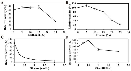
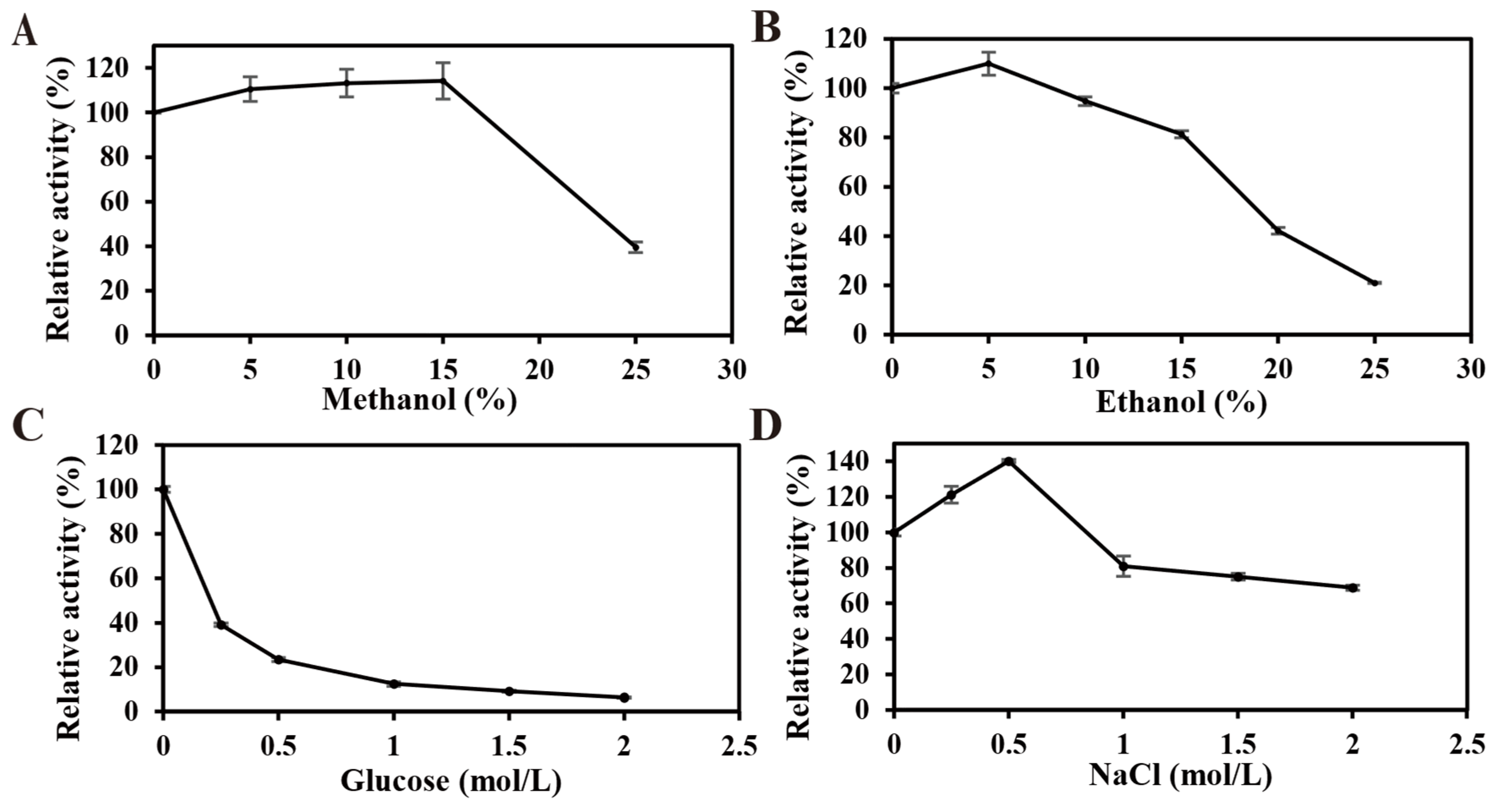
The effects of methanol, ethanol, NaCl, and glucose on LbBgl3 activity are shown in Figure 5. Enzymatic activity initially increased and then decreased with increasing concentrations of methanol or ethanol. At low concentrations, both alcohols slightly enhanced activity. Notably, in the presence of 15% (v/v) methanol, the enzyme still retained 114% of its relative activity, suggesting its potential applicability in the transformation of alcohol-soluble glycosides such as ginsenosides. Salt tolerance followed a similar trend, with maximum activity (~140%) observed at 0.25–0.50 M NaCl. In contrast, LbBgl3 exhibited poor glucose tolerance, a characteristic commonly observed among GH3 family β-glucosidases. Overall, LbBgl3 represents a novel β-glucosidase with tolerance to low concentrations of alcohols and NaCl.
Figure 5.
The solvent tolerance of LbBgl3. (A) Methanol tolerance. (B) Ethanol tolerance. (C) Glucose tolerance. (D) NaCl tolerance. Data are presented as mean ± SD from three independent biological replicates (n = 3).
3.3.4. The Influence of Metal Ions and Chemical Reagents on the Activity of LbBgl3
The effects of various metal ions and chemical reagents on LbBgl3 activity are summarized in Table 3. Specifically, Mg2+ and Zn2+ had no significant effect. In contrast, K+, Cu2+, Mn2+, Pb2+, Fe2+, Fe3+, sodium dodecyl sulfate (SDS), isopropanol, and β-mercaptoethanol exhibited inhibitory effects on LbBgl3 activity. Among these inhibitors, Fe2+ and Fe3+ caused severe inhibition, reducing the relative activity of LbBgl3 to less than 15%. Additionally, Na+, Li+, Cd2+, Co2+, Ni2+, EDTA, methanol and ethanol displayed stimulatory effects on LbBgl3, leading to an increased catalytic activity compared to the control.
Table 3.
The influence of metal ions and chemical reagents on LbBgl3.
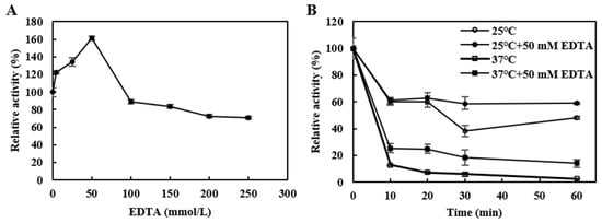
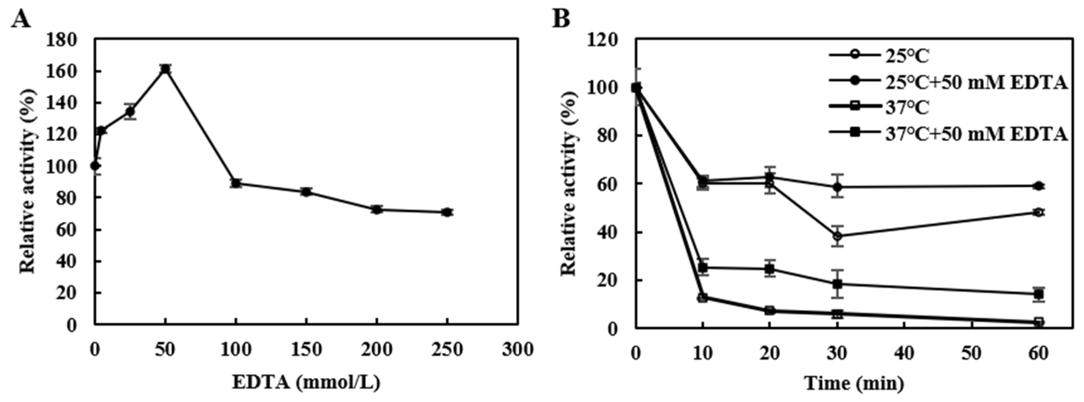
Notably, the metal chelator EDTA enhanced LbBgl3 activity to 122% of the control at 5 mM, which contrasts with most reported β-glucosidases that typically is inhibited by EDTA. A gradual increase in LbBgl3 activity was observed as the EDTA concentration was raised from 5 mM to 50 mM, reaching 161%. Conversely, a sharp decline in enzymatic activity occurred when the EDTA concentration was increased to 100 mM; nevertheless, 71% relative activity was retained even at 250 mM (Figure 6A). Although no studies have to date reported EDTA-induced enhancement of β-glucosidase activity, a similar phenomenon has been documented in other non-metal-dependent enzymes. For instance, in the presence of 5 mM EDTA, the melting temperature (Tm) of endoglucanase from Aspergillus aculeatus increased by 16 °C, and mechanistic studies confirmed that EDTA improved enzyme activity by increasing kcat and enhancing the conformational flexibility [33]. Additionally, a similar phenomenon was observed in cyclomaltodextrinase from Geobacillus thermopakistaniensis, where EDTA enhanced enzyme thermostability, thereby improving catalytic activity [34]. These findings suggest that low-concentrations of EDTA may stabilize the conformation of LbBgl3, which effectively prolongs the thermal inactivation half-life of the enzyme and simultaneously elevates its catalytic activity. This hypothesis is supported by thermostability assays of LbBgl3 in the presence of EDTA: after heat treatment at 25 °C for 60 min, the activity of EDTA-supplemented LbBgl3 remained comparable to that observed after heat treatment for 10 min (Figure 6B,C). This minimal activity loss over extended heating directly reflects the extended thermal inactivation half-life conferred by EDTA supplementation.
Figure 6.
The effect of EDTA on the enzymatic activity of LbBgl3. (A) Effect of EDTA concentration on LbBgl3 activity measured under standard assay conditions. (B) Thermostability of LbBgl3 at 25 °C and 37 °C in pH 5.0 buffer with or without 50 mM EDTA. Residual activities were determined under optimal conditions. Data are presented as mean ± SD from three independent biological replicates (n = 3). Detailed experimental procedures are described in Section 2.5 and Section 2.8.
3.4. In Silico Analysis of EDTA-Mediated Thermostability Enhancement
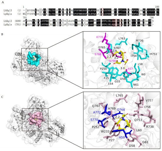
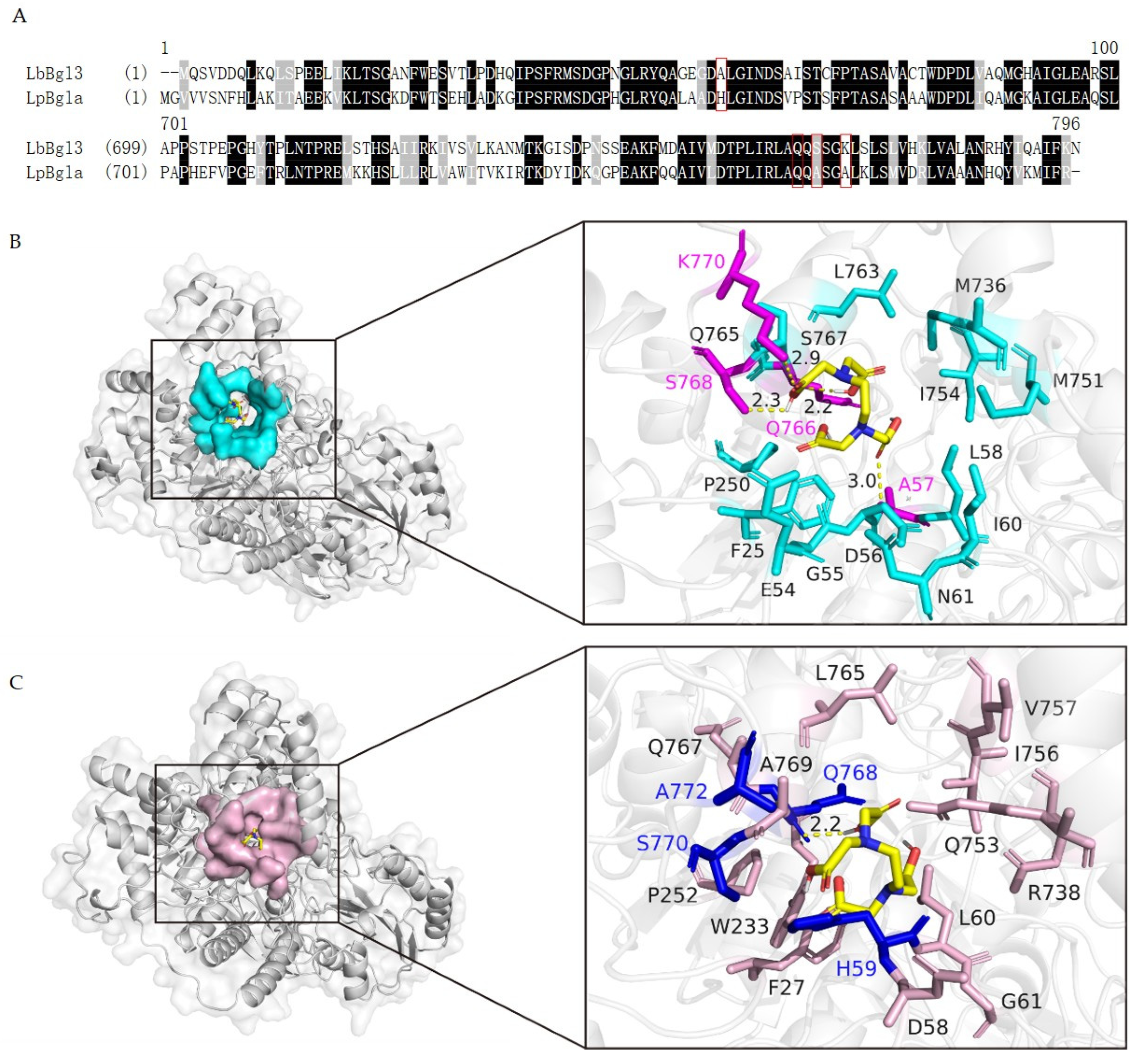
To elucidate the mechanism underlying the EDTA-induced thermostability enhancement of LbBgl3, molecular docking simulations were performed to predict potential intermolecular interactions between EDTA and LbBgl3. The docking results indicated a favorable binding affinity between EDTA and LbBgl3, with a calculated binding energy of −4.3 kcal mol−1 (Figure 7). Specifically, EDTA forms hydrogen bonds with four amino acid residues of LbBgl3, namely Ala57, Gln766, Ser768 and Lys770, which are located near the substrate-binding pocket. Hydrogen bonding networks in protein secondary structures are critical determinants of thermal stability, as these bonds serve to tether conformationally labile surface regions to the structurally stable core domain, thereby significantly strengthening the overall protein structure [35]. Studies have shown that thermophilic proteins contain more hydrogen bonds than their mesophilic homologs [36]. A plausible molecular-level explanation is that the hydrogen bonding interactions between EDTA and its binding residues effectively stabilize the conformational dynamics of the region surrounding the substrate-binding pocket, thereby alleviating the intrinsic thermolability of LbBgl3. Furthermore, LpBgla, a β-glucosidase from L. paracasei TK1501, shares 57.8% sequence identity with LbBgl3, but its enzymatic activity is inhibited in the presence of EDTA. Three-dimensional structural alignment of these two enzymes, combined with molecular docking analyses between each enzyme and EDTA, revealed that the His59, Ser770 and Ala772 residues of LpBgla do not form hydrogen bonds with EDTA. Collectively, these findings further support that the hydrogen bond network formed between EDTA and amino acid residues in the substrate-binding pocket is essential for stabilizing the overall conformation of LbBgl3 and mitigating its inherent thermal instability.
Figure 7.
Sequence and structural differences in enhancing enzyme thermal stability through EDTA. (A) Sequence alignment of LbBgl3 and LpBgla. Differential sites marked with red boxes. (B) Molecular docking of EDTA with LbBgl3, and the cyan sticks represent amino acid residues located within 6 Å distance of EDTA. The magenta sticks represent amino acid binding residues of LbBgl3 (C) Molecular docking of EDTA with LpBgla. The light magenta sticks represent amino acid residues located within 6 Å distance of EDTA, the blue sticks represent aligned residues of LpBgla, the yellow sticks represent EDTA, and the yellow dotted line represents hydrogen bonds.
3.5. Isothermal Titration Calorimetry Reveals Specific Binding Between EDTA and LbBgl3
Given the absence of metal-binding motifs or cofactor requirements in the LbBgl3 sequence, and the fact that EDTA’s enhancing effect did not correlate with the removal of any specific metal ion (as the enzyme remained fully active in metal-free buffer), we opted for ITC to directly probe the EDTA-LbBgl3 interaction rather than pursuing metal content analysis. This approach directly validates the molecular interaction underlying the observed stabilization, as metal content alone would not clarify whether EDTA exerts its effect through metal chelation or through direct binding to the protein.
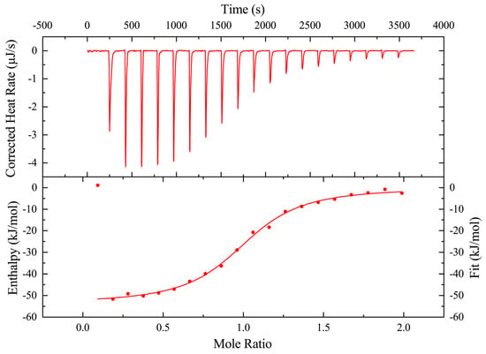
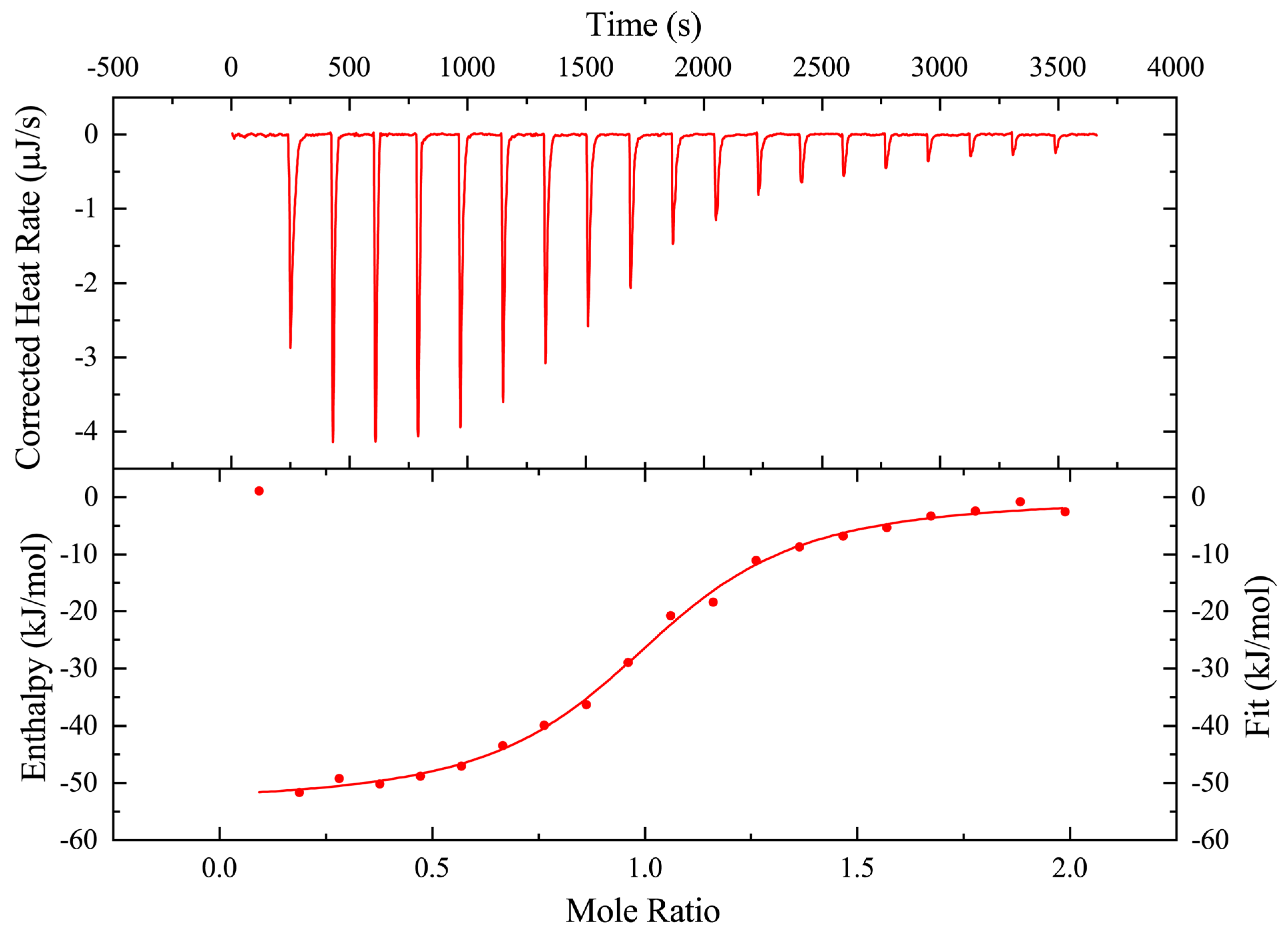
ITC was utilized to directly verify the binding interaction between EDTA and LbBgl3, and the corresponding results are presented in Figure 8. The titration curves revealed that LbBgl3 specifically binds EDTA at a molar ratio of 1:1, with Kd of 1.33 μM, indicative of a relatively strong binding affinity. The binding process was strongly exothermic with ΔH of −53.75 kJ·mol−1, accompanied by reduced ΔS = −67.79 J·mol−1·K−1, reflecting increased ordering of the system upon complex formation. The overall ΔG was calculated to be −33.55 kJ·mol−1, demonstrating that the binding reaction occurs spontaneously. Hydrogen bonds, electrostatic interactions, and van der Waals forces dominate the intermolecular driving forces underlying this binding event. Complementary ITC experiments verified the spontaneous 1:1 specific binding between EDTA and LbBgl3 with strong affinity, which follows an enthalpy-driven mechanism governed by hydrogen bonds, electrostatic and van der Waals interactions; this stable complex formation rationalizes the EDTA-mediated improvement in LbBgl3 thermostability predicted by molecular docking.
Figure 8.
Isothermal titration calorimetry profile for the titration of EDTA against LbBgl3. Titrations were carried out at 298 K. Data are presented as mean ± SD from three independent biological replicates (n = 3).
While metal content analysis is commonly used to evaluate EDTA-mediated metal chelation, the absence of metal dependency and the unique enhancement pattern observed for LbBgl3 suggested a non-classical mechanism. ITC was therefore employed to directly confirm the EDTA-LbBgl3 binding interaction, providing unequivocal evidence for the direct interaction that underpins the observed thermostability enhancement.
4. Conclusions
This study presents the first systematic characterization of a GH3 family β-glucosidase, LbBgl3, from L. buchneri, elucidating its enzymatic characteristics, catalytic active sites, and key structural features. Biochemical characterization identifies LbBgl3 as a cold-adapted, moderately acidophilic β-glucosidase with tolerance to low concentrations of salt and methanol, as well as high specific activity, highlighting L. buchneri as a promising source of novel biocatalysts. A notable finding is the enhancement of LbBgl3 activity and thermostability by EDTA, mediated through hydrogen-bonding interactions that stabilize flexible conformations near the substrate-binding pocket. Future work should employ protein engineering, rational design, and artificial intelligence-aided design to improve its thermostability, thereby facilitating the development of novel, low-carbon, and green industrial applications based on this enzyme.
Author Contributions
H.T. and J.H.: conceptualization, investigation, methodology, software, visualization, and writing—original draft preparation. C.L. and T.L.: methodology and software. M.W. and H.W.: conceptualization, validation, writing—review and editing, supervision, and funding acquisition. P.S.: writing—review, and language editing. All authors have read and agreed to the published version of the manuscript.
Funding
This work was supported by the National Natural Science Foundation of China (32072166), the Agricultural Science and Technology Innovation Project Special Fund of the Chinese Academy of Agricultural Sciences (ASTIP-IBFC), the Key Scientific Research Foundation of Hunan Provincial Education Department (25A0192), the Science and Technology Innovation Program of Hunan Province (2023RC3137) and the nanfan special project of CAAS(YBXM2641).
Institutional Review Board Statement
Not applicable.
Informed Consent Statement
Not applicable.
Data Availability Statement
The original contributions presented in this study are included in the article. Further inquiries can be directed to the corresponding authors.
Conflicts of Interest
The authors declare that they have no known competing financial interests or personal relationships that could have appeared to influence the work reported in this paper.
References
- Sheikh, I.; Mahajan, P.; Kaur, T.; Ahmed, N.; Kour, D. Probiotic potential of endophytic bacteria in functional foods: Opportunities and challenges. Food Biosci. 2025, 72, 107423. [Google Scholar] [CrossRef] [Scilit]
- Dasriya, V.L.; Samtiya, M.; Ranveer, S.; Dhillon, H.S.; Devi, N.; Sharma, V.; Nikam, P.; Puniya, M.; Chaudhary, P.; Chaudhary, V.; et al. Modulation of gut-microbiota through probiotics and dietary interventions to improve host health. J. Sci. Food Agric. 2024, 104, 6359–6375. [Google Scholar] [CrossRef] [Scilit] [PubMed]
- Abedin, M.M.; Chourasia, R.; Phukon, L.C.; Sarkar, P.; Ray, R.C.; Singh, S.P.; Rai, A.K. Lactic acid bacteria in the functional food industry: Biotechnological properties and potential applications. Crit. Rev. Food Sci. Nutr. 2024, 64, 10730–10748. [Google Scholar] [PubMed]
- Ullah, M.; Naeem, M.; Andoh, V.; Khan, M.N.; Han, J.; Rizwan, M.; Hussain, N.; Saeed, M.; Chen, Y.; Chen, H. Lactic acid bacteria as biofactories: Mechanistic insights, engineering strategies, and future horizons for heterologous enzyme expression. J. Agric. Food Chem. 2025, 73, 16028–16048. [Google Scholar] [CrossRef] [Scilit] [PubMed]
- Sadat-Mekmene, L.; Richoux, R.; Aubert-Frogerais, L.; Madec, M.N.; Corre, C.; Piot, M.; Jardin, J.; le Feunteun, S.; Lortal, S.; Gagnaire, V. Lactobacillus helveticus as a tool to change proteolysis and functionality in Swiss-type cheeses. J. Dairy Sci. 2013, 96, 1455–1470. [Google Scholar] [CrossRef] [Scilit] [PubMed]
- Landete José, M.; Ferrer, S.; Monedero, V.; Zúñiga, M. Malic enzyme and malolactic enzyme pathways are functionally linked but independently regulated in Lactobacillus casei BL23. Appl. Environ. Microbiol. 2013, 79, 5509–5518. [Google Scholar] [CrossRef] [Scilit] [PubMed]
- Zhong, F.-L.; Dong, W.-W.; Wu, S.; Jiang, J.; Yang, D.-C.; Li, D.; Quan, L.-H. Biotransformation of gypenoside XVII to compound K by a recombinant β-glucosidase. Biotechnol. Lett. 2016, 38, 1187–1193. [Google Scholar] [CrossRef] [Scilit] [PubMed]
- Singh, G.; Verma, A.K.; Kumar, V. Catalytic properties, functional attributes and industrial applications of β-glucosidases. 3 Biotech 2016, 6, 3. [Google Scholar] [PubMed]
- Srivastava, N.; Rathour, R.; Jha, S.; Pandey, K.; Srivastava, M.; Thakur, V.K.; Sengar, R.S.; Gupta, V.K.; Mazumder, P.B.; Khan, A.F.; et al. Microbial beta glucosidase enzymes: Recent advances in biomass conversation for biofuels application. Biomolecules 2019, 9, 220. [Google Scholar] [CrossRef] [Scilit] [PubMed]
- Magwaza, B.; Amobonye, A.; Pillai, S. Microbial β-glucosidases: Recent advances and applications. Biochimie 2024, 225, 49–67. [Google Scholar] [CrossRef] [Scilit] [PubMed]
- Cantarel, B.L.; Coutinho, P.M.; Rancurel, C.; Bernard, T.; Lombard, V.; Henrissat, B. The carbohydrate-active enzymes database (Cazy): An expert resource for glycogenomics. Nucleic Acids Res. 2009, 37, D233–D238. [Google Scholar] [CrossRef] [Scilit] [PubMed]
- Li, M.; Huang, Z.; Zhang, R.; Zhou, J. Review of probiotics, gut microorganisms, and their enzymes involved in the conversion of ginsenosides. Food Biosci. 2024, 58, 103829. [Google Scholar] [CrossRef] [Scilit]
- Xie, Y.; Wang, Y.; Han, Y.; Zhang, J.; Wang, S.; Lu, S.; Wang, H.; Lu, F.; Jia, L. Complete genome sequence of a novel Lactobacillus paracasei TK1501 and its application in the biosynthesis of isoflavone aglycones. Foods 2022, 11, 2807. [Google Scholar] [CrossRef] [Scilit] [PubMed]
- Hu, Y.; Zhai, L.; Hong, H.; Shi, Z.; Zhao, J.; Liu, D. Study on the biochemical characterization and selectivity of three β-glucosidases from Bifidobacterium adolescentis ATCC15703. Front. Microbiol. 2022, 13, 860014. [Google Scholar] [CrossRef] [Scilit] [PubMed]
- Yuksekdag, Z.; Cinar Acar, B.; Aslim, B.; Tukenmez, U. β-Glucosidase activity and bioconversion of isoflavone glycosides to aglycones by potential probiotic bacteria. Int. J. Food Prop. 2018, 20, S2878–S2886. [Google Scholar]
- Jin, Y.; Wang, P.; Li, F.; Yu, M.; Du, J.; Zhao, T.; Yi, Q.; Tang, H.; Yuan, B. The effects of Lactobacillus plantarum and Lactobacillus buchneri on the fermentation quality, in vitro digestibility, and aerobic stability of Silphium perfoliatum L. Silage. Animals 2024, 14, 2279. [Google Scholar] [CrossRef] [Scilit] [PubMed]
- Jiménez, J.; Doerr, S.; Martínez-Rosell, G.; Rose, A.S.; De Fabritiis, G. DeepSite: Protein-binding site predictor using 3D-convolutional neural networks. Bioinformatics 2017, 33, 3036–3042. [Google Scholar] [CrossRef] [Scilit] [PubMed]
- Lopata, A.; Jójárt, B.; Surányi, É.V.; Takács, E.; Bezúr, L.; Leveles, I.; Bendes, Á.Á.; Viskolcz, B.; Vértessy, B.G.; Tóth, J. Beyond chelation: EDTA Tightly binds Taq DNA polymerase, MutT and dUTPase and directly inhibits dNTPase activity. Biomolecules 2019, 9, 621. [Google Scholar] [CrossRef] [Scilit] [PubMed]
- Thongpoo, P.; McKee, L.S.; Araújo, A.C.; Kongsaeree, P.T.; Brumer, H. Identification of the acid/base catalyst of a glycoside hydrolase family 3 (GH3) β-glucosidase from Aspergillus niger ASKU28. Biochim. Biophys. Acta-Gen. Subj. 2013, 1830, 2739–2749. [Google Scholar] [CrossRef] [Scilit] [PubMed]
- Liu, Y.H.; Zhang, N.; Ma, J.; Zhou, Y.Q.; Wei, Q.; Tian, C.j.; Fang, Y.; Zhong, R.Z.; Chen, G.; Zhang, S.T. Advances in cold-adapted enzymes derived from microorganisms. Front. Microbiol. 2023, 14, 1152847. [Google Scholar] [CrossRef] [Scilit] [PubMed]
- Ahmed, A.; Sumreen, A.; Bibi, A.; Nasim, F.u.H.; Batool, K. In silico approach to elucidate factors associated with GH1 β-glucosidase thermostability. J. Pure Appl. Microbiol. 2019, 13, 1953–1968. [Google Scholar] [CrossRef] [Scilit]
- Marchetti, A.; Orlando, M.; Bombardi, L.; Fusco, S.; Mangiagalli, M.; Lotti, M. Evolutionary history and activity towards oligosaccharides and polysaccharides of GH3 glycosidases from an Antarctic marine bacterium. Int. J. Biol. Macromol. 2024, 275, 133449. [Google Scholar] [CrossRef] [Scilit] [PubMed]
- Pyeon, H.M.; Lee, Y.S.; Choi, Y.L. Cloning, purification, and characterization of GH3 β-glucosidase, MtBgl85, from Microbulbifer thermotolerans DAU221. PeerJ 2019, 7, e7106. [Google Scholar] [PubMed]
- Li, X.; Xia, W.; Bai, Y.; Ma, R.; Yang, H.; Luo, H.; Shi, P. A novel thermostable GH3 β-glucosidase from Talaromyce leycettanus with broad substrate specificity and significant soybean isoflavone glycosides-hydrolyzing capability. BioMed Res. Int. 2018, 2018, 4794690. [Google Scholar] [CrossRef] [Scilit] [PubMed]
- Kim, Y.S.; Lee, C.-J.; Ma, J.Y. Enhancement of active compound, genipin, from Gardeniae Fructus using immobilized glycosyl hydrolase family 3 β-glucosidase from Lactobacillus antri. AMB Express 2017, 7, 64. [Google Scholar] [CrossRef] [Scilit] [PubMed]
- Jaijoi, S.; Kaewsalud, T.; Yakul, K.; Mahatheeranont, S.; Sriyotai, W.; Sommano, S.R.; Rachtanapun, P.; Leksawasdi, N.; Watanabe, M.; Chaiyaso, T. β-Glucosidase from Lacticaseibacillus casei TISTR 1463: Biochemical characterization and application on biotransformation of pigmented leaf extract from Oryza sativa L. (Luem Pua glutinous rice). Biocatal. Agric. Biotechnol. 2025, 64, 103491. [Google Scholar] [CrossRef] [Scilit]
- Siddiqi, M.Z.; Srinivasan, S.; Park, H.Y.; Im, W.-T. Exploration and characterization of novel glycoside hydrolases from the whole genome of Lactobacillus ginsenosidimutans and enriched production of minor ginsenoside Rg3(s) by a recombinant enzymatic process. Biomolecules 2020, 10, 88. [Google Scholar] [CrossRef] [Scilit] [PubMed]
- Ko, J.-A.; Park, J.Y.; Kwon, H.J.; Ryu, Y.B.; Jeong, H.J.; Park, S.J.; Kim, C.Y.; Oh, H.M.; Park, C.S.; Lim, Y.H.; et al. Purification and functional characterization of the first stilbene glucoside-specific β-glucosidase isolated from Lactobacillus kimchi. Enzym. Microb. Technol. 2014, 67, 59–66. [Google Scholar] [CrossRef] [Scilit] [PubMed]
- Xie, Y.; Yan, X.; Li, C.; Wang, S.; Jia, L. Characterization and insight mechanism of an acid-adapted β-Glucosidase from Lactobacillus paracasei and its application in bioconversion of glycosides. Front. Bioeng. Biotechnol. 2024, 12, 1334695. [Google Scholar] [CrossRef] [Scilit] [PubMed]
- Muhammad, A.u.R.; Gumustop, I.; Genel, I.; Kurt, I.C.; Ortakci, F. Comparative genomics of Lentilactobacillus buchneri reveals strain-level hyperdiversity and broad-spectrum CRISPR immunity against human and livestock gut phages. PLoS ONE 2025, 20, e0325832. [Google Scholar] [CrossRef] [Scilit] [PubMed]
- NikkilÄ, P.; Johnsson, T.; Rosenqvist, H.; Toivonen, L. Effect of pH on growth and fatty acid composition of Lactobacillus büchneri and Lactobacillus fermentum. Appl. Biochem. Biotechnol. 1996, 59, 245–257. [Google Scholar] [CrossRef] [Scilit]
- Xia, W.; Bai, Y.; Cui, Y.; Xu, X.; Qian, L.; Shi, P.; Zhang, W.; Luo, H.; Zhan, X.; Yao, B. Functional diversity of family 3 β-glucosidases from thermophilic cellulolytic fungus Humicola insolens Y1. Sci. Rep. 2016, 6, 27062. [Google Scholar] [CrossRef] [Scilit] [PubMed]
- Naika, G.S.; Tiku, P.K. Influence of ethylenediaminetetraacetic acid (EDTA) on the structural stability of endoglucanase from Aspergillus aculeatus. J. Agric. Food Chem. 2011, 59, 7341–7345. [Google Scholar] [CrossRef] [Scilit] [PubMed]
- Aroob, I.; Shaeer, A.; Ahmad, N.; Aslam, M.; Rashid, N. Ethylenediaminetetraacetic acid enhances structural stability and thermotolerance of recombinant cyclomaltodextrinase from Geobacillus thermopakistaniensis at higher temperatures. Biologia 2024, 79, 291–298. [Google Scholar] [CrossRef] [Scilit]
- Al-Mansoori, A.K.K.; Kang, S.Y.; Thangadurai, S.; Rahim, R.A. Exploring enzyme thermostability enhancement through molecular modification: What remains to be known? J. Mol. Struct. 2025, 1337, 141852. [Google Scholar] [CrossRef] [Scilit]
- Tompa, D.R.; Gromiha, M.M.; Saraboji, K. Contribution of main chain and side chain atoms and their locations to the stability of thermophilic proteins. J. Mol. Graph. Model. 2016, 64, 85–93. [Google Scholar] [CrossRef] [Scilit] [PubMed]

Disclaimer/Publisher’s Note: The statements, opinions and data contained in all publications are solely those of the individual author(s) and contributor(s) and not of MDPI and/or the editor(s). MDPI and/or the editor(s) disclaim responsibility for any injury to people or property resulting from any ideas, methods, instructions or products referred to in the content. |
© 2026 by the authors. Licensee MDPI, Basel, Switzerland. This article is an open access article distributed under the terms and conditions of the Creative Commons Attribution (CC BY) license.