


Highlights
- •
- NAD+/NADH regulates cellular redox balance, energy metabolism, and key signaling processes.
- •
- Highly compartmentalized NAD pools exist across organelles with distinct con-centrations and redox states.
- •
- NAD quantification is challenged by methodological variability, rapid intercon-version, and dynamic cellular states.
- •
- NAD+-boosting strategies show therapeutic promise but face significant transla-tional and compartmentalization challenges.
Abstract
Nicotinamide adenine dinucleotide (NAD+) and its reduced form, NADH, are essential coenzymes that play central roles in cellular redox homeostasis, energy metabolism, DNA repair, and signaling. Cellular NAD+ levels are maintained by a dynamic balance between the de novo Preiss–Handler, and salvage synthesis pathways, and consumption by enzymes like sirtuins, PARPs, and CD38. Among these, the nicotinamide Phosphoribosyltransferase (NAMPT)-driven salvage pathway represents the predominant route of NAD+ synthesis. The specific regulation of NAD (NAD+ and NADH) levels across distinct subcellular compartments has emerged as a critical determinant of cellular function but it remains poorly understood. Dysregulation of NAD metabolism is a hallmark of aging and various pathologies, including cancer, neurodegenerative disorders, and metabolic diseases, making strategies to modulate NAD levels a promising therapeutic frontier. This review provides the first integrated overview of NAD concentrations across cellular compartments (cytosol, mitochondria, nucleus, endoplasmic reticulum, Golgi, peroxisomes, and the extracellular space) together with measurement and modulation strategies. We summarize current knowledge on NAD distribution within organelles, address key challenges in accurate quantification, and highlight established and emerging approaches for both global and compartment-specific analysis. Finally, we discuss therapeutic strategies, from NAD+ precursor supplementation to enzyme modulators and gene therapy, highlighting both their translational potential and current limitations in treating diverse diseases and prolonging life and health span.
1. Introduction
Nicotinamide adenine dinucleotide (NAD) in its oxidized form (NAD+) and its reduced form (NADH) are essential coenzymes involved in virtually all aspects of cellular biology. NAD+ and NADH are central to cellular energy metabolism: NAD+ accepts electrons in catabolic pathways like glycolysis and the Tricarboxylic Acid Cycle (TCA) cycle, forming NADH, which then donates electrons to the electron transport chain for ATP generation [1]. Maintaining a physiological NAD+/NADH ratio is essential for efficient mitochondrial respiration and overall cellular energy homeostasis.
Beyond their crucial roles as regulators of cellular redox homeostasis and metabolism, NAD+ and NADH participate in DNA repair, cell cycle progression, gene expression, cell signaling, etc. [1].
The cellular NAD+ pool is tightly regulated through a dynamic balance between synthesis and degradation [2]. NAD+ can be synthesized via three main pathways: the de novo pathway starting from tryptophan, the Preiss–Handler pathway utilizing nicotinic acid (NA) [3], and the predominant salvage pathway, which recycles nicotinamide (NAM) and nicotinamide riboside (NR) [4]. The salvage pathway, primarily catalyzed by nicotinamide phosphoribosyltransferase (NAMPT), is particularly important in mammals for maintaining cellular NAD+ levels [5]. Approximately 85% of total NAD+ is produced through this pathway [6].
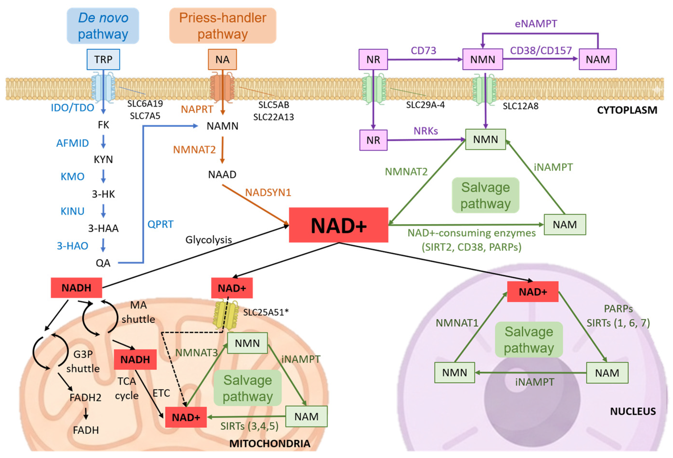
Importantly, two mechanistically distinct processes regulate NAD+ turnover: redox cycling, in which NAD+ is reversibly reduced to NADH by metabolic enzymes, and enzymatic consumption, in which NAD+ is cleaved by NAD+-consuming enzymes (e.g., sirtuins, PARPs, CD38) to produce nicotinamide (NAM), which re-enters the salvage pathway. This distinction is critical for accurately interpreting NAD dynamics and fluxes (Figure 1) [7,8].
Dysregulation of the NAD+/NADH ratio has been implicated in the pathogenesis and progression of numerous age-related diseases [5]. A decline in NAD+ levels is one of the most consistent and evolutionarily conserved hallmarks of aging across species, from yeast to humans, and is observed in virtually all tissues with advancing age [9,10,11]. This age-associated NAD+ depletion—typically ranging from 30 to 70% in old versus young organisms—contributes directly to multiple hallmarks of aging, including mitochondrial dysfunction, impaired DNA repair, loss of proteostasis, deregulated nutrient sensing, chronic inflammation (inflammaging), and cellular senescence [10,11,12]. The decline is driven by multiple interconnected mechanisms, including reduced efficiency of the NAMPT-mediated salvage pathway, increased activity of NAD+-consuming enzymes (notably CD38 and PARPs), and alterations in cellular bioenergetics and redox balance. These changes are tightly linked to broader metabolic rewiring, including impaired glycolysis, mitochondrial dysfunction, and altered lipid metabolism, highlighting the central role of NAD+ in cellular bioenergetics [13,14,15]. In cancer, by contrast, altered metabolic demands lead to dramatically increased NAD+ turnover to sustain rapid proliferation and biosynthetic needs [16]. In neurodegenerative disorders such as Alzheimer’s and Parkinson’s disease, compromised NAD+ availability exacerbates neuronal vulnerability, mitochondrial failure, and impaired proteostasis [17]. Similarly, disrupted NAD+ homeostasis promotes oxidative stress and insulin resistance in metabolic disorders, including obesity [18], type 2 diabetes [19], and cardiovascular diseases [20]. Genetic or pharmacological restoration of NAD+ levels in aged and progeroid models improves mitochondrial function, stem cell activity, neurovascular coupling, physical endurance, and both healthspan and lifespan, establishing NAD+ replenishment as one of the most promising interventions against age-related functional decline [20,21,22].
Accordingly, modulating NAD+ levels and the activity of NAD-metabolizing enzymes has emerged as a promising therapeutic approach to a variety of disorders. However, despite extensive research, a key limitation in the field is the incomplete integration of three critical aspects: (i) subcellular compartmentalization of NAD pools, (ii) methodological challenges in accurately quantifying NAD species, and (iii) the impact of these factors on the efficacy and specificity of therapeutic approaches.
Cellular compartmentalization represents a fundamental cellular strategy to protect and regulate distinct NAD pools. Simply increasing total intracellular NAD+ levels, therefore, may not affect all subcellular pools equally or as desired.
Importantly, emerging evidence suggests that the efficacy of NAD-targeting interventions is highly dependent on the distribution and accessibility of NAD within specific subcellular compartments, which can differentially regulate metabolic and signaling pathways.
For instance, mitochondria play a crucial role in maintaining cellular NAD levels and buffering against shortages in other cellular compartments. Although cells can generally tolerate a drop in overall NAD+, a decline in the mitochondrial NAD+ pool is especially detrimental [23]. Even under stress conditions, mitochondrial NAD+ remains stable longer than NAD+ in the nucleus or cytoplasm. This indicates a built-in protective system where mitochondrial NAD+ is used only as a last resort [24,25,26].
Therefore, effective strategies for controlled NAD modulation require precise quantitative mapping of NAD levels within each compartment. Reliable and accurate measurement of NAD concentrations is equally crucial as the foundation for assessing metabolic dynamics and designing targeted interventions.
In this context, this review aims to address this critical gap by providing an integrated framework that connects NAD compartmentalization, quantification methodologies, and therapeutic modulation strategies. It provides a topical overview of NAD concentrations across different cellular compartments, presents a comprehensive assessment of the technologies available for their measurement, and examines existing approaches for the modulation of NAD metabolism. Together, these elements contribute to establishing a reference point that can guide both fundamental research and the development of therapeutic strategies.
Figure 1.
Integrated overview of NAD+ metabolism and compartmentalization. Schematic representation of NAD+ biosynthesis through the de novo, Preiss–Handler, and salvage pathways. The diagram illustrates the compartmentalized distribution of NAD+ across cytosol, mitochondria, and nucleus, highlighting the organization of distinct subcellular NAD+ pools and their functional interplay with NAD+-consuming enzymes. Extracellular NAD+ metabolism and precursor interconversion pathways are also depicted [14,27]. Abbreviations: NAD+, nicotinamide adenine dinucleotide; NADH, reduced NAD+; NA, nicotinic acid; NAM, nicotinamide; NMN, nicotinamide mononucleotide; NAMN, nicotinic acid mononucleotide; NAAD, nicotinic acid adenine dinucleotide; NR, nicotinamide riboside; NAMPT, nicotinamide phosphoribosyltransferase; NMNAT1–3, nicotinamide mononucleotide adenylyltransferases; NAPRT, nicotinic acid phosphoribosyltransferase; NADSYN1, NAD synthetase 1; NRKs, nicotinamide riboside kinases; CD38/CD157, NAD+ glycohydrolases; CD73, ecto-5′-nucleotidase; SIRT(s), sirtuins; PARP(s), poly(ADP-ribose) polymerases; SLC25A51, mitochondrial NAD+ transporter; TCA cycle, tricarboxylic acid cycle; ETC, electron transport chain. * SLC25A51 has been recently identified as a mitochondrial NAD+ transporter; however, the relative contribution of NAD+ import versus local synthesis remains under investigation.
2. Intracellular and Organelle-Specific NAD+ and NADH Concentrations
Accurate measurement of NAD, both at the cell/tissue and organelle level, is essential for understanding cellular redox homeostasis and metabolism, and roles in physiopathology. This is challenging due to low abundance, rapid interconversion, and compartmentalization [28]. No single method is currently capable of capturing the full complexity of NAD metabolism; instead, each approach involves inherent trade-offs between temporal resolution, spatial localization, sensitivity, and quantitative accuracy. Importantly, these methodological differences can lead to substantial variability in reported NAD concentrations, which must be carefully considered when comparing studies (Figure 2, Table 1 and Table 2) [29].
The concentration of NAD varies significantly not only between mammalian species but also among cell types within the same species [30]. Moreover, NAD levels in cell lines are often reported relative to cell number (e.g., moles per million cells) rather than cell volume, complicating comparisons with other concentration units [31]. In addition, analytical platforms (e.g., LC-MS, enzymatic assays, biosensors) exhibit substantial differences in specificity, sensitivity, dynamic range, and sample preparation requirements, introducing systematic biases across studies. Sample preparation steps, particularly quenching and extraction, can induce artifactual interconversion between NAD+ and NADH, thereby distorting the measured redox state [32,33]. Furthermore, a critical but often overlooked factor is whether measurements reflect total or free NAD pools [28,34]. A significant portion of cellular NAD is protein-bound and tightly associated with enzymatic complexes, especially in mitochondria, while only the free pool is metabolically active [33]. Conventional extraction-based methods predominantly quantify total NAD, potentially overestimating the metabolically available fraction, whereas selective biochemical or imaging-based methods can differentiate between pools, aiding interpretation of redox balance [34,35] (Table 1 and Table 2).
Additionally, disruption of subcellular compartmentalization during sample processing represents a major limitation, as it obscures the true intracellular distribution of NAD species and may lead to misinterpretation of compartment-specific dynamics. Beyond these methodological considerations, NAD levels are highly dynamic and depend on cellular metabolic states, nutritional status, age, and environmental conditions [36].
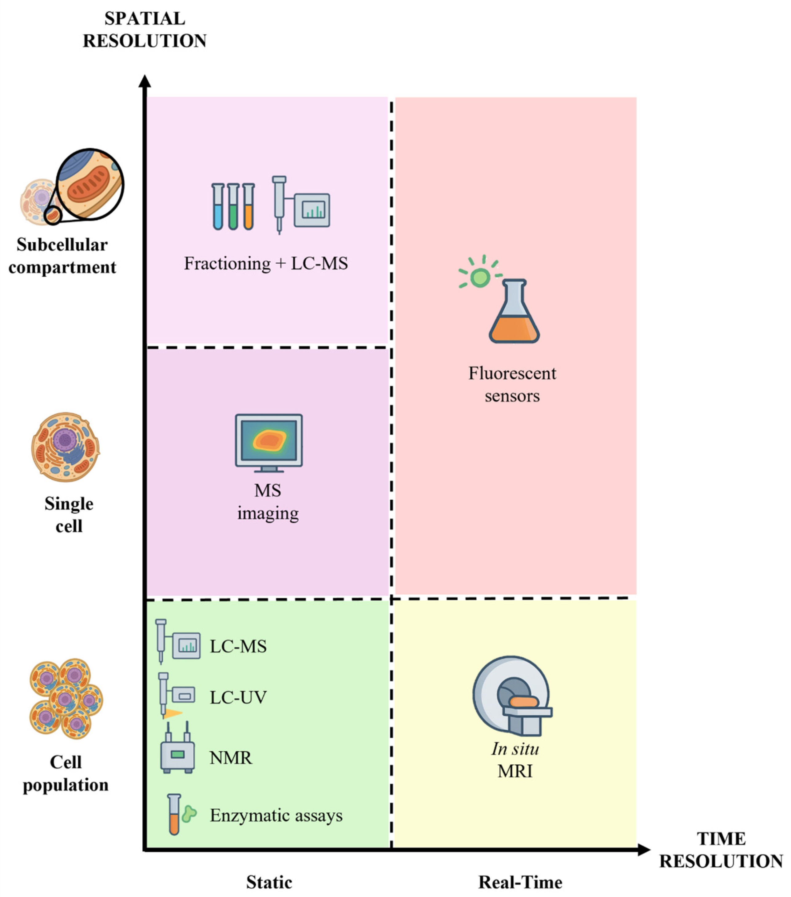
Figure 2.
Comparison of NAD+ quantification methods based on spatial and temporal resolution: in situ Magnetic Resonance Imaging (MRI), Liquid Chromatography tandem Mass Spectrometry (LC-MS), LC-UV, Nuclear Magnetic Resonance (NMR), enzymatic assays, Mass Spectrometry Imaging (MSI), fluorescent sensors, fractioning, and LC-MS [29].
Consequently, measuring the NAD+/NADH ratio is often preferred because it provides a specific readout of the redox state and is generally easier to interpret. From a methodological perspective, ratio-based measurements can partially mitigate absolute quantification biases. However, they remain sensitive to experimental artifacts such as differential degradation or interconversion of NAD species [37].
To study NAD metabolism, many experimental models exist, in vivo, ex vivo, or in vitro [28], but a trade-off is necessary to obtain good results on temporal resolution, tracking changes over time, and spatial resolution, which allows NAD localization within tissues or subcellular compartments [38]. Each technique, therefore, provides a partial and context-dependent view of NAD metabolism, reinforcing the need for integrative approaches that combine complementary methodologies. In Table 1 and Table 2 and Figure 1, the wide range of technologies used for NAD measurement, considering their advantages and limitations, is summarized.
In mammalian cells, total NAD amounts to between approximately 200 and 800 µM [26,39,40,41]. However, reported values vary widely depending on the measurement method and experimental conditions. For example, LC-MS-based studies and biosensor-based approaches can yield substantially different estimates, reflecting methodological biases rather than true biological differences. Notably, substantial discrepancies exist across studies reporting NAD+ concentrations, reflecting methodological rather than purely biological differences. For instance, Yang et al. reported total cellular NAD+ levels of approximately 365 ± 30 µM in HEK293 cells, with mitochondrial concentrations around 245 µM, whereas biosensor-based approaches, such as those used by Cambronne et al., tend to report lower free NAD+ concentrations due to selective detection of metabolically active pools [26,31]. Similarly, LC–MS-based approaches generally yield higher absolute NAD+ concentrations compared to genetically encoded biosensors, which primarily capture relative changes and free NAD+/NADH ratios in live cells. These differences highlight the critical distinction between total and free NAD+ measurements, as well as the impact of extraction procedures and subcellular resolution on quantitative outcomes. Therefore, direct comparison between studies requires careful consideration of both the analytical method and the specific NAD pool being measured, as these discrepancies complicate the establishment of standardized reference values for intracellular and compartment-specific NAD+ concentrations. Importantly, only an estimated 10–25% of this pool exists as free NAD, the metabolically active fraction directly available for redox reactions, enzymatic consumption by sirtuins, PARPs, and CD38, and signaling. In contrast, the majority of NAD is protein-bound, particularly in dehydrogenases, and is less accessible for rapid metabolic regulation. This distinction is critical because most conventional extraction-based methods do not discriminate between these pools, complicating the interpretation of functional NAD+/NADH ratios [41,42]. This balance between free and bound NAD is highly dynamic and depends on different conditions, such as hypoxia or age [43,44].
Approximately 10% of the total NAD pool is phosphorylated by NAD kinases to generate NADP+ and NADPH [4,41,45]. While the NAD+/NADH couple is mainly dedicated to catabolic reactions and mitochondrial energy production, the NADP+/NADPH redox pair primarily supports anabolic processes such as fatty acid, cholesterol, and nucleotide biosynthesis. Moreover, NADPH plays a central role in cellular antioxidant defense by providing reducing power for glutathione reductase and thioredoxin systems, and it serves as an electron donor for NADPH oxidases during immune responses [45,46]. Significantly, the ratios NAD+/NADH and NADP+/NADPH represent key indicators of cellular redox state and metabolic flux. Alterations in these ratios reflect shifts between oxidative and reductive metabolism and have been proposed as potential biomarkers of metabolic dysfunction and disease progression, including conditions characterized by mitochondrial impairment, oxidative stress, or altered biosynthetic demand [45,47].
Importantly, the NADP(H) pool is largely insulated from the NAD(H) pool due to the substrate specificity of most dehydrogenases, allowing independent regulation of catabolic and anabolic redox states. Collectively, NADPH-dependent pathways are particularly important during cellular stress, when biosynthetic, antioxidant, and immune demands increase [46,48].
Table 1.
Whole-cell (intracellular) NAD+ and NADH quantification.
Table 2.
Organelle-Specific NAD+ and NADH Quantification.
Importantly, methodological differences reported in Table 1 and Table 2, including sensitivity, detection limits, and sample preparation requirements, contribute significantly to variability in reported NAD concentrations across studies.
2.1. Organelle-Specific Concentrations and Ratios
NAD levels vary across organelles [31,41,69] (Figure 2). This compartmentalization allows each organelle to maintain distinct NAD+/NADH ratios supporting specific metabolic and signaling functions. Recently, organelle-targeted biosensors and metabolomics workflows enabled the revelation of NAD distributions across [31,42,70]. Estimations of quantitative NAD metabolism parameters for individual organelles are presented in this section (Figure 3).
Figure 3.
Subcellular compartmentalization of NAD+/NADH redox balance shapes metabolic specialization. High NAD+/NADH ratios in the cytosol, nucleus, and peroxisomes support glycolysis, DNA repair, and β-oxidation, whereas a lower ratio in mitochondria favors ATP production through oxidative metabolism (also see Table 3).
2.1.1. Cytosol
In mammalian cells, the free cytosolic concentration is generally estimated to be around 10–100 µM in COS7 cells (Cercopithecus aethiops Renal Cells) [71] and ~100 µM in HEK293T (clonal Human Embryonic Kidney) cells [31] but can reach higher levels in other cell types, such as ~260 µM in A375 human melanoma cells [72]. Considering only free amounts, the NAD+/NADH ratio in the cytoplasm of healthy mammalian tissues is generally very high, often around 700:1, a state that strongly favors glycolysis, lactate dehydrogenase activity, and other dehydrogenase-catalyzed intermediates [73].
2.1.2. Mitochondria
Mitochondria are major sites of NAD+ reduction, where metabolic enzymes of the TCA cycle reduce NAD+ to NADH, which is then re-oxidized by the electron transport chain to support ATP production. A significant portion of the total cellular NAD+ pool, estimated to be between 40% and 70%, resides within the mitochondria, although this proportion varies among cell types [31], e.g., ~70% in cardiac myocytes [73], 50% in neurons, and 30–40% in hepatocytes [74]. Importantly, the mitochondrial NAD pool is relatively distinct from the cytosolic pool, since neither NAD+ nor NADH can freely diffuse across the inner mitochondrial membrane [14]. While mitochondrial NAD+ was long thought to depend primarily on local biosynthesis through the de novo and salvage pathways, the recent identification of SLC25A51 (MCART1) demonstrated that cytosolic NAD+ can also be directly imported into mitochondria. However, the relative contribution of NAD+ import versus intramitochondrial synthesis to mitochondrial NAD homeostasis and cellular physiology remains under investigation. While SLC25A1 has also been proposed to play a role in these processes, its precise contribution to mitochondrial bioenergetics remains a subject of ongoing debate and requires more stringent validation [14,75,76]. In contrast, NADH itself cannot cross the inner mitochondrial membrane. Therefore, the functional coupling between cytosolic and mitochondrial redox states is mediated by redox shuttle systems, primarily the Malate–Aspartate Shuttle (MAS) and the Glycerol-3-Phosphate (G3P) shuttle, which transfer reducing equivalents rather than NAD(H) molecules across compartments. These shuttles regenerate cytosolic NAD+ to sustain glycolysis while simultaneously influencing the NAD+/NADH ratios and redox balance of both cytosolic and mitochondrial compartments, thereby affecting ATP production and cellular metabolism [77,78]. Despite the inherent technical difficulties in quantifying these localized pools, the mitochondrial NAD+/NADH ratio is typically maintained at a lower level compared to the cytosol, ranging from 7 to 8 [79], reflecting the electron transport chain activity, which rapidly re-oxidizes NADH to NAD+. Interestingly, mitochondrial NAD remains stable even when cytosolic NAD+ levels are severely reduced, suggesting that mitochondria maintain a dedicated NAD reservoir to preserve their function under cellular stress conditions [33]. Moreover, mitochondrial NAD+ can compensate for depletion in other cellular compartments, highlighting the role of mitochondria as central regulators of inter-organelle NAD balance [33].
Mitochondrial NAD metabolism is dynamically regulated by the segregation of mitochondrial subpopulations with distinct metabolic functions. When ATP demand increases, pyrroline-5-carboxylate synthase (P5CS), the rate-limiting enzyme for the reductive biosynthesis of proline and ornithine, becomes sequestered in a subset of mitochondria lacking cristae and ATP synthase. These “biosynthetic” mitochondria have high reductive potential, sustaining lower NAD(P)+/NADP(H) ratios, while the complementary “energetic” subpopulation, enriched in ATP synthase and organized cristae, operates with higher NAD+/NADH ratios to maximize oxidative phosphorylation. This segregation is reversible, depends on mitochondrial fusion and fission dynamics and indicates that NAD redox balance within mitochondria changes according to bioenergetic versus biosynthetic needs [80].
2.1.3. Nucleus
The nuclear NAD+ pool is critical for processes such as DNA repair, gene expression, and epigenetic modifications mediated by NAD+-dependent enzymes such as sirtuins and PARPs [5,23]. The free nuclear NAD+ concentration is estimated to be comparable to that of the cytosol, typically around 10–100 µM in COS7 cells and ~100 µM in HEK293T cells [31,71]. Due to the presence of nuclear pore complexes, the free cytosolic and nuclear NAD compartments are generally considered to be in equilibrium, allowing changes in cytosolic redox state to be reflected in the nucleus [71,81].
2.1.4. Endoplasmic Reticulum (ER)
Emerging evidence indicates distinct ER NAD+ pools, separate from the cytosolic one and playing roles in regulating ER function and redox homeostasis [82]. The ER lumen maintains a highly oxidizing environment essential for proper protein folding, and while NADPH is generally considered the primary reductant in the ER, NAD+-dependent processes, such as ADP-ribosylation, also occur within this compartment [69,82]. The concentrations of NAD+ and NADH within the ER have been less extensively quantified compared to mitochondria and the cytosol.
2.1.5. Golgi
NAD+ may also play a critical role in the maintenance of Golgi apparatus structure and function. For instance, NAD+ is required for the disassembly of the Golgi complex by certain inhibitors—such as Brefeldin A— suggesting a role in ADP-ribosylation events within the Golgi [83]. Also, one isoform of the NAD+ synthesizing enzyme, NMNAT2, specifically localizes to the Golgi apparatus, indicating active NAD+ metabolism there [84]. Although the exact NAD concentration and NAD+/NADH ratio within the Golgi apparatus can be less clearly pinpointed than in the cytosol or mitochondria, it is generally hypothesized that the Golgi—being part of the endomembrane system and closely connected to both the ER and cytosol—maintains a relatively high NAD+/NADH ratio, comparable to that of the cytosol. This redox environment would support oxidative modifications and protein-folding processes occurring in this compartment. However, to date, no direct experimental evidence has been reported to substantiate this assumption.
2.1.6. Peroxisomes
Peroxisomes are involved in various lipid metabolism pathways, including fatty acid β-oxidation, and contain NAD-dependent enzymes [69]. Maintenance of NAD homeostasis within peroxisomes seems vital for their function: studies have shown that the peroxisomal NAD+/NADH ratio is closely connected to the cytosolic ratio, with specific redox shuttle systems (e.g., malate/oxaloacetate shuttle and glycerol-3-phosphate shuttle, also pyruvate/lactate [69]) regulating this crosstalk and ensuring the availability of NAD+ for peroxisomal metabolic reactions [69,85].
2.1.7. Extracellular Space
NAD+ is also present in the extracellular space [86]. Probably, the primary source of extracellular NAD+ is the release from dying or damaged cells with compromised cell membrane integrity. However, extracellular NAD+ may also be regulated by pore-forming proteins [86,87,88]. Extracellular NAD+ levels are much lower than intracellular levels, and likely, extracellular NAD+ is metabolized by ectoenzymes with roles in cellular signaling. For instance, CD38 and CD157 (NAD+ glycohydrolases) break down NAD+ into the signaling molecule cyclic ADP-ribose [89,90,91]. Therefore, extracellular NAD+ could represent a targetable pool for modulating signaling pathways, with potential implications in inflammation, immunity, and tissue regeneration, if better understood.
2.2. Tissue-Specific Concentration
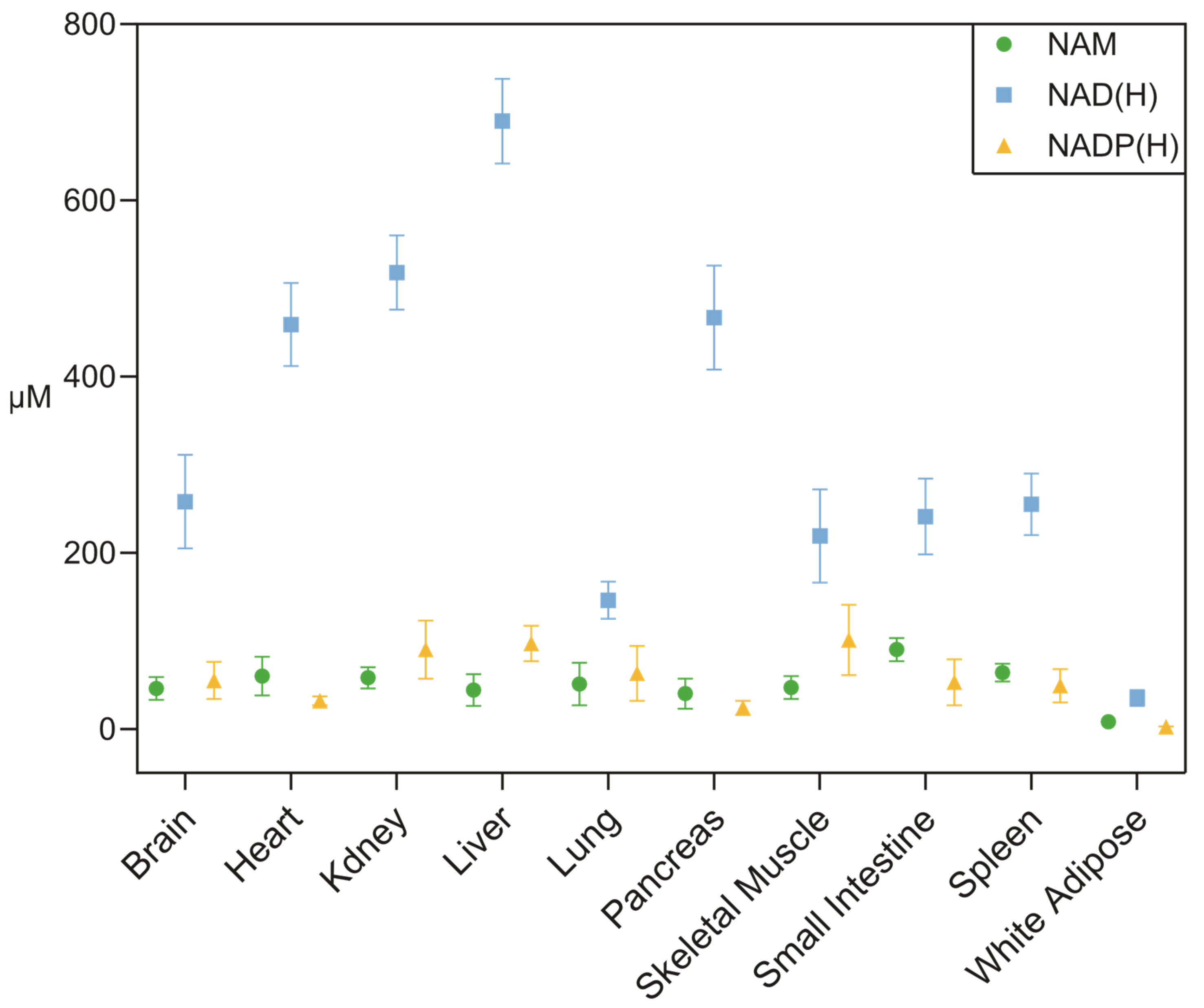
Recently, quantitative metabolomic profiling revealed a pronounced inter-tissue heterogeneity in absolute NAD-related metabolite concentrations in a mouse model, with values ranging from ~690 μM in liver to ~35 μM in adipose tissue (Figure 4) [41]. This gradient suggests that the availability of NAD(H), as well as its related metabolites NAM and NADP(H), depends on tissue-specific biosynthetic capacity, turnover kinetics, and the activity of NAD-consuming enzymes. These differences represent a major challenge for systemic NAD-boosting strategies, since pharmacological or nutritional interventions can have very different effects depending on the metabolic context of each organ [92].
In addition to NAD(H), its precursor nicotinamide (NAM) and the phosphorylated redox pool NADP(H) also display tissue-specific variability (Figure 4). NAM concentrations are generally lower and more homogeneous across tissues, consistent with its role as a circulating intermediate in NAD+ turnover and salvage pathways. In contrast, NADP(H) levels exhibit a distinct distribution, with relatively higher concentrations in metabolically active tissues such as liver and kidney, reflecting its central role in anabolic reactions and redox homeostasis [41].
This variability is largely attributed to differential expression patterns of enzymes involved in NAD+ biosynthesis and recycling pathways [93]. Notably, tissues with high levels of proliferation or immune activity, such as the small intestine and the spleen, exhibit rapid rates of NAD+ turnover, characterized by short half-lives [41]. In contrast, metabolically active but less proliferative tissues, such as skeletal muscle, exhibit significantly slower NAD+ turnover. This highlights that NAD metabolism is highly regulated and adapted to the specific needs of each tissue.
Circulating NAD+ levels in blood and blood cells have been reported to be substantially lower than those observed in solid tissues, further supporting the existence of pronounced inter-compartment variability [94]. However, direct quantitative comparison across studies remains challenging due to methodological heterogeneity.
Figure 4.
Concentrations of NAM, NAD(H), and NADP(H) in murine tissues. Data are expressed as mean ± s.d., n = 4, unit: µM. Data from Liu et al. 2018 [41].
Therefore, successful modulation of NAD metabolism must consider not only subcellular compartmentalization but also inter-organ variability in NAD pool size and turnover, as both factors determine the overall physiological impact of NAD-targeted interventions. In addition, potentially tissue-specific import kinetics of NAD or NAD+ precursors must be considered.
Table 3.
Reported concentration of NAD+, NADH, and [NAD+]/[NADH] ratios in different mammalian cell compartments.
Reported NAD concentrations vary substantially depending on the measurement method, sample preparation, and whether total or free NAD pools are assessed. Therefore, values should be interpreted within the methodological context of each study.
3. Therapeutic Strategies Targeting NAD Metabolism
The pivotal role of NAD+ in cellular energy metabolism, DNA repair, and signaling pathways has made NAD+ metabolism an appealing target for therapeutic interventions across diverse conditions, including age-related diseases, metabolic disorders, and cancers. A broad spectrum of strategies has been developed to either increase NAD+ availability or modulate the activity of enzymes involved in its synthesis and consumption. Importantly, the efficacy of these approaches is increasingly recognized to depend on tissue-specific and subcellular NAD dynamics, which may differentially influence therapeutic outcomes.
3.1. NAD Precursors and Supplements
One of the most widely explored strategies to enhance intracellular NAD+ levels is the administration of NAD+ precursors. Nicotinamide riboside (NR) and nicotinamide mononucleotide (NMN) are particularly prominent in this regard, both serving as direct precursors that can be converted to NAD+ through salvage pathways [10] (Figure 1). Preclinical studies have consistently shown that NR and NMN oral supplementation increases NAD+ levels in multiple tissues, leading to beneficial effects on mitochondrial function, metabolic health, and even lifespan [15].
Clinical studies investigating the effects of NR and NMN supplementation are ongoing and promising. For instance, studies have demonstrated that NR supplementation can safely increase whole blood NAD+ levels in healthy adults [97]. Furthermore, clinical trials investigated the effects of NR and NMN on metabolic parameters, muscle function, and cardiovascular health, with some studies reporting improvements in insulin sensitivity, endurance, and inflammatory markers, particularly in older adults or those with pre-existing metabolic diseases [98,99]. However, despite encouraging results, long-term efficacy, optimal dosing strategies, and tissue-specific responses remain incompletely understood. Despite the promising effects of NAD+ precursors, their efficacy may be limited by bioavailability, tissue-specific uptake, and metabolic conversion [41]. In this context, alternative strategies have been developed to directly enhance endogenous NAD+ biosynthesis.
Among these, small-molecule activators of nicotinamide phosphoribosyltransferase (NAMPT), such as SBI-797812, act by stimulating the rate-limiting step of the NAD+ salvage pathway, thereby increasing intracellular NAD+ production [100]. In contrast to precursor-based approaches, which depend on uptake and enzymatic conversion, NAMPT activators may provide a more direct and potentially more efficient means of boosting NAD+ levels.
Similarly, neuroprotective compounds such as P7C3 have been shown to elevate intracellular NAD+ levels, likely through stabilization of NAMPT activity, thereby supporting neuronal survival under conditions of metabolic and oxidative stress. Preclinical studies indicate that P7C3 can improve mitochondrial function and confer protection in models of neurodegeneration, highlighting its potential relevance in age-related neurological disorders [101,102].
However, despite these promising findings, several limitations remain. The long-term safety, tissue specificity, and potential off-target effects of these compounds are not yet fully understood. Moreover, excessive or non-specific activation of NAD+ biosynthesis may have context-dependent consequences, potentially supporting pathological processes such as tumor metabolism or maladaptive stress responses. Therefore, further investigation is required to determine their translational potential and to assess whether selective modulation of NAD+ synthesis can be achieved in a compartment-specific manner [103,104].
The route of administration appears to play a crucial role in determining the bioavailability and pharmacokinetics of NR and NMN. While NR and NMN can be administered systemically, their metabolic fate is highly context-dependent. Following oral administration, a substantial fraction of NR or NMN is converted to nicotinamide (NAM) in the liver before reaching peripheral tissues, suggesting that systemic NAD+ increases often rely on secondary salvage pathways rather than direct precursor uptake. These findings suggest that the route of administration significantly influences the utilization of these precursors in the body [41,105].
While the liver plays a central role in systemic NAD metabolism through de novo synthesis from tryptophan, many cell types possess the enzymatic machinery required for NAD+ synthesis, albeit at different capacities [5]. This raises an important distinction between cell-autonomous NAD+ production and systemic NAD+ supply. Some tissues rely heavily on circulating precursors (e.g., NAM), whereas others maintain local NAD+ pools through intrinsic biosynthetic pathways. This balance has significant implications for therapeutic strategies, as NAD+ boosting interventions may differentially affect tissues depending on their metabolic autonomy [103,106].
Moreover, the gut microbiome also has a significant impact on NAD+ metabolism by converting precursors such as NR, NMN, and NAM into deamidated metabolites through bacterial enzymes like PncA and PncC [107]. Therefore, only a small proportion of NAD+ precursors reach tissues unmodified [107].
3.2. Enzyme Modulators
Beyond direct NAD+ precursor supplementation, modulating the activity of enzymes involved in NAD+ synthesis or consumption represents another promising therapeutic approach. Inhibitors of NAD+-consuming enzymes, such as CD38 and PARPs, aim to preserve intracellular NAD+ levels by limiting its degradation. CD38 is a major NADase responsible for significant NAD+ degradation, and its inhibition has shown potential in increasing NAD+ levels and improving metabolic function in preclinical models, particularly in the context of aging [108]. Similarly, PARPs are NAD+-dependent enzymes essential for DNA repair; however, excessive PARP activation -such as during extensive DNA damage- can lead to NAD+ depletion. PARP inhibitors, widely used in oncology, may also indirectly preserve NAD+ pools by reducing NAD+ consumption, although their systemic metabolic effects remain to be fully characterized [109,110].
Importantly, modulation of NAD metabolism may interact with other pharmacological treatments. Many commonly used drugs, including antipsychotics [111] and chemotherapeutic agents [112,113], can impair mitochondrial function and cellular bioenergetics, thereby influencing NAD+ turnover and redox balance. As a result, NAD+-boosting strategies may have context-dependent effects, potentially enhancing or counteracting the metabolic impact of concomitant therapies [104].
Alternatively, activating NAD+-dependent enzymes, such as sirtuins, has emerged as another strategy. Sirtuins are a family of deacetylases that require NAD+ for their activity and play crucial roles in regulating metabolism, inflammation, and aging [114]. Sirtuin activators (e.g., resveratrol analogs for SIRT1) aim to enhance sirtuin activity, thereby mimicking the beneficial effects of caloric restriction [115]. These enzyme modulators provide a more targeted and mechanistically specific approach to manipulating NAD+ metabolism, with potential applications in inflammatory conditions, neurodegenerative disorders, and metabolic diseases.
3.3. Gene Therapy and NAD Regulation
Genetic strategies manipulate NAD+ metabolism by directly influencing the expression of key enzymes involved in NAD+ synthesis.
Overexpression of rate-limiting enzymes in NAD+ biosynthesis pathways, such as NAMPT or enzymes in the de novo synthesis pathway, could provide sustained increases in cellular NAD+ levels [9]. In preclinical studies employing viral vectors to deliver genes encoding for NAD+ synthetic enzymes, NAD+ levels were successfully increased, and disease phenotypes in models of metabolic dysfunction and neurodegeneration were ameliorated [116]. While still in early stages of development for applications in humans, these approaches offer the potential for long-term reprogramming of NAD+ homeostasis, although challenges related to delivery efficiency, tissue specificity, and safety remain significant.
Importantly, the success of gene- and enzyme-based NAD-targeting strategies is likely to depend on their ability to modulate NAD+ levels in a compartment-specific manner. Emerging evidence suggests that restoring NAD+ in specific organelles, particularly mitochondria, may be more relevant than globally increasing cellular NAD+, highlighting the need for spatially targeted therapeutic approaches [4,27,117].
4. Targeting NAD-Related Pathways in Aging and Diseases
NAD+ decline represents a central metabolic node linking aging to disease, but its impact is highly dependent on subcellular compartmentalization and tissue-specific bioenergetic demands.
Age-associated NAD+ decline contributes to multiple interconnected cellular dysfunctions, including mitochondrial impairment, genomic instability, deregulated nutrient sensing, and chronic inflammation [24,116]. While the underlying mechanisms–primarily diminished NAMPT activity and heightened consumption by CD38 and PARPs–are well-established and serve as a common upstream driver of NAD+ imbalance across multiple tissues [108,118].
Emerging evidence highlights alterations in subcellular NAD+ distribution as a critical, yet underappreciated, feature of aging [23,27].
In aged cells and tissues, cytosolic and nuclear NAD+ pools often deplete preferentially, whereas mitochondrial NAD+ is relatively preserved through buffering mechanisms, prioritizing oxidative phosphorylation under stress [23,44].
These alterations are tightly linked to widespread changes in cellular bioenergetics, including impaired glycolytic flux (due to reduced GAPDH activity), decreased tricarboxylic acid (TCA) cycle turnover, altered lipid metabolism (including impaired β-oxidation), and compromised mitochondrial respiration, all of which critically depend on NAD+ availability as a central redox cofactor and regulator of metabolic fluxes [17,119].
However, prolonged aging eventually erodes this reserve, exacerbating redox imbalance and energetic failure.
In neurodegenerative disorders such as Alzheimer’s disease (AD) and Parkinson’s disease (PD), reduced NAD+ availability disrupts neuronal bioenergetics at multiple levels, including impaired glycolysis, mitochondrial dysfunction, and defective proteostasis. These alterations converge on reduced ATP production and increased oxidative stress, ultimately enhancing neuronal vulnerability [119,120]. Preclinical studies have shown that NAD+ precursors (NR and NMN) can restore glycolytic flux, TCA cycle activity, and lipid metabolism, thereby improving mitochondrial function, reducing pathology, and ameliorating cognitive deficits in AD and PD models [119,120]. Emerging clinical evidence supports beneficial effects on mitochondrial function and oxidative stress markers, although larger trials focused on cerebral bioenergetics are still needed [17,99].
In the aging brain, NAD+ depletion reduces the activity of NAD+-dependent enzymes, including sirtuins and PARPs, leading to impaired DNA repair and progressive mitochondrial dysfunction. This metabolic failure is both a cause and a consequence of cellular senescence, a state of irreversible cell-cycle arrest that accumulates in the aging CNS. Senescent cells develop a Senescence-Associated Secretory Phenotype (SASP), releasing pro-inflammatory mediators that further disrupt tissue homeostasis. Importantly, upregulation of the NAD+-consuming enzyme CD38 in senescent cells acts as a metabolic “sink,” further depleting NAD+ pools and amplifying bioenergetic dysfunction and inflammation.
Alterations in the NAD+/NADH ratio further constrain oxidative phosphorylation and shift neuronal metabolism toward less efficient energy-producing pathways [17].
Comparable NAD+-dependent metabolic alterations are observed in obesity, type 2 diabetes, and cardiovascular diseases [15,121,122]. In these conditions, reduced NAD+ availability contributes to insulin resistance, impaired mitochondrial fatty acid oxidation, altered glucose metabolism, and chronic inflammation. Altered NAD+/NADH ratios in these diseases reflect systemic redox imbalance and metabolic inflexibility, reinforcing the central role of NAD+ in coordinating energy metabolism across tissues.
Preclinical studies in aged animal models have consistently shown that restoring NAD+ levels improves mitochondrial function, enhances metabolic flexibility, and reduces inflammation. However, despite these shared mechanisms, therapeutic responses to NAD+ modulation can vary substantially depending on tissue type, disease context, and subcellular NAD distribution [99,123,124]. A critical emerging concept is that global increases in NAD+ levels may not uniformly restore cellular function, as distinct subcellular NAD pools are differentially regulated and may respond differently to therapeutic interventions [23].
Importantly, NAD+ modulation exhibits context-dependent effects. While increasing NAD+ levels can restore metabolic and mitochondrial function, excessive or non-targeted NAD+ boosting may also support pathological processes, including tumor metabolism, chronic inflammation, or maladaptive stress responses [125,126]. In addition, NAD-targeting strategies may interact with other pharmacological treatments that influence cellular bioenergetics. For instance, many antipsychotics and psychotropic drugs impair mitochondrial function and disrupt NAD+/NADH redox balance. Therefore, the concomitant administration of NAD+ boosters could lead to synergistic or antagonistic effects on cell survival and energetic recovery, a factor that must be carefully evaluated in clinical settings for patients with neurodegenerative or psychiatric comorbidities [127].
Overall, the impact of NAD+ decline is highly context-dependent and shaped by tissue-specific metabolism and subcellular compartmentalization. As a result, global NAD+ boosting strategies may not uniformly restore cellular function, highlighting the need for spatially and temporally controlled therapeutic approaches [27].
Precise quantification of subcellular NAD+ dynamics will be instrumental in guiding such targeted therapies and enabling more personalized interventions.
5. Challenges and Limitations in NAD Modulation
The preceding sections highlight that NAD metabolism is not only dynamically regulated but also highly compartmentalized across cellular organelles, with distinct pools exhibiting unique concentrations, redox ratios, and responses to stressors. This subcellular organization, together with the tissue-specific regulation of NAD biosynthesis and consumption, poses major challenges to therapeutic modulation. Non-targeted interventions may alter total cellular NAD+ levels without effectively restoring the NAD pools most relevant for mitochondrial function, redox regulation, or stress adaptation, resulting in variable efficacy and possible off-target effects [5,14,73,80]. Moreover, emerging evidence suggests that NAD+ dynamics are influenced by factors such as age, sex, metabolic status, and disease context, implying that uniform NAD-modulating strategies may yield heterogeneous outcomes across individuals [128,129]. Addressing these limitations will require the integration of compartment-specific quantification methods, including biosensors and organelle-resolved analytical approaches, to guide precision interventions and improve translational efficacy [37,70,104].
Despite compelling preclinical evidence and promising initial clinical results, modulating NAD for therapeutic purposes still faces significant challenges and limitations that require careful consideration and ongoing research to refine strategies and mitigate risks.
5.1. Safety Concerns and Side Effects
While NAD+ precursors like NR and NMN have generally shown good safety profiles in short-term human trials, the potential risks of long-term NAD supplementation remain largely unknown. The cellular machinery involved in NAD+ metabolism is incredibly complex and in constant interaction with biochemical processes and molecular pathways. Sustained or excessive increases in NAD+ availability may disrupt this metabolic balance, potentially leading to unintended effects on redox homeostasis, signaling pathways, and cellular adaptation mechanisms [10]. For instance, altering NAD+ metabolism may affect the activity of NAD-dependent enzymes such as sirtuins and PARPs, with downstream consequences for mitochondrial function, inflammatory responses, and metabolic regulation [4].
Furthermore, because NAD+ supports cellular metabolism broadly, excessive or poorly targeted NAD+ boosting could, under certain pathological conditions, facilitate maladaptive responses, including the support of inflammatory or proliferative processes [126,130].
Although preclinical studies have demonstrated clear benefits, chronic NAD+ boosting in humans requires rigorous evaluation to rule out the possibility of cumulative toxicities or subtle physiological shifts that might only manifest over prolonged periods [131].
5.2. Incomplete Clinical Data
A significant limitation in the field is the incomplete clinical data regarding the effects of NAD+ boosting in humans.
Most human studies to date have been relatively small in scale, short in duration, and often conducted in healthy young or middle-aged individuals [104].
There is a critical need for larger, well-controlled, long-term clinical trials involving diverse populations, including elderly individuals and those with specific pre-existing conditions, to fully understand the efficacy and safety of NAD-targeting therapies. Important gaps remain regarding optimal dosing regimens, routes of administration, tissue-specific responses, and the long-term consequences of manipulating NAD metabolism in humans [132,133,134,135]. Furthermore, many studies rely on systemic surrogate markers rather than direct assessments of compartment-specific NAD dynamics or clinically meaningful endpoints, limiting the interpretation of therapeutic benefits.
Without comprehensive clinical evidence, the widespread adoption of NAD-based therapies will remain limited.
5.3. Potential for Resistance and Translation Challenges
Translating promising preclinical findings in NAD-targeting therapies into effective human treatments presents several challenges, including the potential for resistance.
Biological systems can adapt to sustained metabolic interventions, and compensatory mechanisms may reduce the long-term effectiveness of NAD-modulating therapies.
For example, adaptive changes in NAD+ degradation pathways or altered sensitivity of NAD-dependent enzymes may progressively attenuate therapeutic efficacy [10]. Additionally, pharmacokinetic and pharmacodynamic challenges can hinder translation. Ensuring that NAD+ precursors or enzyme modulators reach the appropriate tissues and intracellular compartments at effective concentrations remains a major translational barrier.
The unique metabolic environment of different diseases, such as the tumor microenvironment or the central nervous system, can present significant barriers to effective drug delivery and action, ultimately impacting the success of translating NAD-targeting therapies into widespread clinical use [130,136].
6. Conclusions
Taken together, NAD metabolism is highly complex, responding sensitively to slight intrinsic and extrinsic clues while regulating fundamental cellular processes.
To fully exploit the therapeutic potential of NAD-targeting strategies, a deeper understanding of the pharmacokinetics, pharmacodynamics, and compartment-specific regulation of NAD metabolism is essential. One particularly underexplored aspect concerns how NAD-targeting interventions affect intracellular NAD compartmentalization and the activity of NAD-dependent metabolic pathways within specific organelles [27].
As highlighted throughout this review, the biological effects of NAD modulation cannot be fully understood without considering the distinct regulation of subcellular NAD pools and their role in maintaining cellular bioenergetics and redox homeostasis [4,17].
Addressing these knowledge gaps will be critical for the development of precise and personalized NAD-modulating strategies that maximize therapeutic efficacy while minimizing adverse or context-dependent effects.
Luckily, the recent technological progress is enabling increasingly precise investigations of intracellular NAD dynamics at the organelle level.
In this review, we have summarized key methodologies for measuring NAD within specific organelles, thereby providing a framework to guide future methodological integration and refinement.
The complexity of NAD makes it an attractive therapeutic target but also demands a more integrated understanding of how measurement methodologies, subcellular compartmentalization, and disease-specific metabolic contexts influence therapeutic outcomes.
Future progress will depend on the development of delivery systems and intervention strategies that allow precise spatial targeting, careful dose optimization, and controlled modulation of NAD metabolism across different disease settings [104].
Author Contributions
Conceptualization, G.P., J.B., and F.R.; methodology, M.N., S.S., V.F., G.P., F.R., and J.B.; investigation, M.N., S.S., V.F., G.P., F.R., and J.B.; data curation, M.N., S.S., V.F., G.P., F.R., and J.B.; writing—original draft preparation, M.N., S.S., and V.F.; writing—review and editing, G.P., F.R., and J.B.; visualization, M.N.; supervision, G.P. All authors have read and agreed to the published version of the manuscript.
Funding
No external research funding was received for this work. Article publishing charges were covered by the University of Milan-Bicocca, Department of Medicine and Surgery.
Data Availability Statement
No datasets were generated or analyzed during the current study.
Acknowledgments
The authors thank colleagues and collaborators for helpful discussions. During the preparation of this work, the authors used ChatGPT (OpenAI) and Grok (xAI) in order to improve the language, readability, and clarity of the text. After using these tools/services, the authors reviewed and edited the content as needed and take full responsibility for the content of the published article.
Conflicts of Interest
Giuseppe Paglia reports employment at the University of Milan-Bicocca, Department of Medicine and Surgery. All other authors declare no competing financial interests or personal relationships that could have appeared to influence the work reported in this paper.
Abbreviations
The following abbreviations are used in this manuscript:
| A375 | Human Melanoma Cells |
| ADP | Adenosine Diphosphate |
| ADPR | Adenosine Diphosphate Ribose |
| ATP | Adenosine Triphosphate |
| cADPR | Cyclic Adenosine Diphosphate Ribose |
| CD38 | NAD Glycohydrolase, Cluster of Differentiation 38 |
| CD157 | Cluster of Differentiation 157 (bone marrow stromal antigen-1) |
| COS7 | CV-1 (simian) in Origin with SV40 genes (cell line) |
| DNA | Deoxyribonucleic Acid |
| DRP1 | Dynamin-Related Protein 1 |
| ER | Endoplasmic Reticulum |
| FIS1 | Mitochondrial Fission Protein 1 |
| G3P | Glycerol-3-Phosphate shuttle |
| GAPDH | Glyceraldehyde-3-phosphate dehydrogenase |
| HEK293 | Human Embryonic Kidney 293 cells |
| HEK293T | Human Embryonic Kidney 293 cells expressing SV40 large T antigen |
| HeLa | Human Cervical Cancer Cell Line |
| HPLC | High-Performance Liquid Chromatography |
| LC-MS/MS | Liquid Chromatography Tandem Mass Spectrometry |
| LOD | Limit of Detection |
| LOQ | Limit of Quantification |
| MAS | Malate–Aspartate Shuttle |
| MFF | Mitochondrial Fission Factor |
| MFN1 | Mitofusin 1 |
| MFN2 | Mitofusin 2 |
| MID49 | Mitochondrial Dynamics Protein of 49 kDa |
| MID51 | Mitochondrial Dynamics Protein of 51 kDa |
| MRI | Magnetic Resonance Imaging |
| NA | Nicotinic Acid |
| NAD | Nicotinamide Adenine Dinucleotide (both oxidized and reduced) |
| NAD+ | Nicotinamide Adenine Dinucleotide (oxidized form) |
| NADH | Nicotinamide Adenine Dinucleotide (reduced form) |
| NADP | Nicotinamide Adenine Dinucleotide Phosphate (oxidized form) |
| NADPH | Nicotinamide Adenine Dinucleotide Phosphate (reduced form) |
| NAM | Nicotinamide |
| NAMPT | Nicotinamide Phosphoribosyltransferase |
| NIR | Near-Infrared |
| NMN | Nicotinamide Mononucleotide |
| NMNAT | Nicotinamide Mononucleotide Adenylyltransferase |
| NMR | Nuclear Magnetic Resonance |
| NR | Nicotinamide Riboside |
| OPA1 | Optic Atrophy 1 Protein |
| P5CS | Pyrroline-5-Carboxylate Synthase |
| PARP | Poly(ADP-Ribose) Polymerase |
| PET | Photo-induced electron transfer |
| SARM1 | Sterile Alpha and TIR Motif Containing 1 |
| SASP | Senescence-Associated Secretory Phenotype |
| SIRT1-7 | Sirtuins 1 through 7 |
| SLC25A (MCART1) | Solute Carrier Family 25 Member 51 (Mitochondrial Carrier TripleRepeat 1) |
| SoNar | Sensor of NAD+/NADH Ratio |
| TCA | Tricarboxylic Acid Cycle (Krebs Cycle) |
| UV | Ultraviolet |
References
- Amjad, S.; Nisar, S.; Bhat, A.A.; Shah, A.R.; Frenneaux, M.P.; Fakhro, K.; Haris, M.; Reddy, R.; Patay, Z.; Baur, J.; et al. Role of NAD+ in Regulating Cellular and Metabolic Signaling Pathways. Mol. Metab. 2021, 49, 101195. [Google Scholar] [CrossRef] [Scilit] [PubMed]
- Ferro, V.; Moco, S. NAD+ (Nicotinamide Adenine Dinucleotide, Oxidized Form). Trends Endocrinol. Metab. 2025, 36, 292–293. [Google Scholar] [CrossRef] [Scilit]
- Marletta, A.S.; Massarotti, A.; Orsomando, G.; Magni, G.; Rizzi, M.; Garavaglia, S. Crystal Structure of Human Nicotinic Acid Phosphoribosyltransferase. FEBS Open Bio 2015, 5, 419–428. [Google Scholar] [CrossRef] [Scilit]
- Xie, N.; Zhang, L.; Gao, W.; Huang, C.; Huber, P.E.; Zhou, X.; Li, C.; Shen, G.; Zou, B. NAD+ Metabolism: Pathophysiologic Mechanisms and Therapeutic Potential. Signal Transduct. Target. Ther. 2020, 5, 227. [Google Scholar] [CrossRef] [Scilit]
- Cantó, C.; Menzies, K.J.; Auwerx, J. NAD+ Metabolism and the Control of Energy Homeostasis: A Balancing Act between Mitochondria and the Nucleus. Cell Metab. 2015, 22, 31–53. [Google Scholar] [CrossRef] [Scilit]
- Su, M.; Qiu, F.; Li, Y.; Che, T.; Li, N.; Zhang, S. Mechanisms of the NAD+ Salvage Pathway in Enhancing Skeletal Muscle Function. Front. Cell Dev. Biol. 2024, 12, 1464815. [Google Scholar] [CrossRef] [Scilit] [PubMed]
- Poltronieri, P.; Celetti, A.; Palazzo, L. Mono(ADP-Ribosyl)Ation Enzymes and NAD+ Metabolism: A Focus on Diseases and Therapeutic Perspectives. Cells 2021, 10, 128. [Google Scholar] [CrossRef] [Scilit]
- Katsyuba, E.; Romani, M.; Hofer, D.; Auwerx, J. NAD+ Homeostasis in Health and Disease. Nat. Metab. 2020, 2, 9–31. [Google Scholar] [CrossRef] [Scilit] [PubMed]
- Imai, S.; Guarente, L. NAD+ and Sirtuins in Aging and Disease. Trends Cell Biol. 2014, 24, 464–471. [Google Scholar] [CrossRef] [Scilit]
- Verdin, E. NAD+ in Aging, Metabolism, and Neurodegeneration. Science 2015, 350, 1208–1213. [Google Scholar] [CrossRef] [Scilit]
- Johnson, S.; Imai, S. NAD+ Biosynthesis, Aging, and Disease. F1000Resarch 2018, 7, 132. [Google Scholar] [CrossRef] [Scilit]
- Robinson, A.R.; Yousefzadeh, M.J.; Rozgaja, T.A.; Wang, J.; Li, X.; Tilstra, J.S.; Feldman, C.H.; Gregg, S.Q.; Johnson, C.H.; Skoda, E.M.; et al. Spontaneous DNA Damage to the Nuclear Genome Promotes Senescence, Redox Imbalance and Aging. Redox Biol. 2018, 17, 259–273. [Google Scholar] [CrossRef] [Scilit]
- Chini, C.C.S.; Zeidler, J.D.; Kashyap, S.; Warner, G.; Chini, E.N. Evolving Concepts in NAD+ Metabolism. Cell Metab. 2021, 33, 1076–1087. [Google Scholar] [CrossRef] [Scilit] [PubMed]
- Luongo, T.S.; Eller, J.M.; Lu, M.-J.; Niere, M.; Raith, F.; Perry, C.; Bornstein, M.R.; Oliphint, P.; Wang, L.; McReynolds, M.R.; et al. SLC25A51 Is a Mammalian Mitochondrial NAD+ Transporter. Nature 2020, 588, 174–179. [Google Scholar] [CrossRef] [Scilit] [PubMed]
- Covarrubias, A.J.; Perrone, R.; Grozio, A.; Verdin, E. NAD+ Metabolism and Its Roles in Cellular Processes during Ageing. Nat. Rev. Mol. Cell Biol. 2021, 22, 119–141. [Google Scholar] [CrossRef] [Scilit]
- Chowdhry, S.; Zanca, C.; Rajkumar, U.; Koga, T.; Diao, Y.; Raviram, R.; Liu, F.; Turner, K.; Yang, H.; Brunk, E.; et al. NAD Metabolic Dependency in Cancer Is Shaped by Gene Amplification and Enhancer Remodelling. Nature 2019, 569, 570–575. [Google Scholar] [CrossRef] [Scilit]
- Lautrup, S.; Sinclair, D.A.; Mattson, M.P.; Fang, E.F. NAD+ in Brain Aging and Neurodegenerative Disorders. Cell Metab. 2019, 30, 630–655. [Google Scholar] [CrossRef] [Scilit]
- Yoshino, J.; Mills, K.F.; Yoon, M.J.; Imai, S. Nicotinamide Mononucleotide, a Key NAD+ Intermediate, Treats the Pathophysiology of Diet- and Age-Induced Diabetes in Mice. Cell Metab. 2011, 14, 528–536. [Google Scholar] [CrossRef] [Scilit]
- Cantó, C.; Houtkooper, R.H.; Pirinen, E.; Youn, D.Y.; Oosterveer, M.H.; Cen, Y.; Fernandez-Marcos, P.J.; Yamamoto, H.; Andreux, P.A.; Cettour-Rose, P.; et al. The NAD+ Precursor Nicotinamide Riboside Enhances Oxidative Metabolism and Protects against High-Fat Diet-Induced Obesity. Cell Metab. 2012, 15, 838–847. [Google Scholar] [CrossRef] [Scilit]
- Picciotto, N.E.; Gano, L.B.; Johnson, L.C.; Martens, C.R.; Sindler, A.L.; Mills, K.F.; Imai, S.; Seals, D.R. Nicotinamide Mononucleotide Supplementation Reverses Vascular Dysfunction and Oxidative Stress with Aging in Mice. Aging Cell 2016, 15, 522–530. [Google Scholar] [CrossRef] [Scilit] [PubMed]
- Zhang, H.; Ryu, D.; Wu, Y.; Gariani, K.; Wang, X.; Luan, P.; D’Amico, D.; Ropelle, E.R.; Lutolf, M.P.; Aebersold, R.; et al. NAD+ Repletion Improves Mitochondrial and Stem Cell Function and Enhances Life Span in Mice. Science 2016, 352, 1436–1443. [Google Scholar] [CrossRef] [Scilit]
- Das, A.; Huang, G.X.; Bonkowski, M.S.; Longchamp, A.; Li, C.; Schultz, M.B.; Kim, L.-J.; Osborne, B.; Joshi, S.; Lu, Y.; et al. Impairment of an Endothelial NAD+-H2S Signaling Network Is a Reversible Cause of Vascular Aging. Cell 2018, 173, 74–89.e20. [Google Scholar] [CrossRef] [Scilit] [PubMed]
- Høyland, L.E.; VanLinden, M.R.; Niere, M.; Strømland, Ø.; Sharma, S.; Dietze, J.; Tolås, I.; Lucena, E.; Bifulco, E.; Sverkeli, L.J.; et al. Subcellular NAD+ Pools Are Interconnected and Buffered by Mitochondrial NAD+. Nat. Metab. 2024, 6, 2319–2337. [Google Scholar] [CrossRef] [Scilit] [PubMed]
- Gomes, A.P.; Price, N.L.; Ling, A.J.Y.; Moslehi, J.J.; Montgomery, M.K.; Rajman, L.; White, J.P.; Teodoro, J.S.; Wrann, C.D.; Hubbard, B.P.; et al. Declining NAD+ Induces a Pseudohypoxic State Disrupting Nuclear-Mitochondrial Communication during Aging. Cell 2013, 155, 1624–1638. [Google Scholar] [CrossRef] [Scilit] [PubMed]
- Pittelli, M.; Formentini, L.; Faraco, G.; Lapucci, A.; Rapizzi, E.; Cialdai, F.; Romano, G.; Moneti, G.; Moroni, F.; Chiarugi, A. Inhibition of Nicotinamide Phosphoribosyltransferase. J. Biol. Chem. 2010, 285, 34106–34114. [Google Scholar] [CrossRef] [Scilit]
- Yang, H.; Yang, T.; Baur, J.A.; Perez, E.; Matsui, T.; Carmona, J.J.; Lamming, D.W.; Souza-Pinto, N.C.; Bohr, V.A.; Rosenzweig, A.; et al. Nutrient-Sensitive Mitochondrial NAD+ Levels Dictate Cell Survival. Cell 2007, 130, 1095–1107. [Google Scholar] [CrossRef] [Scilit]
- Cambronne, X.A.; Kraus, W.L. Location, Location, Location: Compartmentalization of NAD+ Synthesis and Functions in Mammalian Cells. Trends Biochem. Sci. 2020, 45, 858–873. [Google Scholar] [CrossRef] [Scilit]
- Braidy, N.; Villalva, M.D.; Grant, R. NADomics: Measuring NAD+ and Related Metabolites Using Liquid Chromatography Mass Spectrometry. Life 2021, 11, 512. [Google Scholar] [CrossRef] [Scilit]
- Chen, W.; Liu, S.; Yang, Y.; Zhang, Z.; Zhao, Y. Spatiotemporal Monitoring of NAD+ Metabolism with Fluorescent Biosensors. Mech. Ageing Dev. 2022, 204, 111657. [Google Scholar] [CrossRef] [Scilit]
- Billington, R.A.; Travelli, C.; Ercolano, E.; Galli, U.; Roman, C.B.; Grolla, A.A.; Canonico, P.L.; Condorelli, F.; Genazzani, A.A. Characterization of NAD Uptake in Mammalian Cells. J. Biol. Chem. 2008, 283, 6367–6374. [Google Scholar] [CrossRef] [Scilit]
- Cambronne, X.A.; Stewart, M.L.; Kim, D.; Jones-Brunette, A.M.; Morgan, R.K.; Farrens, D.L.; Cohen, M.S.; Goodman, R.H. Biosensor Reveals Multiple Sources for Mitochondrial NAD+. Science 2016, 352, 1474–1477. [Google Scholar] [CrossRef] [Scilit]
- Chen, W.W.; Freinkman, E.; Wang, T.; Birsoy, K.; Sabatini, D.M. Absolute Quantification of Matrix Metabolites Reveals the Dynamics of Mitochondrial Metabolism. Cell 2016, 166, 1324–1337.e11. [Google Scholar] [CrossRef] [Scilit] [PubMed]
- Kanamori, K.; de Oliveira, G.; Auxiliadora-Martins, M.; Schoon, R.; Reid, J.; Chini, E. Two Different Methods of Quantification of Oxidized Nicotinamide Adenine Dinucleotide (NAD+) and Reduced Nicotinamide Adenine Dinucleotide (NADH) Intracellular Levels: Enzymatic Coupled Cycling Assay and Ultra-Performance Liquid Chromatography (UPLC)-Mass Spectrometry. Bio. Protoc. 2018, 8, e2937. [Google Scholar] [CrossRef] [Scilit] [PubMed]
- Sadeghi, S.; Fooladi, E.; Malekaneh, M. A Nanocomposite/Crude Extract Enzyme-Based Xanthine Biosensor. Anal. Biochem. 2014, 464, 51–59. [Google Scholar] [CrossRef] [Scilit] [PubMed]
- Becker, W. Fluorescence Lifetime Imaging—Techniques and Applications. J. Microsc. 2012, 247, 119–136. [Google Scholar] [CrossRef] [Scilit]
- Covarrubias, A.J.; Kale, A.; Perrone, R.; Lopez-Dominguez, J.A.; Pisco, A.O.; Kasler, H.G.; Schmidt, M.S.; Heckenbach, I.; Kwok, R.; Wiley, C.D.; et al. Senescent Cells Promote Tissue NAD+ Decline during Ageing via the Activation of CD38+ Macrophages. Nat. Metab. 2020, 2, 1265–1283. [Google Scholar] [CrossRef] [Scilit]
- Hu, Q.; Wu, D.; Walker, M.; Wang, P.; Tian, R.; Wang, W. Genetically Encoded Biosensors for Evaluating NAD+/NADH Ratio in Cytosolic and Mitochondrial Compartments. Cell Rep. Methods 2021, 1, 100116. [Google Scholar] [CrossRef] [Scilit]
- Lane, A.N.; Higashi, R.M.; Fan, T.W.-M. Challenges of Spatially Resolved Metabolism in Cancer Research. Metabolites 2024, 14, 383. [Google Scholar] [CrossRef] [Scilit]
- Yang, Y.; Sauve, A.A. NAD+ Metabolism: Bioenergetics, Signaling and Manipulation for Therapy. Biochim. Biophys. Acta (BBA) Proteins Proteom. 2016, 1864, 1787–1800. [Google Scholar] [CrossRef] [Scilit]
- Trammell, S.A.; Brenner, C. Targeted, LCMS-based metabolomics for quantitative measurement of NAD+ metabolites. Comput. Struct. Biotechnol. J. 2013, 4, e201301012. [Google Scholar] [CrossRef] [Scilit]
- Liu, L.; Su, X.; Quinn, W.J.; Hui, S.; Krukenberg, K.; Frederick, D.W.; Redpath, P.; Zhan, L.; Chellappa, K.; White, E.; et al. Quantitative Analysis of NAD Synthesis-Breakdown Fluxes. Cell Metab. 2018, 27, 1067–1080.e5. [Google Scholar] [CrossRef] [Scilit] [PubMed]
- Sallin, O.; Reymond, L.; Gondrand, C.; Raith, F.; Koch, B.; Johnsson, K. Semisynthetic Biosensors for Mapping Cellular Concentrations of Nicotinamide Adenine Dinucleotides. eLife 2018, 7, e32638. [Google Scholar] [CrossRef] [Scilit] [PubMed]
- Blacker, T.S.; Mann, Z.F.; Gale, J.E.; Ziegler, M.; Bain, A.J.; Szabadkai, G.; Duchen, M.R. Separating NADH and NADPH Fluorescence in Live Cells and Tissues Using FLIM. Nat. Commun. 2014, 5, 3936. [Google Scholar] [CrossRef] [Scilit]
- Dong, Y.; Digman, M.A.; Brewer, G.J. Age- and AD-Related Redox State of NADH in Subcellular Compartments by Fluorescence Lifetime Imaging Microscopy. Geroscience 2019, 41, 51–67. [Google Scholar] [CrossRef] [Scilit]
- Willker, J.E.; Watermann, P.; Dringen, R. Oxidative Stress Induces the Phosphorylation of NAD+ to NADP+ by NAD Kinase in Cultured Primary Rat Astrocytes. Neurochem. Res. 2025, 50, 337. [Google Scholar] [CrossRef] [Scilit] [PubMed]
- Chen, L.; Xing, X.; Zhang, P.; Chen, L.; Pei, H. Homeostatic Regulation of NAD(H) and NADP(H) in Cells. Genes Dis. 2024, 11, 101146. [Google Scholar] [CrossRef] [Scilit]
- Bakalova, R.; Aoki, I.; Zhelev, Z.; Higashi, T. Cellular Redox Imbalance on the Crossroad between Mitochondrial Dysfunction, Senescence, and Proliferation. Redox Biol. 2022, 53, 102337. [Google Scholar] [CrossRef] [Scilit]
- Loscalzo, J. Adaptions to Hypoxia and Redox Stress. Circ. Res. 2016, 119, 511–513. [Google Scholar] [CrossRef] [Scilit]
- Vidugiriene, J.; Leippe, D.; Sobol, M.; Vidugiris, G.; Zhou, W.; Meisenheimer, P.; Gautam, P.; Wennerberg, K.; Cali, J.J. Bioluminescent Cell-Based NAD(P)/NAD(P)H Assays for Rapid Dinucleotide Measurement and Inhibitor Screening. Assay. Drug Dev. Technol. 2014, 12, 514–526. [Google Scholar] [CrossRef] [Scilit]
- Promega Corporation. NAD/NADH-Glo™ Assay Technical Manual; Promega: Madison, WI, USA, 2023. Available online: https://www.promega.com/resources/protocols/technical-manuals/0/nad-nadh-glo-assay-protocol/ (accessed on 11 May 2026).
- Kopel, B.; Prado, F.M.; Guimarães-Ramos, S.L.; de Moura, R.D.; Di Mascio, P.; Hoch, N.C.; de Medeiros, M.H.G.; Souza-Pinto, N.C. de Simultaneous Detection and Quantification of Adenine Nucleotides in Mammalian Cultured Cells by HPLC. ACS Omega 2026, 11, 14306–14315. [Google Scholar] [CrossRef] [Scilit]
- Ishima, T.; Kimura, N.; Kobayashi, M.; Nagai, R.; Osaka, H.; Aizawa, K. A Simple, Fast, Sensitive LC-MS/MS Method to Quantify NAD(H) in Biological Samples: Plasma NAD(H) Measurement to Monitor Brain Pathophysiology. Int. J. Mol. Sci. 2024, 25, 2325. [Google Scholar] [CrossRef] [Scilit] [PubMed]
- Giner, M.P.; Christen, S.; Bartova, S.; Makarov, M.V.; Migaud, M.E.; Canto, C.; Moco, S. A Method to Monitor the NAD+ Metabolome—From Mechanistic to Clinical Applications. Int. J. Mol. Sci. 2021, 22, 10598. [Google Scholar] [CrossRef] [Scilit] [PubMed]
- Pravdova, A.; Kleinert, M.; Henderson, J.; Kafkia, E.; Pladevall-Morera, D.; Yonamine, C.Y.; Treebak, J.T.; Brodiazhenko, T.; Terenin, I.; Zylicz, J.J.; et al. Rapid Analysis of NAD and Other Phosphorylated Metabolites in Complex Biological Samples by Hydrophilic Interaction Liquid Chromatography Coupled with Tandem Mass Spectrometry. Anal. Chem. 2026, 98, 11428–11437. [Google Scholar] [CrossRef] [Scilit] [PubMed]
- Schurman, C.A.; Bons, J.; Kumaar, P.V.A.; Fang, J.; Roberts, A.; Hormazabal, G.V.; Riley, R.R.; Tao, N.; Verdin, E.; Schilling, B. Multiplexed Targeted Spatial Mass Spectrometry Imaging Assays to Monitor Lipids and NAD+ Metabolites in Male CD38 Knockout Mice Exhibiting Improved Metabolism. bioRxiv 2025. [Google Scholar] [CrossRef] [Scilit]
- Zhu, X.; Xu, T.; Peng, C.; Wu, S. Advances in MALDI Mass Spectrometry Imaging Single Cell and Tissues. Front. Chem. 2022, 9, 782432. [Google Scholar] [CrossRef] [Scilit]
- de Graaf, R.A.; Behar, K.L. Detection of Cerebral NAD+ by in Vivo 1H NMR Spectroscopy. NMR Biomed. 2014, 27, 802–809. [Google Scholar] [CrossRef] [Scilit]
- de Graaf, R.A.; De Feyter, H.M.; Brown, P.B.; Nixon, T.W.; Rothman, D.L.; Behar, K.L. Detection of Cerebral NAD+ in Humans at 7T. Magn. Reson. Med. 2017, 78, 828–835. [Google Scholar] [CrossRef] [Scilit]
- Lu, M.; Zhu, X.; Chen, W. In Vivo 31P MRS Assessment of Intracellular NAD Metabolites and NAD+/NADH Redox State in Human Brain at 4 T. NMR Biomed. 2016, 29, 1010–1017. [Google Scholar] [CrossRef] [Scilit]
- Eça, M.; Huber, L.A. Subcellular Fractionation. In Cardiovascular Proteomics; Humana Press: Totowa, NJ, USA, 2007; pp. 73–86. [Google Scholar]
- Abu-Remaileh, M.; Wyant, G.A.; Kim, C.; Laqtom, N.N.; Abbasi, M.; Chan, S.H.; Freinkman, E.; Sabatini, D.M. Lysosomal Metabolomics Reveals V-ATPase- and MTOR-Dependent Regulation of Amino Acid Efflux from Lysosomes. Science 2017, 358, 807–813. [Google Scholar] [CrossRef] [Scilit]
- Yamada, K.; Hara, N.; Shibata, T.; Osago, H.; Tsuchiya, M. The Simultaneous Measurement of Nicotinamide Adenine Dinucleotide and Related Compounds by Liquid Chromatography/Electrospray Ionization Tandem Mass Spectrometry. Anal. Biochem. 2006, 352, 282–285. [Google Scholar] [CrossRef] [Scilit]
- Hung, Y.P.; Albeck, J.G.; Tantama, M.; Yellen, G. Imaging Cytosolic NADH-NAD+ Redox State with a Genetically Encoded Fluorescent Biosensor. Cell Metab. 2011, 14, 545–554. [Google Scholar] [CrossRef] [Scilit] [PubMed]
- Zhao, Y.; Jin, J.; Hu, Q.; Zhou, H.-M.; Yi, J.; Yu, Z.; Xu, L.; Wang, X.; Yang, Y.; Loscalzo, J. Genetically Encoded Fluorescent Sensors for Intracellular NADH Detection. Cell Metab. 2011, 14, 555–566. [Google Scholar] [CrossRef] [Scilit]
- Bilan, D.S.; Matlashov, M.E.; Gorokhovatsky, A.Y.; Schultz, C.; Enikolopov, G.; Belousov, V.V. Genetically Encoded Fluorescent Indicator for Imaging NAD+/NADH Ratio Changes in Different Cellular Compartments. Biochim. Biophys. Acta (BBA) Gen. Subj. 2014, 1840, 951–957. [Google Scholar] [CrossRef] [Scilit] [PubMed]
- Li, T.; Zou, Y.; Liu, S.; Yang, Y.; Zhang, Z.; Zhao, Y. Monitoring NAD(H) and NADP(H) Dynamics during Organismal Development with Genetically Encoded Fluorescent Biosensors. Cell Regen. 2022, 11, 5. [Google Scholar] [CrossRef] [Scilit]
- Yao, L.; Yin, C.; Huo, F. Small-Molecule Fluorescent Probes for Detecting Several Abnormally Expressed Substances in Tumors. Micromachines 2022, 13, 1328. [Google Scholar] [CrossRef] [Scilit] [PubMed]
- Kawaguchi, M.; Ikegawa, S.; Ieda, N.; Nakagawa, H. A Fluorescent Probe for Imaging Sirtuin Activity in Living Cells, Based on One-Step Cleavage of the Dabcyl Quencher. ChemBioChem 2016, 17, 1961–1967. [Google Scholar] [CrossRef] [Scilit]
- Chornyi, S.; Costa, C.F.; IJlst, L.; Fransen, M.; Wanders, R.J.A.; van Roermund, C.W.T.; Waterham, H.R. Human Peroxisomal NAD+/NADH Homeostasis Is Regulated by Two Independent NAD(H) Shuttle Systems. Free Radic. Biol. Med. 2023, 206, 22–32. [Google Scholar] [CrossRef] [Scilit]
- Zou, Y.; Wang, A.; Huang, L.; Zhu, X.; Hu, Q.; Zhang, Y.; Chen, X.; Li, F.; Wang, Q.; Wang, H.; et al. Illuminating NAD+ Metabolism in Live Cells and In Vivo Using a Genetically Encoded Fluorescent Sensor. Dev. Cell 2020, 53, 240–252.e7. [Google Scholar] [CrossRef] [Scilit]
- White, A.T.; Schenk, S. NAD+/NADH and Skeletal Muscle Mitochondrial Adaptations to Exercise. Am. J. Physiol.-Endocrinol. Metab. 2012, 303, E308–E321. [Google Scholar] [CrossRef] [Scilit]
- Gaudino, F.; Manfredonia, I.; Managò, A.; Audrito, V.; Raffaelli, N.; Vaisitti, T.; Deaglio, S. Subcellular Characterization of Nicotinamide Adenine Dinucleotide Biosynthesis in Metastatic Melanoma by Using Organelle-Specific Biosensors. Antioxid. Redox Signal. 2019, 31, 1150–1165. [Google Scholar] [CrossRef] [Scilit]
- Stein, L.R.; Imai, S. The Dynamic Regulation of NAD Metabolism in Mitochondria. Trends Endocrinol. Metab. 2012, 23, 420–428. [Google Scholar] [CrossRef] [Scilit]
- Masia, R.; McCarty, W.J.; Lahmann, C.; Luther, J.; Chung, R.T.; Yarmush, M.L.; Yellen, G. Live Cell Imaging of Cytosolic NADH/NAD+ Ratio in Hepatocytes and Liver Slices. Am. J. Physiol.-Gastrointest. Liver Physiol. 2018, 314, G97–G108. [Google Scholar] [CrossRef] [Scilit]
- Ohanele, C.; Peoples, J.N.; Karlstaedt, A.; Geiger, J.T.; Gayle, A.D.; Ghazal, N.; Sohani, F.; Brown, M.E.; Davis, M.E.; Porter, G.A.; et al. The Mitochondrial Citrate Carrier SLC25A1 Regulates Metabolic Reprogramming and Morphogenesis in the Developing Heart. Commun. Biol. 2024, 7, 1422. [Google Scholar] [CrossRef] [Scilit]
- D’Addabbo, P.; De Grassi, A.; De Luca, D.I.; Scaglione, V.; Francavilla, A.L.; Todisco, S.; Sgobba, M.N.; Guerra, L.; Volpicella, M.; Beheshti, A.; et al. SLC25A Mitochondrial Carriers as Biomarkers and Therapeutic Targets of Spaceflight-Induced Dysfunction: The ADP/ATP Carrier (AAC3) as a Structural Case Study. J. Transl. Med. 2025, 24, 101. [Google Scholar] [CrossRef] [Scilit]
- Contenti, J.; Guo, Y.; Mazzu, A.; Irondelle, M.; Rouleau, M.; Lago, C.; Leva, G.; Tiberi, L.; Ben-Sahra, I.; Bost, F.; et al. The Mitochondrial NADH Shuttle System Is a Targetable Vulnerability for Group 3 Medulloblastoma in a Hypoxic Microenvironment. Cell Death Dis. 2023, 14, 784. [Google Scholar] [CrossRef] [Scilit]
- Veliova, M.; Ferreira, C.M.; Montales, K.; Villalobos, F.; Brownstein, A.J.; Acín-Pérez, R.; Ferreira, G.S.; Stiles, L.; Liesa, M.; Shirihai, O.S.; et al. The Malate–Aspartate Shuttle Supports Thermogenic Lipid Mobilization in Brown Adipocytes. FEBS J. 2025. [Google Scholar] [CrossRef] [Scilit] [PubMed]
- Xiao, W.; Wang, R.-S.; Handy, D.E.; Loscalzo, J. NAD(H) and NADP(H) Redox Couples and Cellular Energy Metabolism. Antioxid. Redox Signal. 2018, 28, 251–272. [Google Scholar] [CrossRef] [Scilit] [PubMed]
- Ryu, K.W.; Fung, T.S.; Baker, D.C.; Saoi, M.; Park, J.; Febres-Aldana, C.A.; Aly, R.G.; Cui, R.; Sharma, A.; Fu, Y.; et al. Cellular ATP Demand Creates Metabolically Distinct Subpopulations of Mitochondria. Nature 2024, 635, 746–754. [Google Scholar] [CrossRef] [Scilit] [PubMed]
- Guo, R.; Yang, S.; Wiesner, H.M.; Li, Y.; Zhao, Y.; Liang, Z.-P.; Chen, W.; Zhu, X.-H. Mapping Intracellular NAD Content in Entire Human Brain Using Phosphorus-31 MR Spectroscopic Imaging at 7 Tesla. Front. Neurosci. 2024, 18, 1389111. [Google Scholar] [CrossRef] [Scilit]
- Girolamo, M.; Fabrizio, G.; Scarpa, E.; Paola, S. NAD+-Dependent Enzymes at the Endoplasmic Reticulum. Curr. Top. Med. Chem. 2013, 13, 3001–3010. [Google Scholar] [CrossRef] [Scilit]
- Mironov, A.; Colanzi, A.; Silletta, M.G.; Fiucci, G.; Flati, S.; Fusella, A.; Polishchuk, R.; Mironov, A.; Di Tullio, G.; Weigert, R.; et al. Role of NAD+ and ADP-Ribosylation in the Maintenance of the Golgi Structure. J. Cell Biol. 1997, 139, 1109–1118. [Google Scholar] [CrossRef] [Scilit]
- Lau, C.; Dölle, C.; Gossmann, T.I.; Agledal, L.; Niere, M.; Ziegler, M. Isoform-Specific Targeting and Interaction Domains in Human Nicotinamide Mononucleotide Adenylyltransferases. J. Biol. Chem. 2010, 285, 18868–18876. [Google Scholar] [CrossRef] [Scilit]
- Al-Saryi, N.A.; Al-Hejjaj, M.Y.; van Roermund, C.W.T.; Hulmes, G.E.; Ekal, L.; Payton, C.; Wanders, R.J.A.; Hettema, E.H. Two NAD-Linked Redox Shuttles Maintain the Peroxisomal Redox Balance in Saccharomyces Cerevisiae. Sci. Rep. 2017, 7, 11868. [Google Scholar] [CrossRef] [Scilit]
- Haag, F.; Adriouch, S.; Braß, A.; Jung, C.; Möller, S.; Scheuplein, F.; Bannas, P.; Seman, M.; Koch-Nolte, F. Extracellular NAD and ATP: Partners in Immune Cell Modulation. Purinergic Signal. 2007, 3, 71. [Google Scholar] [CrossRef] [Scilit]
- Bruzzone, S.; Guida, L.; Zocchi, E.; Franco, L.; Flora, A. De Connexin 43 Hemichannels Mediate Ca2+-regulated Transmembrane NAD+ Fluxes in Intact Cells. FASEB J. 2001, 15, 10–12. [Google Scholar] [CrossRef] [Scilit]
- Fruscione, F.; Scarfì, S.; Ferraris, C.; Bruzzone, S.; Benvenuto, F.; Guida, L.; Uccelli, A.; Salis, A.; Usai, C.; Jacchetti, E.; et al. Regulation of Human Mesenchymal Stem Cell Functions by an Autocrine Loop Involving NAD+ Release and P2Y11-Mediated Signaling. Stem Cells Dev. 2011, 20, 1183–1198. [Google Scholar] [CrossRef] [Scilit]
- Quarona, V.; Zaccarello, G.; Chillemi, A.; Brunetti, E.; Singh, V.K.; Ferrero, E.; Funaro, A.; Horenstein, A.L.; Malavasi, F. CD38 and CD157: A Long Journey from Activation Markers to Multifunctional Molecules. Cytom. B Clin. Cytom. 2013, 84B, 207–217. [Google Scholar] [CrossRef] [Scilit]
- Malavasi, F.; Deaglio, S.; Funaro, A.; Ferrero, E.; Horenstein, A.L.; Ortolan, E.; Vaisitti, T.; Aydin, S. Evolution and Function of the ADP Ribosyl Cyclase/CD38 Gene Family in Physiology and Pathology. Physiol. Rev. 2008, 88, 841–886. [Google Scholar] [CrossRef] [Scilit]
- Fliegert, R.; Heeren, J.; Koch-Nolte, F.; Nikolaev, V.O.; Lohr, C.; Meier, C.; Guse, A.H. Adenine Nucleotides as Paracrine Mediators and Intracellular Second Messengers in Immunity and Inflammation. Biochem. Soc. Trans. 2019, 47, 329–337. [Google Scholar] [CrossRef] [Scilit]
- Azouaoui, D.; Choinière, M.R.; Khan, M.; Sayfi, S.; Jaffer, S.; Yousef, S.; Patten, D.A.; Green, A.E.; Menzies, K.J. Meta-Analysis of NAD(P)(H) Quantification Results Exhibits Variability across Mammalian Tissues. Sci. Rep. 2023, 13, 2464. [Google Scholar] [CrossRef] [Scilit]
- Walzik, D.; Jonas, W.; Joisten, N.; Belen, S.; Wüst, R.C.I.; Guillemin, G.; Zimmer, P. Tissue-specific Effects of Exercise as NAD+ -boosting Strategy: Current Knowledge and Future Perspectives. Acta Physiol. 2023, 237, e13921. [Google Scholar] [CrossRef] [Scilit]
- Balashova, N.V.; Zavileyskiy, L.G.; Artiukhov, A.V.; Shaposhnikov, L.A.; Sidorova, O.P.; Tishkov, V.I.; Tramonti, A.; Pometun, A.A.; Bunik, V.I. Efficient Assay and Marker Significance of NAD+ in Human Blood. Front. Med. 2022, 9, 886485. [Google Scholar] [CrossRef] [Scilit]
- Anderson, K.A.; Madsen, A.S.; Olsen, C.A.; Hirschey, M.D. Metabolic Control by Sirtuins and Other Enzymes That Sense NAD+, NADH, or Their Ratio. Biochim. Biophys. Acta (BBA) Bioenerg. 2017, 1858, 991–998. [Google Scholar] [CrossRef] [Scilit]
- Ying, W. NAD+ /NADH and NADP + /NADPH in Cellular Functions and Cell Death: Regulation and Biological Consequences. Antioxid. Redox Signal. 2008, 10, 179–206. [Google Scholar] [CrossRef] [Scilit]
- Martens, C.R.; Denman, B.A.; Mazzo, M.R.; Armstrong, M.L.; Reisdorph, N.; McQueen, M.B.; Chonchol, M.; Seals, D.R. Chronic Nicotinamide Riboside Supplementation Is Well-Tolerated and Elevates NAD+ in Healthy Middle-Aged and Older Adults. Nat. Commun. 2018, 9, 1286. [Google Scholar] [CrossRef] [Scilit]
- Yoshino, M.; Yoshino, J.; Kayser, B.D.; Patti, G.J.; Franczyk, M.P.; Mills, K.F.; Sindelar, M.; Pietka, T.; Patterson, B.W.; Imai, S.-I.; et al. Nicotinamide Mononucleotide Increases Muscle Insulin Sensitivity in Prediabetic Women. Science 2021, 372, 1224–1229. [Google Scholar] [CrossRef] [Scilit]
- Igarashi, M.; Nakagawa-Nagahama, Y.; Miura, M.; Kashiwabara, K.; Yaku, K.; Sawada, M.; Sekine, R.; Fukamizu, Y.; Sato, T.; Sakurai, T.; et al. Chronic Nicotinamide Mononucleotide Supplementation Elevates Blood Nicotinamide Adenine Dinucleotide Levels and Alters Muscle Function in Healthy Older Men. npj Aging 2022, 8, 5. [Google Scholar] [CrossRef] [Scilit]
- Gardell, S.J.; Hopf, M.; Khan, A.; Dispagna, M.; Hampton Sessions, E.; Falter, R.; Kapoor, N.; Brooks, J.; Culver, J.; Petucci, C.; et al. Boosting NAD+ with a Small Molecule That Activates NAMPT. Nat. Commun. 2019, 10, 3241. [Google Scholar] [CrossRef] [Scilit]
- Wang, G.; Han, T.; Nijhawan, D.; Theodoropoulos, P.; Naidoo, J.; Yadavalli, S.; Mirzaei, H.; Pieper, A.A.; Ready, J.M.; McKnight, S.L. P7C3 Neuroprotective Chemicals Function by Activating the Rate-Limiting Enzyme in NAD Salvage. Cell 2014, 158, 1324–1334. [Google Scholar] [CrossRef] [Scilit]
- Pieper, A.A.; Xie, S.; Capota, E.; Estill, S.J.; Zhong, J.; Long, J.M.; Becker, G.L.; Huntington, P.; Goldman, S.E.; Shen, C.-H.; et al. Discovery of a Proneurogenic, Neuroprotective Chemical. Cell 2010, 142, 39–51. [Google Scholar] [CrossRef] [Scilit]
- Migaud, M.E.; Ziegler, M.; Baur, J.A. Regulation of and Challenges in Targeting NAD+ Metabolism. Nat. Rev. Mol. Cell Biol. 2024, 25, 822–840. [Google Scholar] [CrossRef] [Scilit]
- Poljšak, B.; Kovač, V.; Milisav, I. Current Uncertainties and Future Challenges Regarding NAD+ Boosting Strategies. Antioxidants 2022, 11, 1637. [Google Scholar] [CrossRef] [Scilit]
- Trammell, S.A.J.; Schmidt, M.S.; Weidemann, B.J.; Redpath, P.; Jaksch, F.; Dellinger, R.W.; Li, Z.; Abel, E.D.; Migaud, M.E.; Brenner, C. Nicotinamide Riboside Is Uniquely and Orally Bioavailable in Mice and Humans. Nat. Commun. 2016, 7, 12948. [Google Scholar] [CrossRef] [Scilit]
- Yoshino, J.; Baur, J.A.; Imai, S. NAD+ Intermediates: The Biology and Therapeutic Potential of NMN and NR. Cell Metab. 2018, 27, 513–528. [Google Scholar] [CrossRef] [Scilit]
- Benjamin, C.; Crews, R. Nicotinamide Mononucleotide Supplementation: Understanding Metabolic Variability and Clinical Implications. Metabolites 2024, 14, 341. [Google Scholar] [CrossRef] [Scilit]
- Tarragó, M.G.; Chini, C.C.S.; Kanamori, K.S.; Warner, G.M.; Caride, A.; de Oliveira, G.C.; Rud, M.; Samani, A.; Hein, K.Z.; Huang, R.; et al. A Potent and Specific CD38 Inhibitor Ameliorates Age-Related Metabolic Dysfunction by Reversing Tissue NAD+ Decline. Cell Metab. 2018, 27, 1081–1095.e10. [Google Scholar] [CrossRef] [Scilit]
- Montoni, A.; Robu, M.; Pouliot, É.; Shah, G.M. Resistance to PARP-Inhibitors in Cancer Therapy. Front. Pharmacol. 2013, 4, 18. [Google Scholar] [CrossRef] [Scilit]
- Lord, C.J.; Ashworth, A. PARP Inhibitors: Synthetic Lethality in the Clinic. Science 2017, 355, 1152–1158. [Google Scholar] [CrossRef] [Scilit]
- Chan, S.T.; McCarthy, M.J.; Vawter, M.P. Psychiatric Drugs Impact Mitochondrial Function in Brain and Other Tissues. Schizophr. Res. 2020, 217, 136–147. [Google Scholar] [CrossRef] [Scilit]
- Wan, Y.; He, B.; Zhu, D.; Wang, L.; Huang, R.; Zhu, J.; Wang, C.; Gao, F. Nicotinamide Mononucleotide Attenuates Doxorubicin-Induced Cardiotoxicity by Reducing Oxidative Stress, Inflammation and Apoptosis in Rats. Arch. Biochem. Biophys. 2021, 712, 109050. [Google Scholar] [CrossRef] [Scilit]
- Rashid, M.A.; Oliveros, A.; Kim, Y.S.; Jang, M.-H. Nicotinamide Mononucleotide Prevents Cisplatin-Induced Mitochondrial Defects in Cortical Neurons Derived from Human Induced Pluripotent Stem Cells. Brain Plast. 2022, 8, 143–152. [Google Scholar] [CrossRef] [Scilit] [PubMed]
- Bonkowski, M.S.; Sinclair, D.A. Slowing Ageing by Design: The Rise of NAD+ and Sirtuin-Activating Compounds. Nat. Rev. Mol. Cell Biol. 2016, 17, 679–690. [Google Scholar] [CrossRef] [Scilit] [PubMed]
- Grabowska, W.; Sikora, E.; Bielak-Zmijewska, A. Sirtuins, a Promising Target in Slowing down the Ageing Process. Biogerontology 2017, 18, 447–476. [Google Scholar] [CrossRef] [Scilit]
- Fang, E.F.; Lautrup, S.; Hou, Y.; Demarest, T.G.; Croteau, D.L.; Mattson, M.P.; Bohr, V.A. NAD+ in Aging: Molecular Mechanisms and Translational Implications. Trends Mol. Med. 2017, 23, 899–916. [Google Scholar] [CrossRef] [Scilit]
- Srivastava, S. Emerging Therapeutic Roles for NAD+ Metabolism in Mitochondrial and Age-related Disorders. Clin. Transl. Med. 2016, 5, e25. [Google Scholar] [CrossRef] [Scilit]
- Camacho-Pereira, J.; Tarragó, M.G.; Chini, C.C.S.; Nin, V.; Escande, C.; Warner, G.M.; Puranik, A.S.; Schoon, R.A.; Reid, J.M.; Galina, A.; et al. CD38 Dictates Age-Related NAD Decline and Mitochondrial Dysfunction through an SIRT3-Dependent Mechanism. Cell Metab. 2016, 23, 1127–1139. [Google Scholar] [CrossRef] [Scilit]
- Li, F.; Wu, C.; Wang, G. Targeting NAD Metabolism for the Therapy of Age-Related Neurodegenerative Diseases. Neurosci. Bull. 2024, 40, 218–240. [Google Scholar] [CrossRef] [Scilit] [PubMed]
- Hou, Y.; Lautrup, S.; Cordonnier, S.; Wang, Y.; Croteau, D.L.; Zavala, E.; Zhang, Y.; Moritoh, K.; O’Connell, J.F.; Baptiste, B.A.; et al. NAD+ Supplementation Normalizes Key Alzheimer’s Features and DNA Damage Responses in a New AD Mouse Model with Introduced DNA Repair Deficiency. Proc. Natl. Acad. Sci. USA 2018, 115, E1876–E1885. [Google Scholar] [CrossRef] [Scilit]
- Abdellatif, M.; Sedej, S.; Kroemer, G. NAD+ Metabolism in Cardiac Health, Aging, and Disease. Circulation 2021, 144, 1795–1817. [Google Scholar] [CrossRef] [Scilit]
- Kane, A.E.; Sinclair, D.A. Sirtuins and NAD+ in the Development and Treatment of Metabolic and Cardiovascular Diseases. Circ. Res. 2018, 123, 868–885. [Google Scholar] [CrossRef] [Scilit]
- Radenkovic, D.; Reason; Verdin, E. Clinical Evidence for Targeting NAD Therapeutically. Pharmaceuticals 2020, 13, 247. [Google Scholar] [CrossRef] [Scilit]
- Iqbal, T.; Nakagawa, T. The Therapeutic Perspective of NAD+ Precursors in Age-Related Diseases. Biochem. Biophys. Res. Commun. 2024, 702, 149590. [Google Scholar] [CrossRef] [Scilit]
- Yaku, K.; Okabe, K.; Hikosaka, K.; Nakagawa, T. NAD Metabolism in Cancer Therapeutics. Front. Oncol. 2018, 8, 622. [Google Scholar] [CrossRef] [Scilit] [PubMed]
- Navas, L.E.; Carnero, A. NAD+ Metabolism, Stemness, the Immune Response, and Cancer. Signal Transduct. Target. Ther. 2021, 6, 2. [Google Scholar] [CrossRef] [Scilit]
- Panizzutti, B.; Bortolasci, C.C.; Spolding, B.; Kidnapillai, S.; Connor, T.; Martin, S.D.; Truong, T.T.T.; Liu, Z.S.J.; Gray, L.; Kowalski, G.M.; et al. Effects of Antipsychotic Drugs on Energy Metabolism. Eur. Arch. Psychiatry Clin. Neurosci. 2024, 274, 1125–1135. [Google Scholar] [CrossRef] [Scilit]
- Schwarzmann, L.; Pliquett, R.U.; Simm, A.; Bartling, B. Sex-Related Differences in Human Plasma NAD+/NADH Levels Depend on Age. Biosci. Rep. 2021, 41, BSR20200340. [Google Scholar] [CrossRef] [Scilit]
- Bohr, V.A. Promising Results with NAD Supplementation in Rare Diseases with Premature Aging and DNA Damage. Aging Cell 2026, 25, e70319. [Google Scholar] [CrossRef] [Scilit]
- Yong, J.; Cai, S.; Zeng, Z. Targeting NAD+ Metabolism: Dual Roles in Cancer Treatment. Front. Immunol. 2023, 14, 1269896. [Google Scholar] [CrossRef] [Scilit]
- Song, Q.; Zhou, X.; Xu, K.; Liu, S.; Zhu, X.; Yang, J. The Safety and Antiaging Effects of Nicotinamide Mononucleotide in Human Clinical Trials: An Update. Adv. Nutr. 2023, 14, 1416–1435. [Google Scholar] [CrossRef] [Scilit]
- Elhassan, Y.S.; Kluckova, K.; Fletcher, R.S.; Schmidt, M.S.; Garten, A.; Doig, C.L.; Cartwright, D.M.; Oakey, L.; Burley, C.V.; Jenkinson, N.; et al. Nicotinamide Riboside Augments the Aged Human Skeletal Muscle NAD+ Metabolome and Induces Transcriptomic and Anti-Inflammatory Signatures. Cell Rep. 2019, 28, 1717–1728.e6. [Google Scholar] [CrossRef] [Scilit]
- Dollerup, O.L.; Christensen, B.; Svart, M.; Schmidt, M.S.; Sulek, K.; Ringgaard, S.; Stødkilde-Jørgensen, H.; Møller, N.; Brenner, C.; Treebak, J.T.; et al. A Randomized Placebo-Controlled Clinical Trial of Nicotinamide Riboside in Obese Men: Safety, Insulin-Sensitivity, and Lipid-Mobilizing Effects. Am. J. Clin. Nutr. 2018, 108, 343–353. [Google Scholar] [CrossRef] [Scilit] [PubMed]
- Remie, C.M.; Roumans, K.H.; Moonen, M.P.; Connell, N.J.; Havekes, B.; Mevenkamp, J.; Lindeboom, L.; de Wit, V.H.; van de Weijer, T.; Aarts, S.A.; et al. Nicotinamide Riboside Supplementation Alters Body Composition and Skeletal Muscle Acetylcarnitine Concentrations in Healthy Obese Humans. Am. J. Clin. Nutr. 2020, 112, 413–426. [Google Scholar] [CrossRef] [Scilit] [PubMed]
- Freeberg, K.A.; Udovich, C.C.; Martens, C.R.; Seals, D.R.; Craighead, D.H. Dietary Supplementation with NAD+-Boosting Compounds in Humans: Current Knowledge and Future Directions. J. Gerontol. Ser. A 2023, 78, 2435–2448. [Google Scholar] [CrossRef] [Scilit]
- Curry, A.; White, D.; Cen, Y. Small Molecule Regulators Targeting NAD+ Biosynthetic Enzymes. Curr. Med. Chem. 2022, 29, 1718–1738. [Google Scholar] [CrossRef] [Scilit]
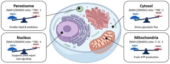
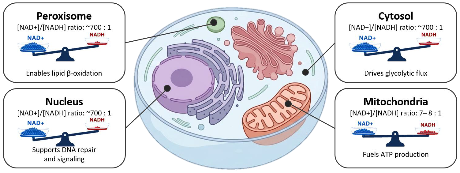
Disclaimer/Publisher’s Note: The statements, opinions and data contained in all publications are solely those of the individual author(s) and contributor(s) and not of MDPI and/or the editor(s). MDPI and/or the editor(s) disclaim responsibility for any injury to people or property resulting from any ideas, methods, instructions or products referred to in the content. |
© 2026 by the authors. Licensee MDPI, Basel, Switzerland. This article is an open access article distributed under the terms and conditions of the Creative Commons Attribution (CC BY) license.