


Abstract
Background: Plants of the genus Nepeta are widely used in ethnomedicine for treating inflammatory disorders due to their rich content of bioactive compounds. This study investigated how extraction temperature specifically affects the bioactive potential of aqueous extracts from wild-grown Nepeta nuda L. Methods: The previously used maceration approach for this plant was applied at 30–60 °C to flowers, leaves, and stems. Phytochemical profiling included spectrophotometric assays, metabolite identification, and quantification. Biological activities reported for this plant were assessed, including antioxidant, anti-inflammatory, antiviral, antiproliferative, and antibacterial capacities. Results: Extraction yield was highest in flowers and leaves, where it increased significantly with rising temperature, while stems were less productive. All plant organs exhibited notable bioactivity falling into two groups: lower temperatures (30 and 40 °C) were optimal for antiviral and anti-inflammatory effects, whereas and higher temperatures (50 and 60 °C) enhanced antioxidant potential. The phytochemical composition, evaluated at representative extraction temperatures, revealed differential accumulation of p-coumaric acid and luteolin in all organs at 40 °C, while extraction at 60 °C corresponded to elevated levels of phenolic compounds. Flower extracts were confirmed to have the richest metabolic composition and were therefore subjected to further investigation. Extracts obtained at 40 °C influenced C1q binding, supporting their anti-inflammatory activity, whereas extraction at 60 °C resulted in stronger antiproliferative activity in colon cancer cell line. Antibacterial effects were similar at both temperatures. Conclusions: These findings highlight the importance of optimizing extraction conditions for future pharmacological applications of N. nuda.
1. Introduction
Phytochemicals produced by plants largely reflect the co-evolution between plants and their surrounding environment, which includes competing plant species, pathogens, herbivores, and various abiotic factors [1,2]. The genus Nepeta has attracted considerable attention in recent years, mainly due to its nepetalactones, which are known to stimulate behavioral responses in felid species [3]. Nepetalactones are monoterpenoids with dual roles as insect pheromones and repellents against phytophagous insects [4]. It has also been suggested that cats may use these plants to repel mosquito attacks [4].
Notably, feline herpesvirus type 1 (FHV-1) is an alphaherpesvirus that causes severe inflammatory diseases in cats [5]. In this context, Hinkov et al. [6] conducted a detailed analysis of aqueous extracts from flowering plants of Nepeta nuda subsp. nuda L., demonstrating antiviral activity against herpesviruses. Nepeta nuda differs from the extensively studied N. cataria (catnip) by the absence or reduced levels of nepetalactones [7]. Nevertheless, nepetalactones and other iridoids have been identified predominantly in N. nuda flowers [8,9]. In traditional medicine, N. nuda has been used to treat inflammatory conditions of the urinary bladder, prostate gland, mammary gland (udder), and wounds [10]. Extraction has typically been performed through decoction (i.e., boiling in water) or as a tincture using a series of alcohol concentrations [11]. Several studies support water as a green and inexpensive solvent that is highly efficient for extracting antioxidants from Nepeta species [8,12,13,14].
Hinkov et al. [6], in their search for molecules associated with antiviral activity, performed NMR (nuclear magnetic resonance) analysis of N. nuda aqueous extracts and identified major phenolic acids, including rosmarinic, chlorogenic, gallic, vanillic, caffeic, protocatechuic, ferulic, and cinnamic acids, as well as flavonoids such as cirsimaritin, chrysoeriol, vanillin, rutin, and quercetin. Primary metabolites included sucrose, α- and β-glucose, and amino acids—alanine, glutamine, leucine, threonine, and valine. Other studies have reported that aqueous extracts from Nepeta species are rich in chlorogenic, ferulic, and rosmarinic acids, which correlate with high antioxidant capacity and antiproliferative activity in human colon carcinoma HT29 cells [12]. Aqueous extracts from N. rtanjensis Diklić et Milojević exhibited in vitro antioxidant, anti-inflammatory, antihyperglycemic, and antimicrobial activities, associated with high levels of rosmarinic acid and monosaccharides such as glucose and fructose [14]. Rosmarinic acid has also been identified in aqueous extracts of N. italica L., where it retained antioxidant and anti-inflammatory effects in an ex vivo model of colon inflammation [13].
Aqueous extracts of N. nuda have been reported to exhibit phytotoxic effects, inhibiting the growth of wheat and cucumber [15]. At the same time, these extracts possess antioxidant, antiproliferative, and antiviral activities [6,8,12,16]. Flowers are generally richer in phenolic antioxidants and iridoids compared to leaves [8,9]. The anti-inflammatory activity of aqueous extracts from N. nuda flowers and leaves has been demonstrated in vitro by inhibition of the inflammation via the classical pathway of complement activation [9]. Consistently, aqueous flower extracts have shown potent anti-inflammatory effects in vivo (rat hind paw model), following both local and systemic administration [17]. Additionally, aqueous leaf extracts exhibited significant anticancer activity against breast, liver, and colon cancer cell lines, with the strongest cytostatic effect observed in colon cancer cells [16].
Previous reports have indicated that the phytochemical composition of N. nuda is significantly influenced by growth conditions, plant organ, and extraction parameters [8,9,18]. We observed that the aqueous extracts exhibiting diverse biological activities were prepared at different extraction temperatures, and the incubation time also varied. For example, phytotoxic (allelopathic) activity was assessed using aerial plant parts subjected to 24 h maceration at room temperature [15]. Antioxidant and antiproliferative activities were evaluated in extracts obtained at higher temperatures, such as 80 °C for 45 min [16] or 60 °C for 24 h [8,16]. Antiviral and anti-inflammatory activities were reported for extracts prepared by maceration at 40 °C for 72 h [6] and for 24 h [9].
Based on existing studies on N. nuda, a research gap remains regarding the effects of extraction temperature and incubation time. To standardize aqueous extraction procedures and gain deeper insight into extract composition and associated biological activities, we conducted a systematic screening of N. nuda extracts prepared through ultrasound-assisted extraction and maceration with a 24 h incubation period in water, following our earlier studies [8,9,16]. Temperatures ranging from 30 to 60 °C were applied to obtain representative extracts corresponding to low and high extraction temperatures for detailed analysis of their phytochemical composition and in vitro biological activities (antioxidant, anti-inflammatory, antiviral, antiproliferative, and antibacterial). To strengthen our observations, we also compared the effect of temperature across different plant organs (flowers, leaves, and stems).
2. Materials and Methods
2.1. Plant Material
Nepeta nuda subsp. nuda L. plants (Bulgaria, Rhodope Mountains, Bekovi Skali, 1320 m a.s.l., coordinates 41.99437188774722, 24.396310265460215) were collected during the flowering period. The voucher specimen (SO108229) has been deposited in the Herbarium of Sofia University “St. Kliment Ohridski,” Sofia, Bulgaria [19].
Plant material from approximately 15–20 plants was air-dried in dark, and flowers, leaves, and stems were collected separately and powdered. Aqueous extracts were prepared using plant material and distilled water in ratio 1 g in 20 mL, followed by treatment in an ultrasound water bath for 20 min and subsequent maceration at 30, 40, 50 and 60 °C for 24 h. After filtration, the water solvent was evaporated through lyophilization at −65 °C (Alpha 1-2 LDplus, Martin Christ Gefriertrocknungsanlagen GmbH, Osterode am Harz, Germany), and the dried extracts were stored at −20 °C to preserve stability. Prior to analyses, the extracts were dissolved in sterile water to obtain a defined stock concentration. For biological activity assays, the extract (100 mg mL−1) was diluted in the respective buffer media to concentrations below 10 mg mL−1). The only exception was the disk diffusion method for antibacterial testing, where 200 mg mL−1 of stock solution was applied directly.
2.2. Phytochemical Content of Aqueous Extracts
Quantification of extract yield, as well as total phenolics and flavonoid content in extracts (prepared at 30, 40, 50, and 60 °C) from different plant organs (flowers, leaves, and stems), was performed according to [8]. The yield was expressed as the percentage of extract weight per gram plant dry weight. The quantification of total phenolics [20] and flavonoids [21] was performed using gallic acid and quercetin as standards, respectively.
Extracts prepared at 40 and 60 °C from plant organs (flowers, leaves, and stems) were subjected to gas chromatography–mass spectrometry (GC–MS) analysis for non-polar (fatty acids and derivatives) and polar (organic acids, amino acids, sugars, and phenolic derivatives) metabolites, as described in detail in [9]. An Agilent GC 7890 GC system (Agilent Technologies, Inc., Santa Clara, CA, USA) coupled with an Agilent MD 5975C mass spectral detector (Agilent Technologies, Inc., Santa Clara, CA, USA) were used. Reference standards included nonadecanoic acid (for the non-polar fraction), ribitol (for the polar fraction), and 3,5-dichloro-4-hydroxybenzoic acid (for the polar phenolic fraction). Relative metabolite concentrations in leaf and stem samples (leaf and stem) were expressed as logarithmic values with the flower extract at 40 °C set as the reference (value = 0). Three technical repetitions were performed for each sample, each comprising material pooled from 10 to 15 plants.
Extracts prepared at 40 and 60 °C from plant organs (flowers, leaves, and stems) were also subjected to metabolic profiling using a Dionex UltiMate 3000 ultra-high-performance liquid chromatography (UHPLC) system coupled with diode-array detector (DAD) and a triple quadrupole mass spectrometer with (±)heated electrospray ionization (UHPLC/DAD/(±)HESI–MS2) (Thermo Fisher Scientific, Basel, Switzerland) for quantification of major phenolic acids (caffeic acid, chlorogenic acid, rosmarinic acid, and p-coumaric acid), flavonoids (apigenin, luteolin, astragalin/kaempferol-3-O-glucoside, and isoorientin/luteolin-6-C-glycoside), and iridoids (nepetanudoside, 1,5,9-epi-deoxyloganic acid, and nepetalactone), as previously described [22].
2.3. Biological Activities of Aqueous Extracts
Extracts prepared at 30, 40, 50 and 60 °C from N. nuda organs (flowers, leaves, and stems) were screened using previously described approaches for assessing DPPH radical scavenging activity [23], antiviral activity [8], anti-inflammatory activity [24], and cytotoxicity [8].
The DPPH-radical-scavenging activity was measured using Trolox as a standard, according to the method in [23].
Antiviral activity and cytotoxicity were evaluated as described in detail in [8]. An acyclovir (ACV)-resistant strain of Simplexvirus humanalpha2 (SvHA2) (family Orthoherpesviridae) (strain DD), obtained from the National Center of Infectious and Parasitic Diseases (NCIPD, Sofia, Bulgaria), was propagated in Madine and Darby bovine kidney (MDBK) cells (obtained from American Type Culture Collection (ATCC, № CCL-22, Manassas, VA, USA)). The maximum nontoxic concentration (MNC) was defined as the highest concentration at which the calculated value for cell viability remained at 100%. The 50% cytotoxic concentration (CC50) was determined as the extract concentration required to reduce MDBK cell viability by 50%. The anti-SvHA2 activity of N. nuda extracts was assessed by measuring cell viability using a colorimetric MTT assay in two experimental setups: (1) simultaneous addition of the extract with cell inoculation and (2) addition of the extract 1 h post-inoculation. The effective concentration (EC50) was determined as the concentration at which 50% of the cells retained their viability. The selectivity index (SI) was calculated as: SI = CC50/EC50.
The anti-inflammatory potential of the extracts (at a concentration of 1 mg mL−1) was evaluated using a hemolytic assay to assess their ability to inhibit the complement system activation (C1q), as described in detail in [24,25].
Extracts prepared at 40 and 60 °C from N. nuda flowers were further evaluated to clarify the role of the complement system (C1q) in the anti-inflammatory response. Accordingly, enzyme-linked immunosorbent assay (ELISA) was performed to assess C1q-IgG (IgG, immunoglobulin) interactions, as described in [26], using heat-aggregated human IgG (HAIgG) as a model of immune complexes.
Extracts prepared at 40 and 60 °C from N. nuda flowers were also tested for anticancer activity against a murine colon adenocarcinoma cell line, Colon 26 (CLS, Eppelheim, Germany), following the procedure described in [16]. A human non-cancerous skin cell line, BJ (ATCC, USA), was used as a control. Cellular metabolic activity and proliferation were evaluated using an MTT assay 72 h after treatment of the cells with N. nuda extracts in a concentration range from 10 to 600 µg mL−1. Half-maximal inhibitory concentrations (IC50) values for both cancer and non-cancerous cell lines were calculated using GraphPad Prism 8 (GraphPad Software, San Diego, CA, USA). For the clonogenic assay, which evaluates the ability of single cells to proliferate and form colonies or clones after treatment, Colon 26 cells were seeded at a density of 1 × 10³ cells per well in 6-well plates. After overnight incubation, the cells were treated with N. nuda extracts prepared at 40 and 60 °C for 7 days. Untreated cells cultured in complete medium served as the negative control. At the end of the treatment period, the cells were fixed and stained with 2% methylene blue in 50% ethanol. Colonies consisting of at least 50 cells were counted under a microscope and the colony-forming ability was expressed as a percentage relative to untreated control cells.
Antimicrobial activity was tested as described in detail in [8]. For qualitative screening, the disk diffusion approach was applied. Extracts were loaded onto sterile paper disks (6 mm in diameter) to obtain the final concentration of 10 mg per disk. Inhibition zones were measured after 24 h incubation period at 37 °C, using tetracycline (30 µg/disk) as a positive control. For quantitative assessment, the minimum inhibitory (MIC) and minimum bactericidal concentration (MBC), expressed as mg mL−1, were determined. A series of two-fold dilutions (50–0.78 mg mL−1, i.e., 50, 25, 12.5, 6.25, 3.125, 1.56, 0.78 mg mL−1) was prepared in 96-well microtiter plates. Bacterial growth was assessed spectrophotometrically for MIC determination. As positive controls, penicillin G (30–0.235 µg ml−1 for Gram-positive bacteria), and gentamicin at concentrations ranging from 20 to 0.156 µg ml−1 for Gram-negative bacteria were used. MBC was determined as the lowest extract concentration inducing 99.9% growth inhibition of the bacterial inoculum after 24 h incubation at 37 °C. To additionally confirm the absence of bacterial growth, 10 µL from the defined MBC were plated on agar MH medium.
The following bacterial strains were purchased from the Bulgarian National Bank for Industrial Microorganisms and Cell Cultures (NBIMCC): the Gram-negative bacteria Acinetobacter calcoaceticus NBIMCC 3730 and Klebsiella pneumoniae NBIMCC 3670 and the Gram-positive bacteria Bacillus subtilis NBIMCC 1709 and Staphylococcus aureus ATCC 25923. The inhibition zones were measured after 24 h incubation period at 37 °C.
2.4. Statistics
The data represent at least three biological replicates, each comprising a representative sample of approximately 10–20 plants. The results are expressed as mean values ± standard error (SE) with n ≥ 3 technical replicates. Statistical comparisons among extraction conditions (30–60 °C) for the studied parameters across all plant organs were performed using two-way analysis of variance (ANOVA), with organ and temperature as factors, followed by Tukey’s post hoc test. Significant differences are indicated by different letters next to the values. Comparisons against control groups were conducted using one-way ANOVA followed by Dunnett’s post hoc test, with significant differences indicated by asterisks. Statistical analyses were carried out using Sigma Plot 11.0 (Systat Software, Inc., Erkrath, Germany), JASP 0.96.0 [27], and GraphPad Prism 8 (GraphPad Software, San Diego, CA, USA). For C1q binding, unpaired t-test with Welch’s correction was applied. The results were considered statistically significant at p < 0.05.
Principal component analyses (PCA) of the metabolites in and bioactivities of the examined variants were performed using prcomp function from stats package [https://search.r-project.org/R/refmans/stats/html/prcomp.html (accessed on 6 May 2026)] in R 4.5.3 [https://cran.r-project.org/ (accessed on 6 May 2026)] following logarithmic normalization of the experimental values. PCA graphs were plotted using the R package ggbiplot 0.6.2 [https://github.com/friendly/ggbiplot (accessed on 6 May 2026)].
3. Results
3.1. The Extraction Temperature Affects the Yield and Composition of N. nuda Aqueous Extracts
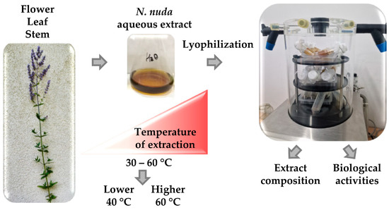
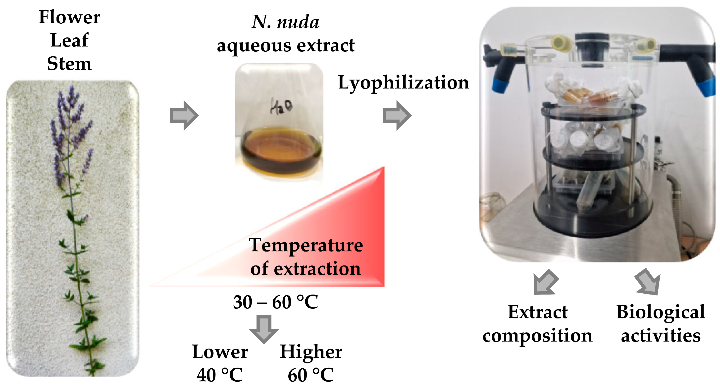
Aqueous extracts from N. nuda flowering plants were prepared at different temperatures (30–60 °C) to elucidate how this factor affects the yield of bioactive metabolites and their bioactive potential (Figure 1).
Figure 1.
Simplified schematic of the experimental setup for the preparation of Nepeta nuda aqueous extracts. A range of extraction temperatures was tested, and two temperatures (40 and 60 °C) were selected for detailed investigation of their effects on metabolite profiles and bioactive properties.
The effect of extraction temperature was monitored in individual plant organs to identify common trends. The extract yield from flowers increased significantly at 60 °C (Table S1). In leaves, the increase occurred at 40 °C, after which the yield remained constant. Stems were the least productive, showing no significant change with increasing temperature.
Total phenolic content appeared highly temperature-sensitive and gradually increased with each tested temperature in flowers and leaves, whereas in stems, a significant accumulation was observed only at 60 °C (Table S1). Compared to total phenolics, flavonoids were the most abundant in leaves and showed a positive correlation with temperature. In flowers, flavonoid content increased at 40 °C and then remained stable at higher temperatures, while in stems, a more pronounced increase was observed at 60 °C.
Based on the screening, we concluded that 30 °C is insufficient for efficient extraction, while 50 °C represents an intermediate condition between 40 and 60 °C in terms of yield and metabolite recovery. Therefore, 40 and 60 °C were selected as representative lower and higher temperatures, respectively, for further detailed investigation (Figure 1). Table 1 highlights the positive correlation between increasing temperature and phenolic content across all plant organs.
Table 1.
Temperature-dependent yield and total phenolic content in aqueous extracts from N. nuda flowers, leaves, and stems. Data are presented as mean values and SE (n ≥ 3). Statistical differences between variants were determined by two-way ANOVA followed by Tukey’s post hoc test and are indicated with different letters. For visualization, values are represented on a color scale ranging from white (minimum) to red (maximum).
For higher resolution, the metabolite composition of the selected extracts prepared at 40 and 60 °C was analyzed using GC–MS (Table S2). A total of 40 polar and six non-polar primary and secondary metabolites were identified. Their relative abundances indicated that flowers and leaves are more enriched in polar metabolites (polyphenolics, amino acids, and sugars), whereas stems contain higher levels of organic acids and non-polar fatty acids. Comparison of extraction temperature dependence across the different organs revealed a trend toward increased metabolite levels upon extraction at 60 °C. Table 2 lists the metabolites showing the most significant changes in the three plant organs.
Table 2.
Metabolites identified by GC–MS in aqueous extracts from N. nuda flowers, leaves, and stems. Relative values are expressed as logarithmic ratios normalized to a control sample (flower extract at 40 °C). Statistical differences between variants for each metabolite were determined by two-way ANOVA followed by Tukey’s post hoc test and are indicated by different letters. For visualization, values for each metabolite are presented on a color scale ranging from white (minimum) to red (maximum). Numbers correspond to metabolites listed in Table S2 (P, polar).
The extraction of fatty acids did not show strong temperature dependence across the examined plant organs (Table S2). Significant differences were observed in flower extracts for palmitic and stearic acids. At 40 °C, higher levels of cysteine, arabinose, and β-gentiobiose were observed (Table 2).
A more precise quantitative characterization of phenolic acids, flavonoids, and iridoids was achieved using a targeted metabolomics approach (Table 3).
Table 3.
UHPLC/DAD/(±)HESI–MS2 quantification of secondary metabolites in aqueous extracts from N. nuda flowers, leaves, and stems. Data are presented as mean ± SE (n ≥ 3) and expressed as µg per 100 mg of dry weight (DW). Statistical differences between variants for each metabolite were determined by two-way ANOVA followed by Tukey’s post hoc test and are indicated by different letters. For visualization, quantitative data are shown on a color scale ranging from white (minimum) to red (maximum), separately for phenolic acids (a), flavonoids (b), and iridoids (c).
At 60 °C, the content of phenolic acids (chlorogenic, caffeic, and rosmarinic) in flowers and leaves was significantly increased when compared to that at 40 °C. A similar trend was observed for the flavonoid isoorientin (luteolin-6-C-glycoside). In contrast, extraction at 40 °C favored the accumulation of more thermosensitive metabolites, including polyphenolics such as p-coumaric acid (in all organs), luteolin (in all organs), and apigenin (in flowers), as well as iridoids such as nepetanudoside and nepetalactone (both in flowers). The content of iridoid 1,5,9-epi-deoxyloganic acid was not significantly affected by extraction temperature.
3.2. The Extraction Temperature Differentially Modulates the Biological Activities of N. nuda Aqueous Extracts
To check the biological activities of N. nuda aqueous extracts, we first evaluated whether their phytochemical composition exerted cytotoxic effects. Cytotoxicity analyses were performed on normal kidney cells. The results did not reveal substantial differences between the variants, although a slight decrease in viability was observed at higher extraction temperatures for leaf and stem extracts (Table 4 and Table S3).
Table 4.
Cytotoxicity of aqueous extracts from N. nuda flowers, leaves, and stems. Madin–Darby bovine kidney (MDBK) cells were exposed to a range of extract concentrations. Data are presented as mean ± SE (n ≥ 3). Statistical differences between variants were determined by two-way ANOVA followed by Tukey’s post hoc test and are indicated by different letters. For visualization, values are shown on a color scale ranging from white (minimum) to red (maximum). CC50 denotes the extract concentration required to reduce cell viability by 50%.
In our previous studies, we reported in vitro biological activities of N. nuda [8,9]. A higher extraction temperature (60 °C) significantly enhanced the antioxidant properties of the aqueous extracts, whereas at a lower temperature (40 °C), anti-inflammatory and antiviral activities were optimal and declined sharply with increasing temperature (Table 5 and Table S4).
Table 5.
Biological activities of aqueous extracts from N. nuda flowers, leaves, and stems. Data about antioxidant activity (DPPH), anti-inflammatory activity (hemolysis and C1q assays), antiviral activity against Simplexvirus humanalpha2 (simultaneous application, SvHA2SA; 1 h post-treatment, SvHA2PA) are presented as mean ± SE (n ≥ 3). Statistical differences between variants were determined by two-way ANOVA followed by Tukey’s post hoc test and are indicated by different letters. For visualization, values for each biological activity are displayed on a color scale ranging from white (minimum) to red (maximum).
Nepeta nuda extracts exhibited stronger antiviral protection when they were added immediately after inoculating the cell monolayer with SvHA2, i.e., SvHA2SA, whereas the activity was weaker when the extracts were added 1 h post-infection, i.e., SvHA2PA (Table 5, Tables S4 and S5). At the same time, one of the most used nucleoside analogs for the treatment of herpes infections, acyclovir (ACV), showed no activity against the same viral model in either experimental setup (Table S6). These results indicate that extracts obtained at temperatures up to 40 °C were the most active against the replication of ACV-resistant SvHA2 strain, likely by interfering with the early stages of the viral replicative cycle.
The selectivity index (SI) reflects the selectivity of an extract toward viral targets relative to cellular toxicity. A plant extract or compound is generally considered to have therapeutic potential when SI ≥ 10 [28]. In the experimental setup involving simultaneous addition of the extracts and the virus, SI values for flower, leaf, and stem extracts obtained at 30 and 40 °C exceeded 10 (Table S5). This criterion was also met by stem extracts obtained at 50 and 60 °C. In contrast, in the post-inoculation setup (addition 1 h after infection), an SI value of 10 was observed only for the stem extract obtained at 30 °C.
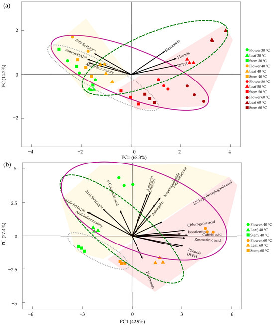
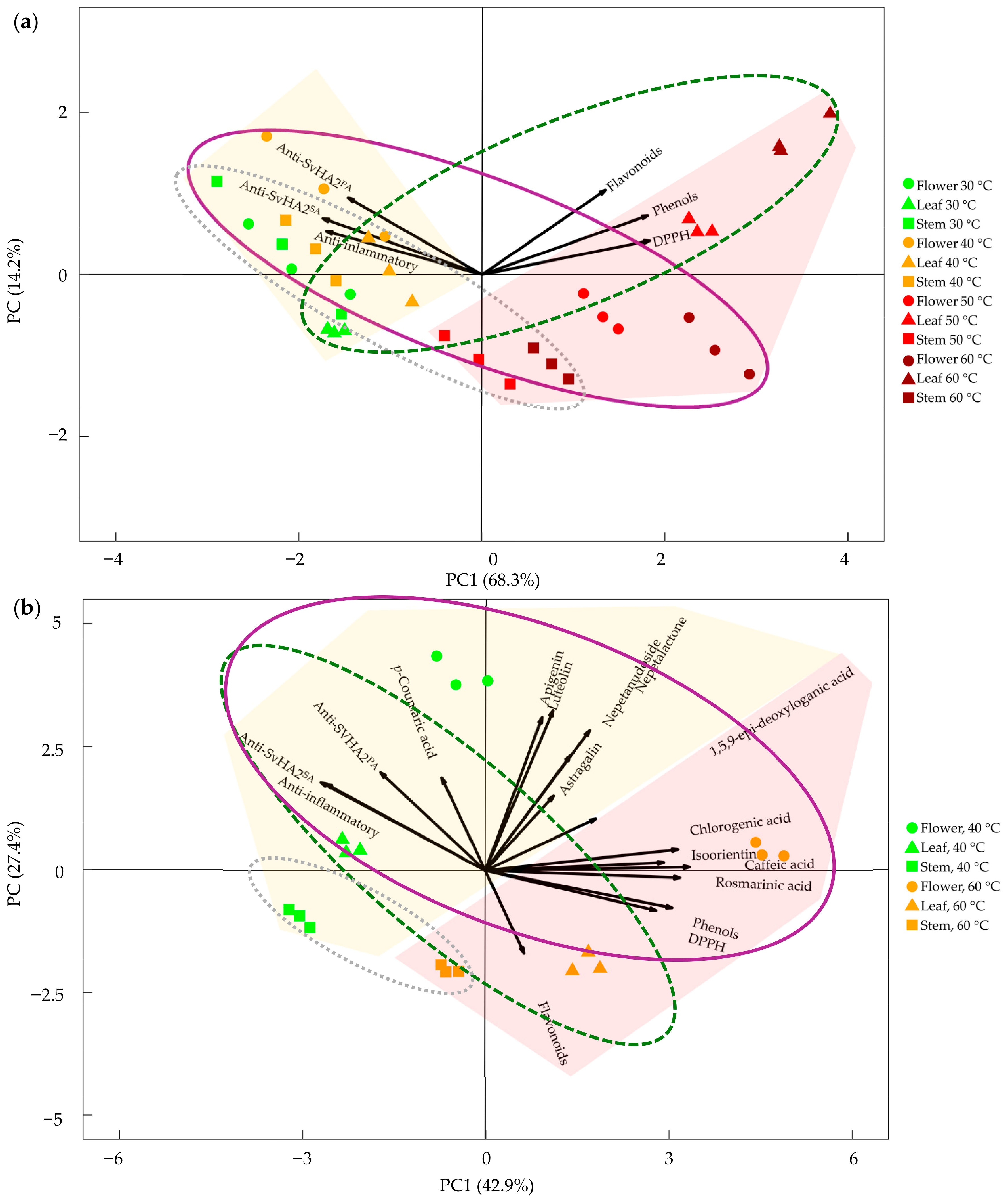
The effect of extraction temperature on the relationship between the tested biological activities and the phytochemical composition in the different N. nuda organs was summarized in Figure 2.
Figure 2.
Interdependence between phytochemical composition and biological activities of aqueous extracts from N. nuda flower, leaf, stem. (a) Principal component analysis (PCA) showing the relationship between total phenolic content and biological activities in extracts prepared across a range of temperatures (30–60 °C). (b) PCA illustrating the interdependence between quantified secondary metabolites and biological activities in extracts prepared at 40 and 60 °C. Antiviral activity against Simplexvirus humanalpha2 (simultaneous application, SvHA2SA; 1 h post-treatment, SvHA2PA), anti-inflammatory activity (hemolysis and C1q assays), and antioxidant activity (DPPH) are shown. Symbols denote plant organs: circles (flowers), triangles (leaves), and squares (stems). Temperature increase is indicated by a color gradient from green to dark brown. Yellow and light brown shaded areas indicate group distributions associated with lower and higher extraction temperatures, respectively. Ellipses represent the distribution of plant organs: solid purple line (flowers), dashed green line (leaves), and dotted gray line (stems).
In the PCA related to biological activities along the extraction temperature gradient (Figure 2a), the first principal component (PC1), accounting for 68.3% of the variance, was associated with all tested parameters. Phenolic content, flavonoids, and DPPH antioxidant capacity increased along PC1, whereas anti-inflammatory and antiviral (SvHA2SA and SvHA2PA) activities decreased. The second principal component (PC2) explained an additional 14.2% of the variance, with all tested parameters increasing along this axis.
Elevation of extraction temperature (from 30 to 60 °C) corresponded to systematic shifts in sample positioning along both principal components, supporting the conclusion that temperature influences extraction efficiency and phytochemical composition. Lower extraction temperatures were associated with stronger anti-inflammatory and antiviral properties, whereas higher temperatures were linked to greater antioxidant capacity.
Clear clustering of samples according to plant organ (as indicated by overlapping ellipses in Figure 2) was not observed overall. However, at lower temperatures, organ groups were more similar, while at higher temperatures, they became more distinctly separated, primarily based on flavonoid content: stem extracts showed the lowest levels, leaf extracts the highest, and flower extracts intermediate levels.
Phenolic content, flavonoids, and DPPH activity were positively correlated, as indicated by their similar vector directions. A similar relationship was observed for anti-inflammatory and antiviral (SvHA2SA and SvHA2PA) activities. Generally, PCA indicated that temperature is the primary driver of variation in the dataset, while plant organ acts as a secondary factor influencing extract composition, particularly at higher temperatures.
The second PCA, focused on extracts prepared at 40 and 60 °C, further supported temperature-dependent clustering of metabolites and associated biological activities (Figure 2b). This analysis also highlighted the contribution of plant organ, with flowers showing the highest metabolite enrichment, followed by leaves and stems.
3.3. Anti-Inflammatory C1q Inhibitory Potential of Aqueous Extracts from N. nuda Flowers
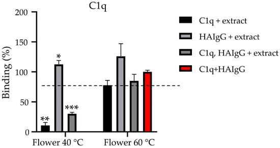
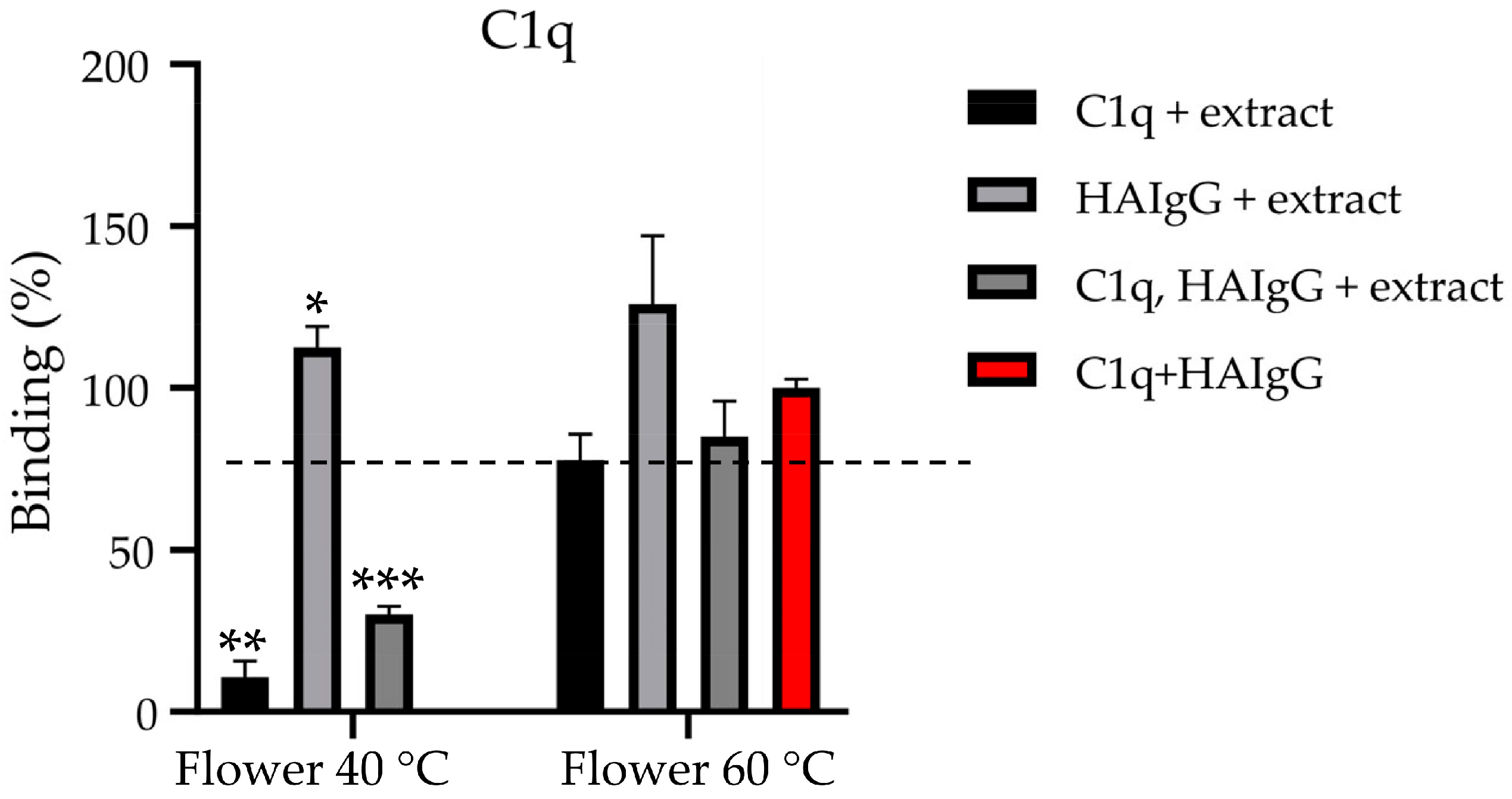
Flowers are generally enriched in metabolites and several reports demonstrated their bioactive potential. In line with this, our previous work on N. nuda aqueous extracts highlighted their anti-inflammatory activity mediated via the complement system (Table 5; [11]). Based on this evidence, and particularly on the inhibitory effect of N. nuda aqueous extracts on the complement system as demonstrated by the hemolysis assay (Table 5), we aimed to further elucidate their effect on the classical complement pathway. Activation of the classical pathway is initiated by the interaction of serum C1q with immune complexes, represented by heat-aggregated human IgG (HAIgG). The role of extraction temperature was investigated using an ELISA by treating C1q and HAIgG individually with N. nuda extracts prepared at 40 and 60 °C, as well as by treating both components prior to their interaction (Figure 3).
Figure 3.
Effect of aqueous extracts of N. nuda flowers (prepared at 40 and 60 °C) on the interaction between C1q and immunoglobulin G (HAIgG). Data are presented as mean ± SD (n = 3). Statistical differences were assessed using an unpaired t-test with Welch’s correction and are indicated by asterisks: * (p ≤ 0.05), ** (p ≤ 0.01), and *** (p ≤ 0.001). The positive control (red) represents the interaction between untreated C1q and HAIgG, defined as 100% binding.
Binding between extract-treated C1q with HAIgG was completely inhibited when the 40 °C extract was used, suggesting a significant conformational change in C1q induced by the plant extract.
In contrast, the interaction between C1q and extract-treated HAIgG was comparable to the positive control. Nevertheless, physiologically relevant scenario involves simultaneous exposure of both C1q and IgG to the extract, which resulted in suppression of inflammatory signaling. Under these conditions, when both proteins were treated with the extract, the formation of C1q–HAIgG complexes decreased approximately fourfold. In contrast, extracts prepared at 60 °C did not exhibit an inhibitory effect on C1q activity.
3.4. Anti-Colon Cancer Potential of Aqueous Extracts from N. nuda Flowers
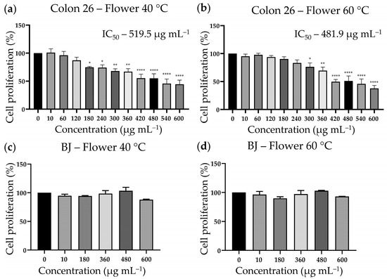
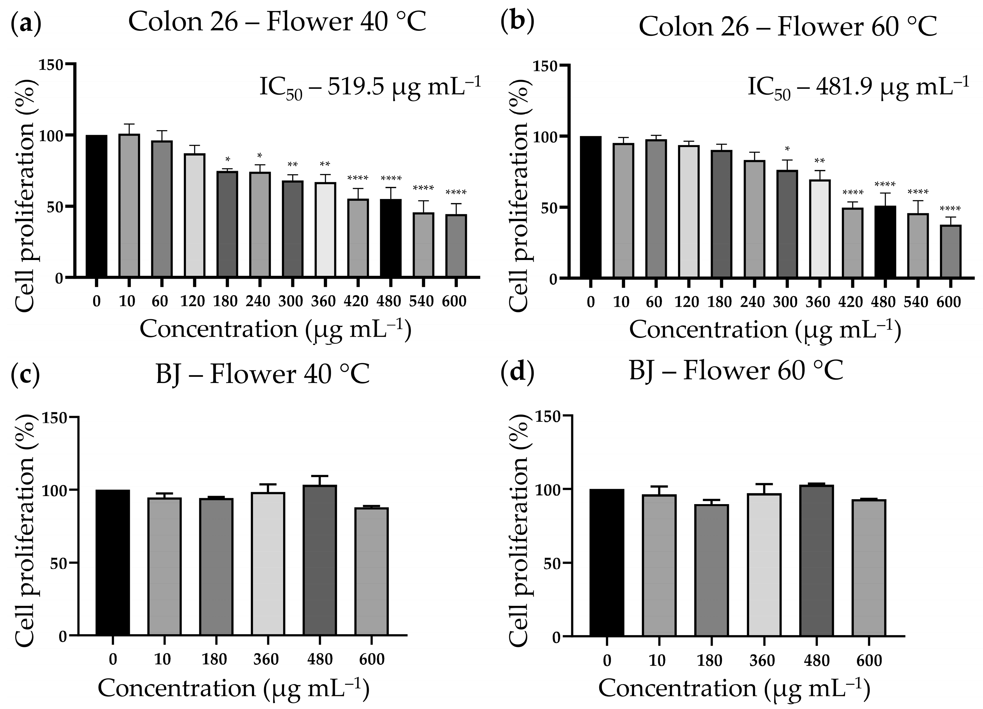
Recently, aqueous extracts from N. nuda leaves were investigated to elucidate the mechanism underlying their antitumour effects on the colon adenocarcinoma cell line, Colon 26 [17]. In the present study, we compared the cytostatic properties of aqueous flower extracts of N. nuda (prepared at 40 and 60 °C) in the Colon 26 cell line and the non-cancerous dermal cell line BJ. The results demonstrated a statistically significant antiproliferative effect of both extracts on Colon 26 cancer cells (Figure 4a,b).
Figure 4.
Antiproliferative activity of aqueous extracts of N. nuda flowers (prepared at 40 and 60 °C). Statistical differences between untreated controls and treated groups were evaluated by one-way ANOVA followed by Dunnett’s post hoc test and are indicated by asterisks (* p ≤ 0.05, ** p ≤ 0.01, and **** p ≤ 0.0001). (a,b) Colon 26 colon adenocarcinoma cell line; (c,d) non-cancerous dermal cell line BJ.
Lower concentrations of the flower extract prepared at 40 °C exerted antiproliferative effect; however, the extract prepared at 60 °C demonstrated a stronger cell growth inhibitory potential—IC50 481.9 µg mL−1 at 60 °C, and IC50 519.5 µg mL−1 at 40 °C. The greatest reduction in cell proliferation was reached at the highest applied concentration (600 µg mL−1), reaching 37.81% and 44.43% of control cell growth levels for extracts prepared at 60 °C and 40 °C, respectively.
Both N. nuda extracts did not show any substantial inhibitory effect on the proliferation of the non-cancerous BJ cell line, indicating high selectivity of their cytostatic anticancer action (Figure 4c,d). Lowest and highest extract concentrations were tested, and at the highest tested concentration (600 µg mL−1), cell proliferation remained at 87.91% for the extract prepared at 40 °C and 93.18% for the extract prepared at 60 °C.
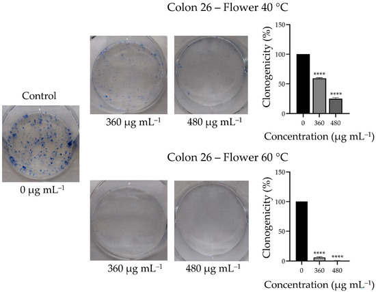
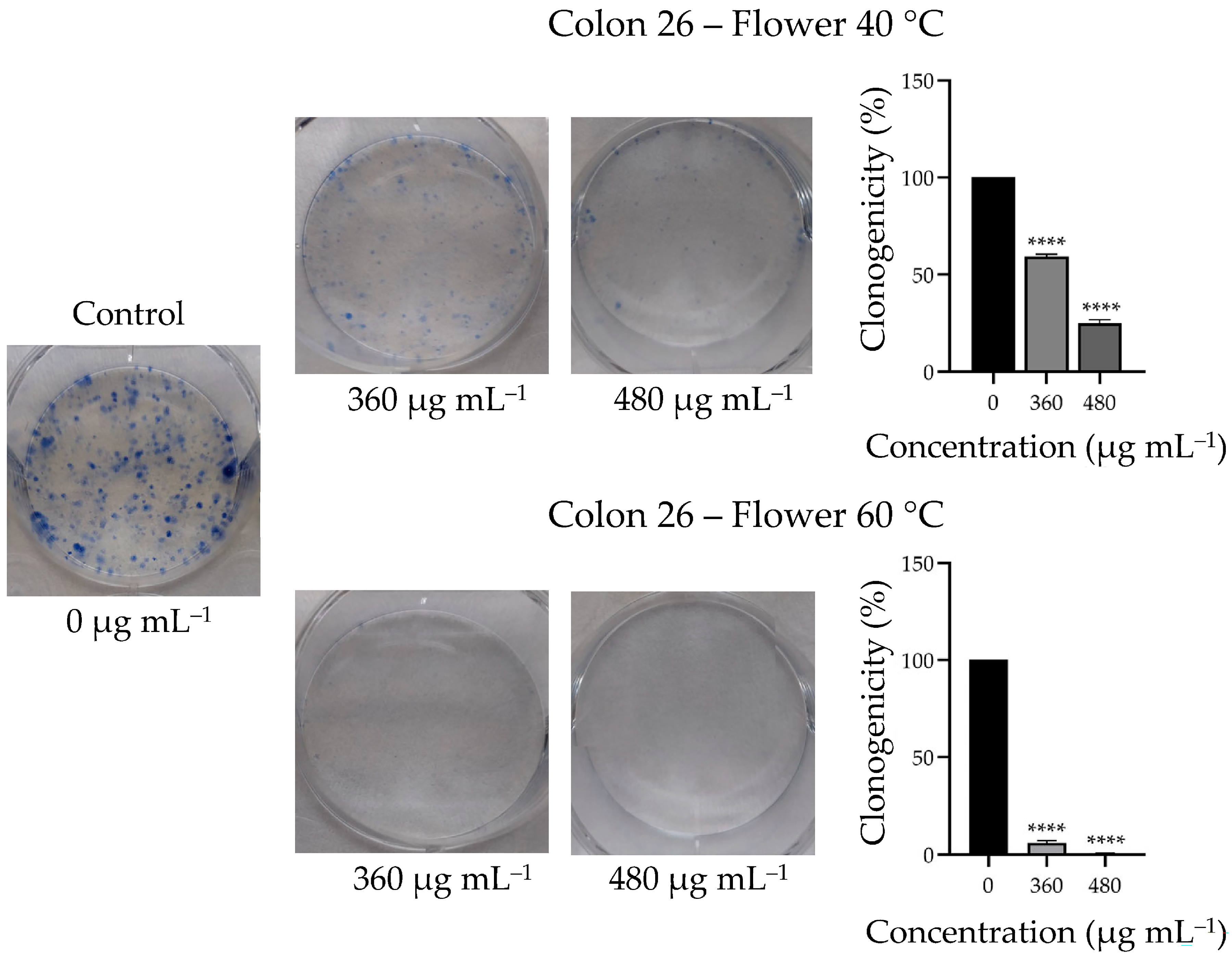
As a next step, the long-term proliferative capacity of colon cancer cells was evaluated following 7-day treatment with flower extracts prepared at 40 and 60 °C at concentrations of 360 and 480 µg mL−1, which are close to the IC50 values determined by the MTT assay. A dose-dependent anticlonogenic effect of the extracts was observed, which was markedly stronger for the extract prepared at 60 °C compared to that at 40 °C (Figure 5).
Figure 5.
Effects of aqueous extracts of N. nuda flowers (prepared at 40 and 60 °C) on the clonogenicity of Colon 26 cells. Cells were treated for 7 days with 360 and 480 µg mL−1 and compared to untreated control. Graphs represent quantitative evaluation of Colon 26 cells clonogenicity. Data are presented as mean ± error bars. Asterisks indicate significant differences compared to the control group, determined by one-way ANOVA followed by Dunnett’s post hoc test (**** p < 0.0001).
At a concentration of 360 µg mL−1, clonogenicity of tumor cells was reduced to 59.32% by the extract prepared at 40 °C, whereas the extract prepared at 60 °C resulted in near-complete inhibition, reducing clonogenicity to 5.93%. At 480 µg mL−1, the colony-forming ability of cancer cells was further reduced to 25.05% and 0.75% for the extracts prepared at 40 °C and 60 °C, respectively.
3.5. Antibacterial Activity of Aqueous Extracts from N. nuda Flowers
Since methanolic extracts of N. nuda have previously been reported to exhibit antibacterial activity against both Gram-negative and Gram-positive bacteria [8], we also evaluated the antibacterial potential of aqueous extracts. Overall, aqueous extracts of N. nuda flowers (prepared at 40 and 60 °C) showed bactericidal activity against three of the tested bacterial strains—A. calcoaceticus, S. aureus, and B. subtilis—while no effect was observed against K. pneumoniae. The results from the qualitative disk diffusion assay demonstrated the strongest inhibitory effects against B. subtilis and A. calcoaceticus at both extraction temperatures (Table 6).
Table 6.
Antibacterial activity of aqueous flower extracts of N. nuda (prepared at 40 and 60 °C). Inhibition zone diameters (disk diffusion assay) and values for minimum inhibitory concentration (MIC) and minimum bactericidal concentration (MBC) are presented. Statistical differences between extracts obtained at 40 and 60 °C were evaluated using Student’s t-test (n > 3) and are indicated by asterisks (** p < 0.001).
The activity of the extracts against S. aureus was more variable. Quantitative analysis revealed MIC values ranging from 6.25 to 25 mg mL−1 and MBC values from 6.25 to 50 mg mL−1. The Gram-positive bacteria B. subtilis and S. aureus exhibited the highest sensitivity, followed by the Gram-negative A. calcoaceticus.
Extraction temperature did not significantly affect antibacterial activity overall, although some trends were observed. Bacillus subtilis showed increased sensitivity to the extract prepared at 40 °C (MIC/MBC = 6.25/6.25 mg mL−1), whereas S. aureus was more sensitive to the extract prepared at 60 °C (MIC/MBC = 6.25/12.5 mg mL−1). In the case of A. calcoaceticus, a significant difference was observed only in the disk diffusion assay, with stronger inhibition exhibited by the extract prepared at 40 °C.
3.6. Metabolite–Bioactivity Correlation
To identify metabolites associated with the temperature-dependent biological activities, a correlation analysis was performed. Only statistically significant correlations are presented in Table 7.
Table 7.
Pearson correlation coefficients between metabolites identified in aqueous extracts of N. nuda flowers and biological activities, including antiviral activity against Simplexvirus humanalpha2 (simultaneous application, SvHA2SA; 1 h post-treatment, SvHA2PA), anti-inflammatory activity (hemolysis and C1q assays), and antioxidant activity (DPPH). Only statistically significant correlations are shown (p < 0.01, n ≥ 3). For visualization, values are presented on a color scale ranging from white (minimum) to red (maximum).
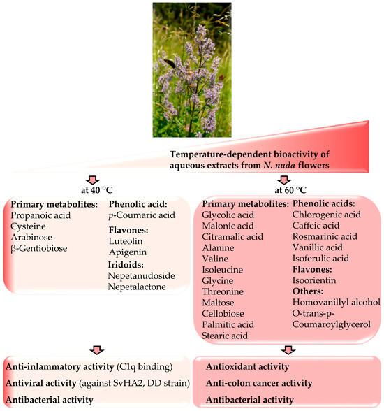
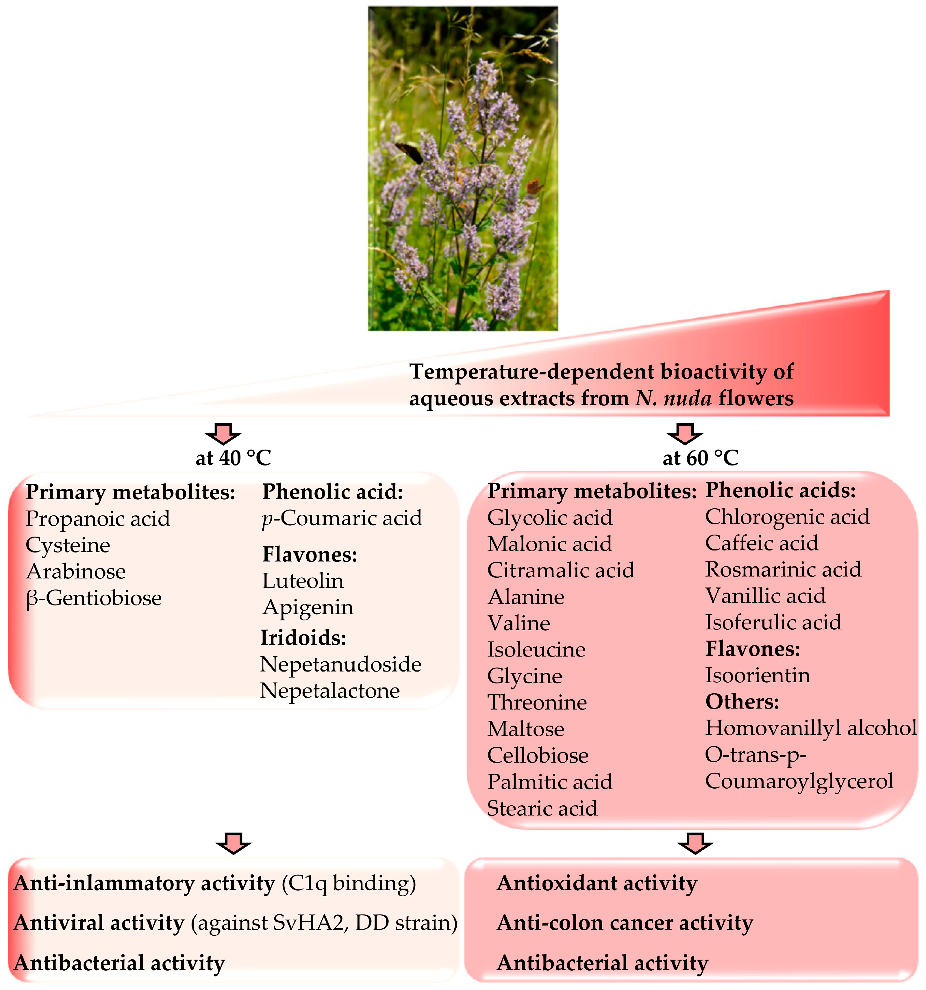
The compositions and biological activities of the extracts prepared at 40 and 60 °C are summarized in Figure 6.
Figure 6.
Model for temperature-dependent bioactivity of aqueous extracts of N. nuda flowers.
4. Discussion
This study investigated how extraction temperature influences the bioactive properties of aqueous extracts of N. nuda. The analyses of flowers, leaves, and stems confirmed bioactivity in all parts, with flowers and leaves exhibiting higher phytochemical content; notably, flowers contained the highest levels of iridoids. Extraction at 40 °C optimized antiviral and anti-inflammatory activities, which were associated with elevated levels of p-coumaric acid, luteolin, apigenin, nepetanudoside, nepetalactone (Figure 6). In contrast, extraction at 60 °C enhanced antioxidant activity, correlating with increased concentrations of caffeic and rosmarinic acids, as well as primary metabolites. Flower extracts obtained at 40 °C affected C1q binding, while those extracted at 60 °C demonstrated stronger antiproliferative effects. Antibacterial activity remained comparable at both temperatures. Overall, extraction temperature shapes the metabolic profile and bioactivity of N. nuda, underscoring the importance of optimizing extraction conditions for pharmacological applications.
4.1. Aqueous Extraction Parameters
A growing emphasis in extraction techniques is on the reduction in hazardous substances, particularly toxic solvents, in order to protect human health and the environment while minimizing energy consumption [29]. Water represents an environmentally friendly solvent, and several studies on Nepeta species have employed maceration as a conventional method [8,12]. A highly efficient alternative—subcritical water extraction (SWE), which involves short exposure to elevated temperature and pressure—has recently gained attention [14,30]. A review of water-based extraction applied to Nepeta species reveals considerable variation in both temperature and duration. Lower-temperature approaches refer to range from room temperature to 40 °C, with or without repeated 24 h extraction cycles [6,8,9,15]. Higher-temperature methods include maceration or boiling at 60 °C and 80 °C (short treatments of approximately 15 min, with or without repetition, or extended up to 24 h) [8,12,13], as well as SWE at 130 °C for 30 min under pressure [14].
Higher temperatures can weaken cell wall integrity, facilitating the release of insoluble polyphenols and flavonoids from the plant matrix [29]. Prolonged extraction time may further enhance this effect by promoting cell wall loosening. Based on previous reports and our earlier work on N. nuda, the present study employed a 24 h maceration approach, which has been shown to be sufficient and effective for extracting bioactive compounds [8,9].
In N. nuda, water has been reported to yield a higher amount of dry extract compared to methanol, ethanol, acetone, or chloroform [8], and our results further demonstrate that extraction yield increases with temperature. At temperatures below 40 °C, the yield was too low, whereas above 60 °C no further increase was observed, which guided the selection of this temperature range. The physicochemical properties of water—such as dielectric constant, surface tension, and viscosity—change with temperature, leading to decreased polarity [31]. Consequently, extraction at higher temperatures becomes less selective and can solubilize moderately polar and even non-polar compounds [32], which may explain higher extract yield observed at 60 °C. The differences in biological activity between lower and higher temperatures suggest thermo-sensitivity of certain compounds or molecular complexes, resulting in selective extraction effects that warrant further investigation.
4.2. Differential Phytochemical Content of Aqueous Extracts
Increasing water temperature enhances vapor pressures, thermal desorption, and diffusion rates of compounds. Elevated temperatures improve the solubility and diffusion of phenolic compounds, thereby accelerating extraction, although they may also lead to compound degradation [29]. Both our results and previous studies indicate that rosmarinic and caffeic acids are more abundant in extracts obtained at moderate to high temperatures [8,12,13,14]. Subcritical water extraction (SWE) has been reported to increase rosmarinic acid yield by up to 41% at elevated temperatures [30]. Notably, rosmarinic and other phenolic acids have also been detected at 40 °C [6], likely due to prolonged extraction (three 24 h cycles), use of whole aboveground plant material, and differences in plant origin.
Regardless of the plant organ, extraction of most phenolic acids (caffeic, chlorogenic, and rosmarinic acids) within this study was more efficient at 60 °C. An exception was p-coumaric acid, the least polar phenolic acid examined, which was more efficiently extracted at 40 °C. This finding was surprising, considering that all targeted phenolic acids belong to the hydroxycinnamic acid group, share structurally similarities, and exhibit comparable physicochemical properties. Furthermore, p-coumaric acid is a key precursor in the biosynthesis of caffeic acid and its derivatives, namely ferulic acid, coniferyl alcohol, and sinapyl alcohol, all of which represent significant building blocks in lignin [33]. It has been suggested that increased phenolic acids content at higher temperatures may result from the cleavage of lignin–phenolic acid bonds or partial lignin degradation [34]. However, this does not appear to apply in the present study, as p-coumaric acid content decreased at 60 °C.
At 60 °C, reduced levels of p-coumaric acid, flavone aglycones (luteolin and apigenin), and monoterpenoids (nepetanudoside and nepetalactone) were observed. Luteolin and p-coumaric acid showed notable variability across all plant organs, whereas other metabolites were more specific to flowers. Thermal degradation is a common explanation for reduced polyphenol content at elevated temperatures [31]. However, the response to temperature appears to be compound-specific and may reflect differences in solubility (polarity) and thermal properties, such as diffusivity and vapor pressure.
Flavones are generally most abundant in N. nuda flowers [8,9], which was confirmed in this study. Flavone aglycones (luteolin and apigenin) were more efficiently extracted at 40 °C. In contrast, flavonoid glycosides—kaempferol-3-O-glucoside (astragalin) and luteolin-6-C-glycoside (isoorientin)—were extracted with similar efficiency at both 40 °C and 60 °C, with minor exceptions. These findings suggest greater thermal stability of glycosylated flavonoids compared to their aglycone counterparts.
Iridoids, characterized by a partially hydrogenated cyclopenta-[c]pyran system, are efficiently extracted using polar solvents such as water [35]. Hot water (50–100 °C) has been shown to be effective for extracting iridoid glycosides, including aucubin and catalpol [36]. This was also supported by the present study, although no significant difference in the extraction efficiency of the predominant N. nuda iridoid, 1,5,9-epi-deoxyloganic acid, was observed between 40 °C and 60 °C. The iridoid glycoside nepetanudoside exhibited slight instability at 60 °C, but only in flowers. The iridoid aglycone nepetalactone is a volatile compound with a boiling point of 71 °C at 0.05 mmHg, making it prone to evaporation even at relatively low temperatures [4]. Therefore, its lower content in samples extracted at 60 °C may be attributed to increased volatilization.
Higher temperatures also promote the extraction of other polar metabolites, including organic acids, amino acids, and sugars. Overall, extract bioactivity likely reflects synergistic or antagonistic interactions among multiple constituents [6,29].
4.3. Differential Biological Activities in Aqueous Extracts
The viral model used in this study is a laboratory-adapted isolate optimized for replication in the employed cell line. The observed antiviral activity is consistent with previous findings on total aqueous extracts of N. nuda (extracted at 40 °C) [6], where experiments targeting different stages of viral replication indicated that the effect is associated with the viral adsorption stage. Based on these findings, it can be assumed that extracts from flowers, leaves, and stems primarily inhibit viral adsorption. Flavones exhibit diverse biological activities in animals, partly determined by their lipophilicity and ability to cross cellular membranes, which depend on the number and position of hydroxyl groups. Luteolin and apigenin, containing four and three hydroxyl groups, respectively, are relatively hydrophilic. In the present study, the correlation between antiviral activity and flavone content supports their role in inhibiting viral adsorption to cell membranes, potentially acting synergistically with other compounds such as cysteine, nepetanudoside, nepetalactone, and p-coumaric acid [37,38,39]. Importantly, the observed activity against an acyclovir-resistant strain suggests that these extracts are not only effective against herpesviruses but also against strains resistant to one of the primary antiviral drugs used in therapy.
Flavones are known to interact with DNA, RNA, and proteins, contributing to a wide range of biological effects [40]. Apigenin, for instance, binds multiple cellular targets, including those in cancer cells. Although flavones are widely studied for their anti-inflammatory, anticancer, and antimicrobial properties, their mechanisms of action remain incompletely understood. In this study, N. nuda flower extracts influenced C1q in the classical complement pathway, possibly through flavones or flavone–protein complexes. Notably, extracts obtained at 40 °C exhibited a pronounced anti-inflammatory effect, in contrast to those obtained at 60 °C. These differences correspond to variations in metabolite composition, with luteolin and p-coumaric acid present at higher levels in the 40 °C extracts across all plant organs. Although neither compound is known to directly interact with C1q or IgG, this study provides, to the best of our knowledge, the first indication that N. nuda extracts may exert anti-inflammatory effects by modulating the initial steps of the classical complement activation pathway.
Flavones and p-coumaric acid also exhibit antioxidant activity [39,40]. Luteolin inhibits xanthine oxidase, while apigenin reduces reactive oxygen species and protects against inflammation-induced damage [40]. Many of their anti-inflammatory and antimicrobial effects are associated with modulation of the Toll-like receptor (TLR)/NF-κB signaling pathway, which regulates key mediators such as tumor necrosis factor (TNF), interleukin-1 (IL-1), and cyclooxygenase-2 (COX-2). The similarities between plant and mammalian defense pathways suggest the presence of evolutionarily conserved molecular targets. Because NF-κB and COX-2 are often upregulated in cancer and inflammatory conditions, flavones may exert their effects through shared mechanisms, including the induction of apoptosis. Moreover, p-coumaric acid seems to act synergistically as it decreases the expression of the inflammatory mediators such as TNF and IL-6 [41].
Glycosides of luteolin and apigenin were previously identified in extracts prepared at higher temperatures [42]. Consistently, luteolin-6-C-glucoside (isoorientin) was more abundant in N. nuda aqueous extracts obtained at 60 °C. In general, flavonoid glycosides exhibit weaker anti-inflammatory activity than their aglycone counterparts, likely due to reduced cellular uptake [42].
In the present study, flower extracts obtained at 40 °C showed weaker antiproliferative activity, which may be related to their flavone profile. Aglycones such as luteolin and apigenin are generally more bioavailable than their glycosylated forms, as they are more readily absorbed in the intestine [43]. In contrast, rosmarinic, chlorogenic, and ferulic acids have been identified as key antiproliferative compounds in N. nuda aqueous extracts [12]. Previous in vitro studies on human cancer cell lines (including breast, colon, and hepatocellular carcinoma) have shown that N. nuda leaf extracts inhibit proliferation and induce apoptosis [16]. In agreement with these findings, the present study demonstrates the antiproliferative potential of flower extracts.
Regarding antibacterial activity, aqueous extracts of Nepeta cataria leaves and stems prepared at room temperature were previously reported to lack activity against S. aureus and B. subtilis [44]. In contrast, in the present study, N. nuda flower extracts obtained at 40 °C exhibited moderate bactericidal activity, particularly against Gram-positive bacteria. This difference may be explained by the presence of an additional outer membrane rich in lipopolysaccharides in Gram-negative bacteria, which limits compound penetration [45]. Interestingly, significant growth inhibition of the Gram-negative bacterium A. calcoaceticus was observed, which was not reported before for aqueous extracts. Increasing the extraction temperature to 60 °C had little effect on antibacterial activity. Methanol and chloroform extracts of N. nuda prepared at 60 °C have demonstrated broader antibacterial activity against A. calcoaceticus, K. pneumoniae, B. cereus, and S. aureus [10,20]. These findings suggest that aqueous extracts have a narrower range of bioactive compounds, and that extraction temperature has a limited impact on their antibacterial efficacy.
5. Conclusions
Standardization of extract preparation is essential not only for reproducibility but also for ensuring consistent extract composition and associated biological activities. In this study, extraction temperature was identified as a major factor influencing the bioactivity of N. nuda aqueous extracts. At lower temperatures, specific metabolites (e.g., luteolin, p-coumaric acid) were preferentially extracted, contributing to enhanced antiviral and anti-inflammatory potential. In contrast, higher temperatures favored the extraction of phenolic acids and a broader range of primary metabolites, resulting in stronger antioxidant and antiproliferative activity (Figure 6). Extraction temperature had a limited impact on antibacterial efficacy. Furthermore, a 24 h maceration approach proved to be efficient for the extraction of bioactive compounds. Future studies exploring additional parameters—such as alternative extraction techniques (e.g., subcritical water extraction and ultrasound-assisted extraction) and shorter extraction times—may provide further optimization for practical applications.
The key contribution of this work lies in establishing a clear relationship between the identified and quantified metabolites and the observed biological activities. These findings provide a solid foundation for future research aimed at identifying specific bioactive compounds and elucidating their mechanisms of action.
Supplementary Materials
The following supporting information can be downloaded at: https://www.mdpi.com/article/10.3390/metabo16050323/s1, Table S1: Temperature-dependent yield and total content of phenolic compounds in aqueous extracts from N. nuda flower, leaf, stem; Table S2: Metabolites identified by GC–MS assay in aqueous extracts from N. nuda flower, leaf and stem; Table S3: Cytotoxicity of aqueous extracts from N. nuda flower, leaf, stem; Table S4: Biological activities of aqueous extracts from N. nuda flower, leaf, stem; Table S5: Data for cell viability (cytotoxicity) of the cell line used treated with and cell protection (antiviral activity) against SvHA2 (DD strain) of aqueous extracts of N. nuda flower, leaf, and stem, obtained at different temperatures; Table S6: Data for cell viability (cytotoxicity) of the cell line used and cell protection (antiviral activity) against SvHA2 (DD strain) by the positive control Acyclovir.
Author Contributions
Conceptualization, M.Z., D.M. (Desislava Mantovska)., Z.Y., I.T., A.H. and Z.G.; methodology, D.M. (Desislava Mantovska), A.K., G.C., I.T., A.H., D.P., Z.G., G.A., D.M. (Danijela Mišić), K.R., M.R., K.S., Z.Y. and M.Z.; software, D.M. (Desislava Mantovska), A.K., G.C., I.T., A.H., Z.G., G.A., D.M. (Danijela Mišić), K.R., M.R., K.S., M.P., Z.Y. and M.Z.; validation, D.M. (Desislava Mantovska), A.K., G.C., I.T., A.H., D.P., Z.G., G.A., D.M. (Danijela Mišić), K.R., M.R., K.S., M.P., Z.Y. and M.Z.; formal analysis, D.M. (Desislava Mantovska), A.K., G.C., I.T., A.H., D.P., Z.G., G.A., D.M. (Danijela Mišić), K.R., M.R., K.S., Z.Y. and M.Z.; investigation, D.M. (Desislava Mantovska), A.K., G.C., I.T., A.H., D.P., Z.G., G.A., D.M. (Danijela Mišić), K.R., M.R., K.S., Z.Y. and M.Z.; resources, D.M. (Desislava Mantovska), I.T., A.H., Z.G., G.A., D.M. (Danijela Mišić), K.R., M.R., K.S., Z.Y. and M.Z.; data curation, D.M. (Desislava Mantovska), A.K., G.C., I.T., A.H., D.P., Z.G., G.A., D.M. (Danijela Mišić), K.R., M.R., K.S., M.P., Z.Y. and M.Z.; writing—original draft preparation, M.Z. and D.M. (Desislava Mantovska); writing—review and editing, D.M. (Desislava Mantovska), G.C., I.T., A.H., Z.G., D.M. (Danijela Mišić), K.R., Z.Y. and M.Z.; visualization, D.M. (Desislava Mantovska), A.K., G.C., I.T., A.H., D.P., Z.G., G.A., D.M. (Danijela Mišić), M.P. and M.Z.; supervision, I.T., A.H., Z.G., D.M. (Danijela Mišić), K.R., K.S., Z.Y. and M.Z.; project administration, D.M. (Desislava Mantovska), G.C., I.T., A.H., Z.G., G.A., D.M. (Danijela Mišić), K.R., M.R., K.S., D.P., Z.Y. and M.Z.; funding acquisition, D.M. (Desislava Mantovska), G.C., I.T., A.H., D.P., Z.G., G.A., D.M. (Danijela Mišić), K.R., M.R., K.S., M.P., Z.Y. and M.Z. All authors have read and agreed to the published version of the manuscript.
Funding
This research was funded by the Bulgarian National Science Fund, grant number KP-06-N56/9/12.11.2021. The research is also supported by the Ministry of Science, Technological Development, and Innovation of the Republic of Serbia (Grant No. 451-03-33/2026-03/200007).
Institutional Review Board Statement
Not applicable.
Informed Consent Statement
Not applicable.
Data Availability Statement
Data are contained within the current article and its Supplementary Materials.
Acknowledgments
Infrastructure support was provided by the Center of Competence “Sustainable Utilization of Bio-resources and Waste of Medicinal and Aromatic Plants for Innovative Bioactive Products” (BIORESOURCES BG) project BG16RFPR002-1.014-0001, funded by the Program “Research, Innovation and Digitization for Smart Transformation” 2021–2027, co-funded by the EU. We thank Svetoslav Anev (University of Forestry, Sofia, Bulgaria) for constructive discussion.
Conflicts of Interest
The authors declare no conflicts of interest. The funders had no role in the design of the study; in the collection, analyses, or interpretation of data; in the writing of the manuscript; or in the decision to publish the results.
Abbreviations
The following abbreviations are used in this manuscript:
| ACV | acyclovir |
| astragalin | kaempferol-3-O-glucoside |
| C1q | complement system acting in the classical pathway of inflammation |
| CC50 | 50% cytotoxicity concentration |
| DW | dry weight |
| GC–MS | gas chromatography–mass spectrometry |
| IgG | immunoglobulin |
| isoorientin | luteolin-6-C-glycoside |
| HAIgG | heat-aggregated human IgG |
| MBC | minimum bactericidal concentration |
| MDBK | Madine and Darby bovine kidney (MDBK) cells |
| MIC | minimum inhibitory concentration |
| NBIMCC | National Bank for Industrial Microorganisms and Cell Cultures |
| PCA | principal component analysis |
| SI | selectivity index |
| SvHA2 | Simplexvirus humanalpha2 |
| SvHA2SA | SvHA2 simultaneous application |
| SvHA2PA | SvHA2 post-infection application |
| SWE | subcritical water extraction (SWE) |
| UHPLC/DAD/(±)HESI–MS2 | ultra-high-performance liquid chromatography/diode-array detector/(±)heated electrospray ionization–tandem mass spectrometry |
References
- Cheng, F.; Cheng, Z. Research progress on the use of plant allelopathy in agriculture and the physiological and ecological mechanisms of allelopathy. Front. Plant Sci. 2015, 6, 1020. [Google Scholar] [CrossRef] [Scilit]
- Mithöfer, A.; Boland, W. Plant defense against herbivores: Chemical aspects. Annu. Rev. Plant Biol. 2012, 63, 431–450. [Google Scholar] [CrossRef] [Scilit]
- Lichman, B.R.; Godden, G.T.; Hamilton, J.P.; Palmer, L.; Kamileen, M.O.; Zhao, D.; Vaillancourt, B.; Wood, J.C.; Sun, M.; Kinser, T.J.; et al. The evolutionary origins of the cat attractant nepetalactone in catnip. Sci. Adv. 2020, 6, eaba0721. [Google Scholar] [CrossRef] [Scilit] [PubMed]
- Uenoyama, R.; Miyazaki, T.; Hurst, J.L.; Beynon, R.J.; Adachi, M.; Murooka, T.; Onoda, I.; Miyazawa, Y.; Katayama, R.; Yamashita, T.; et al. The characteristic response of domestic cats to plant iridoids allows them to gain chemical defense against mosquitoes. Sci. Adv. 2021, 7, eabd9135. [Google Scholar] [CrossRef] [Scilit]
- Thomasy, S.M.; Maggs, D.J. A review of antiviral drugs and other compounds with activity against feline herpesvirus type 1. Vet. Ophthalmol. 2016, 19, 119–130. [Google Scholar] [CrossRef] [Scilit] [PubMed]
- Hinkov, A.; Angelova, P.; Marchev, A.; Hodzhev, Y.; Tsvetkov, V.; Dragolova, D.; Todorov, D.; Shishkova, K.; Kapchina-Toteva, V.; Blundell, R.; et al. Nepeta nuda ssp. nuda L. water extract: Inhibition of replication of some strains of Human Alpha herpes virus (genus Simplex virus) in vitro, mode of action and NMR-based metabolomics. J. Herb. Med. 2020, 21, 100334. [Google Scholar] [CrossRef] [Scilit]
- Mišić, D.; Šiler, B.; Gašić, U.; Avramov, S.; Živković, S.; Nestorović Živković, J.; Milutinović, M.; Tešić, Z. Simultaneous UHPLC/DAD/(+/−)HESI–MS/MS Analysis of Phenolic Acids and Nepetalactones in Methanol Extracts of Nepeta Species: A Possible Application in Chemotaxonomic Studies. Phytochem. Anal. 2015, 26, 72–85. [Google Scholar] [CrossRef] [Scilit]
- Petrova, D.; Gašić, U.; Yocheva, L.; Hinkov, A.; Yordanova, Z.; Chaneva, G.; Mantovska, D.; Paunov, M.; Ivanova, L.; Rogova, M.; et al. Catmint (Nepeta nuda L.) Phylogenetics and Metabolic Responses in Variable Growth Conditions. Front. Plant Sci. 2022, 13, 866777. [Google Scholar] [CrossRef] [Scilit] [PubMed]
- Zaharieva, A.; Rusanov, K.; Rusanova, M.; Paunov, M.; Yordanova, Z.; Mantovska, D.; Tsacheva, I.; Petrova, D.; Mishev, K.; Dobrev, P.I.; et al. Uncovering the Interrelation between Metabolite Profiles and Bioactivity of In Vitro- and Wild-Grown Catmint (Nepeta nuda L.). Metabolites 2023, 13, 1099. [Google Scholar] [CrossRef] [Scilit]
- Kozhuharova, E.; Benbassat, N.; Getov, I. Ethnobotanical records of not yet documented therapeutic effects of some popular Bulgarian medicinal plants. Emir. J. Food Agric. 2014, 26, 647–651. [Google Scholar] [CrossRef] [Scilit]
- Aćimović, M.; Stanković-Jeremić, J.; Cvetković, M. Phyto-pharmacological aspects of Nepeta nuda L.: A systematic review. Nat. Med. Mater. 2020, 40, 75–83. [Google Scholar] [CrossRef] [Scilit]
- Dienaitė, L.; Pukalskienė, M.; Matias, A.A.; Pereira, C.V.; Pukalskas, A.; Venskutonis, P.R. Valorization of six Nepeta species by assessing the antioxidant potential, phytochemical composition and bioactivity of their extracts in cell cultures. J. Funct. Foods 2018, 45, 512–522. [Google Scholar] [CrossRef] [Scilit]
- Acquaviva, A.; Di Simone, S.C.; Nilofar; Bouyahya, A.; Zengin, G.; Recinella, L.; Leone, S.; Brunetti, L.; Uba, A.I.; Guler, O.; et al. Screening for Chemical Characterization and Pharmacological Properties of Different Extracts from Nepeta italica. Plants 2023, 12, 2785. [Google Scholar] [CrossRef] [Scilit] [PubMed]
- Aćimović, M.; Šovljanski, O.; Vučetić, A.; Ivanović, S.; Simić, K.; Tomić, A.; Vulić, J.; Brezo-Borjan, T.; Rat, M.; Švarc Gajić, J. Unveiling the Chemical Composition and Bioactivity of Nepeta rtanjensis Subcritical Water Extract: A Potential Candidate for Pharmaceutical and Food Preservation Applications. Chem. Biodivers. 2025, 22, e01737. [Google Scholar] [CrossRef] [Scilit]
- Dragoeva, A.; Stoyanova, Z.; Koleva, V.; Dragolova, D. Allelopathic activity of Nepeta nuda L. subsp. nuda water extracts. Acta Sci. Nat. 2017, 4, 46–51. [Google Scholar] [CrossRef] [Scilit]
- Gospodinova, Z.; Antov, G.; Stoichev, S.; Zhiponova, M. In Vitro Anticancer Effects of Aqueous Leaf Extract from Nepeta nuda L. ssp. nuda. Life 2024, 14, 1539. [Google Scholar] [CrossRef] [Scilit]
- Keremidarska-Markova, M.; Evtimova-Koeva, V.; Penchev, T.; Doncheva-Stoimenova, D.; Zhiponova, M.; Chichova, M.; Ilieva, B. In Vivo Assessment of Anti-Inflammatory Effects of Aqueous Extracts of Nepeta nuda ssp. nuda L. in Experimental Model of Peripheral Inflammation in Male Long Evans Rats. Life 2025, 15, 1938. [Google Scholar] [CrossRef] [Scilit]
- Petrović, L.; Filipović, B.; Skorić, M.; Šiler, B.; Banjanac, T.; Matekalo, D.; Živković, J.N.; Dmitrović, S.; Dmitrović, S.; Aničić, N.; et al. Milica Molecular background of the diverse metabolic profiles in leaves and inflorescences of naked catmint (Nepeta nuda L.). Front. Plant Sci. 2024, 15, 1452804. [Google Scholar] [CrossRef] [Scilit]
- Petrova, D.; Georgieva, T.; Chaneva, G.; Zhiponova, M. Solvent-dependent antibacterial activity of Nepeta nuda leaf extracts. Ecol. Balk. 2024, 16, 167–173. [Google Scholar]
- Singleton, V.; Orthofer, R.; Lamuela–Raventys, R. Analysis of total phenols and other oxidation substrates and antioxidants by means of Folin–Ciocalteu reagent. Meth. Enzymol. 1999, 299, 152–178. [Google Scholar] [CrossRef] [Scilit]
- Chang, C.C.; Yang, M.H.; Wen, H.M.; Chern, J.C. Estimation of total flavonoid content in propolis by two complementary colorimetric methods. J. Food Drug Anal. 2002, 10, 178–182. [Google Scholar] [CrossRef] [Scilit]
- Zhiponova, M.; Yordanova, Z.; Zaharieva, A.; Ivanova, L.; Gašić, U.; Mišić, D.; Aničić, N.; Skorić, M.; Petrović, L.; Rusanov, K.; et al. Cytokinins enhance the metabolic activity of in vitro-grown catmint (Nepeta nuda L.). Plant Physiol. Biochem. 2024, 214, 108884. [Google Scholar] [CrossRef] [Scilit]
- Brand-Williams, W.; Cuvelier, M.E.; Berset, C. Use of a free radical method to evaluate antioxidant activity. LWT-Food Sci. Technol. 1995, 28, 25–30. [Google Scholar] [CrossRef] [Scilit]
- Tsacheva, I.; Rostan, J.; Iossifova, T.; Vogler, B.; Odjakova, M.; Navas, H.; Kostova, I.; Kojouharova, M.; Kraus, W. Complement inhibiting properties of dragon’s blood from Croton draco. Z. Naturforsch. C J. Biosci. 2004, 59, 528–532. [Google Scholar] [CrossRef] [Scilit] [PubMed]
- Mantovska, D.I.; Zhiponova, M.K.; Georgiev, M.I.; Alipieva, K.; Tsacheva, I.; Simova, S.; Yordanova, Z.P. Biological activity and NMR-fingerprinting of Balkan endemic species Stachys thracica Davidov. Metabolites 2022, 12, 251. [Google Scholar] [CrossRef] [Scilit] [PubMed]
- Zhiponova, M.; Yordanova, Z.; Ivanov, G.; Gašić, U.; Mišić, D.; Alipieva, K.; Mantovska, D.; Cholakova, G.; Kapogianni, A.; Tsacheva, I.; et al. Chemotaxonomy and Bioactive Potential of High-Mountain Plantago atrata. Chem. Biodivers. 2025, 22, e202402433. [Google Scholar] [CrossRef] [Scilit]
- JASP Team. JASP Computer Software, Version 0.96.0; JASP Team: Amsterdam, The Netherlands, 2026.
- Vonthron-Sénécheau, C.; Weniger, B.; Ouattara, M.; Bi, F.T.; Kamenan, A.; Lobstein, A.; Brun, R.; Anton, R. In vitro antiplasmodial activity and cytotoxicity of ethnobotanically selected Ivorian plants. J. Ethnopharmacol. 2003, 87, 221–225. [Google Scholar] [CrossRef] [Scilit]
- Plaskova, A.; Mlcek, J. New insights of the application of water or ethanol-water plant extract rich in active compounds in food. Front. Nutr. 2023, 10, 1118761. [Google Scholar] [CrossRef] [Scilit]
- Atanasova, A.; Petrova, A.; Teneva, D.; Ognyanov, M.; Georgiev, Y.; Nenov, N.; Denev, P. Subcritical Water Extraction of Rosmarinic Acid from Lemon Balm (Melissa officinalis L.) and Its Effect on Plant Cell Wall Constituents. Antioxidants 2023, 12, 888. [Google Scholar] [CrossRef] [Scilit]
- Antony, A.; Farid, M. Effect of temperatures on polyphenols during extraction. Appl. Sci. 2022, 12, 2107. [Google Scholar] [CrossRef] [Scilit]
- Yang, Y.; Hawthorne, S.B.; Miller, D.J. Class-selective extraction of polar, moderately polar, and nonpolar organics from hydrocarbon wastes using subcritical water. Environ. Sci. Technol. 1997, 31, 430–437. [Google Scholar] [CrossRef] [Scilit]
- Martínková, L.; Grulich, M.; Pátek, M.; Křístková, B.; Winkler, M. Bio-based valorization of lignin-derived phenolic compounds: A review. Biomolecules 2023, 13, 717. [Google Scholar] [CrossRef] [Scilit]
- Maillard, M.N.; Berset, C. Evolution of antioxidant activity during kilning: Role of insoluble bound phenolic acids of barley and malt. J. Agric. Food Chem. 1995, 43, 1789–1793. [Google Scholar] [CrossRef] [Scilit]
- Ma, T.; Dong, H.; Xu, L.; Cui, L.; Wang, D.; Wang, X. Effects of inorganic salts on the partition of iridoid glycosides in high-polarity solvent systems and their preparative separation from Lonicerae japonicae flos by high-speed counter-current chromatography. J. Liq. Chromatogr. Relat. Technol. 2019, 42, 654–661. [Google Scholar] [CrossRef] [Scilit]
- Suomi, J.; Sirén, H.; Hartonen, K.; Riekkola, M.-L. Extraction of iridoid glycosides and their determination by micellar electrokinetic capillary chromatography. J. Chromatogr. A 2000, 868, 73–83. [Google Scholar] [CrossRef] [Scilit]
- Romero, L.C.; Aroca, M.Á.; Laureano-Marín, A.M.; Moreno, I.; García, I.; Gotor, C. Cysteine and cysteine-related signaling pathways in Arabidopsis thaliana. Mol. Plant 2014, 7, 264–276. [Google Scholar] [CrossRef] [Scilit]
- Kaewprom, K.; Chen, Y.-H.; Lin, C.-F.; Chiou, M.-T.; Lin, C.-N. Antiviral activity of Thymus vulgaris and Nepeta cataria hydrosols against porcine reproductive and respiratory syndrome virus. Thai J. Vet. Med. 2017, 47, 11. [Google Scholar] [CrossRef] [Scilit]
- Chen, F.; Zhang, X.; Wang, J.; Wang, F.; Mao, J. P-coumaric Acid: Advances in Pharmacological Research Based on Oxidative Stress. Curr. Top. Med. Chem. 2024, 24, 416–436. [Google Scholar] [CrossRef] [Scilit] [PubMed]
- Jiang, N.; Doseff, A.I.; Grotewold, E. Flavones: From Biosynthesis to Health Benefits. Plants 2016, 5, 27. [Google Scholar] [CrossRef] [Scilit]
- Roychoudhury, S.; Sinha, B.; Choudhury, B.P.; Jha, N.K.; Palit, P.; Kundu, S.; Mandal, S.C.; Kolesarova, A.; Yousef, M.I.; Ruokolainen, J.; et al. Scavenging Properties of Plant-Derived Natural Biomolecule Para-Coumaric Acid in the Prevention of Oxidative Stress-Induced Diseases. Antioxidants 2021, 10, 1205. [Google Scholar] [CrossRef] [Scilit] [PubMed]
- Hostetler, G.; Riedl, K.; Cardenas, H.; Diosa-Toro, M.; Arango, D.; Schwartz, S.; Doseff, A.I. Flavone deglycosylation increases their anti-inflammatory activity and absorption. Mol. Nutr. Food Res. 2012, 56, 558–569. [Google Scholar] [CrossRef] [Scilit] [PubMed]
- Ahn-Jarvis, J.H.; Parihar, A.; Doseff, A.I. Dietary Flavonoids for Immunoregulation and Cancer: Food Design for Targeting Disease. Antioxidants 2019, 8, 202. [Google Scholar] [CrossRef] [Scilit]
- Nadeem, A.; Shahzad, H.; Ahmed, B.; Muntean, T.; Waseem, M.; Tabassum, A. Phytochemical profiling of antimicrobial and potential antioxidant plant: Nepeta cataria. Front. Plant Sci. 2022, 13, 969316. [Google Scholar] [CrossRef] [Scilit] [PubMed]
- Sharifi, K.; Sharifi, A. Comparison of antibacterial and antioxidant potentials of pure and nanoemulsified Nepeta pogonosperma essential oil. Food Sci. Nutr. 2023, 11, 1797–1807. [Google Scholar] [CrossRef] [Scilit] [PubMed]

Disclaimer/Publisher’s Note: The statements, opinions and data contained in all publications are solely those of the individual author(s) and contributor(s) and not of MDPI and/or the editor(s). MDPI and/or the editor(s) disclaim responsibility for any injury to people or property resulting from any ideas, methods, instructions or products referred to in the content. |
© 2026 by the authors. Licensee MDPI, Basel, Switzerland. This article is an open access article distributed under the terms and conditions of the Creative Commons Attribution (CC BY) license.