


Abstract
Type 2 diabetes mellitus (T2DM) is frequently complicated by dyslipidemia, which accelerates insulin resistance and the progression of cardiovascular and hepatic diseases. While exercise intervention is a cornerstone of T2DM management, a systems-level understanding of its underlying molecular mechanisms remains incomplete. This article summarizes current evidence to propose that exercise functions as a signaling network regulator, concurrently modulating critical lipid metabolism-related signaling pathways: cyclic adenosine monophosphate (cAMP), phosphatidylinositol 3-kinase–protein kinase B (PI3K–AKT), forkhead box O (FOXO), and mitogen-activated protein kinase (MAPK) signaling pathways. We delineate how dysregulation of these signaling pathways contributes to lipid disorders in T2DM, highlighting their tissue-specific and often bidirectional roles. Subsequently, we detail the molecular adaptations induced by various exercise modalities—from aerobic training to high-intensity intervals—that restore homeostasis of this signaling network. By integrating these findings, we present a novel framework for precision exercise—defined as the tailoring of exercise modality, intensity, and volume based on an individual’s predominant signaling pathway disturbance, assessed via circulating or tissue-specific biomarkers. This framework advocates for future exercise prescriptions to be guided by molecular profiling alongside traditional physiological indicators. This mechanistic insight not only deepens our comprehension of exercise physiology but also paves the way for more effective, personalized strategies to combat T2DM and its metabolic complications.
1. Introduction
The global prevalence of diabetes presents a rapid increase, driven by factors such as population aging and dietary shifts. By 2050, the number of individuals with diabetes worldwide is projected to reach 1.31 billion [1]. Type 2 diabetes mellitus (T2DM), the predominant form, accounts for approximately 90% of all cases [2]. T2DM patients often exhibit pathological features of dyslipidemia, which are closely linked to insulin resistance [3]. When lipid metabolism is abnormal, excess fatty acids cannot be stored normally in adipose tissue, and accumulate in non-adipose organs such as skeletal muscle and liver, where they are converted into triglycerides and ceramides. This process inhibits downstream insulin signaling, ultimately leading to insulin resistance. Moreover, prolonged lipid metabolism disorders readily induce diabetic complications like non-alcoholic fatty liver disease (NAFLD) and coronary heart disease, posing serious threats to patient health [4,5]. Therefore, maintaining normal lipid metabolism is a crucial therapeutic strategy for T2DM and its complications.
Exercise intervention is a safe, cost-effective, and easily implementable foundational interventional strategy. Regular exercise at appropriate intensities has demonstrated favorable outcomes in the management of various conditions, including cardiovascular diseases, osteoporosis, and diabetes [6,7,8]. Exercise also optimizes lipid profiles by regulating triglycerides, total cholesterol, low-density lipoprotein (LDL) cholesterol, and high-density lipoprotein (HDL) cholesterol levels [9]. Additionally, it enhances cholesterol reverse transport capacity and reduces lipid deposition in the liver and skeletal muscle, thereby supporting vascular dilation and preserving endothelial function [10]. Thus, exercise serves as a crucial intervention approach for alleviating dyslipidemia and reversing the progression of T2DM. We delineate how dysregulation of these signaling pathways contributes to lipid disorders in T2DM, highlighting their tissue-specific and often bidirectional roles. Subsequently, we detail the molecular adaptations induced by various exercise modalities—from aerobic training to high-intensity intervals—that restore homeostasis within this signaling network. By mapping exercise-induced adaptations onto specific signaling networks, this review contributes to the emerging framework of “precision exercise,” which aims to tailor interventions based on individual molecular profiles.
2. Prediction and Screening of T2DM-Related Signaling Pathways
2.1. Acquisition of GEO Datasets
We accessed a dataset from the GEO database (https://www.ncbi.nlm.nih.gov/geo/) (GSE267853) containing gene expression profiles from T2DM patients with concomitant lipid metabolism disorders and healthy controls. This dataset includes three groups: healthy mice (8 weeks old), T2DM obese mice (8 weeks old), and T2DM obese mice treated with a vitamin D analog. Although human datasets for T2DM with dyslipidemia are limited, this mouse dataset was selected for its comprehensive representation of glucolipid metabolic disturbances, insulin resistance, and osteoporosis under T2DM pathological conditions, offering greater scientific rigor and data completeness when compared to other available datasets.
2.2. Plotting Volcano Plots and Heatmaps
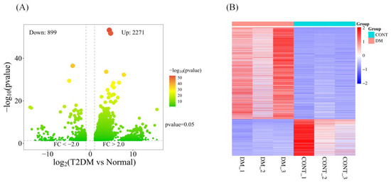
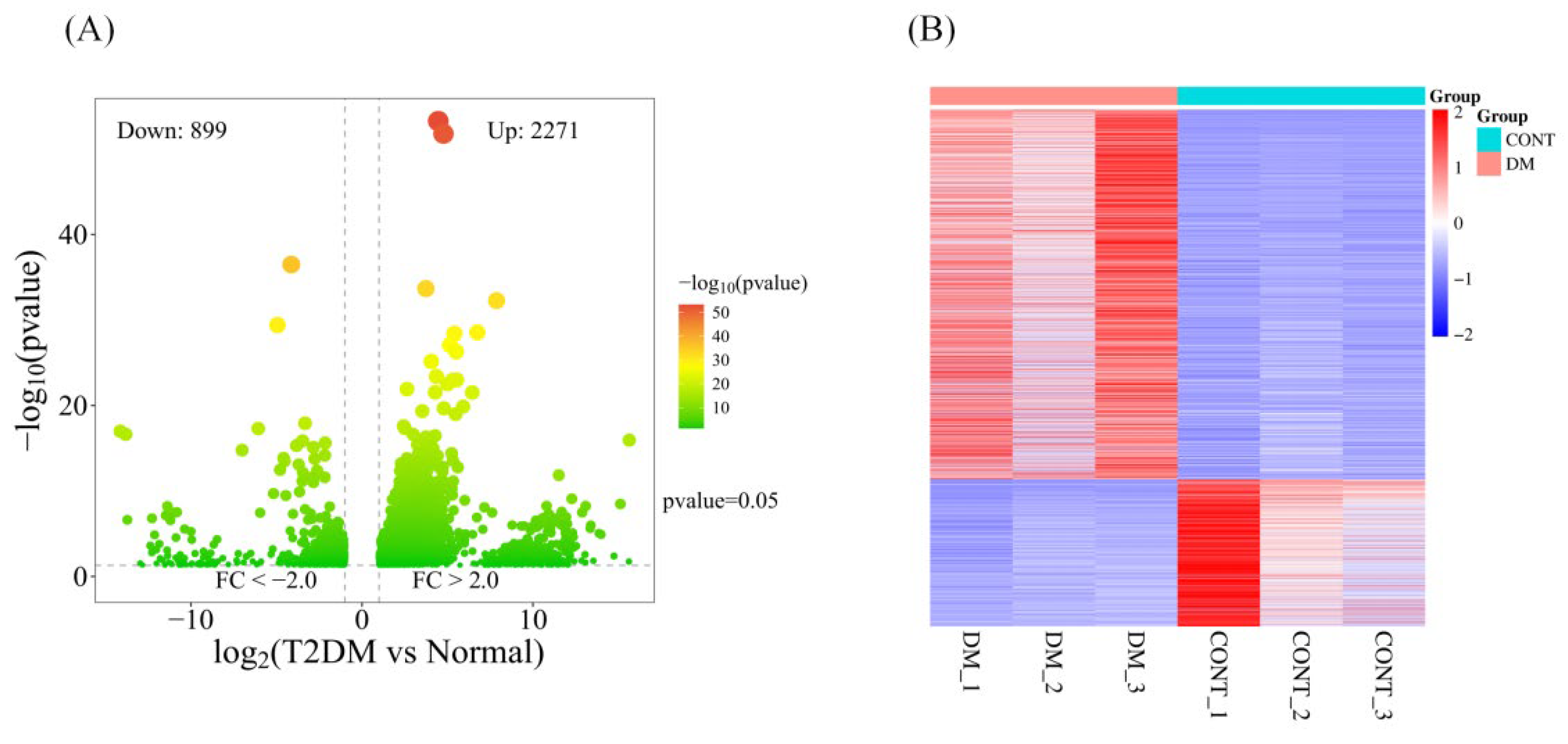
The dataset was analyzed using the WebBioinformatics online platform (https://www.bioinformatics.com.cn/). A volcano plot and heatmap revealed that among 28,945 differentially expressed genes, 3170 genes exhibited significant expression differences, with 2271 upregulated and 899 downregulated genes (Figure 1).
Figure 1.
Volcano plot (A) and heat map (B) of differentially expressed genes.
2.3. GO Functional and KEGG Pathway Enrichment Analysis
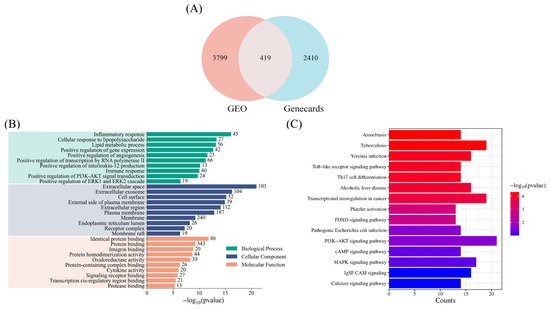
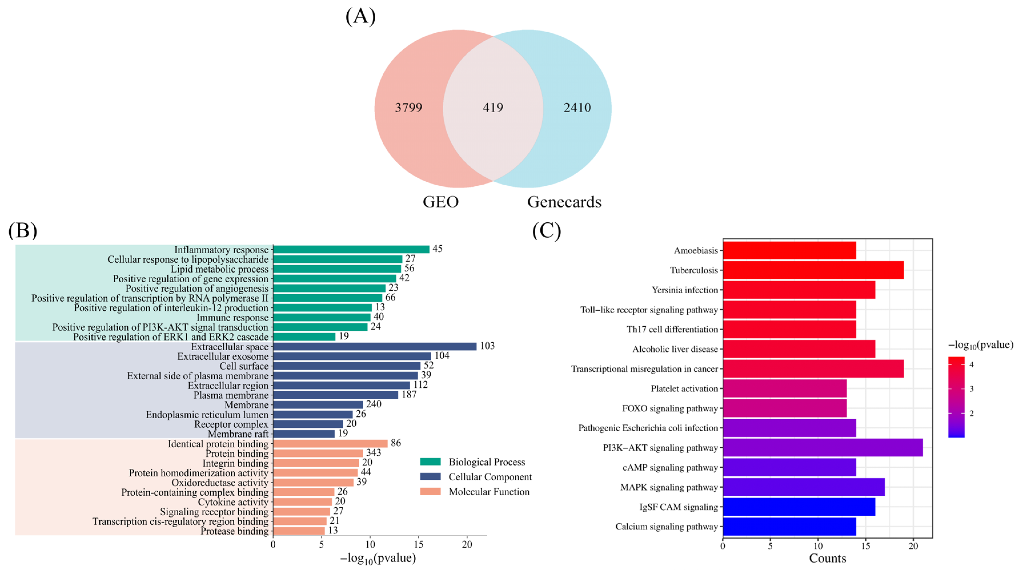
To further screen for potential signaling pathways associated with T2DM-related dyslipidemia and avoid overlooking relevant signaling pathways, we expanded the selection criteria. Genes with |logFC| > 0.5 and p < 0.05 in the GEO dataset were defined as significantly differentially expressed genes. These were then intersected with predicted genes from the Genecards database, yielding 419 overlapping genes, which were visualized in a Venn diagram (Figure 2A). Subsequent Gene Ontology (GO) and Kyoto Encyclopedia of Genes and Genomes (KEGG) enrichment analyses were performed using DAVID Bioinformatics (https://davidbioinformatics.nih.gov/home.jsp).
Figure 2.
Differential expression genes analysis plots. (A) Venn diagram showing the intersection of differentially expressed genes from the GEO dataset (|logFC| > 0.5, p < 0.05) and predicted genes from the Genecards database, yielding 419 overlapping genes. (B) GO enrichment analysis (top 10 terms by p-value in BP, CC, and MF categories) showing significant enrichment of inflammatory response, immune response, extracellular space, and exosomes. (C) KEGG pathway enrichment analysis (p < 0.05, count > 12, followed by biological relevance filtering) identifying TLR, FOXO, PI3K-AKT, cAMP, and MAPK signaling pathways as significantly associated with T2DM-associated dyslipidemia. The TLR signaling pathway was excluded from further analysis due to limited literature on exercise regulation.
GO functional enrichment analysis was performed across three categories: biological process (BP), cellular component (CC), and molecular function (MF). The top 10 terms with the smallest p-values in each category were selected for presentation (Figure 2B). Significant enrichment was observed in terms such as inflammatory response, immune response, extracellular space, and exosomes, indicating that the differentially expressed genes are closely associated with inflammation, immune regulation, and cell signaling processes, and are largely localized to the cell membrane or extracellular environment. Notably, the PI3K-AKT and MAPK signaling pathways showed significant enrichment in this analysis.
For KEGG pathway enrichment analysis, two rounds of screening were applied. The first round used statistical thresholds of p < 0.05 and gene count > 12. The second round was based on biological relevance to T2DM-associated lipid metabolism disorders, with cross-referencing to the GO results. This process identified five significantly enriched signaling pathways: TLR, FOXO, PI3K-AKT, cAMP, and MAPK signaling pathways (Figure 2C). Among these, the TLR signaling pathway was not further analyzed, as current literature on its role in exercise-mediated regulation of T2DM-associated dyslipidemia remains limited. Therefore, this review focuses on the FOXO, PI3K-AKT, cAMP, and MAPK signaling pathways.
3. Lipid Metabolism-Related Signaling Pathways and the Maintenance of Lipid Homeostasis in T2DM
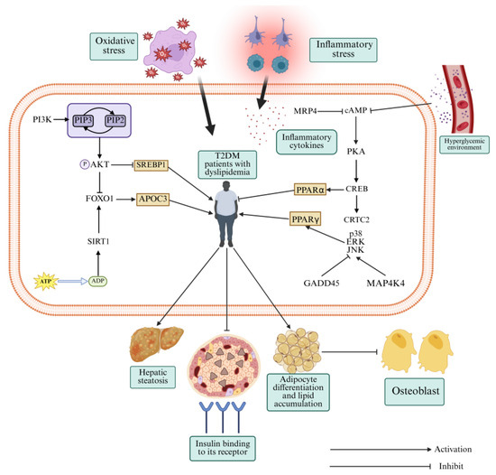
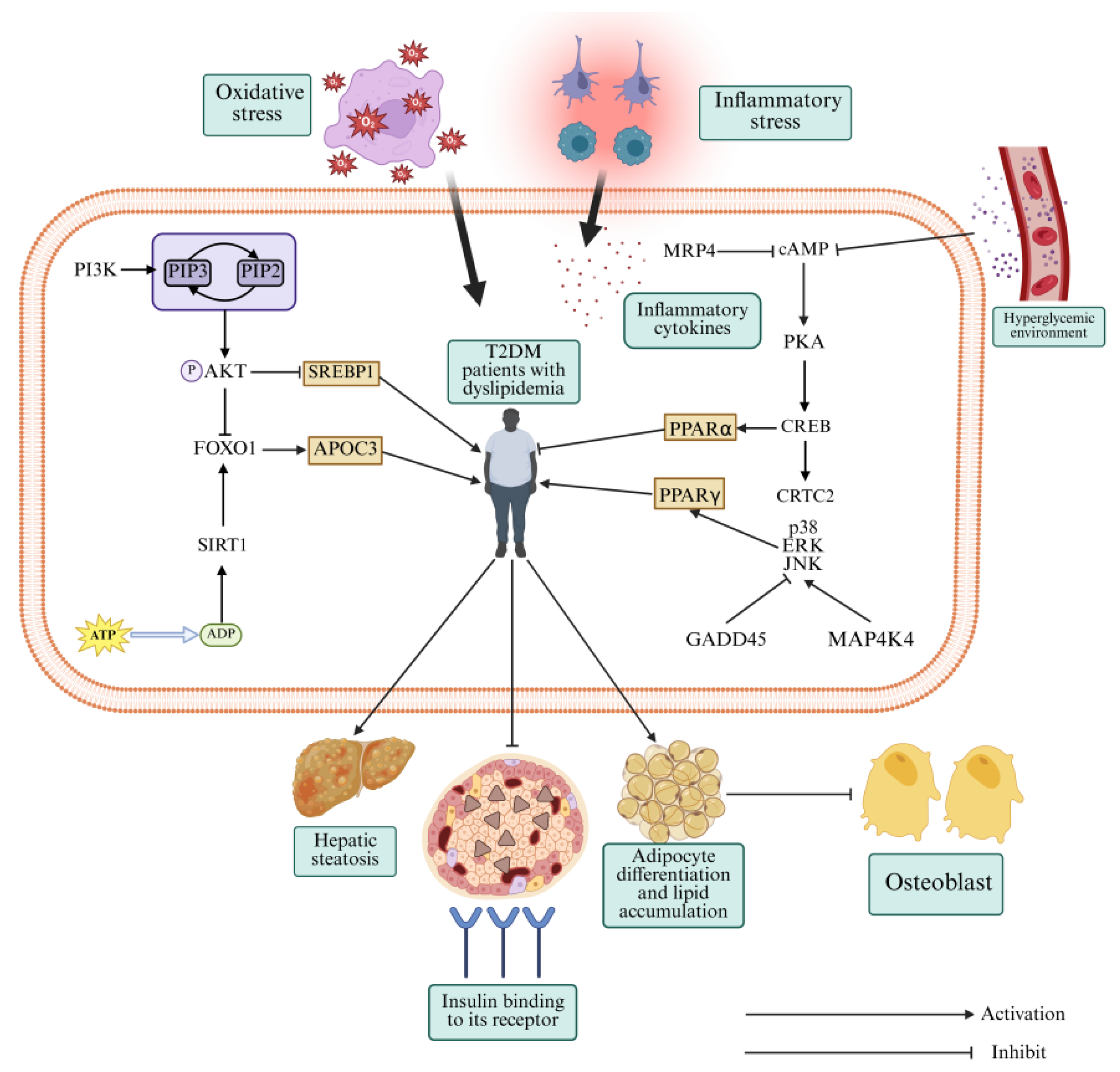
Lipid homeostasis depends on the coordinated action of multiple signaling pathways. From our KEGG enrichment analysis, we focused on four signaling pathways including cAMP, FOXO, PI3K-AKT, and MAPK signaling pathways as core regulators of lipid metabolism in T2DM. MAPK, despite showing weaker enrichment than the other three, is a key downstream effector of insulin signaling. Below, we discuss how each of these pathways contributes to T2DM-associated dyslipidemia (Figure 3).
Figure 3.
Regulatory mechanism of lipid metabolism-related signaling pathways in T2DM with dyslipidemia. cAMP, Cyclic adenosine monophosphate; CREB, cAMP response element-binding protein; JNK, c-Jun N-terminal kinase; PI3K, Phosphatidylinositol-3 kinase; PPARα, Peroxisome proliferator-activated receptor alpha; PPARγ, Peroxisome proliferator-activated receptor gamma; AKT, Protein Kinase B; SIRT1, Sirtuin1; FOXO, Forkhead box O; SREBP1, Sterol regulatory element-binding protein 1; APOC3, Apolipoprotein C3; MRP4, Multidrug resistance-associated protein 4; CRTC2, CREB-regulated transcription coactivator 2; ERK, Extracellular signal-regulated kinase; GADD45, Growth arrest-and DNA damage-inducible protein 45; MAP4K4, Mitogen-activated protein kinase 4.
3.1. cAMP Signaling Pathway and Lipid Metabolism Disorders Associated with T2DM
cAMP is a ubiquitous second messenger [11] that transmits signals primarily by activating protein kinase A (PKA), which ultimately regulates the expression of downstream target genes [12]. The cAMP signaling pathway has been implicated in various diseases, including diabetes, Alzheimer’s disease, and NAFLD [13,14]. Therefore, maintaining normal cAMP signaling pathway is crucial for suppressing lipid metabolism disorders and treating T2DM.
Under physiological conditions, cAMP activates PKA, which phosphorylates cAMP response element-binding protein (CREB), thereby regulating downstream signaling. However, under pathological conditions such as glucolipotoxicity, oxidative stress, and inflammation, the cAMP signaling pathway is often dysregulated in T2DM patients [15]. Multidrug resistance-associated protein 4 (MRP4), an efflux transporter closely associated with glucose and lipid metabolism, plays a crucial role in diabetes and obesity [16]. MRP4 regulates lipid metabolism by suppressing cAMP signaling pathway. Following exogenous knockout of the Mrp4 gene in mice, significant increases in the expression of cAMP, CREB, and CREB-regulated transcription coactivator 2 (CRTC2) expression were observed, along with dyslipidemia and adipose tissue inflammation [17]. This demonstrates that overactivation of cAMP signaling abnormally activates CRTC2, leading to increased lipid synthesis and inflammation, ultimately exacerbating lipid metabolism disorders and insulin resistance. Conversely, a previous study [18] reported that hyperglycemia and insulin resistance suppress cAMP expression in the liver, thereby impairing fatty acid oxidation and inducing hepatic steatosis. Elevating cAMP levels activates AMP-activated protein kinase (AMPK), which phosphorylates peroxisome proliferator-activated receptor alpha (PPARα), ultimately improving hepatic lipid metabolism disorders. Similarly, cAMP signaling is suppressed in the hippocampus and skeletal muscle of T2DM mice [19,20], and appropriate activators can enhance lipid metabolism and mitigate insulin resistance in these tissues. Therefore, cAMP signaling in T2DM exhibits unidirectional and tissue-specific regulation.
cAMP signaling in T2DM displays both suppression and overactivation depending on the tissue context. Both extremes exacerbate dyslipidemia. These observations suggest that restoring normal cAMP signaling by activation or inhibition depending on the tissue could help correct lipid metabolism in T2DM.
3.2. PI3K-AKT Signaling Pathway and Dyslipidemia in T2DM
The PI3K-AKT signaling pathway is a crucial downstream effector of insulin signaling, involved in cell growth, proliferation, differentiation, and apoptosis [21,22]. Normally, PI3K catalyzes the conversion of phosphatidylinositol-4,5-bisphosphate into phatidylinositol-3,4,5-trisphosphate [23], activating AKT phosphorylation, which enhances glucose and lipid metabolism and suppresses insulin resistance, ultimately contributing to the reversal of T2DM progression to some extent.
The PI3K-AKT signaling pathway regulates lipid metabolism by inhibiting lipolysis and promoting lipid storage, thereby enhancing adipose tissue fat-storing capacity [24]. This prevents excess fatty acids from accumulating in non-adipose tissues such as blood and liver, thereby reducing hyperlipidemia and insulin resistance [25]. In T2DM patients, impaired upstream insulin signaling typically suppresses the PI3K-AKT signaling pathway, contributing to lipotoxicity and increasing NAFLD risk [26]. The role of the PI3K-AKT signaling pathway in lipid homeostasis is closely linked to sterol regulatory element-binding protein 1 (SREBP1), a central regulator of lipid uptake and synthesis [27,28]. In high-fat diet (HFD)-induced T2DM rats, hepatic SREBP1 mRNA levels were elevated, while insulin receptor substrate 2 (IRS2), PI3K, and AKT were reduced [29]. Papaya extract treatment also decreased hepatic SREBP1 expression, increased IRS2, PI3K, and AKT levels, and suppressed dyslipidemia. Similarly, gentianin improved hepatic lipid deposition and blood lipid profiles in T2DM mice by activating the PI3K-AKT signaling pathway and suppressing SREBP1 expression [30].
Thus, maintaining normal activation of the PI3K-AKT signaling pathway is a critical therapeutic target for lipid metabolism disorders associated with T2DM. Currently, this signaling pathway has been extensively studied in glucose metabolism [31]; its role in lipid homeostasis remains largely in the preclinical stage. Further clinical studies are needed to validate its therapeutic potential in humans. Additionally, when targeting this signaling pathway, attention may be paid to avoid excessive activation, which may promote tumorigenesis.
3.3. FOXO Signaling Pathway and Dyslipidemia in T2DM
FOXO proteins exist in four forms in mammals: FOXO1, FOXO3, FOXO4, and FOXO6 [32]. The FOXO signaling pathway is involved in cell survival, differentiation, apoptosis, and oxidative stress [33]. FOXO1 is primarily expressed in insulin-responsive tissues such as skeletal muscle, pancreas, and liver, playing a crucial regulatory role in lipid metabolism [34,35]. Thus, maintaining normal FOXO1 expression is essential for alleviating dyslipidemia and insulin resistance in T2DM.
The PI3K-AKT signaling pathway and the deacetylase enzyme Sirtuin 1 (SIRT1) are the primary regulators of FOXO signaling [36,37]. PI3K-AKT signaling activation phosphorylates and inhibits FOXO1, suppressing lipolysis and promoting lipid synthesis [38]. SIRT1, a key regulator of energy and lipid metabolism [39], is activated during exercise or energy restriction via AMPK-mediated increase in nicotinamide adenine dinucleotide levels [40]. SIRT1 deacetylates FOXO1, enhancing its nuclear localization and promoting lipolysis [41]. However, in T2DM, insulin resistance, oxidative stress, and glucolipotoxicity suppress PI3K-AKT signaling, thereby weakening AKT-mediated inhibition of FOXO1. This leads to FOXO1 overexpression, which activates apolipoprotein C-III transcription, thus stimulating triglyceride accumulation in blood and liver [42]. Moreover, SIRT1 expression is downregulated in obesity, as evidenced in HFD-induced obese mice, accompanying upregulated FOXO1 [43]. SIRT1 agonist administration restored normal FOXO1 expression, indicating the unidirectional FOXO1-mediated deacetylation. Similarly, reduced SIRT1 activity shifts the role of FOXO1 in exacerbated dyslipidemia from promoting lipolysis.
Overall, FOXO signaling exerts bidirectional regulation in lipid metabolism. Under normal conditions, AKT maintains FOXO at optimal levels, preventing excessive triglyceride deposition. Appropriate FOXO activation via exercise or other means can accelerate lipolysis and alleviate insulin resistance. Therefore, PI3K-AKT or SIRT1 agonists may be employed to modulate FOXO signaling toward a state beneficial for lipid homeostasis. Current research on this pathway primarily focuses on hepatic lipid metabolism. Consequently, future studies should expand to other tissues such as skeletal muscle.
3.4. MAPK Signaling Pathway and Dyslipidemia in T2DM
MAPK signaling pathway is another crucial regulator in lipid metabolism, comprising three branches: p38, c-Jun N-terminal kinase (JNK), and extracellular signal-regulated kinase (ERK) [44]. The p38/ERK signaling pathway primarily regulates lipid synthesis and metabolism, while JNK is closely associated with insulin signaling [45,46].
Mitogen-activated protein kinase 4 (MAP4K4) and growth arrest- and DNA damage-inducible protein 45 (GADD45) are key regulatory factors of MAPK signaling [47]. A study found that in high-sugar and HFD-induced T2DM mice, the expression of MAP4K4, p38, ERK1/2, and JNK was increased, while GADD45 expression was decreased in the liver. In contrast, artemether intervention normalized these protein levels and enhanced glucose and lipid metabolism [48]. On the other hand, GADD45 activation can promote p38 MAPK signaling in other diseases, thereby exacerbating disease progression [49], suggesting that the regulatory function of GADD45 is pleiotropic or tissue specific. Given that the mechanisms by which protein regulates MAPK signaling to maintain lipid homeostasis remains unclear, subsequent studies should be further explored in different tissues.
Peroxisome proliferator-activated receptor gamma (PPARγ), a transcription factor involved in adipocyte differentiation and lipid storage, is also regulated by MAPK [50]. One previous study reported increased mRNA expression of PPARγ, ERK, p38, and JNK in dyslipidemic mice [51]. Triterpenoid saponin treatment reduced the expression of these factors and restored dyslipodemia, suggesting that triterpenoid saponins may inhibit adipogenic differentiation by suppressing PPARγ expression via MAPK. However, MAPK can also inhibit PPARγ during osteoblast differentiation [52]. PPARγ also exhibits a close association with the PI3K-AKT signaling pathway [53], where its expression enhances PI3K-AKT activation, thereby promoting pancreatic β-cell survival and insulin sensitivity [54].
Taken together, the regulatory relationships among GADD45 and PPARγ, and MAPK signaling pathways exhibit highly context-dependent. Therefore, therapeutic targeting the MAPK signaling should be tissue- and disease stage-specific. For instance, PPARγ activators may be beneficial in the pancreas to enhance insulin signaling, while inhibitors may be preferred in adipose tissue to prevent excessive lipid accumulation.
4. Exercise Regulates Lipid Metabolism-Related Signaling Pathways to Alleviate Dyslipidemia in T2DM
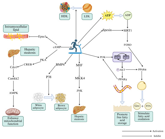
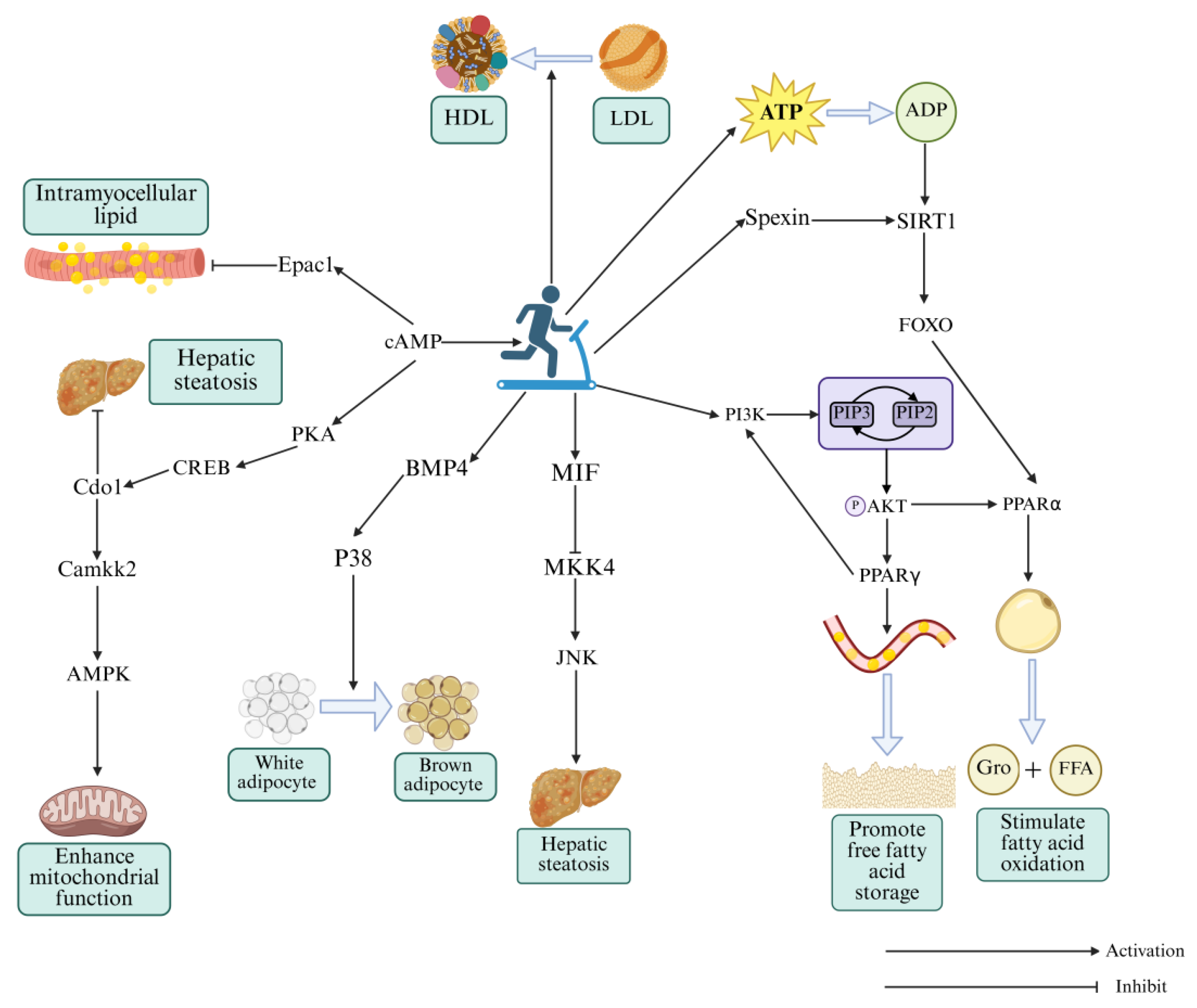
Exercise is a cornerstone of clinical management [55]. Emerging “precision exercise” strategies aim to optimize its therapeutic potential by integrating multi-omics biomarkers and artificial intelligence for individualized risk assessment [56,57]. Unlike many pharmacological interventions, exercise acts through multiple signaling pathways simultaneously, with fewer organ-specific side effects. Although exercise is recognized as an important preventive and therapeutic measure for dyslipidemia in T2DM, its regulatory mechanisms during exercise interventions remain incompletely understood. Below, we summarize the molecular mechanisms by which exercise modulates the four signaling pathways discussed above, providing a theoretical foundation for future clinical research (Figure 4).
Figure 4.
The molecular mechanisms underlying exercise-induced improvement of T2DM with dyslipidemia via regulation of lipid-metabolism related pathways. HDL, high-density lipoprotein; LDL, low-density lipoprotein; cAMP, Cyclic adenosine monophosphate; Epac1, Exchange protein activated directly by cAMP 1; PKA, Protein kinase A; CREB, cAMP response element-binding protein; Cdo1, Cysteine dioxygenase type 1; Camkk2, Ca2+/calmodulin-dependent protein kinase kinase 2; AMPK, AMP-activated protein kinase; BMP4, Bone morphogenetic protein 4; MIF, Macrophage migration inhibitory factor; MKK4, Mitogen-activated protein kinase kinase 4; JNK, c-Jun N-terminal kinase; PI3K, Phosphatidylinositol-3 kinase; PPARα, Peroxisome proliferator-activated receptor alpha; PPARγ, Peroxisome proliferator-activated receptor gamma; AKT, Protein Kinase B; SIRT1, Sirtuin1; FOXO, Forkhead box O.
4.1. Exercise Activates cAMP Signaling Pathway for Restoring Dyslipidemia in T2DM
Abnormal cAMP signaling pathway in T2DM is a key contributor to dyslipidemia. Exercise can precisely regulate cAMP signaling pathway. The liver, a central regulatory organ for glucose and lipid metabolism, exhibits downregulated cAMP signaling pathway in T2DM, leading dyslipidemia and insulin resistance, which mutually reinforce each other and drive the progression of T2DM [58]. An 8-week aerobic exercise intervention improved hepatic lipid deposition and adipocyte degeneration in NAFLD mice by enhancing the cAMP-PKA-CREB signaling axis, promoting the expression of cysteine dioxygenase type 1 (Cdo1) [59]. Cdo1, a key enzyme in taurine synthesis, promotes energy expenditure and lipolysis, suppresses insulin resistance [60], and enhances AMPK signaling via interaction with Ca2+/calmodulin-dependent protein kinase kinase 2 (CaMKK2). Thus, exercise mitigates dyslipidemia through synergistic regulation of the cAMP-AMPK network. Long-term moderate-intensity exercise not only reduces hepatic lipid deposition and insulin resistance, but also enhances mitochondrial function, further promoting glucose and lipid metabolism, thereby reversing T2DM progression. Given the tissue-specific variability in the activation of the cAMP signaling pathway in T2DM patients, and the predominantly suppressed state of the AMPK signaling pathway under T2DM pathological conditions, it can be hypothesized that the cAMP signaling pathway bidirectionally regulates Cdo1 expression. However, whether both excessive and insufficient cAMP signaling suppress Cdo1 expression remains a hypothesis that requires direct experimental validation.
Beyond the cAMP-PKA signaling pathway, the exchange protein activated directly by cAMP 1 (Epac1) also serves as a downstream cAMP effector [61], regulating brown adipose tissue differentiation and energy expenditure [62]. Epac1 expression is suppressed in T2DM [63], but increased in skeletal muscle after moderate-intensity treadmill exercise [64], indicating that exercise enhances lipid oxidation via Epac1 activation. Given that both PKA and Epac1 are suppressed in T2DM skeletal muscle, they likely function synergistically rather than in isolation for maintaining lipid homeostasis.
Moderate-to-low intensity aerobic exercise can enhance lipid oxidation in tissues such as the liver and skeletal muscle by targeting the cAMP signaling pathway, thereby effectively attenuating T2DM-associated dyslipidemia. However, most current evidence is derived from preclinical rodent models, and human validation remains limited. Future studies should explore the effects of higher-intensity and resistance exercise on cAMP signaling, as well as the tissue-specific expression patterns of Cdo1 and Epac1 in patients with different T2DM phenotypes. Additionally, the hypothesized bidirectional regulation of Cdo1 by cAMP signaling requires direct experimental testing.
4.2. Exercise Upregulates PI3K-AKT Signaling Pathway for Attenuating Dyslipidemia in T2DM
Exercise interventions offer advantages over drug therapy in terms of reduced costs and fewer side effects. Aerobic exercise has long been regarded as a key intervention for obesity and insulin resistance [65]. Based on one of previous studies, 8-week aerobic exercise increased hepatic expression of PI3K, AKT and PPARγ in T2DM rats, which is beneficial to improve blood lipid levels, hepatic lipid deposition, and insulin sensitivity [66]. Thus, aerobic exercise activates the PI3K-AKT signaling pathway by PPARγ, enhancing adipocyte storage capacity, and creating a positive feedback loop.
High-intensity interval training (HIIT) also improves insulin sensitivity and HDL function [67]. An 8-week HIIT intervention upregulated PI3K, AKT, IRS-1, and PPARα in T2DM mice, while downregulated PPARγ to attenuate dyslipidemia [68]. These findings indicate that exercise interventions at varying intensities can activate the PI3K-AKT signaling pathway for the mitigation of dyslipidemia although PPARγ expression may vary depending on exercise intensity and disease stage. Notably, HIIT may exacerbate renal fibrosis in T2DM mice [69], highlighting the demand for personalized exercise prescriptions, especially in patients with complications such as diabetic nephropathy.
Collectively, the PI3K-AKT signaling pathway serves as a key target of exercise in regulating T2DM-associated dyslipidemia. Appropriate aerobic exercise should be a primary interventional approach for most T2DM patients, while HIIT should be prescribed with caution in those with complications (e.g., diabetic nephropathy). Notably, the expression of PPARγ in response to exercise shows inconsistent results across different exercise intensities and disease stages. Further studies are needed to clarify the mechanisms underlying these intensity-dependent effects and to determine whether PPARγ could serve as a predictive biomarker for personalized exercise prescription. Most of the current evidence remains preclinical so that clinical trials are urgently needed.
4.3. Exercise Enhances FOXO Signaling Pathway for Mitigating Dyslipidemia in T2DM
Currently, the studies on exercise-mediated FOXO regulation in dyslipidemia are relatively limited. Although FOXO reveals close links to PI3K-AKT and AMPK signaling pathways in T2DM [70,71], it still emerges as a promising therapeutic target during exercise intervention. SIRT1, a critical activator of FOXO, can be affected by intracellular energy changes [72], making exercise a potent modulator of SIRT1/FOXO signaling. According to a previous report [73], endurance exercise enhanced FOXO expression in Drosophila skeletal muscle, rescuing HFD-induced skeletal muscle damage. Even when FOXO expression is reduced, exercise suppresses dyslipidemia via SIRT1 activation, highlighting the redundancy of signaling pathways associated with lipid metabolism. HIIT also regulates FOXO signaling pathway via Spexin, an anorexigenic adipokine that promotes lipid metabolism and insulin sensitivity [74]. Similarly, an 8-week HIIT regimen increased Spexin, SIRT1, FOXO1, and PPARα in T2DM mice, mitigating dyslipidemia and insulin resistance [75]. Because Spexin can enhance SIRT1 expression [76], HIIT may activate the SIRT1/FOXO1 signaling pathway by boosted Spexin.
Together, the FOXO signaling pathway plays a crucial role in exercise-induced restoration of dyslipidemia in T2DM individuals. Aerobic exercise and HIIT have both been shown to activate the SIRT1/FOXO1 signaling axis via Spexin upregulation in preclinical rodent models. However, compared to other three signaling pathways, studies on the FOXO signaling pathway are relatively limited and remain at the preclinical stage. No human clinical trials have yet validated FOXO signaling pathway as a therapeutic target for exercise-mediated lipid regulation. Future studies should prioritize translational research to confirm whether FOXO activation in skeletal muscle and liver translates to improved lipid profiles in T2DM patients, and to determine the optimal exercise modality for FOXO modulation.
4.4. Exercise Triggers MAPK Signaling Pathway for Ameliorating Dyslipidemia in T2DM
MAPK signaling is an evolutionarily conserved pathway that controls cell proliferation, metabolism, and apoptosis [77,78]. Dysregulation of this pathway has been implicated in rheumatoid arthritis, cancer, and T2DM [79,80]. Although previous studies have demonstrated that exercise can reverse disease progression by inhibiting abnormal MAPK signaling pathways, systematic exploration in T2DM accompanied by concomitant lipid metabolism disorders remains limited. Therefore, the following section will summarize studies on exercise-induced activation of the MAPK signaling pathway for rescuing dyslipidemia in T2DM.
Macrophage migration inhibitory factor (MIF), a pro-inflammatory cytokine, can also modulate insulin signaling and lipid metabolism [81]. NAFLD is a chronic metabolic disorder sharing core pathological environments with T2DM, such as insulin resistance and lipid metabolism disorders [82]. A 16-week swimming training upregulated MIF expression in NAFLD mice through inhibiting mitogen-activated protein kinase kinase 4 (MKK4) and suppressing JNK phosphorylation, thereby ameliorating hepatic lipotoxicity [83]. However, MIF is often elevated in T2DM and exhibits context-dependent effects [84,85], warranting further investigation. Moreover, lipid metabolism disorders are frequently accompanied by the whitening and dysfunction of brown adipose tissue [86], a pathological state closely linked to dysregulated bone morphogenetic protein 4 (BMP4)/p38 signaling pathway. It was found that HFD induction can suppress BMP4 and p38 in mouse adipose tissue, but aerobic exercise can rescue the suppression of these signaling pathways [87], thereby stimulating the brown adipose tissue function and lipid metabolism. Interestingly, these findings are contradictory to the conventional view that suppressing excessive p38 activation is beneficial in T2DM, highlighting exercise’s core to counteract pathological stress with physiological activation. It is noteworthy that MIF and BMP4 are part of a broader class of exercise-induced signaling molecules, known as exerkines, which are increasingly recognized as key mediators of exercise benefits, potentially even regulating processes like ferroptosis [88]. Future research should explore whether the amelioration of T2DM-related dyslipidemia by exercise involves exerkine-mediated modulation of cell death and metabolic pathways. The molecular mechanisms described above are summarized to clearly illustrate critical regulatory roles of exercise in T2DM with dyslipidemia (Table 1).
Table 1.
Molecular mechanism by which exercise alleviates T2DM with dyslipidemia through regulating lipid metabolism-related signaling pathways.
Thus, unlike many drugs that broadly inhibit or activate a given signaling pathway, aerobic exercise appears to modulate MAPK signaling in a context- and tissue-specific way, which may be particularly suitable for treating dyslipidemia in T2DM. The divergent roles of p38, JNK, and ERK branches, as well as the context-dependent effects of MIF and BMP4, highlight the need for caution when designing exercise prescriptions targeting this signaling pathway. Future investigations are highly desired to explore the ERK signaling pathway in exercise-induced optimization of lipid metabolism, and to determine whether MAPK-related biomarkers (e.g., MIF, BMP4) can be reliably used to monitor exercise efficacy in clinical settings. Currently, all evidence remains at the preclinical stage.
5. Conclusions
The prevalence of T2DM is increasing rapidly worldwide, and dyslipidemia remains a core pathological feature driving insulin resistance and disease progression. Although novel pharmacotherapies such as mazdutide and lanifibranor have shown promise in clinical trials, their long-term safety remains to be established. Mainstream drugs such as metformin and semaglutide are effective but are often limited by gastrointestinal side effects, hypoglycemia, and variable efficacy. Moreover, when compared with oral pharmacotherapy, exercise compliance poses a major challenge. Therefore, a combinatorial strategy integrating pharmacotherapy with individualized exercise prescription is recommended for optimal T2DM management.
This article synthesizes current evidence demonstrating that exercise acts as a signaling network regulator, concurrently modulating the cAMP, PI3K-AKT, FOXO, and MAPK signaling pathways to restore lipid homeostasis in T2DM. A key contribution of this article is the identification of eight “exercise effector factors” (Cdo1, Epac1, PPARγ, PPARα, SIRT1, Spexin, BMP4, and MIF) that could serve as molecular readouts of signaling pathway-specific exercise adaptations.
Currently, exercise prescriptions for T2DM patients are primarily guided by traditional indicators such as blood glucose, heart rate, and maximal oxygen uptake (VO2max). While these indicators are cost-effective, easy to measure, and provide real-time feedback on exercise safety and intensity, they do not capture whether exercise has truly improved the underlying pathological state of dyslipidemia at the tissue or molecular level. In contrast, the proposed exercise effector factors (summarized in Supplementary Table S1), although more challenging to measure and not suitable for real-time monitoring, could complement traditional indicators by revealing the molecular efficacy of exercise interventions on specific signaling pathways and tissues.
Therefore, clinical translation and future directions may be summarized as follows:
For patients with predominant hepatic cAMP suppression (reflected by low Cdo1 expression), moderate-intensity aerobic exercise may be prioritized.
For those with reduced SIRT1-FOXO activity, HIIT could be considered if no contraindications (e.g., diabetic nephropathy) are present.
The expression levels of these effector factors could be measured at baseline and during follow-up to guide exercise prescription adjustments.
Most mechanistic evidence presented in this article is derived from preclinical animal models; human validation is urgently needed before these biomarkers can be integrated into clinical practice. Furthermore, given the tissue-specific and context-dependent nature of these signaling pathways in T2DM, future studies should clarify the inconsistent findings across different tissues and disease stages. By combining molecular biomarkers with traditional physiological indicators, we envision a future of precision exercise that delivers more effective, personalized, and scientifically grounded interventions for T2DM and its metabolic complications.
Supplementary Materials
The following supporting information can be downloaded at: https://www.mdpi.com/article/10.3390/metabo16040269/s1, Table S1: Comparison of traditional vs. molecular indicators for exercise prescription.
Author Contributions
Manuscript draft writing, T.T.; manuscript reviewing, F.Y., X.L., X.Z. and S.B.; funding acquisition, J.Y. All authors have read and agreed to the published version of the manuscript.
Funding
This work was supported by the National Social Science Foundation (No. 22BTY049) and Guangxi Natural Science Foundation (2025GXNSFBA069048).
Institutional Review Board Statement
Not applicable.
Informed Consent Statement
Not applicable.
Data Availability Statement
No new data were created or analyzed in this study. Data sharing is not applicable to this article.
Acknowledgments
The authors thank the WebBioinformatics online platform (https://www.bioinformatics.com.cn/) for providing data analysis tools, and the DAVID Bioinformatics Resource (https://davidbioinformatics.nih.gov/home.jsp) for gene enrichment analysis. Figures were created using BioRender (https://www.biorender.com/).
Conflicts of Interest
The authors declare no conflicts of interest.
Abbreviations
| T2DM | Type 2 diabetes mellitus |
| cAMP | Cyclic adenosine monophosphate |
| PI3K-AKT | Phosphatidylinositol 3-kinase–protein kinase B |
| FOXO | Forkhead box O |
| MAPK | Mitogen-activated protein kinase |
| NAFLD | Non-alcoholic fatty liver disease |
| PKA | Protein kinase A |
| CREB | cAMP response element-binding protein |
| MRP4 | Multidrug resistance-associated protein 4 |
| CRTC2 | CREB-regulated transcription coactivator 2 |
| AMPK | AMP-activated protein kinase |
| PPARα | Peroxisome proliferator-activated receptor alpha |
| SREBP1 | Sterol regulatory element-binding protein 1 |
| IRS2 | Insulin receptor substrate 2 |
| SIRT1 | Sirtuin1 |
| JNK | c-Jun N-terminal kinase |
| ERK | Extracellular signal-regulated kinase |
| MAP4K4 | Mitogen-activated protein kinase 4 |
| GADD45 | Growth arrest-and DNA damage-inducible protein 45 |
| PPARγ | Peroxisome proliferator-activated receptor gamma |
| Cdo1 | Cysteine dioxygenase type 1 |
| CaMKK2 | Ca2+/calmodulin-dependent protein kinase kinase 2 |
| Epac1 | Exchange protein activated directly by cAMP 1 |
| HIIT | High-intensity interval training |
| MIF | Macrophage migration inhibitory factor |
| MKK4 | Mitogen-activated protein kinase kinase 4 |
| BMP4 | Bone morphogenetic protein 4 |
| GO | Gene Ontology |
| KEGG | Kyoto Encyclopedia of Genes and Genomes |
References
- GBD 2021 Diabetes Collaborators. Global, regional, and national burden of diabetes from 1990 to 2021, with projections of prevalence to 2050: A systematic analysis for the Global Burden of Disease Study 2021. Lancet 2023, 402, 203–234. [Google Scholar] [CrossRef] [Scilit]
- Deng, W.; Zhao, L.; Chen, C.; Ren, Z.; Jing, Y.; Qiu, J.; Liu, D. National burden and risk factors of diabetes mellitus in China from 1990 to 2021: Results from the Global Burden of Disease study 2021. J. Diabetes 2024, 16, e70012. [Google Scholar] [CrossRef] [Scilit] [PubMed]
- Ding, K.; Zhang, Z.; Han, Z.; Shi, L.; Li, X.; Liu, Y.; Li, Z.; Zhao, C.; Cui, Y.; Zhou, L.; et al. Liver ALKBH5 regulates glucose and lipid homeostasis independently through GCGR and mTORC1 signaling. Science 2025, 387, eadp4120. [Google Scholar] [CrossRef] [Scilit] [PubMed]
- Deprince, A.; Haas, J.T.; Staels, B. Dysregulated lipid metabolism links NAFLD to cardiovascular disease. Mol. Metab. 2020, 42, 101092. [Google Scholar] [CrossRef] [Scilit] [PubMed]
- Kim, K.S.; Hong, S.; Han, K.; Park, C.Y. Association of non-alcoholic fatty liver disease with cardiovascular disease and all cause death in patients with type 2 diabetes mellitus: Nationwide population-based study. BMJ 2024, 384, e076388. [Google Scholar] [CrossRef] [Scilit]
- Li, F.; Huang, W.; Zhang, L. Exercise modulates protein acylation to improve cardiovascular diseases. Prog. Biochem. Biophys. 2025, 52, 1453–1467. [Google Scholar] [CrossRef]
- Zhang, N.; Li, S. Research progress on improving osteoporosis by regulating intestinal flora through exercise. Chin. J. Osteoporos. Bone Miner. Dis. 2025, 18, 384–392. [Google Scholar]
- Hashimoto, Y.; Takahashi, F.; Okamura, T.; Hamaguchi, M.; Fukui, M. Diet, exercise, and pharmacotherapy for sarcopenia in people with diabetes. Metabolism 2023, 144, 155585. [Google Scholar] [CrossRef] [Scilit]
- Luo, W.; Gao, W.Y.; Wang, Y.H.; Liu, Y.S.; Zhou, Y.; Ai, L. Aerobic exercise relieves glucose and lipid metabolism disorder after immune stimulation in IR mice by inducing tolerance of trained immunity. Chin. J. Sports Med. 2025, 44, 394–404. [Google Scholar] [CrossRef]
- Suder, A.; Makiel, K.; Targosz, A.; Kosowski, P.; Malina, R.M. Effects of exercise and dietary interventions on asprosin, leptin, and lipid metabolism in males with abdominal obesity, a randomized controlled trial. Sci. Rep. 2024, 14, 28109. [Google Scholar] [CrossRef] [Scilit]
- Yan, C.; Yang, Z.; Chen, P.; Yeh, Y.; Sun, C.; Xie, T.; Huang, W.; Zhang, X. GPR65 sensing tumor-derived lactate induces HMGB1 release from TAM via the cAMP/PKA/CREB pathway to promote glioma progression. J. Exp. Clin. Cancer Res. 2024, 43, 105. [Google Scholar] [CrossRef] [Scilit]
- He, Y.; Liu, S.; Zhang, Y.; Zuo, Y.; Huang, K.; Deng, L.; Liao, B.; Zhong, Y.; Feng, J. Takeda G protein-coupled receptor 5 (TGR5): An attractive therapeutic target for aging-related cardiovascular diseases. Front. Pharmacol. 2025, 16, 1493662. [Google Scholar] [CrossRef] [Scilit] [PubMed]
- Delrue, C.; Speeckaert, R.; Moresco, R.N.; Speeckaert, M.M. Cyclic adenosine monophosphate signaling in chronic kidney disease: Molecular targets and therapeutic potentials. Int. J. Mol. Sci. 2024, 25, 9441. [Google Scholar] [CrossRef] [Scilit] [PubMed]
- Zhang, T.; Luu, M.D.A.; Dolga, A.M.; Eisel, U.L.M.; Schmidt, M. The old second messenger cAMP teams up with novel cell death mechanisms: Potential translational therapeutical benefit for Alzheimer’s disease and Parkinson’s disease. Front. Physiol. 2023, 14, 1207280. [Google Scholar] [CrossRef] [Scilit]
- Ma, Y.; Zhang, H.; Yan, Q.; Wang, P.; Guo, W.; Yu, L. The antidiabetic effect of safflower yellow by regulating the GOAT/ghrelin/GHS-R1a/cAMP/TRPM2 pathway. Sci. Rep. 2025, 15, 5037. [Google Scholar] [CrossRef] [Scilit]
- Donepudi, A.C.; Lee, Y.; Lee, J.Y.; Schuetz, J.D.; Manautou, J.E. Multidrug resistance-associated protein 4 (Mrp4) is a novel genetic factor in the pathogenesis of obesity and diabetes. FASEB J. 2021, 35, e21304. [Google Scholar] [CrossRef] [Scilit]
- Laddha, A.P.; Wu, H.; Lee, J.; Mishra, N.; Manautou, J.E. MRP4 deficiency drives lipid metabolism dysregulation and adipose tissue inflammation through cAMP-CREB-CRTC2 activation. Life Sci. 2025, 379, 123895. [Google Scholar] [CrossRef] [Scilit]
- Lv, S.; Qiu, X.; Li, J.; Liang, J.; Li, W.; Zhang, C.; Zhang, Z.N.; Luan, B. Glucagon-induced extracellular cAMP regulates hepatic lipid metabolism. J. Endocrinol. 2017, 234, 73–87. [Google Scholar] [CrossRef] [Scilit]
- Tang, Y.; Gao, Y.; Nie, K.; Wang, H.; Chen, S.; Su, H.; Huang, W.; Dong, H. Jiao-tai-wan and its effective component-berberine improve diabetes and depressive disorder through the cAMP/PKA/CREB signaling pathway. J. Ethnopharmacol. 2024, 324, 117829. [Google Scholar] [CrossRef] [Scilit]
- Iwai, S.; Kaji, K.; Nishimura, N.; Kubo, T.; Tomooka, F.; Shibamoto, A.; Suzuki, J.; Tsuji, Y.; Fujinaga, Y.; Kitagawa, K.; et al. Glucagon-like peptide-1 receptor agonist, semaglutide attenuates chronic liver disease-induced skeletal muscle atrophy in diabetic mice. Biochim. Biophys. Acta Mol. Basis Dis. 2023, 1869, 166770. [Google Scholar] [CrossRef] [Scilit]
- Ouyang, G.; Zhu, Y.; Ouyang, Z. Investigation of Scutellaria Barbata’s immunological mechanism against thyroid cancer using network pharmacology and experimental validation. Sci. Rep. 2025, 15, 2490. [Google Scholar] [CrossRef] [Scilit] [PubMed]
- Zhou, M.; Konigsberg, W.H.; Hao, C.; Pan, Y.; Sun, J.; Wang, X. Bioactivity and mechanisms of flavonoids in decreasing insulin resistance. J. Enzym. Inhib. Med. Chem. 2023, 38, 2199168. [Google Scholar] [CrossRef] [Scilit]
- Zhang, M.; Xing, Z.Y.; Dong, Z.W.; Qiu, B.Y.; Zheng, J.; Hu, Y.C.; Si, C.Y.; Wang, Y.X. Effects of Shengxian Yixin granules on ventricular remodeling in rats with myocardial infarction by regulating PI3K/AKT signaling pathway. Chin. J. Inf. Tradit. Chin. Med. 2025, 32, 98–104. [Google Scholar] [CrossRef]
- Zhang, Y.; Dong, T.; Wang, M. Lipidomic landscape of lipokines in adipose tissue derived extracellular vesicles. Front. Mol. Biosci. 2023, 10, 1281244. [Google Scholar] [CrossRef] [Scilit] [PubMed]
- Su, W.Y.; Tian, L.Y.; Guo, L.P.; Huang, L.Q.; Gao, W.Y. PI3K signaling-regulated metabolic reprogramming: From mechanism to application. Biochim. Biophys. Acta Rev. Cancer 2023, 1878, 188952. [Google Scholar] [CrossRef] [Scilit]
- Tian, S.; Zhao, H.; Song, H. Shared signaling pathways and targeted therapy by natural bioactive compounds for obesity and type 2 diabetes. Crit. Rev. Food. Sci. Nutr. 2024, 64, 5039–5056. [Google Scholar] [CrossRef] [Scilit]
- Chen, Y.; Wang, W.; Morgan, M.P.; Robson, T.; Annett, S. Obesity, non-alcoholic fatty liver disease and hepatocel-lular carcinoma: Current status and therapeutic targets. Front. Endocrinol. 2023, 14, 1148934. [Google Scholar] [CrossRef] [Scilit]
- Geng, F.; Guo, D. SREBF1/SREBP-1 concurrently regulates lipid synthesis and lipophagy to maintain lipid homeostasis and tumor growth. Autophagy 2024, 20, 1183–1185. [Google Scholar] [CrossRef] [Scilit]
- Roy, J.R.; Janaki, C.S.; Jayaraman, S.; Veeraraghavan, V.P.; Periyasamy, V.; Balaji, T.; Vijayamalathi, M.; Bhuvaneswari, P.; Swetha, P. Hypoglycemic potential of Carica papaya in liver is Mediated through IRS-2/PI3K/SREBP-1c/GLUT2 signaling in high-fat-diet-induced type-2 diabetic male rats. Toxics 2023, 11, 240. [Google Scholar] [CrossRef] [Scilit]
- Xiao, H.; Sun, X.; Lin, Z.; Yang, Y.; Zhang, M.; Xu, Z.; Liu, P.; Liu, Z.; Huang, H. Gentiopicroside targets PAQR3 to activate the PI3K/AKT signaling pathway and ameliorate disordered glucose and lipid metabolism. Acta Pharm. Sin. B 2022, 12, 2887–2904. [Google Scholar] [CrossRef] [Scilit]
- Zhang, Y.; Cao, Y.; Wang, F.; Wang, L.; Xiong, L.; Shen, X.; Song, H. Polysaccharide from Momordica charantia L. alleviates type 2 diabetes mellitus in mice by activating the IRS1/PI3K/Akt and AMPK signaling pathways and regulating the gut microbiota. J. Agric. Food. Chem. 2025, 73, 7298–7309. [Google Scholar] [CrossRef] [Scilit]
- Jamerson, L.E.; Bradshaw, P.C. The roles of white adipose tissue and liver NADPH in dietary restriction-induced longevity. Antioxidants 2024, 13, 820. [Google Scholar] [CrossRef] [Scilit] [PubMed]
- Durand, A.; Bonilla, N.; Level, T.; Ginestet, Z.; Lombès, A.; Guichard, V.; Germain, M.; Jacques, S.; Letourneur, F.; Do Cruzeiro, M.; et al. Type 1 interferons and Foxo1 down-regulation play a key role in age-related T-cell exhaustion in mice. Nat. Commun. 2024, 15, 1718. [Google Scholar] [CrossRef] [Scilit] [PubMed]
- Zhang, Q.; Li, Z.; Liu, X.; Zhao, M. Recombinant humanized IgG1 antibody protects against oxLDL-induced oxidative stress and apoptosis in human monocyte/macrophage THP-1 cells by upregulation of MSRA via Sirt1-FOXO1 axis. Int. J. Mol. Sci. 2022, 23, 11718. [Google Scholar] [CrossRef] [Scilit] [PubMed]
- Wang, Y.; He, W. Improving the dysregulation of FoxO1 activity is a potential therapy for alleviating diabetic kidney disease. Front. Pharmacol. 2021, 12, 630617. [Google Scholar] [CrossRef] [Scilit]
- Fontana, F.; Giannitti, G.; Marchesi, S.; Limonta, P. The PI3K/Akt pathway and glucose metabolism: A dangerous liaison in cancer. Int. J. Biol. Sci. 2024, 20, 3113–3125. [Google Scholar] [CrossRef] [Scilit]
- Luchetti, F.; Nasoni, M.G.; Burattini, S.; Mohammadi, A.; Pagliarini, M.; Canonico, B.; Ambrogini, P.; Balduini, W.; Reiter, R.J.; Carloni, S. Melatonin attenuates ischemic-like cell injury by promoting autophagosome maturation via the Sirt1/FoxO1/Rab7 axis in hippocampal HT22 cells and in organotypic cultures. Cells 2022, 11, 3701. [Google Scholar] [CrossRef] [Scilit]
- Acosta-Martinez, M.; Cabail, M.Z. The PI3K/Akt pathway in meta-inflammation. Int. J. Mol. Sci. 2022, 23, 15330. [Google Scholar] [CrossRef] [Scilit]
- Gupta, S.; Afzal, M.; Agrawal, N.; Almalki, W.H.; Rana, M.; Gangola, S.; Chinni, S.V.; Kumar, K.B.; Ali, H.; Singh, S.K.; et al. Harnessing the FOXO-SIRT1 axis: Insights into cellular stress, metabolism, and aging. Biogerontology 2025, 26, 65. [Google Scholar] [CrossRef] [Scilit]
- Li, Y.; Liu, Y.; Chen, Z.; Tang, K.; Yang, L.; Jiang, Y.; Wang, J.; Huang, P.; Wang, J.; Zheng, P.; et al. Protopanaxadiol ameliorates NAFLD by regulating hepatocyte lipid metabolism through AMPK/SIRT1 signaling pathway. Biomed. Pharmacother. 2023, 160, 114319. [Google Scholar] [CrossRef] [Scilit]
- Yang, J.; Félix-Soriano, E.; Martínez-Gayo, A.; Ibañez-Santos, J.; Sáinz, N.; Martínez, J.A.; Moreno-Aliaga, M.J. SIRT1 and FOXO1 role on MASLD risk: Effects of DHA-rich n-3 PUFA supplementation and exercise in aged obese female mice and in post-menopausal overweight/obese women. J. Physiol. Biochem. 2024, 80, 697–712. [Google Scholar] [CrossRef] [Scilit]
- Wang, Y.; Gao, P.; Wu, Z.; Jiang, B.; Wang, Y.; He, Z.; Zhao, B.; Tian, X.; Gao, H.; Cai, L.; et al. Exploring the therapeutic potential of Chinese herbs on comorbid type 2 diabetes mellitus and Parkinson’s disease: A mechanistic study. J. Ethnopharmacol. 2025, 338, 119095. [Google Scholar] [CrossRef] [Scilit] [PubMed]
- Li, M.; Cai, Y.; Chen, X.; Zhang, L.; Jiang, Z.; Yu, Q. Tamoxifen induced hepatic steatosis in high-fat feeding rats through SIRT1-Foxo1 suppression and LXR-SREBP1c activation. Toxicol. Res. 2022, 11, 673–682. [Google Scholar] [CrossRef] [Scilit] [PubMed]
- Yuan, Y.W.; Ye, Z.F.; Zhang, L.; Ou, L.; Kuang, J.J. The role of macrophage polarization in interacting with other cells in various stages of osteoarthritis. Chin. J. Osteoporos. 2025, 31, 1535–1541. [Google Scholar]
- Wang, Y.; Tian, X.; Wang, Z.; Liu, D.; Zhao, X.; Sun, X.; Tu, Z.; Li, Z.; Zhao, Y.; Zheng, S.; et al. A novel peptide encoded by circ-SLC9A6 promotes lipid dyshomeostasis through the regulation of H4K16ac-mediated CD36 transcription in NAFLD. Clin. Transl. Med. 2024, 14, e1801. [Google Scholar] [CrossRef] [Scilit]
- Yang, K.; Cao, F.; Wang, W.; Tian, Z.; Yang, L. The relationship between HMGB1 and autophagy in the pathogenesis of diabetes and its complications. Front. Endocrinol. 2023, 14, 1141516. [Google Scholar] [CrossRef] [Scilit]
- Ramirez Moreno, M.; Quinton, A.; Jacobsen, E.; Stempor, P.A.; Zeidler, M.P.; Bulgakova, N.A. E-cadherin endocytosis promotes non-canonical EGFR: STAT signaling to induce cell death and inhibit heterochromatinisation. PLoS Genet. 2025, 21, e1011781. [Google Scholar] [CrossRef] [Scilit]
- Chen, L.; Wang, J.; Ren, Y.; Ma, Y.; Liu, J.; Jiang, H.; Liu, C. Artesunate improves glucose and lipid metabolism in db/db mice by regulating the metabolic profile and the MAPK/PI3K/Akt signaling pathway. Phytomedicine 2024, 126, 155382. [Google Scholar] [CrossRef] [Scilit]
- Guo, C.; Li, W.; Liu, Y.; Tao, X.; Mahaman, Y.A.R.; Wang, J.; Liu, R.; Li, S.; Wang, X. EPO deficiency upregulates GADD45b/p38 MAPK axis, mediating schizophrenia-related synaptic and cognitive impairments. Adv. Sci. 2024, 11, e2406979. [Google Scholar] [CrossRef] [Scilit]
- Montaigne, D.; Butruille, L.; Staels, B. PPAR control of metabolism and cardiovascular functions. Nat. Rev. Cardiol. 2021, 18, 809–823. [Google Scholar] [CrossRef] [Scilit]
- Lim, S.H.; Lee, H.S.; Han, H.K.; Choi, C.I. Saikosaponin A and D inhibit adipogenesis via the AMPK and MAPK signaling pathways in 3T3-L1 adipocytes. Int. J. Mol. Sci. 2021, 22, 11409. [Google Scholar] [CrossRef] [Scilit]
- Su, H.; Liu, L.; Yan, Z.; Guo, W.; Huang, G.; Zhuang, R.; Pan, Y. Therapeutic potential of total flavonoids of Rhizoma drynariae: Inhibiting adipogenesis and promoting osteogenesis via MAPK/HIF-1α pathway in primary osteoporosis. J. Orthop. Surg. Res. 2025, 20, 260. [Google Scholar] [CrossRef] [Scilit]
- Huang, G.; Yin, W.; Zhao, X.; Xu, M.; Wang, P.; Li, R.; Zhou, L.; Tang, W.; Jiao, J. Osteoking inhibits apoptosis of BMSCs in osteoporotic rats via PI3K/AKT signaling pathway. J. Ethnopharmacol. 2025, 340, 118961. [Google Scholar] [CrossRef] [Scilit] [PubMed]
- Sinha, K.; Kumar, S.; Rawat, B.; Singh, R.; Purohit, R.; Kumar, D.; Padwad, Y. Kutkin, iridoid glycosides enriched fraction of Picrorrhiza kurroa promotes insulin sensitivity and enhances glucose uptake by activating PI3K/Akt signaling in 3T3-L1 adipocytes. Phytomedicine 2022, 103, 154204. [Google Scholar] [CrossRef] [Scilit] [PubMed]
- Li, S.C.; Gao, D.R.; Laher, I.; Su, Q.S. The effect of myocardial titin on diastolic function in diabetic rats when performing aerobic and resistance training. Chin. Sport Sci. Technol. 2021, 57, 67–73. [Google Scholar] [CrossRef]
- Nasb, M.; Wei, M.H.; Lin, B.X.; Chen, N. Unraveling precision exercise: A journey from efficacy to molecular insights. Adv. Exerc. Health Sci. 2024, 1, 3–15. [Google Scholar] [CrossRef] [Scilit]
- Nasb, M.; Dayoub, L.; Chen, N. Precision prevention in exercise: Integrating OMICS biomarkers and artificial intelligence for injury risk assessment. Adv. Exerc. Health Sci. 2025, 2, 241–253. [Google Scholar] [CrossRef] [Scilit]
- Wang, Y.; Zhang, L.; Xiao, H.; Ye, X.; Pan, H.; Chen, S. Revisiting dietary proanthocyanidins on blood glucose homeostasis from a multi-scale structural perspective. Curr. Res. Food Sci. 2024, 9, 100926. [Google Scholar] [CrossRef] [Scilit]
- Chen, M.; Zhu, J.Y.; Mu, W.J.; Luo, H.Y.; Li, Y.; Li, S.; Yan, L.J.; Li, R.Y.; Guo, L. Cdo1-CaMKK2-AMPK axis confers the protective effects of exercise against NAFLD in mice. Nat. Commun. 2023, 14, 8391. [Google Scholar] [CrossRef] [Scilit]
- Guo, Y.Y.; Li, B.Y.; Xiao, G.; Liu, Y.; Guo, L.; Tang, Q.Q. Cdo1 promotes PPARγ-mediated adipose tissue lipolysis in male mice. Nat. Metab. 2022, 4, 1352–1368. [Google Scholar] [CrossRef] [Scilit]
- Kaur, T.; Weadick, B.; Mace, T.A.; Desai, K.; Odom, H.; Govindarajan, R. Nucleoside transporters and immunosuppressive adenosine signaling in the tumor microenvironment: Potential therapeutic opportunities. Pharmacol. Ther. 2022, 240, 108300. [Google Scholar] [CrossRef] [Scilit]
- Wang, B.; Chen, C.Y.; Liu, J.; Wang, Q.; Zhang, W.; Liu, J.; Tai, A.C.P.; Kai, A.; Ko, B.C.B.; Xu, A.; et al. Epac1 mediates thermogenesis and lipolysis in white adipose tissue via the p38γ-NFAT5 axis in a PKA-independent manner. Clin. Sci. 2025, 139, 649–665. [Google Scholar] [CrossRef] [Scilit] [PubMed]
- Qian, Y.; Wan, L.; Lu, Y.; Ni, W.; Yang, H.; Pan, Y.; Chen, W. Empagliflozin inhibits inflammatory response and alleviates renal injury in type 2 diabetic rats by up-regulating the expression of exchange protein 1 directly activated by cAMP (Epac1). Cell. Mol. Immunol. 2024, 40, 129–134. [Google Scholar]
- So, W.K.; Kim, H.K.; Chen, Y.; Jeong, S.H.; Yeung, P.K.K.; Chow, B.C.K.; Han, J.; Chung, S.K. Exchange protein directly activated by cAMP (Epac1) plays an essential role in stress-induced exercise capacity by regulating PGC-1α and fatty acid metabolism in skeletal muscle. Pflugers Arch. Eur. J. Physiol. 2020, 472, 195–216. [Google Scholar] [CrossRef] [Scilit]
- Mengistu, F.A.; Lake, Y.A.; Andualem, M.E.; Miherete, Y.D.; Zewdie, S.A. Impact of aerobic, resistance, and combined training on cardiometabolic health-related indicators in inactive middle-aged men with excess body weight and obesity. Front. Physiol. 2025, 16, 1519180. [Google Scholar] [CrossRef] [Scilit] [PubMed]
- Song, Y.J.; Ma, C.L.; Xiao, X.; Yang, Y. The moderate-intensity aerobic exercise relieves hepatic glucose and lipid metabolism disorder and inflammation of type 2 diabetic rats by modulating PPARγ/PI3K/AKT pathway. Chin. J. Sports Med. 2023, 42, 48–56. [Google Scholar] [CrossRef]
- Zhu, L.; An, J.; Luu, T.; Reyna, S.M.; Tantiwong, P.; Sriwijitkamol, A.; Musi, N.; Stafford, J.M. Short-term HIIT impacts HDL function differently in lean, obese, and diabetic subjects. Front. Physiol. 2024, 15, 1423989. [Google Scholar] [CrossRef] [Scilit]
- Wang, Y.; Guo, Y.; Xu, Y.; Wang, W.; Zhuang, S.; Wang, R.; Xiao, W. HIIT ameliorates inflammation and lipid metabolism by regulating macrophage polarization and mitochondrial dynamics in the liver of type 2 diabetes mellitus mice. Metabolites 2022, 13, 14. [Google Scholar] [CrossRef] [Scilit]
- Zheng, L.; Qin, R.; Rao, Z.; Xiao, W. High-intensity interval training induces renal injury and fibrosis in type 2 diabetic mice. Life Sci. 2023, 324, 121740. [Google Scholar] [CrossRef] [Scilit]
- Du, X.; Shao, M.H.; Wang, Y.R.; Xv, L. Advances in research on the mechanisms of FOXO in renal mitochondrial autophagy. Med. J. Peking Union Med. Coll. 2025, 16, 973–979. [Google Scholar]
- Afinanisa, Q.; Cho, M.K.; Seong, H.A. AMPK localization: A key to differential energy regulation. Int. J. Mol. Sci. 2021, 22, 10921. [Google Scholar] [CrossRef] [Scilit]
- Feng, Y.; Chen, Y.; Wu, X.; Chen, J.; Zhou, Q.; Liu, B.; Zhang, L.; Yi, C. Interplay of energy metabolism and autophagy. Autophagy 2024, 20, 4–14. [Google Scholar] [CrossRef] [Scilit]
- Jin, J.H.; Wen, D.T.; Chen, Y.L.; Hou, W.Q. Muscle FOXO-specific overexpression and endurance exercise protect skeletal muscle and heart from defects caused by a high-fat diet in young Drosophila. Front. Biosci. (Landmark Ed.) 2023, 28, 16. [Google Scholar] [CrossRef] [Scilit] [PubMed]
- Yu, M.; Ju, M.; Fang, P.; Zhang, Z. Emerging central and peripheral actions of spexin in feeding behavior, leptin resistance and obesity. Biochem. Pharmacol. 2022, 202, 115121. [Google Scholar] [CrossRef] [Scilit]
- Khoramipour, K.; Hosseini, N.S.; Hill, J.W.; Khoramipour, K.; Khoramipour, K.; Izquierdo, S.M.; Lista, S.; Saheli, M. High intensity interval training attenuate insulin resistance in diabetic rats accompanied by improvements in liver metabolism and spexin signaling. Sci. Rep. 2025, 15, 30682. [Google Scholar] [CrossRef] [Scilit] [PubMed]
- Wang, M.; Zhu, Z.; Kan, Y.; Yu, M.; Guo, W.; Ju, M.; Wang, J.; Yi, S.; Han, S.; Shang, W.; et al. Treatment with spexin mitigates diet-induced hepatic steatosis in vivo and in vitro through activation of galanin receptor 2. Mol. Cell. Endocrinol. 2022, 552, 111688. [Google Scholar] [CrossRef] [Scilit] [PubMed]
- Yuan, W.; Shi, Y.; Dai, S.; Deng, M.; Zhu, K.; Xu, Y.; Chen, Z.; Xu, Z.; Zhang, T.; Liang, S. The role of MAPK pathway in gastric cancer: Unveiling molecular crosstalk and therapeutic prospects. J. Transl. Med. 2024, 22, 1142. [Google Scholar] [CrossRef] [Scilit]
- Wu, M.; Mi, B.; Liu, L.; Ma, H.; Jiang, C.; Jiang, S.; Li, Y.; Zhao, Y. Genetic polymorphisms, biomarkers and signaling pathways associated with septic shock: From diagnosis to therapeutic targets. Burns Trauma 2024, 12, tkae006. [Google Scholar] [CrossRef] [Scilit]
- Aihaiti, Y.; Song Cai, Y.; Tuerhong, X.; Ni Yang, Y.; Ma, Y.; Shi Zheng, H.; Xu, K.; Xu, P. Therapeutic effects of naringin in rheumatoid arthritis: Network pharmacology and experimental validation. Front. Pharmacol. 2021, 12, 672054. [Google Scholar] [CrossRef] [Scilit]
- Ullah, R.; Yin, Q.; Snell, A.H.; Wan, L. RAF-MEK-ERK pathway in cancer evolution and treatment. Semin. Cancer Biol. 2022, 85, 123–154. [Google Scholar] [CrossRef] [Scilit]
- Vratarić, M.; Šenk, V.; Bursać, B.; Gligorovska, L.; Ignjatović, D.; Kovačević, S.; Veličković, N.; Djordjevic, A. Fructose diet ameliorates effects of macrophage migration inhibitory factor deficiency on prefrontal cortex inflammation, neural plasticity, and behavior in male mice. Biofactors 2023, 49, 90–107. [Google Scholar] [CrossRef] [Scilit]
- Duan, W.; Shi, R.; Yang, F.; Zhou, Z.; Wang, L.; Huang, Z.; Zang, S. FSTL3 partially mediates the association of increased nonalcoholic fatty liver disease fibrosis risk with acute myocardial infarction in patients with type 2 diabetes mellitus. Cardiovasc. Diabetol. 2023, 22, 297. [Google Scholar] [CrossRef] [Scilit]
- Cui, N.; Li, H.; Dun, Y.; Ripley-Gonzalez, J.W.; You, B.; Li, D.; Liu, Y.; Qiu, L.; Li, C.; Liu, S. Exercise inhibits JNK pathway activation and lipotoxicity via macrophage migration inhibitory factor in nonalcoholic fatty liver disease. Front. Endocrinol. 2022, 13, 961231. [Google Scholar] [CrossRef] [Scilit] [PubMed]
- Wen, X.; Zhang, X.; Wang, W.; Zhao, F.; Xie, M.; Pei, G.; Cao, J.; El-Seedi, H.; Ye, H.; Yang, J.; et al. Effects of Holothuria leucospilota polysaccharide on alleviating diabetic kidney disease through regulating inflammation. Int. J. Biol. Macromol. 2025, 306, 142027. [Google Scholar] [CrossRef] [Scilit] [PubMed]
- Chen, L.; Li, L.; Cui, D.; Huang, Y.; Tong, H.; Zabihi, H.; Wang, S.; Qi, Y.; Lakowski, T.; Leng, L.; et al. Extracellular macrophage migration inhibitory factor (MIF) downregulates adipose hormone-sensitive lipase (HSL) and contributes to obesity. Mol. Metab. 2024, 79, 101834. [Google Scholar] [CrossRef] [Scilit] [PubMed]
- Wang, J.; Jiang, J.; Hu, H.; Chen, L. MCU complex: Exploring emerging targets and mechanisms of mitochondrial physiology and pathology. J. Adv. Res. 2025, 68, 271–298. [Google Scholar] [CrossRef] [Scilit]
- Liu, X.; Jiang, X.; Hu, J.; Ding, M.; Lee, S.K.; Korivi, M.; Qian, Y.; Li, T.; Wang, L.; Li, W. Exercise attenuates high-fat diet-induced PVAT dysfunction through improved inflammatory response and BMP4-regulated adipose tissue browning. Front. Nutr. 2024, 11, 1393343. [Google Scholar] [CrossRef] [Scilit]
- Jia, M.; Li, F.X.; Wu, T.; Chen, N. Exerkines, potential regulators of ferroptosis. J. Sport Health Sci. 2025, 14, 101032. [Google Scholar] [CrossRef] [Scilit]

Disclaimer/Publisher’s Note: The statements, opinions and data contained in all publications are solely those of the individual author(s) and contributor(s) and not of MDPI and/or the editor(s). MDPI and/or the editor(s) disclaim responsibility for any injury to people or property resulting from any ideas, methods, instructions or products referred to in the content. |
© 2026 by the authors. Licensee MDPI, Basel, Switzerland. This article is an open access article distributed under the terms and conditions of the Creative Commons Attribution (CC BY) license.