


Abstract
Background: Cellular senescence contributes to neurodegenerative disease through chronic inflammatory signaling, mitochondrial dysfunction, and metabolic remodeling. Although metabolomic studies have identified numerous metabolic abnormalities in neurodegeneration, their relationship with cellular senescence has not been systematically evaluated. Methods: This systematic review followed PRISMA 2020 guidelines. An artificial intelligence-assisted semantic search using Elicit was supplemented by investigator-led screening and additional PubMed and Embase searches. Twenty-three peer-reviewed studies met predefined eligibility criteria. Data were verified against the original publications and synthesized qualitatively according to recurring metabolic pathways and senescence-associated mechanisms. Results: Four recurrent metabolic themes emerged: (1) disruption of NAD+ homeostasis, purine/nucleotide, and redox metabolism; (2) mitochondrial dysfunction and glycolytic reprogramming; (3) lipid remodeling, including cholesterol, oxysterol, sphingolipid, and lipid-droplet dysregulation; and (4) circulating metabolic signatures with potential translational relevance. These alterations repeatedly converged on senescence-associated features including oxidative stress, mitochondrial dysfunction, chronic inflammatory signaling, and activation of p16INK4a-, p21Cip1-, and SASP-related pathways. In preclinical models, interventions targeting NAD+ metabolism, mitochondrial function, lipid homeostasis, autophagy, or senescent cells attenuated senescence-associated phenotypes and improved metabolic homeostasis. Conclusions: Current evidence supports a close association between metabolic remodeling and cellular senescence in neurodegenerative disease. Rather than a single senescence-specific metabolite, coordinated alterations across interconnected metabolic networks appear to provide more informative indicators of the senescent phenotype. Integrated multi-omic approaches may therefore improve biomarker development and identify metabolism-based therapeutic strategies, although methodological heterogeneity and limited longitudinal human data remain important barriers to clinical translation.
1. Introduction
Cellular senescence is a stress-induced cellular state characterized by stable cell-cycle arrest accompanied by extensive phenotypic, molecular, and metabolic remodeling. Senescence can be triggered by persistent DNA damage, telomere attrition, oxidative stress, mitochondrial dysfunction, proteotoxic stress, and oncogenic signaling. Senescent cells commonly exhibit increased senescence-associated β-galactosidase (SA-β-gal) activity, chromatin remodeling, altered metabolism, and expression of cell-cycle inhibitors such as p16INK4a and p21CIP1 [1,2]. Many also develop a senescence-associated secretory phenotype (SASP), characterized by secretion of pro-inflammatory cytokines, chemokines, growth factors, proteases, extracellular matrix-modifying enzymes, and bioactive lipids [3]. Although transient senescence contributes to processes such as tissue repair and tumor suppression, the long-term persistence of senescent cells disrupts tissue homeostasis through chronic inflammation, altered intercellular communication, and metabolic dysfunction [2,4].
Increasing evidence implicates cellular senescence in both normal brain aging and the pathogenesis of neurodegenerative disease. Senescence-associated phenotypes have been identified in astrocytes, microglia, endothelial cells, neural progenitor cells, and, under certain conditions, post-mitotic neurons, although the molecular features of senescence differ among these cell populations [5,6,7]. Through SASP signaling and other paracrine mechanisms, senescent neural and vascular cells promote chronic neuroinflammation, impair neuronal metabolic support, alter extracellular matrix composition, and increase the vulnerability of neighboring cells. Consistent with a functional contribution, experimental clearance of senescent cells reduces neuroinflammation and improves cognitive function in several models of aging and neurodegeneration [8].
Neurodegenerative disorders, including Alzheimer’s disease (AD), Parkinson’s disease (PD), frontotemporal dementia (FTD), amyotrophic lateral sclerosis (ALS), Huntington’s disease (HD), and multiple sclerosis (MS), are clinically distinct but share numerous pathogenic mechanisms, including mitochondrial dysfunction, oxidative stress, impaired proteostasis, chronic neuroinflammation, lipid dysregulation, and altered cellular energy metabolism [9,10]. Because aging is the greatest risk factor for many of these disorders, cellular senescence has emerged as a potential mechanistic bridge linking age-associated cellular stress with progressive neurodegeneration. However, no single molecular marker reliably defines senescence across tissues or cell types. Instead, senescence is increasingly recognized as a multidimensional cellular state best characterized by integrating phenotypic, molecular, functional, and metabolic features.
Metabolomics and lipidomics provide powerful approaches for capturing these features because they measure the downstream products of cellular physiology. Unlike genomic information, metabolite concentrations dynamically reflect mitochondrial activity, substrate utilization, redox balance, inflammatory signaling, and interactions among neurons, glia, vascular cells, and peripheral tissues [11]. Senescent cells undergo profound metabolic reprogramming to sustain survival and continued SASP production despite irreversible cell-cycle arrest. Reported alterations include dysregulated glycolysis, impaired oxidative phosphorylation, disrupted NAD+ metabolism, amino acid remodeling, altered lipid storage, and changes in sphingolipids, ceramides, cholesterol, and oxysterols [12]. Many of these metabolic changes are detectable in tissues and biofluids, raising the possibility that metabolomic signatures could serve as biomarkers of senescent-cell burden or activity. However, these signatures frequently overlap with metabolic changes associated with aging, inflammation, and neurodegeneration, making their specificity and translational value uncertain.
Although previous reviews have examined metabolomic alterations in aging or neurodegenerative disease and others have summarized the role of cellular senescence in neurodegeneration, the literature directly linking metabolomic remodeling to experimentally defined cellular senescence has not been systematically synthesized. Consequently, it remains unclear whether the metabolic changes reported across neurodegenerative diseases represent disease-specific abnormalities or reflect a conserved metabolic architecture associated with cellular senescence.
The present systematic review addresses this gap by synthesizing evidence linking metabolomic alterations with cellular senescence across neurodegenerative diseases and related experimental models. Specifically, we sought to identify recurrent metabolic pathways associated with senescence, compare findings across diseases, biological models, cell types, and biospecimens, distinguish direct evidence of senescence from broader age-associated metabolic remodeling, and evaluate the potential of these metabolic signatures as mechanistic biomarkers and therapeutic targets in neurodegenerative disease.
2. Methods
2.1. Review Design
This review was conducted and reported in accordance with the Preferred Reporting Items for Systematic Reviews and Meta-Analyses 2020 statement (PRISMA 2020) [13]. The review question was: “What metabolomic biomarkers have been associated with cellular senescence in human or animal models of neurodegenerative disease?” A review protocol was not prospectively registered. The research question and eligibility criteria were defined before investigator-led screening of the records included in the Elicit report. The PRISMA checklist is included as Table S3.
2.2. Information Source and Search Strategy
Literature identification was conducted using Elicit, an artificial intelligence-assisted literature discovery platform that performs semantic searches across records indexed in Semantic Scholar and OpenAlex, in addition to other scholarly sources incorporated by Elicit as Elicit’s indexing evolves [14]. At the time of the search, Elicit reported searching a corpus of more than 138 million academic publications.
The Elicit search was conducted and accessed on 11 June 2026, using the following natural-language research question: “What metabolomic biomarkers have been associated with cellular senescence in human or animal models of neurodegenerative disease?” Results were limited to English-language publications according to Elicit’s native parameters. No restrictions were applied by publication year, disease type, study design, or publication status at the initial search stage. All default Elicit search, screening, extraction, and ranking settings were retained.
2.3. AI-Assisted Screening, Ranking, and Report Generation
The semantic search returned 4975 records that Elicit identified as relevant to the research question. Elicit subsequently applied automated abstract-level and full-text screening criteria to assess relevance.
At the abstract level, Elicit assessed whether records addressed:
- A chronic neurodegenerative disease, including Alzheimer’s disease, Parkinson’s disease, Huntington’s disease, amyotrophic lateral sclerosis, frontotemporal dementia, multiple sclerosis, or another progressive neurodegenerative condition;
- Cellular senescence as a biological process, mechanism, pathway, phenotype, or marker;
- Metabolomics, lipidomics, metabolite profiling, or another form of metabolic analysis;
- A relationship between one or more metabolites or metabolic pathways and cellular senescence;
- Human participants or mammalian experimental models; and
- A metabolomic component rather than an exclusively genomic, transcriptomic, or proteomic investigation.
During its reported full-text screening stage, Elicit further assessed whether studies:
- Examined an established chronic or progressive neurodegenerative disease;
- Explicitly investigated cellular senescence using recognized senescence markers, phenotypes, or mechanisms;
- Used targeted or untargeted metabolomics, lipidomics, metabolite profiling, or related metabolic methods;
- Reported a qualitative or quantitative relationship between metabolites and cellular senescence;
- Used human participants, human tissues, mammalian animal models, or mammalian cell-based systems;
- Represented original research or another study type allowed under the Elicit screening configuration; and
- Focused on chronic neurodegenerative processes rather than exclusively acute neurological conditions.
Elicit considered the screening criteria collectively and applied a holistic relevance assessment rather than treating each criterion as an independent binary exclusion rule. According to the Elicit report, 546 records met the automated full-text screening threshold and proceeded to AI-assisted data extraction.
The 546 records were subsequently ranked according to their screening scores. Because Elicit’s report-generation model could process a maximum of 80 sources, the 80 highest-ranked records were incorporated into the generated report and made available for investigator-led review. Studies ranked below the top 80 were therefore not manually evaluated by the investigators. This approach reflects the maximum number of sources supported by Elicit’s report-generation function and may have excluded additional relevant lower-ranked studies. To mitigate this limitation, supplementary PubMed and Embase searches were performed.
2.4. Investigator-Led Study Selection and Eligibility Criteria
The 80 records included in the Elicit-generated report were exported for investigator-led review. Two investigators independently examined the titles, abstracts, publication types, and available full texts and applied predefined inclusion and exclusion criteria. Final eligibility decisions were based on investigator review of the original publications rather than Elicit’s screening scores or AI-generated summaries. Any disagreements were resolved through discussion and consensus.
Studies were eligible if they investigated an established chronic or progressive neurodegenerative disease, including Alzheimer’s disease, Parkinson’s disease, Huntington’s disease, amyotrophic lateral sclerosis, frontotemporal dementia, multiple sclerosis, or another clearly defined neurodegenerative condition. Stress-induced premature cellular senescence models were eligible when explicitly used to model an established neurodegenerative disease or when they:
- Examined cellular senescence as a biological mechanism, process, phenotype, pathway, or experimentally measured cellular state;
- Used metabolomics, lipidomics, targeted metabolite analysis, untargeted metabolic profiling, or another clearly defined metabolic-analysis method;
- Reported a relationship between metabolites or metabolic pathways and cellular senescence, senescence-associated markers, or senescence-related mechanisms;
- Used human participants, human tissues, mammalian animal models, or mammalian cell-based systems; and
- Were original, peer-reviewed research articles.
Studies were excluded if they:
- Focused on generic accelerated-aging models without a defined neurodegenerative disease context, acute neurological injury, acute infection, or another nonprogressive neurological condition;
- Did not examine cellular senescence;
- Did not include a metabolomic, lipidomic, or metabolic-analysis component;
- Did not evaluate a relationship between metabolism and senescence;
- Used only plant, invertebrate, or other nonmammalian models;
- Were review articles, systematic reviews, meta-analyses, conference abstracts, editorials, commentaries, opinion articles, case reports, or preprints; or
- Reported findings from a participant cohort or experimental dataset that duplicated another included publication without contributing distinct metabolomic or senescence-related analyses.
For full-text eligibility assessment, evidence of cellular senescence included established markers or phenotypes such as senescence-associated β-galactosidase activity, p16INK4a, p21CIP1, p53 pathway activation, persistent DNA-damage signaling, telomere dysfunction, senescence-associated heterochromatin changes, senescence-associated secretory phenotype factors, stable cell-cycle withdrawal, or a validated composite senescence signature. For interpretation of the evidence, studies demonstrating stable cell-cycle arrest together with multiple canonical senescence features were treated as providing more direct evidence of cellular senescence, whereas studies relying primarily on individual molecular markers, inflammatory signatures, or transcriptional associations were described as demonstrating senescence-associated or senescence-like phenotypes. Studies based solely on chronological aging without direct evidence connecting the reported metabolic findings to cellular senescence were not considered sufficient unless the experimental model was explicitly designed and validated as a senescence model.
Duplicate publications were assessed by comparing article title, authors, publication year, and digital object identifier. Potential overlap in human cohorts, animal cohorts, datasets, or experimental samples was evaluated separately from bibliographic duplication. Disagreements regarding eligibility were resolved through discussion and consensus among the investigators. Following eligibility assessment by the first and senior authors, 16 studies identified through Elicit met all inclusion criteria and proceeded to the qualitative synthesis. The excluded articles are listed in the Supplemental Table S1 [15,16,17,18,19,20,21,22,23,24,25,26,27,28,29,30,31,32,33,34,35,36,37,38,39,40,41,42,43,44,45,46,47,48,49,50,51,52,53,54,55,56,57,58,59,60,61,62,63,64,65,66,67,68,69,70,71,72,73,74,75,76,77].
2.5. Data Extraction
Data were organized using a standardized evidence table. Elicit’s large language model-assisted extraction functions were used to generate preliminary summaries of study characteristics and findings. The investigators compared the AI-generated summaries with the original publications and corrected incomplete, unclear, or inaccurate information before inclusion in the final evidence table.
The evidence table included the following fields:
- Study: citation information identifying each publication.
- Disease context: the neurodegenerative disease or disease model investigated.
- Model and sample: the biological model, species, cell type, tissue, cohort, or experimental system used in the study.
- Primary metabolic theme: the principal metabolic pathway, metabolite class, or biological process investigated.
- Major metabolic findings: the principal metabolomic or lipidomic findings reported by the study.
- Senescence evidence: the cellular senescence markers, mechanisms, phenotypes, or experimental evidence linking senescence to the reported metabolic alterations.
The principal metabolic theme examined in each study was recorded together with the major metabolomic findings and the evidence supporting cellular senescence. When studies included multiple experimental models or sample types, these were documented within the evidence table as appropriate. Human, animal, and in vitro findings were distinguished to facilitate comparison across biological systems.
Terminology was harmonized where appropriate to improve consistency in disease names, model systems, metabolic pathways, and senescence-related terminology. Scientific interpretation, final classification of metabolic themes, and assessment of relevance to the review question were performed by the investigators rather than by the AI-assisted extraction system. Disagreements regarding extracted information were resolved through discussion and consensus.
2.6. Secondary Search
To assess the comprehensiveness of the Elicit-based literature search, a secondary search was conducted using PubMed on 30 June 2026. The supplementary PubMed search was also performed to mitigate any ranking bias introduced by Elicit. The PubMed search strategy was designed to identify studies related to cellular senescence, metabolism/metabolomics, and neurodegenerative diseases. The following search string was used:
((“Neurodegenerative Diseases” [Mesh] OR neurodegenerative disease* OR Alzheimer* OR Parkinson* OR Huntington* OR dementia) AND (“Cellular Senescence” [Mesh] OR senescence*) AND (“Metabolomics” [Mesh] OR metabolom* OR metabolomic* OR metabolite* OR “metabolic profiling” OR “metabolic signature”)).
The search returned 141 records [18,25,27,29,31,37,55,64,67,71,72,73,77,78,79,80,81,82,83,84,85,86,87,88,89,90,91,92,93,94,95,96,97,98,99,100,101,102,103,104,105,106,107,108,109,110,111,112,113,114,115,116,117,118,119,120,121,122,123,124,125,126,127,128,129,130,131,132,133,134,135,136,137,138,139,140,141,142,143,144,145,146,147,148,149,150,151,152,153,154,155,156,157,158,159,160,161,162,163,164,165,166,167,168,169,170,171,172,173,174,175,176,177,178,179,180,181,182,183,184,185,186,187,188,189,190,191,192,193,194,195,196,197,198,199,200,201,202,203,204,205,206]. The retrieved studies were then screened using the same inclusion and exclusion eligibility criteria applied during the Elicit search. To compare the performance of the two search strategies, the studies identified through PubMed were evaluated against those included from the Elicit search. Of the 16 peer-reviewed studies included through Elicit, 15 were identified by the PubMed search. The PubMed search also identified two additional relevant studies that were not retrieved through Elicit but met the inclusion criteria and were added to the final qualitative synthesis. The cumulative evidence set comprised 18 studies after the PubMed search, before the additional Embase search. The full-text articles from the PubMed search are listed in Supplemental Table S2.
2.7. Additional Embase Search
To further reduce potential selection bias associated with Elicit’s relevance-ranking procedure and improve the comprehensiveness of the literature search, an additional Embase search was conducted on 10 October 2026. The following search strategy was used: “(‘neurodegenerative disease’/exp OR ‘alzheimer disease’/exp OR alzheimer*:ti,ab,kw OR ‘parkinson disease’/exp OR parkinson*:ti,ab,kw OR ‘huntington disease’/exp OR huntington*:ti,ab,kw OR ‘amyotrophic lateral sclerosis’/exp OR ‘amyotrophic lateral sclerosis’:ti,ab,kw OR ‘frontotemporal dementia’/exp OR ‘frontotemporal dementia’:ti,ab,kw OR ‘multiple sclerosis’/exp OR ‘multiple sclerosis’:ti,ab,kw OR ‘age related macular degeneration’/exp OR ‘age-related macular degeneration’:ti,ab,kw OR ‘neurodegenerative disease*’:ti,ab,kw) AND (‘cell aging’/exp OR senescen*:ti,ab,kw OR ‘cellular senescence’:ti,ab,kw OR ‘senescence-associated secretory phenotype’:ti,ab,kw OR sasp:ti,ab,kw) AND (‘metabolomics’/exp OR ‘lipidomics’/exp OR metabolom*:ti,ab,kw OR lipidom*:ti,ab,kw OR metabolite*:ti,ab,kw OR ‘metabolic profiling’:ti,ab,kw OR ‘metabolic phenotype’:ti,ab,kw OR ‘metabolic pathway*’:ti,ab,kw).”
The Embase search identified 307 records. To facilitate screening, a Python 3.15 title-and-abstract screening script was used to flag records containing predefined terms corresponding to the neurodegenerative diseases specified in the eligibility criteria. The procedure identified 195 records as potentially relevant to the predefined disease scope and 112 records without a matching disease term. Records were not automatically excluded by the computational procedure; all 307 records remained subject to investigator review using the same predefined inclusion and exclusion criteria applied to the Elicit and PubMed searches. Manual review confirmed that none of the 112 records without a matching disease term met the eligibility criteria. Among the 195 records flagged as potentially relevant, 182 were excluded because they did not meet the predefined eligibility criteria, eight corresponded to studies already included through the Elicit and/or PubMed searches, and five additional studies met all eligibility criteria and were added to the qualitative synthesis. This resulted in a total of 23 studies being included in this review.
2.8. Assessment of Study Quality and Risk of Bias
Risk of bias assessment was not performed because the objective of the review was mechanistic synthesis rather than estimation of intervention effects, and the included studies consisted of heterogeneous preclinical, translational, and clinical investigations for which no single validated risk-of-bias assessment tool was appropriate.
2.9. Data Synthesis
Owing to anticipated heterogeneity in disease context, biological model, sample type, metabolomic platform, senescence definition, and reported outcome measures, a quantitative meta-analysis was not performed. Because the objective of the review was to synthesize mechanistic evidence rather than estimate treatment effects, findings were organized according to recurring metabolic pathways instead of intervention type or disease. Accordingly, findings were synthesized qualitatively.
Metabolites and metabolic pathways were grouped according to recurring biological domains identified across the included studies. These domains included lipid remodeling, cholesterol and oxysterol metabolism, glycolysis and lactate metabolism, mitochondrial and tricarboxylic acid cycle dysfunction, NAD+/redox metabolism, amino acid and neurotransmitter pathways, and cell-type-specific metabolic changes. Findings from human, animal, and in vitro studies were compared to identify areas of convergence, divergence, and model-specific effects. The domains emerged inductively after reviewing the evidence table.
A metabolic alteration was treated as more strongly supported when it was reported across multiple independent studies, disease contexts, model systems, or analytical platforms. Findings reported in only one study or one experimental system were interpreted as preliminary. Because the included studies differed substantially in methodology and outcome reporting, no pooled effect estimates were calculated.
2.10. Use of Artificial Intelligence
Elicit was used for semantic literature retrieval, automated relevance screening and ranking, report generation, and preliminary extraction and organization of study information. The investigators were responsible for final eligibility decisions, verification of extracted data against the original publications, classification of findings, and development of the scientific interpretation and narrative synthesis. The literature identification, AI-assisted screening, ranking, and investigator-led study selection process is summarized in Figure 1. Table 1 presents a summary of the studies included in this systematic review.
Figure 1.
Flow diagram of literature identification and study selection. The Elicit semantic search conducted on 11 June 2026, identified 4975 records, of which 546 met the automated full-text screening threshold and 4429 did not. The 80 highest-ranked records entered the Elicit-generated report and underwent investigator-led eligibility assessment; the remaining 466 lower-ranked records did not enter the report and were not manually evaluated. Investigator review excluded 64 records and retained 16 studies. Supplementary PubMed searching identified 141 records and contributed two unique eligible studies, increasing the cumulative total to 18. Embase searching identified 307 records, including 195 flagged as potentially relevant and 112 without a matching disease term that were also manually reviewed. Among the 195 flagged records, 182 were excluded, eight overlapped previously included studies, and five additional unique eligible studies were included. The final qualitative synthesis comprised 23 studies.
Table 1.
Summary of studies included in this systematic review.
The table summarizes the disease contexts, experimental systems, metabolic pathways, senescence measures, and principal findings reported across the 23 studies included in the qualitative synthesis. Abbreviations: AD, Alzheimer’s disease; BBB, blood–brain barrier; CSF, cerebrospinal fluid; D + Q, dasatinib plus quercetin; GBA, glucocerebrosidase gene; GCase, glucocerebrosidase; HPCD, hydroxypropyl-beta-cyclodextrin; LDH, lactate dehydrogenase; NAD+, nicotinamide adenine dinucleotide; PBMC, peripheral blood mononuclear cell; PD, Parkinson’s disease; SA-beta-gal, senescence-associated beta-galactosidase; SASP, senescence-associated secretory phenotype; AMD, age-related macular degeneration; ALS, amyotrophic lateral sclerosis; ADA, adenosine deaminase; SALS, sporadic amyotrophic lateral sclerosis.
3. Results
Twenty-three studies met the eligibility criteria and were included in the qualitative synthesis. The studies encompassed AD, PD, VPS13A disease, ALS, AMD, and related neurodegenerative models and included human cohorts, postmortem tissues, induced pluripotent stem cell-derived cultures, organoids, mammalian animal models, and cultured mammalian cells. Targeted and untargeted metabolomics, lipidomics, and integrated multi-omic approaches were represented. Despite considerable heterogeneity in experimental systems and analytical methodologies, recurring metabolic themes involved NAD+ metabolism and redox regulation, mitochondrial bioenergetics and glycolysis, lipid and cholesterol homeostasis, amino acid metabolism, and circulating metabolic signatures associated with cellular senescence.
3.1. NAD+ Redox Dysregulation and Cellular Senescence in Neurodegenerative Disease
Nicotinamide adenine dinucleotide (NAD+) is a central regulator of cellular metabolism, mitochondrial function, DNA repair, and stress responses [220]. Age-related declines in NAD+ have emerged as a conserved feature of aging tissues and are increasingly recognized as an important contributor to neurodegenerative disease. Recent evidence suggests that disruption of NAD+ homeostasis not only impairs cellular bioenergetics but may also contribute to cellular senescence through mechanisms involving mitochondrial dysfunction, persistent DNA damage, inflammatory signaling, and activation of the SASP [221].
One of the primary regulators of NAD+ metabolism in the aging brain is the NADase CD38 [222]. CD38 hydrolyzes NAD+ to nicotinamide (NAM), and its expression increases with aging and in several neurodegenerative diseases. Basak and colleagues demonstrated that CD38 is highly enriched in midbrain astrocytes and plays a critical role in maintaining NAD+/NAM homeostasis [215]. Using CD38-deficient mice, they showed that loss of CD38 significantly altered astrocytic metabolism, increased tissue NAD+ levels, disrupted mitochondrial bioenergetics, and produced widespread transcriptional changes involving metabolic pathways, oxidative stress responses, astrocyte reactivity, mitochondrial function, and numerous genes previously associated with both PD and AD. Importantly, transcriptomic analysis revealed substantial overlap with previously described astrocyte senescence signatures, although direct senescence markers such as p16INK4a, p21CIP1, or senescence-associated β-galactosidase were not measured [215].
More direct evidence linking NAD+ depletion to cellular senescence comes from studies of AD mouse models. Hou and colleagues demonstrated that APP/PS1 mice exhibit age-dependent reductions in brain NAD+ accompanied by increased neuroinflammation, DNA damage, activation of the cGAS–STING innate immune pathway, and widespread expression of senescence markers [112]. Chronic supplementation with the NAD+ precursor nicotinamide riboside (NR) restored brain NAD+ levels, reduced activation of microglia and astrocytes, suppressed NLRP3 inflammasome signaling, decreased DNA damage, lowered expression of p16INK4a, p21CIP1, and SA-β-gal, and improved cognitive performance. Mechanistically, the authors showed that NAD+ restoration reduced cytoplasmic mitochondrial DNA release through enhanced mitophagy, thereby preventing activation of the cGAS–STING pathway [223]. Knockdown or pharmacologic inhibition of cGAS or STING abolished many of the anti-inflammatory effects of NR [112].
Complementary work has identified CD38 itself as an important mechanistic link between amyloid pathology, NAD+ depletion, and microglial senescence. Hu and colleagues demonstrated that exposure of BV2 microglia to Aβ1−40 induced classical senescence markers, including increased SA-β-gal staining and elevated p16INK4a and p21CIP1 expression, while simultaneously reducing intracellular NAD+, ATP production, mitochondrial membrane potential, and mitochondrial function [207]. These metabolic changes were accompanied by increased CD38 expression and elevated production of the pro-inflammatory cytokines IL-1β, IL-6, and TNF-α. Genetic knockdown of CD38 restored NAD+ levels, improved mitochondrial function, reduced inflammatory cytokine production, and attenuated cellular senescence. Likewise, pharmacologic inhibition of CD38 in APP/PS1 mice improved cerebral NAD+ metabolism, reduced amyloid pathology and neuroinflammation, and significantly improved cognitive performance. CD38 inhibition therefore reduced NAD+ depletion, mitochondrial dysfunction, inflammatory signaling, and senescence-associated phenotypes in these experimental models [207].
Although most studies have focused on AD, similar metabolic mechanisms appear to operate across other neurodegenerative disorders. Riccardi and colleagues investigated VPS13A disease (chorea-acanthocytosis), a rare hereditary neurodegenerative disorder, and demonstrated that loss of VPS13A disrupted autophagy, depleted intracellular NAD+, impaired mitochondrial energy metabolism, increased oxidative damage, and promoted premature aging and cellular senescence in skeletal muscle [157]. The resulting phenotype included activation of unfolded protein response pathways, NF-κB-mediated inflammaging, and accumulation of aging and senescence markers, all of which were substantially improved following rapamycin-mediated restoration of autophagy [157].
Across these studies, reduced NAD+ availability was repeatedly associated with mitochondrial dysfunction, impaired DNA-damage responses, inflammatory signaling, and senescence-associated phenotypes. Interventions that restored NAD+ availability or inhibited NAD+-consuming pathways reduced several of these abnormalities in experimental models. NAD+ dysregulation was therefore a recurrent finding across several included neurodegenerative disease models.
Hall et al. (2026) [219] extended senescence-associated metabolic dysfunction in neurodegenerative disease to purine metabolism in sporadic amyotrophic lateral sclerosis (SALS). Using patient-derived induced astrocytes, LC-MS-based metabolomics demonstrated altered ADA-dependent purine metabolism together with reduced levels of purine-metabolism enzymes including adenosine deaminase (ADA), CD73, and HGPRT. Reduced ADA activity was associated with altered purine and dNTP profiles, including changes in deoxyinosine, dATP, and total purine nucleotide pools. SALS astrocytes also exhibited increased p16INK4a and reduced 53BP1-mediated DNA-repair activity. Experimental inhibition of ADA with pentostatin in control astrocytes reproduced increased p16INK4a expression and impaired 53BP1 responses, while further ADA inhibition in SALS astrocytes increased DNA-damage-associated γH2AX. Purine-metabolism abnormalities were also detected in CSF and postmortem tissue from individuals with SALS, with several CSF purine metabolites showing age-, sex-, and disease-progression-associated relationships [219].
3.2. Energy Metabolism, Glycolytic Reprogramming, and Mitochondrial Dysfunction
Alterations in cellular bioenergetics were among the most recurrent findings across the included studies. Reported changes included impaired mitochondrial respiration, altered glycolytic flux, disrupted pyruvate utilization, ATP depletion, changes in NAD+/NADH balance, lactate accumulation, and increased oxidative stress. These metabolic abnormalities frequently occurred together with p16INK4a-, p21CIP1-, p53-, SA-β-gal-, or SASP-associated changes, suggesting close coupling between energy metabolism and senescence across diverse experimental systems [108,208,214].
3.3. Early Metabolic Dysfunction Precedes Overt Neurodegeneration
Several included studies reported metabolic and senescence-associated abnormalities before overt neurodegenerative pathology or widespread neuronal loss [131,214]. Fang et al. (2025) examined these relationships using the APPNL-F/NL-F knock-in mouse model of AD [214]. At only four months of age, prior to significant amyloid plaque deposition or measurable cognitive decline, these mice exhibited widespread metabolic abnormalities accompanied by increased senescence markers in both peripheral tissues and the hippocampus. Visceral white adipose tissue demonstrated increased expression of Cdkn2a, Cdkn1a, and multiple SASP mediators, including IL-6, TNF-α, MCP-1, IL-10, MIP-1α, and CXCL16, while hippocampal tissue simultaneously displayed elevated p21CIP1, MCP-1, and MIP-1α expression [214].
These early abnormalities were subsequently modified by D + Q treatment in female mice, whereas male mice showed comparatively limited metabolic responses; the intervention findings are described in greater detail below [214]. Lee et al. (2022) examined the effects of trimethylamine N-oxide (TMAO), a gut microbiota-derived metabolite that accumulates during aging and PD, in induced pluripotent stem cell-derived midbrain organoids [131]. Chronic TMAO exposure induced multiple hallmarks of cellular senescence, including increased expression of p53, p21CIP1, and p16INK4a, impaired endoplasmic reticulum homeostasis, reduced lamin A/C expression, decreased H3K9 trimethylation, and suppression of CREB signaling before widespread neuronal loss was observed [131]. These molecular changes were accompanied by impaired BDNF signaling, reduced ERK1/2 and CaMKII activation, decreased dopaminergic neuron survival, diminished dopamine secretion, astrocyte activation, increased inflammatory cytokine production, and pathological phosphorylation of both α-synuclein and tau proteins. These molecular changes occurred before substantial apoptosis or widespread neuronal loss [131].
3.4. Glycolysis–Mitochondrial Coupling and Cellular Senescence
Several included studies identified concurrent alterations in glycolytic flux, pyruvate utilization, mitochondrial respiration, ATP production, and senescence-associated markers [108,211,212]. One mechanism linking impaired bioenergetics to senescence involves disruption of epidermal growth factor receptor (EGFR) signaling. Using adult neural stem cells (NSCs) isolated from prion protein (PrP)-deficient mice, Groveman et al. (2023) demonstrated that loss of PrP resulted in reduced cellular proliferation accompanied by a marked increase in senescence-associated β-galactosidase activity and elevated p21CIP1 expression [108]. These changes were associated with significantly reduced EGFR expression, and pharmacological inhibition of EGFR signaling in wild-type NSCs recapitulated the senescent phenotype. Conversely, supplementation with epidermal growth factor partially reduced the senescent phenotype in PrP-deficient cells [108].
Transcriptomic and metabolomic analyses further revealed that PrP deficiency profoundly altered central carbon metabolism. Genes encoding multiple components of the mitochondrial electron transport chain, particularly Complex I, were broadly downregulated, resulting in impaired oxidative phosphorylation and diminished mitochondrial ATP production. Metabolomic profiling demonstrated accumulation of pyruvate together with depletion of acetyl-CoA and multiple TCA cycle intermediates, indicating failure to efficiently channel glycolytic carbon into mitochondrial oxidative metabolism. This metabolic pattern was accompanied by reduced NAD+, NADH, ATP, and purine nucleotide pools. Functional studies confirmed these molecular findings, demonstrating reduced oxygen consumption, diminished maximal respiratory capacity, and impaired mitochondrial ATP generation in PrP-deficient NSCs [108].
A similar metabolic mechanism has recently been identified in vascular endothelial cells during the preclinical stages of AD. Zhang et al. (2026) demonstrated that sympathetic nervous system hyperactivity increases norepinephrine release within bone tissue, suppressing expression of pyruvate kinase M2 (PKM2), the glycolytic enzyme responsible for catalyzing the conversion of phosphoenolpyruvate to pyruvate [211]. Because endothelial cells rely heavily on glycolysis to satisfy their energetic demands, suppression of PKM2 markedly impaired glycolytic flux, reducing intracellular pyruvate, lactate production, ATP synthesis, and the NAD+/NADH ratio while simultaneously promoting mitochondrial dysfunction-induced senescence (MiDAS). Endothelial cells exposed to norepinephrine exhibited increased expression of p53, p21CIP1, and p16INK4a together with enhanced senescence-associated β-galactosidase activity and nuclear loss of HMGB1, confirming activation of a classical senescence program [211].
Importantly, restoration of glycolytic metabolism reversed these changes. Pharmacological activation of PKM2 using TEPP-46 or supplementation with pyruvate restored glycolytic activity, improved ATP production, normalized metabolic homeostasis, reduced endothelial senescence, and rescued secretion of osteogenic factors required for vascularized bone formation. Likewise, supplementation with nicotinamide mononucleotide (NMN), a precursor of NAD+, restored intracellular NAD+ availability, attenuated MiDAS, reduced senescence-associated β-galactosidase activity, and improved endothelial function [211].
Whereas Groveman et al. (2023) and Zhang et al. (2026) identified impaired glycolysis as the initiating metabolic defect, Zhao et al. (2026) demonstrated that excessive glycolytic activation can likewise promote senescence when mitochondrial metabolism becomes overwhelmed [108,211,212]. Using human induced pluripotent stem cell-derived retinal pigment epithelial (RPE) cells exposed to ferric ammonium citrate, the authors observed a transient surge in glycolytic activity immediately following iron overload. Early increases in pyruvate production, pyruvate dehydrogenase activity, and ATP synthesis were followed by progressive mitochondrial dysfunction characterized by declining ATP levels, reduced NAD+/NADH ratios, mitochondrial membrane depolarization, increased mitochondrial iron accumulation, and excessive reactive oxygen species production. Ultimately, this metabolic transition culminated in robust activation of senescence, including increased expression of Cdkn2a and Cdkn1a, senescence-associated β-galactosidase activity, and multiple SASP components [212].
Pharmacological inhibition of glycolysis with either 2-deoxy-D-glucose or the PKM2 inhibitor PKM2-IN-1 prevented the initial glycolytic surge, reduced mitochondrial ROS production, preserved mitochondrial membrane potential, and substantially attenuated iron-induced senescence. Conversely, inhibition of lactate dehydrogenase using sodium oxamate, which promotes intracellular pyruvate accumulation by blocking pyruvate-to-lactate conversion, exacerbated oxidative stress and accelerated senescence [212].
Across these studies, impaired or dysregulated glycolytic flux was consistently accompanied by mitochondrial dysfunction, reduced ATP production, altered NAD+/NADH balance, oxidative stress, and increased senescence-associated markers. Experimental restoration or modulation of glycolytic and mitochondrial metabolism attenuated several of these abnormalities.
3.5. Lactate-Mediated Epigenetic Remodeling and the Senescent Phenotype
Wei et al. (2023) investigated lactate metabolism and protein lactylation using senescent microglia together with multiple mouse models of aging and AD [208]. Prematurely senescent microglia generated by doxorubicin treatment, as well as hippocampal tissue from naturally aged mice and two AD models (5 × FAD and APP/PS1), exhibited significantly elevated intracellular lactate concentrations compared with controls. Pharmacological inhibition of lactate dehydrogenase reduced intracellular lactate levels, indicating that lactate accumulation was primarily attributable to increased glycolytic metabolism rather than impaired clearance.
Lactylomic analysis demonstrated widespread increases in protein lysine lactylation in senescent microglia, including increased histone H3K18 lactylation. Differentially lactylated proteins were enriched in pathways related to oxidative phosphorylation, lysosomal function, phagocytosis, cell-cycle regulation, and AD [208]. Mechanistic analyses further identified histone H3K18 lactylation as a prominent feature associated with inflammatory signaling.
Chromatin immunoprecipitation sequencing demonstrated increased H3K18 lactylation at the promoters of Rela and NFκB1, accompanied by activation of the canonical NF-κB pathway. RNA sequencing identified increased expression of downstream inflammatory genes, including the SASP-associated cytokines IL-6 and IL-8 [208]. Pharmacologic inhibition of NF-κB signaling reduced lactate-induced IL-6 and IL-8 expression despite continued lactate exposure, supporting a role for H3K18 lactylation upstream of NF-κB activation in this experimental model. Together, these findings showed that increased lactate and H3K18 lactylation accompanied NF-κB activation and increased SASP-associated IL-6 and IL-8 expression in senescent microglia [208].
3.6. Lipid Remodeling and Cholesterol Homeostasis
Cholesterol Homeostasis and Cellular Senescence
Included studies reported associations between disrupted cholesterol trafficking, cholesterol accumulation, oxysterol metabolism, and cellular senescence in AD and related experimental models [177,210]. Single-nucleus transcriptomic analyses of more than 980,000 human brain nuclei demonstrated that AD is characterized by marked activation of cellular senescence pathways within microglia and oligodendrocytes, accompanied by widespread alterations in transcriptomic pathways related to cholesterol transport, biosynthesis, efflux, esterification, and storage [213]. Cholesterol pathway activity strongly correlated with cellular senescence scores at the single-cell level. Similar relationships have been observed across multiple brain cell populations, where expression of cholesterol transport genes, including ABCA1, ABCG1, APOE, and NR1H3 (LXRα), closely tracks with senescence-associated transcriptional signatures in postmortem human AD tissue [177].
In AD, particularly in APOE4-associated models, cholesterol accumulation was associated with lysosomal sequestration of ABCA1, reduced cholesterol efflux, mTORC1 activation, increased p16INK4a and p21CIP1 expression, DNA-damage signaling, and SASP-associated inflammatory changes [177]. Postmortem AD brain tissue similarly showed increased lysosomal ABCA1 localization and senescence-associated markers. In APOE4 transgenic mice, hydroxypropyl-β-cyclodextrin restored ABCA1 trafficking, reduced lysosomal cholesterol accumulation and mTOR activation, decreased neuroinflammation, and improved cognitive performance [177].
In cholesterol-loaded macrophages, intracellular cholesterol accumulation increased LXRα-dependent CD38 expression, accelerated NAD+ degradation, impaired mitochondrial respiration, and increased p16INK4a, p21CIP1, SA-β-gal activity, and SASP factors [210]. NMN supplementation or pharmacologic CD38 inhibition restored NAD+ availability, improved mitochondrial respiration, and reduced several senescence-associated phenotypes [210].
3.7. Microglial Cholesterol Dysregulation During Neurodegeneration
Single-nucleus RNA sequencing demonstrates that senescence is not uniformly distributed across the microglial population but instead preferentially affects distinct functional states. Analysis of more than 160,000 human microglial nuclei revealed that inflammatory, lipid-processing, phagocytic, and homeostatic microglial states progressively acquired senescence-associated transcriptional signatures during AD progression [213]. Notably, these same populations demonstrated coordinated dysregulation of cholesterol transport, efflux, metabolism, and storage pathways, with cholesterol pathway activity exhibiting a strong positive correlation with cellular senescence scores.
Inflammatory microglia showed particularly strong co-activation of cholesterol-associated and senescence-associated pathways. Integration of independent single-cell datasets from the Religious Orders Study and Memory and Aging Project (ROSMAP) and the Seattle Alzheimer’s Disease Brain Cell Atlas (SEA-AD) demonstrated that inflammatory microglia consistently exhibited simultaneous activation of cholesterol-associated pathways and senescence programs in individuals with dementia [213,224,225]. Weighted gene co-expression network analysis identified conserved modules linking cholesterol metabolism, lysosomal trafficking, DNA-damage responses, and senescence [213].
Emerging evidence also implicates cholesterol-rich myelin debris as an important upstream trigger of cholesterol-associated microglial senescence [226]. Myelin debris is exceptionally rich in cholesterol and must be continuously cleared by microglia throughout life [227]. Reanalysis of induced pluripotent stem cell-derived microglia exposed to multiple pathological substrates, including amyloid-β fibrils, apoptotic neurons, synaptosomes, and myelin debris, demonstrated that myelin produced the strongest induction of disease-associated microglia (DAM) while simultaneously activating cellular senescence pathways and cholesterol-associated transcriptional programs [213]. Following myelin exposure, the proportion of senescent microglia nearly doubled, accompanied by increased expression of canonical DAM markers including ABCA1, APOE, GPNMB, and TREM2, as well as increased nuclear CDKN2A expression and characteristic alterations in nuclear morphology associated with cellular senescence [213].
3.8. Lipid-Droplet Remodeling and Cellular Senescence
Rachmian-Cooper et al. (2026) [153] investigated lipid-droplet accumulation as a metabolic feature of cellular senescence using complementary in vitro, mouse, and human datasets. Metabolic and lipidomic profiling of etoposide-induced senescent primary human fibroblasts identified increased glycolysis-related metabolites together with broad lipid remodeling, including marked enrichment of triacylglycerol species, which serve as major precursors of lipid droplets. Senescent fibroblasts also exhibited significantly increased lipid-droplet abundance and elevated expression of the lipid-droplet-associated genes PLIN2 and APOE. Treatment with several AMPK activators reduced lipid-droplet accumulation and was accompanied by decreased IL-8 expression and inflammatory signaling. In 5 × FAD mice, senescent microglia co-expressed canonical senescence markers with increased lipid-droplet markers, including Plin2 and Apoe. Reanalysis of human AD single-nucleus RNA-sequencing data similarly showed increased PLIN2 expression in microglia with high senescence scores [153].
Kadir et al. (2026) further linked lipid-droplet homeostasis with cellular senescence in the neurovascular compartment [218]. Using humanized APOE2, APOE3, and APOE4 knock-in mice together with isogenic human iPSC-derived pericytes, the investigators found that APOE2 was associated with greater pericyte abundance and blood–brain barrier integrity and increased resistance to experimentally induced senescence. Following ionizing-radiation-induced senescence, APOE2 pericytes exhibited reduced SA-β-gal activity, lower p16INK4a and p21CIP1 expression, fewer γH2AX foci, and greater lamin B1 abundance than APOE3 or APOE4 pericytes. Untargeted lipidomics demonstrated APOE-genotype-specific lipid profiles, with APOE2 pericytes showing reduced levels of several phospholipid classes, increased diacylglycerol and triacylglycerol abundance, but fewer intracellular lipid droplets. APOE2 pericytes also exhibited increased lysosomal activity, expression of proteins involved in lipid transport and β-oxidation, greater mitochondrial abundance and oxidative capacity, and higher ATP production. Treatment of APOE3 and APOE4 pericytes with recombinant APOE2 reduced lipid-droplet accumulation, including senescence-associated lipid-droplet accumulation, and improved several measures of pericyte function [218].
3.9. Oxysterols as Senescence-Inducing Lipid Mediators
Oxysterol dysregulation was supported by both included studies and contextual clinical literature. Included studies linked altered oxysterol metabolism with senescence-associated phenotypes in AD-related experimental systems [177,228]. Complementary clinical literature has reported altered circulating oxysterol profiles in AD and MCI, including changes in 27-hydroxycholesterol (27-OHC) and 24S-hydroxycholesterol (24S-OHC), with several measures associated with cognitive status [229,230]. Complementing these findings, postmortem ROSMAP brain tissue showed elevated 7-hydroxycholesterol, particularly in APOE4 carriers, where oxysterol abundance correlated with transcriptomic senescence signatures [177].
Experimental studies further demonstrate that 27-OHC directly induces multiple hallmarks of brain aging. Systemic administration of 27-OHC to mice produces impairments in spatial learning, object recognition memory, and social cognition, accompanied by increased anxiety-like behaviors that resemble early neuropsychiatric manifestations of neurodegenerative disease [228]. Histologically, these behavioral deficits coincide with increased senescence-associated β-galactosidase activity and elevated expression of the cyclin-dependent kinase inhibitors p16INK4a and p21CIP1 within both the hippocampus and prefrontal cortex. Neuronal degeneration is further evidenced by reductions in Nissl bodies and widespread structural abnormalities, indicating that chronic exposure to elevated oxysterol levels accelerates molecular and cellular features of brain aging [228]. Thus, systemic 27-OHC administration was accompanied by behavioral abnormalities, neuronal injury, and increased senescence-associated markers in vivo [228].
Microglia appear particularly susceptible to oxysterol-induced dysfunction. Exposure to 27-OHC produces characteristic dystrophic microglial morphology, including fragmentation and process retraction, while simultaneously increasing expression of p16INK4a and p21CIP1 within IBA1-positive microglia [228]. These senescent microglia exhibit a pronounced shift toward a pro-inflammatory phenotype characterized by increased inducible nitric oxide synthase (iNOS) expression and reduced arginase-1 (Arg1), consistent with polarization toward an M1-like inflammatory state. Similar findings have been reproduced in cultured BV2 microglial cells, where clinically relevant concentrations of 27-OHC induce time-dependent increases in reactive oxygen species, senescence-associated β-galactosidase activity, and inflammatory polarization.
27-OHC exposure was also associated with altered iron homeostasis in microglia. Microglia exposed to 27-OHC accumulate ferritin while exhibiting reduced ferritin light chain expression, indicating pathological intracellular iron overload [228]. Iron accumulation is accompanied by mitochondrial depolarization, reduced glutathione peroxidase-4 (GPX4) expression, increased divalent metal transporter-1 (DMT1), and enhanced oxidative stress, collectively consistent with activation of ferroptosis-associated pathways. Importantly, treatment with the iron chelator deferoxamine restores mitochondrial membrane potential, normalizes iron metabolism, suppresses inflammatory M1 polarization, reduces microglial senescence markers, improves neuronal survival, and rescues cognitive performance in 27-OHC-treated mice [228].
3.10. Sphingolipid Remodeling and Glucocerebrosidase Dysfunction in Parkinson’s Disease
Included PD studies linked GBA/glucocerebrosidase dysfunction and broader lipid remodeling with lysosomal dysfunction, mitochondrial impairment, and senescence-like phenotypes [209,216]. Russo et al. (2024) identified a regulatory pathway connecting the chromatin organizer SATB1, microRNA-22-3p (miR-22-3p), and GBA expression in dopaminergic neurons [209]. Loss of SATB1 increased expression of miR-22-3p, which suppressed translation of GBA mRNA despite relatively preserved transcript abundance, resulting in markedly reduced GCase protein levels and enzymatic activity. Inhibition of miR-22-3p increased GCase in SATB1-deficient cells, whereas a miR-22-3p mimic reduced GCase in wild-type cells, providing functional support for this regulatory relationship [209]. GBA overexpression also reduced SA-β-gal activity in SATB1-deficient neuronal cells and rescued their heightened vulnerability to hydrogen peroxide and 6-hydroxydopamine. In vivo, GBA overexpression mitigated SATB1-knockdown-induced loss of dopaminergic neurons in the substantia nigra [209].
Reduced GCase activity also caused pronounced lysosomal dysfunction. SATB1-deficient neuronal models exhibited accumulation of dysfunctional lysosomes, abnormal membranous structures, and increased intracellular lipid vesicles, while restoration of SATB1 or GBA improved lysosomal abnormalities [209]. SATB1-deficient neurons also accumulated both soluble and insoluble α-synuclein. Overexpression of GBA reduced α-synuclein aggregation, and treatment of human stem cell-derived dopaminergic neurons with recombinant human GCase reduced both LAMP1 and α-synuclein accumulation [209].
Mitochondrial dysfunction represents another major downstream consequence of disruption of the SATB1-GBA pathway. SATB1-deficient neuronal cells demonstrated abnormal mitochondrial ultrastructure, reduced oxygen consumption, diminished ATP production, loss of mitochondrial membrane potential, and impaired oxidative phosphorylation complex integrity [209]. They also exhibited increased parkin-positive mitochondria, persistently elevated PINK1 signaling, and impaired mitochondrial turnover, consistent with accumulation of dysfunctional mitochondria. Importantly, restoration of GBA expression improved oxygen consumption and ATP production, whereas suppression of α-synuclein did not restore these bioenergetic defects [209].
Accumulation of GluCer emerged as the central metabolic consequence of impaired GCase activity. Human stem cell-derived dopaminergic neurons and murine neuronal cells lacking SATB1 exhibited increased intracellular GluCer, whereas recombinant GCase treatment reduced GluCer accumulation [209]. Importantly, direct exposure of otherwise normal neuronal cells to GluCer reproduced several abnormalities observed following SATB1 loss. GluCer treatment increased lysosomal accumulation, disrupted lysosomal gene expression, and produced dose-dependent impairment of mitochondrial oxidative phosphorylation; inhibition of GluCer synthesis partially alleviated the mitochondrial phenotype [209].
GluCer exposure also directly induced a senescence-like state. Increasing concentrations of GluCer increased SA-β-gal-positive cells in human dopaminergic neurons and N2A cells and increased expression of Cdkn2a (p16INK4a), Cdkn1a (p21CIP1), S100A9, and multiple SenMayo SASP-associated genes [209]. GBA overexpression also reduced GluCer-induced SA-β-gal activity.
Complementary evidence from patient-derived GBA-associated PD organoids further linked lipid remodeling with premature cellular senescence. Rosety et al. (2023) [216] generated midbrain organoids from induced pluripotent stem cells carrying the heterozygous GBA-N370S mutation and identified reduced GCase activity, impaired autophagic flux, and mitochondrial dysfunction. Genome-scale metabolic modeling predicted substantial alterations in lipid metabolism, which were supported by lipidomic analysis showing differences in phosphatidylethanolamine, phosphatidylcholine, phosphatidylinositol species, triglycerides, and other lipid classes. GBA-PD organoids also exhibited impaired dopaminergic and general neuronal differentiation together with an expanded population of neural progenitors showing reduced proliferative capacity and S-phase cell-cycle arrest. These progenitors exhibited increased oxidative DNA damage and multiple senescence-associated features, including reduced nuclear LAMINB1 and HP1γ, increased β-galactosidase activity, and accumulation of SA-β-gal-positive cells [216].
3.11. Interventional Studies Targeting Lipid Remodeling and Cholesterol Homeostasis
In APOE4 transgenic mice, hydroxypropyl-β-cyclodextrin (HPCD) reduced brain oxysterol concentrations, restored membrane localization of ABCA1, decreased lysosomal ABCA1 and mTOR activation, reduced markers of cellular senescence and neuroinflammation [177]. These molecular improvements were accompanied by enhanced expression of synaptic proteins and improved cognitive performance without substantially altering total brain cholesterol levels [177]. In cholesterol-overload models, NMN supplementation reduced p16INK4a, p21CIP1, and SASP factors and restored mitochondrial respiration, while pharmacologic CD38 inhibition preserved intracellular NAD+ and attenuated several senescence-associated phenotypes [210]. In 27-hydroxycholesterol-induced neurodegeneration models, deferoxamine reduced intracellular iron accumulation, restored mitochondrial membrane potential, attenuated microglial senescence, normalized inflammatory polarization, improved neuronal survival, and significantly rescued learning and memory deficits [228]. Genetic GBA overexpression or recombinant GCase treatment reduced GluCer accumulation, improved lysosomal function and mitochondrial respiration, and reduced α-synuclein accumulation and senescence-associated phenotypes. Pharmacologic inhibition of GluCer synthesis also attenuated mitochondrial dysfunction and senescence-associated gene expression [209].
3.12. Circulating and Translational Metabolic Signatures
Fluid-Based Metabolic Signatures of Senescence and Senolytic Response
Several included studies evaluated plasma, serum, cerebrospinal fluid, or urine metabolites and lipids as potential markers of senescence-associated disease states or treatment response [104,170,182,214,217,219]. Garbarino et al. (2025) evaluated exploratory fluid biomarkers in five older adults with mild AD enrolled in the SToMP-AD phase 1 trial of D + Q [104]. Although the small open-label study was not designed to establish efficacy, paired plasma and CSF lipidomic analyses identified treatment-associated changes in circulating lipids. Among 194 plasma lipid species, global lipidomic profiles showed separation between baseline and post-treatment samples when normalized to total protein. At the lipid-class level, phosphatidylcholine (PC), lysophosphatidylethanolamine (LPE), and acylcarnitines decreased by approximately 17%, 22%, and 16%, respectively, while lysophosphatidylcholine (LPC) showed a similar downward trend. Nine individual plasma lipid species also differed at an unadjusted p < 0.05, including several PC species, LPC 18:2, and acylcarnitine 14:2, all of which decreased after treatment [104].
The apparent metabolic response depended substantially on normalization strategy. When plasma lipids were expressed per unit volume rather than relative to total protein, the multivariate separation disappeared and triacylglycerides (TAGs) instead emerged as the principal altered lipid class, increasing by 23% after treatment [104]. Long-chain TAG species accounted for much of this response, whereas LPCs continued to show a tendency toward reduction.
Changes within the CSF lipidome were more modest. Global CSF lipid profiles did not separate clearly between baseline and post-treatment samples, and no lipid class changed significantly [104]. At the individual-species level, however, LPC 16:1 decreased by 43% following treatment when normalized to protein, representing the largest treatment-associated effect in CSF. Two PC species increased modestly, while volume normalization largely eliminated significant differences [104].
Other biomarkers provided important context for these lipidomic findings. Urinary metabolites and classical CSF biomarkers, including amyloid-β and tau, remained largely unchanged during the treatment period, whereas peripheral blood mononuclear cells demonstrated reduced expression of multiple inflammatory and stress-responsive genes, including FOS, FOSB, IL1B, IL8, JUN, JUNB, and PTGS2 [104].
3.13. Serum Metabolic Remodeling in Experimental Age-Related Macular Degeneration
Song et al. (2024) [217] examined serum metabolic remodeling in a stress-induced premature senescence-associated model of age-related macular degeneration (AMD). Transcriptomic analysis of retinal tissues from younger individuals with dry AMD identified enrichment of several metabolic pathways, including glycerolipid metabolism, tyrosine metabolism, and pentose and glucuronate interconversions. In sodium iodate-treated middle-aged mice, untargeted serum metabolomics identified 19 metabolites that differed from controls, including increased lysophosphatidylcholine species and reduced 4-guanidinobutanoic acid. Luteoloside treatment partially normalized these metabolic abnormalities, including reductions in several LysoPC species and increases in 4-guanidinobutanoic acid. These metabolic changes occurred alongside reduced serum IL-1β, increased superoxide dismutase activity, preservation of retinal structure, and reduced ocular expression of p53, p21CIP1, Rb1, IL-1β, IL-6, TNF-α, and VEGF [217].
3.14. Multi-Omic Circulating Signatures for Alzheimer’s Disease Detection
Several studies combined circulating proteomic, metabolomic, and transcriptomic measurements to evaluate potential AD-associated signatures [170,182]. Plasma proteomic profiling identified 516 proteins, of which 334 were sufficiently quantified for differential analysis. Twenty-eight candidate proteins differed significantly between AD and cognitively normal participants, including 21 that were increased and seven that were decreased. Five proteins, including ZNF587B, AQR, C-reactive protein (CRP), fibronectin (FN1), and serum amyloid A1 (SAA1), were subsequently evaluated for diagnostic performance. Individually, FN1 and SAA1 demonstrated particularly strong discrimination, with receiver operating characteristic area-under-the-curve (AUC) values of 0.933 and 0.918, respectively, while ZNF587B and AQR produced AUC values of 0.846 and 0.810. CRP showed weaker individual discrimination, with an AUC of 0.692 [182].
The diagnostic signal improved when circulating proteins were considered jointly. A three-dimensional k-nearest neighbors model incorporating ZNF587B, AQR, and CRP yielded an AUC of 0.941 in the training dataset and 1.000 in both the testing and validation datasets [182]. These performance estimates should be interpreted cautiously because they were derived from small internally partitioned datasets, including only nine participants in the testing set and 17 in the validation set. Perfect discrimination in cohorts of this size is particularly vulnerable to model overfitting and sampling variability, and independent external validation in substantially larger cohorts will be required before clinical utility can be inferred. Metabolomic profiling identified phenylacetylglutamine (PAGln) as an additional circulating signature of AD. Serum PAGln was significantly elevated in individuals with AD and distinguished AD from cognitively normal participants with an AUC of 0.900, closely replicating an earlier plasma finding from the same investigators in which PAGln achieved an AUC of 0.914 [182].
Recent work by Tian et al. (2026) further extends this concept by integrating human brain proteomics with transcriptomic, brain metabolomic, and serum metabolomic datasets to examine how senescence-associated molecular programs relate to metabolic remodeling during AD progression [170]. Rather than focusing on individual biomarkers, the investigators identified patient subgroups defined by coordinated expression of cellular senescence proteins and RNA modification regulators. Individuals with the highest senescence-associated risk scores exhibited greater Braak stage, amyloid plaque burden, and cognitive impairment [170]. Importantly, enrichment analyses consistently identified abnormalities in amino acid metabolism, glycerophospholipid metabolism, and other lipid metabolic pathways across both proteomic and metabolomic datasets. Differential metabolite analyses further demonstrated overlapping alterations in brain tissue and serum, including shared changes in phosphatidylcholine metabolites, histidine-related metabolites, and D-amino acid metabolism. Several of these metabolites, including phosphatidylcholine species, lysophosphatidylcholines, kynurenine, and threonine, correlated strongly with senescence-associated hub proteins [170].
3.15. Systemic Metabolic Remodeling Following Senolytic Intervention
In the APPNL-F/NL-F mouse model of AD, Fang et al. (2025) showed that long-term treatment with dasatinib plus quercetin (D + Q) produced widespread metabolic improvements that paralleled reductions in both peripheral and central senescence markers [214]. These effects were largely restricted to female mice.
Female APPNL-F/NL-F mice exhibited evidence of systemic senescence before overt cognitive decline, including increased expression of Cdkn2a and Cdkn1a, and multiple SASP cytokines within visceral white adipose tissue, together with elevated senescence markers in the hippocampus [214]. Monthly administration of D + Q significantly reduced expression of senescence-associated genes and inflammatory cytokines in both tissues, whereas fisetin produced comparatively modest effects.
Metabolic improvements extended well beyond reductions in senescence markers. D + Q-treated female mice displayed significantly lower body weight and adiposity despite unchanged food intake [214]. Visceral, subcutaneous, and retroperitoneal white adipose depots were reduced, accompanied by lower circulating triglycerides, fasting glucose, fed glucose, plasma insulin concentrations, and increased oxygen consumption and energy expenditure. In contrast, glucose tolerance and insulin sensitivity remained largely unchanged [214].
Peripheral metabolic remodeling was accompanied by substantial improvements in neuropathology. D + Q reduced hippocampal expression of p16INK4a, p21CIP1, and TNF-α, decreased senescence-associated β-galactosidase activity, lowered soluble Aβ42 levels and amyloid plaque burden, and improved spatial learning and memory in the Morris water maze [214]. Plasma IL-6, TNF-α, and IL-1β concentrations also declined. These effects were largely absent in male mice and were not reproduced by fisetin.
3.16. Peripheral Tissue Metabolic Alterations in VPS13A Disease
Riccardi et al. (2025) used metabolomic and proteomic profiling of skeletal muscle from patients with VPS13A disease and Vps13a−/− mice to identify a premature muscle-aging phenotype characterized by impaired autophagy, mitochondrial dysfunction, oxidative stress, and extensive metabolic remodeling [157]. Mass spectrometry-based metabolomics of Vps13a−/− skeletal muscle revealed marked depletion of high-energy phosphate metabolites, including ATP, ADP, GTP, GDP, UTP, and phosphocreatine, indicating severe energetic stress [157]. NAD+ and its nicotinate ribonucleotide precursor were also reduced, while the NAD+ breakdown products ADP-ribose and nicotinamide accumulated, consistent with enhanced NAD+ catabolism. Purine breakdown and salvage metabolites were increased, and several free amino acids accumulated, suggesting altered nucleotide and amino acid metabolism in the setting of impaired cellular energetics [157]. Glycolytic intermediates, including bisphosphoglycerate, phosphoglycerate, and phosphoenolpyruvate, were likewise depleted.
Lipid metabolism was also substantially altered. Multiple acylcarnitine species accumulated in Vps13a−/− muscle, which the authors interpreted as evidence of impaired or incomplete fatty acid oxidation [157]. These metabolic abnormalities occurred alongside depletion of the antioxidant metabolites ascorbate and taurine and increased protein oxidation and lipid peroxidation, indicating pronounced redox stress. Proteomic analyses further demonstrated reduced abundance of metabolic enzymes, including PFKP, together with accumulation of ubiquitinated and oxidatively modified proteins, impaired autophagic pathways, and increased pro-apoptotic signaling [157].
Importantly, these metabolic abnormalities occurred in parallel with features of premature skeletal muscle aging. Vps13a−/− muscle exhibited accumulation of NCAM1, impaired autophagic flux, activation of NF-κB and Nrf2 signaling, and increased expression of the pro-inflammatory cytokines IL-6, TNF-α, and IL-1β [157]. Muscle from patients with VPS13A disease similarly demonstrated impaired autophagy, increased protein oxidation, NCAM1 accumulation, and NF-κB activation. Pharmacological activation of autophagy with rapamycin reduced NCAM1, LAMP1, and p62 accumulation in Vps13a−/− mice, supporting a functional relationship between defective autophagy and the premature muscle-aging phenotype [157]. The major metabolic pathways and candidate biomarkers identified across the included studies are summarized in Table 2 and Table 3.
Table 2.
Major metabolic pathways linking metabolic remodeling to cellular senescence.
Table 3.
Circulating metabolic biomarkers and therapeutic implications.
Multiple metabolic pathways converge on mitochondrial dysfunction, oxidative stress, inflammatory signaling, epigenetic remodeling, and activation of canonical senescence programs. Abbreviations: AD, Alzheimer’s disease; ADA, adenosine deaminase; ALS, Amyotrophic lateral sclerosis; D + Q, dasatinib plus quercetin; GBA, glucocerebrosidase gene; GCase, glucocerebrosidase; HPCD, hydroxypropyl-beta-cyclodextrin; LDH, lactate dehydrogenase; NAD+, nicotinamide adenine dinucleotide; NF-kB, nuclear factor kappa B; NMN, nicotinamide mononucleotide; NR, nicotinamide riboside; PD, Parkinson’s disease; ROS, reactive oxygen species; SASP, senescence-associated secretory phenotype.
Candidate circulating biomarkers identified in human and experimental studies are summarized with their proposed diagnostic, stratification, or treatment-monitoring applications. All candidates require validation in larger independent cohorts. Abbreviations: AD, Alzheimer’s disease; ALS, Amyotrophic lateral sclerosis; AQR, Aquarius intron-binding spliceosomal factor; CRP, C-reactive protein; CSF, cerebrospinal fluid; D + Q, dasatinib plus quercetin; FN1, fibronectin 1; PAGln, phenylacetylglutamine; SAA1, serum amyloid A1; ZNF587B, zinc finger protein 587B.
4. Discussion
This systematic review synthesized evidence from 23 studies examining the relationship between metabolomic alterations and cellular senescence in neurodegenerative disease. Despite substantial heterogeneity in disease models, biological systems, metabolomic platforms, and experimental methodologies, four recurring metabolic themes consistently emerged across the literature: (1) disruption of NAD+ homeostasis and redox metabolism, (2) impaired mitochondrial bioenergetics and glycolytic reprogramming, (3) lipid remodeling and cholesterol dyshomeostasis, and (4) circulating metabolic signatures with potential diagnostic and translational relevance [108,112,177,207,208,209,210,213,214,216]. Collectively, these findings support a close and recurrent association between metabolic remodeling and cellular senescence across multiple neurodegenerative disorders, while experimental studies suggest that metabolic dysfunction may contribute to the establishment or maintenance of senescence in specific model systems. Rather than identifying a single metabolite specific to senescence, the available evidence supports a model in which coordinated alterations across interconnected metabolic networks, including NAD+ metabolism, mitochondrial bioenergetics, lipid homeostasis, and inflammatory signaling, more accurately define the senescent phenotype than any individual metabolite alone.
An important concept emerging from this synthesis is that senescence-associated metabolic remodeling is best understood as a coordinated network phenomenon rather than a collection of isolated pathway abnormalities. Across the studies reviewed, disturbances in NAD+ metabolism, mitochondrial bioenergetics, glycolysis, lipid homeostasis, oxidative stress, inflammatory signaling, and proteostasis rarely occurred independently. Instead, these pathways repeatedly interacted through reciprocal feedback loops in which dysfunction in one metabolic system amplified abnormalities in others, progressively stabilizing the senescent phenotype. This systems-level organization may help explain why diverse neurodegenerative diseases converge on remarkably similar senescence-associated metabolic signatures despite differing initiating pathologies.
One of the most striking observations emerging from this review is the remarkable convergence of metabolic mechanisms across otherwise distinct neurodegenerative diseases. Although AD, PD, ALS, AMD, VPS13A disease, and related experimental models arise from different initiating pathological processes, they repeatedly exhibited similar disturbances involving mitochondrial dysfunction, oxidative stress, impaired cellular bioenergetics, NAD+ depletion, lipid remodeling, and chronic inflammatory signaling [108,112,131,157,177,207,209]. These metabolic abnormalities frequently occurred in parallel with activation of canonical senescence-associated features, including increased expression of p16INK4a and p21CIP1, senescence-associated β-galactosidase activity, DNA-damage signaling, and production of SASP-associated inflammatory mediators [112,207,208,222,228]. Although the specific metabolites and pathways varied among experimental systems, the consistency with which these diverse metabolic disturbances converged on a common senescence phenotype suggests that cellular senescence represents a conserved biological response to persistent metabolic stress rather than a disease-specific phenomenon. These observations further support the concept that senescence functions as an integrative stress program activated by multiple forms of metabolic, oxidative, and proteostatic injury, thereby providing a mechanistic framework that may explain common features of neurodegeneration despite substantial differences in disease etiology [1,2].
The findings synthesized in this review further suggest that metabolic dysfunction is not simply a consequence of cellular senescence but may actively participate in its initiation and maintenance. Across multiple experimental systems, disturbances in NAD+ metabolism, mitochondrial respiration, glycolytic regulation, autophagy, and cholesterol homeostasis either preceded or accompanied the development of senescence-associated phenotypes, while restoration of metabolic homeostasis consistently attenuated senescence markers, inflammatory signaling, and mitochondrial dysfunction [108,112,177,187,207,210,222]. Likewise, interventions targeting CD38 activity, NAD+ depletion, glycolytic metabolism, autophagic flux, cholesterol trafficking, glucocerebrosidase activity, or senescent-cell burden improved cellular bioenergetics, reduced oxidative stress, suppressed SASP-associated inflammation, and mitigated senescence-associated phenotypes across diverse preclinical models [112,157,177,207,209,210,211,212,214]. Although direct causal evidence in humans remains limited, the remarkable consistency of these findings supports a model in which metabolic remodeling and cellular senescence participate in a reciprocal feed-forward cycle. Importantly, this feed-forward relationship appears to operate across multiple interconnected metabolic systems rather than within individual pathways, allowing disturbances in mitochondrial function, lipid metabolism, NAD+ homeostasis, glycolysis, autophagy, and inflammatory signaling to reinforce one another over time. The available evidence is consistent with a reciprocal relationship in which metabolic dysfunction may facilitate the establishment or maintenance of senescence, while senescent cells may further amplify metabolic disruption through mitochondrial dysfunction, chronic inflammatory signaling, persistent SASP production, and altered nutrient-sensing pathways [2,12].
mTOR signaling provides an additional framework for integrating many of these apparently distinct metabolic abnormalities. mTORC1 functions as a nutrient-sensing hub that responds to amino-acid and growth-factor availability and coordinates anabolic metabolism, lipid synthesis, mitochondrial activity, and autophagic flux. Recent studies further illustrate how nutrient-responsive mTORC1 signaling couples extracellular metabolic availability to cellular growth and metabolic programs, while broader work on dietary restriction places mTOR together with AMPK and NAD+-dependent sirtuins within a conserved nutrient-sensing network regulating autophagy, proteostasis, mitochondrial function, and stress resistance [231,232,233]. Within the studies included in this review, cholesterol accumulation and lysosomal dysfunction were associated with mTOR activation, whereas rapamycin-mediated enhancement of autophagy improved metabolic and aging-associated abnormalities. These observations support viewing mTOR not as an isolated pathway but as a metabolic hub connecting lipid availability, nutrient sensing, autophagic flux, mitochondrial quality control, and senescence-associated signaling [157,177]. Within this network, AMPK activation during energetic stress favors catabolic and autophagic responses while opposing mTORC1-driven anabolic signaling, whereas NAD+-dependent pathways contribute to mitochondrial maintenance and cellular stress adaptation; disruption of these interconnected responses may therefore amplify mitochondrial dysfunction, impaired proteostasis, and senescence-associated signaling.
Among the metabolic pathways identified, disruption of NAD+ homeostasis emerged as one of the most consistently supported mechanisms linking metabolic dysfunction to cellular senescence across diverse neurodegenerative models. NAD+ depletion was repeatedly associated with impaired mitochondrial respiration, defective DNA repair, oxidative stress, activation of inflammatory signaling pathways, and increased expression of canonical senescence markers, including p16INK4a, p21CIP1, and senescence-associated β-galactosidase activity [112,157,207,210,215]. Mechanistic studies further identified CD38-mediated NAD+ degradation as a key upstream event linking age-associated metabolic stress to chronic inflammation and senescence, while cholesterol-induced activation of the LXRα-CD38 pathway provided an additional connection between lipid dysregulation and NAD+ loss [207,210]. Importantly, restoration of intracellular NAD+ through supplementation with nicotinamide riboside or nicotinamide mononucleotide, as well as pharmacologic inhibition of CD38, consistently improved mitochondrial function, reduced inflammatory signaling, attenuated senescence-associated phenotypes, and, in several models, improved cognitive or cellular function [112,207,210]. Although these interventions remain largely preclinical, the reproducibility of these findings across independent disease models and experimental systems suggests that preservation of NAD+ homeostasis may represent a potential therapeutic strategy for modifying senescence-associated neurodegeneration rather than simply treating its downstream consequences.
Recent findings in sporadic ALS extend this nucleotide-centered metabolic framework beyond NAD+ homeostasis to purine metabolism. Hall et al. identified ADA-dependent purine abnormalities in patient-derived astrocytes together with increased p16INK4a and impaired 53BP1-mediated DNA-repair responses, while pharmacological ADA inhibition reproduced several of these abnormalities in control cells. Importantly, related purine-metabolism alterations were detected in CSF and postmortem tissue from individuals with sporadic ALS, providing translational support for the cellular findings. These observations suggest that disruption of nucleotide availability and purine salvage may represent an additional metabolic route through which impaired DNA repair becomes linked to senescence-associated phenotypes. However, the direct mechanistic evidence derives primarily from patient-derived cellular models, whereas the human biofluid and tissue findings remain associative [219].
A second major finding was the central role of mitochondrial dysfunction and loss of bioenergetic homeostasis in the establishment of cellular senescence. Rather than reflecting isolated defects in ATP production, mitochondrial dysfunction consistently occurred alongside glycolytic remodeling, oxidative stress, altered redox balance, impaired pyruvate utilization, and chronic inflammatory signaling, suggesting disruption of an integrated metabolic network rather than failure of a single pathway [108,157,211,212]. Across multiple experimental systems, impaired coupling between glycolysis and oxidative phosphorylation reduced ATP production, disrupted NAD+/NADH homeostasis, increased mitochondrial reactive oxygen species generation, and promoted activation of canonical senescence pathways involving p53, p21CIP1, and p16INK4a [108,211,212]. Conversely, excessive glycolytic activation under conditions of impaired mitochondrial oxidative capacity likewise culminated in oxidative stress and stable senescence, indicating that both insufficient and dysregulated glycolytic flux can drive cellular aging when metabolic coordination is lost [212]. Collectively, these findings suggest that preservation of metabolic flexibility and efficient coordination between glycolysis and mitochondrial respiration, rather than simple enhancement or suppression of individual metabolic pathways, may be essential for preventing senescence-associated cellular dysfunction. This relationship is also supported by experimental evidence outside the included neurodegenerative disease studies; for example, Özsan et al. showed that streptozotocin-induced metabolic stress in rats increased hippocampal malondialdehyde and the pro-inflammatory cytokines TNF-α, IL-6, and IL-1 while reducing antioxidant defenses including superoxide dismutase, catalase, and glutathione, further illustrating the close coupling between metabolic stress, oxidative imbalance, and inflammatory signaling [234]. Similarly, in an experimental AMD model, treatment-associated normalization of circulating metabolites occurred in parallel with increased antioxidant activity, reduced inflammatory cytokines, and suppression of retinal p53– p21CIP1–Rb1-associated senescence signaling, further supporting the close relationship among systemic metabolism, oxidative stress, inflammation, and cellular senescence [217].
Lipid remodeling likewise emerged as a prominent metabolic feature associated with cellular senescence rather than merely a secondary consequence of neurodegeneration. Across multiple studies, altered cholesterol trafficking, oxysterol accumulation, sphingolipid dysregulation, and phospholipid remodeling were consistently associated with activation of senescence-associated transcriptional programs, particularly within microglia and other glial populations [177,209,213,216,228]. These findings reinforce the growing recognition that lipid metabolism extends well beyond structural membrane maintenance and instead functions as an active regulator of inflammatory signaling, lysosomal integrity, mitochondrial homeostasis, and nutrient-sensing pathways such as mTOR [177,210]. Particularly compelling was the convergence of multiple lipid classes on common downstream mechanisms. Cholesterol accumulation promoted ABCA1 dysfunction, mTOR activation, and CD38-mediated NAD+ depletion, oxysterols induced oxidative stress, iron dyshomeostasis, and microglial senescence, while glucosylceramide accumulation impaired lysosomal function, mitochondrial respiration, and α-synuclein homeostasis [177,209,210,228]. Rather than representing independent metabolic abnormalities, these observations suggest that diverse lipid species converge upon shared pathways involving mitochondrial dysfunction, chronic inflammation, impaired proteostasis, and persistent cellular senescence, thereby providing a mechanistic link between age-associated lipid dysregulation and progressive neurodegeneration.
The findings of Rosety et al. extend this relationship to an earlier developmental stage in GBA-associated PD. Patient-derived midbrain organoids exhibited broad lipidomic alterations together with mitochondrial and autophagic dysfunction, oxidative DNA damage, cell-cycle arrest, and premature senescence of neural progenitors. The accompanying impairment of dopaminergic differentiation suggests that GBA-associated metabolic dysfunction may influence not only the survival of mature neurons but also the developmental state and stress resistance of their precursors [216]. Viewed together with the findings on NAD+ metabolism and mitochondrial dysfunction, these observations further suggest that lipid remodeling functions as one component of a broader metabolic network rather than an isolated pathogenic pathway. Lipid-mediated disturbances in lysosomal function, mitochondrial bioenergetics, nutrient sensing, and inflammatory signaling appear to propagate metabolic dysfunction throughout the cell, reinforcing the establishment and persistence of the senescent phenotype. The findings of Rachmian-Cooper et al. extend this lipid-remodeling framework to lipid-droplet homeostasis. Senescent cells exhibited increased triacylglycerol-associated lipid metabolism and lipid-droplet accumulation, while senescent microglia in both 5 × FAD mice and human AD datasets showed enrichment of lipid-droplet-associated markers. These observations suggest that lipid-droplet accumulation may represent a recurring metabolic feature of the senescent state rather than an isolated consequence of a particular neurodegenerative pathway. The reduction in lipid-droplet abundance and IL-8 following AMPK activation further raises the possibility that altered lipid storage and inflammatory signaling are functionally interconnected. However, because direct metabolomic profiling was performed in experimentally senescent fibroblasts rather than AD brain tissue, the extent to which the same metabolic changes occur directly in human AD microglia remains to be established [153].
Complementary findings from Kadir et al. suggest that efficient lipid processing may be associated with resistance to cellular senescence [218]. APOE2 pericytes accumulated fewer lipid droplets despite increased intracellular triacylglycerol abundance and exhibited enhanced lysosomal activity, lipid transport, fatty-acid β-oxidation, mitochondrial function, and ATP production. These metabolic features were accompanied by reduced susceptibility to senescence and improved blood–brain barrier function compared with APOE3 and APOE4 pericytes. Moreover, recombinant APOE2 reduced lipid-droplet accumulation in APOE3 and APOE4 pericytes, including under senescence-inducing conditions. Considered together with the findings of Rachmian-Cooper et al., these observations suggest that lipid-droplet abundance alone may be less important than the balance among lipid storage, turnover, trafficking, and mitochondrial utilization in determining senescence-associated cellular dysfunction [218].
The translational implications of these findings are substantial but remain at an early stage of development. Because metabolites represent dynamic readouts of cellular physiology, metabolomic and lipidomic profiling offer attractive approaches for monitoring senescence-associated biological activity, disease progression, and therapeutic response rather than simply providing static measures of pathology [104,170,182]. However, the studies included in this review also underscore important barriers to clinical implementation. Few individual metabolites demonstrated sufficient specificity to distinguish cellular senescence from normal aging, systemic inflammation, mitochondrial dysfunction, or neurodegeneration more broadly, and substantial heterogeneity existed across studies with respect to disease populations, biological specimens, metabolomic platforms, analytical methodologies, and definitions of cellular senescence [104,170,182]. This methodological variability complicates direct comparison among studies and currently precludes identification of a universally applicable metabolomic signature of cellular senescence. Instead, the evidence synthesized here suggests that the greatest clinical utility will likely come from integrated multi-omic approaches that combine metabolomic signatures with transcriptomic, proteomic, imaging, and established senescence biomarkers to improve disease classification, patient stratification, and longitudinal monitoring of therapeutic response [104,170,182].
Preclinical findings in AMD provide additional support for the potential value of circulating metabolic profiles as readouts of senescence-associated biology. Song et al. (2024) [217] identified treatment-responsive changes in serum glycerophospholipid and related metabolites in a stress-induced AMD model, including altered lysophosphatidylcholine species and 4-guanidinobutanoic acid, that occurred alongside changes in oxidative stress, inflammatory signaling, and the retinal p53– p21CIP1–Rb1 senescence pathway. These findings suggest that circulating metabolic profiles may reflect intervention-associated changes in tissue senescence, although validation in human AMD cohorts will be necessary before such metabolites can be considered clinically useful biomarkers [217].
Taken together, the evidence synthesized in this review supports a shift in perspective regarding the metabolic basis of cellular senescence. Rather than viewing senescence as the consequence of isolated defects in NAD+ metabolism, mitochondrial function, glycolysis, or lipid homeostasis, the available evidence supports an integrated model in which these pathways form a highly interconnected metabolic network. In this framework, disturbances in one metabolic process propagate throughout the network, creating self-reinforcing cycles of energetic failure, oxidative stress, impaired proteostasis, inflammatory activation, and persistent senescence. This systems-level perspective may help explain both the remarkable consistency of senescence-associated metabolic remodeling across neurodegenerative diseases and the limited success of therapeutic strategies directed at individual downstream abnormalities in isolation.
The strength of evidence supporting these mechanisms differs substantially across experimental systems. Human studies primarily provide associative and translational evidence. For example, circulating and tissue-based multi-omic studies in AD have identified metabolic signatures that correlate with senescence-associated molecular programs, disease severity, or treatment response, but these observations do not establish that the metabolic abnormalities cause cellular senescence in patients [104,170,182]. The SToMP-AD study similarly demonstrated treatment-associated changes in circulating lipid profiles after senolytic therapy, although the small exploratory cohort limits mechanistic inference [104]. Hall et al. extended the human evidence base in sporadic ALS by identifying purine-metabolism abnormalities in cerebrospinal fluid and postmortem tissue; however, the mechanistic link between adenosine deaminase dysfunction, impaired DNA repair, and senescence was established primarily in patient-derived astrocytes rather than directly in vivo [219]. Song et al. (2024) similarly combined human transcriptomic observations with direct metabolomic and senescence measurements in a mouse AMD model; therefore, the human data support the relevance of the identified metabolic pathways, whereas the intervention-associated metabolic and senescence effects remain preclinical [217]. By contrast, the strongest evidence for mechanistic relationships between metabolic remodeling and senescence comes from preclinical animal and cell-based studies in which specific pathways were experimentally manipulated. Studies targeting NAD+ metabolism, CD38 activity, glycolytic flux, cholesterol trafficking, oxysterol exposure, glucocerebrosidase activity, or senescent-cell burden demonstrated that altering these pathways could modify senescence-associated phenotypes, mitochondrial function, inflammatory signaling, or neurodegenerative outcomes [112,177,207,209,210,211,212,214,228]. These findings support potential causal relationships within the experimental systems studied, but their applicability to human disease remains to be established.
In vitro and molecular models provide additional mechanistic resolution but require similar caution in interpretation. Patient-derived iPSC models, organoids, primary cells, and engineered cellular systems have demonstrated associations among GBA dysfunction, lipid remodeling, mitochondrial impairment, lipid-droplet accumulation, and premature senescence [153,209,216,218]. Rosety et al., for example, directly demonstrated lipidomic abnormalities, mitochondrial dysfunction, cell-cycle arrest, and senescence-associated changes in patient-derived GBA-PD midbrain organoids, providing mechanistic evidence in a human cellular model but not direct evidence of these processes in patients in vivo [216]. Rachmian-Cooper et al. likewise demonstrated metabolic and lipid-droplet remodeling in experimentally senescent human fibroblasts and identified corresponding lipid-droplet-associated signatures in senescent microglia from an AD mouse model and human postmortem datasets, although the latter observations remain primarily associative [153]. Kadir et al. similarly used isogenic human iPSC-derived pericytes to demonstrate APOE-genotype-dependent differences in lipid metabolism, lipid-droplet accumulation, and resistance to experimentally induced senescence [218]. Although complementary findings in humanized APOE knock-in mice support an in vivo relationship between APOE genotype, pericyte integrity, and blood–brain barrier function, the direct relationship between lipid metabolism and senescence was established primarily in the cellular model [218]. Such models are particularly useful for testing cell-autonomous mechanisms and rescue strategies, but they do not fully reproduce the multicellular, systemic, and longitudinal features of human neurodegenerative disease. Accordingly, mechanisms demonstrated in these systems should be regarded as biologically plausible and experimentally supported rather than definitively established in patients.
Several limitations of the current evidence base should be considered when interpreting these findings. First, most included studies relied on animal models or in vitro systems, with relatively few investigations conducted in human participants and only a small number incorporating longitudinal clinical data. Consequently, the generalizability of many mechanistic observations to human neurodegenerative disease remains uncertain. Second, substantial heterogeneity existed in disease models, biological specimens, metabolomic platforms, analytical methodologies, and, importantly, the criteria used to define cellular senescence. Because no single biomarker reliably identifies senescent cells across tissues or disease contexts, differences in the use of p16INK4a, p21CIP1, senescence-associated β-galactosidase activity, SASP components, transcriptomic signatures, or composite phenotypes complicate direct comparison among studies [1,2]. Third, many investigations focused on targeted metabolic pathways rather than comprehensive untargeted metabolomic analyses, raising the possibility that additional senescence-associated metabolic networks remain unidentified. Fourth, because most available studies were cross-sectional or mechanistic, the temporal relationship between metabolic remodeling and cellular senescence cannot yet be established with confidence. Well-designed longitudinal human studies integrating metabolomics with complementary molecular measures of senescence will be essential to determine whether specific metabolic alterations precede senescence, arise because of senescence, or participate in reciprocal feed-forward interactions during disease progression. A major limitation of this review is that only the 80 highest-ranked records among the 546 records meeting the Elicit automated full-text screening threshold were manually assessed during the initial Elicit screening stage. Consequently, 466 lower-ranked records were not directly evaluated for eligibility, and potentially eligible studies may therefore have been missed. Because study selection at this stage depended on Elicit’s ranking algorithm, this procedure also introduces a potential ranking or selection bias that could have influenced the composition of the evidence base and the conclusions drawn from it. The subsequent PubMed and Embase searches broadened database coverage but cannot establish that all potentially eligible studies omitted during the initial Elicit stage were recovered.
Despite these limitations, this review has several notable strengths. To our knowledge, it is the first systematic review to specifically synthesize evidence linking metabolomic alterations with cellular senescence across neurodegenerative diseases. By integrating findings from AD, PD, ALS, AMD, VPS13A disease, and related experimental models, this review identifies recurring metabolic mechanisms that may not be readily apparent when individual diseases are considered in isolation. The systematic search strategy, conducted in accordance with PRISMA guidelines and supplemented by investigator verification of study selection, data extraction, and scientific interpretation, enabled a structured synthesis of a rapidly expanding and methodologically diverse literature [13]. Although the heterogeneity of the included studies precluded quantitative meta-analysis, narrative synthesis was well suited to identifying recurring mechanistic themes and highlighting points of convergence across diverse biological systems, experimental models, and metabolomic platforms. These observations provide an integrated framework for understanding how metabolic remodeling contributes to cellular senescence and suggest common biological pathways that may inform future biomarker development and therapeutic investigation.
The findings of this review also highlight several priorities for future investigation. First, longitudinal human studies integrating metabolomics with complementary transcriptomic, proteomic, epigenomic, and imaging approaches will be essential for defining the temporal relationships between metabolic remodeling, cellular senescence, and neurodegenerative disease progression [170,182]. Such studies should incorporate validated molecular and functional measures of senescence to distinguish metabolic changes that initiate senescence from those that arise as secondary consequences of established cellular dysfunction. Second, greater standardization of metabolomic methodologies, including biospecimen collection, analytical platforms, data-processing pipelines, and operational definitions of cellular senescence, will improve reproducibility and facilitate meaningful comparisons across studies [13]. Finally, mechanistic and interventional studies targeting metabolic pathways, including NAD+ metabolism, mitochondrial bioenergetics, lipid homeostasis, autophagy, and senescent-cell burden, will be critical for determining whether metabolic remodeling represents a modifiable contributor to neurodegeneration or primarily reflects downstream consequences of disease progression [112,177,207,209,210,214].
In conclusion, the evidence synthesized in this review supports a close and recurrent association between metabolic remodeling and cellular senescence across neurodegenerative diseases and related experimental models. Rather than reflecting isolated abnormalities in individual metabolic pathways, senescence-associated phenotypes frequently occur alongside coordinated alterations in NAD+ homeostasis, mitochondrial bioenergetics, glycolytic regulation, lipid metabolism, inflammatory signaling, and proteostasis. Across diverse neurodegenerative diseases and experimental systems, these pathways repeatedly converged on shared senescence-associated phenotypes, suggesting that metabolic network dysfunction represents a recurring feature associated with cellular senescence across diverse experimental systems [108,112,177,207,210].
Together, these observations suggest that metabolomic alterations are valuable not only as biomarkers of cellular senescence but also as mechanistic readouts of the biological processes that sustain senescent-cell persistence, providing a framework for future precision medicine approaches targeting metabolic vulnerability in neurodegenerative disease. Although no single metabolite emerged as a universal biomarker of cellular senescence, the collective evidence indicates that coordinated metabolic signatures offer greater mechanistic insight and clinical potential than individual metabolites alone, supporting their use for biomarker development, patient stratification, therapeutic monitoring, and the identification of novel metabolic targets. These findings further suggest that effective interventions may ultimately require restoration of network-wide metabolic homeostasis rather than correction of isolated metabolic abnormalities. Continued integration of metabolomics with complementary multi-omic technologies, standardized measures of cellular senescence, functional validation, and longitudinal clinical investigations will be essential for translating these mechanistic insights into clinically useful strategies for the diagnosis, monitoring, and treatment of neurodegenerative disease. Ultimately, the collective evidence indicates that cellular senescence is defined less by any single metabolic alteration than by a conserved metabolic architecture. Defining this metabolic architecture may provide a useful framework for future biomarker development and therapeutic investigation in neurodegenerative disease.
Supplementary Materials
The following supporting information can be downloaded at https://www.mdpi.com/article/10.3390/metabo16110896/s1: Table S1: Articles excluded from the Elicit search results; Table S2: Full-text PubMed search results; Table S3: PRISMA Checklist.
Author Contributions
Conceptualization, R.L.; Methodology, R.L. and D.D.; Validation, R.L., D.D., L.A. and D.L.S.; Formal Analysis, R.L., D.D. and D.L.S.; Investigation, R.L., D.D., D.L.S. and L.A.; Data Curation, R.L., D.D., D.L.S. and L.A.; Writing—Original Draft Preparation, R.L.; Writing—Review & Editing, R.L., D.D., D.L.S. and L.A.; Visualization, R.L. and D.L.S.; Supervision, R.L.; Project Administration, R.L. All authors have read and agreed to the published version of the manuscript.
Funding
This research received no external funding.
Institutional Review Board Statement
Not applicable.
Informed Consent Statement
Not applicable.
Data Availability Statement
The original contributions presented in this study are included in the article/Supplementary Material. Further inquiries can be directed to the corresponding author.
Acknowledgments
During the preparation of this manuscript/study, the authors used Elicit for the purposes of research article harvesting. The authors have reviewed and edited the output and take full responsibility for the content of this publication.
Conflicts of Interest
The authors declare no conflicts of interest.
References
- Campisi, J.; d’Adda di Fagagna, F. Cellular senescence: When bad things happen to good cells. Nat. Rev. Mol. Cell Biol. 2007, 8, 729–740. [Google Scholar] [CrossRef] [Scilit]
- Childs, B.G.; Durik, M.; Baker, D.J.; van Deursen, J.M. Cellular senescence in aging and age-related disease: From mechanisms to therapy. Nat. Med. 2015, 21, 1424–1435. [Google Scholar] [CrossRef] [Scilit]
- Coppe, J.P.; Patil, C.K.; Rodier, F.; Krtolica, A.; Beausejour, C.M.; Parrinello, S.; Hodgson, J.G.; Chin, K.; Desprez, P.Y.; Campisi, J. A human-like senescence-associated secretory phenotype is conserved in mouse cells dependent on physiological oxygen. PLoS ONE 2010, 5, e9188. [Google Scholar] [CrossRef] [Scilit]
- Rodier, F.; Campisi, J. Four faces of cellular senescence. J. Cell Biol. 2011, 192, 547–556. [Google Scholar] [CrossRef] [Scilit]
- Bhat, R.; Crowe, E.P.; Bitto, A.; Moh, M.; Katsetos, C.D.; Garcia, F.U.; Johnson, F.B.; Trojanowski, J.Q.; Sell, C.; Torres, C. Astrocyte senescence as a component of Alzheimer’s disease. PLoS ONE 2012, 7, e45069. [Google Scholar] [CrossRef] [Scilit]
- Chinta, S.J.; Woods, G.; Demaria, M.; Rane, A.; Zou, Y.; McQuade, A.; Rajagopalan, S.; Limbad, C.; Madden, D.T.; Campisi, J.; et al. Cellular Senescence Is Induced by the Environmental Neurotoxin Paraquat and Contributes to Neuropathology Linked to Parkinson’s Disease. Cell Rep. 2018, 22, 930–940. [Google Scholar] [CrossRef] [Scilit]
- Herdy, J.R.; Traxler, L.; Agarwal, R.K.; Karbacher, L.; Schlachetzki, J.C.M.; Boehnke, L.; Zangwill, D.; Galasko, D.; Glass, C.K.; Mertens, J.; et al. Increased post-mitotic senescence in aged human neurons is a pathological feature of Alzheimer’s disease. Cell Stem Cell 2022, 29, 1637–1652.e1636. [Google Scholar] [CrossRef] [Scilit]
- Ogrodnik, M.; Evans, S.A.; Fielder, E.; Victorelli, S.; Kruger, P.; Salmonowicz, H.; Weigand, B.M.; Patel, A.D.; Pirtskhalava, T.; Inman, C.L.; et al. Whole-body senescent cell clearance alleviates age-related brain inflammation and cognitive impairment in mice. Aging Cell 2021, 20, e13296. [Google Scholar] [CrossRef] [Scilit]
- Heneka, M.T.; Carson, M.J.; El Khoury, J.; Landreth, G.E.; Brosseron, F.; Feinstein, D.L.; Jacobs, A.H.; Wyss-Coray, T.; Vitorica, J.; Ransohoff, R.M.; et al. Neuroinflammation in Alzheimer’s disease. Lancet Neurol. 2015, 14, 388–405. [Google Scholar] [CrossRef] [Scilit]
- Soto, C.; Pritzkow, S. Protein misfolding, aggregation, and conformational strains in neurodegenerative diseases. Nat. Neurosci. 2018, 21, 1332–1340. [Google Scholar] [CrossRef] [Scilit]
- Jove, M.; Portero-Otin, M.; Naudi, A.; Ferrer, I.; Pamplona, R. Metabolomics of human brain aging and age-related neurodegenerative diseases. J. Neuropathol. Exp. Neurol. 2014, 73, 640–657. [Google Scholar] [CrossRef] [Scilit]
- Wiley, C.D.; Campisi, J. From Ancient Pathways to Aging Cells-Connecting Metabolism and Cellular Senescence. Cell Metab. 2016, 23, 1013–1021. [Google Scholar] [CrossRef] [Scilit]
- Page, M.J.; McKenzie, J.E.; Bossuyt, P.M.; Boutron, I.; Hoffmann, T.C.; Mulrow, C.D.; Shamseer, L.; Tetzlaff, J.M.; Akl, E.A.; Brennan, S.E.; et al. The PRISMA 2020 statement: An updated guideline for reporting systematic reviews. BMJ 2021, 372, n71. [Google Scholar] [CrossRef] [Scilit]
- Whitfield, S.; Hofmann, M.A. Elicit: AI literature review research assistant. Public Serv. Q. 2023, 19, 201–207. [Google Scholar] [CrossRef] [Scilit]
- Astarita, G.; Avanesian, A.; Grimaldi, B.; Realini, R.; Justinova, Z.; Panlilio, L.V.; Basit, A.; Goldberg, S.R.; Piomelli, D. Methamphetamine Accelerates Cellular Senescence through Stimulation of De Novo Ceramide Biosynthesis. PLoS ONE 2015, 10, e0116961. [Google Scholar] [CrossRef] [Scilit]
- Akhter, F.; Chen, D.; Akhter, A.; Yan, S.F.; Yan, S.S. Age-dependent accumulation of dicarbonyls and advanced glycation endproducts (AGEs) associates with mitochondrial stress. Free Radic. Biol. Med. 2021, 164, 429–438. [Google Scholar] [CrossRef] [Scilit]
- Cohen, J.; D’Agostino, L.; Wilson, J.; Tuzer, F.; Torres, C. Astrocyte Senescence and Metabolic Changes in Response to HIV Antiretroviral Therapy Drugs. Front. Aging Neurosci. 2017, 9, 281. [Google Scholar] [CrossRef] [Scilit]
- Blaszczyk, J. Energy Metabolism Decline in the Aging Brain; Pathogenesis of Neurodegenerative Disorders. Metabolites 2020, 10, 450. [Google Scholar] [CrossRef] [Scilit]
- Bonaparte, J.; Cook, M.; Dixon, S.; Wright, C.; Hill, W.; Wang, G. Kynurenine metabolites induce microglial cell senescence and stimulate neuroinflammation. Innov. Aging 2023, 7, 1073. [Google Scholar] [CrossRef] [Scilit]
- Cartas-Cejudo, P.; De Miguel, M.; Romero-Murillo, S.; Felizardo-Otalvaro, A.A.; Extramiana, L.; Fernandez-Irigoyen, J.; Santamaria, E. Multi-omics characterization of SIRT3 metabolism and its adaptation to the presence of amyloid-beta oligomers in nasal epithelial cells. bioRxiv 2026. [Google Scholar] [CrossRef] [Scilit]
- Chakraborty, M.; Shi, S.M.; Porter, I.E.; Richard, D.J.; Marinov, G.K.; Murthy, M.N.; Moore, A.A.; Blum, J.L.E.; Natarajan, A.; Jahng, J.W.; et al. Age-related microbiome metabolites alter RNA splicing and chromatin accessibility in the brain. bioRxiv 2026. [Google Scholar] [CrossRef] [Scilit]
- Cheng, F.; Qiu, Y.; Hou, Y.; Wetzel, L.W.; Caldwell, J.; Zhu, X.; Liu, T.; Pieper, A. Systematic characterization of cell type-specific master metabolic regulators in Alzheimer’s disease. Res. Sq. 2025. [Google Scholar] [CrossRef] [Scilit]
- Clark-Matott, J.; Saleem, A.; Dai, Y.; Shurubor, Y.; Ma, X.; Safdar, A.; Beal, M.F.; Tarnopolsky, M.; Simon, D.K. Metabolomic analysis of exercise effects in the POLG mitochondrial DNA mutator mouse brain. Neurobiol. Aging 2015, 36, 2972–2983. [Google Scholar] [CrossRef] [Scilit]
- Cleland, N.R.W.; Potter, G.J.; Buck, C.; Quang, D.; Oldham, D.; Neal, M.; Saviola, A.; Niemeyer, C.S.; Dobrinskikh, E.; Bruce, K.D. Altered Metabolism and DAM-signatures in Female Brains and Microglia with Aging. bioRxiv 2023. [Google Scholar] [CrossRef] [Scilit]
- Currais, A.; Farrokhi, C.; Dargusch, R.; Armando, A.; Quehenberger, O.; Schubert, D.; Maher, P. Fisetin Reduces the Impact of Aging on Behavior and Physiology in the Rapidly Aging SAMP8 Mouse. J. Gerontol. 2018, 73, 299–307. [Google Scholar] [CrossRef] [Scilit]
- Currais, A.; Huang, L.; Petrascheck, M.; Maher, P.; Schubert, D. A chemical biology approach to identifying molecular pathways associated with aging. GeroScience 2021, 43, 353–365. [Google Scholar] [CrossRef] [Scilit]
- Dacomo, L.; Vitola, P.L.; Brunelli, L.; Messa, L.; Micotti, E.; Artioli, L.; Sinopoli, E.; Cecutti, G.; Leva, S.; Gagliardi, S.; et al. Transcriptomic and metabolomic changes might predict frailty in SAMP8 mice. Aging Cell 2024, 23, e14263. [Google Scholar] [CrossRef] [Scilit]
- Fimognari, N.; Hollings, A.; Lam, V.; Tidy, R.J.; Kewish, C.M.; Albrecht, M.A.; Takechi, R.; Mamo, J.C.L.; Hackett, M.J. Biospectroscopic Imaging Provides Evidence of Hippocampal Zn Deficiency and Decreased Lipid Unsaturation in an Accelerated Aging Mouse Model. ACS Chem. Neurosci. 2018, 9, 2774–2785. [Google Scholar] [CrossRef] [Scilit]
- Gao, L.; Zheng, W.; Wu, X.; Du, G.; Qin, X. Baicalein Delays H2O2-Induced Astrocytic Senescence through Inhibition of Senescence-Associated Secretory Phenotype (SASP), Suppression of JAK2/STAT1/NF-κB Pathway, and Regulation of Leucine Metabolism. ACS Chem. Neurosci. 2021, 12, 2320–2335. [Google Scholar] [CrossRef] [Scilit]
- Guan, R.; Zue, Z.; Huang, K.; Zhao, Y.; He, G.; Dai, Y.; Liang, M.; Wen, Y.; Ye, X.; Liu, P.; et al. α-Ketoglutarate Attenuates Oxidative Stress-Induced Neuronal Aging via Modulation of the mTOR Pathway. Pharmaceuticals 2025, 18, 1080. [Google Scholar] [CrossRef] [Scilit]
- Glowacka, P.; Oszajca, K.; Pudlarz, A.; Szemraj, J.; Witusik-Perkowska, M. Postbiotics as Molecules Targeting Cellular Events of Aging Brain—The Role in Pathogenesis, Prophylaxis and Treatment of Neurodegenerative Diseases. Nutrients 2024, 16, 2244. [Google Scholar] [CrossRef] [Scilit]
- Hamilton, L.K.; Dufresne, M.; Joppe, S.E.; Petryszyn, S.; Aumont, A.; Calon, F.; Barnabe-Heider, F.; Furtos, A.; Parent, M.; Chaurand, P.; et al. Aberrant Lipid Metabolism in the Forebrain Niche Suppresses Adult Neural Stem Cell Proliferation in an Animal Model of Alzheimer’s Disease. Cell Stem Cell 2015, 17, 397–411. [Google Scholar] [CrossRef] [Scilit]
- Hill, W.; Kondrikov, D.; Dixon, S.; Wang, G. Inhibiting KMO blocks kynurenine metabolites’ induction of senescence in human neurons, microglia, and 5xFAD mice. Innov. Aging 2025, 9, igaf122.3967. [Google Scholar] [CrossRef] [Scilit]
- Ionescu, R.-B.; Nicaise, A.M.; Reisz, J.A.; Williams, E.C.; Prasad, P.; Dzieciatkowska, M.; Stephenson, D.; Cubero, M.S.; Pirvan, L.; Willis, C.M.; et al. Increased Cholesterol Synthesis Drives Neurotoxicity in Patient Stem Cell-Derived Model of Multiple Sclerosis. bioRxiv 2024. [Google Scholar] [CrossRef] [Scilit]
- James, E.N.; Teh, M.; Li, Y.; Wagner-Bock, C.; Al-Khateeb, Z.F.; Karen-Ng, L.P.; Roberts, T.; Synchyshyn, L.; Lewis, A.; O’Loghlen, A.; et al. Membrane transporter progressive ankylosis protein homologue (ANKH/Ank) partially mediates senescence-derived extracellular citrate and is regulated by DNA damage, inflammation, and ageing. Front. Aging 2025, 6, 1583288. [Google Scholar] [CrossRef] [Scilit]
- Jain, S.; Vohora, D. Decanoic acid, an MCT dietary component, alleviates cognitive impairment, cellular senescence, and promotes autophagy in accelerated aging and neurotoxic mouse models induced by chronic administration of D-galactose and D-galactose/AlCl(3). Front. Aging Neurosci. 2025, 17, 1676926. [Google Scholar] [CrossRef] [Scilit]
- Jiang, N.; Yan, X.; Zhou, W.; Zhang, Q.; Chen, H.; Zhang, Y.; Zhang, X. NMR-based metabonomic investigations into the metabolic profile of the senescence-accelerated mouse. J. Proteome Res. 2008, 7, 3678–3686. [Google Scholar] [CrossRef] [Scilit]
- Kang, S.; Ko, E.Y.; Andrews, A.E.; Shin, J.E.; Nance, K.J.; Barman, P.K.; Heeger, P.S.; Freeman, W.M.; Benayoun, B.A.; Goodridge, H.S. Microglia undergo sex-dimorphic transcriptional and metabolic rewiring during aging. J. Neuroinflammation 2024, 21, 150. [Google Scholar] [CrossRef] [Scilit]
- Khayati, K.; Antikainen, H.; Bonder, E.M.; Weber, G.F.; Kruger, W.D.; Jakubowski, H.; Dobrowolski, R. The amino acid metabolite homocysteine activates mTORC1 to inhibit autophagy and form abnormal proteins in human neurons and mice. FASEB J. 2016, 31, 598–6091. [Google Scholar] [CrossRef] [Scilit]
- Kondrikov, D.; Kondrikova, G.; Dixon, S.; Krieg, C.; Wang, G.; Hill, W. Age-related kynurenine pathway metabolites drive senescent-like changes in neurons: Potential novel ad mechanism. Innov. Aging 2023, 7, 1157. [Google Scholar] [CrossRef] [Scilit]
- Koppel, S.J.; Wilkins, H.M.; Weidling, I.W.; Wang, X.; Menta, B.W.; Swerdlow, R.H. β-Hydroxybutyrate preferentially enhances neuron over astrocyte respiration while signaling cellular quiescence. Mitochondrion 2023, 68, 125–137. [Google Scholar] [CrossRef] [Scilit]
- Larrick, J.W.; Mendelsohn, A.R. Modulation of cGAS-STING Pathway by Nicotinamide Riboside in Alzheimer’s Disease. Rejuvination Res. 2021, 24, 397–402. [Google Scholar] [CrossRef] [Scilit]
- Li, H.; Zheng, L.; Chen, C.; Liu, X.; Zhang, W. Brain Senescence Caused by Elevated Levels of Reactive Metabolite Methylglyoxal on D-Galactose-Induced Aging Mice. Front. Neurosci. 2019, 13, 1004. [Google Scholar] [CrossRef] [Scilit]
- Lian, J.; Xia, L.; Wang, G.; Wu, W.; Yi, P.; Li, M.; Su, X.; Chen, Y.; Li, X.; Dou, F.; et al. Multi-omics evaluation of clinical-grade human umbilical cord-derived mesenchymal stem cells in synergistic improvement of aging related disorders in a senescence-accelerated mouse model. Stem Cell Res. Ther. 2024, 15, 383, Correction in Stem Cell Res. Ther. 2024, 15, 452. [Google Scholar] [CrossRef] [Scilit]
- Lin, Y.; Yao, J.; Chen, Y.; Pang, L.; Li, H.; Cao, Z.; You, K.; Dai, H.; Wu, R. Hippocampal Neurochemical Changes in Senescent Mice Induced with Chronic Injection of D-Galactose and NaNO2: An In Vitro High-Resolution NMR Spectroscopy Study at 9.4T. PLoS ONE 2014, 9, e88562. [Google Scholar] [CrossRef] [Scilit]
- Litvinov, V.V.; Freynd, G.; Stepanova, M. The role of D-amino acids in neurodegeneration associated with Alzheimer’s disease. Clin. Exp. Morphol. 2025, 14, 5–14. [Google Scholar] [CrossRef] [Scilit]
- Mela, V.; Mota, B.C.; Milner, M.; McGinley, A.; Mills, K.H.G.; Kelly, Á.M.; Lynch, M.A. Exercise-induced re-programming of age-related metabolic changes in microglia is accompanied by a reduction in senescent cells. Brain Behav. Immun. 2020, 87, 413–428. [Google Scholar] [CrossRef] [Scilit]
- Orr, M.E.; Gonzales, M.M.; Garbarino, V.R.; Kautz, T.F.; Palavicini, J.P.; Lopez-Cruzan, M.; Dehkordi, S.K.; Matthews, J.; Zare, H.; Xu, P.; et al. Senolytic therapy to modulate the progression of Alzheimer’s Disease (SToMP-AD)—Outcomes from the first clinical trial of senolytic therapy for Alzheimer’s disease. Res. Sq. 2023. [Google Scholar] [CrossRef] [Scilit]
- Otu-Boakye, S.; Natarajan, D.; Plakkot, B.; Raghavendiran, I.; Kiss, T.; Subramanian, M.; Balasubramanian, P. Lipid-laden endothelial cells exhibit a transcriptomic signature linked to blood-brain barrier dysfunction, metabolic reprogramming and increased inflammation in the aging brain. bioRxiv 2025. [Google Scholar] [CrossRef] [Scilit]
- Chen, P.; Wang, Y.; Xie, J.; Lei, J.; Zhou, B. Methylated urolithin A, mitigates cognitive impairment by inhibiting NLRP3 inflammasome and ameliorating mitochondrial dysfunction in aging mice. Neuropharmacology 2024, 252, 109950, Corrigendum in Neuropharmacology 2024, 254, 109990. [Google Scholar] [CrossRef] [Scilit]
- Maria Jose Perez, J.; Lam, A.; Weissleder, C.; Bertoli, F.; Raji, H.; Bosch, M.; Nemazanyy, I.; Kalb, S.; Kehili, M.; Hirschberg, I.; et al. The mitochondrial unfolded protein response in human microglia disrupts neuronal-glial communication and promotes senescence. bioRxiv 2026. [Google Scholar] [CrossRef] [Scilit]
- Plakkot, B.; Turtle, C.; Subramanian, M. Abstract P162: Senolytic Treatment Attenuates Palmitic Acid-Induced Microglial Activation: Therapeutic Implications for Obesity-Induced Sympathoexcitation. Hypertension 2024, 81, AP162. [Google Scholar] [CrossRef] [Scilit]
- Russo, T.; Riessland, M. Lipid accumulation drives cellular senescence in dopaminergic neurons. Aging 2024, 16, 11128–11133. [Google Scholar] [CrossRef] [Scilit]
- Russo, T.; Kolisnyk, B.; Bs, A.; Kim, T.W.; Martin, J.; Plessis-Belair, J.; Ni, J.; Pearson, J.A.; Park, E.J.; Sher, R.B.; et al. The SATB1-MIR22-GBA axis mediates glucocerebroside accumulation inducing a cellular senescence-like phenotype in dopaminergic neurons. bioRxiv 2023. [Google Scholar] [CrossRef] [Scilit]
- Sánchez-Melgar, A.; Albasanz, J.L.; Pallàs, M.; Martín, M. Adenosine Metabolism in the Cerebral Cortex from Several Mice Models during Aging. Int. J. Mol. Sci. 2020, 21, 7300. [Google Scholar] [CrossRef] [Scilit]
- Sánchez-Sánchez, L.; Fernández, R.; Ganfornina, M.D.; Astigarraga, E.; Barreda-Gómez, G. Protective Actions of α-Tocopherol on Cell Membrane Lipids of Paraquat-Stressed Human Astrocytes Using Microarray Technology, MALDI-MS and Lipidomic Analysis. Antioxidants 2022, 11, 2440. [Google Scholar] [CrossRef] [Scilit]
- Smirnov, D.; Eremenko, E.; Stein, D.; Kaluski, S.; Jasinska, W.; Cosentino, C.; Martinez-Pastor, B.; Brotman, Y.; Mostoslavsky, R.; Khrameeva, E.; et al. SIRT6 is a key regulator of mitochondrial function in the brain. Cell Death Dis. 2023, 14, 35. [Google Scholar] [CrossRef] [Scilit]
- Sola, A.D. Dysfunction of Polyamine Metabolism in Down Syndrome Related to Cellular Senescence and β-Amyloid Production. Ph.D. Thesis, University of Denver, Denver, CO, USA, 2024. [Google Scholar] [CrossRef] [Scilit]
- Soriano-Castell, D.; Goujon, M.; Dar, N.; Currais, A.; Maher, P. Acid ceramidase modulates the lipid profile and exacerbates sensitivity to ferroptosis in senescent cells. Res. Sq. 2026. [Google Scholar] [CrossRef] [Scilit]
- Tahir, A.; Balasubramanian, H.B.; Kahr, D.; Haage, F.; Zille, M. Aging-associated metabolomic and lipidomic remodeling in mouse brain endothelial cell senescence in vitro. bioRxiv 2026. [Google Scholar] [CrossRef] [Scilit]
- Tsagkari, D.; Panagiotidou, E.; Tavernarakis, N. The role of lipid metabolism in neuronal senescence. FEBS OpenBio 2025, 16, 857–869. [Google Scholar] [CrossRef] [Scilit]
- Tsang, V.S.K.; Malaspina, A.; Henson, S.M. The metabolic intersection between immunosenescence and neuroinflammation in amyotrophic lateral sclerosis. J. Inflamm. 2025, 22, 36. [Google Scholar] [CrossRef] [Scilit]
- Vos, J.P.; Lopes-Cardozo, M.; Gadella, B.M. Metabolic and functional aspects of sulfogalactolipids. Biochim. Biophys. Acta (BBA)-Lipids Lipid Metab. 1994, 1211, 125–149. [Google Scholar] [CrossRef] [Scilit]
- Wan, H.; Wang, W.; Liu, J.; Zhang, Y.; Yang, B.; Hua, R.; Chen, H.; Chen, S.; Hua, Q. Cochlear metabolomics, highlighting novel insights of purine metabolic alterations in age-related hearing loss. Hear. Res. 2023, 440, 108913. [Google Scholar] [CrossRef] [Scilit]
- Wang, Q.; Duan, L.; Li, X.; Wang, Y.; Guo, W.; Guan, F.; Ma, S. Glucose Metabolism, Neural Cell Senescence and Alzheimer’s Disease. Int. J. Mol. Sci. 2022, 23, 4351. [Google Scholar] [CrossRef] [Scilit]
- Wang, S.; Li, B.; Cai, Z.; Hugo, C.; Li, J.; Sun, Y.; Qian, L.; Remaley, A.T.; Tcw, J.; Chui, H.C.; et al. Cellular senescence induced by cholesterol accumulation is mediated by lysosomal ABCA1 in APOE4 and AD. Res. Sq. 2024. [Google Scholar] [CrossRef] [Scilit]
- Wang, H.; Lian, K.; Han, B.; Wang, Y.; Kuo, S.-H.; Geng, Y.; Qiang, J.; Sun, M.; Wang, M. Age-Related Alterations in the Metabolic Profile in the Hippocampus of the Senescence-Accelerated Mouse Prone 8: A Spontaneous Alzheimer’s Disease Mouse Model. J. Alzheimer’s Dis. 2014, 39, 841–848. [Google Scholar] [CrossRef] [Scilit]
- Ya, J.; Whitby, A.; Bayraktutan, U. Metabolites and Metabolic Functional Changes—Potential Markers for Endothelial Cell Senescence. Biomolecules 2024, 14, 1476. [Google Scholar] [CrossRef] [Scilit]
- Yassine, H.N.; Li, B.; Wang, S.; Ebright, B.; Duro, M.V.V.; Kerman, B.E.; Asante, I.; Arvanitakis, Z.; Louie, S.G. Single cell nuclei transcriptomics of human brains identify a role for homeostatic microglia in cellular senescence in Alzheimer’s disease. Alzheimer’s Dement. 2024, 20, e087171. [Google Scholar] [CrossRef] [Scilit]
- Yu, J.; Li, F.; Chen, X.-j.; Mou, C.; Yao, D.; Bi, Z.; Chen, X.; Du, L.; Feng, Z.; Zhang, X.; et al. Characterizing the metabolomes of microglia, astrocytes, and neurons in aging and Alzheimer’s brains. bioRxiv 2026. [Google Scholar] [CrossRef] [Scilit]
- Zhang, Q.; Li, Y.; Sui, P.; Sun, X.-H.; Gao, Y.; Wang, C.-Y. MALDI mass spectrometry imaging discloses the decline of sulfoglycosphingolipid and glycerophosphoinositol species in the brain regions related to cognition in a mouse model of Alzheimer’s disease. Talanta 2023, 266, 125022. [Google Scholar] [CrossRef] [Scilit]
- Zhang, X.; Gao, Y.; Zhang, S.; Wang, Y.; Pei, X.; Chen, Y.; Zhang, J.; Zhang, Y.; Du, Y.; Hao, S.; et al. Mitochondrial dysfunction in the regulation of aging and aging-related diseases. Cell Commun. Signal. 2025, 23, 290. [Google Scholar] [CrossRef] [Scilit]
- Zhang, Z.; Wang, S.; Rong, R.; Zhang, G.; Li, Z.; Li, Y.; Wang, R.; Liu, Y.; Li, K. Gut microbiota and serum metabolomics unveil the role of Phellinus ribis polysaccharides in improving Alzheimer’s disease symptoms in senescence-accelerated mice. Metab. Brain Dis. 2025, 40, 215. [Google Scholar] [CrossRef] [Scilit]
- Zhao, X.; Yang, X.; Du, C.; Hao, H.; Liu, S.; Liu, G.; Zhang, G.; Fan, K.; Ma, J. Up-regulated succinylation modifications induce a senescence phenotype in microglia by altering mitochondrial energy metabolism. J. Neuroinflammation 2024, 21, 296. [Google Scholar] [CrossRef] [Scilit]
- Zhu, Z.; Quadri, Z.; Crivelli, S.M.; Elsherbini, A.; Zhang, L.; Tripathi, P.; Qin, H.; Roush, E.; Spassieva, S.D.; Nikolova-Karakashian, M.; et al. Regulation of brain aging by neutral sphingomyelinase 2. bioRxiv 2021. [Google Scholar] [CrossRef] [Scilit]
- Zhu, Z.; Quadri, Z.; Crivelli, S.M.; Elsherbini, A.; Zhang, L.; Tripathi, P.; Qin, H.; Roush, E.; Spassieva, S.D.; Nikolova-Karakashian, M.; et al. Neutral Sphingomyelinase 2 Mediates Oxidative Stress Effects on Astrocyte Senescence and Synaptic Plasticity Transcripts. Mol. Neurobiol. 2022, 59, 3233–3253. [Google Scholar] [CrossRef] [Scilit]
- Liu, Y.; Wang, Y.; Liang, Y.; Yang, S.; Deng, Y.; Zeng, S.; Wang, Y.; Shu, Z.; Shuai, Y.; Guo, H. Transcriptomics and metabolomics revealed the effects of Polygonatum Rhizoma polysaccharide on delaying C. elegans senescence and ameliorating Alzheimer’s disease. Int. J. Biol. Macromol. 2025, 327, 147375. [Google Scholar] [CrossRef] [Scilit]
- Adrain, C. Systemic and cellular metabolism: The cause of and remedy for disease? Febs J. 2021, 288, 3624–3627. [Google Scholar] [CrossRef] [Scilit]
- Alves, S.M.; Lisboa-Filho, P.N.; Zilli Vieira, C.L.; Piacenti-Silva, M. Alzheimer’s disease and gut-brain axis: Drosophila melanogaster as a model. Front. Neurosci. 2025, 19, 1543826. [Google Scholar] [CrossRef] [Scilit]
- Amato, M.; Polizzi, A.; Viglianisi, G.; Leonforte, F.; Mascitti, M.; Isola, G. Impact of Periodontitis and Oral Dysbiosis Metabolites in the Modulation of Accelerating Ageing and Human Senescence. Metabolites 2025, 15, 35. [Google Scholar] [CrossRef] [Scilit]
- Amjad, S.; Nisar, S.; Bhat, A.A.; Shah, A.R.; Frenneaux, M.P.; Fakhro, K.; Haris, M.; Reddy, R.; Patay, Z.; Baur, J.; et al. Role of NAD(+) in regulating cellular and metabolic signaling pathways. Mol. Metab. 2021, 49, 101195. [Google Scholar] [CrossRef] [Scilit]
- Ando, A.; Oka, M.; Satomi, Y. Deoxysphingolipids and ether-linked diacylglycerols accumulate in the tissues of aged mice. Cell Biosci. 2019, 9, 61. [Google Scholar] [CrossRef] [Scilit]
- Anton, P.E.; Rutt, L.N.; Capper, C.; Orlicky, D.J.; McCullough, R.L. Profiling the oxylipidome in aged mice after chronic ethanol feeding: Identifying lipid metabolites as drivers of hepatocyte stress. Alcohol 2023, 107, 119–135. [Google Scholar] [CrossRef] [Scilit]
- Atamna, H.; Kumar, R. Protective role of methylene blue in Alzheimer’s disease via mitochondria and cytochrome c oxidase. J. Alzheimer’s Dis. 2010, 20, S439–S452. [Google Scholar] [CrossRef] [Scilit]
- Ávila, F.; Cruz, N.; González, M.A.; Fuentes, E.; Wehinger, S.; Lutz, M. Dietary advanced glycation end products (AGEs): A modifiable risk factor in the prevention of chronic diseases associated with aging? Biochimie 2025, 235, 80–92. [Google Scholar] [CrossRef] [Scilit]
- Bastow, E.L.; Peswani, A.R.; Tarrant, D.S.; Pentland, D.R.; Chen, X.; Morgan, A.; Staniforth, G.L.; Tullet, J.M.; Rowe, M.L.; Howard, M.J.; et al. New links between SOD1 and metabolic dysfunction from a yeast model of amyotrophic lateral sclerosis. J. Cell Sci. 2016, 129, 4118–4129. [Google Scholar] [CrossRef] [Scilit]
- Bashir, B.; Gulati, M.; Vishwas, S.; Gupta, G.; Dhanasekaran, M.; Paudel, K.R.; Chellappan, D.K.; Anand, K.; Negi, P.; Singh, P.K.; et al. Bridging gap in the treatment of Alzheimer’s disease via postbiotics: Current practices and future prospects. Ageing Res. Rev. 2025, 105, 102689. [Google Scholar] [CrossRef] [Scilit]
- Bautista, J.; López-Cortés, A. The microbiome-gerogene axis: A new frontier in precision geromedicine. Front. Aging 2026, 7, 1794192. [Google Scholar] [CrossRef] [Scilit]
- Beard, H.; Dayan, S.; Barthelson, K.; Hewson, L.; O’Keefe, L.V.; Hemsley, K.M. Short-Term Oral Spermidine Supplementation Modifies Aspects of Neurodegenerative Disease in Flies and Mice With MPS III. J. Inherit. Metab. Dis. 2026, 49, e70195. [Google Scholar] [CrossRef] [Scilit]
- Boo, Y.C. Mechanistic Basis and Clinical Evidence for the Applications of Nicotinamide (Niacinamide) to Control Skin Aging and Pigmentation. Antioxidants 2021, 10, 1315. [Google Scholar] [CrossRef] [Scilit]
- Bowirrat, A.; Pinhasov, A.; Bowirrat, A.; Badgaiyan, R. Navigation Between Alzheimer’s Disease (AD) and Its Various Pathophysiological Trajectories: The Pathogenic Link to Neuroimmunology-Genetics and Neuroinflammation. Int. J. Mol. Sci. 2025, 26, 8253. [Google Scholar] [CrossRef] [Scilit]
- Braidy, N.; Liu, Y. NAD+ therapy in age-related degenerative disorders: A benefit/risk analysis. Exp. Gerontol. 2020, 132, 110831. [Google Scholar] [CrossRef] [Scilit]
- Cai, S.; Yang, H.; Wen, B.; Zhu, K.; Zheng, X.; Huang, J.; Wang, Y.; Liu, Z.; Tu, P. Inhibition by microbial metabolites of Chinese dark tea of age-related neurodegenerative disorders in senescence-accelerated mouse prone 8 (SAMP8) mice. Food Funct. 2018, 9, 5455–5462. [Google Scholar] [CrossRef] [Scilit]
- Carmona-Gutierrez, D.; Zimmermann, A.; Kainz, K.; Pietrocola, F.; Chen, G.; Maglioni, S.; Schiavi, A.; Nah, J.; Mertel, S.; Beuschel, C.B.; et al. The flavonoid 4,4′-dimethoxychalcone promotes autophagy-dependent longevity across species. Nat. Commun. 2019, 10, 651. [Google Scholar] [CrossRef] [Scilit]
- Chmielarz, P.; Konovalova, J.; Najam, S.S.; Alter, H.; Piepponen, T.P.; Erfle, H.; Sonntag, K.C.; Schütz, G.; Vinnikov, I.A.; Domanskyi, A. Dicer and microRNAs protect adult dopamine neurons. Cell Death Dis. 2017, 8, e2813. [Google Scholar] [CrossRef] [Scilit]
- Chowdhury, M.R.; Jin, H.K.; Bae, J.S. Diverse Roles of Ceramide in the Progression and Pathogenesis of Alzheimer’s Disease. Biomedicines 2022, 10, 1956. [Google Scholar] [CrossRef] [Scilit]
- Chowen, J.A.; Garcia-Segura, L.M. Role of glial cells in the generation of sex differences in neurodegenerative diseases and brain aging. Mech. Ageing Dev. 2021, 196, 111473. [Google Scholar] [CrossRef] [Scilit]
- Daskalaki, I.; Tavernarakis, N. Mitochondrial biogenesis in organismal senescence and neurodegeneration. Mech. Ageing Dev. 2020, 191, 111345. [Google Scholar] [CrossRef] [Scilit]
- de Medina, P.; Silvente-Poirot, S.; Poirot, M. Oxysterols are potential physiological regulators of ageing. Ageing Res. Rev. 2022, 77, 101615. [Google Scholar] [CrossRef] [Scilit]
- Dongol, A.; Chen, X.; Zheng, P.; Seyhan, Z.B.; Huang, X.F. Quinolinic acid impairs mitophagy promoting microglia senescence and poor healthspan in C. elegans: A mechanism of impaired aging process. Biol. Direct 2023, 18, 86. [Google Scholar] [CrossRef] [Scilit]
- Duarte, C.; Akkaoui, J.; Yamada, C.; Ho, A.; Mao, C.; Movila, A. Elusive Roles of the Different Ceramidases in Human Health, Pathophysiology, and Tissue Regeneration. Cells 2020, 9, 1379. [Google Scholar] [CrossRef] [Scilit]
- Feng, Y.; Lu, Y. The nuclear-mitochondrial crosstalk in aging: From mechanisms to therapeutics. Free Radic. Biol. Med. 2025, 232, 391–397. [Google Scholar] [CrossRef] [Scilit]
- Feng, C.; Shou, Y.; Wu, S.; Mo, H.; Mao, X.; Huang, H.; Lu, Q.; Xia, L.; Lu, L.; Su, Z.; et al. Sea cucumber polypeptide ameliorates aging properties via the brain-gut axis in naturally aging mice. Chin. Med. 2025, 20, 136. [Google Scholar] [CrossRef] [Scilit]
- Garbarino, V.R.; Palavicini, J.P.; Melendez, J.; Barthelemy, N.R.; He, Y.; Kautz, T.F.; Lopez-Cruzan, M.; Mathews, J.J.; Xu, P.; Zhang, B.; et al. Evaluation of exploratory fluid biomarkers from a phase 1 senolytic trial in mild Alzheimer’s disease. Neurotherapeutics 2025, 22, e00591. [Google Scholar] [CrossRef] [Scilit]
- Garbarino, V.R.; Palavicini, J.P.; Melendez, J.; Barthelemy, N.; He, Y.; Kautz, T.F.; Lopez-Cruzan, M.; Mathews, J.J.; Xu, P.; Zhang, B.; et al. Evaluation of Exploratory Fluid Biomarker Results from a Phase 1 Senolytic Trial in Mild Alzheimer’s Disease. Res. Sq. 2024. [Google Scholar] [CrossRef] [Scilit]
- Gerhardt, G.A.; Cass, W.A.; Yi, A.; Zhang, Z.; Gash, D.M. Changes in somatodendritic but not terminal dopamine regulation in aged rhesus monkeys. J. Neurochem. 2002, 80, 168–177. [Google Scholar] [CrossRef] [Scilit]
- Goldstein, D.S.; Kopin, I.J. Linking Stress, Catecholamine Autotoxicity, and Allostatic Load with Neurodegenerative Diseases: A Focused Review in Memory of Richard Kvetnansky. Cell Mol. Neurobiol. 2018, 38, 13–24. [Google Scholar] [CrossRef] [Scilit]
- Groveman, B.R.; Schwarz, B.; Bohrnsen, E.; Foliaki, S.T.; Carroll, J.A.; Wood, A.R.; Bosio, C.M.; Haigh, C.L. A PrP EGFR signaling axis controls neural stem cell senescence through modulating cellular energy pathways. J. Biol. Chem. 2023, 299, 105319. [Google Scholar] [CrossRef] [Scilit]
- Guarente, L.; Sinclair, D.A.; Kroemer, G. Human trials exploring anti-aging medicines. Cell Metab. 2024, 36, 354–376. [Google Scholar] [CrossRef] [Scilit]
- Hardeland, R.; Cardinali, D.P.; Brown, G.M.; Pandi-Perumal, S.R. Melatonin and brain inflammaging. Prog. Neurobiol. 2015, 127–128, 46–63. [Google Scholar] [CrossRef] [Scilit]
- Hasavci, D.; Blank, T. Age-dependent effects of gut microbiota metabolites on brain resident macrophages. Front. Cell Neurosci. 2022, 16, 944526. [Google Scholar] [CrossRef] [Scilit]
- Hou, Y.; Wei, Y.; Lautrup, S.; Yang, B.; Wang, Y.; Cordonnier, S.; Mattson, M.P.; Croteau, D.L.; Bohr, V.A. NAD(+) supplementation reduces neuroinflammation and cell senescence in a transgenic mouse model of Alzheimer’s disease via cGAS-STING. Proc. Natl. Acad. Sci. USA 2021, 118, e2011226118. [Google Scholar] [CrossRef] [Scilit]
- Hruby, A.J.; Higuchi-Sanabria, R. Mitochondrial dysfunction in cellular senescence: A bridge to neurodegenerative disease. npj Aging 2025, 11, 99. [Google Scholar] [CrossRef] [Scilit]
- Im, W.; Ban, J.J.; Chung, J.Y.; Lee, S.T.; Chu, K.; Kim, M. Multidrug resistance protein 1 reduces the aggregation of mutant huntingtin in neuronal cells derived from the Huntington’s disease R6/2 model. Sci. Rep. 2015, 5, 16887. [Google Scholar] [CrossRef] [Scilit]
- Islam, A.; Takeyama, E.; Mamun, M.A.; Sato, T.; Horikawa, M.; Takahashi, Y.; Kikushima, K.; Setou, M. Green Nut Oil or DHA Supplementation Restored Decreased Distribution Levels of DHA Containing Phosphatidylcholines in the Brain of a Mouse Model of Dementia. Metabolites 2020, 10, 153. [Google Scholar] [CrossRef] [Scilit]
- James, E.L.; Michalek, R.D.; Pitiyage, G.N.; de Castro, A.M.; Vignola, K.S.; Jones, J.; Mohney, R.P.; Karoly, E.D.; Prime, S.S.; Parkinson, E.K. Senescent human fibroblasts show increased glycolysis and redox homeostasis with extracellular metabolomes that overlap with those of irreparable DNA damage, aging, and disease. J. Proteome Res. 2015, 14, 1854–1871. [Google Scholar] [CrossRef] [Scilit]
- James, E.L.; Lane, J.A.; Michalek, R.D.; Karoly, E.D.; Parkinson, E.K. Replicatively senescent human fibroblasts reveal a distinct intracellular metabolic profile with alterations in NAD+ and nicotinamide metabolism. Sci. Rep. 2016, 6, 38489. [Google Scholar] [CrossRef] [Scilit]
- James, E.N.L.; Bennett, M.H.; Parkinson, E.K. The induction of the fibroblast extracellular senescence metabolome is a dynamic process. Sci. Rep. 2018, 8, 12148. [Google Scholar] [CrossRef] [Scilit]
- James, E.N.; Sagi-Kiss, V.; Bennett, M.; Mycielska, M.E.; Karen-Ng, L.P.; Roberts, T.; Matta, S.; Dokal, I.; Bundy, J.G.; Parkinson, E.K. Dyskeratosis Congenita Links Telomere Attrition to Age-Related Systemic Energetics. J. Gerontol. Ser. A Biol. Sci. Med. Sci. 2023, 78, 780–789. [Google Scholar] [CrossRef] [Scilit]
- Janer, A.; Prudent, J.; Paupe, V.; Fahiminiya, S.; Majewski, J.; Sgarioto, N.; Des Rosiers, C.; Forest, A.; Lin, Z.Y.; Gingras, A.C.; et al. SLC25A46 is required for mitochondrial lipid homeostasis and cristae maintenance and is responsible for Leigh syndrome. EMBO Mol. Med. 2016, 8, 1019–1038. [Google Scholar] [CrossRef] [Scilit]
- Jiahao, W.; Kameyama, J.; Udono, M.; Katakura, Y. Metabolic changes in the one-carbon metabolism-related amino acids during etoposide-induced cellular senescence of neuronal cells. Cytotechnology 2025, 77, 131. [Google Scholar] [CrossRef] [Scilit]
- Johanson, C.E.; Duncan, J.A., 3rd; Klinge, P.M.; Brinker, T.; Stopa, E.G.; Silverberg, G.D. Multiplicity of cerebrospinal fluid functions: New challenges in health and disease. Cereb. Fluid Res. 2008, 5, 10. [Google Scholar] [CrossRef] [Scilit]
- Kalykaki, M.; Rubio-Tomás, T.; Tavernarakis, N. The role of mitochondria in cytokine and chemokine signalling during ageing. Mech. Ageing Dev. 2024, 222, 111993. [Google Scholar] [CrossRef] [Scilit]
- Karen-Ng, L.P.; James, E.L.; Stephen, A.; Bennett, M.H.; Mycielska, M.E.; Parkinson, E.K. The Extracellular Metabolome Stratifies Low and High Risk Potentially Premalignant Oral Keratinocytes and Identifies Citrate as a Potential Non-Invasive Marker of Tumour Progression. Cancers 2021, 13, 4212. [Google Scholar] [CrossRef] [Scilit]
- Kim, H.J.; Cao, W.; Oh, G.S.; Lee, S.; Shen, A.; Khadka, D.; Lee, S.B.; Sharma, S.; Kim, S.Y.; Choe, S.K.; et al. Augmentation of cellular NAD(+) by NQO1 enzymatic action improves age-related hearing impairment. Aging Cell 2019, 18, e13016. [Google Scholar] [CrossRef] [Scilit]
- Kitaoka, K.; Shimizu, N.; Ono, K.; Chikahisa, S.; Nakagomi, M.; Shudo, K.; Ishimura, K.; Séi, H.; Yoshizaki, K. The retinoic acid receptor agonist Am80 increases hippocampal ADAM10 in aged SAMP8 mice. Neuropharmacology 2013, 72, 58–65. [Google Scholar] [CrossRef] [Scilit]
- Kouga, T.; Miwa, T.; Wei, F.Y.; Sunami, K.; Tomizawa, K. Mitochonic acid 5 mitigates age-related hearing loss progression by targeting defective 2-methylthiolation in mitochondrial transfer RNAs. Front. Cell. Neurosci. 2025, 19, 1541347. [Google Scholar] [CrossRef] [Scilit]
- Krokidis, M.G. Biomarker-Driven Analysis Using High-Throughput Approaches in Neuroinflammation and Neurodegenerative Diseases. In GeNeDis 2020: Genetics and Neurodegenerative Diseases; Advances in Experimental Medicine and Biology; Springer International Publishing: Cham, Switzerland, 2021; Volume 1339, pp. 51–58. [Google Scholar] [CrossRef] [Scilit]
- Lagoa, R.; Rajan, L.; Violante, C.; Babiaka, S.B.; Marques-da-Silva, D.; Kapoor, B.; Reis, F.; Atanasov, A.G. Application of curcuminoids in inflammatory, neurodegenerative and aging conditions—Pharmacological potential and bioengineering approaches to improve efficiency. Biotechnol. Adv. 2025, 82, 108568. [Google Scholar] [CrossRef] [Scilit]
- Larijani, B.; Foroughi-Heravani, N.; Alaei, S.; Rezaei-Tavirani, M.; Alavi-Moghadam, S.; Payab, M.; Goodarzi, P.; Tayanloo-Beik, A.; Aghayan, H.R.; Arjmand, B. Opportunities and Challenges in Stem Cell Aging. Adv. Exp. Med. Biol. 2021, 1341, 143–175. [Google Scholar] [CrossRef] [Scilit]
- Lee, Y.; Kang, J.S.; Ham, O.J.; Son, M.Y.; Lee, M.O. Gut metabolite trimethylamine N-oxide induces aging-associated phenotype of midbrain organoids for the induced pluripotent stem cell-based modeling of late-onset disease. Front. Aging Neurosci. 2022, 14, 925227. [Google Scholar] [CrossRef] [Scilit]
- Li, Q.; Xiao, N.; Zhang, H.; Liang, G.; Lin, Y.; Qian, Z.; Yang, X.; Yang, J.; Fu, Y.; Zhang, C.; et al. Systemic aging and aging-related diseases. Faseb J. 2025, 39, e70430. [Google Scholar] [CrossRef] [Scilit]
- Lin, A.; Wang, T.; Li, C.; Pu, F.; Abdelrahman, Z.; Jin, M.; Yang, Z.; Zhang, L.; Cao, X.; Sun, K.; et al. Association of Sarcopenia with Cognitive Function and Dementia Risk Score: A National Prospective Cohort Study. Metabolites 2023, 13, 245. [Google Scholar] [CrossRef] [Scilit]
- Liu, L.; Liao, X.; Wu, H.; Li, Y.; Zhu, Y.; Chen, Q. Mitophagy and Its Contribution to Metabolic and Aging-Associated Disorders. Antioxid. Redox Signal 2020, 32, 906–927. [Google Scholar] [CrossRef] [Scilit]
- Liu, J.; Yang, J.; Liu, S.; Sun, W.; Xu, B.; Liu, J.; Wei, F. Renshen Shouwu formula alleviates Alzheimer’s disease pathology by modulating tryptophan metabolism and activating the SIRT1 signaling pathway. J. Ethnopharmacol. 2026, 359, 121073. [Google Scholar] [CrossRef] [Scilit]
- Ma, B.Q.; Jia, J.X.; Wang, H.; Li, S.J.; Yang, Z.J.; Wang, X.X.; Yan, X.S. Cannabidiol improves the cognitive function of SAMP8 AD model mice involving the microbiota-gut-brain axis. J. Toxicol. Environ. Health Part A 2024, 87, 471–479. [Google Scholar] [CrossRef] [Scilit]
- Mani, A.K.; Parvathi, V.D.; Ravindran, S. The Anti-Elixir Triad: Non-Synced Circadian Rhythm, Gut Dysbiosis, and Telomeric Damage. Med. Princ. Pract. 2025, 34, 212–225. [Google Scholar] [CrossRef] [Scilit]
- Miller, S.J.; Darji, R.Y.; Walaieh, S.; Lewis, J.A.; Logan, R. Senolytic and senomorphic secondary metabolites as therapeutic agents in Drosophila melanogaster models of Parkinson’s disease. Front. Neurol. 2023, 14, 1271941. [Google Scholar] [CrossRef] [Scilit]
- Motawaj, M.; Peoc’h, K.; Callebert, J.; Arrang, J.M. CSF levels of the histamine metabolite tele-methylhistamine are only slightly decreased in Alzheimer’s disease. J. Alzheimer’s Dis. 2010, 22, 861–871. [Google Scholar] [CrossRef] [Scilit]
- Mycielska, M.E.; James, E.N.; Parkinson, E.K. Metabolic Alterations in Cellular Senescence: The Role of Citrate in Ageing and Age-Related Disease. Int. J. Mol. Sci. 2022, 23, 3652. [Google Scholar] [CrossRef] [Scilit]
- Nemoto, S.; Takeda, K.; Yu, Z.X.; Ferrans, V.J.; Finkel, T. Role for mitochondrial oxidants as regulators of cellular metabolism. Mol. Cell Biol. 2000, 20, 7311–7318. [Google Scholar] [CrossRef] [Scilit]
- Nigro, C.; Leone, A.; Fiory, F.; Prevenzano, I.; Nicolò, A.; Mirra, P.; Beguinot, F.; Miele, C. Dicarbonyl Stress at the Crossroads of Healthy and Unhealthy Aging. Cells 2019, 8, 749. [Google Scholar] [CrossRef] [Scilit]
- Ojo, E.S.; Tischkau, S.A. The Role of AhR in the Hallmarks of Brain Aging: Friend and Foe. Cells 2021, 10, 2729. [Google Scholar] [CrossRef] [Scilit]
- Orrù, V.; Marongiu, M.; Steri, M.; Marongiu, M.; Sidore, C.; Serra, V.; Pala, M.; Olla, S.; Toggia, N.; Floris, M.; et al. Genetic co-regulation of neopterin and Parkinson’s disease. npj Park. Dis. 2026, 12, 69. [Google Scholar] [CrossRef] [Scilit]
- Parkinson, E.K.; James, E.L.; Prime, S.S. Senescence-Derived Extracellular Molecules as Modulators of Oral Cancer Development: A Mini-Review. Gerontology 2016, 62, 417–424. [Google Scholar] [CrossRef] [Scilit]
- Parkinson, J.R.C.; Emsley, R.; Adkins, J.L.T.; Longford, N.; Ozanne, S.E.; Holmes, E.; Modi, N. Clinical and molecular evidence of accelerated ageing following very preterm birth. Pediatr. Res. 2020, 87, 1005–1010. [Google Scholar] [CrossRef] [Scilit]
- Parkinson, E.K.; Adamski, J.; Zahn, G.; Gaumann, A.; Flores-Borja, F.; Ziegler, C.; Mycielska, M.E. Extracellular citrate and metabolic adaptations of cancer cells. Cancer Metastasis Rev. 2021, 40, 1073–1091. [Google Scholar] [CrossRef] [Scilit]
- Poirot, M.; Buñay, J.; Ayadi, S.; Silvente-Poirot, S.; de Medina, P. Liver X receptors and the hallmarks of aging: From molecular mechanisms to therapeutic opportunities. Ageing Res. Rev. 2026, 114, 102967. [Google Scholar] [CrossRef] [Scilit]
- Praveenraj, S.S.; Sonali, S.; Anand, N.; Tousif, H.A.; Vichitra, C.; Kalyan, M.; Kanna, P.V.; Chandana, K.A.; Shasthara, P.; Mahalakshmi, A.M.; et al. The Role of a Gut Microbial-Derived Metabolite, Trimethylamine N-Oxide (TMAO), in Neurological Disorders. Mol. Neurobiol. 2022, 59, 6684–6700. [Google Scholar] [CrossRef] [Scilit]
- Prime, S.S.; Cirillo, N.; Hassona, Y.; Lambert, D.W.; Paterson, I.C.; Mellone, M.; Thomas, G.J.; James, E.N.; Parkinson, E.K. Fibroblast activation and senescence in oral cancer. J. Oral. Pathol. Med. 2017, 46, 82–88. [Google Scholar] [CrossRef] [Scilit]
- Qiao-feng, W.; Ling-ling, G.; Shu-guang, Y.; Qi, Z.; Sheng-feng, L.; Fang, Z.; Hai-yan, Y.; Yong, T.; Xian-zhong, Y. A(1)H NMR-based metabonomic study on the SAMP8 and SAMR1 mice and the effect of electro-acupuncture. Exp. Gerontol. 2011, 46, 787–793. [Google Scholar] [CrossRef] [Scilit]
- Qiu, O.; Zhao, J.; Shi, Z.; Li, H.; Wang, S.; Liao, K.; Tang, M.; Xie, J.; Huang, X.; Zhang, W.; et al. Asparagine endopeptidase deficiency mitigates radiation-induced brain injury by suppressing microglia-mediated neuronal senescence. iScience 2024, 27, 109698. [Google Scholar] [CrossRef] [Scilit]
- Rachmian-Cooper, N.; Shmulevich, R.; Akiva, H.; Pokorna, S.; Dassa, B.; Cherqui, U.; Malitsky, S.; Itkin, M.; Kliger, B.A.; Goliand, I.; et al. Senescent cells accumulate lipid droplets. Aging 2026, 18, 768–786. [Google Scholar] [CrossRef] [Scilit]
- Rajan, P.K.; Sanabria, J.A.; Schade, M.S.; Udoh, U.S.; Gorka, A.; Komal, S.; Pierre, S.V.; Sanabria, J. Sexual Dimorphism in the Initial Apoptotic Switch During MASH Progression in Mice. Int. J. Mol. Sci. 2026, 27, 1501. [Google Scholar] [CrossRef] [Scilit]
- Reiterer, M.; Bruce, L.; Milton, S. Differential Responses of Methionine Sulfoxide Reductases A and B to Anoxia and Oxidative Stress in the Freshwater Turtle Trachemys scripta. Metabolites 2021, 11, 458. [Google Scholar] [CrossRef] [Scilit]
- Ren, C.; Hu, X.; Li, X.; Zhou, Q. Ultra-trace graphene oxide in a water environment triggers Parkinson’s disease-like symptoms and metabolic disturbance in zebrafish larvae. Biomaterials 2016, 93, 83–94. [Google Scholar] [CrossRef] [Scilit]
- Riccardi, V.; Viscomi, C.F.; Sandri, M.; D’Alessandro, A.; Dzieciatkowska, M.; Stephenson, D.; Federti, E.; Hermann, A.; Salviati, L.; Siciliano, A.; et al. Premature skeletal muscle aging in VPS13A deficiency relates to impaired autophagy. Acta Neuropathol. Commun. 2025, 13, 83. [Google Scholar] [CrossRef] [Scilit]
- Roda, E.; De Luca, F.; Ratto, D.; Priori, E.C.; Savino, E.; Bottone, M.G.; Rossi, P. Cognitive Healthy Aging in Mice: Boosting Memory by an Ergothioneine-Rich Hericium erinaceus Primordium Extract. Biology 2023, 12, 196. [Google Scholar] [CrossRef] [Scilit]
- Rojas Velazquez, M.N.; Gousopoulos, E.; Wolf, S.; Zurfluh, C.E.; Lindenblatt, N. Ageing and the lymphatic system: Implications for immunity, brain health, and possible therapeutic interventions. Ageing Res. Rev. 2026, 118, 103126. [Google Scholar] [CrossRef] [Scilit]
- Salminen, A. Activation of aryl hydrocarbon receptor (AhR) in Alzheimer’s disease: Role of tryptophan metabolites generated by gut host-microbiota. J. Mol. Med. 2023, 101, 201–222. [Google Scholar] [CrossRef] [Scilit]
- Santerre, M.; Shrestha, J.; Allen, C.N.S.; Shcherbik, N.; Sawaya, B.E. From Envelope to Encephalopathy: How HIV-1 gp120 Drives Neurocognitive Decline. Viruses 2026, 18, 495. [Google Scholar] [CrossRef] [Scilit]
- Sas, K.; Szabó, E.; Vécsei, L. Mitochondria, Oxidative Stress and the Kynurenine System, with a Focus on Ageing and Neuroprotection. Molecules 2018, 23, 191. [Google Scholar] [CrossRef] [Scilit]
- Scheibye-Knudsen, M. Neurodegeneration in accelerated aging. Dan. Med. J. 2016, 63, B5308. [Google Scholar]
- Schreiber, S.; Bernal, J.; Arndt, P.; Schreiber, F.; Müller, P.; Morton, L.; Braun-Dullaeus, R.C.; Valdés-Hernández, M.D.C.; Duarte, R.; Wardlaw, J.M.; et al. Brain Vascular Health in ALS Is Mediated through Motor Cortex Microvascular Integrity. Cells 2023, 12, 957. [Google Scholar] [CrossRef] [Scilit]
- Sfera, A.; Turturica, E.; Turturica, D.; Ciuperca, I.; Ikhimiukor, D.; Anton, J.J.; Bozza, B.; Nicolson, G. Neuronal lipidome in schizophrenia and metabolic syndrome: Potential interventions. Prog. Neuropsychopharmacol. Biol. Psychiatry 2026, 145, 111647. [Google Scholar] [CrossRef] [Scilit]
- Silaidos, C.V.; Reutzel, M.; Wachter, L.; Dieter, F.; Ludin, N.; Blum, W.F.; Wudy, S.A.; Matura, S.; Pilatus, U.; Hattingen, E.; et al. Age-related changes in energy metabolism in peripheral mononuclear blood cells (PBMCs) and the brains of cognitively healthy seniors. Geroscience 2024, 46, 981–998. [Google Scholar] [CrossRef] [Scilit]
- Smith, J.A.; Leonardi, T.; Huang, B.; Iraci, N.; Vega, B.; Pluchino, S. Extracellular vesicles and their synthetic analogues in aging and age-associated brain diseases. Biogerontology 2015, 16, 147–185. [Google Scholar] [CrossRef] [Scilit]
- Sturmlechner, I.; Ashiqueali, S.A.; Martini, H.; Valdivieso, K.; Franco, A.C.; Guthikonda, P.; Dosch, A.J.; Prieto, L.I.; Asmussen, N.C.; Latham, A.S.; et al. The Sixth Annual Symposium of the Midwest Aging Consortium. J. Gerontol. Ser. A Biol. Sci. Med. Sci. 2026, 81, glaf280. [Google Scholar] [CrossRef] [Scilit]
- Sun, P.; Chen, G.; Guo, Y. Regulatory mechanisms of age-related degenerative diseases: Insights from the gut microbiota-cellular senescence interaction network. Ageing Res. Rev. 2026, 119, 103152. [Google Scholar] [CrossRef] [Scilit]
- Tian, M.; Ye, K.; Chen, X.; Liu, L.; Han, X.; Zheng, T.; Gao, X.; Xia, Q.; Li, F.; Wang, D. Multilayer Proteome and Metabolome-Based Validation Uncovers Combined Regulatory Roles and Predictive Values of 6 RNA Modifications and Cellular Senescence in Alzheimer’s Disease. CNS Neurosci. Ther. 2026, 32, e71021. [Google Scholar] [CrossRef] [Scilit]
- Théry, C.; Witwer, K.W.; Aikawa, E.; Alcaraz, M.J.; Anderson, J.D.; Andriantsitohaina, R.; Antoniou, A.; Arab, T.; Archer, F.; Atkin-Smith, G.K.; et al. Minimal information for studies of extracellular vesicles 2018 (MISEV2018): A position statement of the International Society for Extracellular Vesicles and update of the MISEV2014 guidelines. J. Extracell. Vesicles 2018, 7, 1535750. [Google Scholar] [CrossRef] [Scilit]
- Tsoukalas, D.; Buga, A.M.; Docea, A.O.; Sarandi, E.; Mitrut, R.; Renieri, E.; Spandidos, D.A.; Rogoveanu, I.; Cercelaru, L.; Niculescu, M.; et al. Reversal of brain aging by targeting telomerase: A nutraceutical approach. Int. J. Mol. Med. 2021, 48, 1–11. [Google Scholar] [CrossRef] [Scilit]
- van der Spek, A.; Broer, L.; Draisma, H.H.M.; Pool, R.; Albrecht, E.; Beekman, M.; Mangino, M.; Raag, M.; Nyholt, D.R.; Dharuri, H.K.; et al. Metabolomics reveals a link between homocysteine and lipid metabolism and leukocyte telomere length: The ENGAGE consortium. Sci. Rep. 2019, 9, 11623. [Google Scholar] [CrossRef] [Scilit]
- Waghmare, A.; Rahangdale, S.; Khare, K.; Taksande, B.; Umekar, M.; Mangrulkar, S. Interweaving microglial senescence and gut microbiome dynamics in Alzheimer’s disease—Mechanisms and therapeutic frontiers. Mol. Cell. Neurosci. 2026, 136, 104075. [Google Scholar] [CrossRef] [Scilit]
- Walker, K.A.; Ficek, B.N.; Westbrook, R. Understanding the Role of Systemic Inflammation in Alzheimer’s Disease. ACS Chem. Neurosci. 2019, 10, 3340–3342. [Google Scholar] [CrossRef] [Scilit]
- Wang, H.; Pujos-Guillot, E.; Comte, B.; de Miranda, J.L.; Spiwok, V.; Chorbev, I.; Castiglione, F.; Tieri, P.; Watterson, S.; McAllister, R.; et al. Deep learning in systems medicine. Brief. Bioinform. 2021, 22, 1543–1559. [Google Scholar] [CrossRef] [Scilit]
- Wang, S.; Li, B.; Li, J.; Cai, Z.; Hugo, C.; Sun, Y.; Qian, L.; Tcw, J.; Chui, H.C.; Dikeman, D.; et al. Cellular senescence induced by cholesterol accumulation is mediated by lysosomal ABCA1 in APOE4 and AD. Mol. Neurodegener. 2025, 20, 15. [Google Scholar] [CrossRef] [Scilit]
- Wen, H.; Tian, H.; Liu, C.; Zhang, X.; Peng, Y.; Yang, X.; Chen, F.; Li, J. Metformin and cyanidin 3-O-galactoside from Aronia melanocarpa synergistically alleviate cognitive impairment in SAMP8 mice. Food Funct. 2021, 12, 10994–11008. [Google Scholar] [CrossRef] [Scilit]
- Wu, E.; Li, X.; Ni, Z.; Zhao, F.; Jia, C. Oral microbiome-SASP-aging axis: Mechanisms and targeted intervention strategies for age-related diseases. J. Oral. Microbiol. 2026, 18, 2616138. [Google Scholar] [CrossRef] [Scilit]
- Wu, Z.; Tan, Q.; Xue, F.; Jiang, Z.; Zhao, H.; Zhang, Y.; Liu, Y.; Zhang, Y.; Ding, D.; Chen, H.; et al. Cellular state heterogeneity underlying sex differences in Alzheimer’s disease based on single-cell transcriptome. Alzheimer’s Res. Ther. 2026, 18, 158. [Google Scholar] [CrossRef] [Scilit]
- Yang, Q.; Pang, S.; Zhao, C.; Wang, Y.; Lu, J.; Yue, Z.; Chan, P. Impairment of the trans-Golgi-Lysosomal Pathway Accelerates Dopaminergic Neuronal Senescence in LRRK2(R1627P) Rats. Aging Dis. 2024, 16, 3089–3111. [Google Scholar] [CrossRef] [Scilit]
- Yang, J.; Zhou, Y.; Wang, T.; Li, N.; Chao, Y.; Gao, S.; Zhang, Q.; Wu, S.; Zhao, L.; Dong, X. A multi-omics study to monitor senescence-associated secretory phenotypes of Alzheimer’s disease. Ann. Clin. Transl. Neurol. 2024, 11, 1310–1324. [Google Scholar] [CrossRef] [Scilit]
- Yang, S.; Wang, L.; Liang, X.; Pei, T.; Zeng, Y.; Xie, B.; Wang, Y.; Yang, M.; Wei, D.; Cheng, W. Radix Hedysari Polysaccharides modulate the gut-brain axis and improve cognitive impairment in SAMP8 mice. Int. J. Biol. Macromol. 2025, 306, 141715. [Google Scholar] [CrossRef] [Scilit]
- Yan, X.; Bai, X.; Sun, G.; Duan, Z.; Fu, R.; Zeng, W.; Zhu, C.; Fan, D. Ginsenoside compound K alleviates brain aging by inhibiting ferroptosis through modulation of the ASK1-MKK7-JNK signaling pathway. Phytomedicine 2024, 135, 156239. [Google Scholar] [CrossRef] [Scilit]
- Yu, Y.; Sun, Y.; Zhao, T. The roles of glycerophospholipids in the aging retina and age-related macular degeneration. Graefes Arch. Clin. Exp. Ophthalmol. 2026, 264, 309–323. [Google Scholar] [CrossRef] [Scilit]
- Yu, X.; Tao, J.; Xiao, T.; Duan, X. 4,4′-methylenediphenol reduces Aβ-induced toxicity in a Caenorhabditis elegans model of Alzheimer’s disease. Front. Aging Neurosci. 2024, 16, 1393721. [Google Scholar] [CrossRef] [Scilit]
- Zhang, Y.; Tang, J.; Jiang, C.; Yi, H.; Guang, S.; Yin, G.; Wang, M. Metabolic reprogramming in cancer and senescence. MedComm (2020) 2025, 6, e70055. [Google Scholar] [CrossRef] [Scilit]
- Zhang, Q.; Zhang, W.; Yuan, X.; Peng, X.; Hu, G. Urolithin A in Central Nervous System Disorders: Therapeutic Applications and Challenges. Biomedicines 2025, 13, 1553. [Google Scholar] [CrossRef] [Scilit]
- Zhu, Y.; Fang, X.; Zhang, S.; Liao, Y.; Lin, H.; Chen, P.; Yang, M.; Huang, J.; Wang, X. The Dual Role of Extracellular Vesicles in Aging and Age-Related Diseases: Pathophysiology and Therapeutic Potential. Int. J. Nanomed. 2026, 21, 589123. [Google Scholar] [CrossRef] [Scilit]
- Zia, A.; Farkhondeh, T.; Pourbagher-Shahri, A.M.; Samarghandian, S. The role of curcumin in aging and senescence: Molecular mechanisms. Biomed. Pharmacother. 2021, 134, 111119. [Google Scholar] [CrossRef] [Scilit]
- Wu, C.; Yang, T.; Chen, M.; Guan, S.; Chen, C.; Liu, S. Therapeutic effect of icaritin on cerebral ischemia-reperfusion-induced senescence and apoptosis in an acute ischemic stroke mouse model. Molecules 2022, 27, 5783. [Google Scholar] [CrossRef] [Scilit]
- Wang, D.; Zheng, J.; Sun, X.; Xie, L.; Yang, Y. Study on the Pharmacological Mechanism of Icariin for the Treatment of Alzheimer’s Disease Based on Network Pharmacology and Molecular Docking Techniques. Metabolites 2023, 14, 1. [Google Scholar] [CrossRef] [Scilit]
- Salminen, A. Increased immunosuppression impairs tissue homeostasis with aging and age-related diseases. J. Mol. Med. 2020, 99, 1–20. [Google Scholar] [CrossRef] [Scilit]
- Sala, G.; Cuffaro, L.; Pozzi, F.E.; Andreoni, S.; Bazzini, C.; Conti, E.; Zoia, C.P.; Beretta, S.; Tremolizzo, L.; Bellelli, G.; et al. Multi-pathway blood biomarkers to target and monitor multidimensional prevention of cognitive and functional decline (nested in the IN-TeMPO study framed within the world-wide FINGERS network). Front. Aging Neurosci. 2025, 17, 1581892. [Google Scholar] [CrossRef] [Scilit]
- Proal, A.D.; VanElzakker, M.B. Pathogens accelerate features of human aging: A review of molecular mechanisms. Ageing Res. Rev. 2025, 112, 102865. [Google Scholar] [CrossRef] [Scilit]
- Pinçon, A.; De Montgolfier, O.; Akkoyunlu, N.; Daneault, C.; Pouliot, P.; Villeneuve, L.; Lesage, F.; Levy, B.I.; Thorin-Trescases, N.; Thorin, É.; et al. Non-Alcoholic Fatty Liver Disease, and the Underlying Altered Fatty Acid Metabolism, Reveals Brain Hypoperfusion and Contributes to the Cognitive Decline in APP/PS1 Mice. Metabolites 2019, 9, 104. [Google Scholar] [CrossRef] [Scilit]
- Maiese, K. Agitation, Alzheimer’s disease, and autophagy: Mechanistic insights into aging pathways, gut microbiome, and artificial intelligence. Front. Immunol. 2026, 17, 184628. [Google Scholar] [CrossRef] [Scilit]
- Komatsu, M.; Ueda, Y.; Hiramatsu, M. Different changes in concentrations of monoamines and their metabolites and amino acids in various brain regions by the herbal medicine/Toki-Shakuyaku-San between female and male senescence-accelerated mice (SAMP8). Neurochem. Res. 1999, 24, 825–831. [Google Scholar] [CrossRef] [Scilit]
- Hubert, D.L.; Arnold, K.R.; Greenspan, Z.S.; Pupo, A.; Robinson, R.D.; Chavarin, V.V.; Barter, T.T.; Djukovic, D.; Raftery, D.; Vue, Z.; et al. Selection for Early Reproduction Leads to Accelerated Aging and Extensive Metabolic Remodeling in Drosophila melanogaster. Genome Biol. Evol. 2025, 17, evaf082. [Google Scholar] [CrossRef] [Scilit]
- Huang, S.; Li, J.; Gu, D.; Li, Z.; Chen, H.; Xiong, W. Global research trends in gut microbiota and cellular senescence: A bibliometric and visual analysis from 2015 to 2025. Front. Microbiol. 2025, 16, 1623875. [Google Scholar] [CrossRef] [Scilit]
- Dong, T.; Zhang, Q.; Hambin, M.R.; Wu, M.X. Low-Level Light in Combination with Metabolic Modulators for Effective Therapy of Injured Brain. J. Cereb. Blood Flow. Metab. 2015, 35, 1435–1444. [Google Scholar] [CrossRef] [Scilit]
- Orr, M.E.; Kotkowski, E.; Ramirez, P.; Bair-Kelps, D.; Liu, Q.; Brenner, C.; Schmidt, M.S.; Fox, P.T.; Larbi, A.; Tan, C.; et al. A randomized placebo-controlled trial of nicotinamide riboside in older adults with mild cognitive impairment. Geroscience 2024, 46, 665–682. [Google Scholar] [CrossRef] [Scilit]
- Naeem, A.; McCormack, M.; Lien, R.; Perez, M.S.; Knoer, G.; Garcia, S.; Kausch, M.; Lee, Y.; Carroll, J.; Cole, S.; et al. APOE4 Increases Systemic Inflammation and Alters Metabolic and Neuroprotective Pathways following Multiagent Chemotherapy in Older Human APOE Knock-In Mice. Am. J. Pathol. 2026, 196, 1975–1990. [Google Scholar] [CrossRef] [Scilit]
- Wang, Y.Y.; Zhou, N.; Si, Y.P.; Bai, Z.Y.; Li, M.; Feng, W.S.; Zheng, X.K. A UPLC-Q-TOF/MS-Based Metabolomics Study on the Effect of Corallodiscus flabellatus (Craib) B. L. Burtt Extract on Alzheimer’s Disease. Evid.-Based Complement. Altern. Med. 2021, 2021, 8868690. [Google Scholar] [CrossRef] [Scilit]
- Yang, Y.; Lu, X.; Liu, N.; Ma, S.; Zhang, H.; Zhang, Z.; Yang, K.; Jiang, M.; Zheng, Z.; Qiao, Y.; et al. Metformin decelerates aging clock in male monkeys. Cell 2024, 187, 6358–6378.e6329. [Google Scholar] [CrossRef] [Scilit]
- Zhang, S.; Liu, X.; Xu, S.; Li, W.; Song, J.; Tian, Q.; Du, Y. Multi-Omics Integration Reveals Electroacupuncture Ameliorates Cognitive Impairment in Alzheimer’s Disease via Gut-Brain Axis. Biomolecules 2025, 15, 1486. [Google Scholar] [CrossRef] [Scilit]
- Hu, Y.; Huang, Y.; Xing, S.; Chen, C.; Shen, D.; Chen, J. Abeta promotes CD38 expression in senescent microglia in Alzheimer’s disease. Biol. Res. 2022, 55, 10. [Google Scholar] [CrossRef] [Scilit]
- Wei, L.; Yang, X.; Wang, J.; Wang, Z.; Wang, Q.; Ding, Y.; Yu, A. H3K18 lactylation of senescent microglia potentiates brain aging and Alzheimer’s disease through the NFkappaB signaling pathway. J. Neuroinflamm. 2023, 20, 208. [Google Scholar] [CrossRef] [Scilit]
- Russo, T.; Kolisnyk, B.; Bs, A.; Plessis-Belair, J.; Kim, T.W.; Martin, J.; Ni, J.; Pearson, J.A.; Park, E.J.; Sher, R.B.; et al. The SATB1-MIR22-GBA axis mediates glucocerebroside accumulation inducing a cellular senescence-like phenotype in dopaminergic neurons. Aging Cell 2024, 23, e14077. [Google Scholar] [CrossRef] [Scilit]
- Terao, R.; Lee, T.J.; Colasanti, J.; Pfeifer, C.W.; Lin, J.B.; Santeford, A.; Hase, K.; Yamaguchi, S.; Du, D.; Sohn, B.S.; et al. LXR/CD38 activation drives cholesterol-induced macrophage senescence and neurodegeneration via NAD(+) depletion. Cell Rep. 2024, 43, 114102. [Google Scholar] [CrossRef] [Scilit]
- Zhang, W.; Liu, T.; Rong, X.; Liu, H.; Li, M. Sympathetic overactivity-induced type H endothelial cell senescence contributes to the impairment of vascularized osteogenesis by PKM2-mediated glycolysis in preclinical stages of Alzheimer’s mice. Bone 2026, 207, 117859. [Google Scholar] [CrossRef] [Scilit]
- Zhao, Z.; Ou, Q.; Zhu, J.; He, Z.; Yang, Y.; Jiang, J.; Wang, Q.; Zhou, Y.; Liu, Y.; Zhu, X.; et al. Modulation of Iron-Induced Glucose Metabolic Reprogramming Alleviates Retinal Pigment Epithelial Cell Senescence. Investig. Ophthalmol. Vis. Sci. 2026, 67, 30. [Google Scholar] [CrossRef] [Scilit]
- Li, B.; Wang, S.; Kerman, B.; Hugo, C.; Shwab, E.K.; Shu, C.; Chiba-Falek, O.; Arvanitakis, Z.; Yassine, H.N. Microglial States Are Susceptible to Senescence and Cholesterol Dysregulation in Alzheimer’s Disease. Aging Cell 2025, 24, e70189. [Google Scholar] [CrossRef] [Scilit]
- Fang, Y.; Peck, M.R.; Quinn, K.; Chapman, J.E.; Medina, D.; McFadden, S.A.; Bartke, A.; Hascup, E.R.; Hascup, K.N. Senolytic intervention improves cognition, metabolism, and adiposity in female APP(NL)(-F/NL-F) mice. Geroscience 2025, 47, 1123–1138. [Google Scholar] [CrossRef] [Scilit]
- Basak, S.; Colafrancesco, A.M.; Hernandez, R.D.; Wei, X.; McMeekin, L.J.; Dang, D.; Simmons, M.; Browning, J.L.; Noel, K.; Zhou, F.; et al. Novel Roles for the Ectoenzyme CD38 in the Maintenance of Transcriptional and Metabolic Homeostasis in Astrocytes. Glia 2026, 74, e70112. [Google Scholar] [CrossRef] [Scilit]
- Rosety, I.; Zagare, A.; Saraiva, C.; Nickels, S.; Antony, P.; Almeida, C.; Glaab, E.; Halder, R.; Velychko, S.; Rauen, T.; et al. Impaired neuron differentiation in GBA-associated Parkinson’s disease is linked to cell cycle defects in organoids. npj Park. Dis. 2023, 9, 166. [Google Scholar] [CrossRef] [Scilit]
- Song, Y.; Wei, D.; Wang, Q.; Guo, J.; Zhu, Y.; Shang, E.; Duan, J.-A. Luteoloside mitigates premature age-related macular degeneration by suppressing p53-p21-Rb1 axis: Insights from transcriptomic analysis, serum metabolomics and gut microbiota analysis. J. Pharm. Biomed. Anal. 2024, 248, 116296. [Google Scholar] [CrossRef] [Scilit]
- Kadir, R.R.A.; Bons, J.; Hormazabal, G.V.; Kumaar, P.V.A.; Gerónimo-Olvera, C.; Tshilenge, K.-T.; Pihán, P.; Aguirre, C.G.; Schneider, K.; Roberts, A.; et al. The longevity gene APOE2 enhances pericyte function and reduces lipid droplets. Brain 2026, awag311. [Google Scholar] [CrossRef] [Scilit]
- Hall, B.; Ebrahim, Y.M.; Sharpe, J.L.; Makhija, S.; Ellis, B.C.S.; Wong, K.E.; Walker, H.; Yagüe-Capilla, M.; Timmons, H.O.; Bradley, A.; et al. Adenosine Deaminase-Mediated Purine Dysfunction Leads to DNA Repair Inhibition and Senescence in Sporadic Amyotrophic Lateral Sclerosis. Int. J. Mol. Sci. 2026, 27, 8236. [Google Scholar] [CrossRef] [Scilit]
- Covarrubias, A.J.; Perrone, R.; Grozio, A.; Verdin, E. NAD(+) metabolism and its roles in cellular processes during ageing. Nat. Rev. Mol. Cell Biol. 2021, 22, 119–141. [Google Scholar] [CrossRef] [Scilit]
- Chini, C.C.S.; Cordeiro, H.S.; Tran, N.L.K.; Chini, E.N. NAD metabolism: Role in senescence regulation and aging. Aging Cell 2024, 23, e13920. [Google Scholar] [CrossRef] [Scilit]
- Zhao, X.; Lv, P.; Cai, Z.; Dong, J.; Chen, W.; Sun, L.; Yang, R. Unveiling the role of NAD glycohydrolase CD38 in aging and age-related diseases: Insights from bibliometric analysis and comprehensive review. Front. Immunol. 2025, 16, 1579924. [Google Scholar] [CrossRef] [Scilit]
- Tan, S.; Nepovimova, E.; Valko, M.; Jomova, K.; Wu, Q.; Kuca, K. The cGAS-STING pathway in senescence and aging-related diseases: Mechanisms and therapeutic opportunities. Cell Commun. Signal. 2026, 24, 292. [Google Scholar] [CrossRef] [Scilit]
- Bennett, D.A.; Buchman, A.S.; Boyle, P.A.; Barnes, L.L.; Wilson, R.S.; Schneider, J.A. Religious Orders Study and Rush Memory and Aging Project. J. Alzheimer’s Dis. 2018, 64, S161–S189. [Google Scholar] [CrossRef] [Scilit]
- Hawrylycz, M.; Kaplan, E.S.; Travaglini, K.J.; Bagitto, M.I.; Miller, J.A.; Ng, L.; Close, J.L.; Hodge, R.D.; Long, B.; Mollenkopf, T.; et al. SEA–AD is a multimodal cellular atlas and resource for Alzheimer’s disease. Nat. Aging 2024, 4, 1331–1334. [Google Scholar] [CrossRef] [Scilit]
- Kaye, S.; Gold, A.; Lin, D.; Chen, M.; Zhu, J.; Gao, J. Hypercholesterolemia drives microglial dysfunction and weakens response to amyloid plaques. Exp. Neurol. 2025, 390, 115272. [Google Scholar] [CrossRef] [Scilit]
- Thériault, P.; Rivest, S. Microglia: Senescence Impairs Clearance of Myelin Debris. Curr. Biol. 2016, 26, R772–R775. [Google Scholar] [CrossRef] [Scilit]
- Yu, C.; Wang, W.; Shi, L.; Ma, R.; Zhang, Y.; Zhang, C.; Wang, X.; Wang, T.; Zheng, Y.; Tian, J. 27-Hydroxycholesterol triggers microglial senescence subsequent to iron over-loading contributes to brain aging, suppressed by Deferoxamine. npj Aging 2025, 12, 7. [Google Scholar] [CrossRef] [Scilit]
- Ademowo, O.S.; Dias, I.H.K. Circulating oxysterols in Alzheimer’s disease: A systematic review and meta-analysis. Redox Exp. Med. 2022, 2022, R116–R126. [Google Scholar] [CrossRef] [Scilit]
- An, Y.; Xi, Y.; Wang, T.; Ju, M.; Feng, W.; Guo, Z.; Sun, X.; Yang, K.; Qi, C.; Xiao, R. A panel of altered blood oxysterols in patients with mild cognitive impairment: A novel combined diagnostic marker. Pharmacol. Res. 2025, 213, 107661. [Google Scholar] [CrossRef] [Scilit]
- Fan, J.; Zhang, X.; Zhang, J.; Zhao, T.; Burley, S.K.; Zheng, X.S. PDX1 phosphorylation at S61 by mTORC1 links nutrient signaling to β cell function and metabolic disease. Cell Rep. 2026, 45, 116811. [Google Scholar] [CrossRef] [Scilit]
- Fan, J.; Yuan, Z.; Burley, S.K.; Libutti, S.K.; Zheng, X.S. Amino acids control blood glucose levels through mTOR signaling. Eur. J. Cell Biol. 2022, 101, 151240. [Google Scholar] [CrossRef] [Scilit]
- Fan, J.; Xu, Y. Molecular mechanisms underlying the lifespan and healthspan benefits of dietary restriction across species. Front. Genet. 2026, 17, 1771707. [Google Scholar] [CrossRef] [Scilit]
- Özsan, M.; Düzova, Ü.S.; Dönmez, N. Neuroprotective role of curcumin on the hippocampus against the oxidative stress and inflammation of streptozotocin-induced diabetes in rats. Metab. Brain Dis. 2024, 40, 24. [Google Scholar] [CrossRef] [Scilit]
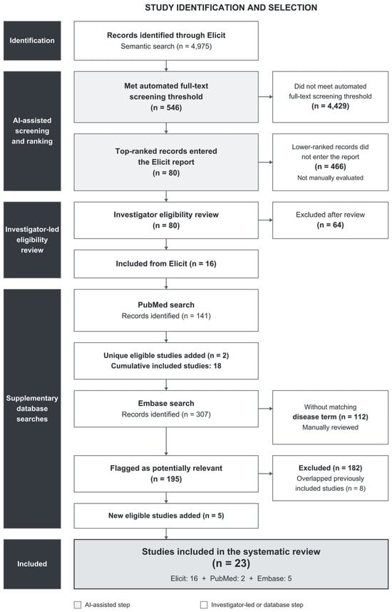
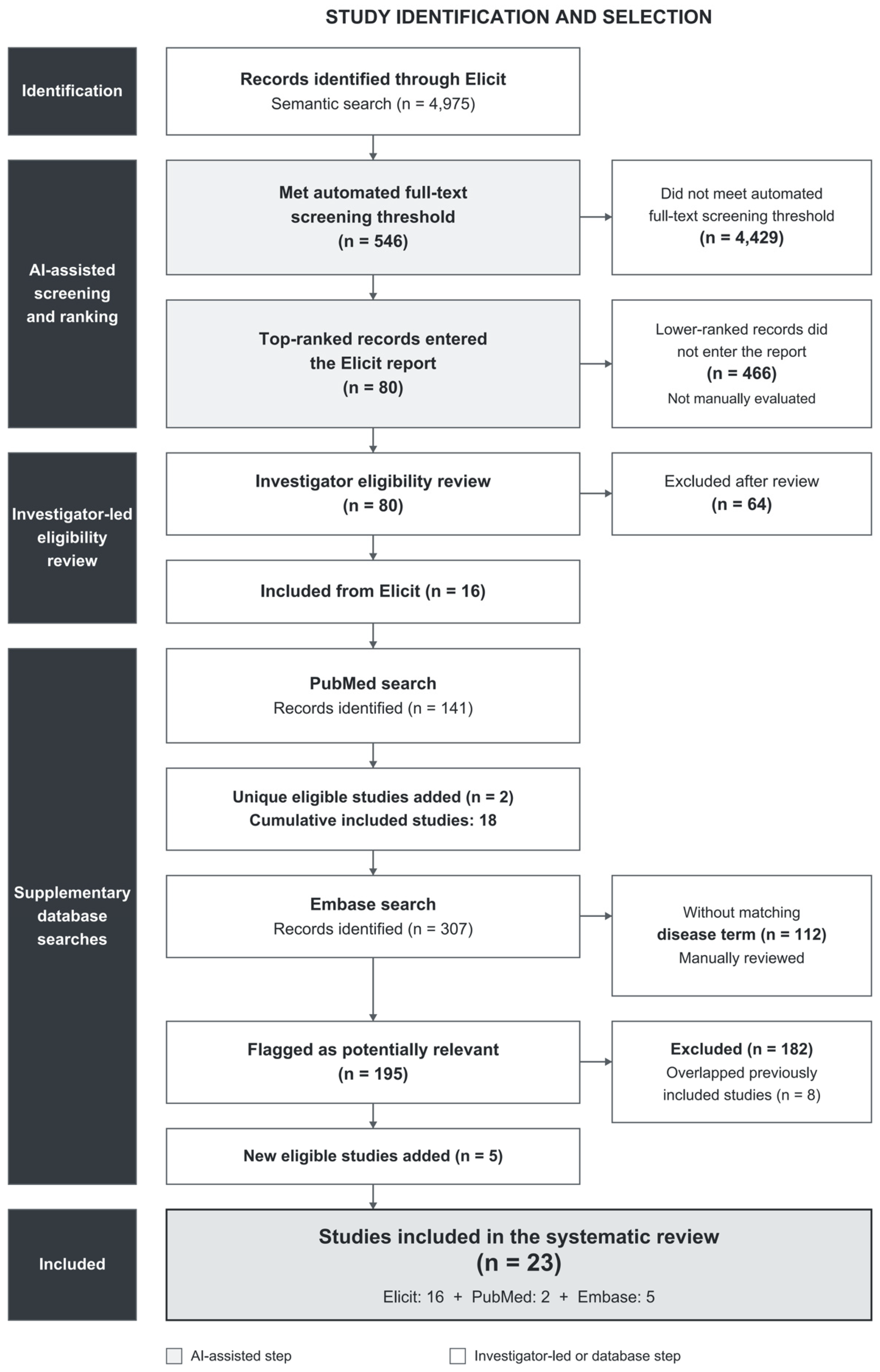

Disclaimer/Publisher’s Note: The statements, opinions and data contained in all publications are solely those of the individual author(s) and contributor(s) and not of MDPI and/or the editor(s). MDPI and/or the editor(s) disclaim responsibility for any injury to people or property resulting from any ideas, methods, instructions or products referred to in the content. |
© 2026 by the authors. Licensee MDPI, Basel, Switzerland. This article is an open access article distributed under the terms and conditions of the Creative Commons Attribution (CC BY) license.