


Abstract
Background: Semaglutide, a glucagon-like peptide-1 (GLP-1) receptor agonist, demonstrates significant anti-obesity and hypoglycemic effects in high-fat-diet (HFD)-induced obese models. However, both clinical and preclinical evidence indicates that semaglutide discontinuation inevitably leads to body weight rebound and progressive metabolic deterioration, severely compromising long-term therapeutic efficacy. Methods: To assess whether Lacticaseibacillus paracasei LP-18 alleviates post-semaglutide metabolic deterioration, we first confirmed its anti-obesity effect in HFD-fed mice, then used a three-stage model: HFD-induced obesity (4 weeks), semaglutide treatment (4 weeks), and withdrawal with or without L. paracasei LP-18 (4 weeks). Results: L. paracasei LP-18 supplementation effectively prevented post-discontinuation weight rebound and reduced cumulative energy intake while maintaining glucose homeostasis through decreased fasting and postprandial blood glucose levels. L. paracasei LP-18 also ameliorated dyslipidemia, reduced liver weight and hepatic lipid deposition, and attenuated white adipose tissue expansion and adipocyte hypertrophy. Mechanistically, L. paracasei LP-18 enriched beneficial taxa (Lacticaseibacillus, Bifidobacterium, Faecalibaculum) and depleted obesity-associated genera (Oribacterium, Anaerotruncus, Dorea), while restoring colonic barrier integrity via increased mucus layer thickness, goblet cell density, and upregulation of Muc2, ZO-1, and Occludin. Conclusions: These findings suggest that L. paracasei LP-18 is a promising probiotic strategy to alleviate post-semaglutide weight rebound and metabolic dysfunction, and its beneficial effects are associated with modulation of gut microbiota and preservation of intestinal barrier integrity.
1. Introduction
Obesity constitutes a global health crisis driven by intricate metabolic dysregulation [1]. It is frequently accompanied by chronic low-grade inflammation and markedly elevates the incidence risk of various secondary comorbidities, including type 2 diabetes mellitus, peripheral neuropathies, cardiovascular diseases, stroke, and multiple malignancies [2]. Global weight loss attempts are growing prevalent. With the advent of highly effective pharmacotherapies, GLP-1 (a glucagon-like peptide-1) receptor agonists and multi-receptor agonists are rapidly reshaping the landscape of obesity treatment. Nineteen agents such as tirzepatide, semaglutide, the cagrilintide–semaglutide combination (CagriSema), orforglipron and mazdutide are used for lifestyle intervention or treatment in patients with obesity, as demonstrated by 262 randomized controlled trials enrolling nearly 100,000 overweight or obese subjects [3]. GLP-1 receptor agonist, significantly reduces body weight, improves glucose tolerance, and ameliorates multiple metabolic parameters in high-fat-diet (HFD)-induced obese rodent models [4,5]. Although low-energy dietary interventions can achieve initial weight reductions of 5–15% [6], post-weight-loss weight regain remains a persistent clinical challenge [7] that severely compromises long-term metabolic benefits, highlighting the urgent need for effective maintenance strategies. Weight rebound stems from interconnected physiological adaptation mechanisms forming a self-sustaining vicious cycle. Initially, disproportionate reduction in resting energy expenditure (REE) triggers hypothalamic–pituitary–adrenal axis activation, remodeling appetite-regulating hormones—elevated ghrelin and reduced leptin stimulate hunger while reducing satiety, promoting increased energy intake [8,9,10]; more critically, these hormonal changes are epigenetically solidified long-term, forming “obesogenic memory” in adipose tissue through persistent macrophage infiltration [11], chronic low-grade inflammation [12], and stable epigenetic alterations [13] including DNA methylation changes [14] and histone modifications maintaining adipogenic potential [15]. These mechanisms exhibit temporal heterogeneity and individual variability—REE reduction and hormonal changes manifest rapidly post-weight loss (1–4 weeks), while epigenetic alterations gradually dominate medium-to-long term (>6 months) [16]. This multi-layered network explains limited efficacy of single-intervention strategies.
The therapeutic benefits of semaglutide are mediated through multiple mechanisms, including enhanced satiety, reduced food intake, and improved insulin sensitivity [17]. However, a critical limitation of GLP-1 receptor agonist therapy is the inevitable weight regain and metabolic deterioration observed upon drug discontinuation [18]. This post-treatment rebound phenomenon represents a significant clinical challenge that severely compromises the long-term sustainability of metabolic benefits, highlighting the urgent need for effective maintenance strategies. The mechanisms underlying metabolic relapse following GLP-1 receptor agonist discontinuation are multifactorial, involving intricate crosstalk among hormonal signaling, central nervous system regulation, and gut microbiota dynamics [19]. Recent studies have demonstrated that gut microbiota dysbiosis serves as a key driver for disrupted metabolic homeostasis after drug withdrawal [20]. GLP-1 analogues have demonstrated significant effects on the composition, richness, and diversity of gut microbiota [21]. The results following semaglutide treatment varied, with Akkermansia muciniphila, known for its positive metabolic functions, increasing [22]; however, microbial diversity decreased; and Odoribacter, Romboutsia, and Ligilactobacillus decreased [23].
Emerging evidence indicates that certain inactivated probiotics can effectively mitigate weight rebound following discontinuation of GLP-1 receptor agonist therapy. Meanwhile, novel therapeutic strategies for obesity prevention and treatment have also been proposed: prebiotics and probiotics are applied to modulate gut microbiota to recapitulate the gut microbial profile of healthy non-obese individuals [24]. Specifically, supplementation with pasteurized Akkermansia muciniphila MucT has demonstrated significant efficacy in inhibiting weight regain, with approximately 40% of supplemented subjects achieving spontaneous and sustained weight loss. This therapeutic effect appears to be more pronounced in individuals exhibiting low baseline gut Akkermansia abundance. The underlying mechanisms involve down-regulation of inflammatory pathways in adipose tissue, enhancement of oxidative metabolism, and increased fecal energy excretion, while maintaining a favorable safety profile [25,26]. These findings represent the first high-quality clinical proof-of-concept for postbiotic-based strategies aimed at weight-loss maintenance.
Lacticaseibacillus paracasei is widely used in fermented dairy products, cereals, and vegetables. It represents a novel probiotic strain with unique and specific functional properties. Previous studies have shown that L. paracasei 36 can restore gut microbiota homeostasis by increasing the abundance of beneficial genera (Ligilactobacillus, Akkermansia) and reducing the abundance of harmful genera (Alistipes, Parasutterella) [27]. L. paracasei K56 can inhibit intracellular lipid accumulation in adipocytes, reduce body weight gain and visceral fat deposition induced by high-fat diet, and improve insulin sensitivity. Furthermore, when combined with α-galacto-oligosaccharides, it further enhances the inhibitory effect against fat accumulation [28,29].
This study mimics real-world post-treatment weight rebound to evaluate whether supplementation with L. paracasei LP-18 can maintain metabolic homeostasis and inhibit weight regain after semaglutide discontinuation by modulating gut microbiota composition and preserving intestinal barrier function. A systematic investigation was performed based on this three-stage animal model, combining histological evaluation and microbiota analysis to elucidate the potential mechanisms underlying the metabolic protection exerted by L. paracasei LP-18.
2. Materials and Methods
2.1. Bacterial Cultivation and Preparation
L. paracasei LP-18 was initially isolated in our laboratory from fecal specimens of healthy human donors. The strain was identified by full-length 16S rRNA gene sequencing and further verified by whole-genome sequencing. It has been deposited at the Guangdong Microbial Culture Collection Center (GDMCC), Guangdong Institute of Microbiology, under accession number GDMCC No. 65551. For cultivation, L. paracasei LP-18 was grown in de Man–Rogosa–Sharpe (MRS) broth at 37 °C for 16 h under anaerobic conditions. To prepare bacterial suspensions for animal experiments, cells were collected by centrifugation, washed twice with sterile phosphate-buffered saline (PBS, pH 7.2), and resuspended in PBS supplemented with 25% (v/v) glycerol to a final concentration of 3 × 10¹⁰ CFU/mL. The suspensions were then stored at −80 °C until further use.
2.2. Animals and Treatment
2.2.1. Animals and Housing
Five- to six-week-old male C57BL/6J mice were procured from Guangdong Vital River Laboratory Animal Technology Co., Ltd. (Foshan, China) The animals were housed under specific pathogen-free conditions in a temperature-controlled room (20~26 °C) under a 12-h light/dark cycle (lights off at 18:00), with unrestricted access to food and water. All animal protocols were reviewed and approved by the Institutional Animal Care and Use Committee of Moon Biotech Co., Ltd. (Guangzhou, China) (approval No. IACUC202411-9O for experiment 1, approval date: 16 November 2024 and IACUC202409-2O for experiment 2, approval date: 12 September 2024) and were conducted in compliance with the Guide for the Care and Use of Laboratory Animals.
2.2.2. Experimental Design
Experiment 1: Anti-Obesity Effect of L. paracasei LP-18 in HFD-Fed Mice
After 1 week of acclimatization, male C57BL/6J mice were fed either a normal chow diet (NCD; D12450B, Research Diets) or a high-fat diet (HFD; D12492, Research Diets) for 4 weeks to induce obesity. The 4-week HFD feeding period was selected based on previous studies demonstrating that C57BL/6J mice develop significant obesity and metabolic dysfunction within 4 weeks on D12492 [30,31,32]. The HFD-fed mice weighting 30~35 g were then randomized by body weight into HFD (n = 5) and LP-18 (n = 5) groups, while NCD mice (n = 5) received normal chow throughout the study. Mice in the LP-18 group received L. paracasei LP-18 by oral gavage at a dose of 6 × 10⁹ CFU/animal once daily for 8 consecutive weeks. The NCD and HFD groups received an equal volume of vehicle buffer. The dose of L. paracasei LP-18 (6 × 10⁹ CFU/animal/day) was selected based on previous studies reporting anti-obesity effects of probiotic strains in HFD-fed mice at doses of 109 CFU/day [32,33,34]. Body weights were recorded weekly. Before the end of the 8-week intervention, an oral glucose tolerance test (OGTT) was performed after 12 h of fasting. Mice received a glucose solution (2 g/kg body weight) by oral gavage, and blood glucose concentrations were measured from the tail vein at 0, 15, 30, 60, 90, and 120 min using a portable glucometer. The area under the curve (AUC) was calculated using the trapezoidal rule.
Experiment 2: L. paracasei LP-18 Prevents Weight Regain After Semaglutide Discontinuation
Stage 1 (HFD-induced obesity phase). After 1 week of acclimatization, male C57BL/6J mice were fed either an NCD or an HFD for 10 weeks to induce obesity. Mice with a body weight of 38~43 g were selected and divided into HFD (n = 8) and HFD-Sema (n = 14) groups; NCD mice (n = 8) received normal chow throughout and served as lean controls.
Stage 2 (semaglutide-induced weight loss phase). The HFD and HFD-Sema groups continued on the high-fat diet. Mice in the HFD-Sema group received daily subcutaneous injections of semaglutide (CAS No. 910463-68-2, Aladdin) at 30 nmol/kg for 28 days (Day 1~28). The NCD and HFD groups received equal volumes of vehicle buffer by subcutaneous injection. On Day 28, the HFD-Sema mice were randomly assigned to two subgroups: Vehicle (n = 7) and LP-18 (n = 7). Body weight was recorded twice weekly, and 24-h food intake was recorded once weekly. Before the end of this stage, an OGTT was performed as described above.
Stage 3 (L. paracasei LP-18-mediated weight regain prevention phase). The HFD, Vehicle, and LP-18 groups continued on the high-fat diet. Mice in the LP-18 group received L. paracasei LP-18 daily by oral gavage (6 × 10⁹ CFU/animal/day, once daily) for 4 weeks. The NCD, HFD, and Vehicle groups received an equal volume of vehicle buffers. The 4-week semaglutide intervention was based on previous preclinical studies in HFD-fed mice [35]. The 4-week LP-18 maintenance period was designed to match the duration of semaglutide treatment, enabling direct comparison between the withdrawal and maintenance phases. Body weight was recorded twice weekly, and 24-h food intake was recorded once weekly. Before the end of this stage, an OGTT was performed as described above.
At the end of each experiment, mice were subjected to overnight fasting and then anesthetized with isoflurane. Orbital venous blood was collected under terminal anesthesia, after which the mice were immediately euthanized via cervical dislocation. This animal study was conducted under blind conditions for group allocation, experimental procedures, outcome assessment, and data analysis. Briefly, all experimental groups were marked only with alphanumeric codes, and researchers were kept unaware of the treatment assignments. All outcome assessments were performed by investigators blinded to group allocation. Serum and tissue samples were labelled using randomized codes with no information about experimental groups. Data analysis was also completed by investigators who were blinded to group assignments. The following serum biochemical parameters were determined by Wuhan Servicebio Technology Co., Ltd. (Wuhan, China) using an automated biochemical analyzer: total cholesterol (CHO), triglycerides (TG), low-density lipoprotein (LDL), high-density lipoprotein (HDL), alanine aminotransferase (ALT), aspartate aminotransferase (AST), blood urea nitrogen (BUN), and creatinine (CRE).
2.3. Histological Staining and Analysis
Liver and epididymal adipose tissue samples were fixed in 4% paraformaldehyde, dehydrated, embedded in paraffin, and sectioned. The sections were stained with hematoxylin and eosin (H&E) for morphological assessment. Hepatic lipid droplet area fraction (%) was quantified from H&E-stained liver sections, and mean adipocyte diameter (μm) was measured in epididymal adipose tissue sections. In addition, frozen liver sections were stained with Oil Red O to visualize neutral lipid accumulation, and the hepatic Oil Red O-positive area fraction (%) was quantified. Colon tissues were fixed and stained with Alcian blue to visualize acidic mucins. Mucus layer thickness (μm) and goblet cell density (cells/mm2) were measured on Alcian blue-stained colon sections. Images were acquired using a digital slide scanner (KFBIO, Yuyao, China, KF-FL-400) and analyzed with Halo 3.4.2986 (Indica Labs, Albuquerque, NM, USA). Histopathological assessment was carried out by Ningbo Yangming Medical Inspection Laboratory Co., Ltd. (Ningbo, China).
2.4. Quantitative Real-Time PCR
Total RNA was isolated from epididymal adipose tissue with the MiniBEST Universal RNA Extraction Kit (Takara,9767, Dalian, China) following the manufacturer’s protocol. For cDNA synthesis, 1 μg of total RNA was reverse-transcribed using the PrimeScript™ RT Reagent Kit with gDNA Eraser (Takara, RR047A). Quantitative real-time PCR was conducted on an Applied Biosystems QuantStudio 3 system with TB Green® Premix Ex Taq™ II (Takara, RR820A). Primers were synthesized by Sangon Biotech (Shanghai, China), and their sequences are listed in Supplementary Table S1. Relative gene expression was determined by the 2−ΔΔCt method, using B2m and Gapdh as internal controls for epididymal adipose tissue and colon tissue, respectively, with the NCD group as the calibrator.
2.5. 16S rRNA Sequencing and Analysis
Fecal genomic DNA was extracted using the CTAB method. The V3–V4 region of the bacterial 16S rRNA gene was amplified with primers 341F (CCTAYGGGRBGCASCAG) and 806R (GGACTACNNGGGTATCTAAT). Each 15-μL PCR reaction contained Phusion High-Fidelity PCR Master Mix, 0.2 μM each primer, and ~10 ng DNA template. Thermal cycling was: 98 °C for 1 min; 30 cycles of 98 °C for 10 s, 50 °C for 30 s, and 72 °C for 30 s; and 72 °C for 5 min. Amplicons were purified with magnetic beads, assessed on an Agilent 5400 Fragment Analyzer (Agilent Technologies, Santa Clara, CA, USA), pooled at equal ratios, and used for library construction. Libraries were quantified by Qubit fluorometry and qPCR, and sequenced on the DNBSEQ-G99 platform at Novogene (Beijing, China).
Paired-end reads were assigned to samples according to their unique barcodes and trimmed to remove barcode and primer sequences. Quality filtering of the raw reads was performed using fastp (v0.23.1) to obtain high-quality clean reads [36]. Subsequently, the clean reads were merged using FLASH (v1.2.11; http://ccb.jhu.edu/software/FLASH/ (accessed on 15 Augst 2026.)) [37]. Then, chimera removal and denoising were performed on the merged sequences using the DADA2 plugin in QIIME 2 to obtain initial amplicon sequence variants (ASVs) [38]. To investigate the phylogenetic relationships among ASVs and differences in dominant species among samples or groups, multiple sequence alignment was performed using QIIME 2. ASV abundance was normalized by rarefying to the minimum sequencing depth (i.e., the sample with the fewest sequences). Taxonomic annotation of ASV representative sequences was performed using QIIME 2 with the Greengenes2 database (2024.09), and taxonomic information for each ASV from phylum to species level was obtained [39]. Based on the normalized feature table and taxonomic annotation results, the relative abundance of each sample was calculated at six taxonomic levels (phylum, class, order, family, genus, and species), and an abundance profile (relative abundance table) was generated.
To analyze the diversity, richness, and evenness of communities within samples, alpha diversity was calculated using seven indices in QIIME 2, including Observed ASVs, Chao1, Shannon, Faith_pd, and Pielou’s evenness. To evaluate the complexity of community composition and compare differences among samples or groups, beta diversity was calculated based on Jaccard distance in QIIME 2. Principal coordinate analysis (PCoA) based on Jaccard distance was performed to visualize differences in microbial community structure among samples, with results displayed using the ade4 and ggplot2 packages in R (v4.0.3). Differential taxa in fecal microbiota were identified using LEfSe, which applies linear discriminant analysis to estimate the effect size of each differentially abundant feature, with an LDA score threshold of 2 and a significance level of p < 0.05.
2.6. Statistical Analysis
Statistical analyses and graph generation were performed using GraphPad Prism (version 10.2.3). Data are presented as mean ± standard deviation (SD). Normality was evaluated using the Shapiro–Wilk test for sample sizes below five and the Kolmogorov–Smirnov test for samples of five or more. Homogeneity of variances was examined using the Brown–Forsythe test. For two-group comparisons, a two-tailed unpaired t-test was applied when data met normality and equal variance assumptions; otherwise, the Mann–Whitney U test was used. For multiple-group comparisons, one-way ANOVA followed by Dunnett’s post hoc test (with the HFD group as control) was used for parametric data with equal variances. Welch’s ANOVA followed by Dunnett’s T3 post hoc test was applied to parametric data with unequal variances. Repeated measures data (e.g., body weight and blood glucose during OGTT) were analyzed by two-way repeated measures ANOVA followed by Dunnett’s multiple comparisons test. For all two-way ANOVA analyses, the main effect of each factor and the factor-by-factor interaction effect were explicitly reported. Post hoc multiple comparisons were performed only when the interaction effect was significant (p < 0.05); when the interaction was not significant, main effects were interpreted without post hoc comparisons, following standard conventions for factorial design [40,41]. Non-parametric data were analyzed using the Kruskal–Wallis test followed by Dunnett’s multiple comparison test. A p-value < 0.05 was considered statistically significant.
3. Results
3.1. L. paracasei LP-18 Supplementation Attenuates Body Weight Regain and Reduces Cumulative Energy Intake After Semaglutide Withdrawal
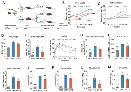
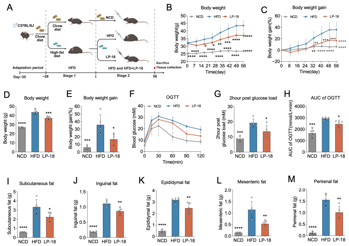
To evaluate the anti-obesity effect of L. paracasei LP-18, mice were fed an HFD for 4 weeks to induce obesity and then randomized into HFD and LP-18 groups, with NCD mice as controls. Body weight was recorded weekly during the 8-week intervention (Figure 1A). Body weight was significantly lower in the LP-18 group than in the HFD group during the maintenance phase, with a significant time-by-treatment interaction (p < 0.0001, Figure 1B). Similarly, body weight gain was significantly attenuated in the LP-18 group compared with the HFD group, again with a significant time-by-treatment interaction (p < 0.0001, Figure 1C). HFD-fed mice showed a progressive increase in body weight compared with NCD mice (Figure 1B,C). L. paracasei LP-18 supplementation significantly attenuated body weight gain (Figure 1B,C). At the end of the intervention, the average body weight of the LP-18 group is significantly lower than that of the HFD group (p < 0.001; Figure 1D). Consistently, the percentage of body weight gain was significantly reduced in the LP-18 group compared with the HFD group (p < 0.05; Figure 1E). An OGTT was performed before the end of the intervention. For blood glucose during the OGTT, L. paracasei LP-18 treatment significantly lowered overall glucose levels across the time course, as indicated by a significant main effect of treatment, without a significant time-by-treatment interaction (Figure 1F). The significant main effect of treatment indicates that L. paracasei LP-18 lowered overall blood glucose levels across the OGTT time course. Consistently, the 2-h post-load blood glucose level was significantly decreased in the LP-18 group compared with the HFD group (p < 0.05; Figure 1G). Accordingly, the AUC of OGTT was significantly reduced in the LP-18 group relative to the HFD group (p < 0.05; Figure 1H). These results indicate that L. paracasei LP-18 improves glucose tolerance in HFD-fed obese mice. At the end of the experiment, L. paracasei LP-18 supplementation significantly reduced the weights of subcutaneous, inguinal, epididymal, mesenteric, and perirenal fat compared with the HFD group (all p < 0.05; Figure 1I–M). These results established that L. paracasei LP-18 possesses independent metabolic regulatory capacity, prompting us to investigate whether L. paracasei LP-18 could serve as a maintenance strategy to prevent metabolic relapse after the discontinuation of a potent anti-obesity pharmacotherapy.
Figure 1.
L. paracasei LP-18 supplementation attenuates body weight gain, improves glucose tolerance, and reduces adiposity in HFD-induced obese mice. (A) Schematic representation of the experimental design and grouping. Mice were randomly assigned to three groups: normal chow diet (NCD, n = 5), high-fat diet (HFD, n = 5), and HFD plus LP-18 (LP-18, n = 5). L. paracasei LP-18 was administered daily by oral gavage concurrently with the initiation of HFD feeding for 8 weeks (from Day 1 to Day 56). (B) Body weight throughout the experimental period. (C) Body weight gain over time. (D) Body weight on Day 56. (E) Body weight gain on Day 56. (F) Blood glucose excursion curves during the OGTT. (G) 2-h post-load blood glucose concentrations. (H) Area under the curve (AUC) of OGTT. (I) Subcutaneous fat weight. (J) Inguinal fat weight. (K) Epididymal fat weight. (L) Mesenteric fat weight. (M) Perirenal fat weight. Data are presented as mean ± SD. For (B), two-way ANOVA revealed significant main effects of time (F = 22.89, p < 0.0001) and treatment (F = 311.9, p < 0.0001), as well as a significant time-by-treatment interaction (F = 4.505, p < 0.0001); post hoc comparisons were performed using Dunnett’s multiple comparisons test versus the HFD group. For (C), two-way ANOVA revealed significant main effects of time (F = 21.48, p < 0.0001) and treatment (F = 45.76, p < 0.0001), as well as a significant time-by-treatment interaction (F = 3.717, p < 0.0001); post hoc comparisons were performed using Dunnett’s multiple comparisons test versus the HFD group. For (F), two-way ANOVA revealed significant main effects of time (F = 55.56, p < 0.0001) and treatment (F = 50.39, p < 0.0001) but no significant time-by-treatment interaction (F = 1.795, p = 0.0769); therefore, no post hoc multiple comparisons were performed. For (D,E,G–M), one-way ANOVA followed by Dunnett’s multiple comparisons test was used versus the HFD group. * p < 0.05, ** p < 0.01, *** p < 0.001, **** p < 0.0001 vs. HFD group.
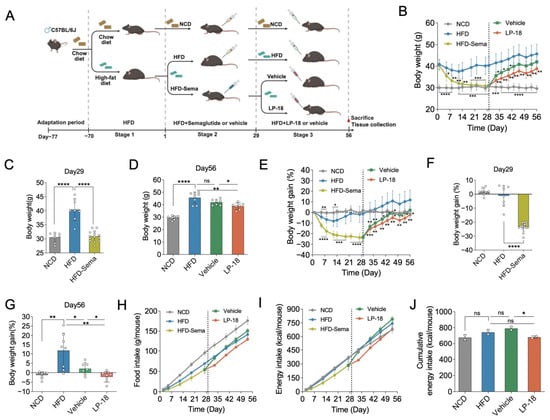
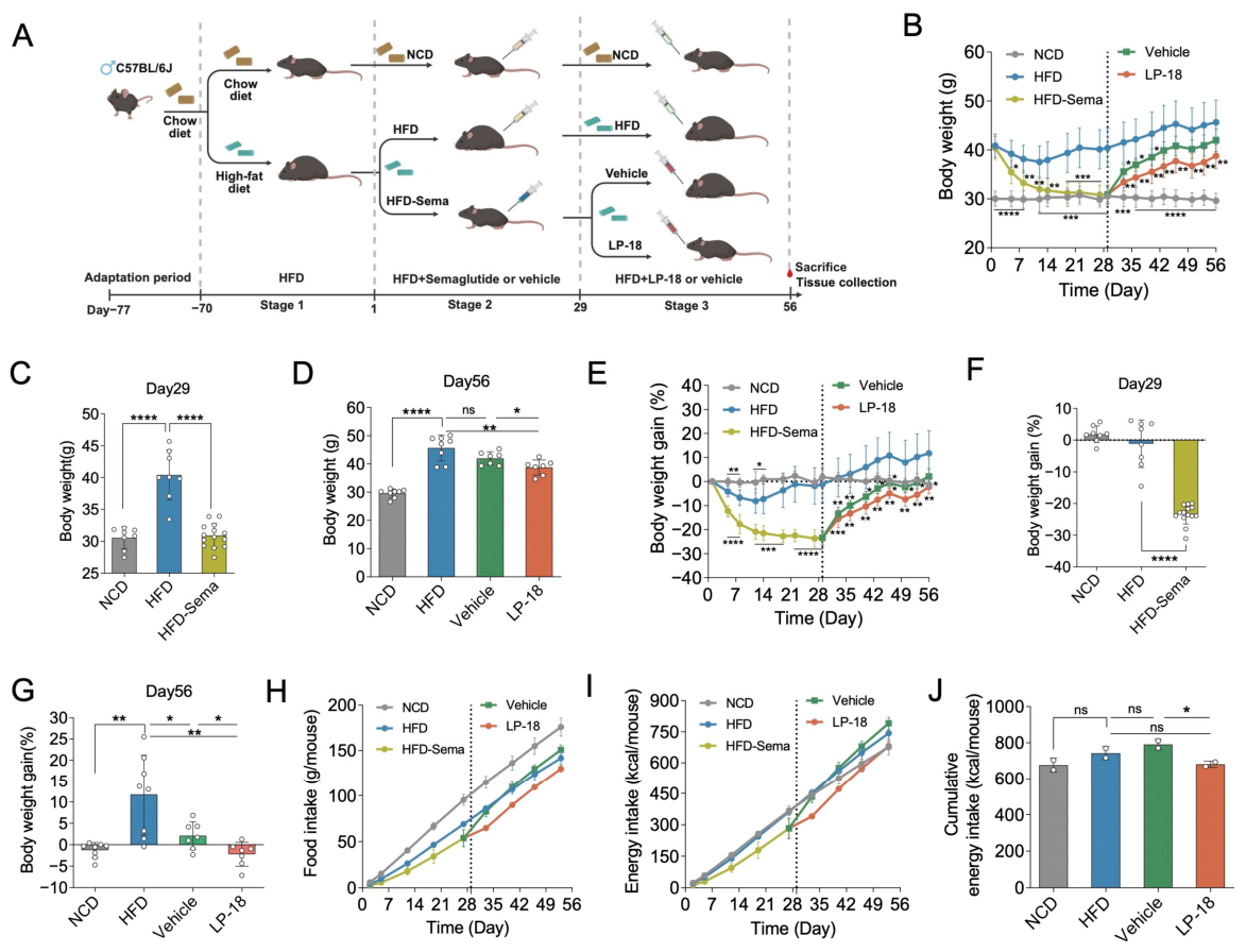
To address this question, we employed a three-stage experimental design that recapitulates the clinical trajectory of GLP-1 receptor agonist therapy: HFD-induced obesity establishment (4 weeks) → semaglutide intervention (4 weeks) → drug withdrawal with or without L. paracasei LP-18 maintenance (4 weeks) (Figure 2A). This staged approach allowed us to distinguish the effects ofLP-18 as a maintenance strategy from its direct therapeutic actions, and to specifically assess its capacity to counteract post-withdrawal metabolic relapse—a phenomenon that closely mimics the clinical reality faced by patients discontinuing GLP-1 receptor agonists. Following the initial 4-week HFD feeding, mice developed significant obesity with markedly elevated body weight compared to NCD controls. The subsequent 4-week semaglutide treatment effectively reduced body weight in HFD-fed mice, resulting in significantly lower absolute body weight and body weight gain relative to the HFD group (p < 0.0001; Figure 2B,C,E,F), confirming the potent anti-obesity efficacy of semaglutide. Upon drug cessation, however, vehicle-treated mice exhibited a rapid and progressive weight rebound. Following semaglutide discontinuation, L. paracasei LP-18 supplementation substantially blunted post-drug body-weight regain. Two-way ANOVA performed on body-weight data across the maintenance phase (Day 29–56) identified significant main effects of time and treatment, together with a significant time-by-treatment interaction (all p < 0.0001). Post-hoc testing demonstrated that body weight remained significantly lower in the LP-18 group relative to the HFD group throughout the maintenance period (Figure 2B,D). Starting from Day 43, body weight in the Vehicle group was indistinguishable from the HFD group (ns versus HFD; Figure 2B,D). At Day 56, the LP-18 group exhibited significantly lower body weight compared with Vehicle-treated controls (p < 0.05; Figure 2D). Consistent with these findings, analysis of cumulative body-weight gain also returned significant main effects of time and treatment, as well as a significant time-by-treatment interaction (all p < 0.0001). Post-hoc comparisons confirmed reduced body-weight gain in L. paracasei LP-18-treated animals versus the HFD group over the maintenance phase (Figure 2E,G). No significant difference in body-weight gain was observed between Vehicle and HFD animals at Day 56 (ns versus HFD; Figure 2E,G). Accordingly, L. paracasei LP-18 treatment led to markedly lower cumulative weight gain relative to Vehicle controls at Day 56 (p < 0.05; Figure 2G). Dynamic body weight gain curves further confirmed that L. paracasei LP-18 slowed the regrowth trajectory throughout the entire 4-week maintenance period (Figure 1E).
Figure 2.
L. paracasei LP-18 supplementation attenuates weight regain and modulates energy intake after semaglutide discontinuation in HFD-induced obese mice. (A) Schematic representation of the experimental design and grouping. Mice were fed a normal chow diet (NCD, n = 8) or a high-fat diet (HFD, n = 8). HFD-fed mice received semaglutide for 4 weeks (HFD-Sema, n = 14). After drug withdrawal, the HFD-Sema mice were randomly assigned to receive either vehicle (Vehicle, n = 7) or LP-18 (LP-18, n = 7) by oral gavage for 4 weeks. (B) Body weight throughout the experimental period. (C) Body weight on Day 29. (D) Body weight on Day 56. (E) Body weight gain over time. (F) Body weight gain on Day 29. (G) Body weight gain on Day 56. (H) Daily food intake during the intervention period. (I) Daily energy intake. (J) Cumulative energy intake over the intervention. Data are presented as mean ± SD. For (B), two-way ANOVA revealed a significant time × treatment interaction (F = 23.80, p < 0.0001), a significant main effect of time (F = 152.7, p < 0.0001), and a significant main effect of treatment (F = 33.95, p < 0.0001); post hoc comparisons were performed using Dunnett’s multiple comparisons test versus the HFD group. For (E), two-way ANOVA revealed a significant time × treatment interaction (F = 22.32, p < 0.0001), a significant main effect of time (F = 135.4, p < 0.0001), and a significant main effect of treatment (F = 14.22, p < 0.0001); post hoc comparisons were performed using Dunnett’s multiple comparisons test versus the HFD group. For (C,D,F,G,J), statistical significance was assessed using two-tailed unpaired Student’s t-test compared with the corresponding control group (HFD for panels (C,F); HFD or Vehicle for panels (D,G,J)). * p < 0.05, ** p < 0.01, *** p < 0.001, and **** p < 0.0001 vs. HFD or Vehicle group.
To explore whether reduced food intake contributed to this anti-regain effect, we monitored daily food consumption and calculated cumulative energy intake. Longitudinal food intake curves showed comparable trends across groups (Figure 2H,I), but cumulative energy intake by day 56 was significantly lower in L. paracasei LP-18-treated mice than in Vehicle controls (p < 0.05; Figure 2J), suggesting that L. paracasei LP-18 partially counteracts weight rebound through energy intake modulation.
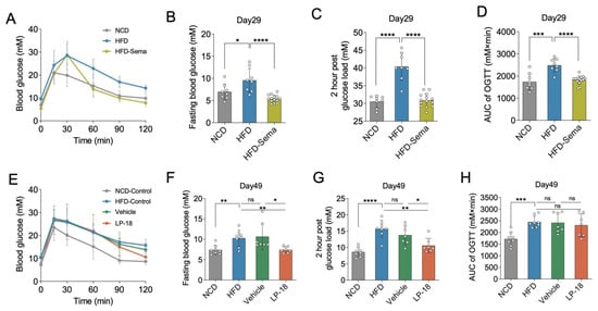
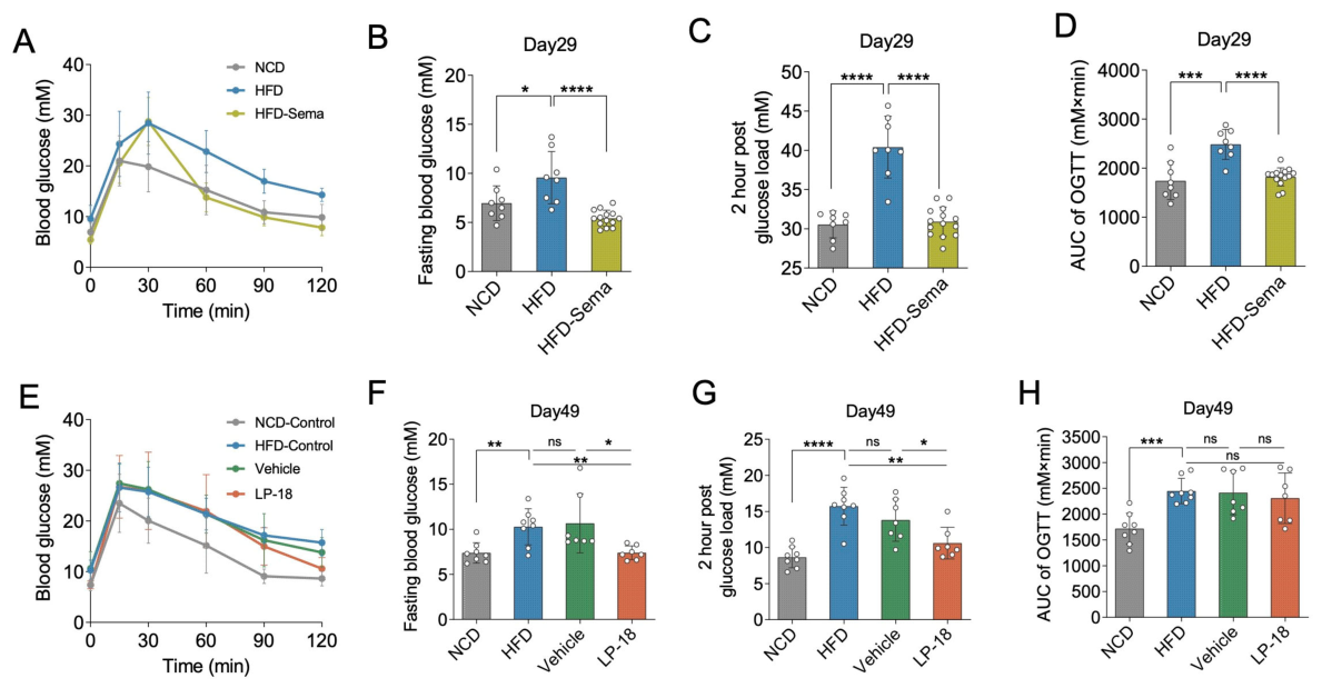
We next assessed whether L. paracasei LP-18 preserved glycemic homeostasis after semaglutide withdrawal. Oral glucose tolerance tests (OGTT) were performed at two critical time points: immediately before drug withdrawal (day 29) and after 3 weeks of maintenance intervention (day 49). On day 29, HFD-fed mice exhibited overt glucose intolerance characterized by elevated fasting glucose (p < 0.05 vs. NCD; Figure 3B), higher 2-h post-load glucose (p < 0.0001 vs. NCD; Figure 3C), and increased OGTT-derived area under the curve (AUC; p < 0.0001 vs. NCD; Figure 3D). Semaglutide treatment robustly corrected all these abnormalities (p < 0.0001 vs. HFD for fasting glucose, 2-h glucose, and AUC; Figure 3A–D), confirming its potent hypoglycemic efficacy. On day 49, after 3 weeks of drug withdrawal, vehicle-treated mice showed a relapse of glycemic dysregulation, with fasting glucose and 2-h post-load glucose remaining elevated at levels comparable to HFD controls (ns vs. HFD; Figure 3E–G). L. paracasei LP-18 supplementation, however, significantly lowered both fasting blood glucose (p < 0.05 vs. Vehicle; Figure 2F) and postprandial glucose excursion (p < 0.05 vs. Vehicle; Figure 2G). Although the OGTT-AUC in the LP-18 group exhibited a declining trend, the difference did not reach statistical significance (ns vs. Vehicle; Figure 2H). Collectively, these data demonstrate that L. paracasei LP-18 acts as a maintenance intervention that partially preserves semaglutide’s glycemic benefits—specifically by controlling fasting and postprandial hyperglycemia—rather than serving as a substitute for the potent and comprehensive glucose-lowering effects of the drug itself.
Figure 3.
L. paracasei LP-18 supplementation improves glycemic control after semaglutide discontinuation in HFD-induced obese mice. (A) Blood glucose excursion curves during the OGTT on Day 29. (B) Fasting blood glucose levels on Day 29. (C) 2-h post-load blood glucose concentrations on Day 29. (D) Area under the curve (AUC) of the Day-29 OGTT. (E) Blood glucose excursion curves during the OGTT on Day 49. (F) Fasting blood glucose levels on Day 49. (G) 2-h post-load blood glucose concentrations on Day 49. (H) Area under the curve (AUC) of the Day-49 OGTT. Data are presented as mean ± SD. Statistical significance was assessed using two-tailed unpaired Student’s t-test compared with the corresponding control group (HFD for panels (B–D); HFD or Vehicle for panels (F–H)). * p < 0.05, ** p < 0.01, *** p < 0.001, and **** p < 0.0001 vs. HFD or Vehicle group.
3.2. L. paracasei LP-18 Alleviates Obesity-Associated Dyslipidemia, Hepatic Steatosis, and White Adipose Tissue Expansion
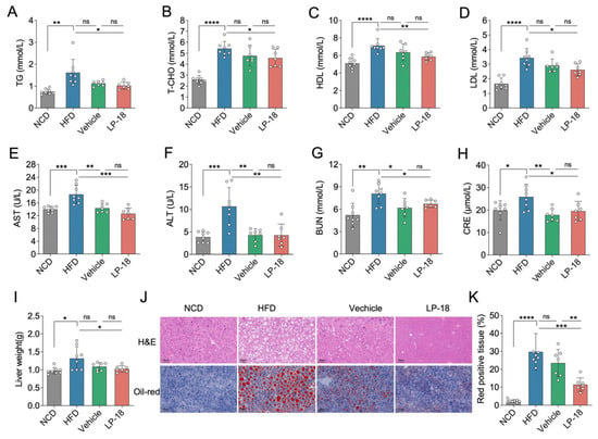
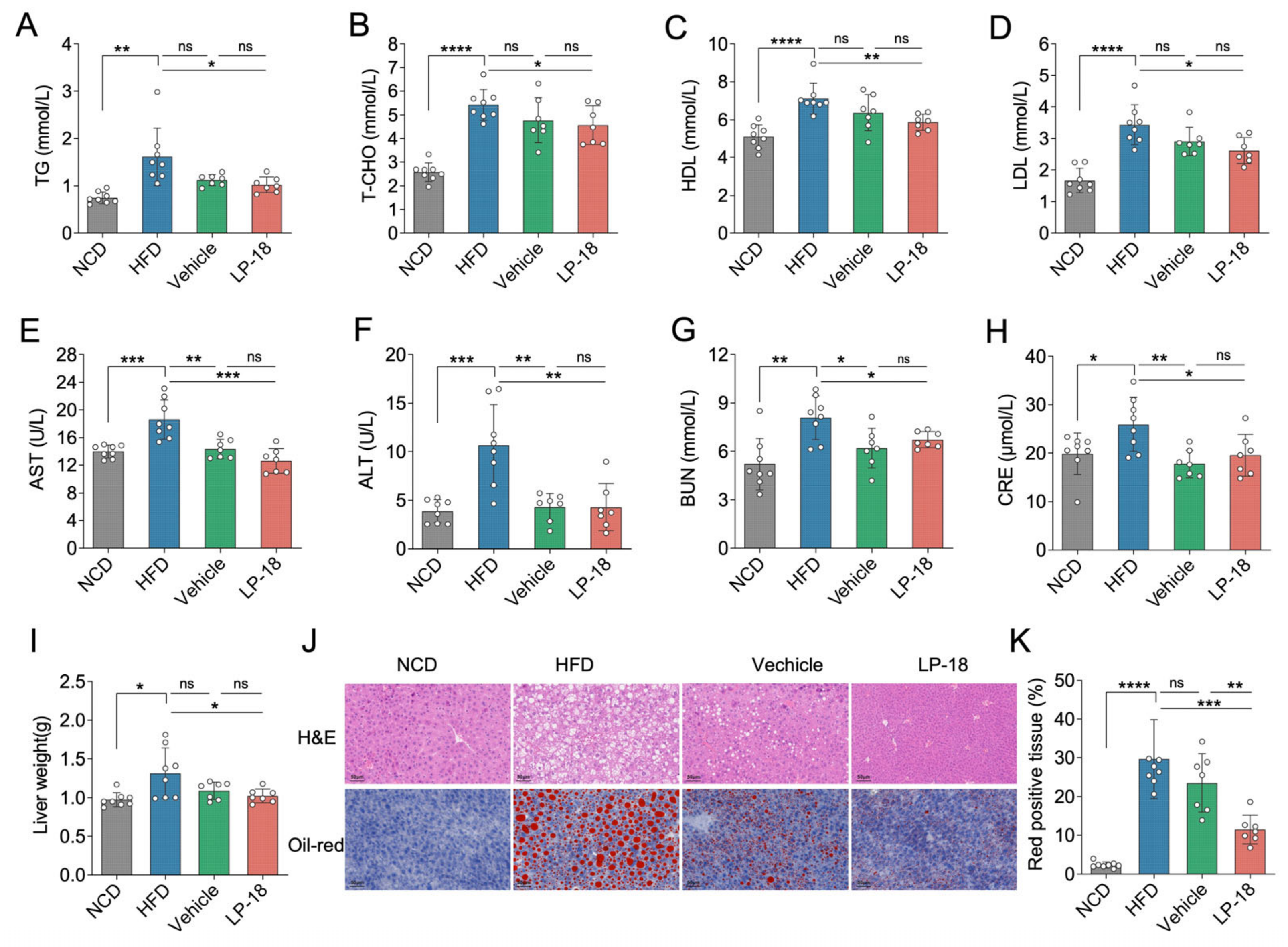
To evaluate the systemic metabolic protection conferred by L. paracasei LP-18 after semaglutide discontinuation, we assessed serum biochemical parameters and hepatic/adipose tissue morphology. HFD feeding markedly elevated serum TG, T-CHO, HDL, and LDL levels compared with NCD controls. After semaglutide withdrawal, vehicle-treated mice exhibited dyslipidemia, with all four parameters remaining elevated at levels comparable to HFD controls (ns vs. HFD; Figure 4A–D). L. paracasei LP-18 supplementation, however, significantly reduced serum TG, T-CHO, HDL, and LDL levels compared with the HFD group (p < 0.05 vs. Vehicle; Figure 4A–D). These results suggest that L. paracasei LP-18 ameliorates HFD-induced dyslipidemia after semaglutide discontinuation. HFD feeding markedly elevated serum AST, ALT, BUN, and CRE levels compared with NCD controls. After semaglutide withdrawal, both Vehicle and LP-18 groups showed significant reductions in all four parameters compared with HFD-fed mice, with no significant differences between Vehicle and LP-18 groups (Figure 4E–H), indicating that semaglutide withdrawal restored hepatic and renal function markers to normal level, and L. paracasei LP-18 helped sustain the recovery after semaglutide withdrawal.
Figure 4.
L. paracasei LP-18 supplementation ameliorates dyslipidemia, hepatorenal function, and hepatic lipid accumulation after semaglutide withdrawal in HFD-fed mice. (A) Serum triglyceride (TG) levels. (B) Serum total cholesterol (T-CHO) levels. (C) Serum high-density lipoprotein cholesterol (HDL) levels. (D) Serum low-density lipoprotein cholesterol (LDL) levels. (E) Serum aspartate aminotransferase (AST) levels. (F) Serum alanine aminotransferase (ALT) levels. (G) Serum blood urea nitrogen (BUN) levels. (H) Serum creatinine (CRE) levels. (I) Liver weight. (J) Representative H&E and Oil Red O staining images of liver tissue (scale bar: 50 μm). (K) Quantification of hepatic Oil Red O-positive area fraction (%). Data are presented as mean ± SD. Statistical significance was assessed using two-tailed unpaired Student’s t-test compared with the HFD or Vehicle group. * p < 0.05, ** p < 0.01, *** p < 0.001, and **** p < 0.0001 vs. HFD or Vehicle group.
HFD feeding induced significant hepatomegaly compared with NCD controls. After semaglutide withdrawal, liver weight in the Vehicle group remained elevated at levels comparable to HFD controls (ns vs. HFD; Figure 4I). L. paracasei LP-18 supplementation, however, significantly reduced absolute liver weight compared with the Vehicle group (p < 0.05; Figure 4I). To further confirm hepatic lipid accumulation, H&E and Oil Red O staining was performed. HFD feeding markedly increased the Oil Red O-positive area compared with NCD controls, and this elevation persisted in vehicle-treated mice after semaglutide withdrawal (ns vs. HFD; Figure 4J,K). In contrast, L. paracasei LP-18 supplementation significantly reduced the Oil Red O-positive area compared with both HFD and Vehicle groups (p < 0.05; Figure 4J,K), consistent with the liver weight and further supporting the protective effects of L. paracasei LP-18 against hepatic steatosis.
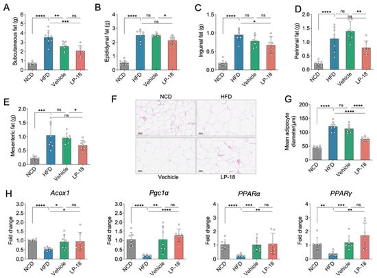
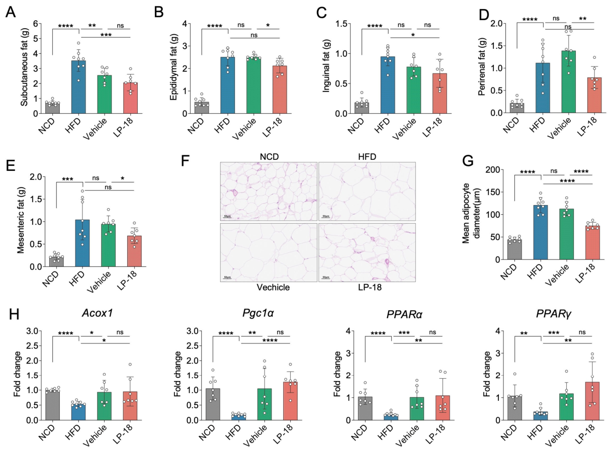
In obesity, adipose tissue dysfunction is typically characterized by excessive expansion of white adipose tissue (WAT) and adipocyte hypertrophy. HFD feeding significantly increased the weights of subcutaneous, epididymal, inguinal, perirenal, and mesenteric adipose depots compared with NCD controls (all p < 0.001). After semaglutide withdrawal, vehicle-treated mice maintained elevated WAT depot weights comparable to HFD-fed mice (ns vs. HFD), including epididymal, inguinal, perirenal, and mesenteric depots. In contrast, L. paracasei LP-18 supplementation maintained WAT mass at significantly lower levels than the HFD group (p < 0.001 for subcutaneous fat, p < 0.05 for inguinal fat; Figure 5A,C), or than the Vehicle group (p < 0.05 for epididymal fat and mesenteric fat; p < 0.01 for perirenal fat, Figure 5B,D,E), suggesting that L. paracasei LP-18 sustains semaglutide-induced improvements in adipose mass.
Figure 5.
L. paracasei LP-18 supplementation reduces adiposity and adipose lipid accumulation after semaglutide discontinuation in HFD-induced obese mice. (A) Subcutaneous fat weight. (B) Epididymal fat weight. (C) Inguinal fat weight. (D) Perirenal fat weight. (E) Mesenteric fat weight. (F) Representative (H&E) staining images of epididymal adipose tissue (scale bar: 50 μm). (G) Quantification of mean adipocyte diameter in epididymal adipose tissue (μm). (H) Relative mRNA expression levels of ACOX1, Pgc1α, PPARα, and PPARγ in epididymal adipose tissue. Data are presented as mean ± SD. Statistical significance was assessed using two-tailed unpaired Student’s t-test compared with the HFD or Vehicle group. * p < 0.05, ** p < 0.01, *** p < 0.001, and **** p < 0.0001 vs. HFD or Vehicle group.
H&E staining further revealed that HFD feeding markedly increased mean adipocyte diameter (p < 0.0001 vs. NCD; Figure 5F,G). This hypertrophy persisted in Vehicle mice (ns vs. HFD) but was significantly attenuated by L. paracasei LP-18 compared with Vehicle, and also versus HFD group (p < 0.0001; Figure 4F,G). To gain molecular insights into adipose tissue remodeling, we quantified the expression of genes involved in lipid metabolism and energy homeostasis, including Acox1, Pgc1a, PPARα, and PPARγ. HFD feeding significantly downregulated the expression of all four genes relative to the NCD group (all p < 0.01; Figure 5H). Following semaglutide withdrawal, these genes remained significantly up-regulated in Vehicle mice compared with HFD-fed mice (all p < 0.05; Figure 5H), indicating that semaglutide-activated oxidative pathways were partially sustained after drug cessation. Supplementation with L. paracasei LP-18 induced upward trends in the expression of partial genes relative to the Vehicle group, yet these changes did not reach statistical significance (ns vs. Vehicle for all genes; Figure 5H). Collectively, these results suggest that L. paracasei LP-18 preserves the semaglutide-mediated restoration of adipose oxidative gene programs.
3.3. L. paracasei LP-18 Reshapes Gut Microbiota Composition by Enriching Beneficial Taxa While Depleting Obesity-Associated Genera
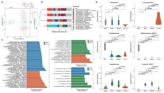
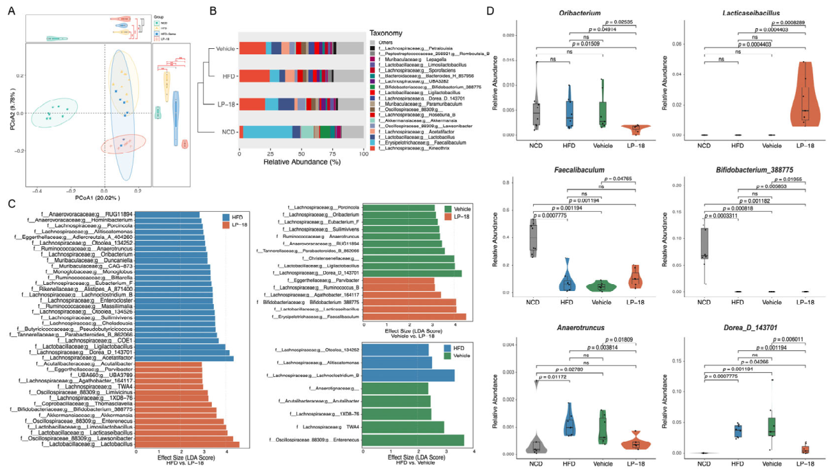
Given the pivotal role of gut microbiota in regulating host energy metabolism, we performed 16S rRNA gene sequencing on fecal samples to assess whether L. paracasei LP-18 modulated the intestinal microbial community. We first evaluated α-diversity using four complementary metrics. HFD feeding resulted in higher Shannon index and Pielou’s evenness compared with NCD, and these elevations persisted in vehicle-treated mice after drug withdrawal (Supplementary Figure S1A–D). No significant differences in any α-diversity metric were detected between the Vehicle and LP-18 groups (ns for all indices), suggesting that L. paracasei LP-18 did not broadly alter overall community richness or evenness. We next assessed β-diversity via PCoA (Figure 6A). A clear separation based on Jaccard distance was observed between the NCD and HFD groups, confirming the profound impact of long-term HFD on gut microbial ecology. After semaglutide withdrawal, the microbial profile of vehicle-treated mice remained clustered with the HFD group, indicating persistence of the obesogenic-associated community structure. Notably, L. paracasei LP-18 significantly shifted the community structure away from the HFD/Vehicle cluster and partially toward the NCD profile, demonstrating that L. paracasei LP-18 effectively remodels overall gut microbial composition, likely through selective enrichment and depletion of specific taxa.
Figure 6.
L. paracasei LP-18 supplementation remodels gut-microbiota composition in HFD-induced obese mice. (A) Principal coordinate analysis (PCoA) plots based on Jaccard distance showing β-diversity of the gut microbial community among the NCD, HFD, Vehicle, and LP-18 groups (n = 7–8 per group). (B) Bar plots showing the relative abundance of gut microbiota at the genus level (top 20 genera) across the four groups. (C) Lefse analysis identifying differentially abundant taxa between groups (LDA score > 2, p < 0.05). Only taxa with significant differences among HFD, Vehicle, and LP-18 groups are shown. (D) Relative abundances of Lacticaseibacillus, Faecalibaculum, Bifidobacterium, Agathobacter, Oribacterium, Ligilactobacillus, Anaerotruncus, and Dorea across the NCD, HFD, Vehicle, and LP-18 groups.
Genus-level analysis further illustrates the microbial reconfiguration induced by L. paracasei LP-18 (Figure 6B). Linear discriminant effect size (LEfSe) analysis identified multiple genera as L. paracasei LP-18-responsive differential taxa (LDA score > 2, p < 0.05). Quantitative comparison revealed that L. paracasei LP-18 significantly enriched beneficial commensals, including Lacticaseibacillus (p < 0.001), Bifidobacterium (p < 0.001), and Faecalibaculum (p < 0.01), relative to both the HFD and Vehicle groups (Figure 6C). Notably, Lacticaseibacillus is the genus to which L. paracasei LP-18 belongs; Lacticaseibacillus, Bifidobacterium, and Faecalibaculum have been associated with anti-obesity effects and improved metabolic parameters [40,41,42]. These results indicate that L. paracasei LP-18 shifts the gut microbiota toward a composition linked to improved metabolic health. Concurrently, HFD feeding markedly expanded the relative abundances of Oribacterium [43], Anaerotruncus [44] and Dorea [45]-taxa previously associated with obesity and metabolic dysfunction-which remained highly enriched in vehicle-treated mice after semaglutide withdrawal. L. paracasei LP-18 significantly suppressed these taxa (p < 0.05 vs. Vehicle; Figure 6D). Collectively, these data demonstrate that L. paracasei LP-18 selectively enriches beneficial commensals and depletes obesity-related pathobionts, reconfiguring the gut microbiota toward a more metabolically favorable composition without disrupting overall microbial diversity.
3.4. L. paracasei LP-18 Preserves Colonic Barrier Integrity by Restoring Mucus Layer and Upregulating Tight Junction Genes
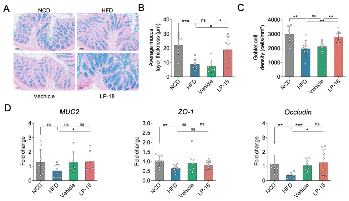
Intestinal barrier dysfunction is a critical link between gut microbiota dysbiosis and systemic metabolic inflammation. Given the profound microbial remodeling induced by L. paracasei LP-18, we next examined whether L. paracasei LP-18 preserved colonic barrier integrity after semaglutide discontinuation. Alcian blue histological staining of distal colon sections revealed that HFD feeding caused severe erosion of the colonic mucus layer, with markedly reduced average mucus thickness compared with NCD controls (p < 0.001; Figure 7A). Vehicle-treated mice after semaglutide withdrawal maintained a thin and disrupted mucus layer, with no significant recovery relative to the HFD group (ns). In striking contrast, L. paracasei LP-18 supplementation significantly restored mucus layer thickness (p < 0.05 vs. Vehicle; Figure 7B), indicating effective repair of the physical mucus barrier. Consistent with this restoration, goblet cell density-the primary source of mucin production-was significantly decreased in HFD mice compared with NCD controls (p < 0.01; Figure 7C) and remained depressed in vehicle-treated mice (ns vs. HFD). L. paracasei LP-18 treatment markedly elevated goblet cell density (p < 0.01 vs. Vehicle; Figure 7C), suggesting that L. paracasei LP-18 promotes the differentiation or survival of mucus-secreting cells.
Figure 7.
L. paracasei LP-18 supplementation preserves colonic mucus barrier and tight junction integrity after semaglutide discontinuation in HFD-induced obese mice. (A) Representative Alcian blue staining photographs of colon tissue (scale bar: 50 μm). (B) Average mucus layer thickness (μm). (C) Goblet cell density (cells/mm2). (D) Relative mRNA expression levels of tight junction (ZO-1 and Occludin) and mucin genes (MUC2) in colon tissue. Data are presented as mean ± SD. Statistical significance was assessed using two-tailed unpaired Student’s t-test compared with the HFD or Vehicle group. * p < 0.05, ** p < 0.01, and *** p < 0.001 vs. HFD or Vehicle group.
We further quantified the transcript levels of colonic mucin and tight junction-related genes by quantitative real-time PCR. HFD feeding significantly downregulated the expression of the core mucin gene MUC2, as well as tight junction components ZO-1 (zonula occludens-1), and occludin (Figure 7). After semaglutide discontinuation, this transcriptional suppression persisted in vehicle-treated mice, with expression levels of MUC2 and ZO-1 comparable to those in the HFD group (ns). L. paracasei LP-18 supplementation, however, significantly upregulated the mRNA expression of Muc2 (p < 0.05), and Occludin (p < 0.05) relative to HFD controls (Figure 7D), confirming the restoration of intestinal epithelial barrier function at the molecular level. Taken together, these findings suggest that L. paracasei LP-18 preserves colonic mucus barrier integrity after semaglutide discontinuation through coordinated restoration of the physical mucus layer, replenishment of goblet cells, and upregulation of barrier-related gene expression, providing a potential mechanistic link between gut microbiota remodeling and systemic metabolic protection.
4. Discussion
GLP-1 receptor agonists, exemplified by semaglutide, exert potent weight-lowering and glycemia-regulating effects in both preclinical obesity models and clinical obese populations [46]. However, post-discontinuation weight rebound, and recurrent metabolic dysfunction represent common clinical dilemmas that severely limit the long-term therapeutic sustainability of GLP-1 agonists [6]. Developing effective maintenance strategies to sustain metabolic benefits after semaglutide withdrawal is therefore of great clinical significance [47]. Weight rebound was observed in 71.4% of female subjects after semaglutide discontinuation, accompanied by gut dysbiosis, altered bile acid metabolism and rebound of hypothalamic orexigenic signals [48]. In the present study, we systematically demonstrated that oral supplementation with L. paracasei LP-18 effectively attenuated post-semaglutide weight regain and metabolic deterioration in HFD-induced obese mice. Mechanistically, L. paracasei LP-18 remodeled gut microbial community composition and preserved colonic mucus barrier integrity, thereby improving energy metabolism, glucose homeostasis, hepatic lipid metabolism, and systemic lipid profiles. This study provides a novel probiotic-based adjuvant strategy for long-term metabolic maintenance after GLP-1 agonist discontinuation.
Consistent with previous studies, our results verified the robust short-term metabolic efficacy of semaglutide in obese mice [49]. Four-week semaglutide intervention significantly reduced body weight, corrected hyperglycemia, and improved glucose tolerance in HFD-induced obese mice [50]. Nevertheless, drug withdrawal led to obvious weight rebound and progressive metabolic disorder in Vehicle mice, recapitulating the clinical phenomenon of post-GLP-1 agonist metabolic relapses [51]. Notably, L. paracasei LP-18 supplementation effectively restrained post-discontinuation weight regrowth and reduced cumulative energy intake, indicating that L. paracasei LP-18 modulates energy balance to alleviate obesity relapses. In terms of glycemic regulation, L. paracasei LP-18 significantly reduced fasting and postprandial blood glucose but failed to significantly alter OGTT-AUC compared with Vehicle controls. This partial metabolic improvement suggests that L. paracasei LP-18 acts as a maintenance intervention rather than a substitute for semaglutide. Unlike the potent and comprehensive hypoglycemic effect of semaglutide, L. paracasei LP-18 exerts mild but sustainable glycemic protection, which is more suitable for long-term metabolic stabilization after drug withdrawal. It should be noted that L. paracasei LP-18 was evaluated as a maintenance strategy after semaglutide withdrawal, not as a replacement for semaglutide. L. paracasei LP-18 is a food-grade probiotic strain intended for use as a functional food ingredient rather than a therapeutic drug. Mice were fed an HFD for 4 weeks to induce obesity and then randomized to HFD or LP-18 groups for an 8-week intervention; this is therefore an intervention model in already obese mice. The improvements support the potential of L. paracasei LP-18 as a dietary strategy for body weight management in obesity, but the single dose and 8-week intervention do not establish a therapeutic effect. Dose–response studies, longer-term interventions, and clinical trials would be required to support any therapeutic claim.
Obesity and HFD exposure inevitably trigger ectopic lipid deposition, hepatic steatosis, and hepatic tissue damage, which further aggravate systemic metabolic disorders [52]. Our serum biochemical and histological results demonstrated that L. paracasei LP-18 supplementation significantly reduced circulating AST and ALT levels, alleviated hepatic lipid accumulation and hepatomegaly, and improved HFD-induced dyslipidemia after semaglutide discontinuation. Excessive adipocyte hypertrophy and hepatic lipid droplet aggregation are core pathological drivers of insulin resistance and obese metabolic complications [53]. Therefore, the inhibitory effects of L. paracasei LP-18 on adipocyte overgrowth and hepatic steatosis constitute key tissue-level mechanisms for its metabolic protective activity. Of note, L. paracasei LP-18 showed no additional improvement in renal function markers compared with the Vehicle group, indicating a tissue-specific protective effect of L. paracasei LP-18, which preferentially targets adipose tissue and liver rather than renal tissue under current experimental conditions.
Gut microbiota represents a pivotal regulator of host energy metabolism, intestinal barrier homeostasis, and gut–liver axis crosstalk [54,55]. Consistent with its metabolic protective effects, L. paracasei LP-18 significantly remodeled gut microbial structure without altering overall microbial α-diversity. This phenomenon indicates that L. paracasei LP-18 does not change the overall richness and uniformity of gut microbiota but specifically modulates the abundance of functional microbial taxa. At the genus level, L. paracasei LP-18 significantly enriched beneficial commensal bacteria including Lacticaseibacillus and Bifidobacterium [56], which are well-documented to improve intestinal barrier function [57], inhibit chronic inflammation [40], and regulate lipid metabolism [58]. Meanwhile, L. paracasei LP-18 depleted obesity-promoting taxa such as Agathobacter, Dorea, and Oribacterium, which are closely associated with fat accumulation and metabolic dysfunction [59,60]. Such targeted microbial remodeling is highly consistent with the improved systemic metabolic phenotype, confirming that gut microbiota modulation is a core mechanism underlying L. paracasei LP-18-mediated metabolic protection.
Intestinal mucus barrier destruction is a key initiating factor of HFD-induced metabolic disorders [61]. Impaired mucus structure and reduced goblet cell density lead to increased intestinal permeability, triggering metabolic endotoxemia and low-grade systemic inflammation, which further exacerbates obesity, insulin resistance, and hepatic steatosis [62]. Our results confirmed that the intestinal barrier defect persisted after semaglutide discontinuation in Vehicle mice. In contrast, L. paracasei LP-18 supplementation effectively restored colonic mucus layer thickness, increased goblet cell density, and upregulated the expression of mucin and tight junction-related genes, thereby stabilizing intestinal barrier integrity. Combined with microbial profiling results, we speculate that L. paracasei LP-18 remodels beneficial gut microbial composition, which further repairs intestinal barrier function, blocks inflammatory infiltration, and ultimately improves systemic metabolic homeostasis, forming a complete gut-microbiota-barrier-metabolism regulatory axis.
This study has several limitations that need to be acknowledged. First, limitations related to the animal experimental setting. All experiments were conducted using male C57BL/6J mice, and it remains unclear whether the metabolic-protective effects of L. paracasei LP-18 can be reproduced in female mice. In addition, high-fat diet was continuously provided after semaglutide withdrawal in our mouse model. This experimental setup was deliberately applied to evaluate the efficacy of L. paracasei LP-18 under persistent obesogenic pressure, yet it cannot fully recapitulate real-world clinical conditions where patients usually maintain dietary restrictions after GLP-1 agonist discontinuation. Follow-up animal studies incorporating dietary modification are required to better mimic clinical scenarios. Second, limitations concerning mechanistic interpretation. Although we observed L. paracasei LP-18-driven modulation of gut microbiota alongside metabolic benefits, causal links between reshaped gut microbiota and improved metabolic phenotypes have not been definitively established. Further evidence from fecal microbiota transplantation or antibiotic-mediated microbiota depletion assays is therefore needed. Moreover, this work mainly focuses on phenotypic characterization and preliminary mechanistic observations. The detailed molecular signaling cascades connecting microbial metabolites, intestinal barrier restoration and metabolic improvement still warrant deeper investigation. Third, limitations regarding translational relevance. All conclusions are derived from rodent-based experiments. Whether these preclinical findings can be extrapolated to humans awaits validation in prospective clinical intervention trials.
In summary, the present study demonstrates that L. paracasei LP-18 supplementation effectively inhibits post-semaglutide weight rebound and metabolic deterioration in HFD-induced obese mice. The protective effects are mechanistically attributed to the remodeling of gut microbial composition and the maintenance of colonic mucus barrier integrity. This study innovatively proposes a probiotic maintenance strategy for solving post-GLP-1 agonist metabolic relapse, providing a new theoretical basis and experimental reference for the long-term clinical management of obesity.
5. Conclusions
GLP-1 receptor agonists such as semaglutide produce profound short-term weight loss, yet weight rebound and metabolic relapse after treatment cessation remain key barriers to durable obesity treatment. Our work demonstrates that oral supplementation with L. paracasei LP-18 can alleviate post-semaglutide weight regain and metabolic dysfunction in HFD-induced obese mice. Distinct from the strong but transient effects of GLP-1 agonists, this probiotic confers modest but persistent metabolic protection, supporting its potential application as a maintenance intervention rather than a substitute for anti-obesity pharmacotherapy. Important limitations of this study include its animal only design and the unresolved causal link between L. paracasei LP-18 shaped gut microbiota and the observed metabolic benefits. Future work is needed to validate these findings in humans and to dissect the underlying mechanisms. In summary, this study provides a probiotic-based strategy to retain metabolic gains after GLP-1 agonist discontinuation and offers preclinical insights for developing long-term obesity management regimens.
Supplementary Materials
The following supporting information can be downloaded at: https://www.mdpi.com/article/10.3390/metabo16110832/s1. Refer to Web version on PubMed Central for supplementary material. Figure S1. Effects of L. paracasei LP-18 supplementation on gut microbiota alpha diversity in HFD-induced obese mice; Table S1. Primers used in this paper.
Author Contributions
W.C. and P.K. Writing—review and editing, Writing—original draft, Methodology, Data curation, Visualization, Formal analysis, Conceptualization. B.H., W.X., C.Z., and Z.L. Writing—review and editing, Methodology, Data curation, Visualization. A.J.C. Writing—review and editing, Conceptualization, Supervision. Y.X. Writing—review and editing, Conceptualization, Supervision, Funding acquisition, Project administration. All authors have read and agreed to the published version of the manuscript.
Funding
This work was supported by the Chinese Academy of Sciences Science and Technology Service Network Program (STS)-Huangpu Special Project (grant number: STS-HP-202303).
Institutional Review Board Statement
All animal experiments were approved by the Institutional Animal Care and Use Committee of Moon Biotech Co., Ltd. (approval numbers: IACUC202411-9O and IACUC202409-2O, approval dates: 16 November 2024 and 12 September 2024, respectively) and were carried out in compliance with the NIH Guide for the Care and Use of Laboratory Animals.
Informed Consent Statement
Human fecal specimens were obtained from volunteers who had provided written informed consent. Given that the sampling procedure was non-invasive and all samples were anonymized, the need for formal ethical approval was waived in accordance with applicable local regulations and institutional policies.
Data Availability Statement
The raw metagenomic sequencing data have been deposited in the NCBI Sequence Read Archive (SRA) under BioProject accession PRJNA1527082. For peer review, the data can be accessed via the reviewer link: https://dataview.ncbi.nlm.nih.gov/object/PRJNA1527082?reviewer=tsu7ibsnp2e6h42709eepruti2 (accessed on 11 September 2026). The data will be released upon publication.
Acknowledgments
Special thanks are extended to all the staff of animal center of Moon (Guangzhou) Biotech Co., Ltd. for their excellent work on animal maintenance and experiments.
Conflicts of Interest
W.C., P.K., B.H., W.X., C.Z., Z.L., A.J.C., and Y.X. are employees of Moon (Guangzhou) Biotech Co., Ltd., which provided funding for this research. The authors declare no other competing interests.
References
- Piche, M.E.; Tchernof, A.; Despres, J.P. Obesity Phenotypes, Diabetes, and Cardiovascular Diseases. Circ. Res. 2020, 126, 1477–1500. [Google Scholar] [CrossRef] [Scilit] [PubMed]
- Pellegata, N.S.; Berriel Diaz, M.; Rohm, M.; Herzig, S. Obesity and cancer-extracellular matrix, angiogenesis, and adrenergic signaling as unusual suspects linking the two diseases. Cancer Metastasis Rev. 2022, 41, 517–547. [Google Scholar] [CrossRef] [Scilit] [PubMed]
- Nong, K.; Shi, Q.; Xie, X.; Wang, Y.; Agarwal, A.; Guyatt, G.H.; Zhang, H.; Gao, Y.; Khunti, K.; Le Roux, C.W.; et al. Comparative effects of drugs for adults with overweight or obesity: Systematic review and network meta-analysis. BMJ 2026, 394, e372161. [Google Scholar] [CrossRef] [Scilit] [PubMed]
- Moszak, M.; Szulinska, M.; Bogdanski, P. You Are What You Eat-The Relationship between Diet, Microbiota, and Metabolic Disorders-A Review. Nutrients 2020, 12, 1096. [Google Scholar] [CrossRef] [Scilit] [PubMed]
- Rubino, D.; Abrahamsson, N.; Davies, M.; Hesse, D.; Greenway, F.L.; Jensen, C.; Lingvay, I.; Mosenzon, O.; Rosenstock, J.; Rubio, M.A.; et al. Effect of Continued Weekly Subcutaneous Semaglutide vs Placebo on Weight Loss Maintenance in Adults With Overweight or Obesity: The STEP 4 Randomized Clinical Trial. JAMA 2021, 325, 1414–1425. [Google Scholar] [CrossRef] [Scilit] [PubMed]
- Wilding, J.P.H.; Batterham, R.L.; Davies, M.; Van Gaal, L.F.; Kandler, K.; Konakli, K.; Lingvay, I.; McGowan, B.M.; Oral, T.K.; Rosenstock, J.; et al. Weight regain and cardiometabolic effects after withdrawal of semaglutide: The STEP 1 trial extension. Diabetes Obes. Metab. 2022, 24, 1553–1564. [Google Scholar] [CrossRef] [Scilit] [PubMed]
- Budini, B.; Luo, S.; Tam, M.; Stead, I.; Lee, A.; Akrami, A.; Vidal-Puig, A.; Park, A. Trajectory of weight regain after cessation of GLP-1 receptor agonists: A systematic review and nonlinear meta-regression. EClinicalMedicine 2026, 93, 103796. [Google Scholar] [CrossRef] [Scilit] [PubMed]
- Ostendorf, D.M.; Melanson, E.L.; Caldwell, A.E.; Creasy, S.A.; Pan, Z.; MacLean, P.S.; Wyatt, H.R.; Hill, J.O.; Catenacci, V.A. No consistent evidence of a disproportionately low resting energy expenditure in long-term successful weight-loss maintainers. Am. J. Clin. Nutr. 2018, 108, 658–666. [Google Scholar] [CrossRef] [Scilit] [PubMed]
- Pereira, L.V.; Coutinho, M.A.P.; Hortiz, D.; Xavier, A.L.; Lima, R.P.A. Metabolic Adaptation and Weight Regain in Obesity Treatment: The Central Role of Nutrition in the Era of Bariatric Surgery and GLP-1-Based Pharmacotherapy. Nutrients 2026, 18, 1725. [Google Scholar] [CrossRef] [Scilit] [PubMed]
- Leidy, H.J.; Mattes, R.D.; Campbell, W.W. Effects of acute and chronic protein intake on metabolism, appetite, and ghrelin during weight loss. Obesity 2007, 15, 1215–1225. [Google Scholar] [CrossRef] [Scilit] [PubMed]
- Hinte, L.C.; Castellano-Castillo, D.; Ghosh, A.; Melrose, K.; Gasser, E.; Noe, F.; Massier, L.; Dong, H.; Sun, W.; Hoffmann, A.; et al. Author Correction: Adipose tissue retains an epigenetic memory of obesity after weight loss. Nature 2025, 643, E24. [Google Scholar] [CrossRef] [Scilit] [PubMed]
- Navarro, M.D.C.; Hinchado, M.D.; Bote, E.; Galvez, I.; Otero, E.; Palomino-Segura, M.; Martin-Cordero, L.; Ortega, E. Obesogenic Inflammatory Memory: A New Concept Related to the Dangerous Effects of Weight Cycling. Biomolecules 2026, 16, 193. [Google Scholar] [CrossRef] [Scilit] [PubMed]
- Caslin, H.L.; Cottam, M.A.; Pinon, J.M.; Boney, L.Y.; Hasty, A.H. Weight cycling induces innate immune memory in adipose tissue macrophages. Front. Immunol. 2022, 13, 984859. [Google Scholar] [CrossRef] [Scilit] [PubMed]
- Mahmoud, A.M. An Overview of Epigenetics in Obesity: The Role of Lifestyle and Therapeutic Interventions. Int. J. Mol. Sci. 2022, 23, 1341. [Google Scholar] [CrossRef] [Scilit] [PubMed]
- Hinte, L.C.; Castellano-Castillo, D.; Ghosh, A.; Melrose, K.; Gasser, E.; Noe, F.; Massier, L.; Dong, H.; Sun, W.; Hoffmann, A.; et al. Adipose tissue retains an epigenetic memory of obesity after weight loss. Nature 2024, 636, 457–465. [Google Scholar] [CrossRef] [Scilit] [PubMed]
- Hall, K.D.; Farooqi, I.S.; Friedman, J.M.; Klein, S.; Loos, R.J.F.; Mangelsdorf, D.J.; O’Rahilly, S.; Ravussin, E.; Redman, L.M.; Ryan, D.H.; et al. The energy balance model of obesity: Beyond calories in, calories out. Am. J. Clin. Nutr. 2022, 115, 1243–1254. [Google Scholar] [CrossRef] [Scilit] [PubMed]
- Wilding, J.P.H.; Batterham, R.L.; Calanna, S.; Davies, M.; Van Gaal, L.F.; Lingvay, I.; McGowan, B.M.; Rosenstock, J.; Tran, M.T.D.; Wadden, T.A.; et al. Once-Weekly Semaglutide in Adults with Overweight or Obesity. N. Engl. J. Med. 2021, 384, 989–1002. [Google Scholar] [CrossRef] [Scilit] [PubMed]
- Pi-Sunyer, X.; Astrup, A.; Fujioka, K.; Greenway, F.; Halpern, A.; Krempf, M.; Lau, D.C.; le Roux, C.W.; Violante Ortiz, R.; Jensen, C.B.; et al. A Randomized, Controlled Trial of 3.0 mg of Liraglutide in Weight Management. N. Engl. J. Med. 2015, 373, 11–22. [Google Scholar] [CrossRef] [Scilit] [PubMed]
- Cani, P.D.; Van Hul, M. Gut microbiota in overweight and obesity: Crosstalk with adipose tissue. Nat. Rev. Gastroenterol. Hepatol. 2024, 21, 164–183. [Google Scholar] [CrossRef] [Scilit] [PubMed]
- Turnbaugh, P.J.; Ley, R.E.; Mahowald, M.A.; Magrini, V.; Mardis, E.R.; Gordon, J.I. An obesity-associated gut microbiome with increased capacity for energy harvest. Nature 2006, 444, 1027–1031. [Google Scholar] [CrossRef] [Scilit] [PubMed]
- Gofron, K.K.; Wasilewski, A.; Malgorzewicz, S. Effects of GLP-1 Analogues and Agonists on the Gut Microbiota: A Systematic Review. Nutrients 2025, 17, 1303. [Google Scholar] [CrossRef] [Scilit] [PubMed]
- Feng, J.; Teng, Z.; Yang, Y.; Liu, J.; Chen, S. Effects of semaglutide on gut microbiota, cognitive function and inflammation in obese mice. PeerJ 2024, 12, e17891. [Google Scholar] [CrossRef] [Scilit] [PubMed]
- Mao, T.; Zhang, C.; Yang, S.; Bi, Y.; Li, M.; Yu, J. Semaglutide alters gut microbiota and improves NAFLD in db/db mice. Biochem. Biophys. Res. Commun. 2024, 710, 149882. [Google Scholar] [CrossRef] [Scilit] [PubMed]
- Cerdó, T.; García-Santos, J.; G. Bermúdez, M.; Campoy, C. The Role of Probiotics and Prebiotics in the Prevention and Treatment of Obesity. Nutrients 2019, 11, 635. [Google Scholar] [CrossRef] [Scilit] [PubMed]
- Mount, S.; Canfora, E.E.; Jocken, J.W.; Umanets, A.; Hul, G.; Coenjaerds, M.; Aldaz Laquidain, P.; Adriaens, M.E.; Holst, J.J.; Jardon, K.M.; et al. Pasteurized Akkermansia muciniphila Muc(T) for weight loss maintenance in people with overweight and obesity: A controlled randomized trial. Nat. Med. 2026, 32, 2107–2116. [Google Scholar] [CrossRef] [Scilit] [PubMed]
- Huang, B.; Chen, Z.; Xue, W.; Pu, Z.; Zhou, Y.; Koay, S.S.N.; Kong, P.; Zhao, Y.; Tai, L.; Lan, Z.; et al. Synergistic Anti-Obesity Effect of Akkermansia muciniphila AKM Lab-01 and Garcinia cambogia Extract via Gut Microbiota Remodeling in Diet-Induced Obese Mice. Food Sci. Nutr. 2026, 14, e72140. [Google Scholar] [CrossRef] [Scilit] [PubMed]
- Wang, F.; Wu, L.; Li, X.; Yao, Y.; Fu, A.; Yang, H.; Xiao, Y.; Li, W. Lacticaseibacillus paracasei 36 attenuates D-GalN/LPS-induced acute liver injury in mice via suppressing the TLR4/NF-kappaB/MAPK pathway and NLRP3 inflammasome activation through modulating the intestinal microbiota. J. Adv. Res. 2026, 84, 123–142. [Google Scholar] [CrossRef] [Scilit] [PubMed]
- Kadeer, G.; Fu, W.; He, Y.; Feng, Y.; Liu, W.H.; Hung, W.L.; Feng, H.; Zhao, W. Effect of different doses of Lacticaseibacillus paracasei K56 on body fat and metabolic parameters in adult individuals with obesity: A pilot study. Nutr. Metab. 2023, 20, 16. [Google Scholar] [CrossRef] [Scilit] [PubMed]
- Miao, Z.; Zheng, H.; Liu, W.H.; Cheng, R.; Lan, H.; Sun, T.; Zhao, W.; Li, J.; Shen, X.; Li, H.; et al. Lacticaseibacillus paracasei K56 Attenuates High-Fat Diet-Induced Obesity by Modulating the Gut Microbiota in Mice. Probiotics Antimicrob. Proteins 2023, 15, 844–855. [Google Scholar] [CrossRef] [Scilit] [PubMed]
- Fryklund, C.; Neuhaus, M.; Moren, B.; Borreguero-Munoz, A.; Lundmark, R.; Stenkula, K.G. Expansion of the Inguinal Adipose Tissue Depot Correlates With Systemic Insulin Resistance in C57BL/6J Mice. Front. Cell Dev. Biol. 2022, 10, 942374. [Google Scholar] [CrossRef] [Scilit] [PubMed]
- Teeple, K.; Rajput, P.; Gonzalez, M.; Han-Hallett, Y.; Fernandez-Juricic, E.; Casey, T. High fat diet induces obesity, alters eating pattern and disrupts corticosterone circadian rhythms in female ICR mice. PLoS ONE 2023, 18, e0279209. [Google Scholar] [CrossRef] [Scilit] [PubMed]
- Kang, S.W.; Choi, S. Anti-obesity effect of Bifidobacterium adolescentis SPM2022, isolated from Korean adult. Food Sci. Biotechnol. 2026, 35, 239–250. [Google Scholar] [CrossRef] [Scilit] [PubMed]
- Galindev, U.; Erdenebold, U.; Batnasan, G.; Ganzorig, O.; Batdorj, B. Anti-obesity effects of potential probiotic Lactobacillus strains isolated from Mongolian fermented dairy products in high-fat diet-induced obese rodent model. Braz. J. Microbiol. 2024, 55, 2501–2509. [Google Scholar] [CrossRef] [Scilit] [PubMed]
- Jacouton, E.; Mondot, S.; Langella, P.; Bermudez-Humaran, L.G. Impact of Oral Administration of Lactiplantibacillus plantarum Strain CNCM I-4459 on Obesity Induced by High-Fat Diet in Mice. Bioengineering 2023, 10, 1151. [Google Scholar] [CrossRef] [Scilit] [PubMed]
- Xiang, J.; Qin, L.; Zhong, J.; Xia, N.; Liang, Y. GLP-1RA Liraglutide and Semaglutide Improves Obesity-Induced Muscle Atrophy via SIRT1 Pathway. Diabetes Metab. Syndr. Obes. 2023, 16, 2433–2446. [Google Scholar] [CrossRef] [Scilit] [PubMed]
- Bokulich, N.A.; Subramanian, S.; Faith, J.J.; Gevers, D.; Gordon, J.I.; Knight, R.; Mills, D.A.; Caporaso, J.G. Quality-filtering vastly improves diversity estimates from Illumina amplicon sequencing. Nat. Methods 2013, 10, 57–59. [Google Scholar] [CrossRef] [Scilit] [PubMed]
- Magoc, T.; Salzberg, S.L. FLASH: Fast length adjustment of short reads to improve genome assemblies. Bioinformatics 2011, 27, 2957–2963. [Google Scholar] [CrossRef] [Scilit] [PubMed]
- Wang, Y.; Guo, H.; Gao, X.; Wang, J. The Intratumor Microbiota Signatures Associate With Subtype, Tumor Stage, and Survival Status of Esophageal Carcinoma. Front. Oncol. 2021, 11, 754788. [Google Scholar] [CrossRef] [Scilit] [PubMed]
- McDonald, D.; Jiang, Y.; Balaban, M.; Cantrell, K.; Zhu, Q.; Gonzalez, A.; Morton, J.T.; Nicolaou, G.; Parks, D.H.; Karst, S.M.; et al. Greengenes2 unifies microbial data in a single reference tree. Nat. Biotechnol. 2024, 42, 715–718. [Google Scholar] [CrossRef] [Scilit] [PubMed]
- Molina-Tijeras, J.A.; Hidalgo-Garcia, L.; Lopez-Escanez, L.; Vezza, T.; Diez-Echave, P.; Garcia-Garcia, J.; Ruiz-Malagon, A.J.; Rodriguez-Sojo, M.J.; Romero, M.; Robles-Vera, I.; et al. Modulation of the gut microbiota by Lacticaseibacillus paracasei reduces adipogenesis and metabolic dysregulation in high-fat diet-induced obese mice. Biochem. Pharmacol. 2026, 253, 118264. [Google Scholar] [CrossRef] [Scilit] [PubMed]
- Yin, C.; Li, C.; Fan, X.; Shi, D.; Li, J.; Qiu, J.; Yao, F.; Yu, W.; Gao, H. Schizophyllum commune polysaccharide ameliorates diet-induced obesity by regulating the Faecalibaculum rodentium/indole-3-lactic acid/microRNA axis. Carbohydr. Polym. 2026, 371, 124436. [Google Scholar] [CrossRef] [Scilit] [PubMed]
- Gao, D.; Dong, Y.; Jia, Z.; Bian, C.; Zhu, J.; Wu, Y.; Fang, S.; Gu, S. Bifidobacterium Breve BBr60 Improves Obesity Via the Gut Microbiota-Short-Chain Fatty Acid-IL-27/GLP-1 Axis: Evidence from a Randomized, Double-Blind, Placebo-Controlled Trial. Probiotics Antimicrob. Proteins 2026, 18, 6264–6275. [Google Scholar] [CrossRef] [Scilit] [PubMed]
- Shibl, A.A.; Denekew, T.W.; Ahmad, A.R.; Abdelrazig, S.; Leonor, C.E.; Utenova, L.; Zhang, G.; AbdelBaqi, M.; Malla, Y.; Arshad, M.; et al. Integrative multi-omics analysis reveals oral microbiome-metabolome signatures of obesity. Cell Rep. 2026, 45, 116819. [Google Scholar] [CrossRef] [Scilit] [PubMed]
- Zikou, E.; Koliaki, C.; Makrilakis, K. The Role of Fecal Microbiota Transplantation (FMT) in the Management of Metabolic Diseases in Humans: A Narrative Review. Biomedicines 2024, 12, 1871. [Google Scholar] [CrossRef] [Scilit] [PubMed]
- Pinart, M.; Dotsch, A.; Schlicht, K.; Laudes, M.; Bouwman, J.; Forslund, S.K.; Pischon, T.; Nimptsch, K. Gut Microbiome Composition in Obese and Non-Obese Persons: A Systematic Review and Meta-Analysis. Nutrients 2022, 14, 12. [Google Scholar] [CrossRef] [Scilit] [PubMed]
- Alabduljabbar, K.; Al-Najim, W.; le Roux, C.W. The Impact Once-Weekly Semaglutide 2.4 mg Will Have on Clinical Practice: A Focus on the STEP Trials. Nutrients 2022, 14, 2217. [Google Scholar] [CrossRef] [Scilit] [PubMed]
- Berg, S.; Stickle, H.; Rose, S.J.; Nemec, E.C. Discontinuing glucagon-like peptide-1 receptor agonists and body habitus: A systematic review and meta-analysis. Obes. Rev. 2025, 26, e13929. [Google Scholar] [CrossRef] [Scilit] [PubMed]
- Wang, N.; Guo, H.; Song, L.; Wang, J.; Hu, S.; Wang, M.; Zhang, D.; Jing, Y.; Zhang, Y.; Wang, M.; et al. Post-semaglutide weight regain in females with obesity: Associations with gut microbiota, bile acid metabolism, and central nervous system. Diabetes Obes. Metab. 2026, 28, 3903–3916. [Google Scholar] [CrossRef] [Scilit] [PubMed]
- Rakhat, Y.; Wang, L.; Han, W.; Rustemova, A.; Kulzhanova, N.; Yamada, Y.; Yabe, D.; Seino, Y.; Yada, T. Oral Semaglutide under Human Protocols and Doses Regulates Food Intake, Body Weight, and Glycemia in Diet-Induced Obese Mice. Nutrients 2023, 15, 3765. [Google Scholar] [CrossRef] [Scilit] [PubMed]
- Yue, L.; Chen, S.; Ren, Q.; Niu, S.; Pan, X.; Chen, X.; Li, Z.; Chen, X. Effects of semaglutide on vascular structure and proteomics in high-fat diet-induced obese mice. Front. Endocrinol. 2022, 13, 995007. [Google Scholar] [CrossRef] [Scilit] [PubMed]
- Tzang, C.C.; Wu, P.H.; Luo, C.A.; Chen, Z.T.; Lee, Y.T.; Huang, E.S.; Kang, Y.F.; Lin, W.C.; Tzang, B.S.; Hsu, T.C. Metabolic rebound after GLP-1 receptor agonist discontinuation: A systematic review and meta-analysis. EClinicalMedicine 2025, 90, 103680. [Google Scholar] [CrossRef] [Scilit] [PubMed]
- de Paula Simino, L.A.; de Fante, T.; Figueiredo Fontana, M.; Oliveira Borges, F.; Torsoni, M.A.; Milanski, M.; Velloso, L.A.; Souza Torsoni, A. Lipid overload during gestation and lactation can independently alter lipid homeostasis in offspring and promote metabolic impairment after new challenge to high-fat diet. Nutr. Metab. 2017, 14, 16. [Google Scholar] [CrossRef] [Scilit] [PubMed]
- Longo, M.; Zatterale, F.; Naderi, J.; Parrillo, L.; Formisano, P.; Raciti, G.A.; Beguinot, F.; Miele, C. Adipose Tissue Dysfunction as Determinant of Obesity-Associated Metabolic Complications. Int. J. Mol. Sci. 2019, 20, 2358. [Google Scholar] [CrossRef] [Scilit]
- Albillos, A.; de Gottardi, A.; Rescigno, M. The gut-liver axis in liver disease: Pathophysiological basis for therapy. J. Hepatol. 2020, 72, 558–577. [Google Scholar] [CrossRef] [Scilit] [PubMed]
- de Vos, W.M.; Tilg, H.; Van Hul, M.; Cani, P.D. Gut microbiome and health: Mechanistic insights. Gut 2022, 71, 1020–1032. [Google Scholar] [CrossRef] [Scilit] [PubMed]
- Gai, Z.; Liao, W.; Huang, Y.; Dong, Y.; Feng, H.; Han, M. Effects of Bifidobacterium BL21 and Lacticaseibacillus LRa05 on gut microbiota in type 2 diabetes mellitus mice. AMB Express 2023, 13, 97. [Google Scholar] [CrossRef] [Scilit] [PubMed]
- Kaewarsar, E.; Chaiyasut, C.; Lailerd, N.; Makhamrueang, N.; Peerajan, S.; Sirilun, S. Effects of Synbiotic Lacticaseibacillus paracasei, Bifidobacterium breve, and Prebiotics on the Growth Stimulation of Beneficial Gut Microbiota. Foods 2023, 12, 3847. [Google Scholar] [CrossRef] [Scilit] [PubMed]
- Krumbeck, J.A.; Rasmussen, H.E.; Hutkins, R.W.; Clarke, J.; Shawron, K.; Keshavarzian, A.; Walter, J. Probiotic Bifidobacterium strains and galactooligosaccharides improve intestinal barrier function in obese adults but show no synergism when used together as synbiotics. Microbiome 2018, 6, 121. [Google Scholar] [CrossRef] [Scilit] [PubMed]
- Takiishi, T.; Fenero, C.I.M.; Camara, N.O.S. Intestinal barrier and gut microbiota: Shaping our immune responses throughout life. Tissue Barriers 2017, 5, e1373208. [Google Scholar] [CrossRef] [Scilit] [PubMed]
- Wang, J.; Ji, H.; Wang, S.; Liu, H.; Zhang, W.; Zhang, D.; Wang, Y. Probiotic Lactobacillus plantarum Promotes Intestinal Barrier Function by Strengthening the Epithelium and Modulating Gut Microbiota. Front. Microbiol. 2018, 9, 1953. [Google Scholar] [CrossRef] [Scilit] [PubMed]
- Schroeder, B.O.; Birchenough, G.M.H.; Pradhan, M.; Nystrom, E.E.L.; Henricsson, M.; Hansson, G.C.; Backhed, F. Obesity-associated microbiota contributes to mucus layer defects in genetically obese mice. J. Biol. Chem. 2020, 295, 15712–15726. [Google Scholar] [CrossRef] [Scilit] [PubMed]
- Rohr, M.W.; Narasimhulu, C.A.; Rudeski-Rohr, T.A.; Parthasarathy, S. Negative Effects of a High-Fat Diet on Intestinal Permeability: A Review. Adv. Nutr. 2020, 11, 77–91. [Google Scholar] [CrossRef] [Scilit] [PubMed]
Disclaimer/Publisher’s Note: The statements, opinions and data contained in all publications are solely those of the individual author(s) and contributor(s) and not of MDPI and/or the editor(s). MDPI and/or the editor(s) disclaim responsibility for any injury to people or property resulting from any ideas, methods, instructions or products referred to in the content. |
© 2026 by the authors. Licensee MDPI, Basel, Switzerland. This article is an open access article distributed under the terms and conditions of the Creative Commons Attribution (CC BY) license.