


Abstract
Endothelial dysfunction is a fundamental catalyst of atherosclerosis and cardiovascular mortality in type 2 diabetes mellitus (T2DM). While sodium-glucose co-transporter 2 inhibitors (SGLT2is) provide robust cardioprotection, whether these benefits stem from direct vasculature effects or from secondary systemic hemodynamics remains an active translational frontier. At the cellular level, preclinical data suggests that SGLT2is may modulate homeostatic pathways by suppressing NADPH oxidase-mediated oxidative stress, attenuating NF-κB/NLRP3-driven inflammatory cascades, and preserving endothelial nitric oxide synthase (eNOS) activity. In clinical cohorts, these molecular actions correlate with heterogeneous changes in flow-mediated dilation (FMD) and reductions in arterial stiffness. However, translating these cellular findings to macrovascular outcomes is highly confounded by concurrent volume contraction, blood pressure lowering and metabolic shifts.
1. Introduction
In patients with type 2 diabetes mellitus (T2DM), vascular health is severely compromised by chronic hyperglycemia, insulin resistance, and lipotoxicity, which disrupt the homeostatic balance of the endothelium, shifting it towards a pro-inflammatory and pro-thrombotic phenotype [1,2,3]. This endothelial impairment serves to further accelerate the progression of atherosclerosis, increasing the risk of cardiovascular complications. Landmark cardiovascular and renal outcome trials have established the efficacy of sodium-glucose cotransporter 2 inhibitors (SGLT2i), demonstrating significant reductions in major adverse cardiovascular events (MACE), heart failure hospitalizations and chronic kidney disease (CKD) [4,5,6,7,8,9].
While traditional therapeutic strategies focus solely on intensive glycemic control, macrovascular residual risk frequently persists [10]. This observation suggests that the clinical utility of SGLT2is may extend beyond proximal renal tubular glucose clearance [11]. Emerging translational evidence hints at a potential role for these agents in modulating endothelial signaling pathways [12,13,14,15]. SGLT2is appear to exert pleiotropic endothelial-protective properties, potentially acting as auxiliary vascular modifiers. However, whether these localized vascular actions occur fully independent of systemic metabolic improvements or represent a subordinate phenomenon remains a topic for future investigations.
In this communication, we explore the connection between laboratory data and clinical observations by evaluating how SGLT2i therapy modulates oxidative stress, localized in-flammation, and eNOS coupling. Furthermore, we contextualize how these cellular modifications contribute to observable human macrovascular and microvascular findings, specifically changes in conduit artery flow-mediated dilation, indices of arterial stiffness, and long-term cardiorenal outcomes.
2. Methodology
A comprehensive literature search was conducted using PubMed/MEDLINE, Scopus and Web of Science databases for articles published until August 2026, using combinations of terms including ‘SGLT2 inhibitors’, ‘endothelial dysfunction’, ‘FMD’, and ‘arterial stiffness’, prioritizing peer-reviewed primary cell models, meta-analyses, and clinical trials. Boolean operators (AND, OR) were used to refine the search. Original peer-reviewed research articles, encompassing in vitro primary cell lines, in vivo animal models, human observational data, and clinical trials, along with comprehensive meta-analyses, were prioritized for inclusion. Studies were selected if their primary outcomes detailed the molecular, cellular, microvascular, or macrovascular cardiorenal consequences of SGLT2 inhibitor intervention. Exclusion criteria consisted of non-English language publications and studies that did not explicitly focus on cardiovascular, metabolic, or renal outcomes. The final selection, evaluation, and data extraction process were performed independently by the authors, focusing on mapping key mechanistic pathways and evaluating the translation of preclinical observations into clinical outcomes.
3. Mechanistic Insights
3.1. Oxidative Stress Suppression
At the cellular level, the endothelium of the blood vessels in T2DM patients is heavily affected by the presence of free radicals, primarily mediated by the overactivation of nicotinamide adenine dinucleotide phosphate (NADPH) oxidase (NOX) isoforms, particularly NOX2 and NOX4, alongside increased mitochondrial superoxide generation [14,16]. This excessive production of reactive oxygen species (ROS) outpaces antioxidant defenses, giving rise to lipid peroxidation, protein oxidation, and the activation of downstream detrimental signaling cascades [17,18]. Preclinical models indicate that SGLT2i intervention may help break this cycle of oxidative injury [19]. Experimental evidence demonstrates that use of empagliflozin or dapagliflozin correlates with a transcriptional downregulation of key NOX sub-units, thereby reducing baseline cytosolic ROS production [20]. Concurrently, SGLT2is have been observed to optimize mitochondrial efficiency and preserve mitochondrial membrane potential, mitigating the leakage of premature electrons and subsequent mitochondrial superoxide formation [21,22]. By dampening both enzymatic and organelle-sourced oxidative bursts, SGLT2i therapy preserves endothelial structural integrity and maintains a more favorable intracellular redox equilibrium, providing a clear mechanistic basis for direct vascular protection.
3.2. Inflammatory Attenuation
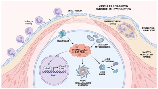
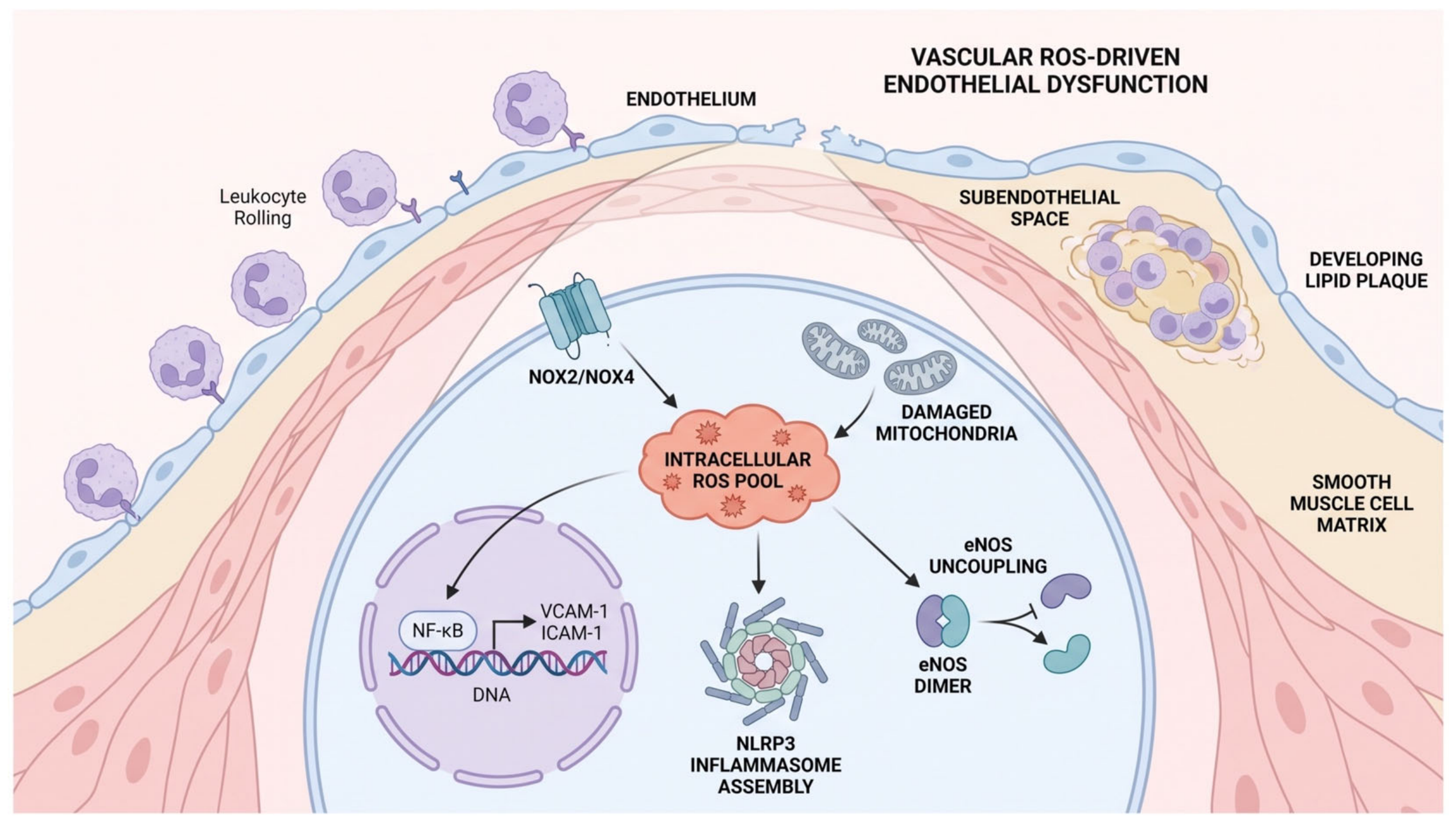
Chronic metabolic stress within the vessels initiates an inflammatory response that accelerates vascular remodeling and plaque vulnerability. Central to this process is the activation of the nuclear factor-kappa B (NF-κB) signaling pathway, which is upregulated by upstream oxidative stress and advanced glycation end-products (AGEs) [23,24]. Upon activation, NF-κB translocates to the nucleus, driving the transcription of pro-inflammatory cytokines such as interleukin-1 beta (IL-1β), interleukin-6 (IL-6), and tumor necrosis factor-alpha (TNF-α) alongside endothelial adhesion molecules, including vascular cell adhesion molecule-1 (VCAM-1) and intercellular adhesion molecule-1 (ICAM-1) [25,26]. These adhesion molecules facilitate the tethering, rolling, and transendothelial migration of circulating leukocytes into the subendothelial space, a foundational step in atherogenesis (Figure 1).
Figure 1.
Mechanisms of Vascular ROS-Driven Endothelial Dysfunction and Leukocyte Migration in Type 2 Diabetes. Chronic metabolic stress upregulates NOX2/NOX4 isoforms and damages mitochondrial structures, expanding the intracellular reactive oxygen species (ROS) pool. This oxidative stress drives the nuclear translocation of NF-κB, inducing the genomic transcription and surface expression of endothelial adhesion molecules VCAM-1 and ICAM-1. Concurrently, cytosolic ROS triggers NLRP3 inflammasome assembly, while uncoupled eNOS homodimers generate further superoxide (O2∙−), ultimately leading to cell junction disruption, leukocyte rolling, and subendothelial lipid plaque development. Black arrows indicate activation or pathways; T-bar lines indicate inhibition; cell types and tissue layers are color-coded for visual distinction.
Accumulating translational data suggest that SGLT2i treatment may attenuate this inflammatory cascade. In endothelial cell lines and diabetic rodent models, SGLT2i administration has been associated with suppression of NF-κB phosphorylation and nuclear translocation [27,28,29]. This molecular inhibition correlates with a downstream reduction in the expression of VCAM-1 and ICAM-1, thereby limiting leukocyte-endothelial interactions [30,31]. Emerging evidence indicates that SGLT2is modulate NLRP3 inflammasome activation, a key macromolecular complex responsible for IL-1β maturation, via systemic substrate shifts [32,33]. By inducing a state of fasting-mimicry, SGLT2is stimulate hepatic ketogenesis, mildly elevating circulating beta-hydroxybutyrate (β-HB) [34]. Within vascular cells, β-HB acts as an active signaling modifier that upregulates SIRT1, which subsequently deacetylates and inactivates the p65 subunit of NF-κB to suppress cytokine transcription [35,36]. Additionally, β-HB prevents the structural assembly of the NLRP3 inflammasome [32,37]. By suppressing these pathways, SGLT2is may blunt the chronic vascular inflammation that underpins accelerated macrovascular disease in T2DM [15,16].
3.3. eNOS Stabilization and NHE1 Inhibition
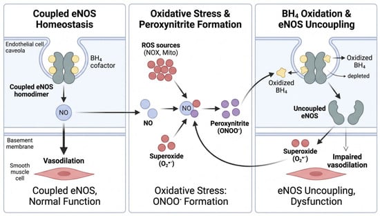
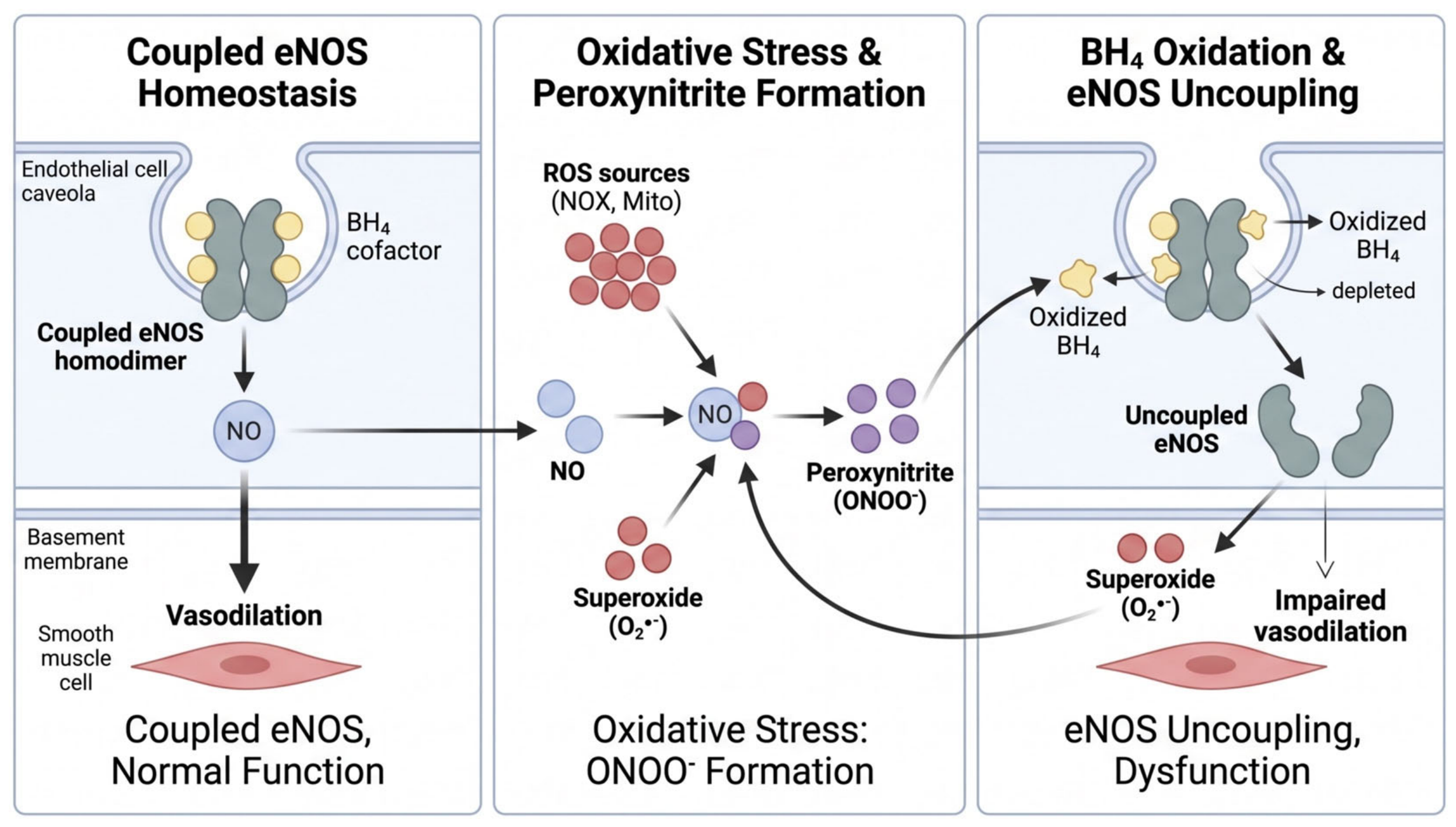
A defining hallmark of diabetic vasculopathy is the loss of endothelial nitric oxide synthase (eNOS) activity, which reduces nitric oxide (NO) and impairs endothelial vasodilation. Under physiological conditions, eNOS produces basal levels of NO, maintaining vascular tone, inhibiting platelet aggregation, and suppressing smooth muscle cell proliferation [38,39]. However, in the high-oxidative environment of T2DM, excessive reactive oxygen species (ROS) particularly superoxide (O2−) rapidly react with NO to form peroxynitrite (ONOO−) [39,40]. This highly reactive nitrogen species oxidizes tetrahydrobiopterin (BH4), an essential cofactor for eNOS [41]. Deprived of BH4, the eNOS enzyme undergoes “uncoupling,” a dysfunctional enzymatic state wherein it paradoxically generates further superoxide instead of NO, creating a feedback loop of endothelial injury [41,42] (Figure 2).
Figure 2.
Cellular Cascade of Coupled eNOS Homeostasis Versus Peroxynitrite-Induced eNOS Uncoupling. (Left Panel) Under physiological conditions, the coupled eNOS homodimer utilizes the essential cofactor tetrahydrobiopterin (BH4) within endothelial caveolae to produce basal nitric oxide (NO), promoting smooth muscle cell vasodilation. (Middle Panel) Under diabetic oxidative stress, excessive superoxide (O2−) rapidly reacts with NO to form highly reactive peroxynitrite (ONOO−). (Right Panel) Peroxynitrite oxidizes and depletes local BH4 reserves, causing functional enzymatic uncoupling of the eNOS enzyme; uncoupled eNOS paradoxically generates further superoxide instead of NO, impairing vasodilation and driving vascular injury. Grey shading contrasts coupled vs. uncoupled states; red/purple spheres denote reactive radical species; arrows trace biochemical progression; muscle cells are represented in light red.
Experimental models suggest that SGLT2i therapy may disrupt this uncoupling cascade and support eNOS function [43,44,45,46]. By suppressing upstream NOX activation, SGLT2is reduce consumption of BH4, stabilizing the functional state of eNOS [44]. Furthermore, molecular studies indicate that SGLT2is stimulate eNOS enzymatic activity via the activation of the adenosine monophosphate-activated protein kinase (AMPK) and protein kinase B (Akt) pathways, promoting the regulatory phosphorylation of eNOS at its activating site [47,48]. The resulting restoration of NO has been proposed to normalize local vasodilation and reinforce the integrity of the vessel wall [49]. Nevertheless, because these laboratory findings are mapped in isolated myocardial, renal or lymphocyte models, uniform replication in the human blood vessels remains unproven. Translational evidence points towards inhibition of the sodium-hydrogen exchanger 1 (NHE1) as a parallel pathway for vascular preservation [50,51,52]. In diabetes, endothelial NHE1 hyperactivation prompts intracellular sodium and calcium overload, driving mitochondrial dysfunction and apoptosis [53,54]. Preclinical models indicate that specific gliflozins, particularly empagliflozin, inhibit vascular NHE1, restoring ion homeostasis [52]. This pathway, however, varies significantly across individual SGLT2i agents. Individual gliflozins manifest highly divergent affinity profiles for different NHE isoforms, thus NHE modulation represents a compound-specific pharmacodynamic trait rather than a uniform class effect [51,55]. A consolidated overview of these molecular pathways and cellular mechanisms under diabetic conditions is summarized in Table 1.
Table 1.
An overview of the interplay between diabetic vascular damage and SGLT2i intervention.
4. Clinical Implications
The translational bridge between cellular modifications and macrovascular preservation is clinically evaluated through non-invasive surrogates of vascular health, primarily flow-mediated dilation (FMD) and indices of arterial stiffness. While early landmark outcome trials like EMPA-REG OUTCOME and DECLARE-TIMI 58 established systemic macrovascular benefits, granular physiological sub-studies and dedicated prospective cohorts have provided direct insight into localized conduit and elastic artery dynamics [4,5]. To interpret these data accurately, translational research requires a clear distinction between basic science pathways and long-term clinical outcomes. While these domains provide overlapping insights, a favorable shift in one cannot be interpreted as a confirmation in the other. Thus, although the clinical efficacy of SGLT2is in non-diabetic cohorts suggests that their effects go beyond proximal tubule inhibition, these outcomes reflect an integrated response heavily driven by systemic fluid and pressure changes rather than isolated endothelial cell protection (Table 2).
4.1. Flow-Mediated Dilation (FMD)
Clinical assessment of endothelium-dependent vasodilation via brachial artery FMD serves as a proxy marker of real-time endothelial nitric oxide (NO) bioavailability [47,48]. In patients with T2DM, who characteristically manifest blunt or absent hyperemic reactive dilation, SGLT2i therapy has demonstrated heterogeneous functional results.
In the DEFENCE Trial, which evaluated dapagliflozin versus metformin in early-stage T2DM over 16 weeks, there was no statistically significant difference in the primary endpoint of overall baseline-adjusted change in brachial artery FMD between the two groups [56]. However, a pre-specified analysis revealed a significant improvement in FMD exclusively in patients with poorly controlled diabetes (baseline HbA1c > 7%) [56]. This specific benefit correlated with a significant reduction in urinary 8-OHdG markers, suggesting that SGLT2i-mediated oxidative mitigation is most functionally relevant in states of severe metabolic and oxidative stress [56]. Parallel subacute trends were described by Balleza-Alejandri et al. (2024) in a double-blind trial demonstrating that an acute, 7-day administration of empagliflozin or dapagliflozin significantly increased brachial artery FMD from baseline in patients with T2DM [57]. While these findings suggest a rapid class effect on the dynamic, shear-stress-responsive properties of the endothelial wall, interpretation remains restricted by the brief study duration, sample size, and notable baseline clinical imbalances [57]. More broadly, systematic reviews confirm that across unselected clinical cohorts, SGLT2is yield modest and highly heterogeneous FMD modifications, underscoring a low-to-moderate overall certainty of evidence [58,59,60].
Clinical FMD responses are not uniform across trial populations. The landmark EMPA-HEART CardioLink-6 trial evaluated the impact of empagliflozin (10 mg/day) over 6 months in patients with T2DM and established coronary artery disease, failing to demonstrate a statistically significant improvement in brachial artery FMD compared to placebo [61]. This discrepancy suggests that while gliflozins may modulate dynamic, shear-stress-responsive endothelial properties in early-stage metabolic dysfunction or lower-risk cohorts, they do not consistently override or reverse structural macrovascular damage in advanced, established atherothrombotic disease where intensive cardioprotective medications and mechanical hemodynamic loading fluctuations drive vascular behavior [62,63].
4.2. Arterial Stiffness and Wave Reflection
Concurrently, structural and functional stiffening of the large elastic arteries, quantified via the gold-standard carotid-femoral pulse wave velocity (cfPWV) and the cardio-ankle vascular index (CAVI), represents an independent predictor of incident heart failure and cardiovascular mortality [64,65]. The therapeutic use of SGLT2is has shown an association with reductions in central vascular impedance. For instance, a pooled analysis of Phase III trials specifically scrutinized the acute and intermediate effects of empagliflozin on arterial stiffness parameters in patients with T2DM. While the primary findings demonstrated consistent shifts in central hemodynamics over 12-week and 24-week timelines, parallel findings indicated a stabilizing or reducing effect on peripheral vascular resistance and ambulatory arterial stiffness [66,67,68].
However, the capacity of these agents to achieve true structural destiffening remains highly debated. A systematic review and meta-analysis by Wei et al. (2022) demonstrated that while SGLT2i therapy significantly improved peripheral vascular resistance, it failed to identify a statistically significant overall reduction in the cfPWV [59]. This underscores that clinical improvements in vascular impedance may stem primarily from acute mechanical volume contraction and blood pressure reduction, rather than true structural matrix remodeling (such as reductions in advanced glycation end-products or active vascular smooth muscle cell modulation) of the elastic arterial wall [59,63,69,70,71].
4.3. Nephrology Outcome Trials
While localized measurements like FMD and cfPWV reflect subacute changes in vascular physiology, landmark large-scale outcome trials within the nephrology sector indicate that pathways correspond to microvascular and macrovascular preservation in the kidneys. This cardiorenal protective class effect has been mapped across three major clinical trials. The CREDENCE Trial, evaluating canagliflozin in patients with T2DM and diabetic nephropathy, was the first dedicated renal outcome trial to demonstrate a significant 30% reduction in the risk of the primary composite cardiorenal endpoint (progression to end-stage kidney disease, doubling of serum creatinine, or renal/cardiovascular death) [7].
The DAPA-CKD Trial expanded the therapeutic horizon by including chronic kidney disease (CKD) patients with and without T2DM [8]. Dapagliflozin demonstrated a significant 39% reduction in the primary composite renal endpoint alongside significant reductions in all-cause mortality and cardiovascular hospitalizations, indicating that the foundational mechanisms of SGLT2i-mediated vascular and tissue preservation can operate independently of ambient baseline glycemic states [8]. Finally, the EMPA-KIDNEY Trial scrutinized a broad spectrum of CKD populations, showing that empagliflozin reduced the risk of kidney disease progression or cardiovascular death by 28% [9]. Notably, EMPA-KIDNEY included patients with lower baseline kidney function (eGFR down to 20 mL/min/1.73 m2) and lower levels of albuminuria, supporting the agent’s role as a broad-spectrum vascular stabilizer [9].
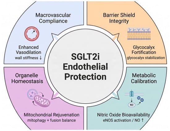
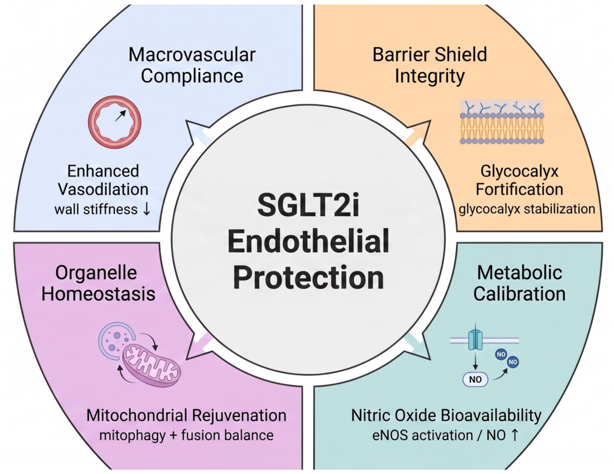
The cardiorenal protection highlighted in these trials is reinforced at the microvascular level through the preservation of the endothelial filtration barrier, specifically the endothelial glycocalyx (Figure 3). The glycocalyx regulates the permeability of the endothelium and limits leukocyte adhesion [72,73]. In T2DM, chronic glucotoxicity degrades the barrier, giving rise to protein leakage, podocyte loss and worsening glomerular injury [74,75]. Emerging preclinical data indicate that SGLT2i therapy helps prevent glycocalyx shedding and supports its structural thickness [76]. By shielding this endothelial gel layer, SGLT2is mitigate microvascular permeability, directly contributing to the observable reductions in albuminuria and long-term filtration decline demonstrated across the CREDENCE and DAPA-CKD cohorts [7,8].
Figure 3.
Pleiotropic Pathways and Macrovascular Benefits of SGLT2i-Mediated Endothelial Protection. This diagram maps the multi-targeted systemic impacts of SGLT2i therapy, organizing them into four distinct functional axes: organelle homeostasis (mitochondrial rejuvenation via mitophagy and fusion balance), nitric oxide bioavailability (eNOS activation and enhanced NO generation), barrier shield integrity (endothelial glycocalyx fortification and stabilization), and macrovascular compliance (enhanced vasodilation paired with a reduction in arterial wall stiffness). The diagram features four color-coded quadrants, blue, orange, purple, and green, representing distinct therapeutic axes; outward block arrows indicate downstream effects of central SGLT2i action, while small black arrows denote increased or decreased parameters.
Ultimately, these cumulative macrovascular and nephrology outcomes demonstrate that the molecular hallmarks of SGLT2i action, which encompass oxidative, inflammatory, and enzymatic pathways, correlate favorably with overall macrovascular compliance and endothelial responsiveness (Figure 3).
4.4. Limitations and Clinical Confounders
The translational evidence connecting SGLT2is to direct endothelial protection possesses several distinct boundaries. Mechanistically, primary basic science data exhibit notable variability across individual agents; for example, specific pathway interactions (such as NHE1 inhibition) represent compound-specific traits rather than uniform class effects [51,52]. Clinically, human studies evaluating vascular surrogate markers are severely constrained by small sample sizes and brief treatment durations, leaving long-term structural changes in the arterial wall largely unverified. Most importantly, non-invasive clinical vascular measurements like FMD, cfPWV, and CAVI are highly sensitive to acute fluctuations in blood pressure, volume status, systemic glycemic control, and body weight [77]. Because SGLT2i therapy simultaneously modulates all of these metabolic and fluid parameters via osmotic diuresis and natriuresis, isolating an intrinsic, direct endothelial stabilization effect in human cohorts remains a complex clinical challenge.
Table 2.
Stratification of clinical trials and systematic evidence evaluating SGLT2i vascular and cardiorenal endpoints.
5. Conclusions
Accumulating translational and clinical evidence suggests that SGLT2 inhibitors possess vascular-modulating properties, potentially complementing their established systemic cardiorenal effects. Mechanistically, preclinical models demonstrate that these agents sustain nitric oxide bioavailability by suppressing oxidative stress, NF-κB-mediated inflammation, and eNOS uncoupling. In human cohorts, these cellular actions correlate with changes in flow-mediated dilation and reduced arterial stiffness, underpinning some of the macrovascular and renal protection observed in landmark outcome trials. However, to definitively isolate these direct endothelial benefits from systemic hemodynamic confounders, such as osmotic diuresis and blood pressure reduction, the cardiorenal research community should transition toward more targeted clinical trial designs. Specifically, future protocols should prioritize flow-mediated dilation and pulse wave velocity assessments conducted under strictly volume- and blood pressure-clamped conditions. Furthermore, head-to-head mechanistic trials comparing SGLT2is directly against pure loop or thiazide diuretics are essential to differentiate mechanical volume unloading from active endothelial cell stabilization. Elucidating these precise pathways remains vital to addressing the high residual cardiorenal risk persisting in this population.
Author Contributions
Conceptualization, L.D. and A.C.-R.; methodology, L.D., A.C.-R. and O.-J.O.; writing—original draft, L.D., A.C.-R. and O.-J.O.; writing—review and editing, A.C.-R. and O.-J.O. All authors have read and agreed to the published version of the manuscript.
Funding
This research received no external funding.
Institutional Review Board Statement
Not applicable.
Informed Consent Statement
Not applicable.
Data Availability Statement
No new data were created or analyzed in this study. Data sharing is not applicable to this article.
Acknowledgments
During the preparation of this work, the authors used ChatGPT version 5.2 strictly for English copy-editing, syntax refinement, and clarity improvement of text originally drafted by the authors. The authors reviewed, verified, edited all outputs manually and take full academic responsibility for the integrity of the scientific content.
Conflicts of Interest
The authors declare no conflicts of interest.
References
- van Sloten, T.T.; Henry, R.M.A.; Dekker, J.M.; Nijpels, G.; Unger, T.; Schram, M.T.; Stehouwer, C.D.A. Endothelial Dysfunction Plays a Key Role in Increasing Cardiovascular Risk in Type 2 Diabetes. Hypertension 2014, 64, 1299–1305. [Google Scholar] [CrossRef] [Scilit] [PubMed]
- Low Wang, C.C.; Hess, C.N.; Hiatt, W.R.; Goldfine, A.B. Clinical Update: Cardiovascular Disease in Diabetes Mellitus. Circulation 2016, 133, 2459–2502. [Google Scholar] [CrossRef] [Scilit] [PubMed]
- Tabit, C.E.; Chung, W.B.; Hamburg, N.M.; Vita, J.A. Endothelial dysfunction in diabetes mellitus: Molecular mechanisms and clinical implications. Rev. Endocr. Metab. Disord. 2010, 11, 61–74. [Google Scholar] [CrossRef] [Scilit] [PubMed]
- Zinman, B.; Wanner, C.; Lachin, J.M.; Fitchett, D.; Bluhmki, E.; Hantel, S.; Mattheus, M.; Devins, T.; Johansen, O.E.; Woerle, H.J.; et al. Empagliflozin, Cardiovascular Outcomes, and Mortality in Type 2 Diabetes. N. Engl. J. Med. 2015, 373, 2117–2128. [Google Scholar] [CrossRef] [Scilit] [PubMed]
- Wiviott, S.D.; Raz, I.; Bonaca, M.P.; Mosenzon, O.; Kato, E.T.; Cahn, A.; Silverman, M.G.; Zelniker, T.A.; Kuder, J.F.; Murphy, S.A.; et al. Dapagliflozin and Cardiovascular Outcomes in Type 2 Diabetes. N. Engl. J. Med. 2019, 380, 347–357. [Google Scholar] [CrossRef] [Scilit] [PubMed]
- Neal, B.; Perkovic, V.; Mahaffey, K.W.; de Zeeuw, D.; Fulcher, G.; Erondu, N.; Shaw, W.; Law, G.; Desai, M.; Matthews, D.R.; et al. Canagliflozin and Cardiovascular and Renal Events in Type 2 Diabetes. N. Engl. J. Med. 2017, 377, 644–657. [Google Scholar] [CrossRef] [Scilit] [PubMed]
- Perkovic, V.; Jardine, M.J.; Neal, B.; Bompoint, S.; Heerspink, H.J.L.; Charytan, D.M.; Edwards, R.; Agarwal, R.; Bakris, G.; Bull, S.; et al. Canagliflozin and Renal Outcomes in Type 2 Diabetes and Nephropathy. N. Engl. J. Med. 2019, 380, 2295–2306. [Google Scholar] [CrossRef] [Scilit] [PubMed]
- Heerspink, H.J.L.; Stefánsson, B.V.; Correa-Rotter, R.; Chertow, G.M.; Greene, T.; Hou, F.-F.; Mann, J.F.E.; McMurray, J.J.V.; Lindberg, M.; Rossing, P.; et al. Dapagliflozin in Patients with Chronic Kidney Disease. N. Engl. J. Med. 2020, 383, 1436–1446. [Google Scholar] [CrossRef] [Scilit] [PubMed]
- Herrington, W.G.; Staplin, N.; Wanner, C.; Green, J.B.; Hauske, S.J.; Emberson, J.R.; Preiss, D.; Judge, P.; Mayne, K.J.; The EMPA-KIDNEY Collaborative Group; et al. Empagliflozin in Patients with Chronic Kidney Disease. N. Engl. J. Med. 2023, 388, 117–127. [Google Scholar] [CrossRef] [Scilit] [PubMed]
- Poonoosamy, J.; Lopes, P.; Huret, P.; Dardari, R.; Penfornis, A.; Thomas, C.; Dardari, D. Impact of Intensive Glycemic Treatment on Diabetes Complications—A Systematic Review. Pharmaceutics 2023, 15, 1791. [Google Scholar] [CrossRef] [Scilit] [PubMed]
- Checa-Ros, A.; Okojie, O.J.; Steib, N.; Bellasi, A.; Salvador-Martínez, P.; Fortea, I.; D’MArco, L. SGLT2 inhibitors and their role in reducing adiposopathy and inflammation in diabetes and non-diabetes CKD patients. Int. Urol. Nephrol. 2025, 58, 2721–2730. [Google Scholar] [CrossRef] [Scilit] [PubMed]
- Scisciola, L.; Cataldo, V.; Taktaz, F.; Fontanella, R.A.; Pesapane, A.; Ghosh, P.; Franzese, M.; Puocci, A.; De Angelis, A.; Sportiello, L.; et al. Anti-inflammatory role of SGLT2 inhibitors as part of their anti-atherosclerotic activity: Data from basic science and clinical trials. Front. Cardiovasc. Med. 2022, 9, 1008922. [Google Scholar] [CrossRef] [Scilit] [PubMed]
- Deedwania, P. SGLT2 Inhibitors: The Dawn of a New Era in Cardio-Metabolic Therapeutics. Am. J. Cardiovasc. Drugs 2022, 22, 1–4. [Google Scholar] [CrossRef] [Scilit] [PubMed]
- Zhang, Y.; Murugesan, P.; Huang, K.; Cai, H. NADPH oxidases and oxidase crosstalk in cardiovascular diseases: Novel therapeutic targets. Nat. Rev. Cardiol. 2020, 17, 170–194. [Google Scholar] [CrossRef] [Scilit] [PubMed]
- Checa-Ros, A.; Okojie, O.J.; D’Marco, L. SGLT2 Inhibitors: Multifaceted Therapeutic Agents in Cardiometabolic and Renal Diseases. Metabolites 2025, 15, 536. [Google Scholar] [CrossRef] [Scilit] [PubMed]
- Hamilton, S.J.; Watts, G.F. Endothelial Dysfunction in Diabetes: Pathogenesis, Significance, and Treatment. Rev. Diabet. Stud. 2013, 10, 133–156. [Google Scholar] [CrossRef] [Scilit] [PubMed]
- de la Lastra, J.M.P.; Andrés, C.M.C.; Munguira, E.B.; Juan, C.A.; Pérez-Lebeña, E. Pathophysiology of reactive oxygen species (ROS). Arch. Toxicol. 2026, 100, 475–513. [Google Scholar] [CrossRef] [Scilit] [PubMed]
- Jomova, K.; Raptova, R.; Alomar, S.Y.; Alwasel, S.H.; Nepovimova, E.; Kuca, K.; Valko, M. Reactive oxygen species, toxicity, oxidative stress, and antioxidants: Chronic diseases and aging. Arch. Toxicol. 2023, 97, 2499–2574. [Google Scholar] [CrossRef] [Scilit] [PubMed]
- Chambers, J.M.; Croteau, D.; Pimentel, D.R.; Gower, A.C.; Panagia, M.; Baka, T.; Qin, F.; Luptak, I.; Colucci, W.S. SGLT2 inhibitor upregulates myocardial genes for oxidative phosphorylation and fatty acid metabolism in Gαq-mice. J. Mol. Cell. Cardiol. Plus 2025, 12, 100296. [Google Scholar] [CrossRef] [Scilit] [PubMed]
- Urbanek, K.; Cappetta, D.; Bellocchio, G.; Coppola, M.A.; Imbrici, P.; Telesca, M.; Donniacuo, M.; Riemma, M.A.; Mele, E.; Cianflone, E.; et al. Dapagliflozin protects the kidney in a non-diabetic model of cardiorenal syndrome. Pharmacol. Res. 2023, 188, 106659. [Google Scholar] [CrossRef] [Scilit] [PubMed]
- Pham, L.T.T.; Mangmool, S.; Parichatikanond, W. Sodium-Glucose Cotransporter 2 (SGLT2) Inhibitors: Guardians against Mitochondrial Dysfunction and Endoplasmic Reticulum Stress in Heart Diseases. ACS Pharmacol. Transl. Sci. 2024, 7, 3279–3298. [Google Scholar] [CrossRef] [Scilit] [PubMed]
- Pérez-Flores, I.; López-Pastor, A.R.; Gómez-Pinedo, U.; Gómez-Infantes, A.; Espino-Paisán, L.; Romero, N.C.; de la Higuera, M.A.M.; Rodríguez-Cubillo, B.; Gómez-Delgado, I.; Sánchez-Fructuoso, A.I.; et al. Mitochondrial Changes Induced by SGLT2i in Lymphocytes from Diabetic Kidney Transplant Recipients: A Pilot Study. Int. J. Mol. Sci. 2025, 26, 3351. [Google Scholar] [CrossRef] [Scilit] [PubMed]
- Checa-Ros, A.; D’Marco, L. Molecular mechanisms linking adipose tissue browning to reduced cardiovascular risk. Atherosclerosis 2025, 411, 120564. [Google Scholar] [CrossRef] [Scilit] [PubMed]
- Salazar, J.; Navarro, C.; Ortega, Á.; Nava, M.; Morillo, D.; Torres, W.; Hernández, M.; Cabrera, M.; Angarita, L.; Ortiz, R.; et al. Advanced Glycation End Products: New Clinical and Molecular Perspectives. Int. J. Environ. Res. Public Health 2021, 18, 7236. [Google Scholar] [CrossRef] [Scilit] [PubMed]
- Hoffmann, A.; Cheng, G.; Baltimore, D. NF-κB: Master regulator of cellular responses in health and disease. Immun. Inflamm. 2025, 1, 2. [Google Scholar] [CrossRef] [Scilit] [PubMed]
- Xia, Y.F.; Liu, L.P.; Zhong, C.P.; Geng, J.G. NF-κB Activation for Constitutive Expression of VCAM-1 and ICAM-1 on B Lymphocytes and Plasma Cells. Biochem. Biophys. Res. Commun. 2001, 289, 851–856. [Google Scholar] [CrossRef] [Scilit] [PubMed]
- Mancini, S.J.; Boyd, D.; Katwan, O.J.; Strembitska, A.; Almabrouk, T.A.; Kennedy, S.; Palmer, T.M.; Salt, I.P. Canagliflozin inhibits interleukin-1β-stimulated cytokine and chemokine secretion in vascular endothelial cells by AMP-activated protein kinase-dependent and -independent mechanisms. Sci. Rep. 2018, 8, 5276. [Google Scholar] [CrossRef] [Scilit] [PubMed]
- Maekawa, H.; Zhou, Y.; Aoi, Y.; Fain, M.E.; Kaminski, D.S.; Kong, H.; Sebo, Z.L.; Chakrabarty, R.P.; Howard, B.C.; Andersen, G.; et al. SGLT2 inhibition protects kidney function by SAM-dependent epigenetic repression of inflammatory genes under metabolic stress. J. Clin. Investig. 2025, 135, e188933. [Google Scholar] [CrossRef] [Scilit] [PubMed]
- Uthman, L.; Kuschma, M.; Römer, G.; Boomsma, M.; Kessler, J.; Hermanides, J.; Hollmann, M.W.; Preckel, B.; Zuurbier, C.J.; Weber, N.C. Novel Anti-inflammatory Effects of Canagliflozin Involving Hexokinase II in Lipopolysaccharide-Stimulated Human Coronary Artery Endothelial Cells. Cardiovasc. Drugs Ther. 2021, 35, 1083–1094. [Google Scholar] [CrossRef] [Scilit] [PubMed]
- Park, S.H.; Farooq, M.A.; Gaertner, S.; Bruckert, C.; Qureshi, A.W.; Lee, H.-H.; Benrahla, D.; Pollet, B.; Stephan, D.; Ohlmann, P.; et al. Empagliflozin improved systolic blood pressure, endothelial dysfunction and heart remodeling in the metabolic syndrome ZSF1 rat. Cardiovasc. Diabetol. 2020, 19, 19. [Google Scholar] [CrossRef] [Scilit] [PubMed]
- Mroueh, A.; Fakih, W.; Kerth, S.; Kikuchi, S.; Aboueddahab, C.; Gong, D.-S.; Choi, M.; Nicolas, A.; Fass, S.; Trimaille, A.; et al. Synergistic SGLT2 and GLP-1R targeting alleviates systemic inflammation-induced and M1 monocyte-driven endothelial dysfunction in coronary artery disease. Cardiovasc. Diabetol. 2026, 25, 292. [Google Scholar] [CrossRef] [Scilit] [PubMed]
- Kim, S.R.; Lee, S.G.; Kim, S.H.; Kim, J.H.; Choi, E.; Cho, W.; Rim, J.H.; Hwang, I.; Lee, C.J.; Lee, M.; et al. SGLT2 inhibition modulates NLRP3 inflammasome activity via ketones and insulin in diabetes with cardiovascular disease. Nat. Commun. 2020, 11, 2127. [Google Scholar] [CrossRef] [Scilit] [PubMed]
- Byrne, N.J.; Matsumura, N.; Maayah, Z.H.; Ferdaoussi, M.; Takahara, S.; Darwesh, A.M.; Levasseur, J.L.; Jahng, J.W.S.; Vos, D.; Parajuli, N.; et al. Empagliflozin Blunts Worsening Cardiac Dysfunction Associated With Reduced NLRP3 (Nucleotide-Binding Domain-Like Receptor Protein 3) Inflammasome Activation in Heart Failure. Circ. Heart Fail. 2020, 13, e006277. [Google Scholar] [CrossRef] [Scilit] [PubMed]
- Kim, J.H.; Lee, M.; Kim, S.H.; Kim, S.R.; Lee, B.; Kang, E.S.; Cha, B.; Cho, J.W.; Lee, Y. Sodium-glucose cotransporter 2 inhibitors regulate ketone body metabolism via inter-organ crosstalk. Diabetes Obes. Metab. 2019, 21, 801–811. [Google Scholar] [CrossRef] [Scilit] [PubMed]
- Newman, J.C.; Verdin, E. β-Hydroxybutyrate: A Signaling Metabolite. Annu. Rev. Nutr. 2017, 37, 51–76. [Google Scholar] [CrossRef] [Scilit] [PubMed]
- Yang, H.; Zhang, W.; Pan, H.; Feldser, H.G.; Lainez, E.; Miller, C.; Leung, S.; Zhong, Z.; Zhao, H.; Sweitzer, S.; et al. SIRT1 Activators Suppress Inflammatory Responses through Promotion of p65 Deacetylation and Inhibition of NF-κB Activity. PLoS ONE 2012, 7, e46364. [Google Scholar] [CrossRef] [Scilit] [PubMed]
- Li, Y.; Liu Squan Chu, C.; Yang, J. The interplay of ketones and succinate: Seeking metabolic balance and novel therapeutic strategies in myocardial hypertrophy and heart failure. iScience 2026, 29, 115217. [Google Scholar] [CrossRef] [Scilit] [PubMed]
- Zhao, Y.; Vanhoutte, P.M.; Leung, S.W.S. Vascular nitric oxide: Beyond eNOS. J. Pharmacol. Sci. 2015, 129, 83–94. [Google Scholar] [CrossRef] [Scilit] [PubMed]
- Bhatti, J.S.; Sehrawat, A.; Mishra, J.; Sidhu, I.S.; Navik, U.; Khullar, N.; Kumar, S.; Bhatti, G.K.; Reddy, P.H. Oxidative stress in the pathophysiology of type 2 diabetes and related complications: Current therapeutics strategies and future perspectives. Free Radic. Biol. Med. 2022, 184, 114–134. [Google Scholar] [CrossRef] [Scilit] [PubMed]
- Caturano, A.; Rocco, M.; Tagliaferri, G.; Piacevole, A.; Nilo, D.; Di Lorenzo, G.; Iadicicco, I.; Donnarumma, M.; Galiero, R.; Acierno, C.; et al. Oxidative Stress and Cardiovascular Complications in Type 2 Diabetes: From Pathophysiology to Lifestyle Modifications. Antioxidants 2025, 14, 72. [Google Scholar] [CrossRef] [Scilit] [PubMed]
- Janaszak-Jasiecka, A.; Płoska, A.; Wierońska, J.M.; Dobrucki, L.W.; Kalinowski, L. Endothelial dysfunction due to eNOS uncoupling: Molecular mechanisms as potential therapeutic targets. Cell. Mol. Biol. Lett. 2023, 28, 21. [Google Scholar] [CrossRef] [Scilit] [PubMed]
- Edgar, K.S.; Cunning, C.; Gardiner, T.A.; McDonald, D.M. BH4 supplementation reduces retinal cell death in ischaemic retinopathy. Sci. Rep. 2023, 13, 21292. [Google Scholar] [CrossRef] [Scilit] [PubMed]
- Hasan, R.; Lasker, S.; Hasan, A.; Zerin, F.; Zamila, M.; Parvez, F.; Rahman, M.; Khan, F.; Subhan, N.; Alam, A. Canagliflozin ameliorates renal oxidative stress and inflammation by stimulating AMPK–Akt–eNOS pathway in the isoprenaline-induced oxidative stress model. Sci. Rep. 2020, 10, 14659. [Google Scholar] [CrossRef] [Scilit] [PubMed]
- Lopez, M.d.C.A.; Lax, A.; Hernandez Vicente, A.; Vicente, A.H.; Guillen, E.S.; Hernandez-Martinez, A.; del Palacco, M.J.F.; Bayes-Genis, A.; Figal, D.A.P. Empagliflozin improves post-infarction cardiac remodeling through GTP enzyme cyclohydrolase 1 and irrespective of diabetes status. Sci. Rep. 2020, 10, 13553. [Google Scholar] [CrossRef] [Scilit] [PubMed]
- Juni, R.P.; Kuster, D.W.D.; Goebel, M.; Helmes, M.; Musters, R.J.; van der Velden, J.; Koolwijk, P.; Paulus, W.J.; van Hinsbergh, V.W. Cardiac Microvascular Endothelial Enhancement of Cardiomyocyte Function Is Impaired by Inflammation and Restored by Empagliflozin. JACC Basic. Transl. Sci. 2019, 4, 575–591. [Google Scholar] [CrossRef] [Scilit] [PubMed]
- Juni, R.P.; Al-Shama, R.; Kuster, D.W.D.; van der Velden, J.; Hamer, H.M.; Vervloet, M.G.; Eringa, E.C.; Koolwijk, P.; van Hinsbergh, V.W. Empagliflozin restores chronic kidney disease–induced impairment of endothelial regulation of cardiomyocyte relaxation and contraction. Kidney Int. 2021, 99, 1088–1101. [Google Scholar] [CrossRef] [Scilit] [PubMed]
- Moens, A.L.; Goovaerts, I.; Claeys, M.J.; Vrints, C.J. Flow-Mediated Vasodilation. Chest 2005, 127, 2254–2263. [Google Scholar] [CrossRef] [Scilit] [PubMed]
- Flammer, A.J.; Anderson, T.; Celermajer, D.S.; Creager, M.A.; Deanfield, J.; Ganz, P.; Hamburg, N.M.; Lüscher, T.F.; Shechter, M.; Taddei, S.; et al. The Assessment of Endothelial Function. Circulation 2012, 126, 753–767. [Google Scholar] [CrossRef] [Scilit] [PubMed]
- Venkatesan, S.; Smirne, C.; Aquino, C.I.; Surico, D.; Remorgida, V.; Pour, M.M.O.; Pirisi, M.; Grossini, E. Nitric Oxide Signaling in Cardiovascular Physiology and Pathology: Mechanisms, Dysregulation, and Therapeutic Frontiers. Int. J. Mol. Sci. 2026, 27, 629. [Google Scholar] [CrossRef] [Scilit] [PubMed]
- Li, X.; Römer, G.; Kerindongo, R.P.; Hermanides, J.; Albrecht, M.; Hollmann, M.W.; Zuurbier, C.J.; Preckel, B.; Weber, N.C. Sodium Glucose Co-Transporter 2 Inhibitors Ameliorate Endothelium Barrier Dysfunction Induced by Cyclic Stretch through Inhibition of Reactive Oxygen Species. Int. J. Mol. Sci. 2021, 22, 6044. [Google Scholar] [CrossRef] [Scilit] [PubMed]
- Chung, Y.J.; Park, K.C.; Tokar, S.; Eykyn, T.R.; Fuller, W.; Pavlovic, D.; Swietach, P.; Shattock, M.J. Off-target effects of sodium-glucose co-transporter 2 blockers: Empagliflozin does not inhibit Na+/H+ exchanger-1 or lower [Na+]i in the heart. Cardiovasc. Res. 2021, 117, 2794–2806. [Google Scholar] [CrossRef] [Scilit] [PubMed]
- Chen, S.; Wang, Q.; Bakker, D.; Hu, X.; Zhang, L.; van der Made, I.; Tebbens, A.M.; Kovácsházi, C.; Giricz, Z.; Brenner, G.B.; et al. Empagliflozin prevents heart failure through inhibition of the NHE1-NO pathway, independent of SGLT2. Basic. Res. Cardiol. 2024, 119, 751–772. [Google Scholar] [CrossRef] [Scilit] [PubMed]
- Wu, Y.; Zhang, M.; Liu, R.; Zhao, C. Oxidative Stress-Activated NHE1 Is Involved in High Glucose-Induced Apoptosis in Renal Tubular Epithelial Cells. Yonsei Med. J. 2016, 57, 1252. [Google Scholar] [CrossRef] [Scilit] [PubMed]
- Jiang, K.; Su, F.; Deng, R.; Xu, Y.; Qin, A.; Yuan, X.; Xing, D.; Chen, Y.; Wang, D.; Shen, L.; et al. Cardiomyocyte-specific NHE1 overexpression confers protection against myocardial infarction during hyperglycemia. Cardiovasc. Diabetol. 2025, 24, 184. [Google Scholar] [CrossRef] [Scilit] [PubMed]
- Chung, Y.J.; Park, K.C.; Tokar, S.; Eykyn, T.R.; Fuller, W.; Pavlovic, D.; Swietach, P.; Shattock, M.J. SGLT2 inhibitors and the cardiac Na+/H+ exchanger-1: The plot thickens. Cardiovasc. Res. 2021, 117, 2702–2704. [Google Scholar] [CrossRef] [Scilit] [PubMed]
- Shigiyama, F.; Kumashiro, N.; Miyagi, M.; Ikehara, K.; Kanda, E.; Uchino, H.; Hirose, T. Effectiveness of dapagliflozin on vascular endothelial function and glycemic control in patients with early-stage type 2 diabetes mellitus: DEFENCE study. Cardiovasc. Diabetol. 2017, 16, 84. [Google Scholar] [CrossRef] [Scilit] [PubMed]
- Balleza Alejandri, L.R.; Grover Páez, F.; González Campos, E.; Campos, E.G.; Becerra, C.G.R.; Muñóz, E.G.C.; González, S.P.; Zavala, M.G.R.; Roa, A.S.R.; Rico, D.O.S.; et al. Empagliflozin and Dapagliflozin Improve Endothelial Function in Mexican Patients with Type 2 Diabetes Mellitus: A Double-Blind Clinical Trial. J. Cardiovasc. Dev. Dis. 2024, 11, 182. [Google Scholar] [CrossRef] [Scilit] [PubMed]
- Sridharan, K.; Sivaramakrishnan, G. Sodium Glucose Cotransporter-2 Inhibitors Improve Endothelial Function and Arterial Stiffness in Diabetic Individuals: A Systematic Review and Network Meta-Analysis. Curr. Vasc. Pharmacol. 2025, 23, 272–280. [Google Scholar] [CrossRef] [Scilit] [PubMed]
- Wei, R.; Wang, W.; Pan, Q.; Guo, L. Effects of SGLT-2 Inhibitors on Vascular Endothelial Function and Arterial Stiffness in Subjects With Type 2 Diabetes: A Systematic Review and Meta-Analysis of Randomized Controlled Trials. Front. Endocrinol. 2022, 13, 826604. [Google Scholar] [CrossRef] [Scilit] [PubMed]
- Haider, S.H.; Irfan, A.; Sheikh, S.M.; Larik, M.O.; Raza, A.; Khabir, Y. Evaluating the Safety and Efficacy of SGLT-2 Inhibitors on Reducing Cardiovascular and Renal Mortality, Morbidity and Inflammatory Outcomes in Various Patient Populations: A Systematic Review and Meta-Analysis of 92 920 Patients. Clin. Med. Insights Cardiol. 2025, 19, 11795468251347777. [Google Scholar] [CrossRef] [Scilit] [PubMed]
- Verma, S.; Mazer, C.D.; Yan, A.T.; Mason, T.; Garg, V.; Teoh, H.; Zuo, F.; Quan, A.; Farkouh, M.E.; Fitchett, D.H.; et al. Effect of Empagliflozin on Left Ventricular Mass in Patients With Type 2 Diabetes Mellitus and Coronary Artery Disease. Circulation 2019, 140, 1693–1702. [Google Scholar] [CrossRef] [Scilit] [PubMed]
- Irace, C.; Casciaro, F.; Scavelli, F.B.; Oliverio, R.; Cutruzzolà, A.; Cortese, C.; Gnasso, A. Empagliflozin influences blood viscosity and wall shear stress in subjects with type 2 diabetes mellitus compared with incretin-based therapy. Cardiovasc. Diabetol. 2018, 17, 52. [Google Scholar] [CrossRef] [Scilit] [PubMed]
- Góes-Santos, B.R.; Castro, P.C.; Girardi, A.C.C.; Antunes-Correa, L.M.; Davel, A.P. Vascular effects of SGLT2 inhibitors: Evidence and mechanisms. Am. J. Physiol.-Cell Physiol. 2025, 329, C1150–C1160. [Google Scholar] [CrossRef] [Scilit] [PubMed]
- Namba, T.; Masaki, N.; Takase, B.; Adachi, T. Arterial Stiffness Assessed by Cardio-Ankle Vascular Index. Int. J. Mol. Sci. 2019, 20, 3664. [Google Scholar] [CrossRef] [Scilit] [PubMed]
- Aimagambetova, B.; Ariko, T.; Merritt, S.; Rundek, T. Arterial stiffness measured by pulse wave velocity correlated with cognitive decline in hypertensive individuals: A systematic review. BMC Neurol. 2024, 24, 393. [Google Scholar] [CrossRef] [Scilit] [PubMed]
- Chilton, R.; Tikkanen, I.; Cannon, C.P.; Crowe, S.; Woerle, H.J.; Broedl, U.C.; Johansen, O.E. Effects of empagliflozin on blood pressure and markers of arterial stiffness and vascular resistance in patients with type 2 diabetes. Diabetes Obes. Metab. 2015, 17, 1180–1193. [Google Scholar] [CrossRef] [Scilit] [PubMed]
- Ott, C.; Jumar, A.; Striepe, K.; Friedrich, S.; Karg, M.V.; Bramlage, P.; Schmieder, R.E. A randomised study of the impact of the SGLT2 inhibitor dapagliflozin on microvascular and macrovascular circulation. Cardiovasc. Diabetol. 2017, 16, 26. [Google Scholar] [CrossRef] [Scilit] [PubMed]
- Cherney, D.Z.; Perkins, B.A.; Soleymanlou, N.; Har, R.; Fagan, N.; Johansen, O.; Woerle, H.-J.; von Eynatten, M.; Broedl, U.C. The effect of empagliflozin on arterial stiffness and heart rate variability in subjects with uncomplicated type 1 diabetes mellitus. Cardiovasc. Diabetol. 2014, 13, 28. [Google Scholar] [CrossRef] [Scilit] [PubMed]
- Liu, L.; Ni, Y.Q.; Zhan, J.K.; Liu, Y.S. The Role of SGLT2 Inhibitors in Vascular Aging. Aging Dis. 2021, 12, 1323. [Google Scholar] [CrossRef] [Scilit] [PubMed]
- Li, S.; Wang, L.; Wang, P.; Xu, X.; Guo, Y. Dapagliflozin improves cardiac function and reduces adverse events in myocardial infarction: A meta-analysis in diabetic and non-diabetic populations. Front. Endocrinol. 2025, 16, 1594861. [Google Scholar] [CrossRef] [Scilit] [PubMed]
- González-Clemente, J.M.; García-Castillo, M.; Gorgojo-Martínez, J.J.; Jiménez, A.; Llorente, I.; Matute, E.; Tejera, C.; Izarra, A.; Lecube, A. Beyond the Glycaemic Control of Dapagliflozin: Impact on Arterial Stiffness and Macroangiopathy. Diabetes Ther. 2022, 13, 1281–1298. [Google Scholar] [CrossRef] [Scilit] [PubMed]
- Hu, Z.; Cano, I.; D’Amore, P.A. Update on the Role of the Endothelial Glycocalyx in Angiogenesis and Vascular Inflammation. Front. Cell Dev. Biol. 2021, 9, 734276. [Google Scholar] [CrossRef] [Scilit] [PubMed]
- Delgadillo, L.F.; Marsh, G.A.; Waugh, R.E. Endothelial Glycocalyx Layer Properties and Its Ability to Limit Leukocyte Adhesion. Biophys. J. 2020, 118, 1564–1575. [Google Scholar] [CrossRef] [Scilit] [PubMed]
- Nakamichi, R.; Hayashi, K.; Itoh, H. Effects of High Glucose and Lipotoxicity on Diabetic Podocytes. Nutrients 2021, 13, 241. [Google Scholar] [CrossRef] [Scilit] [PubMed]
- Li, X.; Zhang, Y.; Xing, X.; Li, M.; Liu, Y.; Xu, A.; Zhang, J. Podocyte injury of diabetic nephropathy: Novel mechanism discovery and therapeutic prospects. Biomed. Pharmacother. 2023, 168, 115670. [Google Scholar] [CrossRef] [Scilit] [PubMed]
- Cooper, S.; Teoh, H.; Campeau, M.A.; Verma, S.; Leask, R.L. Empagliflozin restores the integrity of the endothelial glycocalyx in vitro. Mol. Cell. Biochem. 2019, 459, 121–130. [Google Scholar] [CrossRef] [Scilit] [PubMed]
- Higashi, Y. Noninvasive Assessment of Vascular Function. JACC Asia 2024, 4, 898–911. [Google Scholar] [CrossRef] [Scilit] [PubMed]

Disclaimer/Publisher’s Note: The statements, opinions and data contained in all publications are solely those of the individual author(s) and contributor(s) and not of MDPI and/or the editor(s). MDPI and/or the editor(s) disclaim responsibility for any injury to people or property resulting from any ideas, methods, instructions or products referred to in the content. |
© 2026 by the authors. Licensee MDPI, Basel, Switzerland. This article is an open access article distributed under the terms and conditions of the Creative Commons Attribution (CC BY) license.