


Abstract
4-Thiazolidinone derivatives, a group of compounds with a broad spectrum of activity and anticancer properties, are used in medicine as anti-inflammatory, antiviral, antibacterial, antidiabetic, and blood pressure-lowering drugs; some of them are now in phase II and III clinical trials as anticancer drugs. Here, we evaluated the anticancer potential of 4-thiazolidinones (2b, 12b, 14b) targeting tubulin and metastasis-related pathways in breast cancer models. Molecular docking revealed that compound 14b exhibited the strongest binding affinity to the paclitaxel-binding site of β-tubulin, outperforming 2b and 12b, though slightly weaker than Taxol. Biological assays confirmed significant tubulin inhibition, with 14b reducing β-tubulin levels up to eight-fold in MCF-7 cells and two-fold in MDA-MB-231 cells. All compounds promoted tubulin polymerization, showing paclitaxel-like activity, with comparable the maximum velocity (Vmax) values. Cell cycle analysis demonstrated G2/M arrest in MCF-7 cells and S-phase accumulation in MDA-MB-231 cells. Overall, compound 14b demonstrated the most promising multitarget anticancer activity in vitro.
1. Introduction
Breast cancer is the second most common cancer worldwide, after lung cancer with 0.67 million deaths in 2022 [1]. Estrogen-positive breast cancer is the most common, accounting for 70–75% of diagnoses [1,2]. Currently, cancer treatment includes radiotherapy, surgical removal of the tumor, targeted therapy, immunotherapy, and chemotherapy [3]. Cancer cells which divide rapidly as compared to normal cells are more susceptible to tubulin-inhibiting agents [4]. Microtubules, as elements of the cytoskeleton in eukaryotic cells, play a vital role in cell division, signal transmission, mitosis, cell motility, and the maintenance of cell morphology. Hence, microtubules have become a key target in drug design and cancer treatment [5]. Among the compounds widely used in cancer treatment are anti-mitotic compounds, such as toxoids (paclitaxel and docetaxel) and vinca alkaloids (vincristine and vinblastine) [6]. However, the development of new chemotherapy drugs for breast cancer is essential to overcome treatment resistance and improve survival, as many tumors eventually become refractory to existing therapies and show limited response rates [7]. Moreover, novel agents are needed to reduce systemic toxicity and enhance therapeutic efficacy, enabling more durable and personalized treatment outcomes for patients. 4-thiazolidinones represent a promising backbone for novel drugs used in therapeutic applications not only for anticancer drugs but also for antidiabetic, anticonvulsant, anti-inflammatory, and blood pressure-lowering agents [8]. Furthermore, thiazolidinones are characterized by a multidirectional mechanism of action as PPARγ agonists, tubulin inhibitors, reactive oxygen species (ROS) generators, mitogen-activated protein kinase (MAPK) activators, and inhibitors of Bcl-2/Bcl-xL, which makes them promising compounds for overcoming limitations such as drug resistance and the limited bioavailability of administered chemotherapy [8,9,10].
We have previously analyzed 15 new 4-thiazolidinones with a 5-nitrofuranpropenylidene moiety at C5 position of the core heterocycle and various carboxylic acid moieties at the N3 position [11]. Compounds 2b, 12b, and 14b, with 2-propanoic acid, 3-propanoic acid, and 6-hexanoic acid, respectively, attached to the N3-position of the 4-thiazolidinone core, were identified as the most promising compounds, exhibiting the highest cytotoxicity against two breast cancer cell lines, MCF-7 and MDA-MB-231, and inducing apoptosis in them [11]. This effect was dose- and time-dependent, especially against the MDA-MB-231 cell line, in which the cytotoxicity of the compounds was highest after 72 h (Figures S1–S3). We assayed the compounds against proliferating non-tumor cells (BJ cell line, which represents fibroblasts). As expected, their half-maximal inhibitory concentration (IC50) values were similar to those of breast cancer cells (Figure S4). Indeed, microtubule-targeting agents exert their antitumor activity primarily by disrupting microtubule dynamics required for mitotic spindle assembly and chromosome segregation. Consequently, these agents preferentially affect actively dividing cells, irrespective of whether they are malignant or normal. Rapidly proliferating normal tissues, such as the bone marrow, gastrointestinal epithelium, hair follicles, and germinal cells, are therefore highly susceptible, explaining the characteristic toxicities of neutropenia, mucositis, alopecia, and infertility. In contrast, quiescent (G0) or slowly proliferating cells are generally less sensitive [12]. To obtain additional information on the mechanism of action of 2b, 12b, and 14b, based on the revealed results from DNA biosynthesis, which clearly indicate that the compounds affect the genetic material of the cell and that, as previously described, 4-thiazolidinone derivatives have tubulin inhibiting properties [13], we decided to evaluate the potential activity against tubulin of the compounds and observed that they inhibit tubulin polymerization and decrease β-tubulin concentration in breast cancer cells.
2. Materials and Methods
2.1. Studied Compounds
Studied compounds 2b, 12b, and 14b were synthesized in accordance with the protocol described previously [11].
2.2. Cell Culture
Human breast cancer cell lines (MCF-7 and MDA-MB-231) used in this study were obtained from the American Type Culture Collection (ATCC) (Manassas, VA, USA). Cells were cultured in Dulbecco’s Modified Eagle Medium (DMEM) (Corning, Kennebunk, ME, USA) with 10% fetal bovine serum (FBS) (Gibco, Grand Island, NY, USA) and 1% penicillin-streptomycin antibacterial solution (Corning, Kennebunk, ME, USA). All cells were cultured in tissue culture dishes (Sarstedt, Numbrecht, Germany) at 37 °C in an atmosphere with 5% CO2. Before detachment, cells were washed with phosphate-buffered saline (PBS) (Corning, Kennebunk, ME, USA), and then 0.05% trypsin containing 0.02% EDTA (Corning, Kennebunk, ME, USA) was added. A Scepter 3.0 cell counter (Merck Millipore, Burlington, MA, USA) was used to calculate the number of cells before seeding them for the tests.
2.3. Determination of β-Tubulin Concentrations
Using a SimpleStep Enzyme-Linked Immunosorbent Assay (ELISA) kit (Abcam, Cambridge, UK) we determined the concentration of β-tubulin protein. The level of β-tubulin protein in cells treated with compounds 2b, 12b, and 14b was determined in both tested cell lines at 1 and 5μM and 5 and 10μM for the MCF-7 and MDA-MB-231 lines, respectively. Doxorubicin, as a reference compound, was used in two concentrations, 1 and 5μM, in both cell lines. The test was performed according to the protocol included with the appropriate manufacturer’s kit.
2.4. Cell Cycle Analysis
To conduct cell cycle analysis, we seeded cells in 12-well plates and added compounds 2b, 12b, and 14b at 1, 5, and 5, 10 μM for the MCF-7 and MDA-MB-231 cells, respectively, and incubated the cells for 24 h. After this time, the cells were harvested, centrifuged at room temperature (RT), washed with PBS, and then 2 mL of ice-cold 70% ethanol was added to each sample, after which the samples were and frozen overnight. The next day, the cells were centrifuged, washed with PBS, and then resuspended in 300 μL of PBS, and 5 μL of RNAse and 5 μL of propidium iodide were added. After a 10 min incubation, the cells were analyzed using a BD Accuri C6 Plus (Becton Dickinson Biosciences, San Jose, CA, USA) flow cytometer. The obtained results were analyzed using Floreada.io (https://floreada.io; accessed on 16 September 2025).
2.5. Tubulin Polymerization Assay
To conduct the tubulin polymerization assay, the manufacturer’s protocol for Cytoskeleton (Cat# BK006P) was used. The plate was pre-warmed at 37 °C, then the tested compounds (2b, 12b, 14b), the positive control Paclitaxel, and the negative control Combretastatin A4 (CA4) were prepared at a 10-fold concentration in the assay buffer. An amount of 10 µL of the prepared solutions were pipetted into a 96-well plate it. The concentrations of all tested compounds were 10 µM. Assay buffer was added as a control. Afterwards, 100 µL of the diluted tubulin solution (containing 3 mg/mL of tubulin in 80 mM PIPES, pH 6.9, 2 mM MgCl2, 0.5 mM EGTA, 1 mM GTP, and 7% glycerol), was pipetted into the 96-well plate, which was immediately placed in the plate reader. Absorbance was measured at 340 nm every minute for 61 readings using a Tecan Spark multimode plate reader (Tecan, Männedorf, Switzerland).
2.6. Molecular Docking
Molecular docking analysis was performed using Schrödinger Suite (2024-4 release) software. The structure of the protein of β-tubulin bound with Taxol (5SYF [14]) was downloaded from the Protein Data Bank (PDB; http://www.rcsb.org/; accessed on 10 June 2024). Proteins and ligands were prepared for docking as documented in our previous work [15]. The studied compounds were docked in the grid of the original ligand via Glide standard precision docking in the Schrödinger suit. Obtained results are displayed as docking scores in kcal/mol. Visualization of docked structures and molecular interactions was performed using the Maestro interface of the Schrödinger suit.
2.7. Confocal Microscopy
MCF-7 and MDA-MB-231 cells were seeded on glass coverslips in 6-well plates at a concentration of 4 × 10⁵ cells per well and incubated overnight. Afterwards cells were treated with 2b, 12b, 14b at 5 and CA4 at 0.1 µM concentrations. Control was treated with vehicle substance—DMSO for 24 h. Cells were fixed with 4% paraformaldehyde for 15 min, permeabilized with 0.1% Triton X-100 for 2 min, and blocked with 2% Bovine Serum Albumin (BSA) for 30 min at room temperature. Subsequently, the cells were incubated with the anti-β-tubulin antibody conjugated to Alexa Fluor 647 (EPR19591, Abcam, Cambridge, UK) (1:1000 dilution) for 1 h at room temperature in the dark. After staining, 10 µg/mL 4′,6-diamidino-2-phenylindole (DAPI) (Sigma-Aldrich, St. Louis, MO, USA) was applied for 5 min. Imaging was performed using a Stellaris 5 system (Leica Microsystems, GmbH, Wetzlar, Germany), and data analysis was carried out with ImageJ software version 1.54k.
3. Results
3.1. Molecular Docking Assessment of Compounds Binding Affinity Towards Tubulin
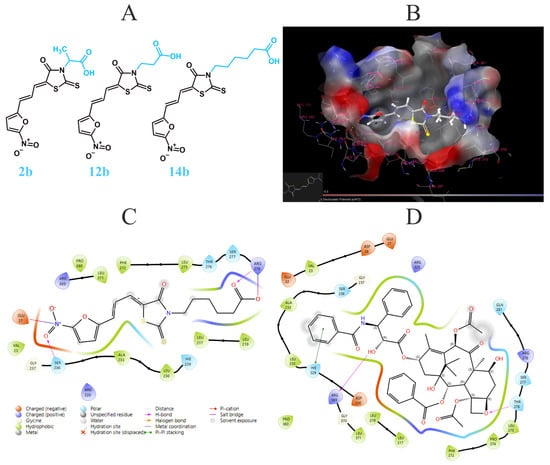
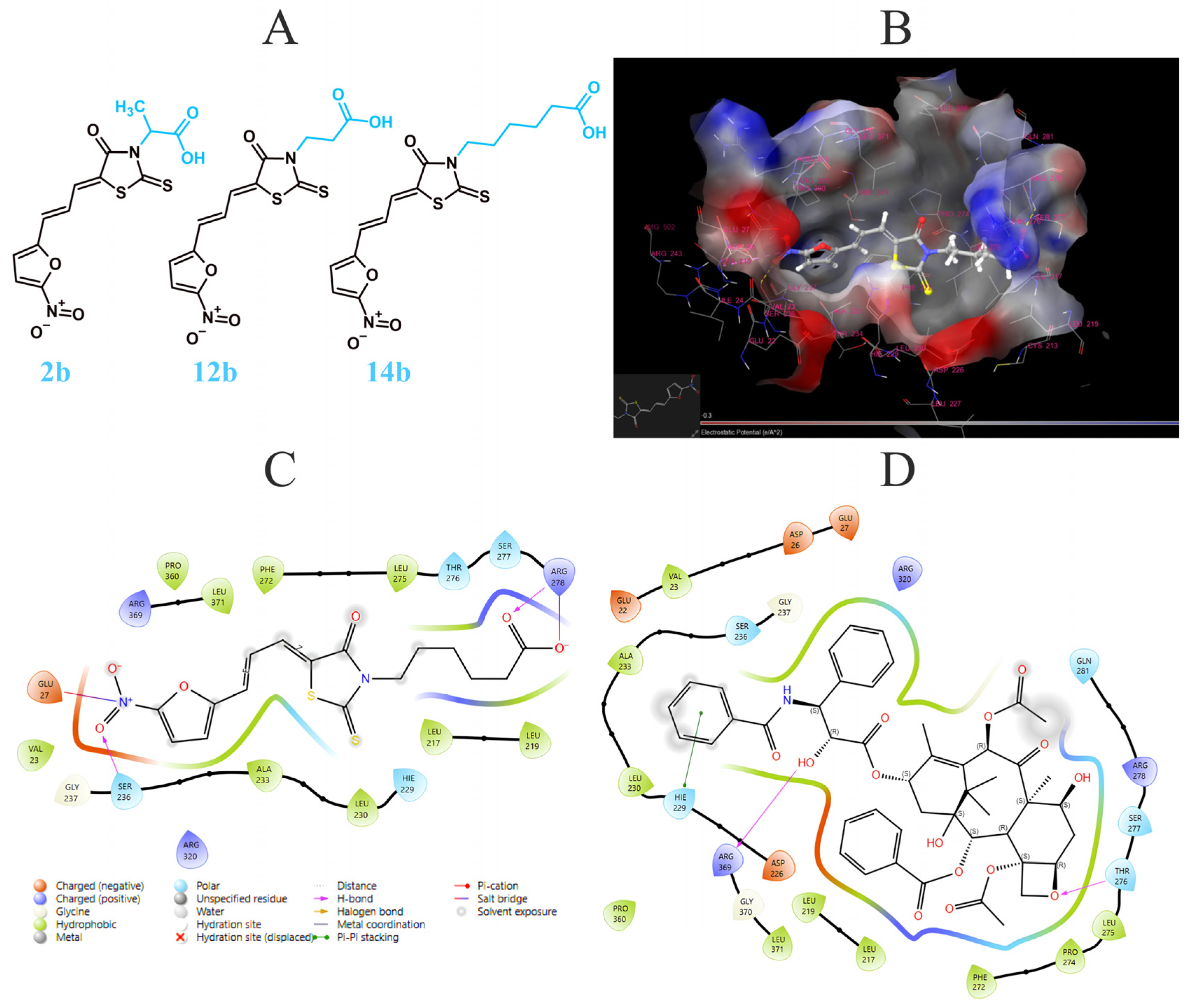
Molecular docking was applied to analyze the effect of the tested compounds 2b, 12b, 14b on paclitaxel binding site. To perform this analysis, we utilized tubulin complex with paclitaxel from the PDB (5SYF). The docking site for this analysis was generated in the place of the original ligand (Figure 1). The compound 14b exhibited the most promising results with −6.829 kcal/mol, outperforming both 2b (−5.916 kcal/mol) and 12b (−5.624 kcal/mol). Those results, however, were lower than that of Taxol (−7.709 kcal/mol; Table 1; Figure 1).
Figure 1.
Analyzed 4-thiazolidinone derivatives: (A) representation of molecular docking of compound 14b in Taxol binding site; (B) β-tubulin amino acids interactions with analyzed compounds at Taxol binding site: 14b (C), original ligand (D).
Table 1.
Molecular docking results for the compounds 2b, 12b, 14b, and Taxol (5SYF). Results are displayed in kcal/mol. OL—original ligand.
Analyzing the molecular interactions of compounds 2b, 12b, 14b, and the original ligand (Taxol), certain observations can be made. Taxol had the highest number of interactions with amino acids and produced the highest docking score. Compound 14b had the second-best docking score and demonstrated more interactions than the other two 4-thiazolidinone derivatives, which can be attributed to its longer carbon chain. Notably, 14b formed two salt bridges with GLU 27 and ARG 278 at both ends of the structure, while Taxol displayed only positively charged interaction with those residues. However, the original ligand compensated for this by exhibiting a π–π stacking interaction with HIE 229 and two H-bonds with ARG 369 and THR 276. In contrast, 14b had either a polar or a positively charged contact with those residues. Thus, the two compounds exhibit different crucial molecular interactions at the Taxol binding site rather than mimicking each other. Compounds 2b and 12b also exhibited interesting interactions with amino acids at the Taxol binding site. For instance, similarly to Taxol, 12b showcased one π–π stacking interaction, albeit with a different amino acid (PHE 272), formed two H-bonds (ARG 278 and HIE 229) and formed one salt bridge (ARG 278) with amino acids different from those interacting with both 14b and Taxol. Altogether, 12b exhibited a notably weaker docking score than 14b. In comparison, 2b displayed a profile more similar to that of 14b, with two H-bonds with SER 236 and THR 276 and, more importantly, one salt bridge with GLU 27 involving the nitrogen atom of the nitrofuran moiety. With those interactions, 2b displayed a docking score similar to 12b. This might suggest that GLU 27 in the Taxol binding site is an important residue for 4-thiazolidinones with nitrofuran moieties (Table 2, Figure 1, Figure S5, Supplementary Materials).
Table 2.
Molecular interactions of 2b, 12b, 14b, and Taxol with β-tubulin in the Taxol binding pocket.
3.2. Inhibition of Tubulin Protein Concentration in MCF-7 and MDA-MB-231
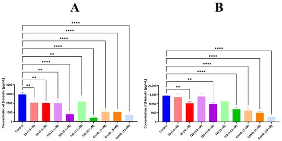
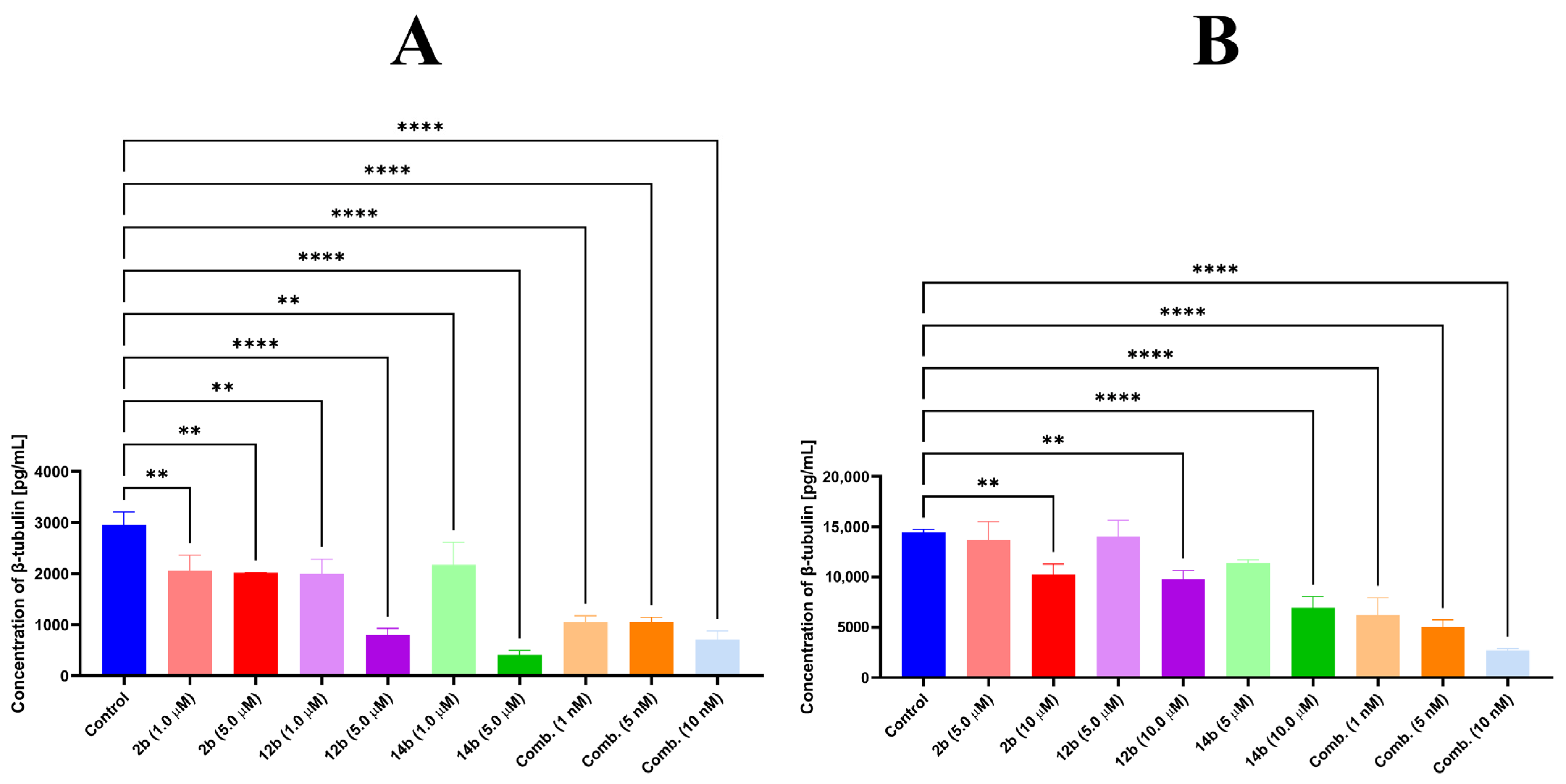
Tubulin inhibitors affect its synthesis, turnover, and degradation [16,17]. We therefore decided to analyze the effects of the compounds 2b, 12b, and 14b at concentrations of 1, 5, and 10 µM on tubulin concentrations in MCF-7 and MDA-MB-231 breast cancer cells by ELISA [18] (Figure 2). Combretastatin A4, a potent tubulin inhibitor, was used as a reference compound. Compound 14b at 5 µM reduced β-tubulin levels eight-fold vs. untreated control at 24 h (412.67 ± 83.28 vs. 2950.33 ± 255.6 pg/mL, respectively) in MCF-7 cells. In MDA-MB-231 cells, 14b at a concentration of 10 µM was also most active in MDA-MB-231, reducing β-tubulin levels two-fold (6947.67 ± 1117.0 vs. 14,445.3 ± 284.5 pg/mL).
Figure 2.
Effect of compounds 2b, 12b, and 14b on β-tubulin concentration. ELISA test in MCF-7 (A) and MDA-MB-231 (B) cell lines incubated with either compounds 2b, 12b, and 14b or combretastatin A4 for 24 h. ANOVA tests were performed to demonstrate statistical significance. ** p ≤ 0.01; **** p ≤ 0.0001. Data are shown as means ± SD; N = 3.
3.3. Tested Compounds Promoted Tubulin Polymerization
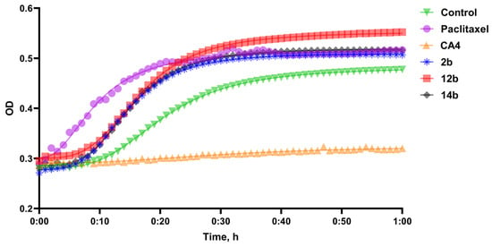
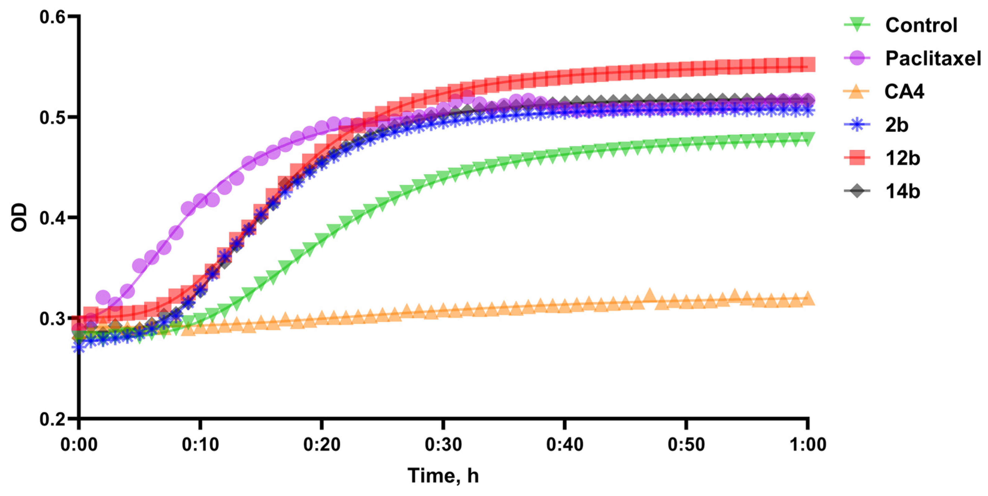
As the compounds 2b, 12b, and 14b reduced tubulin protein concentration in both MCF-7 and MDA-MB-231 cell lines, we further analyzed their effects on tubulin polymerization. Different drugs may either inhibit tubulin polymerization (Combretastatin A4) or promote it (Paclitaxel) [19]. To analyze the effects of 2b, 12b, and 14b, we carried out an in vitro tubulin polymerization assay (Figure 3) that analyzes fluctuations in optical density (OD) caused by the polymerization of tubulin over one hour, with one reading per minute. Tubulin polymerizing agents cause a spike in optical density, while tubulin depolymerizing compounds decrease OD in comparison to the control. We measured Vmax for each compound by calculating the highest value of the slope created by non-linear regression for OD values. All three compounds and a reference compound promoted polymerization of tubulin, with Vmax values of 15.54 ± 2.11, 14.26 ± 0.51, 15.68 ± 1.16, and 15.88 ± 0.72 mOD/min for 2b, 12b, 14b, and Paclitaxel, respectively (Table 3).
Figure 3.
Effect of compounds 2b, 12b, 14b, paclitaxel, CA4, and the control (DMSO) on tubulin polymerization, mOD/min. N = 2.
Table 3.
Effect of compounds 2b, 12b, 14b, paclitaxel, and CA4 on tubulin polymerization: Vmax (mOD/min) and percentage inhibition in comparison to the untreated control, in %.
3.4. Compounds 2b, 12b, and 14b Induce Cell Cycle Arrest
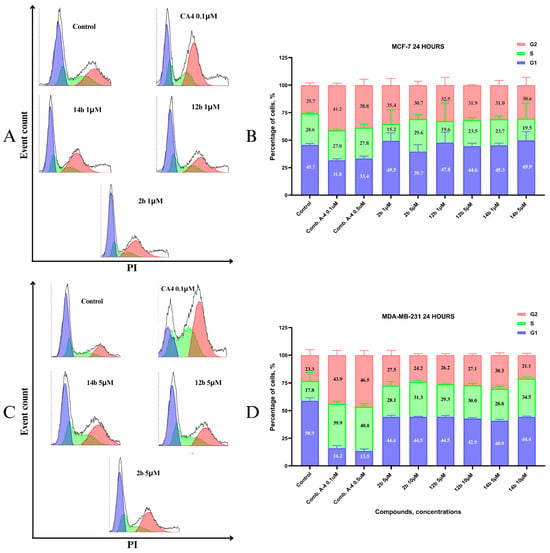
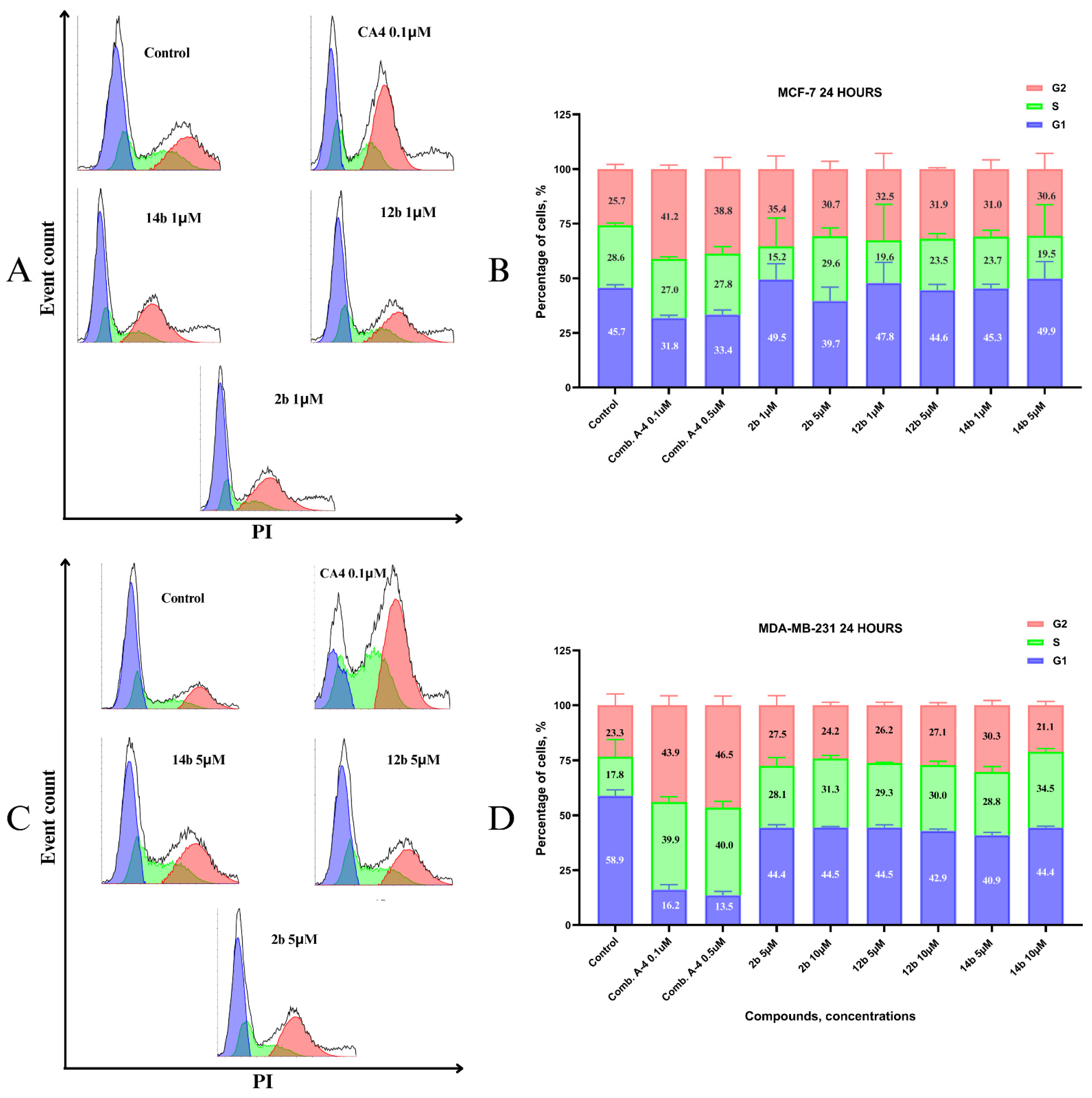
Tubulin inhibition affects cell cycle; therefore, we analyzed the cell cycles of MCF-7 and MDA-MB-231 cells treated with compounds 2b, 12b, and 14b at concentrations of 1 to 10 µM and CA4 at 0.1 and 0.5 µM as a reference compound that arrests cells primarily in the G2/M phase. CA4 arrested MCF-7 cells in the G2/M phase, with 41.2% and 38.8% at 0.1 and 0.5 µM, respectively, in comparison with 25.7% of cells in the G2/M phase in the control. Compounds 2b, 12b, and 14b also arrested cells in the G2/M phase, albeit to a lesser degree (30.6% to 35.4% of cells in the G2/M phase). Similar effects of CA4 were observed in MDA-MB-231 cells, with 43.9% and 46.5% in G2/M cells at 0.1 and 0.5 µM, respectively, while compounds 2b, 12b, and 14b mostly arrested MDA-MB-231 cells in the S phase (28.1% to 34.5% vs. 17.8% of cells in the S phase cells in the untreated control) (Figure 4). Microtubules are increasingly recognized as regulators of nuclear organization, intracellular trafficking of replication factors, and checkpoint signaling. In MDA-MB-231 cells, S phase arrest has previously been observed with the microtubule-disrupting agent 9-(4-vinylphenyl) noscapine [20].
Figure 4.
Representative cytograms of cell cycle assessment via flow cytometry after 24 h of treatment with compounds 2b, 12b, 14b, and CA4 in MCF-7 cells (A) and MDA-MB-231 cells (C). Graphical representation of cell cycle assessment in MCF-7 cells (B) and MDA-MB-231 cells(D). Blue—G1 phase, green—S phase, and red—G2 phase of the cell cycle. N = 3.
3.5. Compounds 2b, 12b, and 14b Affect Microtubule Cytoskeleton
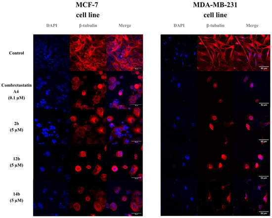
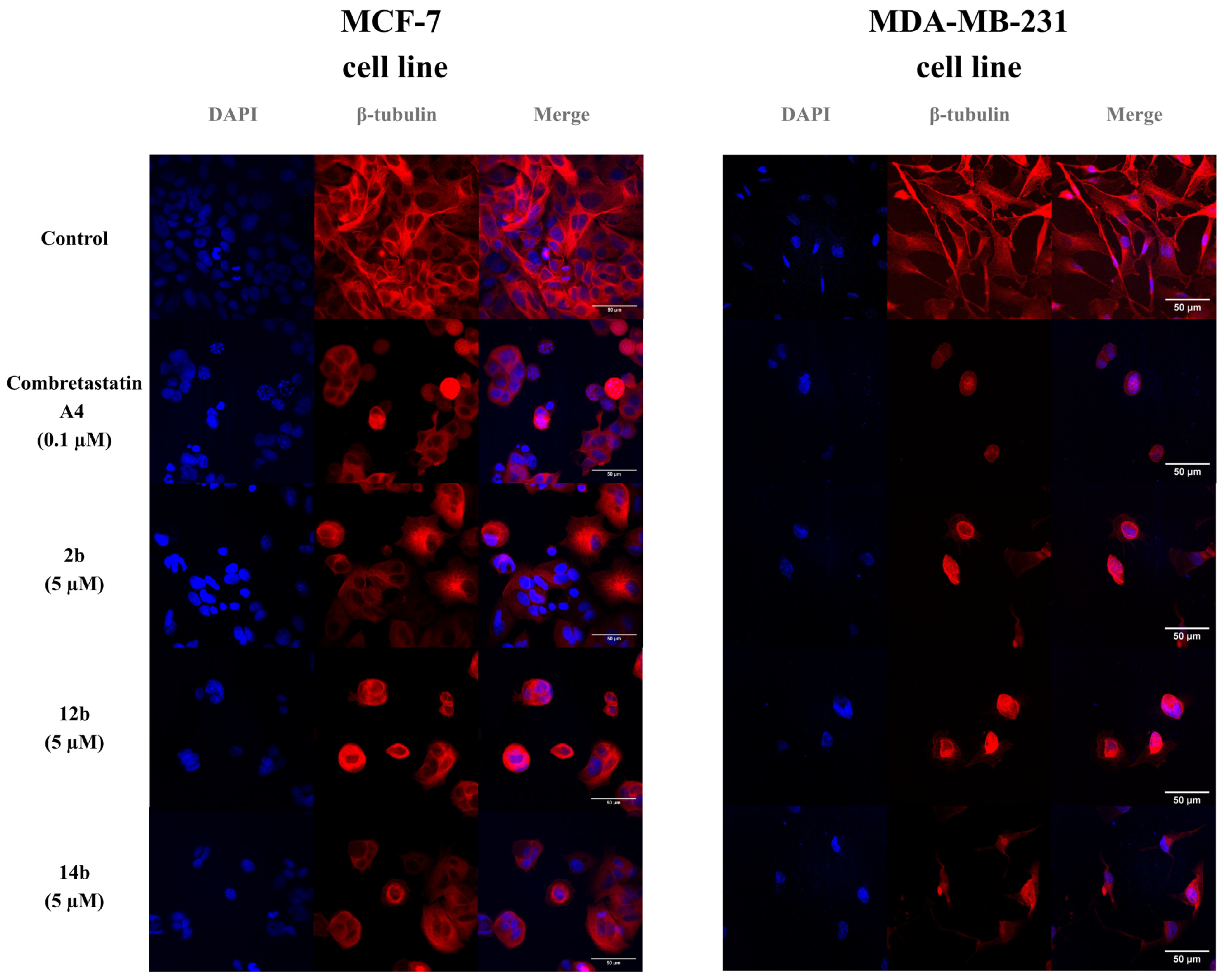
Cells were treated with either combretastatin A4 or compounds 2b, 12b, and 14b, followed by fixation and immunofluorescence staining for β–tubulin (red) and counterstaining with DAPI (blue) (Figure 5). In the control cells, both MCF-7 and MDA-MB-231 exhibited an extensive and well-organized microtubule network in the cytoplasm. Treatment with combretastatin A4 or compounds 2b, 12b, and 14b resulted in marked disruption of the microtubule architecture, accompanied by cell rounding and reduced organization in the β–tubulin pattern. The observed changes are consistent with the tubulin-targeting activity of the tested compounds.
Figure 5.
Representative confocal microscope images of MCF-7 and MDA-MB-231 cell lines treated with combretastatin A4 and compounds 2b, 12b, and 14b. Blue (left) indicates cell nuclei stained with DAPI, red indicates tubulin (middle), and the merged channels are presented on the (right). Scale bar: 50µM.
4. Discussion
In this study, we analyzed tubulin-inhibiting properties of 2b, 12b, and 14b in MCF-7 and MDA-MB-231 breast cancer cell lines. We first assessed β-tubulin concentration after treatment using the ELISA method. While molecular docking results showed lower binding affinity for 14b in comparison to Taxol (−6.829 vs. −7.709 kcal/mol), the in vitro analysis showed a much more promising result, with 14b causing an eight-fold reduction in β-tubulin levels in MCF-7 cells. This contributes to the existing limitation of the molecular docking assessment of potential binding affinity of novel structures towards the expected molecular targets, which may not correspond the actual biological affinity in living cells. Thus, such results should be treated as theoretical in nature and have to be confirmed via in vitro and/or in vivo assessment. All compounds significantly (up to eight-fold) inhibited β-tubulin concentration in both cell lines. As there are two main ways compounds can affect tubulin, namely, by decreasing its polymerization, similarly to Combretastatin A4, or by increasing its polymerization (Paclitaxel), we used the tubulin polymerization assay to demonstrate that our compounds exhibited Paclitaxel-like effects and increased tubulin polymerization (Figure 6). When microtubule polymerization occurs, there is a decrease in the concentration of free, unpolarized β-tubulin, which we observed in the ELISA test [21]. Additionally, by visualizing β-tubulin via confocal microscopy, we found that the tested compounds disrupted the organization of microtubules in cancer cells in both tested lines. Further research will be carried out to determine the exact mechanism of tubulin inhibition by the compounds 2b, 12b, and 14b, and whether they can achieve antitumor efficacy within an acceptable safety margin in vivo. Given their promising anticancer activity against MCF-7 and MDA-MB-231 breast cancer cell lines, further evaluation of their safety profile is warranted. Tubulin-targeting agents traditionally have a narrow therapeutic window because microtubules are ubiquitous across all eukaryotic cells. Tubulin-targeting drugs primarily target rapidly dividing cells to stop cancer growth; therefore, it is not surprising that they also target dividing non-cancerous cells. Since most healthy adult cells are in a G0 phase of the cell cycle, they are largely spared from the drug’s mechanisms. Conversely, the IC50 values of our compounds against normal cells are similar to those of doxorubicin, another cytostatic drug commonly used in the treatment of breast cancer. In vitro studies have demonstrated the enormous potential of 4-thiazolidinone derivatives for multi-targeted cancer cell control across multiple cell lines. Given these promising results, the next step would be to conduct safety testing of the tested compounds in vitro and in animal models.
Figure 6.
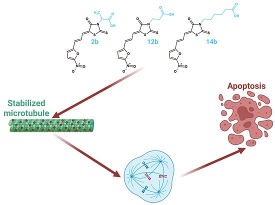
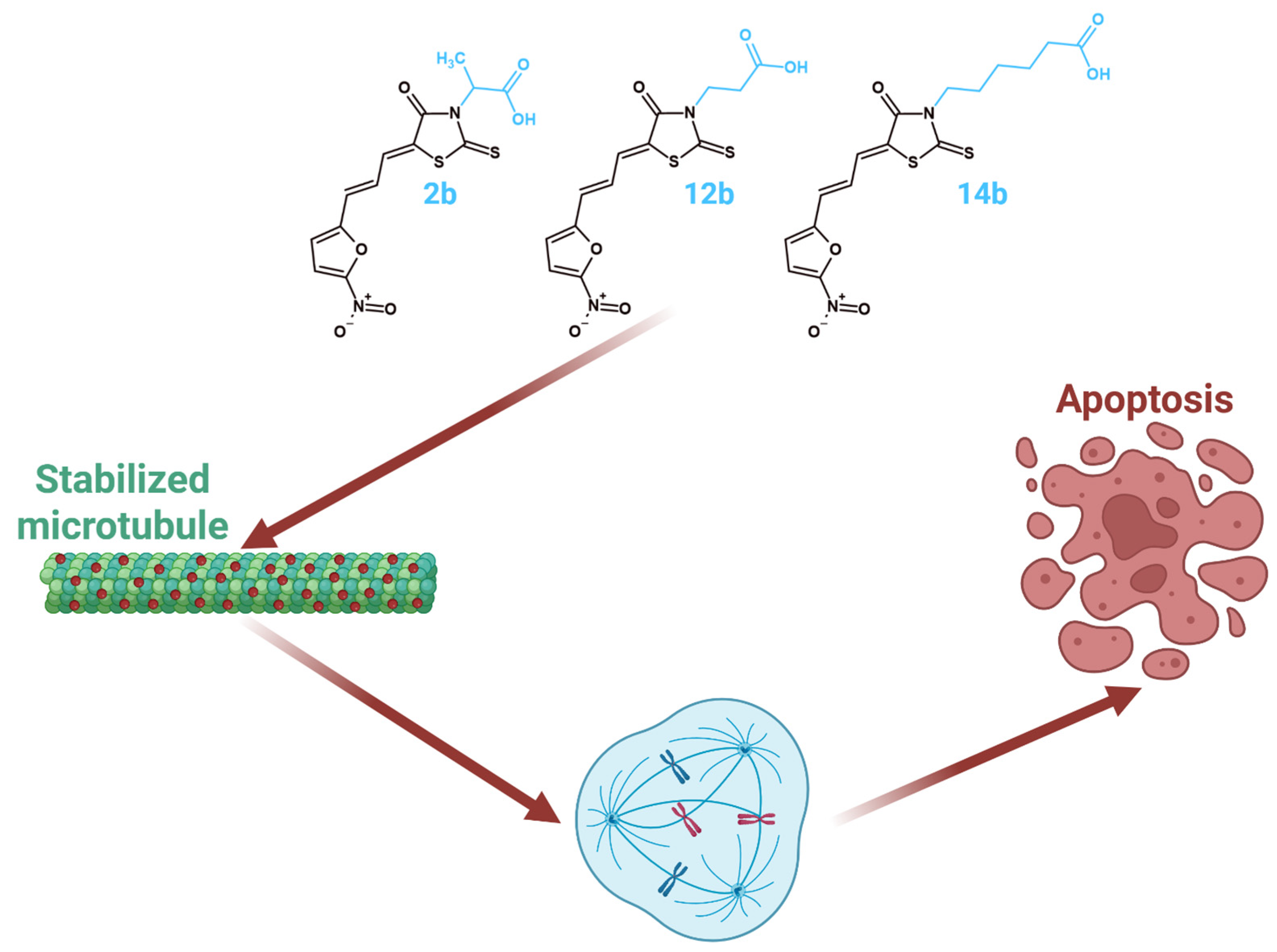
Compounds 2b, 12b, and 14b inhibit tubulin and promote apoptosis.
5. Conclusions
The three most active derivatives, 2b, 12b, and 14b, from the series of 5-((5-nitrofuran-2-yl)allylidene)-2-thioxo-4-thiazolidinones with cytotoxic properties against cancer cells were examined in a series of studies for tubulin inhibition on the MCF-7 and MDA-MB-231 breast cancer lines. Promising molecular docking results encouraged further analysis of those compounds’ effects against tubulin. A series of in vitro experiments were performed to provide further confirmation of the activity of 2b, 12b, and 14b on tubulin. An in vitro kinetic tubulin polymerization assay and ELISA assessment of tubulin protein concentrations post-treatment showed that all tested compounds exhibited tubulin-polymerizing capabilities and reduced tubulin protein levels post-treatment.
Supplementary Materials
The following supporting information can be downloaded at: https://www.mdpi.com/article/10.3390/scipharm94040087/s1.
Author Contributions
Conceptualization, M.P., R.D., K.B., A.B. and R.L.; methodology, M.P., R.D., O.K.S.-R., M.B.-K., Y.V. and A.G.; validation, M.P., R.D., K.B., A.B. and R.L.; formal analysis, K.B., A.B., Y.V. and R.L.; investigation, M.P., R.D., O.K.S.-R., M.B.-K. and A.G.; resources, K.B., A.B. and R.L.; data curation, M.P., R.D. and A.G.; writing—original draft preparation, M.P. and R.D.; writing—review and editing, M.P., R.D., K.B., A.B., Y.V. and R.L.; visualization, M.P., O.K.S.-R. and R.D.; supervision, K.B., A.B. and R.L.; project administration, K.B., A.B. and R.L.; funding acquisition, K.B., A.B. and R.L. All authors have read and agreed to the published version of the manuscript.
Funding
The research leading to these results has received funding from the Medical University of Białystok (Grants B.SUB.24.242 and B.SUB.23.158 (M.P.)) and from the National Research Foundation of Ukraine, under the project numbers 2023.05/0021 and 2023.03/0104.
Institutional Review Board Statement
Not applicable.
Informed Consent Statement
Not applicable.
Data Availability Statement
Data are contained within the article and Supplementary Materials.
Conflicts of Interest
The authors declare no conflicts of interest.
Abbreviations
The following abbreviations are used in this manuscript:
| Bcl-2 | B-cell lymphoma 2 |
| Bcl-xL | B-cell lymphoma-extra large |
| CA4 | Combretastatin A4 |
| BSA | Bovine serum albumin |
| DAPI | 4′,6-diamidino-2-phenylindole |
| DMEM | Dulbecco’s Modified Eagle Medium |
| DMSO | Dimethyl Sulfoxide |
| DNA | Deoxyribonucleic acid |
| DOX | Doxorubicin |
| EDTA | Ethylenediaminetetraacetic Acid |
| EGTA | Ethylene glycol-bis(beta-aminoethyl ether)-N,N,N’,N’-tetraacetic acid |
| ELISA | Enzyme-Linked Immunosorbent Assay |
| FBS | Fetal bovine serum |
| GTP | Guanosine triphosphate |
| MAPK | Mitogen-Activated Protein Kinase |
| IC50 | Half-maximum inhibitory concentration |
| OD | Optical density |
| PBS | Phosphate-Buffered Saline |
| PDB | Protein Data Bank |
| PIPES | piperazine-N,N′-bis(2-ethanesulfonic acid |
| PPARγ | Peroxisome proliferator-activated receptor gamma |
| ROS | Reactive Oxygen Species |
| RT | Room temperature |
| SD | Standard deviation |
| Vmax | Maximum velocity |
| ATCC | the American Type Culture Collection |
References
- Filho, A.M.; Laversanne, M.; Ferlay, J.; Colombet, M.; Piñeros, M.; Znaor, A.; Parkin, D.M.; Soerjomataram, I.; Bray, F. The GLOBOCAN 2022 Cancer Estimates: Data Sources, Methods, and a Snapshot of the Cancer Burden Worldwide. Int. J. Cancer 2025, 156, 1336–1346. [Google Scholar] [CrossRef] [Scilit]
- Mayrovitz, H.N. (Ed.) Breast Cancer; Exon Publications: Brisbane, Australia, 2022; ISBN 9780645332032. [Google Scholar]
- Zugazagoitia, J.; Guedes, C.; Ponce, S.; Ferrer, I.; Molina-Pinelo, S.; Paz-Ares, L. Current Challenges in Cancer Treatment. Clin. Ther. 2016, 38, 1551–1566. [Google Scholar] [CrossRef] [Scilit]
- Godinho, S.A.; Kwon, M.; Pellman, D. Centrosomes and Cancer: How Cancer Cells Divide with Too Many Centrosomes. Cancer Metastasis Rev. 2009, 28, 85–98. [Google Scholar] [CrossRef] [Scilit]
- Shuai, W.; Wang, G.; Zhang, Y.; Bu, F.; Zhang, S.; Miller, D.D.; Li, W.; Ouyang, L.; Wang, Y. Recent Progress on Tubulin Inhibitors with Dual Targeting Capabilities for Cancer Therapy. J. Med. Chem. 2021, 64, 7963–7990. [Google Scholar] [CrossRef] [Scilit]
- Sigalapalli, D.K.; Pooladanda, V.; Singh, P.; Kadagathur, M.; Guggilapu, S.D.; Uppu, J.L.; Tangellamudi, N.D.; Gangireddy, P.K.; Godugu, C.; Bathini, N.B. Discovery of Certain Benzyl/Phenethyl Thiazolidinone-Indole Hybrids as Potential Anti-Proliferative Agents: Synthesis, Molecular Modeling and Tubulin Polymerization Inhibition Study. Bioorg. Chem. 2019, 92, 103188. [Google Scholar] [CrossRef] [Scilit]
- Nikolaou, M.; Pavlopoulou, A.; Georgakilas, A.G.; Kyrodimos, E. The Challenge of Drug Resistance in Cancer Treatment: A Current Overview. Clin. Exp. Metastasis 2018, 35, 309–318. [Google Scholar] [CrossRef] [Scilit]
- Roszczenko, P.; Holota, S.; Szewczyk, O.K.; Dudchak, R.; Bielawski, K.; Bielawska, A.; Lesyk, R. 4-Thiazolidinone-Bearing Hybrid Molecules in Anticancer Drug Design. Int. J. Mol. Sci. 2022, 23, 13135. [Google Scholar] [CrossRef] [Scilit]
- Kaur Manjal, S.; Kaur, R.; Bhatia, R.; Kumar, K.; Singh, V.; Shankar, R.; Kaur, R.; Rawal, R.K. Synthetic and Medicinal Perspective of Thiazolidinones: A Review. Bioorg. Chem. 2017, 75, 406–423. [Google Scholar] [CrossRef] [Scilit]
- Nirwan, S.; Chahal, V.; Kakkar, R. Thiazolidinones: Synthesis, Reactivity, and Their Biological Applications. J. Heterocycl. Chem. 2019, 56, 1239–1253. [Google Scholar] [CrossRef] [Scilit]
- Podolak, M.; Horishny, V.; Dudchak, R.; Gornowicz, A.; Czarnomysy, R.; Mural, D.; Holota, S.; Bielawski, K.; Lesyk, R.; Bielawska, A. Synthesis and Anticancer Activity Evaluation of New 5-((5-Nitrofuran-2-Yl)Allylidene)-2-Thioxo-4-Thiazolidinones. Pharmaceuticals 2025, 18, 1598. [Google Scholar] [CrossRef] [Scilit]
- Weaver, B.A. How Taxol/Paclitaxel Kills Cancer Cells. Mol. Biol. Cell 2014, 25, 2677–2681. [Google Scholar] [CrossRef] [Scilit] [PubMed]
- Soni, J.P.; Chilvery, S.; Sharma, A.; Reddy, G.N.; Godugu, C.; Shankaraiah, N. Design, Synthesis and in Vitro Cytotoxicity Evaluation of Indolo–Pyrazoles Grafted with Thiazolidinone as Tubulin Polymerization Inhibitors. RSC Med. Chem. 2023, 14, 549–562. [Google Scholar] [CrossRef] [Scilit]
- Kellogg, E.H.; Hejab, N.M.A.; Howes, S.; Northcote, P.; Miller, J.H.; Díaz, J.F.; Downing, K.H.; Nogales, E. Insights into the Distinct Mechanisms of Action of Taxane and Non-Taxane Microtubule Stabilizers from Cryo-EM Structures. J. Mol. Biol. 2017, 429, 633–646. [Google Scholar] [CrossRef] [Scilit]
- Dudchak, R.; Podolak, M.; Sydorenko, I.; Czarnomysy, R.; Gornowicz, A.; Karpenko, O.; Holota, S.; Bielawska, A.; Bielawski, K.; Lesyk, R. 5-Ene-2-Arylaminothiazol-4(5H)-Ones Induce Apoptosis in Breast Cancer Cells. Cells 2025, 14, 861. [Google Scholar] [CrossRef] [Scilit]
- Huff, L.M.; Sackett, D.L.; Poruchynsky, M.S.; Fojo, T. Microtubule-Disrupting Chemotherapeutics Result in Enhanced Proteasome-Mediated Degradation and Disappearance of Tubulin in Neural Cells. Cancer Res. 2010, 70, 5870–5879. [Google Scholar] [CrossRef] [Scilit]
- Cleveland, D.W.; Lopata, M.A.; Sherline, P.; Kirschner, M.W. Unpolymerized Tubulin Modulates the Level of Tubulin MRNAs. Cell 1981, 25, 537–546. [Google Scholar] [CrossRef] [Scilit]
- Otto, A.M. Microtubules and DNA Replication. Int. Rev. Cytol. 1987, 109, 113–158. [Google Scholar] [CrossRef] [Scilit]
- Zhu, T.; Wang, S.-H.; Li, D.; Wang, S.-Y.; Liu, X.; Song, J.; Wang, Y.-T.; Zhang, S.-Y. Progress of Tubulin Polymerization Activity Detection Methods. Bioorg. Med. Chem. Lett. 2021, 37, 127698. [Google Scholar] [CrossRef] [Scilit]
- Cheriyamundath, S.; Mahaddalkar, T.; Kantevari, S.; Lopus, M. Induction of Acetylation and Bundling of Cellular Microtubules by 9-(4-Vinylphenyl) Noscapine Elicits S-Phase Arrest in MDA-MB-231 Cells. Biomed. Pharmacother. 2017, 86, 74–80. [Google Scholar] [CrossRef] [Scilit]
- Orr, G.A.; Verdier-Pinard, P.; McDaid, H.; Horwitz, S.B. Mechanisms of Taxol Resistance Related to Microtubules. Oncogene 2003, 22, 7280–7295. [Google Scholar] [CrossRef] [Scilit]

Disclaimer/Publisher’s Note: The statements, opinions and data contained in all publications are solely those of the individual author(s) and contributor(s) and not of MDPI and/or the editor(s). MDPI and/or the editor(s) disclaim responsibility for any injury to people or property resulting from any ideas, methods, instructions or products referred to in the content. |
© 2026 by the authors. Published by MDPI on behalf of the Österreichische Pharmazeutische Gesellschaft. Licensee MDPI, Basel, Switzerland. This article is an open access article distributed under the terms and conditions of the Creative Commons Attribution (CC BY) license.