


Abstract
Three series of benzylidene cinnamoylhydrazides (21 compounds) were synthesized and evaluated for cytotoxic activity against malignant and non-malignant cell lines. Among the compounds tested, 2d and 3c were the most active against Jurkat and CEM leukemia cells, with CC50 values in the low micromolar range. Both compounds were more potent against these leukemia cell lines than the reference compounds 5-fluorouracil, curcumin, and melphalan. However, 2d and 3c showed only modest selectivity over non-malignant Hs27 fibroblasts, with selectivity index values of 2.50 and 2.09, respectively, in CEM cells. Further studies in CEM cells showed that treatment with 2d and 3c was associated with apoptosis, caspase activation, increased ROS accumulation, mitochondrial membrane depolarization, and changes in cell-cycle distribution, with the most notable change being an increase in the sub-G0-G1 population. The compounds also showed growth-inhibitory and cytotoxic activity against several cell lines in the NCI-60 panel. Semi-empirical PM3 calculations suggested possible relationships between cytotoxic activity, electronic polarization of the α,β-unsaturated carbonyl system, and molecular dimensions. However, these relationships were not consistent across the series and should be considered preliminary. Overall, 2d and 3c represent useful hit structures for further investigation, although additional structural optimization and biological studies will be needed to improve their selectivity and better define their mechanisms of action.
1. Introduction
The discovery and development of novel antineoplastic agents with improved selectivity and reduced off-target toxicity remain important goals in medicinal chemistry. A major focus of our research program has been the design of conjugated unsaturated carbonyl compounds that may act as thiol-reactive hits [1]. It is hypothesized that such compounds preferentially interact with cellular thiols, such as glutathione or cysteine residues in proteins, rather than the amino or hydroxyl groups found in nucleic acids [2,3,4]. This mechanism could theoretically reduce the mutagenic and carcinogenic risks associated with traditional DNA-alkylating agents [5].
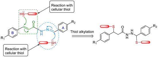
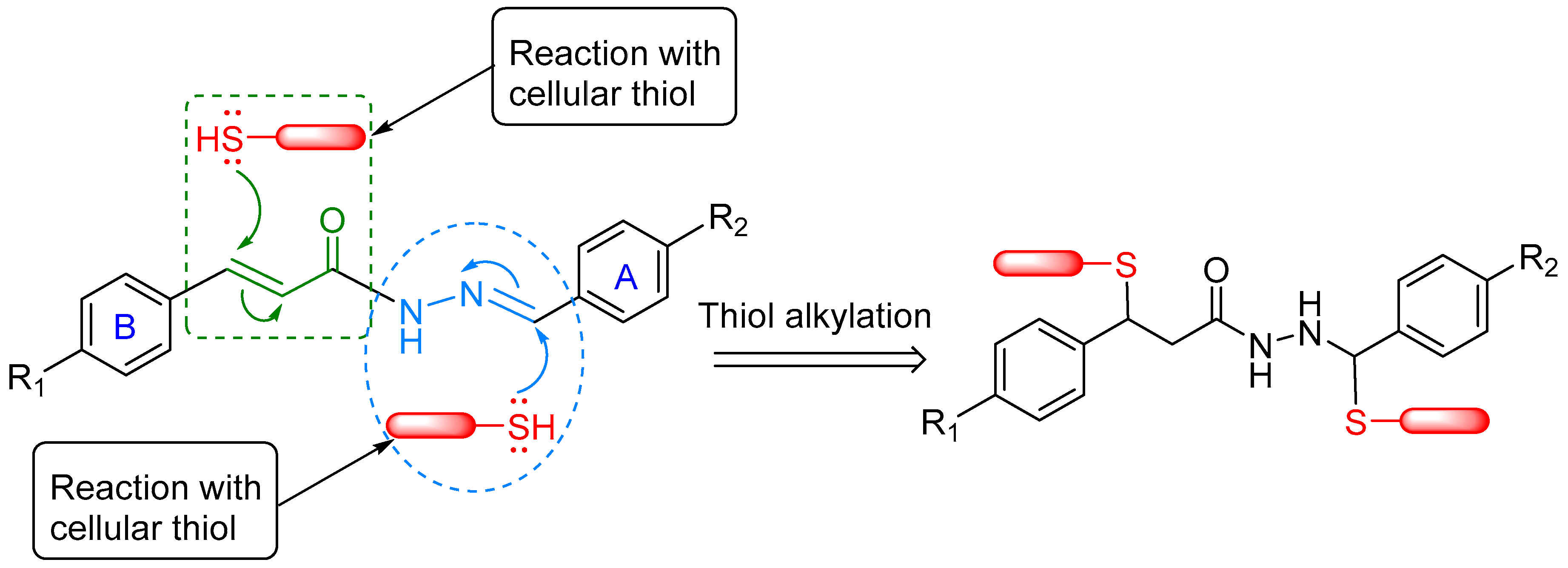
The present study describes the design and synthesis of three series of benzylidene cinnamoylhydrazides (Series 1–3) and examines their cytotoxic potential and structure–activity relationships (SAR). These compounds contain two potentially electrophilic sites, namely C7 of the α,β-unsaturated carbonyl system (adjacent to ring B) and C10 of the hydrazone moiety (adjacent to ring A), as shown in Figure 1. Both α,β-unsaturated carbonyl compounds [4,6,7] and N-acylhydrazones [8,9,10,11,12] have attracted considerable interest in drug design because of their synthetic accessibility and diverse biological activities, including anticancer activity. The presence of these two potentially electrophilic sites led us to hypothesize that the compounds might interact with cellular thiols, thereby contributing to their cytotoxic effects. However, thiol reactivity remains a working hypothesis in the present study. Other mechanisms associated with cytotoxicity, including apoptosis and alterations in cell-cycle progression, were also investigated.
Figure 1.
Possible modes of action of a representative compound with cellular constituents.
The three series were designed by varying the electronic, hydrophobic, and steric properties of aryl rings A and B (Figure 1) to examine how these changes affect the electrophilic properties of the scaffold. In Series 1, both aryl rings were modified, whereas Series 2 and Series 3 were designed to examine the effects of substituents on ring A or ring B separately. We were particularly interested in determining how these substituents affect the atomic charges at C7 (adjacent to ring B) and C10 (adjacent to ring A), the two predicted electrophilic sites. Semi-empirical PM3 calculations were therefore used to examine possible relationships between electronic polarization and cytotoxic activity. Based on these considerations, compounds 1a–e, 2a–i, and 3a–g were prepared.
Among the 21 compounds investigated in this study, 11 are novel derivatives, whereas 10 have been previously reported in different contexts [13,14,15,16]. For example, some cinnamoylhydrazide derivatives have previously been investigated for antitrypanosomal and antiviral activities [13,14,15,16]. The present work extends these earlier studies by systematically evaluating the three series against malignant and non-malignant cell lines and investigating potential mechanisms associated with the cytotoxicity of the most promising hit compounds. Specifically, their effects on apoptosis, ROS accumulation, mitochondrial membrane potential, caspase activation, and cell-cycle distribution were investigated.
2. Materials and Methods
2.1. Chemistry
2.1.1. General
Melting points were determined using an Electrochemical 1A9100 digital apparatus (Cole-Parmer Ltd., Stone, Staffordshire, UK) and are reported uncorrected. 1H and 13C NMR spectra were recorded on a Bruker Avance III 500 MHz spectrometer (Bruker Scientific LLC, Billerica, MA, USA) in either DMSO-d6 or CDCl3; chemical shifts (δ) are reported in ppm relative to tetramethylsilane (TMS). High-resolution mass spectrometry (HRMS) was performed on a Thermo Scientific Finnigan LTQ Orbitrap XL (Thermo Fisher Scientific, Waltham, MA, USA) using ESI in positive ion mode. Compound purity was verified using an Agilent 1200 HPLC equipped with a diode-array detector (Agilent Technologies, Inc., Santa Clara, CA, USA), and all final products exceeded 95% purity. HPLC analysis was performed using a Hypersil Gold column (column dimensions: 100 mm × 4.6 mm, particle size: 5 μm, Thermo Fisher Scientific, Waltham, MA, USA) and a linear gradient mobile phase starting at 65% water and 35% acetonitrile and increasing to 100% acetonitrile over 4 min with a flow rate of 1.0 mL/min, total run time 6 min. The column was re-equilibrated with a 1 mL/min flow of a 65% water/35% acetonitrile mixture for 1 min before injecting the next sample. 5 μL of solution in methanol of each sample was injected, and compounds were detected by UV absorbance at 250 nm. All reagents and solvents were used as received without further purification.
2.1.2. General Procedure for the Synthesis of Substituted E-Cinnamic Acids (6b–h)
A mixture of substituted benzaldehyde (8.0 mmol), malonic acid (1.83 g, 17.6 mmol), pyridine (3 mL), and 5 drops of piperidine was refluxed at 115 °C for 3 h. When the reaction was complete (checked with TLC), the hot reaction mixture was poured into a beaker containing a mixture of 60 mL 1 M HCl and ice. The precipitate formed was collected by vacuum filtration, washed with ice-cold water, and dried in the oven at 50 °C to give substituted E-cinnamic acid 6b–h with 80–90% yield.
2.1.3. General Procedure for the Synthesis of Cinnamoylhydrazides (7a–h)
To a solution of the corresponding cinnamic acid 6 (2.5 mmol) in dichloromethane (10 mL) and DMF (20 μL) in a 25 mL pear-shaped flask was added SOCl2 (5.0 mmol, 365 μL) dropwise under argon gas. The mixture was stirred overnight (about 16 h) at room temperature. After purging the reaction flask well with argon gas, the acyl chloride solution was collected using a syringe. The solution was then slowly added dropwise to a 50 mL round-bottom flask containing a stirred solution of hydrazine monohydrate (12.5 mmol, 626 mg) in acetonitrile (10 mL), with the flask cooled in an ice bath. When the addition was complete, the reaction mixture was allowed to warm to room temperature and stirred for 3 h. After that, the reaction mixture was transferred to a separatory funnel, treated with 50 mL 5% aqueous NaHCO3, and extracted with ethyl acetate (3 × 25 mL). The organic layers were combined and treated with anhydrous Na2SO4. After filtration, the solvents were removed by rotary evaporation, yielding the corresponding cinnamoylhydrazides 7a–h in 70–80% yield.
2.1.4. General Procedure for the Synthesis of N′-((E)-Benzylidene)cinnamohydrazides 1a–e, 2a–i, and 3a–g
To a solution of the corresponding cinnamoylhydrazides (7) (1.0 mmol) in ethanol (15 mL) was added a solution of the corresponding benzaldehyde (4) (2.0 mmol; except for 4-nitrobenzaldehyde, which was 1.0 mmol) in 5 mL of ethanol and 3–4 drops of glacial acetic acid. The resultant mixture was refluxed at 85 °C for 3 h. After that, the reaction mixture was concentrated using a rotary evaporator by evaporating 40–50% ethanol, forming a precipitate. The reaction mixture was cooled in an ice bath, and the precipitate was collected using vacuum filtration. The precipitate was dried in an oven at 50 °C to yield N′-((E)-benzylidene)cinnamohydrazides 1–3 in 85–90% yield.
Among the 10 known compounds (1a, 1c, 1d, 2a–e, 3b, and 3c), 1H NMR and melting points data for 1a, 1d, and 2a–e were previously reported [13,15]. However, compounds 1c, 3b, and 3c were previously reported as reaction intermediates without their spectral and melting point data [16]. Therefore, the NMR and melting points for compounds 1b, 1c, 1e, 2f–i, and 3a–g were reported below for the first time. The present data for compounds 1a, 1d, and 2a–e were similar to the previously reported data.
N′-((E)-Benzylidene)cinnamohydrazide (1a): Compound 1a was obtained as a white powder, yield 85.0%, melting point (MP): 227.6–228.0 °C, 1a exists in equilibrium between two stable conformers in NMR solution. 1H NMR (DMSO-d6): δ 11.75 & 11.58 (s, 1H, CONH), 8.31 & 8.12 (s, 1H, N=CH), 7.81 (m, 3H, 2 × Ar-H & 1 × CH=CHCO), 7.67–7.65 (s, br, 2H, Ar-H), 7.48 (s, br, 6H, ArH), 7.72 & 6.78 (d, 1H, CH=CHCO, 15.0 Hz) ppm. 13C NMR (DMSO-d6): δ 166.5, 162.0, 147.2, 143.7, 142.6, 141.1, 135.3, 135.1, 134.8, 134.7, 130.5, 130.4, 130.3, 129.5, 129.4, 129.3, 129.2, 128.7, 128.2, 127.6, 127.4, 120.7, 117.6 ppm. HRMS (ESI): m/z 273.0993 ([M + Na]+), calcd 273.0998 for C16H14N2O23Na.
N′-((E)-4-Chlorobenzylidene)-4-chlorocinnamohydrazide (1b): Compound 1b was obtained as a white powder, yield 89.3%, melting point (MP): 212.5–212.8 °C, 1b exists in equilibrium between two stable conformers in NMR solution. 1H NMR (DMSO-d6): δ 11.79 & 11.63 (s, 1H, CONH), 8.29 & 8.09 (s, 1H, N=CH), 7.85–7.78 (m, 3H, 2 × Ar-H & 1 × CH=CHCO), 7.73–7.69 (m, 2H, Ar-H), 7.54 (s, br, 4H, ArH), 7.63 & 6.76 (d, 1H, CH=CHCO, 15.0 Hz) ppm. 13C NMR (DMSO-d6): δ 166.4, 161.8, 146.0, 142.6, 141.4, 139.8, 135.0, 134.8, 134.7, 134.2, 134.1, 133.7, 133.6, 130.5, 129.9, 129.5, 129.4, 129.3, 129.2, 129.1, 121.5, 118.3 ppm. HRMS (ESI): m/z 341.0215 ([M + Na]+), calcd 341.0219 for C16H12N2O35Cl223Na.
N′-((E)-4-Methoxybenzylidene)-4-methoxycinnamohydrazide (1c): Compound 1c was obtained as a needle-shaped crystalline compound, yield 91.5%, melting point (MP): 225.9–226.8 C. 1c exists in equilibrium between two stable conformers in NMR solution. 1H NMR (DMSO-d6): δ 11.53 & 11.35 (s, 1H, CONH), 8.23 & 8.04 (s, 1H, N=CH), 7.75–7.70 (m, 3H, 2 × Ar-H & 1 × CH=CHCO), 7.65–7.61 (m, 2H, Ar-H), 7.04 (s, br, 4H, ArH), 7.50 & 6.60 (d, 1H, CH=CHCO, 15.0 Hz), 3.84 (s, 6H, OCH3) ppm. 13C NMR (DMSO-d6): δ 166.6, 162.1, 161.2, 161.1, 161.0, 146.7, 143.2, 142.1, 140.5, 130.3, 129.8, 129.1, 128.9, 128.0, 127.8, 127.4, 127.3, 118.3, 115.2, 114.9, 114.8, 114.7, 55.7 ppm. HRMS (ESI): m/z 333.1205 ([M + Na]+), calcd 333.1209 for C18H18N2O323Na.
N′-((E)-4-Methylbenzylidene)-4-methylcinnamohydrazide (1d): Compound 1d was obtained as a needle-shaped crystalline compound, yield 87.3%, melting point (MP): 235.0–235.5 C. 1d exists in equilibrium between two stable conformers in NMR solution. 1H NMR (DMSO-d6): δ 11.59 & 11.43 (s, 1H, CONH), 8.26 & 8.08 (s, 1H, N=CH), 7.67–7.55 (br, 5H, 4 × Ar-H & 1 × CH=CHCO), 7.29 (s, br, 4H, ArH), 7.50 & 6.70 (d, 1H, CH=CHCO, 15.0 Hz), 2.38 (s, 6H, Ar-CH3) ppm. 13C NMR (DMSO-d6): δ 166.5, 162.0, 147.0, 143.7, 142.5, 140.9, 140.3, 140.0, 132.6, 132.4, 132.1, 132.0, 130.1, 130.0, 129.9, 129.8, 128.7, 128.2, 127.6, 127.4, 119.7, 116.6, 21.5 ppm. HRMS (ESI): m/z 301.1305 ([M + Na]+), calcd 301.1311 for C18H18N2O23Na.
N′-((E)-4-Nitrobenzylidene)-4-nitrocinnamohydrazide (1e): Compound 1e was obtained as a white powder, yield 84.5%, melting point (MP): 281.9–282.6 °C, 1e exists in equilibrium between two stable conformers in NMR solution. 1H NMR (DMSO-d6): δ 12.14 & 12.02 (s, 1H, CONH), 8.41 & 8.22 (s, 1H, N=CH), 8.33 (s, br, 4H, ArH), 8.12–7.96 (m, br, 4H, 4 × Ar-H), 7.84–7.80 (br, 1H, CH=CHCO), 7.80 & 6.96 (d, 1H, CH=CHCO, 15.0 Hz) ppm. HRMS (ESI): m/z 341.0880 ([M + H]+), calcd 341.0881 for C16H13N4O5.
N′-((E)-4-Chlorobenzylidene)cinnamohydrazide (2a): Compound 2a was obtained as a white powder, yield 88.5%, melting point (MP): 202.1–202.5 °C, 2a exists in equilibrium between two stable conformers in NMR solution. 1H NMR (DMSO-d6): δ 11.79 & 11.63 (s, 1H, CONH), 8.29 & 8.10 (s, 1H, N=CH), 7.85–7.80 (m, 3H, 2 × Ar-H & 1 × CH=CHCO), 7.72–7.62 (m, 2H, Ar-H), 7.54–7.48 (m, br, 5H, ArH), 7.71 & 6.76 (d, 1H, CH=CHCO, 15.0 Hz) ppm. 13C NMR (DMSO-d6): δ 166.6, 162.0, 145.9, 142.8, 142.4, 141.2, 135.3, 135.1, 135.0, 134.7, 133.8, 133.6, 130.5, 130.4, 129.5, 129.4, 129.3, 129.2, 129.1, 128.7, 120.6, 117.5 ppm. HRMS (ESI): m/z 307.0604 ([M + Na]+), calcd 307.0609 for C16H13N2O35Cl23Na.
N′-((E)-4-Fluorobenzylidene)cinnamohydrazide (2b): Compound 2b was obtained as a white powder, yield 85.4%, melting point (MP): 189.0–189.3 °C, 2b exists in equilibrium between two stable conformers in NMR solution. 1H NMR (DMSO-d6): δ 11.74 & 11.58 (s, 1H, CONH), 8.30 & 8.11 (s, 1H, N=CH), 7.88–7.79 (m, 3H, 2 × Ar-H & 1 × CH=CHCO), 7.72–7.63 (m, 2H, Ar-H), 7.48 (s, br, 3H, ArH), 7.32 (s, br, 2H, ArH), 7.71 & 6.77 (d, 1H, CH=CHCO, 15.0 Hz) ppm. 13C NMR (DMSO-d6): δ 166.5, 164.6, 164.4, 162.6, 162.4, 162.0, 146.1, 142.6, 142.5, 141.1, 135.3, 135.2, 131.4, 131.3, 130.5, 130.3, 129.8, 129.7, 129.6, 129.5, 129.5, 129.4, 128.7, 128.2, 120.7, 117.6, 116.4, 116.2 ppm. HRMS (ESI): m/z 291.0898 ([M + Na]+), calcd 291.0904 for C16H13N2OF23Na.
N′-((E)-4-Methoxybenzylidene)cinnamohydrazide (2c): Compound 2c was obtained as a needle-shaped crystalline compound, yield 89.0%, melting point (MP): 216.8–218.1 C. 2c exists in equilibrium between two stable conformers in NMR solution. 1H NMR (DMSO-d6): δ 11.58 & 11.42 (s, 1H, CONH), 8.25 & 8.06 (s, 1H, N=CH), 7.78–7.66 (m, 5H, 4 × Ar-H & 1 × CH=CHCO), 7.48 (s, br, 3H, Ar-H), 7.05 (s, br, 2H, ArH), 7.73 & 6.76 (d, 1H, CH=CHCO, 15.0 Hz), 3.84 (s, 3H, OCH3) ppm. 13C NMR (DMSO-d6): δ 166.3, 161.8, 161.4, 161.2, 147.2, 143.6, 142.3, 140.7, 135.3, 130.2, 129.4, 129.2, 128.9, 128.6, 128.1, 127.4, 121.1, 118.0, 114.8, 55.8 ppm. HRMS (ESI): m/z 303.1098 ([M + Na]+), calcd 303.1104 for C17H16N2O223Na.
N′-((E)-4-Methylbenzylidene)cinnamohydrazide (2d): Compound 2d was obtained as a white powder, yield 90.7%, melting point (MP): 214.8–215.7 °C, 2d exists in equilibrium between two stable conformers in NMR solution. 1H NMR (DMSO-d6): δ 11.54 & 11.38 (s, 1H, CONH), 8.30 & 8.09 (s, 1H, N=CH), 7.77–7.67 (br, 5H, 4 × Ar-H & 1 × CH=CHCO), 7.48 (s, br, 3H, ArH), 7.30 (s, br, 2H, ArH), 7.67 & 6.76 (d, 1H, CH=CHCO, 15.0 Hz), 2.38 (s, 3H, Ar-CH3) ppm. 13C NMR (DMSO-d6): δ 166.4, 161.9, 147.3, 143.8, 142.5, 140.9, 140.4, 140.0, 135.4, 135.2, 132.1, 132.0, 130.5, 130.3, 129.9, 129.5, 129.4, 128.7, 128.2, 127.6, 127.4, 120.9, 117.8, 21.5 ppm. HRMS (ESI): m/z 287.1149 ([M + Na]+), calcd 287.1154 for C17H16N2O23Na.
N′-((E)-4-Nitrobenzylidene)cinnamohydrazide (2e): Compound 2e was obtained as a white powder, yield 85.5%, melting point (MP): 226.9–227.8 °C, 2e exists in equilibrium between two stable conformers in NMR solution. 1H NMR (DMSO-d6): δ 12.01 & 11.85 (s, 1H, CONH), 8.38 & 8.19 (s, 1H, N=CH), 8.31 (s, br, 2H, ArH), 8.08–8.01 (m, 2H, Ar-H), 7.81–7.63 (m, 3H, 2 × Ar-H & 1 × CH=CHCO), 7.49 (s, br, 3H, ArH), 7.77 & 6.78 (d, 1H, CH=CHCO, 15.0 Hz) ppm. 13C NMR (DMSO-d6): δ 166.8, 162.2, 148.2, 148.1, 144.7, 143.2, 141.8, 141.3, 141.1, 141.0, 135.1, 135.0, 130.7, 130.5, 129.5, 129.4, 128.8, 128.5, 128.3, 124.5, 120.3, 117.2 ppm. HRMS (ESI): m/z 318.0843 ([M + Na]+), calcd 318.0849 for C16H13N3O323Na.
N′-((E)-4-Ethylbenzylidene)cinnamohydrazide (2f): Compound 2f was obtained as a white powder, yield 92.0%, 2f exists in equilibrium between two stable conformers in NMR solution. 1H NMR (DMSO-d6): δ 11.68 & 11.51 (s, 1H, CONH), 8.26 & 8.08 (s, 1H, N=CH), 7.78 (d, 1H, CH=CHCO, 15 Hz), 7.72–7.66 (m, 4H, ArH), 7.49–7.44 (m, 3H, ArH), 7.32 (d, 2H, ArH, 8 Hz), 7.69 & 6.76 (d, 1H, CH=CHCO, 15.0 Hz), 2.66 (q, 2H, Ar-CH2, 8 Hz), 1.22 (t, 3H, CH3, 8 Hz) ppm. 13C NMR (DMSO-d6): δ 166.4, 161.9, 147.3, 146.6, 146.3, 143.8, 142.5, 141.0, 135.3, 135.2, 132.3, 132.2, 130.5, 130.3, 129.5, 129.4, 128.7, 128.2, 127.7, 127.4, 120.7, 117.7, 28.6, 15.8 ppm. HRMS (ESI): m/z 301.1313 ([M + Na]+), calcd 301.1311 for C18H18N2O23Na.
N′-((E)-2-Methylbenzylidene)cinnamohydrazide (2g): Compound 2g was obtained as a white crystalline powder, yield 87.7%. 2g exists in equilibrium between two stable conformers in NMR solution. 1H NMR (DMSO-d6): δ 11.75 & 11.49 (s, 1H, CONH), 8.56 & 8.39 (s, 1H, N=CH), 7.90 (dd, 1H, ArH, 8 Hz), 7.76–7.60 (m, 3H, 2 × Ar-H & 1 × CH=CHCO), 7.48–7.46 (m, 3H, ArH), 7.34–7.27 (m, 3H, ArH), 7.62 & 6.75 (d, 1H, CH=CHCO, 16.0 Hz), 2.47 (s, 3H, Ar-CH3) ppm. 13C NMR (DMSO-d6): δ 166.4, 161.9, 145.8, 142.8, 142.5, 141.1, 137.3, 137.0, 135.3, 135.1, 132.7, 132.6, 131.4, 131.3, 130.5, 130.4, 130.2, 129.9, 129.5, 129.4, 128.6, 128.2, 126.7, 126.6, 126.4, 120.7, 117.7, 19.8, 19.5 ppm. HRMS (ESI): m/z 287.1155 ([M + Na]+), calcd 287.1155 for C17H16N2O23Na.
N′-((E)-3-Methylbenzylidene)cinnamohydrazide (2h): Compound 2h was obtained as a white crystalline powder, yield 83.5%. 2h exists in equilibrium between two stable conformers in NMR solution. 1H NMR (DMSO-d6): δ 11.74 & 11.54 (s, 1H, CONH), 8.26 & 8.09 (s, 1H, N=CH), 7.79 (d, 1H, ArH, 8 Hz), 7.72–7.67 (m, 2H, 1 × Ar-H & 1 × CH=CHCO), 7.62–7.54 (m, 2H, ArH), 7.49–7.46 (m, 3H, ArH), 7.37 (t, 1H, ArH, 8 Hz), 7.27 (d, 1H, ArH, 8 Hz), 7.62 & 6.77 (d, 1H, CH=CHCO, 16.0 Hz), 2.38 (s, 3H, Ar-CH3) ppm. 13C NMR (DMSO-d6): δ 166.4, 162.0, 147.3, 144.0, 142.6, 141.1, 138.6, 138.5, 135.3, 135.1, 134.7, 134.6, 131.3, 131.0, 130.5, 130.4, 129.5, 129.4, 129.2, 129.1, 128.7, 128.2, 128.0, 127.9, 125.0, 124.5, 120.7, 117.6, 21.4, 21.3 ppm. HRMS (ESI): m/z 287.1156 ([M + Na]+), calcd 287.1155 for C17H16N2O23Na.
N′-((E)-4-Carboxybenzylidene)cinnamohydrazide (2i): Compound 2i was obtained as a white powder, yield 82.7%, 2i exists in equilibrium between two stable conformers in NMR solution. 1H NMR (DMSO-d6): δ 13.05 (s, br, 1H, COOH), 11.89 & 11.71 (s, 1H, CONH), 8.35 & 8.17 (s, 1H, N=CH), 8.05 (d, 2H, ArH, 8 Hz), 7.94–7.88 (m, 2H, ArH), 7.81 (m, 1H, CH=CHCO), 7.73–7.68 (dd, 2H, ArH, 8 & 4 Hz), 7.48 (m, br, 3H, ArH), 7.62 & 6.78 (d, 1H, CH=CHCO, 16.0 Hz) ppm. 13C NMR (DMSO-d6): δ 167.4, 166.7, 162.1, 146.0, 143.0, 142.6, 141.4, 140.6, 138.8, 135.2, 135.1, 132.1, 132.0, 130.6, 130.4, 130.2, 129.5, 129.4, 128.8, 128.3, 127.6, 127.4, 120.5, 117.4 ppm. HRMS (ESI): m/z 295.1079 ([M + H]+), calcd 295.1077. for C17H15N2O3.
N′-((E)-Benzylidene)-4-chlorocinnamohydrazide (3a): Compound 3a was obtained as a white powder, yield 90.0%, melting point (MP): 216.1–216.7 °C, 3a exists in equilibrium between two stable conformers in NMR solution. 1H NMR (DMSO-d6): δ 11.72 & 11.57 (s, 1H, CONH), 8.30 & 8.11 (s, 1H, N=CH), 7.82–7.66 (m, 5H, 4 × Ar-H & 1 × CH=CHCO), 7.54–7.49 (m, 5H, Ar-H), 7.71 & 6.76 (d, 1H, CH=CHCO, 16.0 Hz) ppm. 13C NMR (DMSO-d6): δ 166.3, 161.8, 147.4, 143.8, 141.2, 139.7, 134.9, 134.8, 134.7, 134.3, 134.2, 130.5, 130.4, 130.3, 129.9, 129.5, 129.4, 129.3, 127.6, 127.4, 121.7, 118.5 ppm. HRMS (ESI): m/z 307.0601 ([M + Na]+), calcd 307.0609 for C16H13N2O35Cl23Na.
N′-((E)-Benzylidene)-4-methoxycinnamohydrazide (3b): Compound 3b was obtained as needle shaped crystalline compound, yield 86.7%, melting point (MP): 221.0–221.8 °C, 3b exists in equilibrium between two stable conformers in NMR solution. 1H NMR (DMSO-d6): δ 11.66 & 11.48 (s, 1H, CONH), 8.29 & 8.10 (s, 1H, N=CH), 7.80–7.62 (m, 5H, 4 × Ar-H & 1 × CH=CHCO), 7.47 (s, br, 3H, Ar-H), 7.04 (s, br, 2H, ArH), 7.70 & 6.62 (d, 1H, CH=CHCO, 15.0 Hz), 3.83 (s, 3H, OCH3) ppm. 13C NMR (DMSO-d6): δ 166.8, 162.3, 161.3, 161.2, 146.8, 143.3, 142.4, 140.9, 134.9, 134.8, 130.4, 130.2, 129.9, 129.3, 128.0, 127.7, 127.5, 127.3, 118.1, 114.9, 55.8 ppm. HRMS (ESI): m/z 303.1100 ([M + Na]+), calcd 303.1104 for C17H16N2O223Na.
N′-((E)-Benzylidene)-4-methylcinnamohydrazide (3c): Compound 3c was obtained as a white powder, yield 91.4%, melting point (MP): 241.7–242.6 °C, 3c exists in equilibrium between two stable conformers in NMR solution. 1H NMR (DMSO-d6): δ 11.69 & 11.53 (s, 1H, CONH), 8.29 & 8.11 (s, 1H, N=CH), 7.80–7.77 (br, 2H, 1 × Ar-H & 1 × CH=CHCO), 7.68 (br, 2H, ArH), 7.57 (s, br, 1H, ArH), 7.48 (s, br, 3H, ArH), 7.30 (s, br, 2H, ArH), 7.62 & 6.71 (d, 1H, CH=CHCO, 15.0 Hz), 2.38 (s, 3H, Ar-CH3) ppm. 13C NMR (DMSO-d6): δ 166.6, 162.1, 147.0, 143.5, 142.6, 141.1, 140.4, 140.2, 134.8, 134.7, 132.6, 132.4, 130.5, 130.3, 130.1, 130.0, 129.3, 129.3, 128.7, 128.2, 127.6, 127.4, 119.6, 116.4, 21.5 ppm. HRMS (ESI): m/z 287.1150 ([M + Na]+), calcd 287.1154 for C17H16N2O23Na.
N′-((E)-Benzylidene)-4-nitrocinnamohydrazide (3d): Compound 3d was obtained as a white powder, yield 85.5%, melting point (MP): 205.9–208.2 °C, 3d exists in equilibrium between two stable conformers in NMR solution. 1H NMR (DMSO-d6): δ 11.86 & 11.71 (s, 1H, CONH), 8.31 (s, br, 2H, ArH), 8.12–8.08 (m, br, 1H, N=CH), 7.94 (m, 1H, Ar-H), 7.82–7.77 (m, 4H, 3 × Ar-H & 1 × CH=CHCO), 7.48 (s, br, 3H, ArH), 7.90 & 6.94 (d, 1H, CH=CHCO, 15.0 Hz) ppm. 13C NMR (DMSO-d6): δ 165.9, 161.2, 148.3, 148.2, 147.8, 144.2, 141.8, 141.7, 140.1, 138.6, 134.6, 134.5, 131.6, 130.7, 130.4, 129.8, 129.3, 127.7, 127.5, 125.1, 124.6, 124.5, 123.5, 122.0 ppm. HRMS (ESI): m/z 318.0843 ([M + Na]+), calcd 318.0849 for C16H13N3O323Na.
N′-((E)-Benzylidene)-4-ethylcinnamohydrazide (3e): Compound 3e was obtained as a white powder, yield 84.0%, 3e exists in equilibrium between two stable conformers in NMR solution. 1H NMR (DMSO-d6): δ 11.71 & 11.52 (s, 1H, CONH), 8.29 & 8.10 (s, 1H, N=CH), 7.81–7.76 (m, 2H, ArH), 7.71–7.66 (m, 1H, CH=CHCO), 7.62–7.58 (m, 2H, ArH), 7.50–7.48 (m, 3H, ArH), 7.32 (m, 2H, ArH), 7.69 & 6.72 (d, 1H, CH=CHCO, 16.0 Hz), 2.68 (m, 2H, Ar-CH2), 1.22 (t, 3H, CH3, 8 Hz) ppm. 13C NMR (DMSO-d6): δ 166.6, 162.1, 147.0, 146.6, 146.5, 143.6, 142.6, 141.1, 134.8, 134.7, 132.8, 132.7, 130.5, 130.3, 129.3, 128.9, 128.9, 128.8, 128.3, 127.6, 127.4, 119.7, 116.6, 28.5, 15.8 ppm. HRMS (ESI): m/z 301.1313 ([M + Na]+), calcd 301.1311 for C18H18N2O23Na.
N′-((E)-Benzylidene)-2-methylcinnamohydrazide (3f): Compound 3f was obtained as white crystalline powder, yield 91.4%, 3f exists in equilibrium between two stable conformers in NMR solution. 1H NMR (DMSO-d6): δ 11.76 & 11.61 (s, 1H, CONH), 8.31 & 8.12 (s, 1H, N=CH), 7.99–7.88 (d, 1H, CH=CHCO, 16 Hz), 7.78 (d, 2H, Ar-H, 8 Hz), 7.64–7.54 (m, 1H, ArH), 7.49–7.47 (m, 3H, ArH), 7.33–7.31 (m, 3H, ArH), 7.82 & 6.68 (d, 1H, CH=CHCO, 16.0 Hz), 2.45 & 2.44 (s, 3H, Ar-CH3) ppm. 13C NMR (DMSO-d6): δ 166.6, 162.0, 147.3, 143.8, 139.9, 138.5, 137.7, 137.5, 134.8, 134.6, 134.1, 134.0, 131.3, 131.2, 130.5, 130.3, 130.2, 130.1, 129.3, 127.6, 127.3, 127.0, 126.6, 121.9, 118.7, 19.9, 19.8 ppm. HRMS (ESI): m/z 287.1156 ([M + Na]+), calcd 287.1155 for C17H16N2O23Na.
N′-((E)-Benzylidene)-3-methylcinnamohydrazide (3g): Compound 3g was obtained as white crystalline powder, yield 84.5%, 3g exists in equilibrium between two stable conformers in NMR solution. 1H NMR (DMSO-d6): δ 11.73 & 11.55 (s, 1H, CONH), 8.29 & 8.12 (s, 1H, N=CH), 7.81–7.77 (m, 2H, ArH), 7.72–7.58 (m, 2H, 1 × Ar-H & 1 × CH=CHCO), 7.49–7.45 (m, 4H, ArH), 7.39–7.34 (m, 1H, ArH), 7.28–7.25 (t, 1H, ArH, 8 Hz), 7.72 & 6.75 (d, 1H, CH=CHCO, 16.0 Hz), 2.39 & 2.37 (s, 3H, Ar-CH3) ppm. 13C NMR (DMSO-d6): δ 167.0, 162.4, 147.6, 144.2, 143.2, 141.6, 139.1, 135.6, 135.5, 135.2, 135.1, 131.7, 131.5, 130.9, 130.7, 129.8, 129.7, 129.7, 129.7, 129.1, 128.0, 127.8, 126.2, 125.9, 121.0, 117.8, 21.8 ppm. HRMS (ESI): m/z 287.1156 ([M + Na]+), calcd 287.1155 for C17H16N2O23Na.
2.1.5. Procedure for the Synthesis of 5-(4-Methoxyphenyl)pyrazolidin-3-one (9)
To a solution of ethyl 4-methoxycinnamate (8) (412 mg, 2.0 mmol) in 5 mL ethanol, hydrazine monohydrate (16.0 mmol) was added, followed by 3 drops of glacial acetic acid. The reaction mixture was refluxed at 85 °C for 20 h. The solvent was then removed by rotary evaporation. The residue was redissolved in 10 mL of ethyl acetate, transferred to a separatory funnel, and washed with 10 mL of water. The organic phase was dried over anhydrous sodium sulfate, filtered, and evaporated using a rotary evaporator to yield 332 mg of crude 5-(4-methoxyphenyl)pyrazolidin-3-one (9). The crude product was analyzed by 1H NMR. The NMR data were identical to the previously reported data [17]. Crude yield 86.5%, 1H NMR (CDCl3): δ 2.39 (1H, dd, J = 14.0, 8.0 Hz), 2.93 (1H, dd, J = 14.0, 7.7 Hz), 3.76 (3H, s), 4.25 (1H, s), 4.57 (1H, t, J = 7.5 Hz), 6.80 (2H, d, J = 8.6 Hz), 7.09 (2H, d, J = 8.6 Hz), 9.30 (1H, s).
2.1.6. Calculation of Charge Density
All quantum chemical calculations were performed using the Spartan Student software package v8.0.6 [18]. The three-dimensional structures of all compounds in the series were built in silico. To obtain optimized geometries and atomic charges, each structure was subjected to a geometry optimization calculation to find the lowest-energy conformation. These calculations were performed using the semi-empirical PM3 method. To simulate a physiological environment, the Equilibrium Geometry was calculated in water as the solvent. The resulting atomic charges for specific atoms (C-7, C-8, and C-10) were extracted from the optimized structures for use in the structure–activity relationship analysis.
2.2. Biological Assays
2.2.1. Cell Lines and Culture Conditions
RPMI-1640 culture medium (Hyclone, Logan, UT, USA), supplemented with 10% fetal bovine serum (FBS), 100 U/mL penicillin and 100 μg/mL streptomycin (Lonza, Walkersville, MD, USA), was used for the lymphoma-leukemia CEM and Jurkat cell lines. Dulbecco’s Modified Eagle’s Medium (DMEM; CORNING, Corning, NY, USA), supplemented with 10% fetal bovine serum (FBS) and 100 U/mL of penicillin and 100 μg/mL of streptomycin, was used to culture the non-malignant Hs27 fibroblast cell line. All three cell lines were grown at 37 °C in a 5% CO2 humidified atmosphere. All cell lines were purchased from ATCC. The ATCC catalog numbers for Jurkat, CEM, and HS27 are TIB-152, CCL-119, and CRL-1634, respectively. Culture media, supplements, incubation conditions, and passage range are based strictly on handling instructions provided by ATCC. Mycoplasma-testing is done routinely following standard procedure, and we have never had mycoplasma contamination. After thawing cell vials, the cells were not used for more than 10 passages to ensure they were not exhausted or altered/mutated.
2.2.2. Differential Nuclear Staining Assay (DNS)
To determine the cytotoxic activity of compound series 1–3 in cancerous and non-cancerous cell lines, the DNS assay was used [19]. 10,000 cells per well were seeded in 96-well plates at a volume of 100 μL of media per well. The plates were then incubated overnight. Cells were then treated with a concentration gradient ranging from 1 μM to 40 μM. DMSO (1% v/v) was used as the vehicle control, untreated cells as the negative control, and H2O2 (1 mM) as the positive control. Independent biological experiments were performed three times, and each experiment had three technical replicates. After treatment, plates were incubated for 48 h. Two hours before the end of the 48-h period, a mixture of Hoescht 33342 and propidium iodide (PI) in phosphate-buffered saline (PBS) was added to each well. After incubation, the plates were read using the ImageXpress PICO bioimager (Molecular Devices, San Jose, CA, USA). This instrument provides a percentage of cell death and total cell count per well.
2.2.3. Cytotoxic Concentration 50% (CC50) and Selective Cytotoxicity Values
The CC50 values, or cytotoxic concentration at which treated compounds kill 50% of the cell population, were obtained by using the cytotoxicity percentage acquired from a secondary screening and a linear interpolation equation (https://www.johndcook.com/interpolator.html, accessed on 6 January 2025) [19]. Cells were exposed to each compound for 48 h at various concentrations, then a dose–response curve for each compound was used to determine the CC50 of the corresponding compound. Dose–response curves are provided as the Supplementary Materials.
To determine the CC50 values after 24 h of treatment, CEM cells were exposed to a concentration range of compounds 2d and 3c for 24 h under the conditions described above. The resulting concentration-response data were used to calculate the 24-h CC50 values by linear interpolation. The resulting values were 4.33 μM for 2d and 4.18 μM for 3c. The corresponding concentration-response curves are provided in the Supplementary Materials. The 24-h CC50 and 2× CC50 concentrations were used in most experiments to determine the dose–response effects of each compound on cells.
The selectivity index (SI) values represent the ability of compounds 2d and 3c to selectively kill cancer cells without significantly damaging normal, non-cancerous cells. The SI was calculated by dividing the CC50 value for Hs27 cells by the CC50 value for the Jurkat and CEM leukemia cell lines, respectively.
2.2.4. Annexin V-FITC Propidium Iodide Assay
The externalization of phosphatidylserine, which is an indicator of early-stage apoptosis, was measured using the Annexin V-FITC and PI kit (Beckman Coulter, Miami, FL, USA) [19]. CCRF-CEM cells were seeded in a 24-well plate at a density of 50,000 cells/well in 1 mL of medium per well and incubated overnight at 37 °C. The next day, treatments (2d CC50, 2d CC50 × 2, 3c CC50, 3c CC50 × 2, 1 mM H2O2, 1% v/v DMSO) were added, and untreated cells were used as the negative control. Independent biological experiments were performed three times, and each experiment had three technical wells. After treatment, the plate was incubated for 24 h in optimal cell culture conditions (37 °C, in a 5% CO2-humidified atmosphere). Cells were harvested into flow cytometry tubes and centrifuged for 5 min at 1200 rpm at 4 °C. The supernatant of each sample was discarded carefully without disturbing the pellet. 500 μL of PBS was added to each sample, and they were centrifuged once again with the same settings; the supernatant was discarded afterward. The cells were then resuspended in 100 μL of a mixture containing 1× binding buffer, 1 μL of Annexin V-FITC, and 2.5 μL of PI per sample. The samples were then incubated for 30 min on ice in the dark. After incubation, the samples were carefully vortexed at low speed and immediately read on the flow cytometer.
2.2.5. Mitochondrial Depolarization Assay
To examine the effects of compounds 2d and 3c on mitochondrial membrane potential, CCRF-CEM cells were evaluated after treatment using the MitoProbe JC-1 assay kit (Molecular Probes, Eugene, OR, USA; M34152) [19]. CCRF-CEM cells were seeded at a density of 50,000 cells/well in 1 mL of media per well in a 24-well plate and incubated overnight at 37 °C. The next day, treatments (2d CC50, 2d CC50 × 2, 3c CC50, 3c CC50 × 2, 1 mM H2O2, 1% v/v DMSO) were added, and untreated cells were used as the negative control. Independent biological experiments were performed three times, and each experiment had three technical wells. The plate was incubated for 6 h under optimal cell culture conditions (37 °C, in a 5% CO2-humidified atmosphere). Samples were then harvested into flow cytometry tubes and centrifuged for 5 min at 1200 rpm at 4 °C. The supernatant was discarded, and the cells were resuspended in 500 uL of PBS containing 5 μL of the JC-1 fluorescent dye and incubated for 30 min at 37 °C. After incubation, 1 mL of PBS was added to each sample, and cells were centrifuged again using the same centrifuge settings previously described. The supernatant was discarded, and the cells were resuspended in 500 μL of PBS and read in the flow cytometer.
2.2.6. Reactive Oxygen Species (ROS) Assay
To identify if compounds 2d and 3c induced accumulation of ROS in CCRF-CEM cells, carboxy-H2DCFDA (6-carboxy-2′,7′-dichlorodihydrofluorescein diacetate) fluorescein reagent (Invitrogen; C400, Thermo Fisher Scientific, Eugene, OR, USA) was used [19]. CCRF-CEM cells were seeded at a density of 50,000 cells/well in 1 mL of media per well in a 24-well plate and incubated overnight at 37 °C. The next day, treatments (2d CC50, 2d CC50 × 2, 3c CC50, 3c CC50 × 2, 1 mM H2O2, 1% v/v DMSO) were added, and untreated cells were used as the negative control. Independent biological experiments were performed three times, and each experiment had three technical wells. The plate was then incubated for 18 h under optimal cell culture conditions (37 °C, in a 5% CO2-humidified atmosphere). Samples were then harvested into flow cytometry tubes and centrifuged for 5 min at 1200 rpm at 4 °C. The supernatant was discarded, and cells were resuspended in 1 mL of pre-warmed PBS with carboxy-H2DCFDA to a final concentration of 10 μM. The samples were incubated for 1 h in optimal cell culture conditions, then centrifuged using the same settings previously described. The supernatant was discarded, and cells were resuspended in 500 μL of PBS, allowed to recover for 30 min, and immediately analyzed by flow cytometry.
2.2.7. Caspase-3/7 Detection Assay
Activation of caspase-3/7 in cells was monitored using the NucView 488 caspase-3/7 enzyme-substrate (Biotium, Hayward, CA, USA) [19]. CCRF-CEM cells were seeded at a density of 50,000 cells/well in 1 mL of media per well in a 24-well plate and incubated overnight at 37 °C. The next day, treatments (2d CC50, 2d CC50 × 2, 3c CC50, 3c CC50 × 2, 1 mM H2O2, 1% v/v DMSO) were added, and untreated cells were used as the negative control. Independent biological experiments were performed three times, and each experiment had three technical wells. The plate was then incubated for 6 h under optimal cell culture conditions (37 °C, in a 5% CO2-humidified atmosphere). Samples were then harvested into flow cytometry tubes and centrifuged for 5 min at 1200 rpm at 4 °C. The supernatant was discarded, and the cells were resuspended in 500 μL of PBS containing the NucView 488 caspase-3/7 enzyme-substrate at a concentration of 5 μm. The samples were then incubated at room temperature in the dark for 30 min. Afterward, 300 uL of pre-warmed PBS was added to each sample, and cells were immediately analyzed by flow cytometry.
2.2.8. Analysis of Cell Cycle
To analyze the effect of compounds 2d and 3c on the progression of the cell cycle, total cellular DNA content was measured [19]. CCRF-CEM cells were seeded at a density of 25,000 cells/well in 1 mL of media per well in a 24-well plate and incubated overnight at 37 °C. The next day, lower treatments {2d (0.54, 1.08, and 2.16 μM), or 3c (0.52, 1.05, and 2.09 μM), 100 μM Etoposide, 1% v/v DMSO} were added; untreated cells were used as the negative control. Independent biological experiments were performed three times, and each experiment had three technical wells. Lower concentrations were used to prevent excessive DNA fragmentation and/or cell death. The plate was then incubated for 72 h under optimal cell culture conditions (37 °C, in a 5% CO2-humidified atmosphere). After the incubation period, the cells were centrifuged, decanted, and resuspended in 200 μL of nuclear isolation media (NIM)-DAPI solution (Beckman Coulter), and cells were then immediately analyzed in the flow cytometer.
2.2.9. NCI-60 HTS384 Screening
Cell viability was evaluated using the NCI-60 HTS384 high-throughput screening methodology at the National Cancer Institute [20]. Cells were seeded into white 384-well plates at densities optimized for each cell line (250–2500 cells per well) in RPMI-1640 medium supplemented with 5% fetal bovine serum and 2 mM L-glutamine (40 µL per well). After a 24 h incubation at 37 °C in a humidified atmosphere containing 5% CO2 to allow cell attachment, test compounds were transferred to assay plates using acoustic dispensing (100 nL per well) to achieve the desired final concentrations with a final DMSO concentration of 0.25% (v/v). Plates were incubated for 72 h under the same conditions. Cell viability was then quantified by addition of CellTiter-Glo reagent (40 µL per well, Promega Corporation, Madison, WI, USA) followed by luminescence measurement using a plate reader, which correlates with intracellular ATP levels as an indicator of metabolically active cells. Vehicle controls and cytotoxic reference compounds were included on each plate for normalization and quality control. Percent growth relative to vehicle-treated controls and time-zero measurements was calculated, and dose–response curves were used to determine GI50 (50% growth inhibition), TGI (total growth inhibition), and LC50 values (50% lethal concentration).
3. Results and Discussion
3.1. Synthesis and Structural Characterization
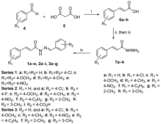
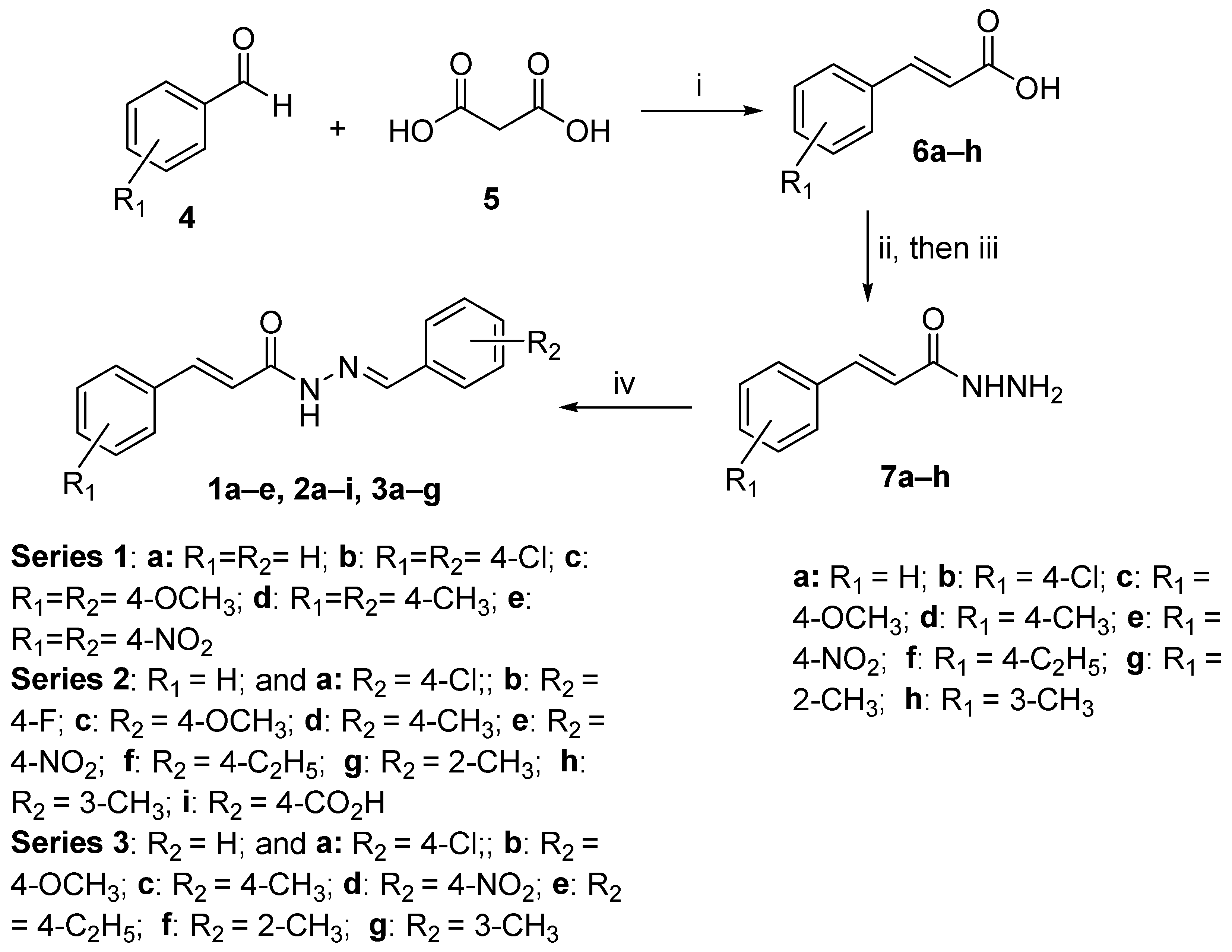
Compounds 1a–e, 2a–i, and 3a–g were prepared from E-cinnamic acids (6a–h) using previously reported procedures, as portrayed in Scheme 1. The non-commercially available E-cinnamic acids (6b–h) were prepared by following the Knoevenagel–Doebner reaction of the corresponding benzaldehyde with malonic acid (5) in a pyridine/piperidine mixture under reflux conditions [13]. The cinnamic acids (6a–h) were then converted to the corresponding cinnamoylhydrazides (7a–h) by treatment with thionyl chloride (SOCl2) in dichloromethane, followed by reaction with hydrazine in acetonitrile at room temperature [21].
Scheme 1.
Synthesis of benzylidene cinnamoylhydrazides 1–3. Reagents and conditions: (i) pyridine, piperidine, reflux; (ii) SOCl2, CH2Cl2; (iii) NH2NH2, CH3CN; (iv) R2-benzaldehyde, AcOH, EtOH, reflux.
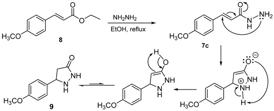
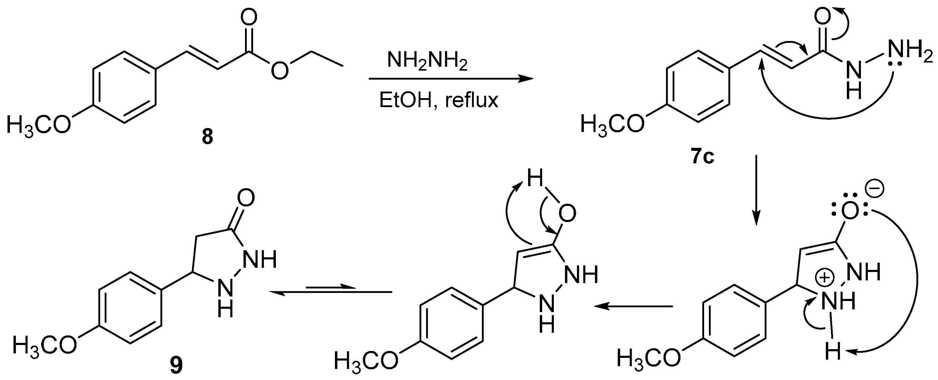
An initial attempt was made to prepare 4-methoxycinnamoylhydrazide (7c) from ethyl 4-methoxycinnamate (8) by reaction with hydrazine in refluxing ethanol. Under these conditions, however, the expected hydrazide 7c was not obtained; instead, cyclization occurred to form compound 9, as shown in Scheme 2 [17]. The 1H NMR spectrum of 9 was consistent with the previously reported data [17]. Therefore, cinnamoylhydrazides 7a–h were prepared using the SOCl2/hydrazine route as shown in Scheme 1. Finally, condensation of the cinnamoylhydrazides 7a–h with appropriate benzaldehydes in ethanol in the presence of acetic acid afforded benzylidene cinnamoylhydrazides 1a–e, 2a–i, and 3a–g [22].
Scheme 2.
Cyclization of hydrazide 7c to give compound 9 under reflux conditions [17].
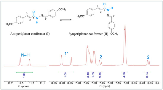
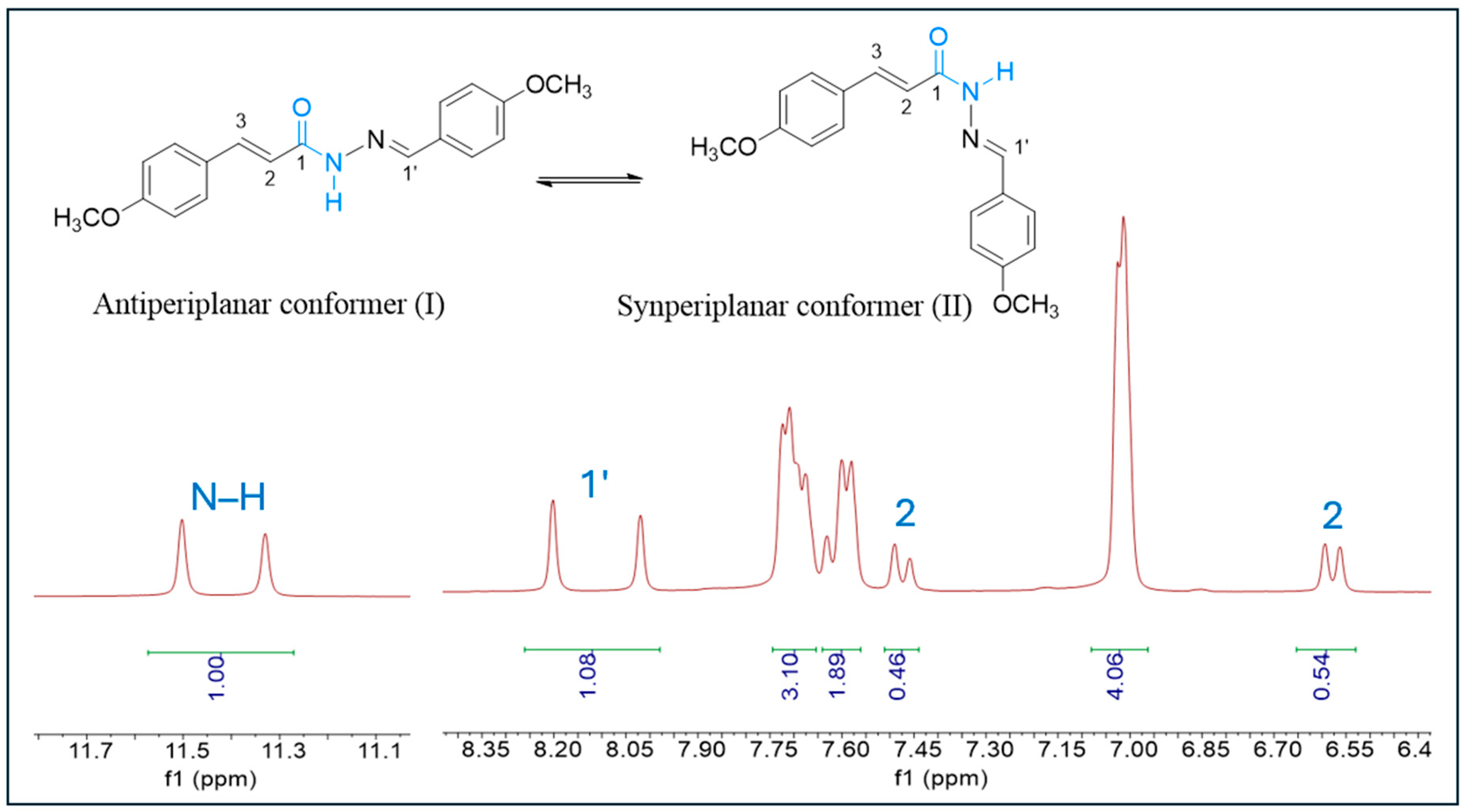
The synthesized compounds were characterized by NMR spectroscopy and high-resolution mass spectrometry. The 1H NMR spectra showed that compounds in Series 1–3 exist in equilibrium between two stable conformers (I and II) in solution, likely because of restricted rotation around the amide bond (Figure 2) [23,24]. As a result, some proton signals, particularly those corresponding to NH and positions 1′ and 2, appeared as two sets of signals (Figure 2).
Figure 2.
1H NMR spectrum of compound 1c. Because of equilibrium between the two conformers (I and II), the proton signals corresponding to N–H, 1′, and 2 appear as two signals of nearly equal intensity.
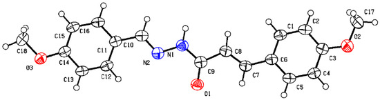
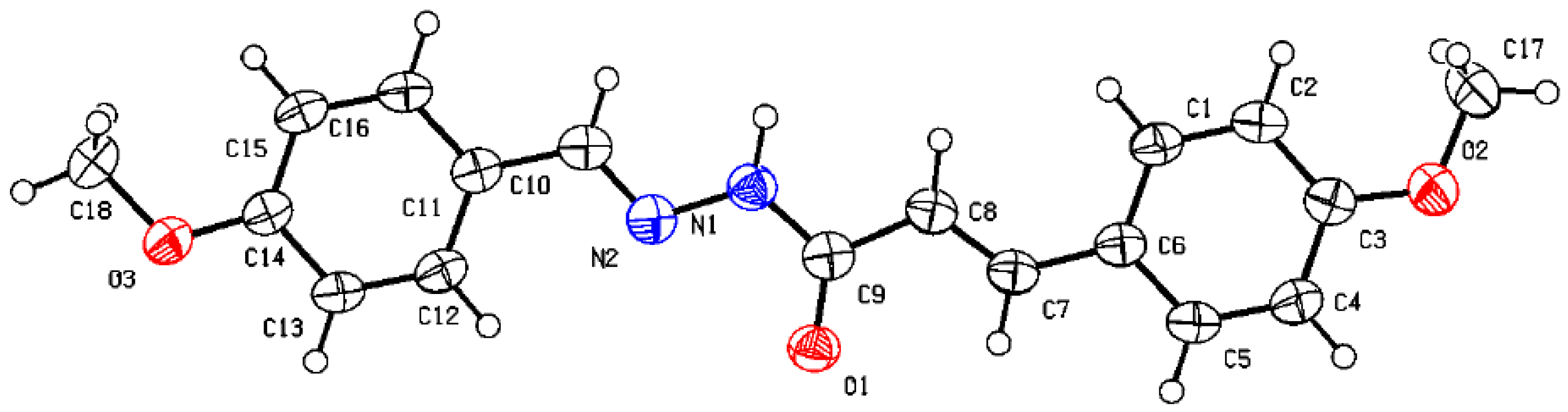
Compound 1c was also examined by X-ray crystallography to obtain additional structural information [25]. The ORTEP diagram is shown in Figure 3. In the solid state, 1c adopts an almost planar conformation, with both double bonds in the E configuration. The C5–C6–C7–C8 and C1–C6–C7–C8 torsion angles are 175.2° and −6.0°, respectively, whereas the N2–C10–C11–C16 and N2–C10–C11–C12 torsion angles are −174.7° and 5.4°, respectively. These values indicate that the two aryl rings are nearly coplanar with their adjacent unsaturated groups. Although 1c itself was inactive in biological assays, its crystal structure provides useful structural information about this scaffold. However, the solid-state conformation of 1c may not represent the preferred conformations of 2d, 3c, or other compounds under biological conditions.
Figure 3.
The ORTEP diagram of compound 1c [25].
3.2. Cytotoxic Evaluation
Compounds 1a–e, 2a–i, and 3a–g were evaluated for cytotoxic activity against Jurkat and CEM leukemia cells, and the results are presented in Table 1. CC50 values were acquired after 48 h. The reference drugs 5-fluorouracil, curcumin, and melphalan were evaluated under the same conditions. Among the compounds tested, 2d and 3c were most active, with CC50 values in the low micromolar range. Compound 2d showed CC50 values of 2.93 ± 0.35 µM against Jurkat cells and 1.59 ± 0.04 µM against CEM cells. The corresponding values for 3c were 3.40 ± 0.36 and 3.09 ± 0.33 µM, respectively.
Table 1.
Cytotoxic evaluation of compounds 1a–e, 2a–i, 3a–g, and anticancer drugs against human T-cell lymphoma Jurkat and CEM cells after 48 h.
Structural changes around 2d and 3c had a marked effect on cytotoxic activity. For example, compounds 2f–i, which are structurally related to 2d, were less active than 2d. Similarly, compounds 3e–g were less active than 3c. These results suggest that relatively small structural changes in either aryl ring can substantially affect the cytotoxic activity of this scaffold.
Under the conditions used in this study, 2d and 3c were more potent against the leukemia cell lines than the reference compounds 5-fluorouracil, curcumin, and melphalan. However, greater cytotoxic potency does not necessarily indicate better therapeutic potential, particularly when selectivity toward malignant cells is limited. Compounds 2d and 3c were therefore also evaluated against non-malignant Hs27 fibroblasts. Their CC50 values were 3.97 ± 0.18 and 6.46 ± 0.15 µM, respectively, giving selectivity index (SI) values of 2.50 for 2d and 2.09 for 3c with CEM, whereas SI values were 1.35 for 2d and 1.90 for 3c with Jurkat. Thus, both compounds showed only modest selectivity for the leukemia cells over Hs27 cells. Although additional “normal” cell lines could have been tested, the modest selectivity indicates that the compounds are not highly selective toward cancer cells. This is an important limitation of the present study. Moreover, because only one non-malignant cell line was examined, no general conclusions about tumor selectivity can be drawn from these results. Further structural optimization and evaluation against additional non-malignant cell lines will be needed to determine whether the selectivity of this class of compounds can be improved.
Based on their activity against Jurkat and CEM cells, 2d and 3c were selected for further evaluation using the NCI-60 HTS384 screening platform [15,16]. Selected results with LC50 < 100 μM are summarized in Table 2. The complete NCI-60 screening results, including concentration/growth curves for each cell line, are provided in the Supplementary Materials.
Table 2.
Evaluation of the antiproliferative effects of compounds 2d and 3c against representative cell lines from the NCI-60 human tumor cell line panel (HTS384 screen) *.
Both compounds showed growth-inhibitory and cytotoxic activity against several cell lines in the NCI-60 panel. Notable activity was observed against several leukemia cell lines. For example, 2d showed GI50 values of 0.195, 0.206, 0.177, and 0.145 µM against HL-60(TB), MOLT-4, RPMI-8226, and SR cells, respectively. Compound 3c also showed submicromolar GI50 values against these cell lines. Activity was also observed against selected non-small-cell lung cancer, colon cancer, melanoma, ovarian cancer, and breast cancer cell lines. These findings show that the cytotoxic effects of 2d and 3c are not limited to the two leukemia cell lines used in the initial screening. However, because their activity varied considerably across the NCI-60 panel, these results should not be considered evidence of broad-spectrum antitumor activity.
3.3. Mechanistic Evaluation of Hit Compounds and
3.3.1. Apoptosis and Caspase Activation
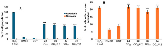
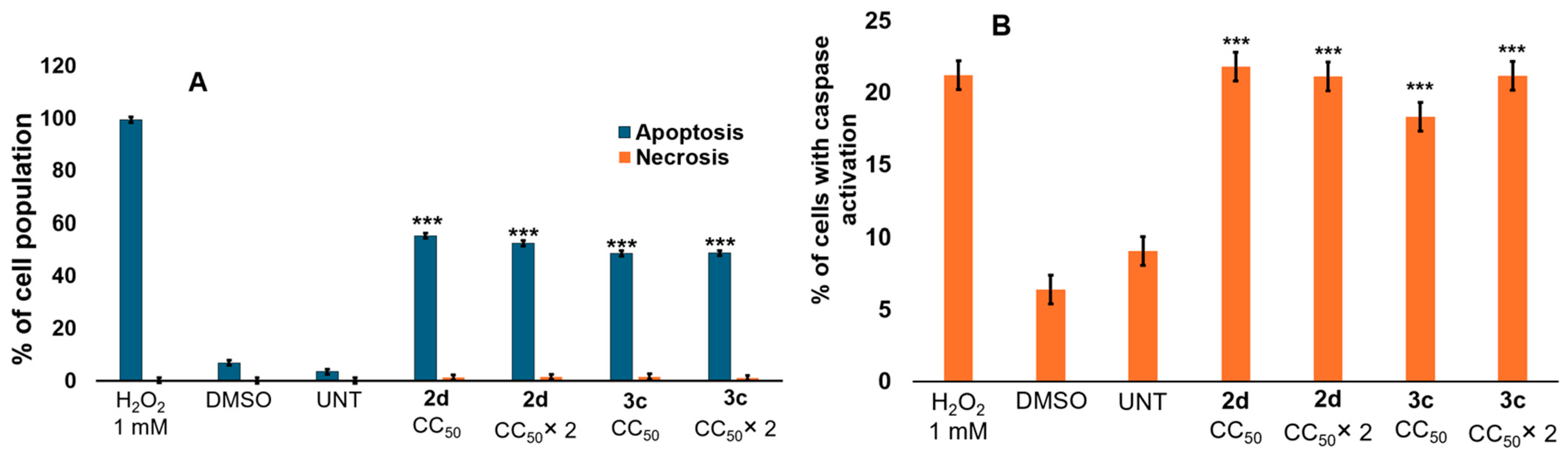
To investigate the cellular effects associated with the cytotoxicity of 2d and 3c, phosphatidylserine externalization was examined in CEM cells using Annexin V-FITC/PI staining followed by flow cytometry. As shown in Figure 4A, treatment with either compound increased the apoptotic cell population compared with the vehicle and untreated controls. The increase was observed at both the CC50 and 2× CC50 concentrations. Under these experimental conditions, apoptosis represented the major form of cell death observed, whereas necrosis remained relatively low.
Figure 4.
Compounds 2d and 3c induce apoptotic cell death and caspase activation in CEM cells. (A) Phosphatidylserine externalization was evaluated by Annexin V-FITC/PI staining and flow cytometry. Treatment with 2d or 3c increased the apoptotic cell population in CEM cells. The percentages shown represent total apoptosis (early and late apoptosis). (B) Caspase activation was observed after 6 h of treatment with 2d or 3c, as determined by NucView 488 fluorescence and flow cytometry. For both experiments, cells were treated with 2d CC50 (4.33 μM) and 2× CC50 (8.66 μM) or with 3c CC50 (4.18 μM) and 2× CC50 (8.36 μM). DMSO (1%) was used as the vehicle control, H2O2 (1 mM) as the positive control, and untreated cells (Unt) as the negative control. The 24-h CC50 values were used. Bars represent the mean of three independent biological experiments, each performed with three technical replicates, and error bars indicate the standard deviation. Statistical significance was determined by one-way ANOVA followed by Dunnett’s multiple-comparisons test, comparing each compound’s CC50 and CC50 × 2 with the DMSO control. One-way ANOVA and Dunnett’s test results were F(2,24) = 305.21, p < 0.001, d = 2.39 for 2d and F(2,24) = 178.02, p < 0.001, d = 2.39 for 3c in the Annexin-V assay. One-way ANOVA and Dunnett’s test results were F(2,24) = 50.33, p < 0.001, d = 2.39 for 2d and F(2,23) = 41.86, p < 0.001, d = 2.398 for 3c in the Caspase-3/7 assay. *** p < 0.001.
Caspase activation was also examined using the NucView 488 assay. Treatment with 2d or 3c increased the proportion of cells positive for the caspase substrate compared with the vehicle control (Figure 4B). Together with the Annexin V-FITC/PI results, these findings support the involvement of apoptosis in the cytotoxic effects of 2d and 3c. Caspase-3 is an important executioner caspase involved in apoptotic cell death [26,27]. However, activation of an executioner caspase alone cannot distinguish between the intrinsic and extrinsic apoptotic pathways. Therefore, although these results support the involvement of apoptosis, they do not establish which apoptotic pathway is responsible.
3.3.2. ROS Accumulation and Mitochondrial Membrane Depolarization
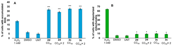
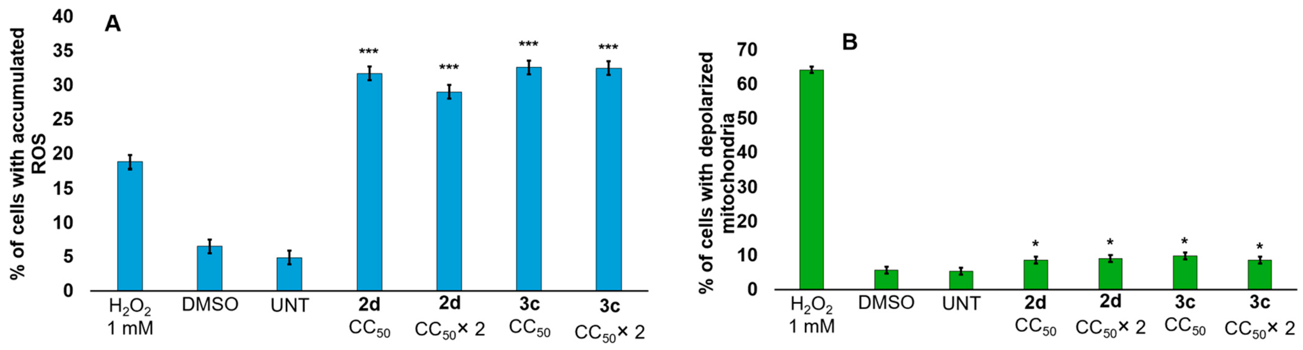
The effects of 2d and 3c on intracellular ROS levels were examined using carboxy-H2DCFDA. As shown in Figure 5A, treatment with either compound markedly increased H2DCFDA fluorescence compared with the vehicle and untreated controls, indicating an increase in intracellular oxidative activity. The effects of 2d and 3c on mitochondrial membrane potential were also investigated using the JC-1 assay. Treatment with 2d or 3c increased the population of cells showing mitochondrial membrane depolarization (Figure 5B). These results indicate that exposure to the compounds is associated with both increased oxidative stress and mitochondrial dysfunction.
Figure 5.
Compounds 2d and 3c increase ROS accumulation and mitochondrial membrane depolarization in CEM cells. (A) Increased ROS accumulation was observed in CEM cells after 18 h of treatment with 2d or 3c. Cells were stained with carboxy-H2DCFDA and analyzed by flow cytometry. (B) Mitochondrial membrane depolarization was observed after 6 h of treatment with 2d or 3c, as determined using the JC-1 assay and flow cytometry. For both experiments, cells were treated with 2d at CC50 (4.33 μM) and 2× CC50 (8.66 μM) or with 3c at CC50 (4.18 μM) and 2× CC50 (8.36 μM). DMSO (1%) was used as the vehicle control, H2O2 (1 mM) as the positive control, and untreated cells (Unt) as the negative control. The 24-h CC50 values were used. Bars represent the mean of three independent biological experiments, each performed with three technical replicates, and error bars indicate the standard deviation. Statistical significance was determined by one-way ANOVA followed by Dunnett’s multiple-comparisons test, comparing each compound’s CC50 and CC50 × 2 with the DMSO control. One-way ANOVA and Dunnett’s test results were F(2,15) = 73.03, p < 0.001, d = 2.49 for 2d and F(2,15) = 125.2, p < 0.001, d = 2.49 for 3c in the ROS assay. One-way ANOVA and Dunnett’s test results were F(2,22) = 4.35, p < 0.05, d = 2.41 for 2d and F(2,24) = 5.4, p < 0.05, d = 2.39 for 3c in the JC-1 assay. * p < 0.05, *** p < 0.001.
Taken together with the Annexin V-FITC/PI and caspase results, these observations are consistent with apoptotic cell death associated with oxidative stress and mitochondrial dysfunction. However, the present experiments do not establish that ROS accumulation is responsible for mitochondrial depolarization or apoptosis. In particular, ROS-scavenger rescue experiments were not performed, and no experiments involving caspase inhibition, caspase-9 activation, or cytochrome c release were conducted. Therefore, the present data are not sufficient to establish a ROS-dependent intrinsic apoptotic pathway.
3.3.3. Effects on Cell-Cycle Distribution
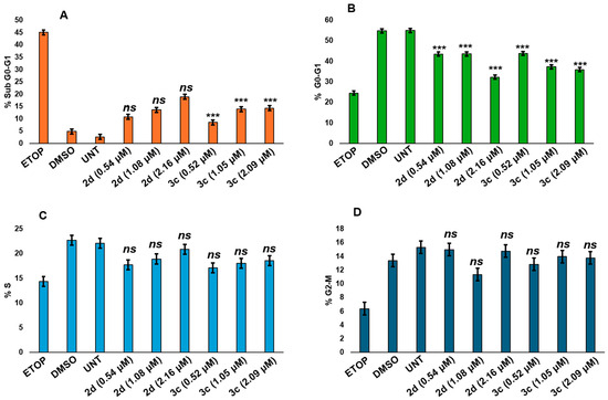
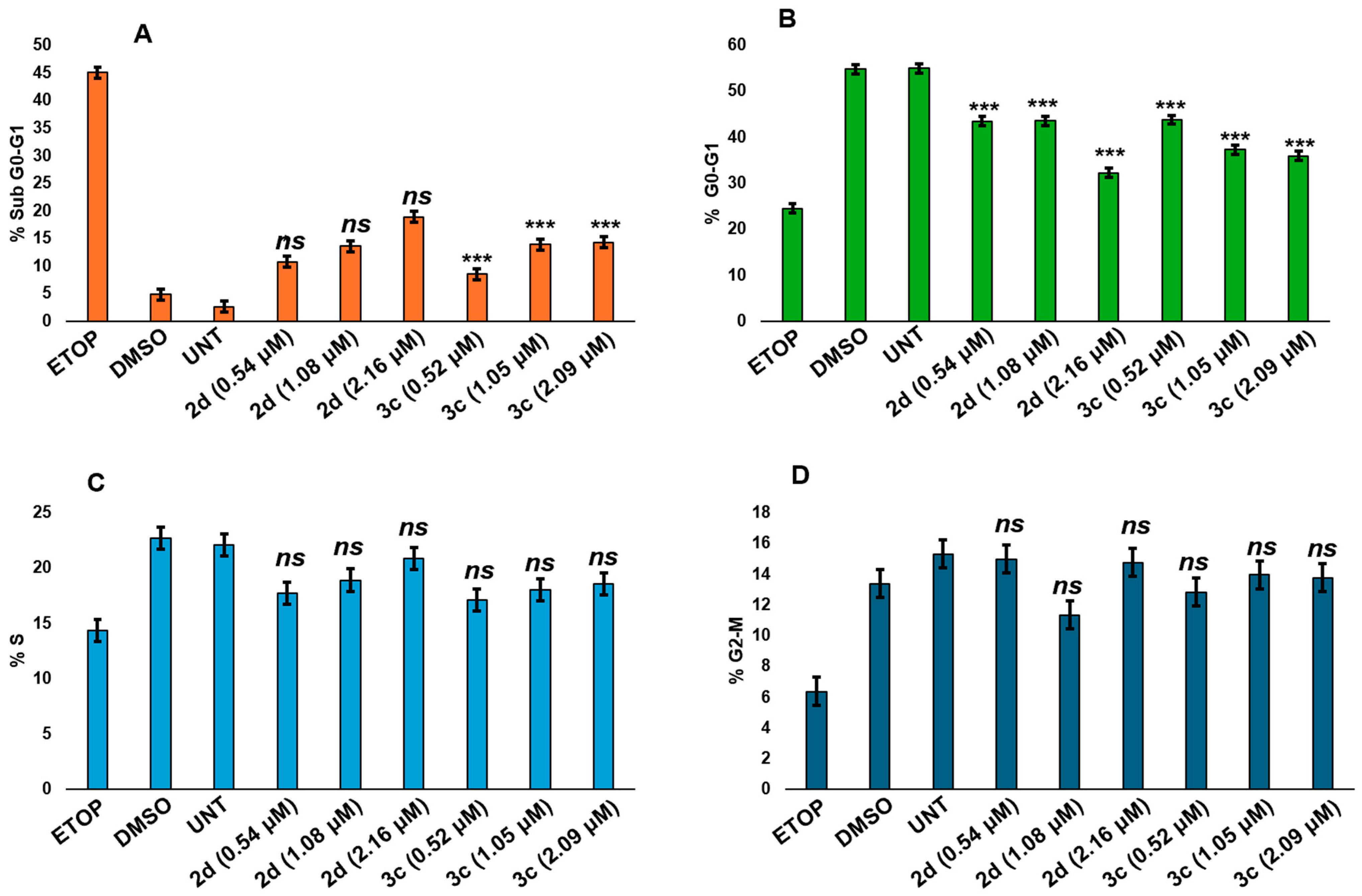
The effects of 2d and 3c on cell-cycle distribution in CEM cells were examined after 72 h of treatment (Figure 6). Treatment with both compounds altered the distribution of cells among the different DNA-content populations. The most evident change was an increase in the sub-G0-G1 population, particularly after treatment with 3c, which is consistent with DNA fragmentation associated with cell death. In contrast, the G0-G1 population was significantly reduced following treatment with both compounds. The S-phase population also tended to decrease after treatment, although these changes were not statistically significant. Similarly, no statistically significant changes were observed in the G2-M population for either compound. The increase in the sub-G0-G1 population is consistent with the apoptotic effects observed in the Annexin V/PI and caspase assays. In DNA-content analysis, the sub-G0-G1 population can arise from loss of fragmented DNA from apoptotic cells and is therefore commonly used as an indicator of DNA fragmentation associated with apoptosis [28]. However, the cell-cycle data alone do not establish a specific cell-cycle arrest mechanism. Taken together with the apoptosis data, the present results suggest that 2d and 3c primarily promote cell death accompanied by changes in cell-cycle distribution, rather than causing a clear arrest at the G2-M or S phase.
Figure 6.
Compounds 2d and 3c alter cell-cycle distribution in CEM cells, with the most notable change being an increase in the sub-G0/G1 population. Changes in cell-cycle distribution were evaluated after 72 h of treatment. For 2d, cells were treated with 0.54, 1.08, and 2.16 μM; for 3c, cells were treated with 0.52, 1.05, and 2.09 μM. Treatment with 2d or 3c increased the sub-G0/G1 population (A) and reduced the G0/G1 population (B). The S-phase population (C) showed no statistically significant change, and no statistically significant change was observed in the G2/M population (D). The increase in sub-G0/G1 cells is consistent with DNA fragmentation. Cells were stained with NIM-DAPI and analyzed by flow cytometry. DMSO (1%) was used as the vehicle control, and etoposide (100 μM) was included as a positive control. Statistical significance was determined by one-way ANOVA followed by Dunnett’s multiple-comparisons test. These results were F(3,20) = 2.41, ns, d = 2.6 for 2d and F(3,20) = 10.1, p < 0.001, d = 2.6 for 3c in Sub G0-G1. F(3,20) = 10.52, p < 0.001, d = 2.6 for 2d and F(3,20) = 23.33, p < 0.001, d = 2.6 for 3c in G0-G1. F(3,20) = 2.87, ns, d = 2.6 for 2d and F(3,20) = 2.5, ns, d = 2.6 for 3c in S. F(3,20) = 0.21, ns, d = 2.6 for 2d and F(3,20) = 0.018, ns, d = 2.6 for 3c in G2-M. *** p < 0.001, ns = not significant.
3.4. Preliminary Structure–Activity Relationships
To investigate whether the electronic properties of the compounds might be related to their cytotoxic activity, atomic charges at C7, C8, and C10 were calculated using the semi-empirical PM3 method in a simulated aqueous environment. The calculated values and molecular lengths are presented in Table 3.
Table 3.
Charge densities on C7, C8, and C10 carbons, and molecular length of compounds 1–3.
The difference in calculated charge between C7 and C8 (ΔQ) provides an estimate of the polarization of the α,β-unsaturated carbonyl system. Comparison of these values with the cytotoxicity data revealed some qualitative trends. For example, the active compounds 2d and 3c had ΔQ values of 0.431 and 0.519, respectively, whereas the less active compounds 3d and 1e had lower values of 0.305 and 0.328, respectively. Greater polarization of an α,β-unsaturated carbonyl system could increase its susceptibility to nucleophilic attack [29,30]. However, the relationship between ΔQ and cytotoxic activity was not consistent throughout the series. For example, 1c, 3f, and 3g showed relatively high calculated polarization but were inactive or considerably less active than 2d and 3c. Thus, the calculated charge difference alone cannot explain the observed cytotoxic activity.
A possible relationship between molecular length and cytotoxic activity was also considered. The two most active compounds, 2d and 3c, had calculated molecular lengths of 15.37 and 15.33 Å, respectively, whereas some compounds with substantially longer or shorter molecular lengths showed lower activity. These observations suggest that molecular dimensions may contribute to cytotoxic activity together with electronic properties. However, these relationships should be regarded as preliminary qualitative trends rather than established structure–activity relationships. No statistical correlation or validated computational model was developed, and several compounds did not follow the apparent trends. Other physicochemical properties, such as lipophilicity, steric accessibility, cell permeability, and metabolic stability, may also contribute to the differences in cytotoxic activity among these compounds.
The calculated electronic properties also do not demonstrate that these compounds react selectively with cellular thiols. Although the α,β-unsaturated carbonyl system provides a possible electrophilic site for nucleophilic attack, direct thiol-binding or glutathione-adduct experiments were not performed. Therefore, interaction with cellular thiols remains a working hypothesis that requires experimental verification.
4. Conclusions
In this study, three series of benzylidene cinnamoylhydrazides were synthesized and evaluated for their cytotoxic activity. Compounds 2d and 3c were the most active hits against the leukemia cell lines and also showed activity against several other tumor cell lines in the NCI-60 panel. Further studies in CEM cells showed that their cytotoxic effects were associated with apoptosis, caspase activation, mitochondrial membrane depolarization, increased oxidative activity, and changes in cell-cycle distribution.
However, the present results do not establish a direct causal relationship among ROS accumulation, mitochondrial dysfunction, caspase activation, and apoptosis. Similarly, the proposed interaction of these compounds with cellular thiols remains a working hypothesis because thiol reactivity was not examined experimentally. The computational studies also suggest possible relationships between electronic and structural properties and cytotoxic activity, but these observations should be considered preliminary.
Although 2d and 3c showed good cytotoxic activity, their selectivity for leukemia cells over non-malignant Hs27 fibroblasts was modest. Therefore, these compounds are best considered hit structures for further investigation rather than selective anti-leukemic agents. Further structural optimization, testing against additional non-malignant cell lines, and more detailed mechanistic studies will be needed to determine whether their potency and selectivity can be improved.
Supplementary Materials
The following supporting information can be downloaded at https://www.mdpi.com/article/10.3390/scipharm94040086/s1, [31,32,33,34].
Author Contributions
Conceptualization, M.H., R.J.A. and J.R.D.; methodology, M.H., S.M.M., J.P.-Z., J.J.W., J.N.D. and R.J.A.; software, M.H.; validation, M.H., R.J.A. and J.R.D.; formal analysis, M.H., R.J.A. and J.R.D.; investigation, M.H., S.M.M., J.P.-Z., J.J.W., J.N.D. and R.J.A.; resources, M.H. and R.J.A.; data curation, M.H., R.J.A. and J.R.D.; writing—original draft preparation, M.H., R.J.A. and J.R.D.; writing—review and editing, M.H., J.P.-Z., R.J.A. and J.R.D.; visualization, M.H. and R.J.A.; supervision, M.H., R.J.A. and J.R.D.; project administration, M.H., R.J.A. and J.R.D.; funding acquisition, M.H., R.J.A. and J.R.D. All authors have read and agreed to the published version of the manuscript.
Funding
Financial support for this study was provided by the Office of the Executive Vice Chancellor for Academic Affairs, Indiana University Kokomo, which awarded a Summer Faculty Fellowship and Grant-in-Aid to M. Hossain; NIH grant (1R16GM149379) to R.J. Aguilera and the core facilities of the Border Biomedical Research Center, funded by the Research Centers at Minority Institutions grant (5U54MD007592) from the National Institute on Minority Health and Health Disparities; the Maunders McNeil Foundation Inc. (418431) for a grant to J. R. Dimmock.
Institutional Review Board Statement
Not applicable.
Informed Consent Statement
Not applicable.
Data Availability Statement
All data produced in this study are contained in the manuscript text and Supplementary Materials.
Acknowledgments
The authors gratefully acknowledge Maren Pink, Indiana University Molecular Structure Center, Indiana University, Bloomington, for assistance with the collection and analysis of the X-ray crystallographic data. We also acknowledge support for the acquisition of the Bruker Venture D8 diffractometer through the Major Scientific Research Equipment Fund, provided by the President of Indiana University and the Office of the Vice President for Research. The authors also sincerely acknowledge the Developmental Therapeutics Program (DTP) of the National Cancer Institute (NCI), USA, for conducting the NCI-60 human tumor cell line screening (HTS384).
Conflicts of Interest
The authors declare no conflicts of interest.
References
- Hossain, M.; Enci, C.E.; Dimmock, J.R.; Das, U. Discovery and Investigation of 1-[4-(2-Aminoethoxy)Phenylcarbonyl]-3,5-Bis-(Benzylidene)-4-Piperidones as Candidate Antineoplastic Agents: Our Last 15 Years Study. Curr. Med. Chem. 2021, 28, 2453–2464. [Google Scholar] [CrossRef] [Scilit] [PubMed]
- Mutus, B.; Wagner, J.D.; Talpas, C.J.; Dimmock, J.R.; Phillips, O.A.; Reid, R.S. 1-p-chlorophenyl-4,4-dimethyl-5-diethylamino-1-penten-3-one hydrobromide, a sulfhydryl-specific compound which reacts irreversibly with protein thiols but reversibly with small molecular weight thiols. Anal. Biochem. 1989, 177, 237–243. [Google Scholar] [CrossRef] [Scilit] [PubMed]
- Baluja, M.G.; Municio, A.M.; Vega, S. Reactivity of α,β-Unsaturated Ketones toward Sulfhydryl Compounds and Their Antifungal Activity. Chem. Ind. 1964, 2053–2054. [Google Scholar]
- Hossain, M.; Das, U.; Dimmock, J.R. Recent advances in α, β-unsaturated carbonyl compounds as mitochondrial toxins. Eur. J. Med. Chem. 2019, 183, 111687. [Google Scholar] [CrossRef] [Scilit] [PubMed]
- Benvenuto, J.A.; Connor, T.H.; Monteith, D.K.; Laidlaw, J.L.; Adams, S.C.; Matney, T.S.; Theiss, J.C. Degradation and inactivation of antitumor drugs. J. Pharm. Sci. 1993, 82, 988–991. [Google Scholar] [CrossRef] [Scilit]
- Amslinger, S. The tunable functionality of alpha,beta-unsaturated carbonyl compounds enables their differential application in biological systems. ChemMedChem 2010, 5, 351–356. [Google Scholar] [CrossRef] [Scilit] [PubMed]
- Das, U.; Sharma, R.K.; Dimmock, J.R. 1,5-diaryl-3-oxo-1,4-pentadienes: A case for antineoplastics with multiple targets. Curr. Med. Chem. 2009, 16, 2001–2020. [Google Scholar] [CrossRef] [Scilit] [PubMed]
- Antoniolli, G.; de Moraes, G.R.; Deruvo, C.; Altomare, C.D.; Coelho, F.; de Candia, M. N-Acylhydrazones: Applications and advances in drug discovery. Eur. J. Med. Chem. Rep. 2026, 16, 100317. [Google Scholar] [CrossRef] [Scilit]
- Kassab, A.E. Anticancer agents incorporating the N-acylhydrazone scaffold: Progress from 2017 to present. Arch. Pharm. 2023, 356, e2200548. [Google Scholar] [CrossRef] [Scilit] [PubMed]
- Socea, L.I.; Barbuceanu, S.F.; Pahontu, E.M.; Dumitru, A.C.; Nitulescu, G.M.; Sfetea, R.C.; Apostol, T.V. Acylhydrazones and Their Biological Activity: A Review. Molecules 2022, 27, 8719. [Google Scholar] [CrossRef] [Scilit] [PubMed]
- Hossain, M.; Hall, S.C.; Wiggington, P.J.; Roth, S.M.; Das, S.; Das, U.; Roayapalley, P.K.; Dimmock, J.R. Cytotoxic benzylidene hydrazides of terephthalic acid and related compounds. Pharmazie 2022, 77, 90–94. [Google Scholar] [CrossRef] [Scilit]
- Hossain, M.; Filbrun, R.A.; Roth, S.M.; Sakagami, H.; Amano, S.; Nagase, H.; Satoh, K.; Dimmock, J.R. Cytostatic Benzylidene-isophthaloylhydazides With Diminished Neurotoxicity. Anticancer Res. 2026, 46, 1183–1195. [Google Scholar] [CrossRef] [Scilit] [PubMed]
- Carvalho, S.A.; Feitosa, L.O.; Soares, M.; Costa, T.E.; Henriques, M.G.; Salomao, K.; de Castro, S.L.; Kaiser, M.; Brun, R.; Wardell, J.L.; et al. Design and synthesis of new (E)-cinnamic N-acylhydrazones as potent antitrypanosomal agents. Eur. J. Med. Chem. 2012, 54, 512–521. [Google Scholar] [CrossRef] [Scilit] [PubMed]
- Carvalho, S.A.; Kaiser, M.; Brun, R.; da Silva, E.F.; Fraga, C.A. Antiprotozoal activity of (E)-cinnamic N-acylhydrazone derivatives. Molecules 2014, 19, 20374–20381. [Google Scholar] [CrossRef] [Scilit] [PubMed]
- Tardugno, R.; Giancotti, G.; De Burghgraeve, T.; Delang, L.; Neyts, J.; Leyssen, P.; Brancale, A.; Bassetto, M. Design, synthesis and evaluation against Chikungunya virus of novel small-molecule antiviral agents. Bioorg. Med. Chem. 2018, 26, 869–874. [Google Scholar] [CrossRef] [Scilit] [PubMed]
- Chatrabhuji, P.M.; Vyas, K.B.; Nimavat, K.S.; Undavia, N.K. Synthesis and antibacterial activity of some new thiazolidines. Elixir. Int. J. 2013, 54A, 12778–12780. [Google Scholar]
- Sulistyowaty, M.I.; Nugroho, A.E.; Putra, G.S.; Ekowati, J.; Budiati, T. Syntheses, Molecular Docking Study and Anticancer Activity Examination of p-Methoxycinnamoyl Hydrazides. Int. J. Pharm. Clin. Res. 2016, 8, 623–627. [Google Scholar]
- Spartan Student Software Package, version 8.0.6; Wavefunction, Inc.: Irvine, CA, USA, 2021.
- Gutierrez, D.A.; Contreras, L.; Villanueva, P.J.; Borrego, E.A.; Moran-Santibanez, K.; Hess, J.D.; DeJesus, R.; Larragoity, M.; Betancourt, A.P.; Mohl, J.E.; et al. Identification of a Potent Cytotoxic Pyrazole with Anti-Breast Cancer Activity That Alters Multiple Pathways. Cells 2022, 11, 254. [Google Scholar] [CrossRef] [Scilit] [PubMed]
- National Cancer Research Institute. HTS384 Screening Methodology. Available online: https://dctd.cancer.gov/drug-discovery-development/assays/high-throughput-screening-services/nci60/screening-methodology.pdf (accessed on 31 January 2025).
- Fregnan, A.M.; Brancaglion, G.A.; Galvão, A.F.C.; Costa, C.O.D.S.; Moreira, D.R.M.; Soares, M.B.P.; Bezerra, D.P.; Silva, N.C.; Morais, S.M.S.; Oliver, J.C.; et al. Synthesis of piplartine analogs and preliminary findings on structure–antimicrobial activity relationship. Med. Chem. Res. 2017, 26, 603–614. [Google Scholar] [CrossRef] [Scilit]
- Lima, P.C.; Lima, L.M.; da Silva, K.C.; Leda, P.H.; de Miranda, A.L.; Fraga, C.A.; Barreiro, E.J. Synthesis and analgesic activity of novel N-acylarylhydrazones and isosters, derived from natural safrole. Eur. J. Med. Chem. 2000, 35, 187–203. [Google Scholar] [CrossRef] [Scilit] [PubMed]
- Palla, G.; Predieri, G.; Domiano, P.; Vignali, C.; Turner, W. Conformational behavior and E/Z isomerization of N-acyl and N-aroylhydrazones. Tetrahedron 1986, 42, 3649–3654. [Google Scholar]
- Kummerle, A.E.; Raimundo, J.M.; Leal, C.M.; da Silva, G.S.; Balliano, T.L.; Pereira, M.A.; de Simone, C.A.; Sudo, R.T.; Zapata-Sudo, G.; Fraga, C.A.; et al. Studies towards the identification of putative bioactive conformation of potent vasodilator arylidene N-acylhydrazone derivatives. Eur. J. Med. Chem. 2009, 44, 4004–4009. [Google Scholar] [CrossRef] [Scilit] [PubMed]
- CCDC 2580006 Contains the Supplementary Crystallographic Data for This Paper. These Data Can Be Obtained Free of Charge from The Cambridge Crystallographic Data Centre. Available online: www.ccdc.cam.ac.uk/structures (accessed on 10 August 2026).
- Porter, A.G.; Janicke, R.U. Emerging roles of caspase-3 in apoptosis. Cell Death Differ. 1999, 6, 99–104. [Google Scholar] [CrossRef] [Scilit] [PubMed]
- Elmore, S. Apoptosis: A review of programmed cell death. Toxicol. Pathol. 2007, 35, 495–516. [Google Scholar] [CrossRef] [Scilit] [PubMed]
- Plesca, D.; Mazumder, S.; Almasan, A. DNA damage response and apoptosis. Methods Enzymol. 2008, 446, 107–122. [Google Scholar] [CrossRef] [Scilit] [PubMed]
- Roseli, R.B.; Keto, A.B.; Krenske, E.H. Mechanistic aspects of thiol additions to Michael acceptors: Insights from computations. WIREs Comput. Mol. Sci. 2022, 13, e1636. [Google Scholar] [CrossRef] [Scilit]
- Paasche, A.; Schiller, M.; Schirmeister, T.; Engels, B. Mechanistic study of the reaction of thiol-containing enzymes with alpha,beta-unsaturated carbonyl substrates by computation and chemoassays. ChemMedChem 2010, 5, 869–880. [Google Scholar] [CrossRef] [Scilit] [PubMed]
- SAINT, version 8.40A; Bruker AXS: Madison, WI, USA, 2020.
- Krause, L.; Herbst-Irmer, R.; Sheldrick, G.M.; Stalke, D. Comparison of silver and molybdenum microfocus X-ray sources for single-crystal structure determination. J. Appl. Crystallogr. 2015, 48, 3–10. [Google Scholar] [CrossRef] [Scilit] [PubMed]
- Sheldrick, G.M. SHELXT—Integrated space-group and crystal-structure determination. Acta Crystallogr. Sect. A Found. Adv. 2015, 71, 3–8. [Google Scholar] [CrossRef] [Scilit] [PubMed]
- Sheldrick, G.M. Crystal structure refinement with SHELXL. Acta Crystallogr. Sect. C Struct. Chem. 2015, 71, 3–8. [Google Scholar] [CrossRef] [Scilit]

Disclaimer/Publisher’s Note: The statements, opinions and data contained in all publications are solely those of the individual author(s) and contributor(s) and not of MDPI and/or the editor(s). MDPI and/or the editor(s) disclaim responsibility for any injury to people or property resulting from any ideas, methods, instructions or products referred to in the content. |
© 2026 by the authors. Published by MDPI on behalf of the Österreichische Pharmazeutische Gesellschaft. Licensee MDPI, Basel, Switzerland. This article is an open access article distributed under the terms and conditions of the Creative Commons Attribution (CC BY) license.