


Abstract
Cancer is still one of the major causes of death globally. While there are many cancer therapies, they often come with significant adverse effects and limitations. A lot of studies have shown that using natural products can improve the efficacy of conventional therapies and decrease their toxicity. Natural products and their derivatives encompassing unmodified molecules, semi-synthetic analogs, and synthetic structures featuring natural pharmacophores, represent a major foundation for approved anticancer agents. Phenolic compounds are a large family of naturally occurring compounds found in plants and have attracted a lot of attention for their potential anticancer properties. Preclinical studies have shown that phenolic compounds have anticancer activity through different mechanisms, mainly by modulating inflammation, which plays a critical role in cancer development. Inflammatory processes in cancer involve complex molecular mechanisms regulated by both oncogenic and tumor-suppressing transcription factors. This review focuses on how phenolic compounds regulate transcription factors, such as nuclear factor kappa-light-chain-enhancer of activated B-cells (NF-κB), nuclear factor erythroid 2-related factor 2 (Nrf2), and signal transducer and activator of transcription 3 (STAT3), which are critical in cancer-related gene expression, ultimately leading to the inhibition of cancer cell proliferation, invasion, and metastasis. Furthermore, phenolic compounds have been shown to synergize with conventional anticancer drugs, enhancing their efficacy and reducing side effects. This review investigates the role of phenolic compounds in regulating transcription factors in cancer animal models, highlighting both the therapeutic potential and limitations of these compounds in cancer prevention and treatment.
1. Introduction
Cancer is a multifactorial disease, a state characterized by uncontrolled cell growth and division in any tissue [1]. About 90% of cases have their pathogenesis attributed to mutations and environmental factors; on the other hand, epigenetic changes play a very crucial role [1]. According to global statistics from 2022, the new cases of cancer were approximately 20 million, and cancer deaths amounted to 9.7 million [2,3,4]. Analysis of this data estimates that about one in five men or women get cancer during a lifetime, and about one in nine men and one in 12 women die of it [2]. Regarding childhood cancer, studies show that it has a better prognosis, since the 5-year survival rate for children is 80%, whereas in adults it is 68% [1]. In recent years, there has been a decrease in death rates in many types of cancer, possibly due to the early detection methods and the numerous treatment options [5]. However, cancer remains the second leading cause of death worldwide, following cardiovascular disease [6]. The current research is focused on developing more effective cancer therapies with reduced adverse toxicity [7]. In this context, natural products have regained significant attention [8]; however, their safety profile is not intrinsic but highly dependent on the specific chemical structure, dosage, matrix formulation, bioavailability, and exposure duration. The medicinal use of natural products dates back to antiquity, with the first references being made in Mesopotamia in 2600 B.C. [9]. A vast diversity of secondary metabolites has been identified across plant, microbial, and marine species, with phenolic compounds representing one of the most abundant and biologically active classes of natural products in the plant kingdom [10]. Accumulating evidence suggests that the anticancer effects of phenolic compounds are also related to their capacity to modulate important oncogenic and inflammatory transcription factors. Nuclear factor kappa-light-chain-enhancer of activated B-cells (NF-κB), nuclear factor erythroid 2-related factor 2 (Nrf2) and signal transducer and activator of transcription 3 (STAT3) form a tripartite network that maintains tumor survival, inflammation and cellular redox homeostasis. While several reviews have addressed the general anticancer properties of dietary phenolic compounds or their impact on individual signaling pathways, a unified analysis explicitly focusing on the interconnected NF-κB, Nrf2, and STAT3 tripartite axis remains limited. This review addresses this critical gap by providing a structured, comparative synthesis of how distinct structural classes of phenolic compounds concurrently modulate this core signaling network. Furthermore, we critically analyze the context-dependent roles of these transcription factors in cancer progression and address translational considerations, particularly regarding the physiological relevance of tested concentrations and inherent bioavailability challenges.
Literature Search Strategy and Selection Criteria
To ensure a comprehensive, transparent and reproducible synthesis of the current evidence, a literature search was carried out to identify all relevant articles published up to July 2026 in three major electronic databases, PubMed/MEDLINE, Scopus, and Web of Science. The search strategy included combinations of Medical Subject Headings (MeSH) terms and free-text keywords with Boolean logic. The first search query consisted of a combination of terms for phenolic compounds (e.g., “phenolic compounds”, “resveratrol”, “pterostilbene”, “catechins”, “EGCG”, “genistein”, “xanthohumol”, “curcumin”, “luteolin”, “quercetin”, “apigenin”) and terms for the target signaling pathways (e.g., “NF-κB”, “Nrf2”, “STAT3”, “transcription factors”) and cancer context (e.g., “cancer”, “neoplasm”, “carcinogenesis”, “chemoresistance”, “synergy”). High-impact peer-reviewed journals’ original research articles (in vitro cell culture models and in vivo animal studies), relevant human clinical trials and mechanistic reviews in English were included. Studies without specific mechanistic insight into NF-κB, Nrf2, or STAT3 pathways were excluded. Studies using uncharacterized crude plant extracts without quantified phenolic compound composition were excluded. The non-peer-reviewed literature (e.g., conference abstracts, preprints, editorials) and studies published in languages other than English were excluded. Articles were critically evaluated according to methodological quality, experimental rigor, and physiological relevance of concentrations/doses tested.
2. Phenolic Compounds
Phenolic compounds are organic compounds produced by secondary metabolism in plants. Phenolic compounds contain at least one benzene ring linked to one or more hydroxyl groups, which establish an aromatic or aliphatic structure [10]. One of the important characteristics of these compounds is their large structural diversity from simple phenols to complex polyphenolic compounds [10]. Phenolic compounds may be divided into four major classes—phenolic acids, flavonoids, stilbenes and lignans—depending upon the number of phenol rings they contain, and on the structural elements linking these rings together [11]. Specifically, phenolic acids consist of hydroxybenzoic (C6-C1) and hydroxycinnamic (C6-C3) structures; flavonoids share a diphenylpropane (C6-C3-C6) backbone with two aromatic rings linked via a heterocyclic pyran/pyrone ring; stilbenes possess a 1,2-diphenylethylene (C6-C2-C6) skeleton; and lignans are formed by the oxidative dimerization of two phenylpropanoid (C6-C3) units. As secondary metabolites, they are not essential for the survival of the producing organism, but they do play a facilitating role. In the botanical world, phenolic compounds are the plants’ internal defense against pathogens and ultraviolet (UV) light [11]. On the other hand, as food items, these compounds are well known for their ability to extend shelf life, owing to their antioxidant activity [12]. Concerning health benefits, phenolic compounds are mainly known to be free-radical scavengers; that is, they act as antioxidants [12]. These compounds may also show anti-inflammatory effects through the inhibition of transcription factors responsible for the expression of inflammatory biomarkers [12]. This has been well investigated in different inflammatory diseases, including Alzheimer’s disease, inflammatory bowel disease, rheumatoid arthritis, and cancer [13]. Moreover, the improvement in cardiovascular health has also been associated with phenolic compounds, since they inhibit angiotensin-converting enzyme (ACE) [13]. It is of high importance to clarify that most of these findings are derived from in vitro or in vivo preclinical studies and by using concentrations much higher than those generally contained in the human diet [14]. This is because, although phenolic compounds are found in a variety of foods such as fruits, vegetables, cereals, and beverages, they have a relatively low bioavailability since they are recognized as xenobiotics by the body [15]. Bioavailability refers to the extent to which a substance has been processed and absorbed, making it possible for it to carry out its biological functions at the specified site of action. Advanced drug delivery technologies are being extensively investigated to improve pharmaceutical relevance and get over these bioavailability obstacles. These methods include polymeric nanoparticles and micelles for controlled targeted release, phospholipid complexes that enhance membrane permeability, lipid-based nanocarriers (such as liposomes, solid lipid nanoparticles, and nanoemulsions) that shield phenolic compounds from gastrointestinal enzymatic degradation, and prodrug strategies (such as reversible esterification of phenolic hydroxyl groups) that prevent rapid phase II metabolic inactivation [14,16]. The metabolism of each compound is highly variable between individuals and depends on several factors, such as the molecular weight and chemical composition of the natural product, the food matrix, interaction with other nutrients, the digestive system, and the gut microbiome of the consumer [17]. There are no specific recommendations for the intake of individual phenolic compounds at particular doses for specific health benefits because of the complex metabolism of phenolic compounds and their mechanisms of action, which are not yet fully understood [14]. However, the epidemiological evidence strongly supports the consumption of a diet rich in phenolic compound-containing foods [14]. Therefore, a diet rich in a diverse variety of polyphenol-containing foods is highly recommended [14].
3. Overview of Key Transcription Factors Altered in Cancer
3.1. Overview of Transcription Factors
Transcription factors are regulatory proteins that identify and interact with specific DNA sequences, known as promoters and enhancers [18]. They can be classified into two main categories: the general, and the specific transcription factors [19,20,21,22,23,24,25]. General transcription factors bind to the core promoter and help RNA polymerase II to also bind to the promoter [19,20,21,22,23,24,25]. This way, the pre-initiation transcription complex is formed. In a cell-free environment, this is sufficient for the initiation of transcription; however, within the complex cellular environment, additional regulatory elements are required [19,20,21,22,23,24,25]. Specific transcription factors are gene-specific and act as activators or repressors of a gene’s transcription [19,20,21,22,23,24,25]. When they act as activators, they bind to the enhancer and assist in the formation of the active transcription complex, which includes the pre-initiation transcription complex bound to specific transcription factors and the enhancer, as well as the mediator, which helps link these elements together [19,20,21,22,23,24,25]. Less frequently, transcription factors act as repressors of gene expression and bind to a region similar to the enhancer, called the silencer [19,20,21,22,23,24,25].
3.2. NF-κB: A Core Regulator of Inflammation and Cell Survival
Nuclear factor kappa-light-chain-enhancer of activated B-cells (NF-κB) is a five-member family of transcription factors with an important role in immune and inflammatory processes [26,27,28,29,30,31,32,33,34]. NF-κB contributes to the regulation of activation, survival, differentiation, and apoptosis of immune cells and T-inflammatory cells [26,27,28,29,30,31,32,33,34]. Also, NF-κB regulates the expression of various pro-inflammatory genes, including those encoding cytokines and chemokines, like tumor necrosis factor-alpha (TNF-α) and interleukin-1 beta (IL-1β). NF-κB participates in inflammasome regulation [34,35]. Under physiological conditions, in the absence of stimuli, NF-κB is inactive in the cytoplasm, bound to inhibitory proteins, the most well-known of which are called IkappaB (IκB) kinase [36]. The precursor proteins of NF-κΒ subunit 1 (NF-κB1) and NF-κΒ subunit 2 (NF-κB2), known as p105 and p100, respectively, can exert a similar inhibitory effect due to the similarity in their structure [35]. Once NF-κB is released from these proteins, it migrates to the nucleus, where it performs its function. Its activation can follow two types of pathways: the canonical and the non-canonical [37].
NF-κB is a transcription factor that is frequently associated with the pathogenesis of diseases [26,27,28,29,30,31,32,33,34]. Cancer is no exception, as deregulation of the NF-κB pathway has been reported in various types of cancer, both solid and hematologic, where it appears to support the proliferation and survival of cancer cells [26,27,28,29,30,31,32,33,34]. In particular, cancer cells’ mutations in NF-κB’s positive regulators cause it to become persistently activated, which can target oncogene promoters and produce a positive feedback loop [26,27,28,29,30,31,32,33,34]. Due to a lack of knowledge regarding the mechanism of action of the NF-κB transcription factor and the incapacity of its inhibitors to function as an effective stand-alone anticancer treatment, NF-κB inhibitors have not been approved for therapeutic use, despite numerous attempts to develop them for the treatment of cancer in animal models [38,39,40,41,42,43,44]. Nevertheless, recent research has shown conflicting results, demonstrating that NF-κB can also have tumor-suppressive effects in specific cancers [26,27,28,29,30,31,32,33,34]. The transcriptional activation of the Fas ligand (CD178, CD95L or Apo-1L) is one such pathway. Additionally, the synergy between Nrf2 and NF-κB/p65 (NF-κB/p65/RelA) was found to prevent necrosis, inflammation, and liver fibrosis, protecting against the development of hepatocellular adenoma [45,46,47,48]. The nuclear factor of kappa light polypeptide gene enhancer in B-cells inhibitor alpha (IκBα) was overexpressed to prevent NF-κB activation, and oncogenic invasive epidermal growth caused by Ras protein was observed, resembling squamous cell carcinoma [49]. Finally, the deletion of the nuclear factor of kappa light polypeptide gene enhancer in B-cells inhibitor beta (IKKβ) may enhance the activation of the c-Jun N-terminal kinase (JNK) family members, including JNK1, which contributes to the development of hepatocellular carcinoma [49]. Thus, the relationship between NF-κB and cancer remains unclear [50].
3.2.1. The Canonical Pathway
Various stimuli involved in processes such as inflammation, immune response, cell differentiation, apoptosis and survival activate the canonical NF-κB pathway [34]. Activation can be induced, for instance, by binding members of the tumor necrosis factor receptor (TNFR) superfamily, cytokine receptors, pattern recognition receptors (PRRs), T-cell receptors and B-cell receptors [34]. This pathway requires the IkB kinase (IKK) complex, a multi-subunit kinase which is composed of two catalytic subunits (IKKα and IKKβ) and a regulatory subunit (NF-κB essential modulator, or IKKγ) [34]. Numerous factors, such as cytokines, growth factors, microbiological components, and stressors, might activate this IKK complex [34]. Upon stimulation, IKK phosphorylates and degrades the inhibitory proteins IκBα. This allows NF-κB to be released and translocate to the nucleus, primarily as p50/RelA and p50/c-Rel dimers [35]. In the nucleus, NF-κB induces the activation of target genes, a process that, although rapid, is transient, as NF-κB also promotes the expression of negative regulators such as IkBα, A20, and p105 [34].
3.2.2. The Non-Canonical Pathway
Alternatively, the non-canonical NF-κB pathway is selective, has a more focused biological action, and reacts to particular stimuli like the binding of a subset of TNF receptor superfamily members, such as receptor activator of nuclear factor kappa B (RANK), cluster of differentiation 40 (CD40), B-cell activating factor receptor (BAFF-R), and lymphotoxin-beta receptor (LTBR) [31]. A further difference between the two pathways is that the non-canonical pathway does not involve the phosphorylation of IκBα, but rather phosphorylation of p100 [34,35,38]. NF-κB-inducing kinase (NIK) is a crucial protein in this process because it phosphorylates p100 by activating and collaborating with IKKα [34,35,37]. As a result, mature NF-ΚB2 is produced and translocates as a p52/RelB complex to the nucleus [34,35,37].
3.3. Nrf2: Dual Roles in Oxidative Stress and Chemoresistance
Nuclear factor erythroid 2-related factor 2 (Nrf2) is a transcription factor with a dual role in cancer [51,52,53,54,55,56]. Although it is generally known to protect against oxidative stress, it has been shown to exert this protective action in cancer cells as well, with the result that they use it as a defense mechanism [51,52,53,54,55,56]. Specifically, the hyperactivation of Nrf2 in various types of cancer has been found to support the proliferation, growth, and avoidance of apoptosis and cell death of cancer cells [51,52,53,54,55,56]. Thus, in some cases, Nrf2 contributes to the resistance to chemotherapy and radiotherapy [57].
3.3.1. Nrf2 in Oxidative Stress Regulation
Nrf2 belongs to the Cap‘n’collar (CNC) family, a highly conserved group of basic leucine zipper (bZIP) transcription factors, and is structurally composed of 605 amino acids with seven functional domains, known as Neh1-Neh7 [58]. In the absence of stimuli, Nrf2 is retained in the cytoplasm bound to its inhibitory protein, kelch-like ECH-associated protein 1 (Keap1) [51]. Activation occurs in response to various oxidative stress-related stimuli, causing Nrf2 to dissociate from Keap1 and translocate to the nucleus [51]. There, it forms a heterodimer with small musculoaponeurotic fibrosarcoma (Maf) proteins and binds to the antioxidant response element (ARE), which acts as an enhancer for the expression of various antioxidant enzymes [59]. Beyond regulating antioxidant enzymes, Nrf2 is also involved in the expression of phase II detoxifying enzymes and in mitochondrial function regulation, specifically in beta-oxidation and expression of the complex V α-subunit (ATP synthase) [59,60].
3.3.2. Nrf2 and NF-κB Cross-Talk and Interactions
Τhe transcription factors Nrf2 and NF-κB operate through compensatory coordination to maintain redox homeostasis in cells, which controls how cells react to stress and inflammation [46]. In particular, elevated Nrf2 expression causes the cell to produce more heme oxygenase-1 (HO-1), which raises the amounts of phase II enzymes and blocks the NF-κB inhibitory protein IκBα from degrading, hence preventing NF-κB activation [46]. In a similar mechanism, NF-κB inhibits the binding of CREB-binding protein (CBP), a transcriptional coactivator of Nrf2, and, consequently, Nrf2 expression [58].
3.4. STAT3 Signaling: Activation, Dysregulation, and Role in Oncogenesis
STAT3 is critically involved in the process of oncogenesis through modulation of the expression of cell cycle regulators and genes that promote cancer such as those that are part of the anti-apoptotic B-cell lymphoma 2 (Bcl-2) family, and myeloid cell leukemia 1 (Mcl-1) [61]. It is believed that about 70% of human malignancies have an overexpression of STAT3 [26,62,63,64,65,66,67,68,69,70]. However, some data indicate that activated STAT3 may have tumor-suppressive activities in specific settings, even if the hyperactivation of STAT3 is largely linked to increasing tumor growth [26,62,63,64,65,66,67,68,69,70].
STAT proteins are a group of transcription factors located in the cytoplasm that transmit signals from the cell membrane to the nucleus [71]. In the absence of stimuli, STAT proteins exist in the cytoplasm as monomers or unphosphorylated dimers [71]. Upon extracellular ligand binding, transmembrane receptors undergo conformational changes that activate kinases such as Janus kinases (JAKs) [68,72,73,74]. These kinases induce the phosphorylation of tyrosine residues on the intracellular receptor complex, where STATs then bind and are phosphorylated, allowing them to form dimers and translocate to the nucleus to activate target gene transcription [75]. These genes are associated with functions such as the activation, survival, differentiation, and apoptosis of cells [71,75]. STAT deactivation can occur through the dephosphorylation of STAT proteins or their receptors by nuclear tyrosine phosphatases (PTPs), the protein inhibitors of activated STAT (PIAS) proteins, which act as inhibitors by binding STATs and preventing nuclear entry and DNA binding, and the suppressor of cytokine signaling (SOCS) family members, which function as negative regulators of STAT signaling [71,75].
4. Phenolic Compounds and Cancer: Mechanisms of Action
4.1. The Interplay of Inflammation, Oxidative Stress, and Epigenetic Alterations in Cancer Pathogenesis
The body uses inflammation as a natural defense mechanism when it encounters endogenous or external stressors, like tissue damage or infection [76,77]. The two main types of inflammation are acute and chronic [76]. Acute inflammation, a brief but temporary immune response, aims to eliminate the cause and restore homeostasis [76]. It is characterized by vasodilation and the recruitment of leukocytes and other immune molecules to the affected tissues [76]. If the acute inflammatory response fails to restore homeostasis, the continued stimulation of the immune system will lead to the establishment of chronic inflammation, which is associated with the physiological and pathological progression of various diseases, such as atherosclerosis, Alzheimer’s disease, depression, and cancer [78]. Acute-phase proteins, reactive oxygen and nitrogen species, and pro- and anti-inflammatory cytokines are all produced as part of the systemic inflammatory response [79]. Consequently, cellular antioxidant capability is diminished, and oxidative stress is generated [79].
The imbalance between oxidants and antioxidants in the cell, which results in an excess of free radicals, is known as oxidative stress [80]. Free radicals, being unstable substances, are highly reactive [80]. Thus, they react with proteins and fatty acids in the cell membrane, causing them to be nonfunctional. Moreover, free radicals can damage DNA and/or cause mutations to it, which may lead to the development of cancer [80]. There are four main stages in cancer, all of which involve inflammation: initiation, progression, metastasis, and resistance to pharmacotherapy [1]. Notably, cancer-related inflammation is recognized as the seventh hallmark of cancer [81]. Oxidative stress and inflammation can serve as stimuli for the activation of transcription factors involved in the expression of pro-inflammatory genes [82]. These redox-sensitive transcription factors are often altered in several types of cancer [82].
Although each cell has the entire organism’s gene pool, the specialization that each cell gains throughout cellular differentiation allows it to carry out particular tasks [21]. As a result, only genes associated with their specific function are expressed simultaneously in all cell types [21]. By regulating the expression of specific genes, this is achieved. Gene expression is the process by which the information contained in a gene is converted into a functional output [21]. Gene expression can be influenced by a variety of epigenetic factors, including exposure to environmental pollutants, sleep habits, alcohol consumption, smoking, exercise, and diet [22]. Cell division is one way that epigenetic modifications can spread, even though they are reversible and do not alter the DNA sequence [23]. Natural products, especially phenolic compounds, can alter gene expression through a number of mechanisms, including transcription factor regulation, direct interactions with cellular components, chromatin structural alterations, and interactions with non-coding RNA [24]. Since transcription factors are the primary regulators of transcription and, consequently, gene expression, the current research focuses on their regulation [24].
4.2. Mechanisms of Action of Phenolic Compounds: Targeting Transcription Factors and Signaling Pathways
Phenolic compounds can act as protective agents against tumor initiation [83,84,85]. Some mechanisms that have been proposed include the regulation of enzymes, like the carcinogen-detoxifying phase II enzymes and heme-containing phase I enzymes, as well as obstructing the activity of the mutagen-transforming enzymes [86]. Moreover, phenolic compounds can prevent the formation of genotoxic molecules and DNA adducts [86]. Anti-inflammatory effects of phenolic compounds can reduce inflammatory components, like interleukin-6 (IL-6), and inhibit the activation of transcription factors. For example, Oroxylin A was shown to suppress the IL-6-mediated STAT3 signaling pathway [87]. Other anticancer mechanisms involve their antioxidant activity [86]. Several phenolic compounds, like luteolin and epigallocatechin-3-gallate (EGCG), are able to activate the Nrf2 antioxidant pathway, which regulates the production of antioxidant enzymes that stop free radicals from causing cellular harm [88,89]. Through the Extracellular signal-regulated kinase 1 and 2—Ribosomal S6 kinase—Neuronal nitric oxide Synthase (ERK1/2-RSK-nNOS) axis, resveratrol, another phenolic compound, can prevent oxidation. Through the AMP-activated protein kinase/sirtuin 1 (AMPK/SIRT1) pathway, quercetin, a phenolic molecule, can also lessen oxidative stress [1]. Flavonoids have anti-inflammatory effects, enhance immunity, and can modulate NF-κB, p53, mitogen-activated protein kinase (MAPK), inflammasome, and JAK-STAT pathways, which are usually altered in cancer [90]. The consumption of a diet enriched with anthocyanins, flavonols, flavanones, and hydrolyzed tannins has been associated with a reduction in oncogenesis [1]. Both their efficacy and their mode of action differ. Their chemical structure is one of the main factors affecting their activity. In particular, more anticancer activity has been linked to the presence of aromatic rings, a short unsaturated fatty acid side chain, and more hydroxyl groups [91].
4.3. Specific Examples of Bioactive Phenolic Compounds
4.3.1. Resveratrol
Numerous edible plants, including peanuts, berries, blackberries, strawberries, and grapes, contain the phenolic compound resveratrol. Although fruits represent the primary dietary source of this nutrient, resveratrol is found in significantly higher concentrations in red wine compared to white wine. However, its composition varies depending on the different cultivars, ripeness levels, production and processing methods, etc. Its therapeutic properties are believed to be achieved through a daily intake of 30–150 mg [92]. Resveratrol appears to regulate transcription factors involved in cancer pathogenesis. Specifically, studies have demonstrated its involvement in the transcription and translation of STAT3 proteins. By blocking the acetylation of STAT3 and demethylating a number of tumor-suppressor gene promoters, resveratrol treatment reduced oncogenesis in mice. Additionally, resveratrol, due to its ability to cross the blood–brain barrier, has been shown in glioblastoma-affected rats to be a promising anticancer drug. Nrf2 signaling is also impacted by resveratrol. Resveratrol treatment raised Nrf2 expression in a study of rats with breast cancer, which resulted in an increase in antioxidant enzymes and a decrease in inflammation and oxidative stress in breast tissues [1]. Resveratrol has the ability to decrease NF-κB activity and enhance cancer cell death by reducing IκB phosphorylation, p65 phosphorylation, acetylation, and nuclear translocation [93]. In addition to the impact of resveratrol in glioblastoma and breast cancer, this compound has also exhibited preclinical promise in lung malignancies. In particular, the profound antitumor efficacy of resveratrol was observed at a dose of 250 mg/kg upon oral or systemic administration in orthotopic non-small-cell lung cancer (NSCLC) rat models. This intervention was effective in suppressing tumor growth and progression, further highlighting the broad-spectrum anticancer profile of this phenolic compound across different tumor types [94].
4.3.2. Pterostilbene
Pterostilbene is a phenolic compound widely found in grapes, blueberries, and almonds [95]. Although it is chemically similar to resveratrol, it appears to have some advantages, since it has a higher absorption, bioavailability, and half-life (105 min vs. 14 min) [96]. Pterostilbene can inhibit tumor growth, invasion, and metastasis. Its anticancer efficacy has been demonstrated in a variety of malignancies, including breast, colon, and liver carcinomas, as well as melanoma. In mice with melanoma, pterostilbene administration (30 mg/kg) led to decreased proliferation and increased cell death. Nrf2 was also decreased, as well as all Nrf2-dependent enzymes tested, except γ-glutamyl transpeptidase 1 (GGT1) and NADPH oxidase 1 (NOX1). The reduction in Nrf2 signaling was attributed to an increase in Keap1, a negative regulator of Nrf2. These findings indicated that pterostilbene either induced the activation of Keap1 and/or decreased its (proteasome-independent) degradation [97]. In another study, the administration of pterostilbene prevented the induction of skin cancer in mice exposed to UV light by activating Nrf2 [1].
4.3.3. Catechins
Catechins are a subfamily of phenolic compounds, which are abundant in green tea. Treatment with green tea extract in rats with prostate cancer lowered NF-κΒ activity, with the results being even more promising in the group receiving a combination of green tea extract treatment and aerobic exercise [98]. The most well-known catechin is EGCG, which exerts anticancer activity through its anti-proliferative, anti-angiogenic, and pro-apoptotic properties both in vitro and in vivo [99]. When administered at the dose of 100 mg/kg in mice with bladder cancer (SW780 cells), it decreased tumor volume and tumor weight by downregulating NF-κΒ [100]. Another catechin, epigallocatechin (EGC) 100 mg/kg, was shown to suppress tumor volume and tumor weight in mice with colorectal cancer (SW620 cells) through the downregulation of STAT3 signaling [101].
4.3.4. Genistein
Genistein is a phytoestrogen, mainly found in soybeans and soy-derived foods [102,103]. In a 2019 study, C57BL/6 mice were injected with diethylnitrosamine (DEN) to induce hepatocellular carcinoma (HCC). The group that was fed with 80 mg/kg/day genistein showed a reduction in both the incidence and burden of HCC, while the tumor count remained unaffected. Inflammatory markers like TNF-α and IL-6 were significantly lower in the genistein group, possibly due to a lower expression of IκBα and therefore lower NF-κB activity. Tumor proliferation was decreased and apoptosis was increased in the genistein group [104]. The findings of research examining the mechanism by which genistein suppresses the production of pro-inflammatory cytokines showed that genistein could inhibit the activation of NF-κB by preventing its TNF-α-induced translocation to the nucleus. Genistein also inhibited the phosphorylation of IκB kinase-α/β and IκBα [105].
4.3.5. Xanthohumol
Xanthohumol is a phenolic compound, found in hops and beer, which was shown to suppress proliferation and activate caspase cascades in vitro in colon cancer cells [106,107]. In vivo breast cancer models receiving 100 μM xanthohumol solution ad libitum in drinking water for 60 days showed reductions in NF-κB signaling, as well as reduced levels of IκBα and IL-1β, factors typically elevated in breast cancer. This data indicates an anti-inflammatory and possibly anti-angiogenic impact [108]. Similar findings were also reported in Kaposi’s sarcoma animal models, where xanthohumol demonstrated anti-apoptotic and anti-angiogenic effects through NF-κB. This mechanism was studied in vitro, where it was found that xanthohumol blocked NF-κB activation by inhibiting IκBα phosphorylation, therefore preventing NF-κB translocation to the nucleus [109]. Anti-angiogenic action through NF-κB inhibition was also reported in pancreatic cancer xenograft mouse models treated with 10 mg/kg/week xanthohumol for 5 weeks [110]. The mechanisms by which xanthohumol regulates NF-κB were studied extensively in leukemia cells. Xanthohumol was found to suppress both constitutive and inducible NF-κB activation. The most effective inhibitory effect was achieved with 50 μM xanthohumol. The mechanisms of action included inhibition of IKK through blocking IKK activation by TNF-α and by reacting with cysteine residue 179 in the IKK-β subunit, inhibition of TNF-α-induced p65 phosphorylation and translocation to the nucleus, and direct blocking of NF-κB binding to DNA [111].
4.3.6. Curcumin
Turmeric contains a polyphenol called curcumin, which has strong anticancer effects [112]. Two dosages of curcumin (0.2% and 0.6%) dramatically decreased the incidence and multiplicity of noninvasive and invasive adenocarcinomas in rats with colon cancer in an in vivo investigation. These benefits were dose-dependent. In another study, rats were fed with dietary curcumin (500 ppm) during the initiation and post-initiation stages. As a result, the incidence of esophageal carcinogenesis was inhibited by 27 and 33%, respectively [113]. Similarly, liposomal curcumin at 20 mg/kg three times a week for 4 weeks inhibited the tumor growth of pancreatic cancer cells in a xenograft model by up to 42 percent [114]. Curcumin exerts these effects mainly by inhibiting NF-κB and angiogenesis [113]. In a study with mice, a curcumin-enriched diet prevented lung cancer induced by benzo(a)pyrene exposure. This action was attributed to the reduced activity of NF-κB and MAPK [1]. In another study, orthotopic mouse models of pancreatic cancer followed a diet containing curcumin at 0.6% for 6 weeks. The antitumor effects of curcumin were found to be related to the inhibition of the NF-κB pathway [115]. Another study showed that curcumin 1% in diet before and during weekly nontypeable Haemophilus influenzae (NTHi) exposure suppresses the progression of Kirsten rat sarcoma viral oncogene homolog (K-ras)-induced lung cancer in mice by inhibiting intrinsic and extrinsic inflammation and by direct anti-tumoral effects [116]. Though these results seem promising, the potential therapeutic application of curcumin is still debated due to its relatively poor bioavailability in humans, even when administered at high dosage (12 g/day) [112].
4.3.7. Luteolin
Luteolin is a flavonoid mainly found in plant foods, which has the potential to block angiogenesis and tumor cell metastasis [117]. Luteolin has shown tumor growth inhibitory effects in vivo through the activation of Src homology 2 domain-containing phosphatase-1 (SHP-1), a protein tyrosine phosphatase that dephosphorylates STAT3 [118]. Luteolin is also able to increase STAT3 protein degradation in vitro in several cancer cells, thus limiting the activation of STAT3 [117]. Luteolin was also found to regulate NF-κB. In lung cancer cells, luteolin administration enhanced the expression of c-Jun N-terminal kinase and downregulated the expression of NF-κB. As a result, cancer cell apoptosis induced by tumor necrosis factor was activated. Luteolin can also inhibit NF-κB TNF-α-regulated nuclear translocation [117]. Luteolin has a dual role in modulating the Nrf2 pathway depending on context. It usually acts as a Nrf2 activator to increase antioxidant defenses in healthy tissues, but it may act as a Nrf2 inhibitor in the tumor microenvironment. For example, luteolin treatment (10, 40, or 80 mg/kg for 14 days) in non-small-cell lung cancer (NSCLC) xenografts significantly downregulated Nrf2 expression, which enhanced the susceptibility of cancer cells to oxidative stress and suppressed tumor growth [119]. This effect was linked to decreased cell proliferation, reduced Nrf2 expression, and lowered levels of antioxidant enzymes in the tumor tissues. Luteolin’s specific action through the Nrf2 pathway is proven because luteolin reduced the expression of NADPH quinone oxidoreductase 1 (NQO1), a downstream target of Nrf2, in a dose- and time-dependent manner, only in the liver of wild-type mice but not in Nrf2 knockout mice [119].
4.3.8. Quercetin
Quercetin is a flavonoid, widely found in a variety of fruits and vegetables with potent anticancer activity [120,121]. Rats with colorectal cancer were treated with 50 mg/kg quercetin, which led to the induction of the Nrf2/Keap1 pathway, therefore modulating precarcinogenic events like oxidative stress and DNA damage. Enzymic and non-enzymic antioxidants were increased as well [122]. Quercetin 25 mg/kg also showed potential benefits in cancer cachexia when administered to multiple intestinal neoplasia Apc/(Min/+) mouse models of colorectal cancer [123]. Additionally, daily administration of 100 mg/kg quercetin for 21 consecutive days reduced tumor size and tumor weight in mice bearing A375 cells. The same dosage of quercetin was used to assess the antimetastatic effect of quercetin in vivo in melanoma mice models. The results showed that, on day 24, the number of metastatic lung nodules was notably lower in the quercetin treatment group. The main mechanism of action was found to be the inhibitory effect of quercetin on STAT3 signaling by blockage of its phosphorylation and nuclear translocation [124].
4.3.9. Apigenin
Apigenin is a flavonoid that can be found in many fruits, vegetables and herbs in the form of glycoside or aglycone [125,126]. Treatment with 20 and 50 μg/day in mice with prostate cancer decreased tumor volume and abolished metastasis [127]. The main mechanism of action was the inhibition of NF-κΒ through various routes, including blockage of phosphorylation and degradation of IκBα, by inhibiting IKK activation and interfering with NF-κΒ/DNA binding [127]. In another study, a 5-day treatment with apigenin at 1.56 and 6.25 μM in mouse skin epidermal JB6 P+ cells upregulated Nrf2 cellular expression and nuclear translocation, suggesting that apigenin could act as a preventative and therapeutic agent against skin cancer [128].
5. Synergistic Effects and Toxicity Reduction in Phenolic Compounds in Cancer Combination Treatments
Additionally, phenolic compounds have been investigated as possible agents to increase the effectiveness of well-known anticancer medications [129]. For example, in cancer animal models, curcumin administration plus programmed death-ligand 1 (PD-L1) inhibition improved T-cell responses and reversed tumor-induced immunosuppression, enhancing antitumor effects synergistically [130]. Several traditional chemotherapeutic medications, including doxorubicin, cisplatin, and paclitaxel, have shown comparable outcomes. Curcumin also exhibited a synergistic effect with cisplatin in vivo in rats with fibrosarcoma, modulating tumor markers. When curcumin was administered in mice bearing fibrosarcoma along with radiotherapy treatment, the resistance towards radiotherapy was reduced, as indicated by the significant inhibition of radiation-induced ERK and NF-κB expression [113]. Quercetin, another phenolic compound, has shown the ability to not only support the anticancer activity of drugs but also to reduce their toxic side effects. In a 2019 study, breast cancer mice models were divided into four groups: control, quercetin, cisplatin, and cisplatin–quercetin. The findings demonstrated that, while quercetin treatment had comparable outcomes to the control group, the combination of quercetin and cisplatin resulted in a 54% reduction in tumor volume when compared to the cisplatin group. Additionally, it was discovered that the group that received both quercetin and cisplatin had less kidney damage than the group that only received cisplatin. Thus, quercetin decreased the renal toxicity induced by cisplatin [131]. In another study, the combination of quercetin and 2-methoxyestradiol showed enhanced antitumor effects on prostate cancer in vivo and a reduction in side effects of either quercetin or 2-methoxyestradiol alone [132]. Luteolin has a synergistic effect with paclitaxel, which can effectively reduce p-STAT3 in mouse breast cancer cells and induce breast tumor regression in mice injected subcutaneously with MDA-MB-231 cells together with paclitaxel. Finally, combination therapy with luteolin and paclitaxel significantly reduced tumor size and tumor weight in an orthotopic tumor model of MDA-MB-231 cells in nude mice [133]. Caffeic acid is yet another phenolic compound that has been studied for its synergistic effect. In vitro, in A2780cisR cells, the combined administration of 50 μM caffeic acid and 5 μM cisplatin increased the caspase action, leading to enhancement of the apoptotic cascade [134]. In ovarian carcinoma cell lines, caffeic acid, when co-administered with cisplatin, led to a higher cytotoxicity and exhibited an inhibitory effect on glutathione S-transferase and glutathione reductase, enzymes that are known to be involved in the resistance of cells towards cisplatin. However, when caffeic acid was administered before cisplatin, the outcomes were different. By increasing Nrf2 activity, caffeic acid pretreatment exerts a cytoprotective effect against the cytotoxic effects of cisplatin. This process draws attention to a possible problem in the treatment of cancer since antioxidants might occasionally reduce the effectiveness of chemotherapy medications like cisplatin [134]. These findings highlight that the co-administration of phenolic compounds with conventional chemotherapeutics involves complex pharmacodynamic and pharmacokinetic considerations. Pharmacodynamically, the timing and order of therapy (sequential vs. simultaneous exposure) are critical elements that determine whether phenolic compounds cause unwanted cytoprotection or synergistic chemosensitization, as demonstrated by caffeic acid. From a pharmacokinetic standpoint, phenolic compounds such as quercetin and EGCG are active modulators of phase I/II drug-metabolizing enzymes (particularly cytochrome P450 isoforms like CYP3A4) and efflux transporters, including P-glycoprotein (P-gp/ABCB1) and Breast Cancer Resistance Protein (BCRP/ABCG2). Such interactions can alter the systemic clearance, plasma levels, and toxicity profiles of co-administered antineoplastic agents. Finally, rigorous evaluation using quantitative models (such as the Combination Index) is required in preclinical settings to clearly distinguish true pharmacodynamic synergy from simple additive or hyper-additive effects.
6. Current Perspective on Phenolic Component Research: Progress and Challenges
This review demonstrates how phenolic components control transcription factors along many different pathways, such as the control of oxidative stress and inflammation. Anti-inflammatory and antioxidant qualities are among what phenolic components are known for. In certain conditions, however, they have been known to act as pro-oxidants, interfering with the activation of redox-sensitive transcription factors, leading to changes in certain indicators of inflammation. Phenolic components act at the cellular level by interacting with kinases that regulate transcription factor action [86]. Redox-sensitive transcription factors, like Nrf2 and NF-κB, are typically located in the cytoplasm attached to inhibitory proteins in inactivated forms [35,51]. Phenolic components have the ability to either block the release of the transcription factors or interfere with these inhibitory proteins, leading them to degrade, therefore releasing the transcription factors and allowing them to translocate to the nucleus [97,104,127]. In terms of STATs’ activation, which requires JAKs, phenolic components block STAT phosphorylation by JAKs or promote dephosphorylation to inhibit STAT activity [1,118,135]. Furthermore, phenolic components may disrupt the direct binding of transcription factors to DNA, preventing them from influencing gene expression, and can also prevent the translocation of transcription factors into the nucleus, ensuring that they remain in the cytoplasm in an inactive state [127]. Phenolic components have a number of antitumor benefits when it comes to cancer, such as decreased proliferation, increased apoptosis, and anti-inflammatory, anti-angiogenic, and antimetastatic properties [97,104,110,113,119]. The impact of phenolic components on transcription factors and cancer, the range of mechanisms by which phenolic components regulate transcription factors, and the upregulation or downregulation of particular transcription factors in cancer are all covered in detail in Table 1, Table 2 and Table 3 and Figure 1 of this review. However, translating these preclinical mechanisms into clinical practice faces major hurdles, primarily due to the gap between high in vitro effective doses and low systemic bioavailability in humans. Overcoming these clinical limitations will require standardized human trials using bioavailable formulations and validated biomarkers of transcription factor activity.
Table 1.
Antitumor activity of phenolic components through regulation of transcription factors in animal models.
Table 2.
Mechanisms of action of dietary phenolic compounds in transcription factor regulation.
Table 3.
Regulation of transcription factors by phenolic compounds in various cancer types.
Figure 1.
Modulation of NF-κB, Nrf2, and STAT3 signaling in cancer by phenolic compounds. NF-κB Pathway: xtracellular LPS or TNF-α activates IKK, which phosphorylates IκBα, leading to its degradation in the 26S proteasome. The p50/p65 dimer translocates to the nucleus, binds to κB sites, and promotes inflammation and survival (cyclin D1, Bcl-2, MMP-9, VEGF, COX-2, IL-6). Phenolic compounds (xanthohumol, curcumin, resveratrol, EGCG) block this pathway at multiple levels. Nrf2 Pathway: Under normal conditions, Keap1 targets Nrf2 for proteasomal degradation. Phenolic compounds (luteolin, apigenin, curcumin) modify Keap1 cysteines to release Nrf2, which moves to the nucleus, binds to sMaf and ARE sequences, and upregulates cytoprotective enzymes (HO-1, NQO1, GST, SOD). STAT Pathway: Cytokines activate JAK, which phosphorylates (P) STAT3. Resveratrol also blocks STAT3 acetylation (L) at Lys685. Active STAT3 forms dimers, enters the nucleus, binds to GAS sites, and drives oncogenesis (cyclin D1, c-Myc, Survivin, Mcl-1, Bcl-xL). Phenolic compounds inhibit its phosphorylation, acetylation, or promote its degradation. Molecular Cross-talk: Nrf2-induced HO-1 directly inhibits NF-κB inflammation. In contrast, p-STAT3 and p65 collaborate to drive cancer progression, a synergy that curcumin successfully counteracts. Abbreviations: NF-κB: nuclear factor kappa-light-chain-enhancer of activated B-cells (master inflammatory regulator). TNF-α: tumor necrosis factor-alpha (pro-inflammatory cytokine). LPS: lipopolysaccharide (bacterial inflammatory endotoxin). IKK: IκB kinase (upstream activating enzyme complex). IκBα: nuclear factor of kappa light polypeptide gene enhancer in B-cells inhibitor alpha (cytoplasmic inhibitor of NF-κΒ). p50/p65: core protein subunits forming the functional NF-κB heterodimer. 26S Proteasome: cellular multi-protein machinery responsible for protein degradation. κB: binding site (specific DNA promoter sequence for NF-κB). EGCG: epigallocatechin gallate. Cyclin D1: cell cycle progression and proliferation regulator. Bcl-2: B-cell lymphoma 2 (anti-apoptotic/survival protein). MMP-9: matrix metalloproteinase-9 (extracellular matrix degradation and metastasis enzyme). VEGF: vascular endothelial growth factor (angiogenesis/blood vessel growth factor). COX-2: cyclooxygenase-2 (inducible pro-inflammatory enzyme). IL-6: Interleukin-6 (pro-inflammatory/pro-tumorigenic cytokine). Nrf2: nuclear factor erythroid 2-related factor 2 (master antioxidant transcription factor). Keap1: Kelch-like ECH-associated protein 1 (oxidative stress sensor and Nrf2 inhibitor). sMaf: small Maf proteins (essential nuclear dimerization partners for Nrf2). ARE: antioxidant response element (regulatory DNA sequence for cytoprotective genes). HO-1: heme oxygenase-1 (antioxidant and anti-inflammatory enzyme). NQO1: NADPH quinone dehydrogenase 1 (phase II detoxification enzyme). GST: glutathione S-transferase (electrophilic detoxification enzyme). SOD: superoxide dismutase (free-radical scavenging enzyme). STAT3: signal transducer and activator of transcription 3 (oncogenic transcription factor). JAK: Janus kinase (upstream receptor-associated tyrosine kinases). P: Phosphorylation. Lys685: The specific amino acid residue targeted for STAT3 acetylation. GAS: interferon-gamma activated sequence (target DNA binding site for STAT3). c-Myc: master oncogenic transcription factor driving rapid proliferation. Survivin: Inhibitor of Apoptosis (IAP) family protein. Mcl-1: myeloid cell leukemia-1 (pro-survival Bcl-2 family member). Bcl-xL: B-cell lymphoma-extra large (anti-apoptotic protein).
7. Conclusions and Clinical Implications
Phenolic components are beneficial compounds, having a wide range of pharmacological applications and bioactivities such as antioxidant, anti-inflammatory and anticancer activities [13]. They have some advantages over conventional chemical entities, including a potentially favorable safety profile when appropriately dosed and the ability to retain bioactivity in inaccessible areas [1]. It is crucial to differentiate between their various potential uses from the standpoint of clinical translation: (i) chemoprevention, in which dietary phenolic compounds reduce oxidative damage and early-stage carcinogenesis in healthy or high-risk populations; (ii) adjunctive therapy, in which they improve the therapeutic index of conventional chemotherapeutic agents through synergistic sensitization and reduction in off-target toxicity; and (iii) direct anticancer therapy, in which high-dose or targeted formulations have intrinsic cytotoxic, anti-proliferative, and pro-apoptotic effects on malignant cells. However, there are some limitations regarding the use of phenolic compounds in research. One of the major problems that is expensive and time-consuming is the difficulty in the identification, isolation and optimization of these compounds [8,148]. Also, individual metabolic differences and variations in the composition of different cultivars hinder the determination of recommended dietary levels, and the absorption of phenolic compounds is sometimes limited [14,15,17]. It may be challenging to forecast how phenolic compounds may impact individual transcription factors without inadvertently changing other signaling pathways due to their complex and non-site-specific effects. This complexity is further complicated by the fact that transcription factors have various functions in cancer, acting as both tumor-suppressive and oncogenic agents in a variety of cancer types [1,50,57,61]. Notably, under specific conditions, such as high doses or particular cellular environments, phenolic compounds can exert paradoxical pro-oxidant or toxic effects, with some studies even suggesting potential mutagenic risks [19,20]. Given these challenges, future research should focus on understanding how each transcription factor functions specifically in different types of cancer and how phenolic compounds regulate transcription factors in vivo. This will enable the investigation of possible interactions with other pathways. Since human toxicity cannot always be accurately predicted from animal studies, more human toxicity research is also required. The same is true for clinical research evaluating phenolic compounds’ bioactivity in various cancer types. Researching phenolic compound bioavailability through the development of chemical analogs or drug delivery systems that improve absorption is also crucial. It is also worthwhile to look into the synergistic benefits of phenolic compounds and anticancer drugs due to demonstrated synergistic therapeutic effects while mitigating drug-induced toxicity. Overall, phenolic compounds appear to be promising as cancer therapeutic agents; however, further research is required to completely understand their toxicity, metabolism, mechanistic effects, and interactions with other nutrients or drugs.
Author Contributions
Conceptualization and Design, A.A.P.; Investigation and Analysis, E.R., E.K.K., K.D., M.D., A.D. and A.A.P.; Validation, M.D., A.D. and A.A.P.; Interpretation, M.D., A.D. and A.A.P.; Writing—original draft preparation, E.R. and A.A.P.; Writing—review and editing, E.R., E.K.K., K.D., M.D., A.D. and A.A.P.; Supervision, A.A.P. All authors have read and agreed to the published version of the manuscript.
Funding
This research received no external funding.
Institutional Review Board Statement
Not applicable.
Informed Consent Statement
Not applicable.
Data Availability Statement
All data and analysis are available within the manuscript and/or upon request to the corresponding author.
Conflicts of Interest
The authors declare no conflicts of interest.
References
- Rajagopal, C.; Lankadasari, M.B.; Aranjani, J.M.; Harikumar, K.B. Targeting oncogenic transcription factors by polyphenols: A novel approach for cancer therapy. Pharmacol. Res. 2018, 130, 273–291. [Google Scholar] [CrossRef] [Scilit] [PubMed]
- Bray, F.; Laversanne, M.; Sung, H.; Ferlay, J.; Siegel, R.L.; Soerjomataram, I.; Jemal, A. Global cancer statistics 2022: GLOBOCAN estimates of incidence and mortality worldwide for 36 cancers in 185 countries. CA Cancer J. Clin. 2024, 74, 229–263. [Google Scholar] [CrossRef] [Scilit] [PubMed]
- Siegel, R.L.; Kratzer, T.B.; Wagle, N.S.; Sung, H.; Jemal, A. Cancer statistics, 2026. CA Cancer J. Clin. 2026, 76, e70043. [Google Scholar] [CrossRef] [Scilit] [PubMed]
- Parums, D.V. Editorial: Recently Identified Global Trends in Cancer Incidence and Mortality and Modifiable Cancer Risk Factors. Med. Sci. Monit. 2026, 32, e953221. [Google Scholar] [CrossRef] [Scilit] [PubMed]
- Dede, Z.; Tumer, K.; Kan, T.; Yucel, B. Current Advances and Future Prospects in Cancer Immunotherapeutics. Medeni. Med. J. 2023, 38, 88–94. [Google Scholar] [CrossRef] [Scilit] [PubMed]
- Wang, H.; Naghavi, M.; Allen, C.; Barber, R.M.; Bhutta, Z.A.; Carter, A.; Casey, D.C.; Charlson, F.J.; Chen, A.Z.; Coates, M.M.; et al. Global, regional, and national life expectancy, all-cause mortality, and cause-specific mortality for 249 causes of death, 1980-2015: A systematic analysis for the Global Burden of Disease Study 2015. Lancet 2016, 388, 1459–1544. [Google Scholar] [CrossRef] [Scilit] [PubMed]
- Kaur, R.; Bhardwaj, A.; Gupta, S. Cancer treatment therapies: Traditional to modern approaches to combat cancers. Mol. Biol. Rep. 2023, 50, 9663–9676. [Google Scholar] [CrossRef] [Scilit] [PubMed]
- Atanasov, A.G.; Zotchev, S.B.; Dirsch, V.M.; Supuran, C.T. Natural products in drug discovery: Advances and opportunities. Nat. Rev. Drug Discov. 2021, 20, 200–216. [Google Scholar] [CrossRef] [Scilit] [PubMed]
- Dias, D.A.; Urban, S.; Roessner, U. A historical overview of natural products in drug discovery. Metabolites 2012, 2, 303–336. [Google Scholar] [CrossRef] [Scilit] [PubMed]
- Lin, D.; Xiao, M.; Zhao, J.; Li, Z.; Xing, B.; Li, X.; Kong, M.; Li, L.; Zhang, Q.; Liu, Y.; et al. An Overview of Plant Phenolic Compounds and Their Importance in Human Nutrition and Management of Type 2 Diabetes. Molecules 2016, 21, 1374. [Google Scholar] [CrossRef] [Scilit] [PubMed]
- Pandey, K.B.; Rizvi, S.I. Plant polyphenols as dietary antioxidants in human health and disease. Oxidative Med. Cell. Longev. 2009, 2, 270–278. [Google Scholar] [CrossRef] [Scilit] [PubMed]
- Munekata, P.E.S.; Pateiro, M.; Bellucci, E.R.B.; Domínguez, R.; da Silva Barretto, A.C.; Lorenzo, J.M. Chapter Five-Strategies to increase the shelf life of meat and meat products with phenolic compounds. In Advances in Food and Nutrition Research; Granato, D., Ed.; Academic Press: Cambridge, MA, USA, 2021; Volume 98, pp. 171–205. [Google Scholar]
- Liu, W.; Cui, X.; Zhong, Y.; Ma, R.; Liu, B.; Xia, Y. Phenolic metabolites as therapeutic in inflammation and neoplasms: Molecular pathways explaining their efficacy. Pharmacol. Res. 2023, 193, 106812. [Google Scholar] [CrossRef] [Scilit] [PubMed]
- D’Archivio, M.; Filesi, C.; Varì, R.; Scazzocchio, B.; Masella, R. Bioavailability of the polyphenols: Status and controversies. Int. J. Mol. Sci. 2010, 11, 1321–1342. [Google Scholar] [CrossRef] [Scilit] [PubMed]
- Selby-Pham, S.N.B.; Miller, R.B.; Howell, K.; Dunshea, F.; Bennett, L.E. Physicochemical properties of dietary phytochemicals can predict their passive absorption in the human small intestine. Sci. Rep. 2017, 7, 1931. [Google Scholar] [CrossRef] [Scilit] [PubMed]
- Hu, Y.; Lin, Q.; Zhao, H.; Li, X.; Sang, S.; McClements, D.J.; Long, J.; Jin, Z.; Wang, J.; Qiu, C. Bioaccessibility and bioavailability of phytochemicals: Influencing factors, improvements, and evaluations. Food Hydrocoll. 2023, 135, 108165. [Google Scholar] [CrossRef] [Scilit]
- Karaś, M.; Jakubczyk, A.; Szymanowska, U.; Złotek, U.; Zielińska, E. Digestion and bioavailability of bioactive phytochemicals. Int. J. Food Sci. Technol. 2017, 52, 291–305. [Google Scholar] [CrossRef] [Scilit]
- Lambert, S.A.; Jolma, A.; Campitelli, L.F.; Das, P.K.; Yin, Y.; Albu, M.; Chen, X.; Taipale, J.; Hughes, T.R.; Weirauch, M.T. The Human Transcription Factors. Cell 2018, 172, 650–665. [Google Scholar] [CrossRef] [Scilit] [PubMed]
- Bak, M.J.; Das Gupta, S.; Wahler, J.; Suh, N. Role of dietary bioactive natural products in estrogen receptor-positive breast cancer. Semin. Cancer Biol. 2016, 40–41, 170–191. [Google Scholar] [CrossRef] [Scilit] [PubMed]
- Mennen, L.I.; Walker, R.; Bennetau-Pelissero, C.; Scalbert, A. Risks and safety of polyphenol consumption. Am. J. Clin. Nutr. 2005, 81, 326S–329S. [Google Scholar] [CrossRef] [Scilit] [PubMed]
- Mitsis, T.; Efthimiadou, A.; Bacopoulou, F.; Vlachakis, D.; Chrousos, G.P.; Eliopoulos, E. Transcription factors and evolution: An integral part of gene expression (Review). World Acad. Sci. J. 2020, 2, 3–8. [Google Scholar] [CrossRef] [Scilit]
- Burris, H.H.; Baccarelli, A.A. Environmental epigenetics: From novelty to scientific discipline. J. Appl. Toxicol. 2014, 34, 113–116. [Google Scholar] [CrossRef] [Scilit] [PubMed]
- John, R.M.; Rougeulle, C. Developmental Epigenetics: Phenotype and the Flexible Epigenome. Front. Cell Dev. Biol. 2018, 6, 130. [Google Scholar] [CrossRef] [Scilit] [PubMed]
- Mierziak, J.; Kostyn, K.; Boba, A.; Czemplik, M.; Kulma, A.; Wojtasik, W. Influence of the Bioactive Diet Components on the Gene Expression Regulation. Nutrients 2021, 13, 3673. [Google Scholar] [CrossRef] [Scilit] [PubMed]
- Borggrefe, T.; Yue, X. Interactions between subunits of the Mediator complex with gene-specific transcription factors. Semin. Cell Dev. Biol. 2011, 22, 759–768. [Google Scholar] [CrossRef] [Scilit] [PubMed]
- Fan, Y.; Mao, R.; Yang, J. NF-κB and STAT3 signaling pathways collaboratively link inflammation to cancer. Protein Cell 2013, 4, 176–185. [Google Scholar] [CrossRef] [Scilit] [PubMed]
- Guo, Q.; Jin, Y.; Chen, X.; Ye, X.; Shen, X.; Lin, M.; Zeng, C.; Zhou, T.; Zhang, J. NF-κB in biology and targeted therapy: New insights and translational implications. Signal Transduct. Target. Ther. 2024, 9, 53. [Google Scholar] [CrossRef] [Scilit] [PubMed]
- Hayden, M.S.; Ghosh, S. Shared principles in NF-kappaB signaling. Cell 2008, 132, 344–362. [Google Scholar] [CrossRef] [Scilit] [PubMed]
- Hoesel, B.; Schmid, J.A. The complexity of NF-κB signaling in inflammation and cancer. Mol. Cancer 2013, 12, 86. [Google Scholar] [CrossRef] [Scilit] [PubMed]
- Lawrence, T. The nuclear factor NF-kappaB pathway in inflammation. Cold Spring Harb. Perspect. Biol. 2009, 1, a001651. [Google Scholar] [CrossRef] [Scilit] [PubMed]
- Sun, S.C. Non-canonical NF-κB signaling pathway. Cell Res. 2011, 21, 71–85. [Google Scholar] [CrossRef] [Scilit] [PubMed]
- Sun, S.C. The noncanonical NF-κB pathway. Immunol. Rev. 2012, 246, 125–140. [Google Scholar] [CrossRef] [Scilit] [PubMed]
- Williams, L.M.; Gilmore, T.D. Looking Down on NF-κB. Mol. Cell. Biol. 2020, 40, e00104-20. [Google Scholar] [CrossRef] [Scilit] [PubMed]
- Yu, H.; Lin, L.; Zhang, Z.; Zhang, H.; Hu, H. Targeting NF-κB pathway for the therapy of diseases: Mechanism and clinical study. Signal Transduct. Target. Ther. 2020, 5, 209. [Google Scholar] [CrossRef] [Scilit] [PubMed]
- Liu, T.; Zhang, L.; Joo, D.; Sun, S.-C. NF-κB signaling in inflammation. Signal Transduct. Target. Ther. 2017, 2, 17023. [Google Scholar] [CrossRef] [Scilit] [PubMed]
- Kulms, D.; Schwarz, T. NF-kappaB and cytokines. Vitam. Horm. 2006, 74, 283–300. [Google Scholar] [CrossRef] [Scilit] [PubMed]
- Peng, C.; Ouyang, Y.; Lu, N.; Li, N. The NF-κB Signaling Pathway, the Microbiota, and Gastrointestinal Tumorigenesis: Recent Advances. Front. Immunol. 2020, 11, 1387. [Google Scholar] [CrossRef] [Scilit] [PubMed]
- Das, R.; Mehta, D.K.; Dhanawat, M. Medicinal Plants in Cancer Treatment: Contribution of Nuclear Factor- Kappa B (NF-kB) Inhibitors. Mini Rev. Med. Chem. 2022, 22, 1938–1962. [Google Scholar] [CrossRef] [Scilit] [PubMed]
- Dolcet, X.; Llobet, D.; Pallares, J.; Matias-Guiu, X. NF-kB in development and progression of human cancer. Virchows Arch. Int. J. Pathol. 2005, 446, 475–482. [Google Scholar] [CrossRef] [Scilit] [PubMed]
- Ivanenkov, Y.A.; Balakin, K.V.; Lavrovsky, Y. Small molecule inhibitors of NF-kB and JAK/STAT signal transduction pathways as promising anti-inflammatory therapeutics. Mini Rev. Med. Chem. 2011, 11, 55–78. [Google Scholar] [CrossRef] [Scilit] [PubMed]
- Liang, W.J.; Yang, H.W.; Liu, H.N.; Qian, W.; Chen, X.L. HMGB1 upregulates NF-kB by inhibiting IKB-α and associates with diabetic retinopathy. Life Sci. 2020, 241, 117146. [Google Scholar] [CrossRef] [Scilit] [PubMed]
- Mulero, M.C.; Bigas, A.; Espinosa, L. IκBα beyond the NF-kB dogma. Oncotarget 2013, 4, 1550–1551. [Google Scholar] [CrossRef] [Scilit] [PubMed]
- Pordanjani, S.M.; Hosseinimehr, S.J. The Role of NF-kB Inhibitors in Cell Response to Radiation. Curr. Med. Chem. 2016, 23, 3951–3963. [Google Scholar] [CrossRef] [Scilit] [PubMed]
- Srivastava, A.K.; Srivastava, S.; Kumar, V.; Ghosh, S.; Yadav, S.; Malik, R.; Roy, P.; Prasad, R. Identification and mechanistic exploration of structural and conformational dynamics of NF-kB inhibitors: Rationale insights from in silico and in vitro studies. J. Biomol. Struct. Dyn. 2024, 42, 1485–1505. [Google Scholar] [CrossRef] [Scilit] [PubMed]
- Ahmed, S.M.; Luo, L.; Namani, A.; Wang, X.J.; Tang, X. Nrf2 signaling pathway: Pivotal roles in inflammation. Biochim. Biophys. Acta Mol. Basis Dis. 2017, 1863, 585–597. [Google Scholar] [CrossRef] [Scilit] [PubMed]
- El-Shitany, N.A.; Eid, B.G. Icariin modulates carrageenan-induced acute inflammation through HO-1/Nrf2 and NF-kB signaling pathways. Biomed. Pharmacother. 2019, 120, 109567. [Google Scholar] [CrossRef] [Scilit] [PubMed]
- Mundkar, M.; Bijalwan, A.; Soni, D.; Kumar, P. Neuroprotective potential of Moringa oleifera mediated by NF-kB/Nrf2/HO-1 signaling pathway: A review. J. Food Biochem. 2022, 46, e14451. [Google Scholar] [CrossRef] [Scilit] [PubMed]
- Sivandzade, F.; Prasad, S.; Bhalerao, A.; Cucullo, L. NRF2 and NF-қB interplay in cerebrovascular and neurodegenerative disorders: Molecular mechanisms and possible therapeutic approaches. Redox Biol. 2019, 21, 101059. [Google Scholar] [CrossRef] [Scilit] [PubMed]
- Brown, M.; Cohen, J.; Arun, P.; Chen, Z.; Van Waes, C. NF-kappaB in carcinoma therapy and prevention. Expert Opin. Ther. Targets 2008, 12, 1109–1122. [Google Scholar] [CrossRef] [Scilit] [PubMed]
- Xia, L.; Tan, S.; Zhou, Y.; Lin, J.; Wang, H.; Oyang, L.; Tian, Y.; Liu, L.; Su, M.; Wang, H.; et al. Role of the NFκB-signaling pathway in cancer. OncoTargets Ther. 2018, 11, 2063–2073. [Google Scholar] [CrossRef] [Scilit] [PubMed]
- Adinolfi, S.; Patinen, T.; Jawahar Deen, A.; Pitkänen, S.; Härkönen, J.; Kansanen, E.; Küblbeck, J.; Levonen, A.L. The KEAP1-NRF2 pathway: Targets for therapy and role in cancer. Redox Biol. 2023, 63, 102726. [Google Scholar] [CrossRef] [Scilit] [PubMed]
- He, F.; Antonucci, L.; Karin, M. NRF2 as a regulator of cell metabolism and inflammation in cancer. Carcinogenesis 2020, 41, 405–416. [Google Scholar] [CrossRef] [Scilit] [PubMed]
- Menegon, S.; Columbano, A.; Giordano, S. The Dual Roles of NRF2 in Cancer. Trends Mol. Med. 2016, 22, 578–593. [Google Scholar] [CrossRef] [Scilit] [PubMed]
- Rojo de la Vega, M.; Chapman, E.; Zhang, D.D. NRF2 and the Hallmarks of Cancer. Cancer Cell 2018, 34, 21–43. [Google Scholar] [CrossRef] [Scilit] [PubMed]
- Sánchez-Ortega, M.; Carrera, A.C.; Garrido, A. Role of NRF2 in Lung Cancer. Cells 2021, 10, 1879. [Google Scholar] [CrossRef] [Scilit] [PubMed]
- Su, H.; Yang, F.; Fu, R.; Li, X.; French, R.; Mose, E.; Pu, X.; Trinh, B.; Kumar, A.; Liu, J.; et al. Cancer cells escape autophagy inhibition via NRF2-induced macropinocytosis. Cancer Cell 2021, 39, 678–693.e611. [Google Scholar] [CrossRef] [Scilit] [PubMed]
- Zimta, A.-A.; Cenariu, D.; Irimie, A.; Magdo, L.; Nabavi, S.M.; Atanasov, A.G.; Berindan-Neagoe, I. The Role of Nrf2 Activity in Cancer Development and Progression. Cancers 2019, 11, 1755. [Google Scholar] [CrossRef] [Scilit] [PubMed]
- Saha, S.; Buttari, B.; Panieri, E.; Profumo, E.; Saso, L. An Overview of Nrf2 Signaling Pathway and Its Role in Inflammation. Molecules 2020, 25, 5474. [Google Scholar] [CrossRef] [Scilit] [PubMed]
- Pan, H.; Wang, H.; Zhu, L.; Mao, L.; Qiao, L.; Su, X. The Role of Nrf2 in Migration and Invasion of Human Glioma Cell U251. World Neurosurg. 2013, 80, 363–370. [Google Scholar] [CrossRef] [Scilit] [PubMed]
- de Oliveira, M.R. Chapter 24—Sulforaphane and its modulation of brain redox status: The mitochondria as a target. In Oxidative Stress and Dietary Antioxidants in Neurological Diseases; Martin, C.R., Preedy, V.R., Eds.; Academic Press: Cambridge, MA, USA, 2020; pp. 377–391. [Google Scholar] [CrossRef] [Scilit]
- Sajjadi-Dokht, M.; Merza Mohamad, T.A.; Sulaiman Rahman, H.; Suliman Maashi, M.; Danshina, S.; Shomali, N.; Solali, S.; Marofi, F.; Zeinalzadeh, E.; Akbari, M.; et al. MicroRNAs and JAK/STAT3 signaling: A new promising therapeutic axis in blood cancers. Genes Dis. 2022, 9, 849–867. [Google Scholar] [CrossRef] [Scilit] [PubMed]
- Aigner, P.; Just, V.; Stoiber, D. STAT3 isoforms: Alternative fates in cancer? Cytokine 2019, 118, 27–34. [Google Scholar] [CrossRef] [Scilit] [PubMed]
- Arun, S.; Patel, P.K.; Lakshmanan, K.; Rajangopal, K.; Swaminathan, G.; Byran, G. Targeting STAT3 Enzyme for Cancer Treatment. Mini Rev. Med. Chem. 2024, 24, 1252–1261. [Google Scholar] [CrossRef] [Scilit] [PubMed]
- Chen, Q.; Hu, Q.; Chen, Y.; Shen, N.; Zhang, N.; Li, A.; Li, L.; Li, J. PRMT6 methylation of STAT3 regulates tumor metastasis in breast cancer. Cell Death Dis. 2023, 14, 655. [Google Scholar] [CrossRef] [Scilit] [PubMed]
- Hu, Y.; Dong, Z.; Liu, K. Unraveling the complexity of STAT3 in cancer: Molecular understanding and drug discovery. J. Exp. Clin. Cancer Res. 2024, 43, 23. [Google Scholar] [CrossRef] [Scilit] [PubMed]
- Ma, J.H.; Qin, L.; Li, X. Role of STAT3 signaling pathway in breast cancer. Cell Commun. Signal. 2020, 18, 33. [Google Scholar] [CrossRef] [Scilit] [PubMed]
- Ouyang, S.; Li, H.; Lou, L.; Huang, Q.; Zhang, Z.; Mo, J.; Li, M.; Lu, J.; Zhu, K.; Chu, Y.; et al. Inhibition of STAT3-ferroptosis negative regulatory axis suppresses tumor growth and alleviates chemoresistance in gastric cancer. Redox Biol. 2022, 52, 102317. [Google Scholar] [CrossRef] [Scilit] [PubMed]
- Yu, H.; Lee, H.; Herrmann, A.; Buettner, R.; Jove, R. Revisiting STAT3 signalling in cancer: New and unexpected biological functions. Nat. Rev. Cancer 2014, 14, 736–746. [Google Scholar] [CrossRef] [Scilit] [PubMed]
- Yu, H.; Pardoll, D.; Jove, R. STATs in cancer inflammation and immunity: A leading role for STAT3. Nat. Rev. Cancer 2009, 9, 798–809. [Google Scholar] [CrossRef] [Scilit] [PubMed]
- Zou, S.; Tong, Q.; Liu, B.; Huang, W.; Tian, Y.; Fu, X. Targeting STAT3 in Cancer Immunotherapy. Mol. Cancer 2020, 19, 145. [Google Scholar] [CrossRef] [Scilit] [PubMed]
- Verhoeven, Y.; Tilborghs, S.; Jacobs, J.; De Waele, J.; Quatannens, D.; Deben, C.; Prenen, H.; Pauwels, P.; Trinh, X.B.; Wouters, A.; et al. The potential and controversy of targeting STAT family members in cancer. Semin. Cancer Biol. 2020, 60, 41–56. [Google Scholar] [CrossRef] [Scilit] [PubMed]
- Agashe, R.P.; Lippman, S.M.; Kurzrock, R. JAK: Not Just Another Kinase. Mol. Cancer Ther. 2022, 21, 1757–1764. [Google Scholar] [CrossRef] [Scilit] [PubMed]
- Johnson, D.E.; O’Keefe, R.A.; Grandis, J.R. Targeting the IL-6/JAK/STAT3 signalling axis in cancer. Nat. Rev. Clin. Oncol. 2018, 15, 234–248. [Google Scholar] [CrossRef] [Scilit] [PubMed]
- Zyuz’kov, G.N.; Miroshnichenko, L.A.; Kotlovskaya, L.Y.; Chaikovsky, A.V. The Role of JAKs and STAT3 in Regulation of Regenerative-Competent Cells of the Nervous Tissue in β-Amyloid-Induced Neurodegeneration. Bull. Exp. Biol. Med. 2022, 173, 419–423. [Google Scholar] [CrossRef] [Scilit] [PubMed]
- Awasthi, N.; Liongue, C.; Ward, A.C. STAT proteins: A kaleidoscope of canonical and non-canonical functions in immunity and cancer. J. Hematol. Oncol. 2021, 14, 198. [Google Scholar] [CrossRef] [Scilit] [PubMed]
- Arulselvan, P.; Fard, M.T.; Tan, W.S.; Gothai, S.; Fakurazi, S.; Norhaizan, M.E.; Kumar, S.S. Role of Antioxidants and Natural Products in Inflammation. Oxidative Med. Cell. Longev. 2016, 2016, 5276130. [Google Scholar] [CrossRef] [Scilit] [PubMed]
- Satapathy, T.; Patel, N.; Sahu, P.; Satapathy, A. Decoding inflammatory signaling networks: From molecular mechanisms to therapeutic targets. Adv. Biomark. Sci. Technol. 2025, 7, 204–221. [Google Scholar] [CrossRef] [Scilit]
- Anderton, H.; Wicks, I.P.; Silke, J. Cell death in chronic inflammation: Breaking the cycle to treat rheumatic disease. Nat. Rev. Rheumatol. 2020, 16, 496–513. [Google Scholar] [CrossRef] [Scilit] [PubMed]
- Kostenko, V.; Akimov, O.; Gutnik, O.; Kostenko, H.; Kostenko, V.; Romantseva, T.; Morhun, Y.; Nazarenko, S.; Taran, O. Modulation of redox-sensitive transcription factors with polyphenols as pathogenetically grounded approach in therapy of systemic inflammatory response. Heliyon 2023, 9, e15551. [Google Scholar] [CrossRef] [Scilit] [PubMed]
- Nemat, K.; Yadollah, S.; Mahdi, M. Chronic Inflammation and Oxidative Stress as a Major Cause of Age- Related Diseases and Cancer. Recent Pat. Inflamm. Allergy Drug Discov. 2009, 3, 73–80. [Google Scholar] [CrossRef] [Scilit] [PubMed]
- Colotta, F.; Allavena, P.; Sica, A.; Garlanda, C.; Mantovani, A. Cancer-related inflammation, the seventh hallmark of cancer: Links to genetic instability. Carcinogenesis 2009, 30, 1073–1081. [Google Scholar] [CrossRef] [Scilit] [PubMed]
- Ahmed, A.U.; Williams, B.R.G.; Hannigan, G.E. Transcriptional Activation of Inflammatory Genes: Mechanistic Insight into Selectivity and Diversity. Biomolecules 2015, 5, 3087–3111. [Google Scholar] [CrossRef] [Scilit] [PubMed]
- Singaravelan, N.; Tollefsbol, T.O. Polyphenol-Based Prevention and Treatment of Cancer Through Epigenetic and Combinatorial Mechanisms. Nutrients 2025, 17, 616. [Google Scholar] [CrossRef] [Scilit] [PubMed]
- Rabbani, S.A.; Sharma, S.; El-Tanani, M.; Khurana, S.; Saini, M.; Yadav, M.; Kumar, R.; El-Tanani, Y. Plant-Derived Polyphenols in Cancer Therapy: Bridging Molecular Mechanisms and Bioavailability Toward Clinical Translation. Pharmaceutics 2026, 18, 737. [Google Scholar] [CrossRef] [Scilit] [PubMed]
- Meer, N.; Elamin, A.; Mohammed, F.; Bashir, F.; Tayyem, R. Dietary polyphenols as anticancer agents: A review of recent advances and mechanisms. Nutr. Health 2026, 32, 799–817. [Google Scholar] [CrossRef] [Scilit] [PubMed]
- Abdal Dayem, A.; Choi, H.Y.; Yang, G.-M.; Kim, K.; Saha, S.K.; Cho, S.-G. The Anti-Cancer Effect of Polyphenols against Breast Cancer and Cancer Stem Cells: Molecular Mechanisms. Nutrients 2016, 8, 581. [Google Scholar] [CrossRef] [Scilit] [PubMed]
- Yang, X.; Zhang, F.; Wang, Y.; Cai, M.; Wang, Q.; Guo, Q.; Li, Z.; Hu, R. Oroxylin A Inhibits Colitis-associated Carcinogenesis Through Modulating the IL-6/STAT3 Signaling Pathway. Inflamm. Bowel Dis. 2013, 19, 1990–2000. [Google Scholar] [CrossRef] [Scilit] [PubMed]
- Zuo, Q.; Wu, R.; Xiao, X.; Yang, C.; Yang, Y.; Wang, C.; Lin, L.; Kong, A.-N. The dietary flavone luteolin epigenetically activates the Nrf2 pathway and blocks cell transformation in human colorectal cancer HCT116 cells. J. Cell. Biochem. 2018, 119, 9573–9582. [Google Scholar] [CrossRef] [Scilit] [PubMed]
- Enkhbat, T.; Nishi, M.; Yoshikawa, K.; Jun, H.; Tokunaga, T.; Takasu, C.; Kashihara, H.; Ishikawa, D.; Tominaga, M.; Shimada, M. Epigallocatechin-3-gallate Enhances Radiation Sensitivity in Colorectal Cancer Cells Through Nrf2 Activation and Autophagy. Anticancer Res. 2018, 38, 6247–6252. [Google Scholar] [CrossRef] [Scilit] [PubMed]
- Ponte, L.G.S.; Pavan, I.C.B.; Mancini, M.C.S.; da Silva, L.G.S.; Morelli, A.P.; Severino, M.B.; Bezerra, R.M.N.; Simabuco, F.M. The Hallmarks of Flavonoids in Cancer. Molecules 2021, 26, 2029. [Google Scholar] [CrossRef] [Scilit] [PubMed]
- Anantharaju, P.G.; Gowda, P.C.; Vimalambike, M.G.; Madhunapantula, S.V. An overview on the role of dietary phenolics for the treatment of cancers. Nutr. J. 2016, 15, 99. [Google Scholar] [CrossRef] [Scilit] [PubMed]
- Xu, Y.; Fang, M.; Li, X.; Wang, D.; Yu, L.; Ma, F.; Jiang, J.; Zhang, L.; Li, P. Contributions of Common Foods to Resveratrol Intake in the Chinese Diet. Foods 2024, 13, 1267. [Google Scholar] [CrossRef] [Scilit] [PubMed]
- Xu, L.; Botchway, B.O.A.; Zhang, S.; Zhou, J.; Liu, X. Inhibition of NF-κB Signaling Pathway by Resveratrol Improves Spinal Cord Injury. Front. Neurosci. 2018, 12, 690. [Google Scholar] [CrossRef] [Scilit] [PubMed]
- Wang, H.; Jia, R.; Lv, T.; Wang, M.; He, S.; Zhang, X. Resveratrol Suppresses Tumor Progression via Inhibiting STAT3/HIF-1α/VEGF Pathway in an Orthotopic Rat Model of Non-Small-Cell Lung Cancer (NSCLC). OncoTargets Ther. 2020, 13, 7057–7063. [Google Scholar] [CrossRef] [Scilit] [PubMed]
- Chauhan, A.; Islam, A.U.; Prakash, H.; Singh, S. Phytochemicals targeting NF-κB signaling: Potential anti-cancer interventions. J. Pharm. Anal. 2022, 12, 394–405. [Google Scholar] [CrossRef] [Scilit] [PubMed]
- Wang, Y.-J.; Chen, R.-J. Chapter 27—Pterostilbene Protection and Bladder Cancer Cells. In Cancer; Preedy, V., Ed.; Academic Press: San Diego, CA, USA, 2014; pp. 271–281. [Google Scholar] [CrossRef] [Scilit]
- Benlloch, M.; Obrador, E.; Valles, S.L.; Rodriguez, M.L.; Sirerol, J.A.; Alcácer, J.; Pellicer, J.A.; Salvador, R.; Cerdá, C.; Sáez, G.T.; et al. Pterostilbene Decreases the Antioxidant Defenses of Aggressive Cancer Cells In Vivo: A Physiological Glucocorticoids- and Nrf2-Dependent Mechanism. Antioxid. Redox Signal. 2015, 24, 974–990. [Google Scholar] [CrossRef] [Scilit] [PubMed]
- Saedmocheshi, S.; Saghebjoo, M.; Vahabzadeh, Z.; Sheikholeslami-Vatani, D. Aerobic Training and Green Tea Extract Protect against N-methyl-N-nitrosourea-induced Prostate Cancer. Med. Sci. Sports Exerc. 2019, 51, 2210–2216. [Google Scholar] [CrossRef] [Scilit] [PubMed]
- Talib, W.H.; Awajan, D.; Alqudah, A.; Alsawwaf, R.; Althunibat, R.; Abu AlRoos, M.; Al Safadi, A.; Abu Asab, S.; Hadi, R.W.; Al Kury, L.T. Targeting Cancer Hallmarks with Epigallocatechin Gallate (EGCG): Mechanistic Basis and Therapeutic Targets. Molecules 2024, 29, 1373. [Google Scholar] [CrossRef] [Scilit] [PubMed]
- Luo, K.-W.; Wei, C.; Lung, W.-Y.; Wei, X.-Y.; Cheng, B.-H.; Cai, Z.-M.; Huang, W.-R. EGCG inhibited bladder cancer SW780 cell proliferation and migration both in vitro and in vivo via down-regulation of NF-κB and MMP-9. J. Nutr. Biochem. 2017, 41, 56–64. [Google Scholar] [CrossRef] [Scilit] [PubMed]
- Luo, K.-W.; Ye, W.; Li, N.; Cheng, B.-H. Tea polyphenol EGC suppresses colorectal cancer cell proliferation both in vitro and in vivo via downregulation of STAT3. J. Funct. Foods 2024, 112, 105977. [Google Scholar] [CrossRef] [Scilit]
- Yu, L.; Rios, E.; Castro, L.; Liu, J.; Yan, Y.; Dixon, D. Genistein: Dual Role in Women’s Health. Nutrients 2021, 13, 3048. [Google Scholar] [CrossRef] [Scilit] [PubMed]
- Intharuksa, A.; Arunotayanun, W.; Na Takuathung, M.; Chaichit, S.; Prasansuklab, A.; Chaikhong, K.; Sirichanchuen, B.; Chupradit, S.; Koonrungsesomboon, N. Daidzein and Genistein: Natural Phytoestrogens with Potential Applications in Hormone Replacement Therapy. Int. J. Mol. Sci. 2025, 26, 6973. [Google Scholar] [CrossRef] [Scilit] [PubMed]
- Lee, S.R.; Kwon, S.W.; Lee, Y.H.; Kaya, P.; Kim, J.M.; Ahn, C.; Jung, E.-M.; Lee, G.-S.; An, B.-S.; Jeung, E.-B.; et al. Dietary intake of genistein suppresses hepatocellular carcinoma through AMPK-mediated apoptosis and anti-inflammation. BMC Cancer 2019, 19, 6. [Google Scholar] [CrossRef] [Scilit] [PubMed]
- Li, J.; Li, J.; Yue, Y.; Hu, Y.; Cheng, W.; Liu, R.; Pan, X.; Zhang, P. Genistein suppresses tumor necrosis factor α-induced inflammation via modulating reactive oxygen species/Akt/nuclear factor κB and adenosine monophosphate-activated protein kinase signal pathways in human synoviocyte MH7A cells. Drug Des. Dev. Ther. 2014, 8, 315–323. [Google Scholar] [CrossRef] [Scilit] [PubMed]
- Pan, L.; Becker, H.; Gerhäuser, C. Xanthohumol induces apoptosis in cultured 40-16 human colon cancer cells by activation of the death receptor- and mitochondrial pathway. Mol. Nutr. Food Res. 2005, 49, 837–843. [Google Scholar] [CrossRef] [Scilit] [PubMed]
- Azuero, M.; Wenceslau, C.F.; Tan, W. Xanthohumol: Mechanistic Actions and Emerging Evidence as a Multi-Target Natural Nutraceutical. Nutrients 2026, 18, 520. [Google Scholar] [CrossRef] [Scilit] [PubMed]
- Monteiro, R.; Calhau, C.; Silva, A.O.e.; Pinheiro-Silva, S.; Guerreiro, S.; Gärtner, F.; Azevedo, I.; Soares, R. Xanthohumol inhibits inflammatory factor production and angiogenesis in breast cancer xenografts. J. Cell. Biochem. 2008, 104, 1699–1707. [Google Scholar] [CrossRef] [Scilit] [PubMed]
- Albini, A.; Dell’Eva, R.; Vené, R.; Ferrari, N.; Buhler, D.R.; Noonan, D.M.; Fassina, G. Mechanisms of the antiangiogenic activity by the hop flavonoid xanthohumol: NF-kappaB and Akt as targets. FASEB J. 2006, 20, 527–529. [Google Scholar] [CrossRef] [Scilit] [PubMed]
- Saito, K.; Matsuo, Y.; Imafuji, H.; Okubo, T.; Maeda, Y.; Sato, T.; Shamoto, T.; Tsuboi, K.; Morimoto, M.; Takahashi, H.; et al. Xanthohumol inhibits angiogenesis by suppressing nuclear factor-κB activation in pancreatic cancer. Cancer Sci. 2018, 109, 132–140. [Google Scholar] [CrossRef] [Scilit] [PubMed]
- Harikumar, K.B.; Kunnumakkara, A.B.; Ahn, K.S.; Anand, P.; Krishnan, S.; Guha, S.; Aggarwal, B.B. Modification of the cysteine residues in IκBα kinase and NF-κB (p65) by xanthohumol leads to suppression of NF-κB–regulated gene products and potentiation of apoptosis in leukemia cells. Blood 2009, 113, 2003–2013. [Google Scholar] [CrossRef] [Scilit] [PubMed]
- Dei Cas, M.; Ghidoni, R. Dietary Curcumin: Correlation between Bioavailability and Health Potential. Nutrients 2019, 11, 2147. [Google Scholar] [CrossRef] [Scilit] [PubMed]
- Mullaicharam, A.R.; Maheswaran, A. Pharmacological effects of curcumin. Int. J. Nutr. Pharmacol. Neurol. Dis. 2012, 2, 92–99. [Google Scholar] [CrossRef] [Scilit]
- Hosseini, M.; Hassanian, S.M.; Mohammadzadeh, E.; ShahidSales, S.; Maftouh, M.; Fayazbakhsh, H.; Khazaei, M.; Avan, A. Therapeutic Potential of Curcumin in Treatment of Pancreatic Cancer: Current Status and Future Perspectives. J. Cell. Biochem. 2017, 118, 1634–1638. [Google Scholar] [CrossRef] [Scilit] [PubMed]
- Bimonte, S.; Barbieri, A.; Palma, G.; Luciano, A.; Rea, D.; Arra, C. Curcumin Inhibits Tumor Growth and Angiogenesis in an Orthotopic Mouse Model of Human Pancreatic Cancer. BioMed Res. Int. 2013, 2013, 810423. [Google Scholar] [CrossRef] [Scilit] [PubMed]
- Moghaddam, S.J.; Barta, P.; Mirabolfathinejad, S.G.; Ammar-Aouchiche, Z.; Garza, N.T.; Vo, T.T.; Newman, R.A.; Aggarwal, B.B.; Evans, C.M.; Tuvim, M.J.; et al. Curcumin inhibits COPD-like airway inflammation and lung cancer progression in mice. Carcinogenesis 2009, 30, 1949–1956. [Google Scholar] [CrossRef] [Scilit] [PubMed]
- Rauf, A.; Wilairatana, P.; Joshi, P.B.; Ahmad, Z.; Olatunde, A.; Hafeez, N.; Hemeg, H.A.; Mubarak, M.S. Revisiting luteolin: An updated review on its anticancer potential. Heliyon 2024, 10, e26701. [Google Scholar] [CrossRef] [Scilit] [PubMed]
- Yin, Q.; Wang, L.; Yu, H.; Chen, D.; Zhu, W.; Sun, C. Pharmacological Effects of Polyphenol Phytochemicals on the JAK-STAT Signaling Pathway. Front. Pharmacol. 2021, 12, 716672. [Google Scholar] [CrossRef] [Scilit] [PubMed]
- Chian, S.; Thapa, R.; Chi, Z.; Wang, X.J.; Tang, X. Luteolin inhibits the Nrf2 signaling pathway and tumor growth in vivo. Biochem. Biophys. Res. Commun. 2014, 447, 602–608. [Google Scholar] [CrossRef] [Scilit] [PubMed]
- Mirza, M.A.; Mahmood, S.; Hilles, A.R.; Ali, A.; Khan, M.Z.; Zaidi, S.A.A.; Iqbal, Z.; Ge, Y. Quercetin as a Therapeutic Product: Evaluation of Its Pharmacological Action and Clinical Applications—A Review. Pharmaceuticals 2023, 16, 1631. [Google Scholar] [CrossRef] [Scilit] [PubMed]
- Zhai, Z.; Yang, F.; Feng, R.; Zhang, H.; Wang, J.; Zuo, H.; Yang, X.; Ye, M.; Xu, H.; Hu, J.; et al. Quercetin’s antitumor effect in bladder cancer: Synergistic regulation of gut microbiota and l-serine metabolic pathway. Acta Pharm. Sin. B 2026. [Google Scholar] [CrossRef] [Scilit]
- Darband, S.G.; Sadighparvar, S.; Yousefi, B.; Kaviani, M.; Ghaderi-Pakdel, F.; Mihanfar, A.; Rahimi, Y.; Mobaraki, K.; Majidinia, M. Quercetin attenuated oxidative DNA damage through NRF2 signaling pathway in rats with DMH induced colon carcinogenesis. Life Sci. 2020, 253, 117584. [Google Scholar] [CrossRef] [Scilit] [PubMed]
- Velázquez, K.T.; Enos, R.T.; Narsale, A.A.; Puppa, M.J.; Davis, J.M.; Murphy, E.A.; Carson, J.A. Quercetin supplementation attenuates the progression of cancer cachexia in ApcMin/+ mice. J. Nutr. 2014, 144, 868–875. [Google Scholar] [CrossRef] [Scilit] [PubMed]
- Cao, H.H.; Tse, A.K.; Kwan, H.Y.; Yu, H.; Cheng, C.Y.; Su, T.; Fong, W.F.; Yu, Z.L. Quercetin exerts anti-melanoma activities and inhibits STAT3 signaling. Biochem. Pharmacol. 2014, 87, 424–434. [Google Scholar] [CrossRef] [Scilit] [PubMed]
- Nozhat, Z.; Heydarzadeh, S.; Memariani, Z.; Ahmadi, A. Chemoprotective and chemosensitizing effects of apigenin on cancer therapy. Cancer Cell Int. 2021, 21, 574. [Google Scholar] [CrossRef] [Scilit] [PubMed]
- Li, L.; Zhang, K.; Lai, B.; Qiu, X.; Ren, J.; Lin, L.; Shi, R.; Liu, W.; Sedibe, M.; Bao, J.; et al. Apigenin and apigenin-7-O-β-d-glucoside enhance pepper defense against Phytophthora capsici by inhibiting pathogen growth and reproduction. Phytopathol. Res. 2026, 8, 21. [Google Scholar] [CrossRef] [Scilit]
- Shukla, S.; Shankar, E.; Fu, P.; MacLennan, G.T.; Gupta, S. Suppression of NF-κB and NF-κB-Regulated Gene Expression by Apigenin through IκBα and IKK Pathway in TRAMP Mice. PLoS ONE 2015, 10, e0138710. [Google Scholar] [CrossRef] [Scilit] [PubMed]
- Paredes-Gonzalez, X.; Fuentes, F.; Su, Z.Y.; Kong, A.N. Apigenin reactivates Nrf2 anti-oxidative stress signaling in mouse skin epidermal JB6 P + cells through epigenetics modifications. AAPS J. 2014, 16, 727–735. [Google Scholar] [CrossRef] [Scilit] [PubMed]
- Acharya, B.; Mishra, D.P.; Vellingiri, D.; Sahu, P.K. Synergistic potential of medicinal plant–derived phytochemicals and nanotechnology-based delivery systems in modern cancer therapy. Next Nanotechnol. 2026, 9, 100516. [Google Scholar] [CrossRef] [Scilit]
- Hayakawa, T.; Sugiyama, J.; Yaguchi, T.; Imaizumi, A.; Kawakami, Y. Enhanced anti-tumor effects of the PD-1/PD-L1 blockade by combining a highly absorptive form of NF-kB/STAT3 inhibitor curcumin. J. Immunother. Cancer 2014, 2, P210. [Google Scholar] [CrossRef] [Scilit]
- Liu, H.; Lee, J.I.; Ahn, T.-G. Effect of quercetin on the anti-tumor activity of cisplatin in EMT6 breast tumor-bearing mice. Obstet. Gynecol. Sci. 2019, 62, 242–248. [Google Scholar] [CrossRef] [Scilit] [PubMed]
- Qureshi, A.A.; Zuvanich, E.G.; Khan, D.A.; Mushtaq, S.; Silswal, N.; Qureshi, N. Proteasome inhibitors modulate anticancer and anti-proliferative properties via NF-kB signaling, and ubiquitin-proteasome pathways in cancer cell lines of different organs. Lipids Health Dis. 2018, 17, 62. [Google Scholar] [CrossRef] [Scilit] [PubMed]
- Yang, M.-Y.; Wang, C.-J.; Chen, N.-F.; Ho, W.-H.; Lu, F.-J.; Tseng, T.-H. Luteolin enhances paclitaxel-induced apoptosis in human breast cancer MDA-MB-231 cells by blocking STAT3. Chem.-Biol. Interact. 2014, 213, 60–68. [Google Scholar] [CrossRef] [Scilit] [PubMed]
- Sirota, R.; Gibson, D.; Kohen, R. The timing of caffeic acid treatment with cisplatin determines sensitization or resistance of ovarian carcinoma cell lines. Redox Biol. 2017, 11, 170–175. [Google Scholar] [CrossRef] [Scilit] [PubMed]
- Song, S.; Su, Z.; Xu, H.; Niu, M.; Chen, X.; Min, H.; Zhang, B.; Sun, G.; Xie, S.; Wang, H.; et al. Correction: Luteolin selectively kills STAT3 highly activated gastric cancer cells through enhancing the binding of STAT3 to SHP-1. Cell Death Dis. 2018, 9, 787. [Google Scholar] [CrossRef] [Scilit] [PubMed]
- Ren, Z.; Wang, L.; Cui, J.; Huoc, Z.; Xue, J.; Cui, H.; Mao, Q.; Yang, R. Resveratrol inhibits NF-B signaling through suppression of p65 and IB kinase activities. Die Pharm.-Int. J. Pharm. Sci. 2013, 68, 689–694. [Google Scholar] [CrossRef] [Scilit]
- Li, T.; Fu, X.; Liu, B.; Wang, X.; Li, J.; Zhu, P.; Niu, X.; Bai, J.; Liu, Y.; Lu, X.; et al. Luteolin binds Src, promotes STAT3 protein ubiquitination and exerts anti-melanoma effects in cell and mouse models. Biochem. Pharmacol. 2022, 200, 115044. [Google Scholar] [CrossRef] [Scilit] [PubMed]
- Cao, H.-H.; Chu, J.-H.; Kwan, H.-Y.; Su, T.; Yu, H.; Cheng, C.-Y.; Fu, X.-Q.; Guo, H.; Li, T.; Tse, A.K.-W.; et al. Inhibition of the STAT3 signaling pathway contributes to apigenin-mediated anti-metastatic effect in melanoma. Sci. Rep. 2016, 6, 21731. [Google Scholar] [CrossRef] [Scilit] [PubMed]
- Choi, K.-C.; Lee, Y.-H.; Jung, M.G.; Kwon, S.H.; Kim, M.-J.; Jun, W.J.; Lee, J.; Lee, J.M.; Yoon, H.-G. Gallic Acid Suppresses Lipopolysaccharide-Induced Nuclear Factor-κB Signaling by Preventing RelA Acetylation in A549 Lung Cancer Cells. Mol. Cancer Res. 2009, 7, 2011–2021. [Google Scholar] [CrossRef] [Scilit] [PubMed]
- Găman, A.M.; Egbuna, C.; Găman, M.-A. Chapter 6—Natural bioactive lead compounds effective against haematological malignancies. In Phytochemicals as Lead Compounds for New Drug Discovery; Egbuna, C., Kumar, S., Ifemeje, J.C., Ezzat, S.M., Kaliyaperumal, S., Eds.; Elsevier: Amsterdam, The Netherlands, 2020; pp. 95–115. [Google Scholar] [CrossRef] [Scilit]
- Zhong, L.X.; Zhang, Y.; Wu, M.L.; Liu, Y.N.; Zhang, P.; Chen, X.Y.; Kong, Q.Y.; Liu, J.; Li, H. Resveratrol and STAT inhibitor enhance autophagy in ovarian cancer cells. Cell Death Discov. 2016, 2, 15071. [Google Scholar] [CrossRef] [Scilit] [PubMed]
- Zhao, M.; Ma, J.; Zhu, H.-Y.; Zhang, X.-H.; Du, Z.-Y.; Xu, Y.-J.; Yu, X.-D. Apigenin inhibits proliferation and induces apoptosis in human multiple myeloma cells through targeting the trinity of CK2, Cdc37 and Hsp90. Mol. Cancer 2011, 10, 104. [Google Scholar] [CrossRef] [Scilit] [PubMed]
- Fouzder, C.; Mukhuty, A.; Kundu, R. Kaempferol inhibits Nrf2 signalling pathway via downregulation of Nrf2 mRNA and induces apoptosis in NSCLC cells. Arch. Biochem. Biophys. 2021, 697, 108700. [Google Scholar] [CrossRef] [Scilit] [PubMed]
- Mahmoudi, S.; Ghorbani, M.; Sabzichi, M.; Ramezani, F.; Hamishehkar, H.; Samadi, N. Targeted hyaluronic acid-based lipid nanoparticle for apigenin delivery to induce Nrf2-dependent apoptosis in lung cancer cells. J. Drug Deliv. Sci. Technol. 2019, 49, 268–276. [Google Scholar] [CrossRef] [Scilit]
- Wu, F.; Cui, L. Resveratrol suppresses melanoma by inhibiting NF-κB/miR-221 and inducing TFG expression. Arch. Dermatol. Res. 2017, 309, 823–831. [Google Scholar] [CrossRef] [Scilit] [PubMed]
- Xia, Y.; Yuan, M.; Li, S.; Thuan, U.T.; Nguyen, T.T.; Kang, T.W.; Liao, W.; Lian, S.; Jung, Y.D. Apigenin Suppresses the IL-1β-Induced Expression of the Urokinase-Type Plasminogen Activator Receptor by Inhibiting MAPK-Mediated AP-1 and NF-κB Signaling in Human Bladder Cancer T24 Cells. J. Agric. Food Chem. 2018, 66, 7663–7673. [Google Scholar] [CrossRef] [Scilit] [PubMed]
- Hawley, C.; Mankins, C.; Byrd, S.K. Effects of the Bioflavonoid Quercetin on TLR4 Expression and NFKb activation in A375 Melanoma Cells. FASEB J. 2020, 34, 1. [Google Scholar] [CrossRef] [Scilit]
- Chaachouay, N.; Zidane, L. Plant-Derived Natural Products: A Source for Drug Discovery and Development. Drugs Drug Candidates 2024, 3, 184–207. [Google Scholar] [CrossRef] [Scilit]
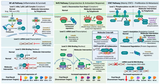
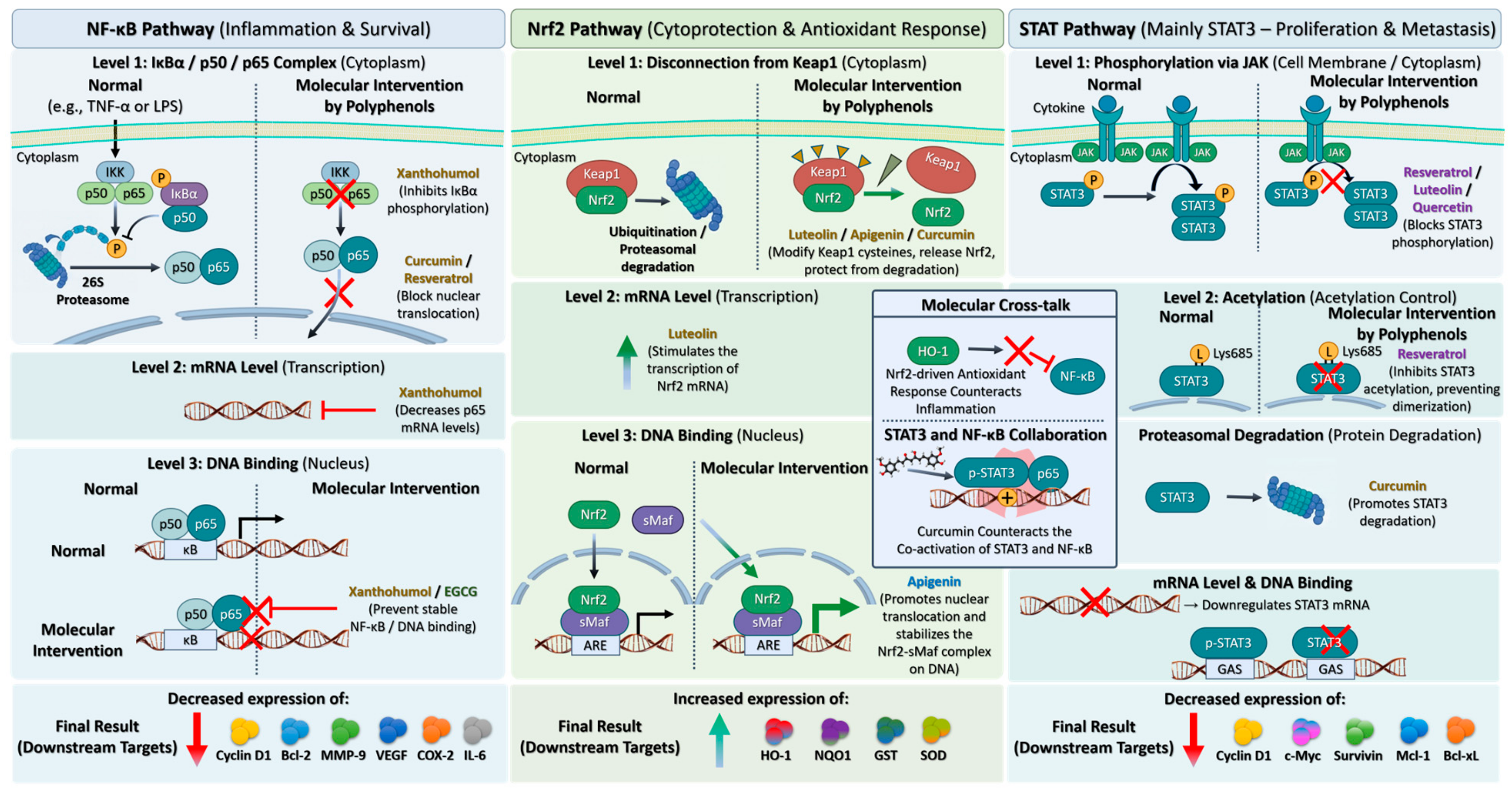

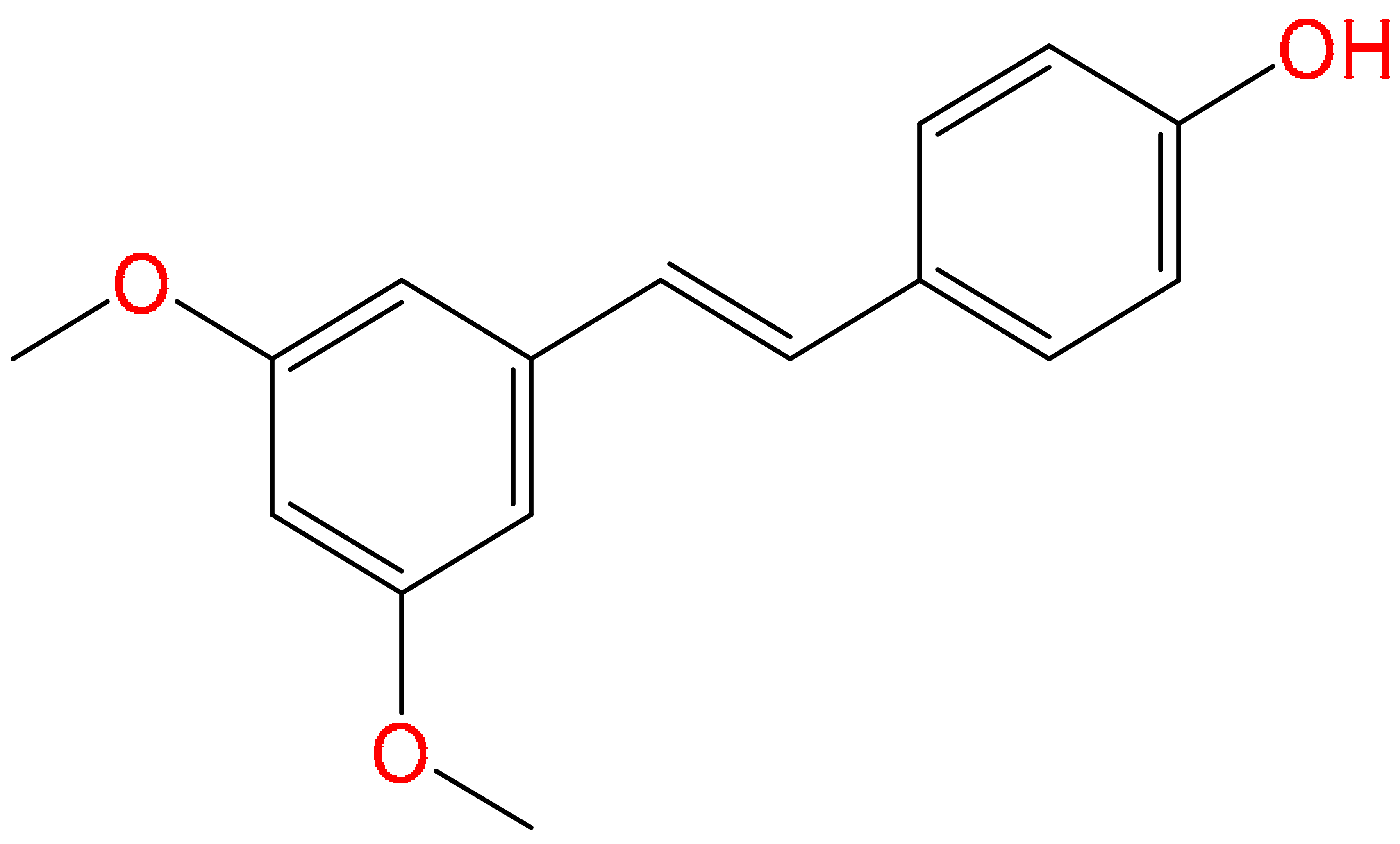
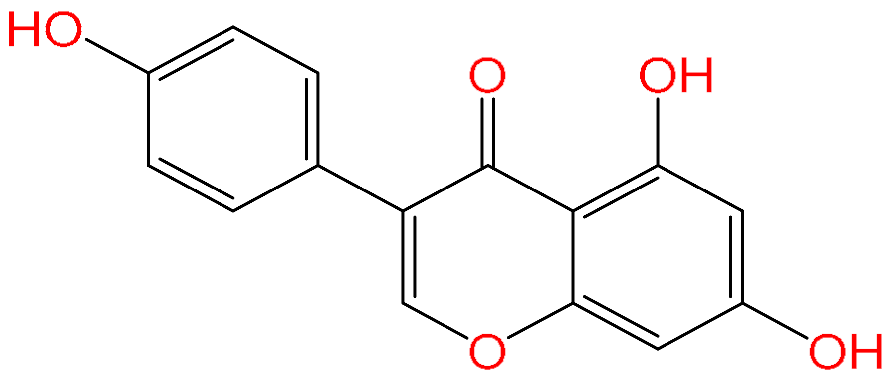
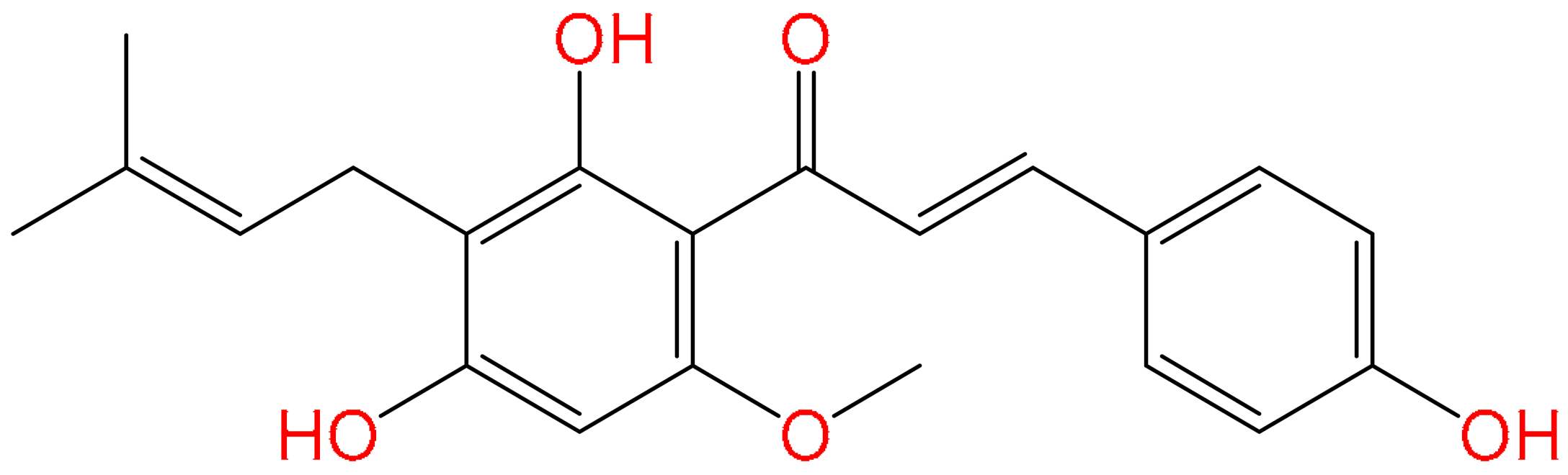
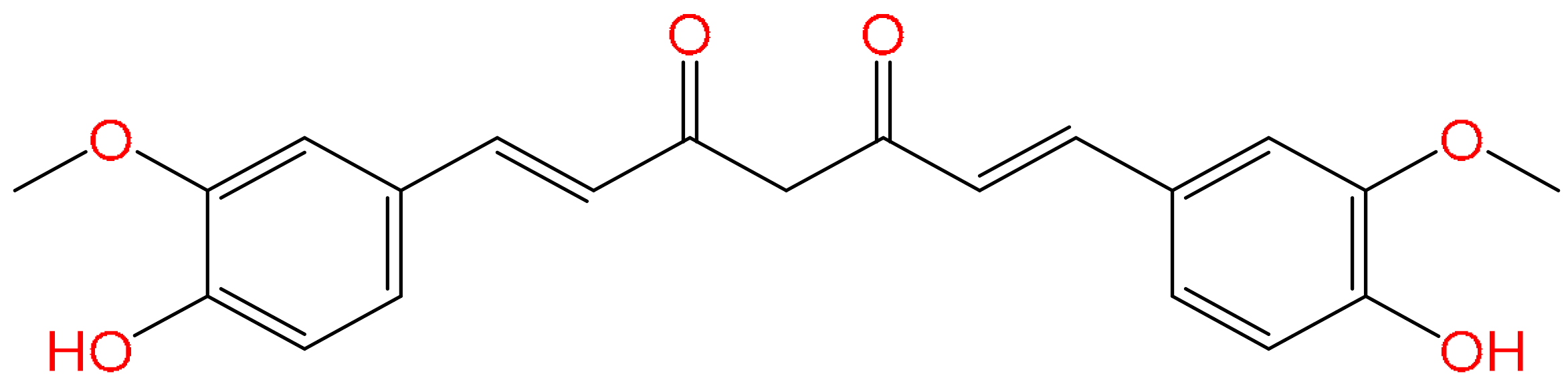
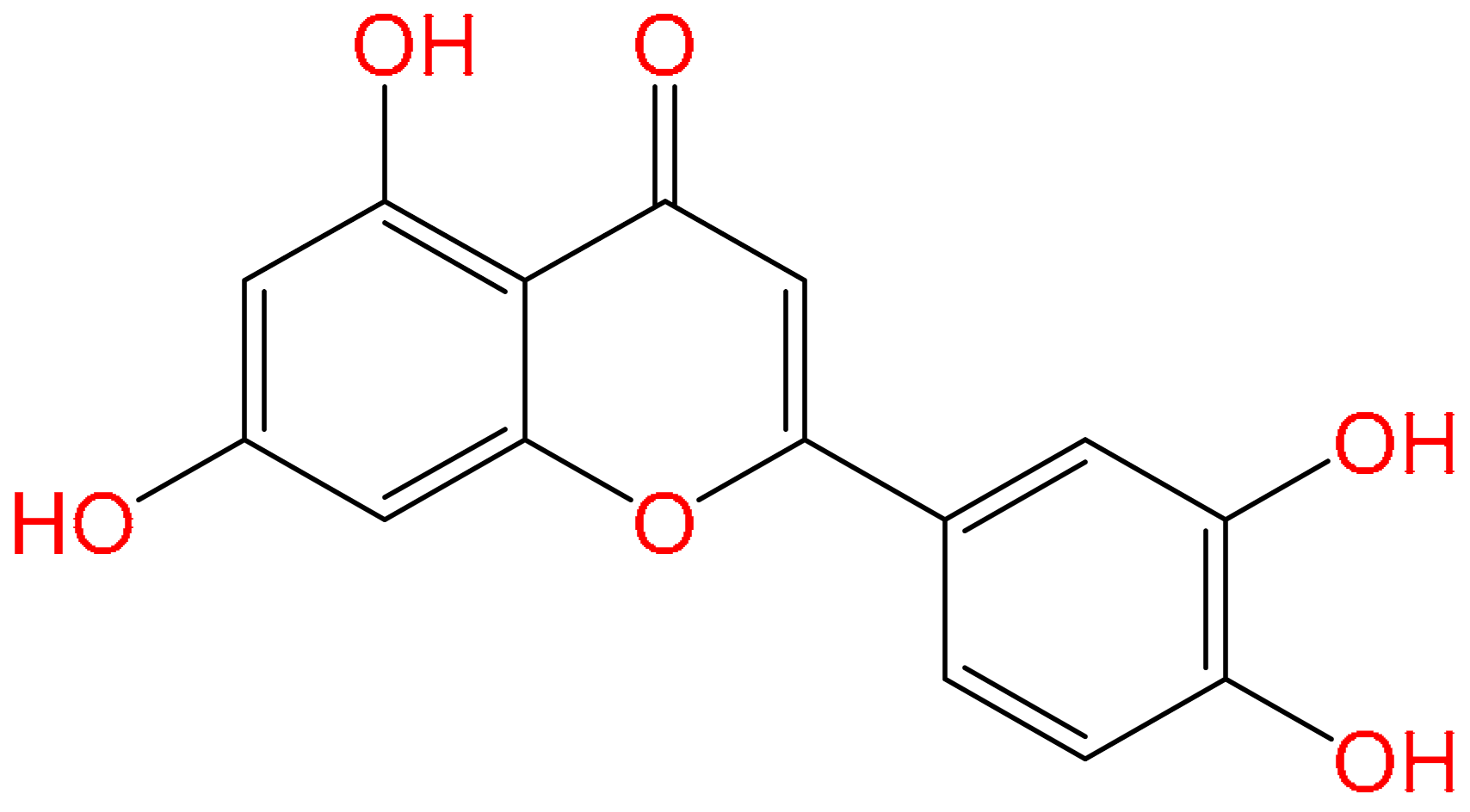
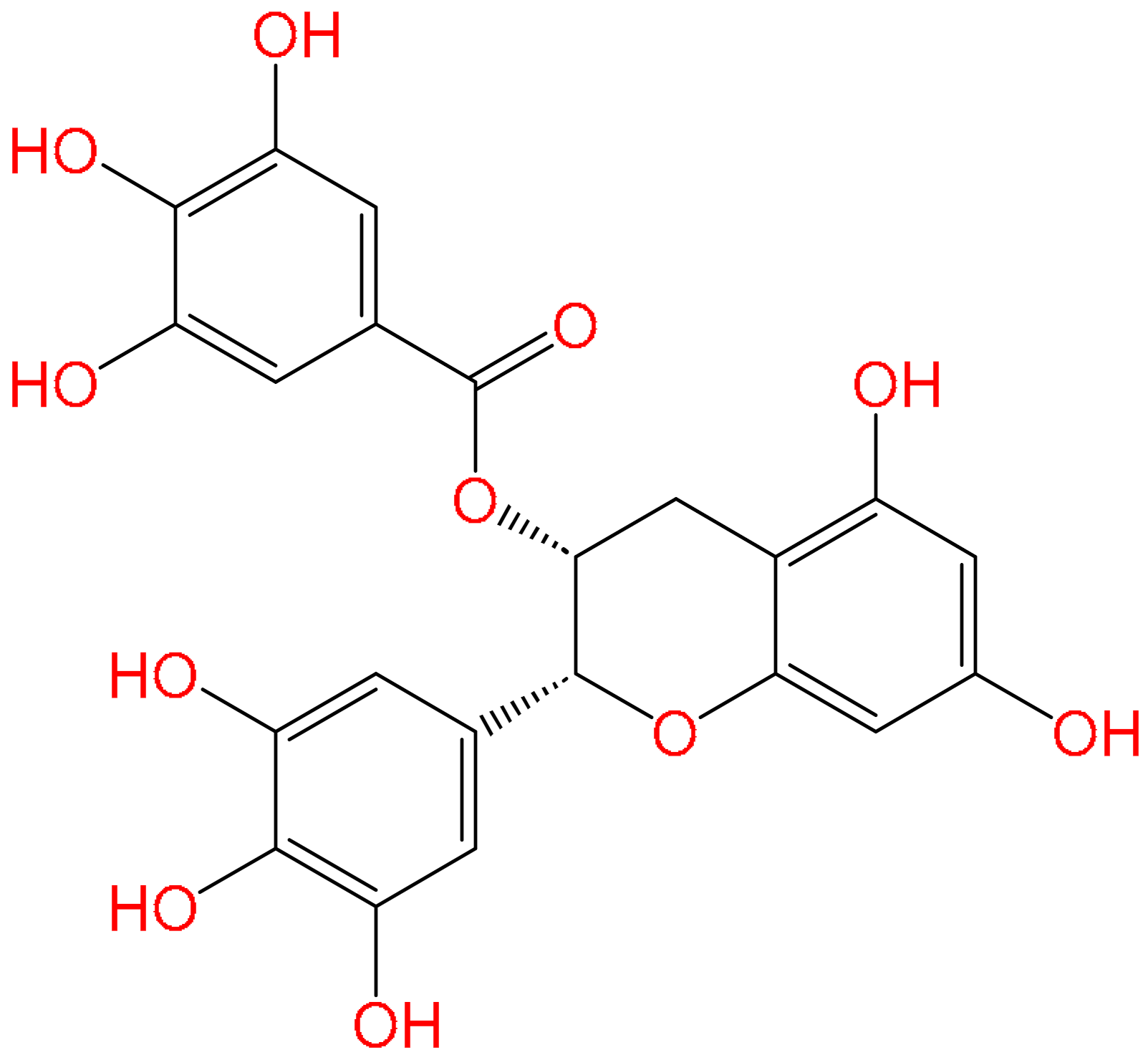
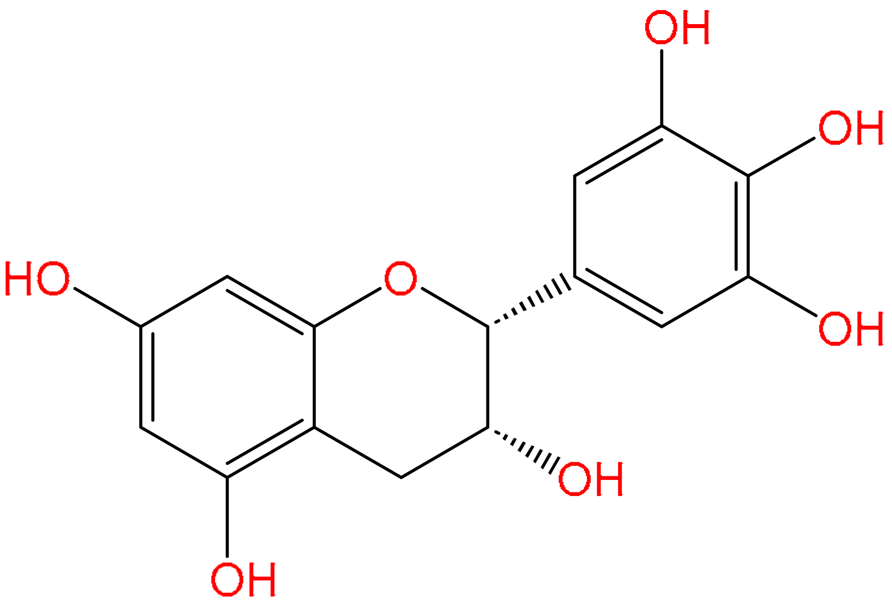
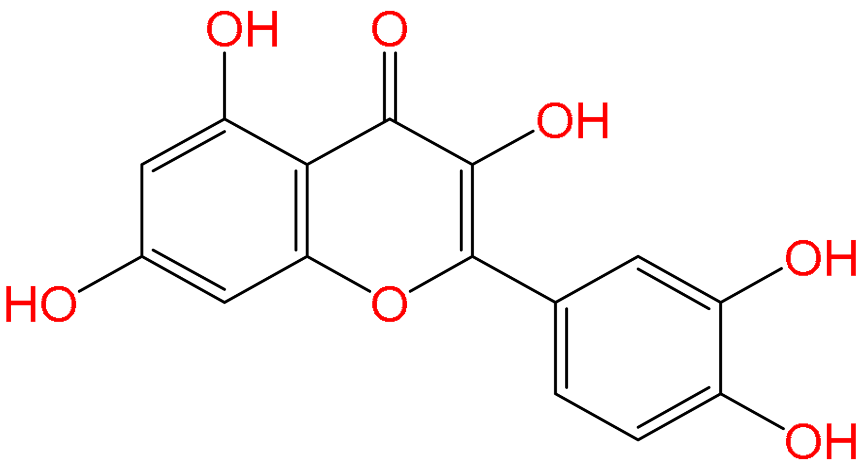
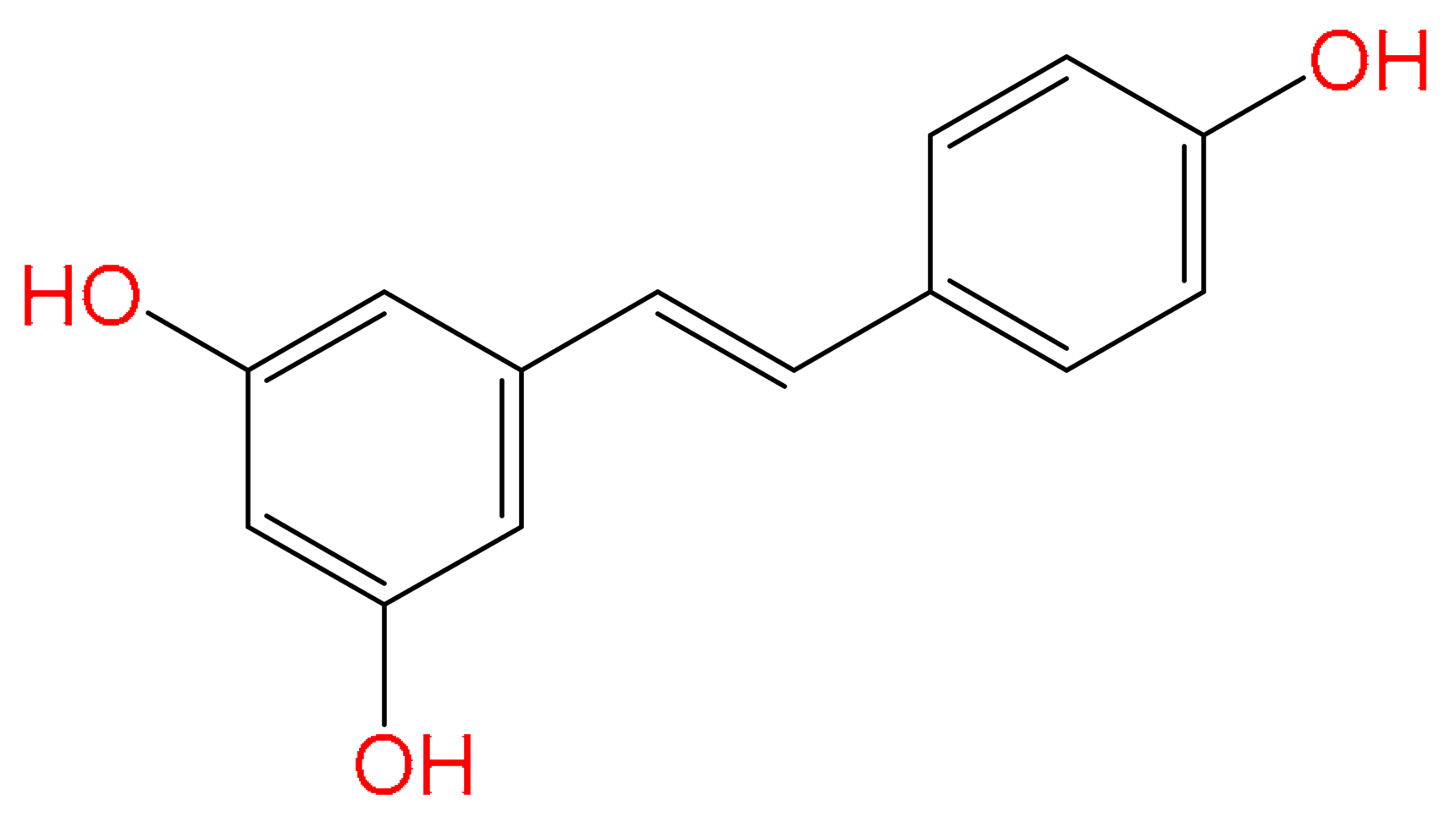
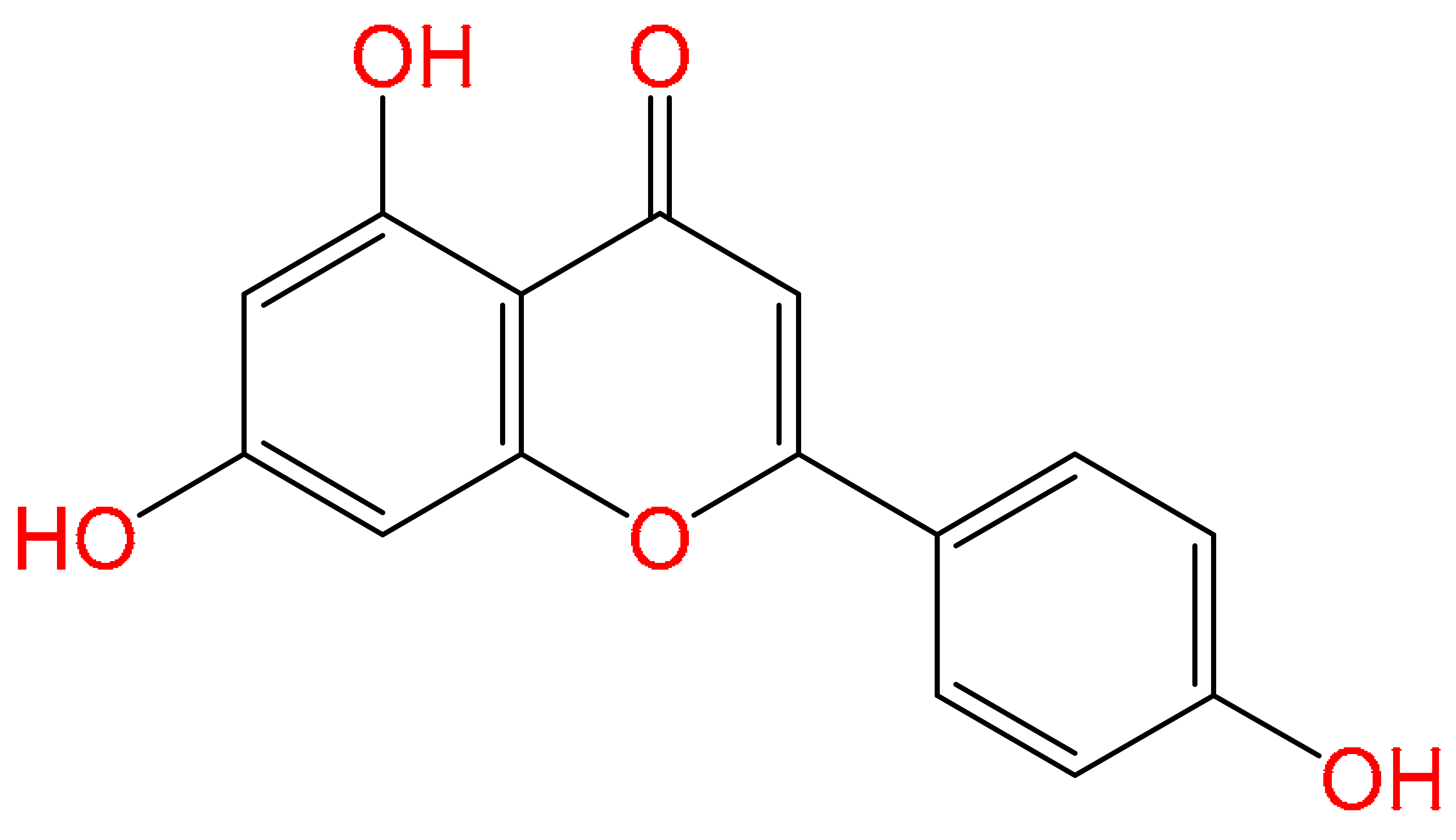
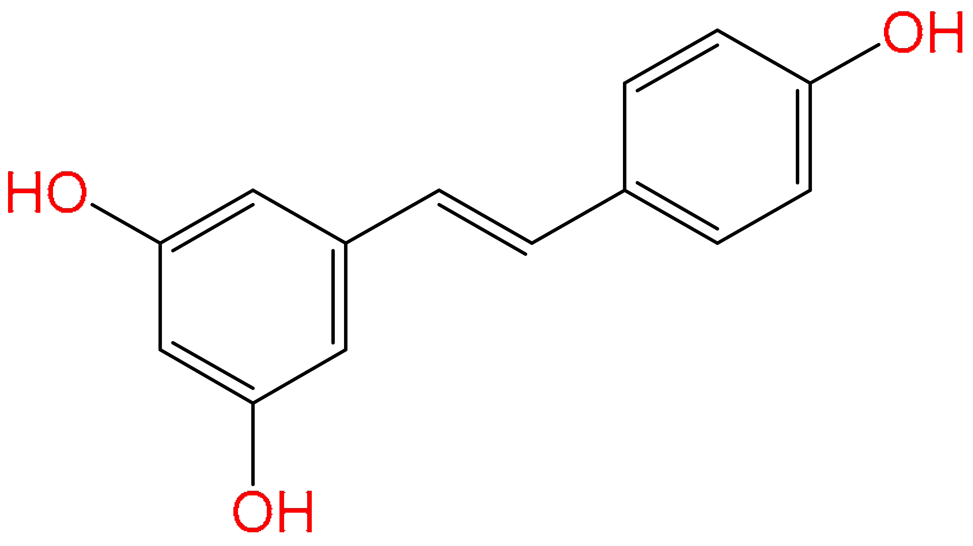
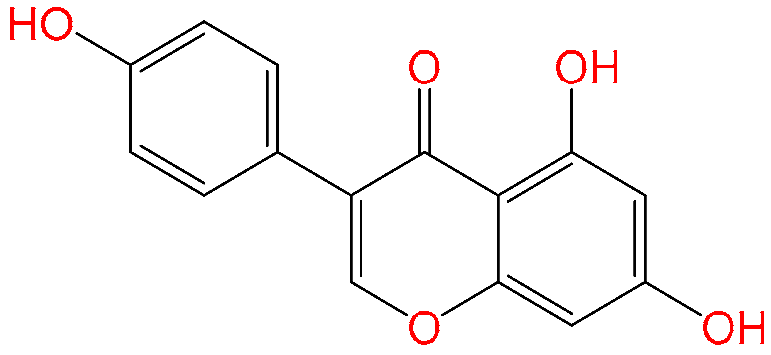
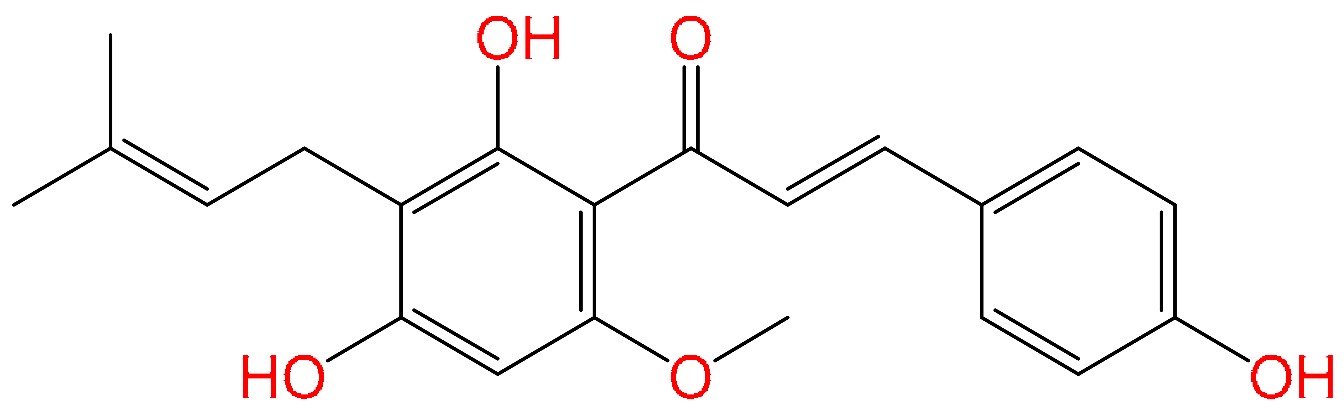
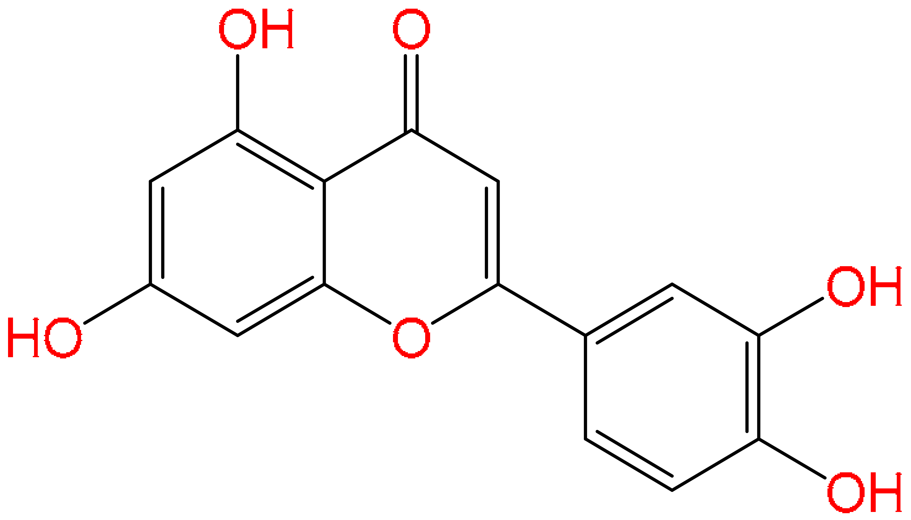
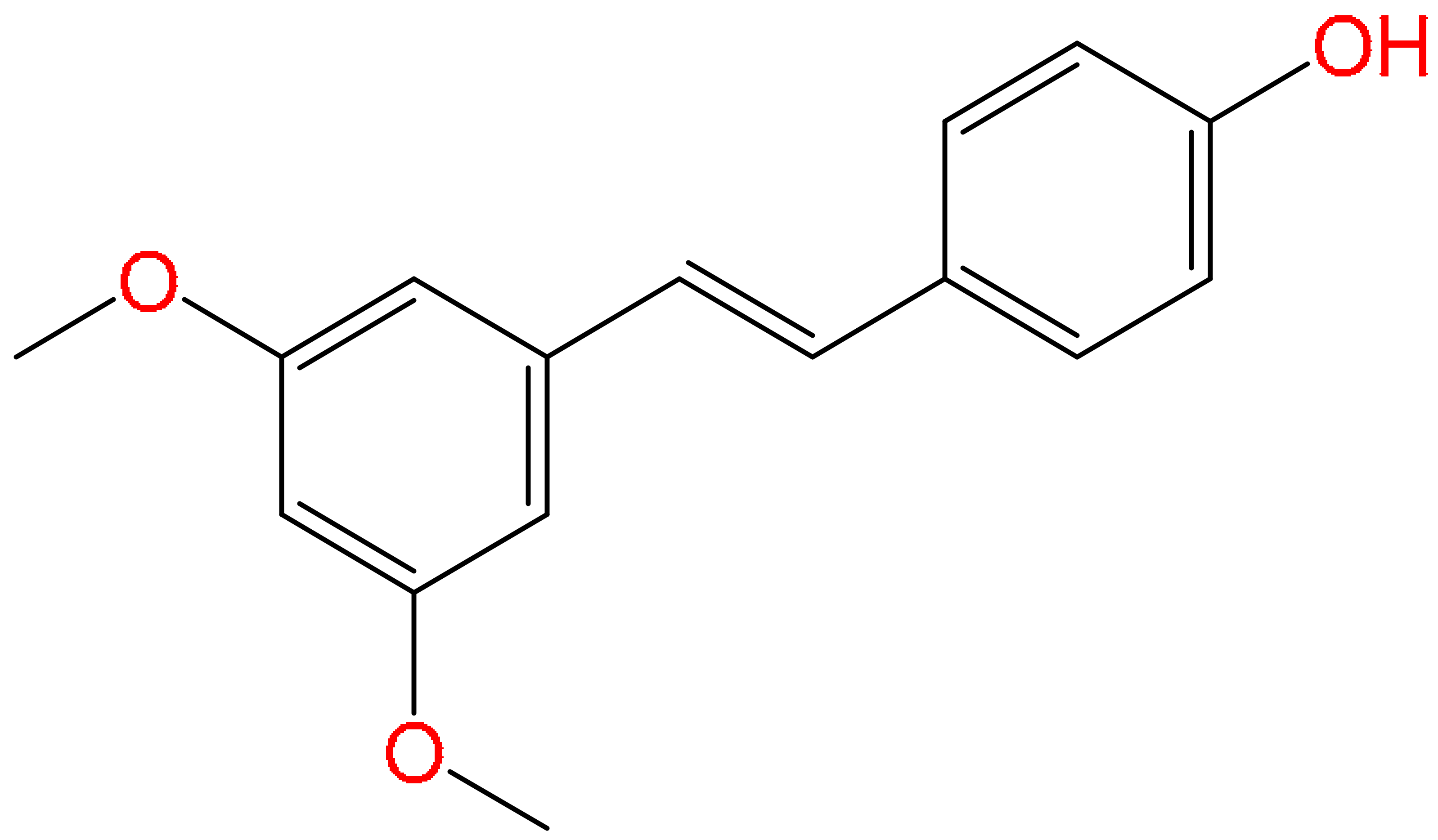
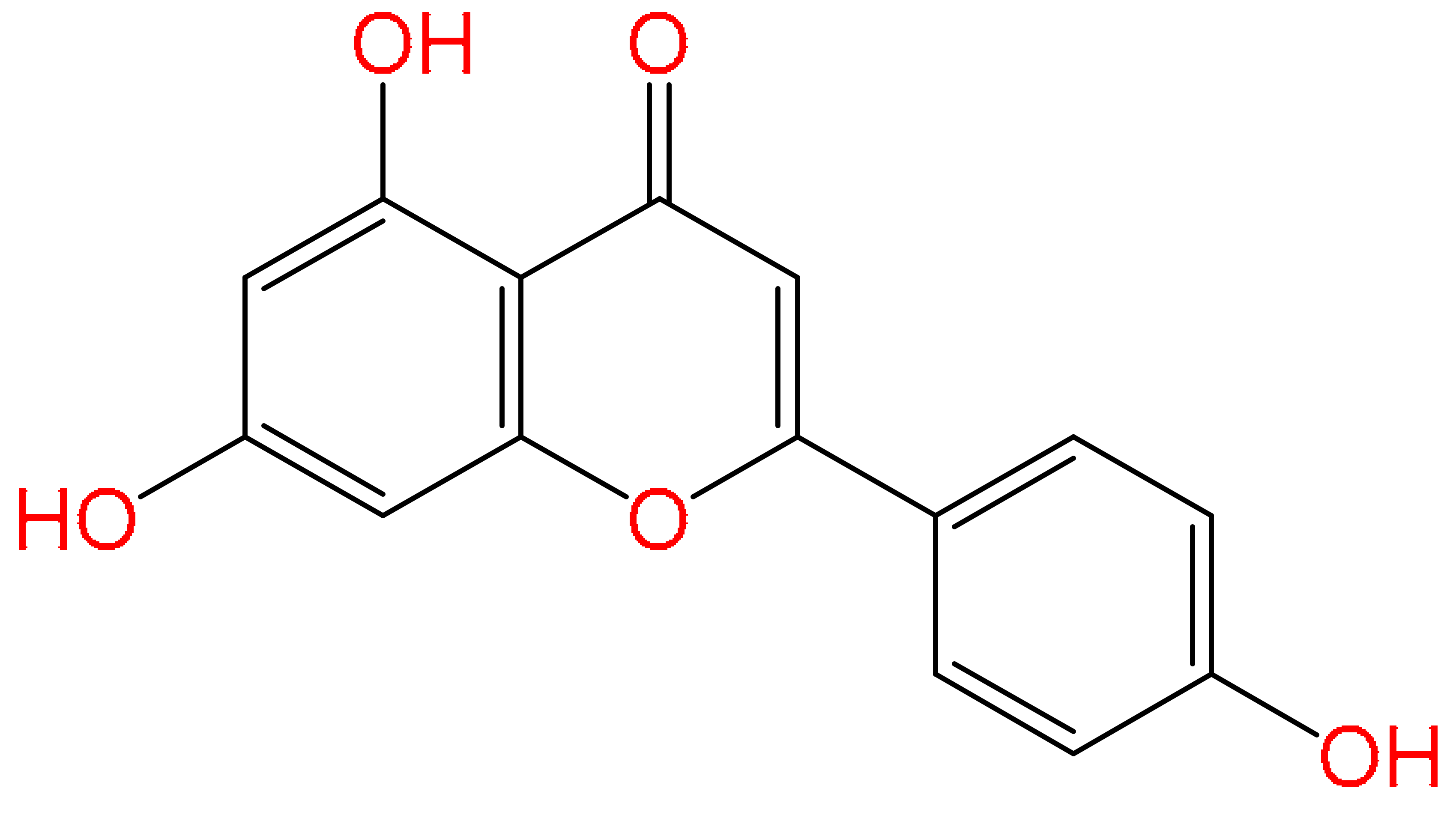
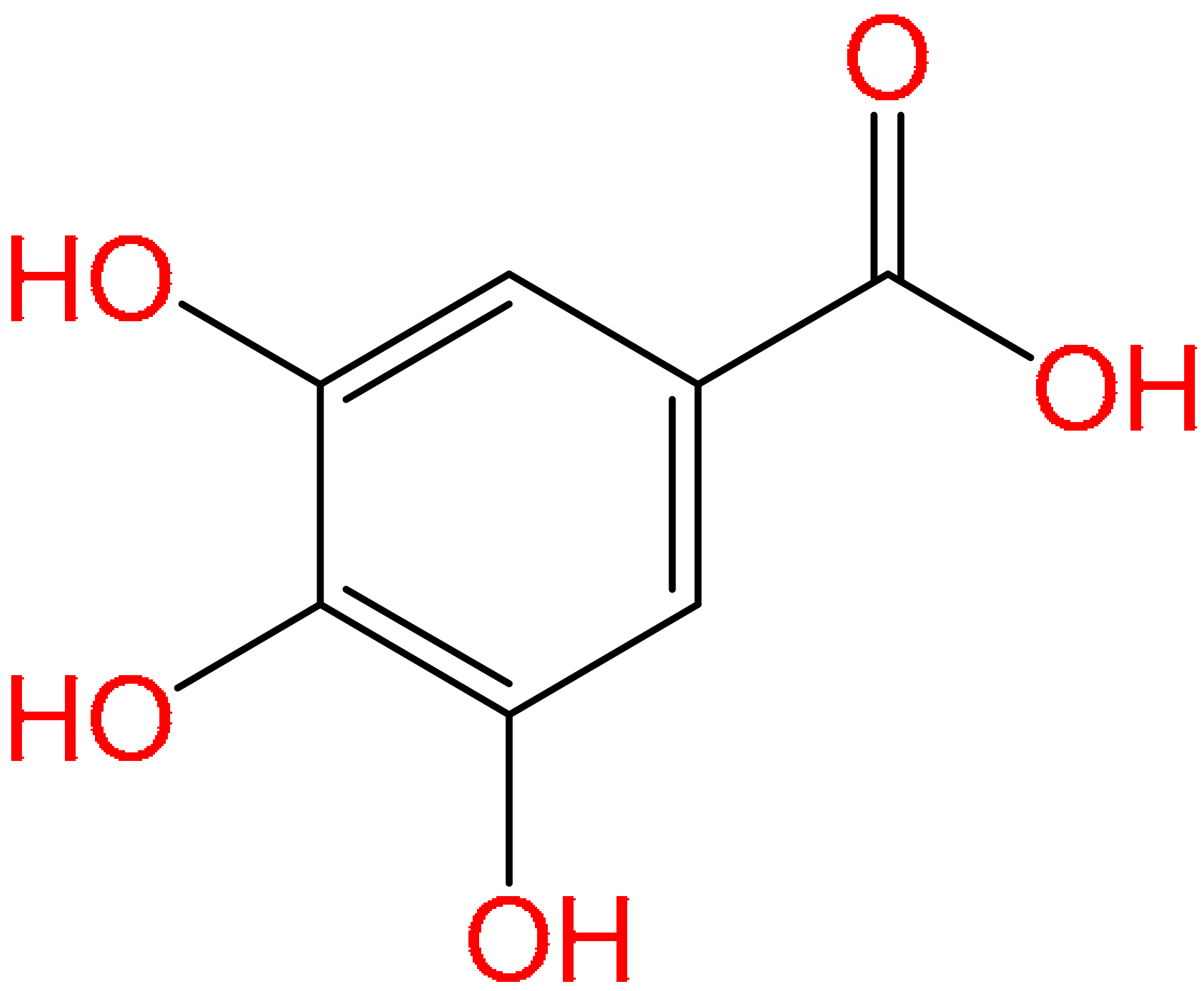
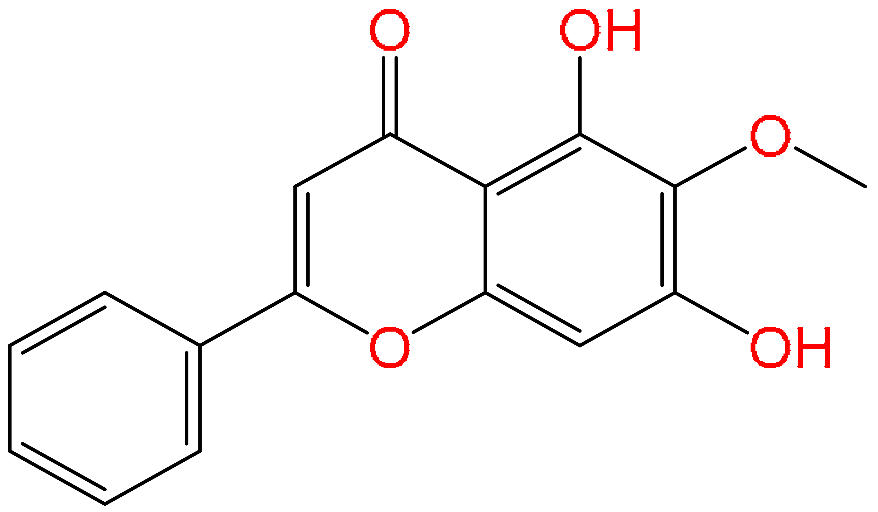
Disclaimer/Publisher’s Note: The statements, opinions and data contained in all publications are solely those of the individual author(s) and contributor(s) and not of MDPI and/or the editor(s). MDPI and/or the editor(s) disclaim responsibility for any injury to people or property resulting from any ideas, methods, instructions or products referred to in the content. |
© 2026 by the authors. Published by MDPI on behalf of the Österreichische Pharmazeutische Gesellschaft. Licensee MDPI, Basel, Switzerland. This article is an open access article distributed under the terms and conditions of the Creative Commons Attribution (CC BY) license.