


Abstract
Background/Objectives: In the last decade, the prevalence of metabolic-associated fatty liver disease (17–46%) and non-alcoholic fatty pancreas disease (NAFPD) (16–33%) has increased due to their association with obesity, both predictors of early atherosclerosis and metabolic risk. Computed tomography (CT) has been proposed as a diagnostic method. Currently, the factors associated with NAFPD have not been fully described. The aim of this study is to describe the prevalence and association of NAFPD and liver steatosis in patients with very high cardiovascular risk. Methods: A retrospective evaluation was conducted on the medical records of patients classified as very high cardiovascular risk who had undergone a CT scan. NAFPD was determined by the difference in pancreatic and splenic attenuation (−1.9), while liver steatosis was identified by hepatic attenuation <40. Bivariate and multivariate analyses were performed to determine the independent factors associated with NAFPD. Results: 169 medical records were collected; 68.6% (n = 116) were men, with a median age of 70 [IQR 61–78] years and 25.8 [IQR 23.7–28.7] kg/m2 of body mass index. According to the CT scans, 80.5% (n = 136) presented NAFPD, 24.3% (n = 41) had liver steatosis, and 21.3% (n = 36) had both. In the multivariate analysis, liver steatosis, abnormal levels of aspartate aminotransferase, and being overweight were independent factors associated with NAFPD. Conclusions: In a very high cardiovascular-risk population, the prevalence of NAFPD is high, and it is independently associated with the presence of liver steatosis.
1. Introduction
Pancreatic steatosis was described for the first time in 1926 [1]. Since then, different terms have been suggested to describe the accumulation of fat in the pancreas, or pancreatic steatosis. One of them is Non-alcoholic Fatty Pancreas Disease (NAFPD), which is used to describe a subtype of pancreatic steatosis associated with obesity and metabolic syndrome (MetS), in the absence of excessive alcohol consumption [2].
The pathophysiology of NAFPD is associated with a positive energy balance, resulting in the accumulation of free fatty acids in various organs, including the liver, heart, and vascular endothelium. This excess fat triggers the release of proinflammatory cytokines, promoting systemic inflammation and insulin resistance (IR) [3]. The prevalence of NAFPD in the adult population has been estimated between 16% and 33% [4]. The primary factors associated with the accumulation of pancreatic fat include MetS, age, and the presence of Metabolic Dysfunction-Associated Steatotic Liver Disease (MASLD) [5]. The latter is considered part of the MetS spectrum [6,7] and has gained significant importance due to its risk of progressing to hepatic fibrosis and its association with extrahepatic diseases such as type 2 diabetes mellitus (DM) and cardiovascular diseases [5,8,9]. Currently, it is recognized as the foremost cause of chronic liver disease worldwide. [8,10]. NAFPD has been linked to an increased risk of developing arterial hypertension (OR 1.67; 95% CI, 1.32–2.10; p < 0.0001), DM (OR 2.08; 95% CI, 1.44–3.00; p = 0.0001), and MetS (OR 2.37; 95% CI, 2.07–2.71; p < 0.0001) [11,12].
The reference standard for the diagnosis of pancreatic steatosis is detecting pancreatic fat ≥6.2% through histology [13]; likewise, the diagnosis of liver steatosis is confirmed with >5% of fat in a biopsy of hepatic tissue [10]. However, the invasive nature of biopsy limits its use in clinical practice; therefore, non-invasive imaging techniques, such as computed tomography (CT), have become the preferred alternative and have been shown to correlate well with histopathology in both scenarios. For pancreatic steatosis detection by CT, a cutoff value of −1.9 of the attenuation-based index, which estimates the difference between pancreatic and splenic attenuation (P-S), has a sensitivity of 80% and a specificity of 42%; on the other hand, the pancreas-spleen attenuation ratio (P/S), with a cutoff value of 0.9, shows a sensitivity of 70% and specificity of 58% [13,14].
Regarding the determination of liver steatosis, CT has demonstrated high performance in detecting liver steatosis compared to biopsy, with a sensitivity of 72% and a specificity of 88% [15].
The clinical significance of diagnosing NAFPD lies in its association with other diseases, such as chronic pancreatitis (OR 3.96; 95% CI, 2.04–7.66) [16], pancreatic cancer (OR 18.027; 95% CI, 7.28–44.58) [17], alterations in insulin secretion, and DM [12], as well as MASLD [18]. Furthermore, its association with MetS and an increased cardiovascular risk (CVR) has been reported [12,19]. A recent meta-analysis that included 49,329 patients found that NAFPD was significantly associated with an increased risk of MetS (RR = 2.25; 95% CI, 2.00–2.53; p < 0.0001), central obesity (RR = 1.91; 95% CI, 1.67–2.19; p < 0.0001) and MASLD (RR = 2.49; 95% CI, 2.06–3.02; p < 0.0001) [20]. Regarding CVR, the leading cause of mortality in patients with MetS and MASLD, a study by Kul et al. [21] found that epicardial adipose tissue, a marker of ectopic fat, and aortic intima-media thickness, an indirect indicator of subclinical atherosclerosis, were significantly higher in subjects with NAFPD.
Since 2018, the European Society of Cardiology, the European Atherosclerosis Association and the American Heart Association have proposed the “very high cardiovascular risk” definition, which includes patients with a history of one major atherosclerotic cardiovascular disease: recent acute coronary syndrome, a history of myocardial infarction, a history of ischemic stroke, or symptomatic peripheral arterial disease in addition to at least one high-risk condition such as age > 65 years, familial hypercholesterolemia, a history of prior artery bypass surgery, DM, hypertension, chronic kidney disease, current smoking, persistently elevated low-density lipoprotein (despite treatment with statin and ezetimibe), and a history of congestive heart failure [22,23]. These patients are characterized by a persistent inflammatory status, which, in combination with metabolic abnormalities, may lead to an increased risk of liver and pancreatic steatosis [24,25].
Despite the increasing evidence suggesting that NAFPD may negatively impact metabolism, predisposing individuals to MetS, MASLD, and the development of subclinical atherosclerosis [26]. The role of pancreatic steatosis remains an ongoing debate since there is evidence that suggests no relationship between pancreatic-liver steatosis and CVR. Specific risk factors for pancreatic steatosis have not been fully described; therefore, evaluating populations with very high CVR, such as patients in coronary care units, could provide evidence on the prevalence of this entity and its relationship with CVR, as well as comparing it with liver steatosis prevalence. This study aims to describe the prevalence and association of NAFPD and liver steatosis in patients with very high CVR.
2. Materials and Methods
2.1. Study Population
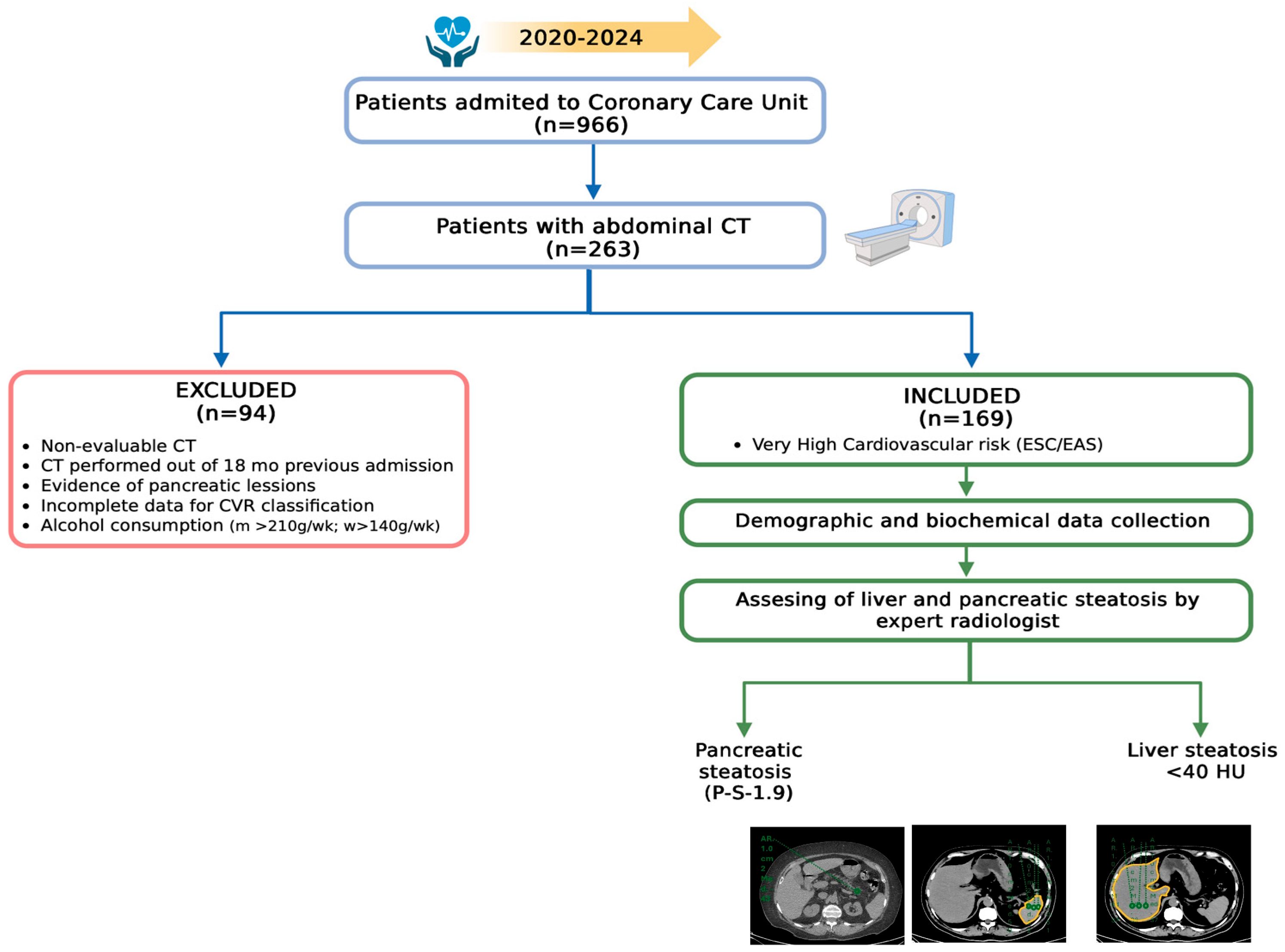
This is a retrospective medical record revision of adult patients (>18 years) admitted to the Coronary Care Unit of a tertiary care private hospital in Mexico City from 2020 to 2024 that met criteria for group 1 of very high CVR: documented clinical atherosclerotic cardiovascular disease, according to the European Society of Cardiology and the European Atherosclerosis Society [23], and had a non-contrast abdominal CT scan performed during their admission or within 18 months prior to the cardiovascular event for any reason (Figure 1).
Figure 1.
Diagram of selection criteria. CT Computed Tomography; CVR Cardiovascular Risk; m men; w woman; ESC European Society of Cardiology; EAS European Atherosclerosis Society; P-S Difference between pancreatic and splenic attenuation; HU Hounsfield units. Created in BioRender. Juarez-Hernández, E. (2025). https://BioRender.com/li8d2vn.
Biochemical data of blood glucose, lipids, and liver function tests were recorded and dichotomized according to abnormal values as follows: blood glucose > 100 mg/dL; triglycerides > 150 mg/dL; high-density lipoprotein (HDL) < 40 mg/dL for men and < 60 mg/dL for women; cholesterol > 200 mg/dL, aspartate aminotransferase (AST) > 47 U/L; alanine aminotransferase (ALT) > 40 U/L; and gamma-glutamyl transpeptidase (GGT) > 50 U/L. The exclusion criteria were non-evaluable CT images, evidence of pancreatic lesions, and incomplete data for CVR assessment; also, records of patients with excessive alcohol consumption (>210 g/week for men and >140 g/week for women), as referred to in the medical records, were excluded (Figure 1).
2.2. MASLD and NAFPD Assessment
Radiologists from the diagnostic imaging service reviewed the CT images to exclude pancreatic lesions, ensuring the integrity of the pancreatic parenchyma and vascular structures. A single expert radiologist assessed the presence of pancreatic steatosis using the attenuation index, based on both contrast-enhanced and non-contrast CT images. Three regions of interest (ROIs) with areas of 1.0 cm2 in different pancreatic sections were selected using a dedicated 3D workstation. The mean of pancreatic attenuation from the three ROI measurements was considered representative for each subject. We used the average of three cm2 ROIs from different spleen areas on non-enhanced CT images for the splenic attenuation. A cutoff value of −1.9 for P-S and a cutoff value of 0.9 for P/S were used to detect pancreatic steatosis. Meanwhile, liver steatosis was assessed using non-contrast abdominal tomography, where liver steatosis was defined as a hepatic attenuation of <40 Hounsfield units.
2.3. Statistical Analysis
The frequency and percentage of categorical variables, as well as the median and interquartile ranges (IQR) of continuous data, were estimated for descriptive analysis.
Given the high prevalence of NAFPD (80.5%), Prevalence Ratios (PR) and 95% confidence intervals were estimated using Poisson regression with robust (sandwich) variance. The variables analyzed included gender; overweight (Body Mass Index (BMI) 25–29.9 kg/m2) and obesity (BMI ≥ 30 kg/m2); presence of DM; high blood pressure; dyslipidemia, as documented in medical records of diagnosis or treatment; history of smoking; and dichotomized biochemical values. (glucose, triglycerides, HDL, LDL, total cholesterol, AST, ALT, and GGT) Variables with p < 0.10 in the univariate analysis were included in the multivariate model; p < 0.05 was considered statistically significant. Model calibration was evaluated using the Hosmer–Lemeshow test, and discrimination was assessed using the area under the ROC curve (AUC). Collinearity was assessed using the Variance Inflation Factor. Statistical analyses were performed using IBM SPSS Statistics, version 21.0.
3. Results
A total of 169 medical records met the inclusion criteria and were included in the analysis. 68.6% (n = 116) were male, with a median age of 70 [IQR 61–78] years and 25.8 [IQR 23.7–28.7] kg/m2 of BMI. DM was present in 32% (n = 54) of the patients; 60.9% (n = 103) had hypertension, 26.6% (n = 45) had dyslipidemia, and 26.6% (n = 45) had a history of smoking. According to BMI, 39.6% (n = 67) of patients were overweight, and obesity was present in 19.5% (n = 33). Regarding biochemical variables, the medians of glucose and triglycerides were 109 [IQR 96–134] mg/dL and 112 [IQR 84–144] mg/dL, respectively. The general characteristics of the patients are presented in Table 1.
Table 1.
General characteristics of the population.
Regarding previous history of cardiovascular events, 30.8% (n = 52) had a history of acute myocardial infarction, 3.6% (n = 6) had experienced a cerebrovascular event, 39.6% (n = 67) had acute coronary syndrome, and 45.6% (n = 77) had ischemic cardiopathy.
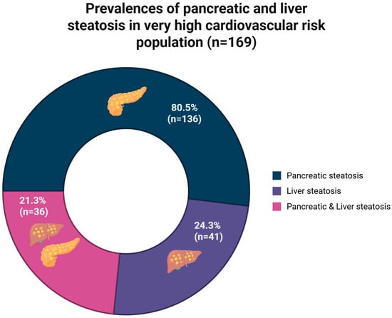
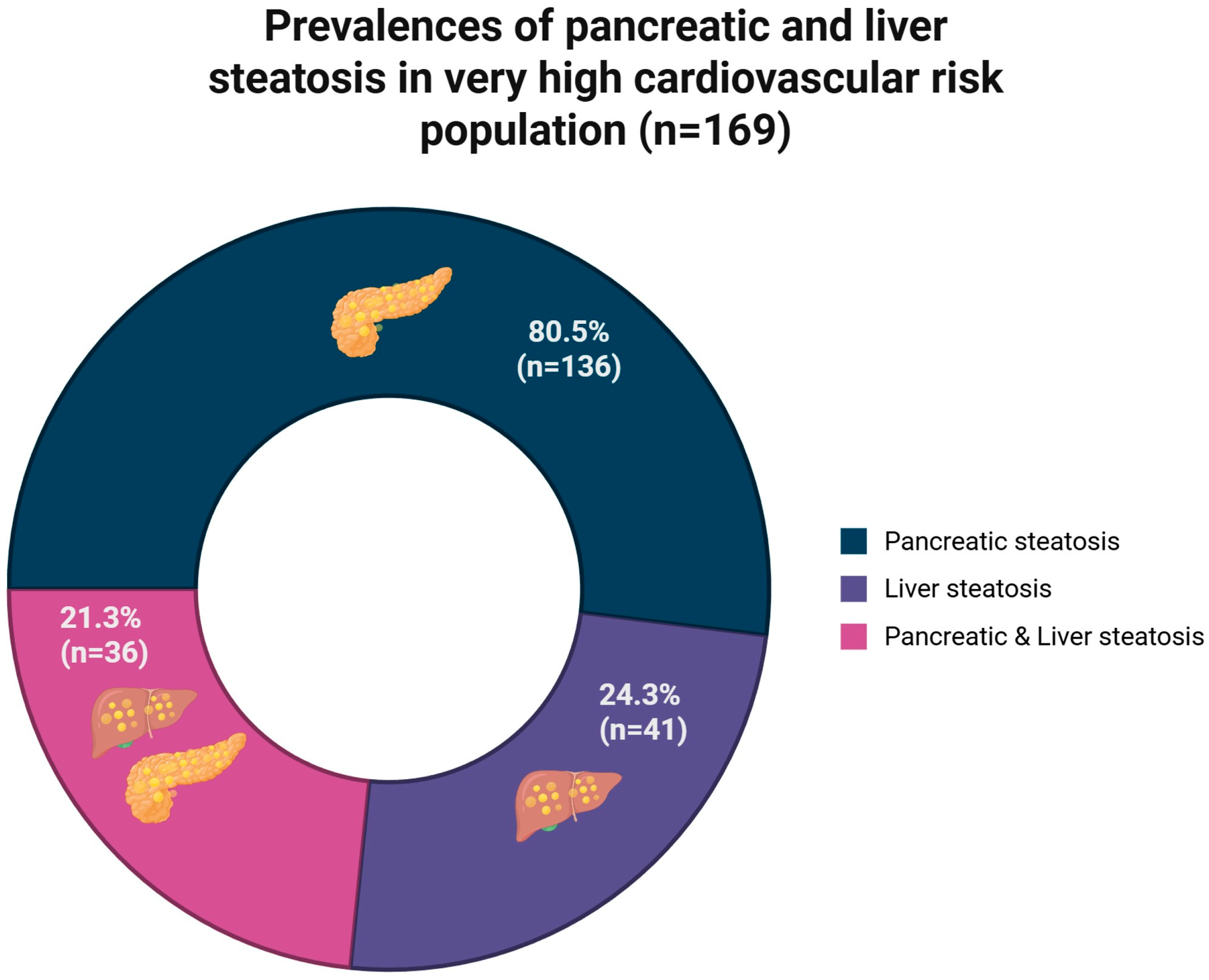
According to CT, 80.5% (n = 136) had NAFPD (P-S as the diagnostic reference), 24.3% (n = 41) had liver steatosis, and 21.3% (n = 36) had both (Figure 2). Meanwhile, the prevalence of NAFPD was 61.9% under the P/S criteria.
Figure 2.
Prevalences of pancreatic steatosis, liver steatosis, and both. Created in BioRender. Juárez-Hernández, E. (2025). https://BioRender.com/k95v303.
In the univariate analysis, liver steatosis (OR 6.23, 95% CI 1.42–27.31; p = 0.006), overweight (OR 0.40, 95% CI 0.19–0.87; p = 0.028), and elevated AST > 47 U/L (OR 0.44, 95% CI 0.19–0.98; p = 0.047) showed significant associations with NAFPD and were included in the multivariate model (Table 2).
Table 2.
Univariate and multivariate analysis of factors associated with NAPFD.
In the multivariate Poisson regression model, liver steatosis was independently associated with NAFPD (PR 1.21, 95% CI 1.07–1.36; p = 0.002), as was overweight (PR 0.83, 95% CI 0.71–0.98; p = 0.029). Elevated AST did not retain independent significance (PR 0.84, 95% CI 0.68–1.04; p = 0.114). The model showed adequate calibration (Hosmer–Lemeshow χ2(8) = 3.89, p = 0.867) and acceptable discrimination (AUC = 0.715).
The model was adjusted by BMI ≥ 25 kg/m2; in the Poisson regression model, only liver steatosis showed a significant independent association (PR 1.22 (95% CI 1.08–1.37; p = 0.001)); overweight and AST > 47 did not retain a significant association.
When univariate and multivariate analyses were performed using P/S as NAFPD diagnostic criteria (prevalence 61.9% (n = 104)); only overweight showed a significant association (OR 0.46, 95% CI 0.24–0.86; p = 0.08); however, the presence of DM (OR 1.88, 95% CI 0.93–3.80; p = 0.08) and smoking (OR 1.97, 95% CI 0.96–4.04; p = 0.08) were included in the multivariate analysis. Finally, DM (OR 1.30, 95% CI 1.04–1.62; p = 0.023) and overweight (OR 0.73, 95% CI 0.56–0.02; p = 0.020) were independently associated with NAFPD; however, discrimination of the model was moderate (AUC 0.627).
4. Discussion
In our study, we analyzed 169 patients admitted to the Coronary Care Unit, most of whom were men of advanced age. We found a high prevalence of pancreatic steatosis, and 21.3% of these cases coexist with liver steatosis.
The epidemiology of NAFPD has not been fully established due to the lack of specifically defined diagnostic criteria and methods; however, the prevalence of NAFPD in the USA has been estimated to be between 16% and 35% [4]. In a multicentric study, Sezgin et al. found a prevalence of 68.9% (n = 1700) in an outpatient reference clinic [27]. In our population, the prevalence of NAFPD was higher than previously reported (80.5%); however, it is essential to note that our study was conducted in a population with higher metabolic risk and does not consist of an obese population.
Nowadays, the metabolic implications of pancreatic steatosis are not fully understood; evidence shows an interaction between pancreatic steatosis and other organs, with effects on inflammatory signaling pathways. The recent study conducted by Antony et al. [28] included 37 patients who underwent a total or distal pancreatectomy due to evidence of pancreatic neoplasms. After twelve months of follow-up, it was observed that there was a significant increase in the incidence and severity of liver steatosis in patients who underwent a total pancreatectomy (p = 0.002). The authors conclude that the development of liver steatosis after pancreatic resections results from various factors, including pancreatic exocrine insufficiency, loss of duodenal hormones, portal endotoxemia, micronutrient deficiencies, and post-pancreatectomy diarrhea.
On the other hand, confirming the interaction between the pancreas and different organs and pathways, it is well known that patients with DM and obesity have impairments in β-pancreatic cell function, which is associated with IR. Oe et al. [29] conducted a study on patients with DM and obesity who underwent sleeve gastrectomy to determine the impact of bariatric surgery on β-pancreatic cell function. After one year of follow-up, a significant improvement was observed in the insulin secretion-sensitivity index-2 (0.91 ± 0.39 to 1.4 ± 0.7, p < 0.01). This finding highlights the interaction of the pancreas with multiple metabolic pathways.
In patients with very high CVR, the high prevalence of liver steatosis and NAFPD does not seem coincidental. Given the multifactorial etiology, insulin resistance, which drives excessive lipolysis in visceral adipose tissue, appears to be a key factor in the development of steatosis in both organs, with a continuous flow of free fatty acids to the liver and pancreas that simultaneously activates hepatic de novo lipogenesis and induces lipotoxic dysfunction of pancreatic β cells through the accumulation of diacylglycerols and ceramides [12,30].
The very high CVR in the presence of liver steatosis/NAFPD results from the synergistic convergence of these pathways—insulin resistance, lipotoxicity, hepatic atherogenic dyslipidemia, systemic inflammation, intestinal dysbiosis, and pancreatic dysfunction—which can lead to an exponential increase in CVR, highlighting the importance of both types of steatosis that can become potential therapeutic targets [31,32,33].
The results of these studies suggest that pancreatic steatosis is an underestimated entity that plays a role in regulating visceral fat accumulation. Probably, the presence of pancreatic steatosis is another factor involved in metabolic impairment, increasing liver steatosis, inflammation, and IR.
Regarding liver steatosis, in our study, the prevalence was also high; this elevated prevalence may be due to our sample comprising patients with very high CVR and a high prevalence of comorbidities, such as DM and hypertension. Additionally, 39.4% of the patients had a history of acute myocardial infarction, and 18.2% had heart failure conditions that contribute to a pro-inflammatory state and IR, which are associated with the development of NAFPD [4]. In a similar population, Yilmaz et al. [34] observed that liver steatosis, evaluated by ultrasound, could predict the presence and severity of coronary arterial diseases according to the Gensini score, showing significantly higher values between liver steatosis grades 0 and 2 and 3 (9.8 ± 11.9 vs. 56.7 ± 26.4, p ≤ 0.0001). These patients also exhibited significantly elevated levels of AST (27.0 ± 15.9 vs. 28.3 ± 12.3, p = 0.02). Despite these important results, liver steatosis was evaluated using ultrasound, a method that is operator-dependent and has lower diagnostic accuracy compared to CT [35].
The presence and progression of visceral fat accumulation, including hepatic and pancreatic steatosis, are attributed to a state of chronic low-grade inflammation that involves the production of proinflammatory cytokines in adipose tissue. In this process, it seems that splenic volume plays a significant role. The spleen has been associated with inflammatory diseases, but specifically in metabolic-inflammatory diseases such as obesity and liver steatosis, it seems that the spleen’s immune system has pathogenic connections between low-grade chronic inflammation, IR, and the development of liver steatosis due to shared metabolic activities of the liver-spleen axis; therefore, ultrasonography measurement of spleen volume has been proposed to improve liver steatosis diagnosis [36].
This study achieved NAFPD diagnosis through CT, using the P-S, which has a higher sensitivity than the P/S. Despite this, according to the P/S, the prevalence of NAFPD was lower (61.9%, n = 104). Although CT is not the standard diagnostic reference [37], it has been described that this technique correlates significantly with histological findings (r = −0.622, p < 0.01 for P-S) and also has been shown to have a strong inverse correlation with other image diagnostic methods such as magnetic resonance (Spearman coefficient −0.755) [38,39] regardless of patient factors, with the ability to evaluate the entire pancreas [40]. Therefore, it could be used to assess pancreatic fat in large population studies [14].
Cardiovascular disease is the leading cause of mortality in patients with MASLD; the underlying mechanism may involve metabolic dysfunction and an increased pancreatic fatty acid load in individuals with fatty liver, predisposing them to pancreatic fat accumulation, in a bidirectional way [3]; given this relationship, it is not unusual to assume that patients with NAPFD have an increased risk of cardiovascular alterations. In a study that included one hundred patients with MASLD, confirmed through liver biopsy, and thirty-four controls, Ozturk et al. observed significant correlations between the presence of fatty pancreas (evaluated by ultrasound) and higher carotid intima-media thickness and carotid-femoral pulse wave velocity; these correlations were maintained even after confounder adjustment [41]. In 324 liver steatosis patients, Marti-Aguado et al. [31] observed that pancreatic steatosis (evaluated by magnetic resonance imaging) was independently associated with a high risk of cardiovascular disease (OR: 3.15, 95% CI 1.63–6.09; p = 0.001). Regarding the very high CVR population, which includes DM patients, pancreatic fat has been proposed as an endocrine modulator of IR [42]; therefore, the presence of pancreatic steatosis has been related to an incidence of DM (adjusted OR per 10 Hounsfield units lower attenuation: 1.32 [95% CI 1.06–1.63], p = 0.012) [43]. In a prospective cohort study of 42,599 patients, Dong et al. [44] observed that pancreatic fat, as quantified by magnetic resonance imaging, independently increases the risk of DM by 33.7%; furthermore, pancreatic fat also increases the risk of acute and chronic pancreatitis (298.2% and 97.6%, respectively). In patients with very high CVR, the presence of NAPFD could be an additional risk for increased severity or progression of MetS, as well as pancreatic abnormalities.
Under conditions of fatty acid excess, when subcutaneous adipose tissue reaches its storage capacity, fatty acids are deposited in ectopic sites, such as the pancreas, liver, and other organs. This ectopic fat triggers systemic inflammation and IR and correlates significantly, according to sex, with different biomarkers of MetS, such as triglycerides (r = 0.46 w r = 0.37 m), HDL (r = 0.35 w r = 0.33 m), and blood glucose (r = 0.34 w r = 0.19 m) [3,45]. Due to these correlations, NAFPD has been linked to MetS and obesity [2]. In our study, the prevalence of MetS in patients with NAFPD was 52.5% (n = 42/80); in addition to the relationship between ectopic fat accumulation and the development of metabolic diseases and their complications, the presence of pancreatic steatosis has been particularly associated with local complications such as chronic pancreatitis (OR 3.96 [95% CI 2.04, 7.66]) [16] and pancreatic cancer (OR 18.027 [95% CI 7.28–44.58]) [17].
All of the results provide evidence for the association between metabolic abnormalities and NAFPD. However, this association may deteriorate with the addition of CVR, a condition inherently linked to metabolic impairment. This situation is particularly pertinent in current epidemiological contexts characterized by the increasing prevalence of obesity. Overweight/obesity is linked to increased levels of inflammatory cytokines (interleukin-6 and C-reactive protein) and significantly reduced levels of adiponectin, which is associated with coronary artery calcification and stroke [46]. Inflammation represents a pivotal mechanism implicated in cardiovascular diseases and their associated comorbidities, including DM and metabolic syndrome MetS. Both obesity and cardiometabolic abnormalities appear to be significant determinants influencing the progression of pancreatic steatosis, thereby exacerbating pancreatic stiffness. Specifically, glucose impairments have been proposed as stronger predictors of elevated stiffness in patients with pancreatic fat infiltration [47].
One of the consistent findings of our study is the inverse association between overweight and NAFPD; this result could be associated with the average age of our patients (70 years), where sarcopenia and visceral redistribution of fat with age are prevalent [48]. BMI in the overweight range may represent a metabolically less adverse phenotype than normal weight or obesity, possibly due to better nutritional status and lower chronic systemic inflammation. However, BMI is a poor surrogate for visceral adiposity [49], as it is not a parameter that distinguishes between muscle mass and fat mass, so the measurement of ectopic pancreatic fat accumulation correlates more strongly with visceral adiposity than with BMI itself [50,51].
Our study has limitations, including its retrospective design and the methods used to detect steatosis. Notwithstanding, at this moment, there are no specific guidelines for the diagnosis of pancreatic steatosis, and the reference standard for diagnosis is a procedure that represents significant risks and complications for the patient (pancreatic biopsy). Although CT has shown good concordance with pancreatic and liver biopsy, other non-invasive methods have better performance, particularly magnetic resonance and transient elastography in liver steatosis [52], However, due to the retrospective nature of our study and the setting of the patients, the detection of pancreatic or liver steatosis was not an intentional focus in managing the clinical scenario. Despite the very high CVR nature of our population, the median values of blood lipids were within normal ranges. Even when the history of dyslipidemia showed a significant association with the presence of NAFPD in univariate analysis, this association was not maintained in the multivariate analysis. This could be explained by the fact that these patients could already be under pharmacological treatment, which is also a characteristic of patients classified as very high CVR.
Currently, the evidence regarding the association between pancreatic steatosis and CVR remains controversial. Consequently, this study aims to evaluate the factors associated with pancreatic steatosis in patients who have already been identified as having very high CVR, rather than investigating steatosis as a potential risk factor; our results show a significant association with liver steatosis in these patients. Future prospective studies are necessary to highlight the importance of pancreatic steatosis, not only as an additional CVR factor but also as a standalone disease that could be associated with pancreatic diseases, liver steatosis, and MetS.
It is imperative to acknowledge that the correlation between CVR and pancreatic steatosis remains controversial. Independent of the limitations, our results show a significant association between the presence of pancreatic and liver steatosis in patients with very high CVR, which supports the need for additional prospective studies to explain the role of NAFPD in both the general population and in patients with high metabolic risk more precisely.
Liver steatosis and CVR are significant targets for clinical trials; however, according to our results and the emerging evidence regarding NAFPD, this could be considered an additional pathology that deserves inclusion in trial designs. Although the evidence is limited, it appears to be the tip of the iceberg in describing a probable new metabolic pathway.
5. Conclusions
In a very high CVR population, NAFPD has a high prevalence, and it is independently associated with the presence of liver steatosis.
Author Contributions
Conceptualization, I.L.-M.; methodology, I.L.-M. and V.T.-C. and E.J.-H.; formal analysis, E.J.-H., I.L.-M.; investigation, R.G.-M.; resources, M.U.; data curation, J.S.-Z., R.G.-M., C.A.T.-A.; writing—original draft preparation, R.G.-M.; writing—review and editing, I.L.-M., G.C.-N., and E.J.-H.; supervision, M.U.; project administration, M.U. All authors have read and agreed to the published version of the manuscript.
Funding
This research received no external funding.
Institutional Review Board Statement
The study was conducted in accordance with the Declaration of Helsinki, and approved by the Institutional Ethics Committee of MEDICA SUR S.A.B. DE C.V. (2023-EXT-727, date of approval: 20 April 2023).
Informed Consent Statement
Informed consent was obtained from all subjects involved in the study.
Data Availability Statement
The data presented in this study are available on request from the corresponding authors. The data are not publicly available due to privacy and ethics.
Acknowledgments
The authors would like to thank Ana Luisa Ordoñez-Vázquez and Alejandra Mijangos-Trejo; Monserrat Berrospe-Alfaro and Luis Eduardo Cazeres-Villareal for their support in data collection.
Conflicts of Interest
The authors declare no conflicts of interest.
Abbreviations
The following abbreviations are used in this manuscript:
| NAFPD | Nonalcoholic Fatty Pancreas Disease |
| MetS | Metabolic Syndrome |
| IR | Insulin Resistance |
| MASLD | Metabolic Dysfunction-Associated Steatotic Liver Disease |
| DM | Type 2 Diabetes Mellitus |
| CT | Computed Tomography |
| P-S | Pancreatic and Splenic attenuation |
| P/S | Pancreas/spleen attenuation ratio |
| CVR | Cardiovascular Risk |
| HDL | High Density Lipoprotein |
| AST | Aspartate Aminotransferase |
| ALT | Alanine Aminotransferase |
| GGT | Gamma-Glutamyl Transpeptidase |
| ROI | Regions of Interest |
| IQR | Interquartile Range |
| BMI | Body Mass Index |
References
- Schaefer, J.H. The Normal Weight of the Pancreas in the Adult Human Being: A Biometric Study. Anat. Rec. 1926, 32, 119–132. [Google Scholar] [CrossRef] [Scilit]
- Ramkissoon, R.; Gardner, T.B. Pancreatic Steatosis: An Emerging Clinical Entity. Am. J. Gastroenterol. 2019, 114, 1726–1734. [Google Scholar] [CrossRef] [Scilit]
- Britton, K.A.; Fox, C.S. Ectopic Fat Depots and Cardiovascular Disease. Circulation 2011, 124, e837–e841. [Google Scholar] [CrossRef] [Scilit]
- Habas, E.; Farfar, K.; Habas, E.; Rayani, A.; Elzouki, A.N. Extended Review and Updates of Nonalcoholic Fatty Pancreas Disease. Saudi J. Med. Med. Sci. 2024, 12, 284–291. [Google Scholar] [CrossRef] [Scilit]
- Eslam, M.; Newsome, P.N.; Sarin, S.K.; Anstee, Q.M.; Targher, G.; Romero-Gomez, M.; Zelber-Sagi, S.; Wai-Sun Wong, V.; Dufour, J.F.; Schattenberg, J.M.; et al. A New Definition for Metabolic Dysfunction-Associated Fatty Liver Disease: An International Expert Consensus Statement. J. Hepatol. 2020, 73, 202–209. [Google Scholar] [CrossRef] [Scilit]
- Khan, R.S.; Bril, F.; Cusi, K.; Newsome, P.N. Modulation of Insulin Resistance in Nonalcoholic Fatty Liver Disease. Hepatology 2019, 70, 711–724. [Google Scholar] [CrossRef] [Scilit]
- Tomizawa, M.; Kawanabe, Y.; Shinozaki, F.; Sato, S.; Motoyoshi, Y.; Sugiyama, T.; Yamamoto, S.; Sueishi, M. Triglyceride Is Strongly Associated with Nonalcoholic Fatty Liver Disease among Markers of Hyperlipidemia and Diabetes. Biomed. Rep. 2014, 2, 633–636. [Google Scholar] [CrossRef] [Scilit] [PubMed]
- Lin, H.; Zhang, X.; Li, G.; Wong, G.L.H.; Wong, V.W.S. Epidemiology and Clinical Outcomes of Metabolic (Dysfunction)-Associated Fatty Liver Disease. J. Clin. Transl. Hepatol. 2021, 9, 972–982. [Google Scholar] [CrossRef] [Scilit] [PubMed]
- Zhou, F.; Zhou, J.; Wang, W.; Zhang, X.J.; Ji, Y.X.; Zhang, P.; She, Z.G.; Zhu, L.; Cai, J.; Li, H. Unexpected Rapid Increase in the Burden of NAFLD in China From 2008 to 2018: A Systematic Review and Meta-Analysis. Hepatology 2019, 70, 1119–1133. [Google Scholar] [CrossRef] [Scilit]
- Rinella, M.E.; Lazarus, J.V.; Ratziu, V.; Francque, S.M.; Sanyal, A.J.; Kanwal, F.; Romero, D.; Abdelmalek, M.F.; Anstee, Q.M.; Arab, J.P.; et al. A Multisociety Delphi Consensus Statement on New Fatty Liver Disease Nomenclature. J. Hepatol. 2023, 79, 1542–1556. [Google Scholar] [CrossRef]
- Singh, R.G.; Yoon, H.D.; Wu, L.M.; Lu, J.; Plank, L.D.; Petrov, M.S. Ectopic Fat Accumulation in the Pancreas and Its Clinical Relevance: A Systematic Review, Meta-Analysis, and Meta-Regression. Metabolism 2017, 69, 1–13. [Google Scholar] [CrossRef] [Scilit] [PubMed]
- Wagner, R.; Eckstein, S.S.; Yamazaki, H.; Gerst, F.; Machann, J.; Jaghutriz, B.A.; Schürmann, A.; Solimena, M.; Singer, S.; Königsrainer, A.; et al. Metabolic Implications of Pancreatic Fat Accumulation. Nat. Rev. Endocrinol. 2021, 18, 43–54. [Google Scholar] [CrossRef] [Scilit]
- Kim, H.J.; Byun, J.H.; Park, S.H.; Shin, Y.M.; Kim, P.N.; Ha, H.K.; Lee, M.G. Focal Fatty Replacement of the Pancreas: Usefulness of Chemical Shift MRI. AJR Am. J. Roentgenol. 2007, 188, 429–432. [Google Scholar] [CrossRef] [Scilit]
- Kim, S.Y.; Kim, H.; Cho, J.Y.; Lim, S.; Cha, K.; Lee, K.H.; Kim, Y.H.; Kim, J.H.; Yoon, Y.S.; Han, H.S.; et al. Quantitative Assessment of Pancreatic Fat by Using Unenhanced CT: Pathologic Correlation and Clinical Implications. Radiology 2014, 271, 104–112. [Google Scholar] [CrossRef] [Scilit]
- Haghshomar, M.; Antonacci, D.; Smith, A.D.; Thaker, S.; Miller, F.H.; Borhani, A.A. Diagnostic Accuracy of CT for the Detection of Hepatic Steatosis: A Systematic Review and Meta-Analysis. Radiology 2024, 313, e241171. [Google Scholar] [CrossRef] [Scilit]
- Fujii, M.; Ohno, Y.; Yamada, M.; Kamada, Y.; Miyoshi, E. Impact of Fatty Pancreas and Lifestyle on the Development of Subclinical Chronic Pancreatitis in Healthy People Undergoing a Medical Checkup. Environ. Health Prev. Med. 2019, 24, 10. [Google Scholar] [CrossRef] [Scilit] [PubMed]
- Desai, V.; Patel, K.; Sheth, R.; Barlass, U.; Chan, Y.M.; Sclamberg, J.; Bishehsari, F. Pancreatic Fat Infiltration Is Associated with a Higher Risk of Pancreatic Ductal Adenocarcinoma. Visc. Med. 2020, 36, 220–226. [Google Scholar] [CrossRef] [Scilit] [PubMed]
- Van Geenen, E.J.M.; Smits, M.M.; Schreuder, T.C.M.A.; Van Der Peet, D.L.; Bloemena, E.; Mulder, C.J.J. Nonalcoholic Fatty Liver Disease Is Related to Nonalcoholic Fatty Pancreas Disease. Pancreas 2010, 39, 1185–1190. [Google Scholar] [CrossRef] [Scilit]
- Dite, P.; Blaho, M.; Bojkova, M.; Jabandziev, P.; Kunovsky, L. Nonalcoholic Fatty Pancreas Disease: Clinical Consequences. Dig. Dis. 2020, 38, 143–149. [Google Scholar] [CrossRef] [Scilit]
- Bi, Y.; Wang, J.L.; Li, M.L.; Zhou, J.; Sun, X.L. The Association between Pancreas Steatosis and Metabolic Syndrome: A Systematic Review and Meta-Analysis. Diabetes. Metab. Res. Rev. 2019, 35, e3142. [Google Scholar] [CrossRef] [Scilit]
- Kul, S.; Karadeniz, A.; Dursun, I.; Şahin, S.; Çirakoglu, Ö.F.; Sayin, M.R.; Turan, T.; Ateş, A.H. Non-Alcoholic Fatty Pancreas Disease Is Associated with Increased Epicardial Adipose Tissue and Aortic Intima-Media Thickness. Acta Cardiol. Sin. 2019, 35, 118–125. [Google Scholar] [CrossRef] [Scilit]
- Grundy, S.M.; Stone, N.J.; Bailey, A.L.; Beam, C.; Birtcher, K.K.; Blumenthal, R.S.; Braun, L.T.; de Ferranti, S.; Faiella-Tommasino, J.; Forman, D.E.; et al. 2018 AHA/ACC/AACVPR/AAPA/ABC/ACPM/ADA/AGS/APhA/ASPC/NLA/PCNA Guideline on the Management of Blood Cholesterol: Executive Summary. J. Am. Coll. Cardiol. 2019, 73, 3168–3209. [Google Scholar] [CrossRef] [Scilit]
- Tokgözoğlu, L.; Casula, M.; Pirillo, A.; Catapano, A.L. Similarities and Differences between European and American Guidelines on the Management of Blood Lipids to Reduce Cardiovascular Risk. Atheroscler. Suppl. 2020, 42, e1–e5. [Google Scholar] [CrossRef] [Scilit]
- Cheng, J.; Wang, H.-Y.; Song, C.; Qiao, Z.; Bian, X.; Yin, D.; Feng, L.; Zhu, C.; Yang, M.; Gao, G.; et al. Optimizing Long-Term Cardiovascular Risk Stratification: Integrating Biomarkers into the Very High-Risk ASCVD Definition in Asian Patients. Am. J. Prev. Cardiol. 2025, 22, 100965. [Google Scholar] [CrossRef] [Scilit] [PubMed]
- Wong, N.D. Identifying the Very-High-Risk Atherosclerotic Cardiovascular Disease Patient. J. Am. Coll. Cardiol. 2019, 74, 2508–2510. [Google Scholar] [CrossRef] [Scilit]
- Rojas-Martínez, R.; Aguilar-Salinas, C.A.; Romero-Martínez, M.; Castro-Porras, L.; Gómez-Velasco, D.; Mehta, R. Trends in the Prevalence of Metabolic Syndrome and Its Components in Mexican Adults, 2006–2018. Salud Publica Mex. 2021, 63, 713–724. [Google Scholar] [CrossRef] [Scilit]
- Sezgin, O.; Yaraş, S.; Cindoruk, M.; Kasap, E.; Ünal, H.; Köksal, A.Ş.; Yıldırım, A.E.; Özşeker, B.; Oruç, N.; Soytürk, M.; et al. Prevalence of Pancreatic Steatosis and Its Associated Factors in Turkey: A Nation-Wide Multicenter Study. Turk. J. Gastroenterol. 2024, 35, 239–254. [Google Scholar] [CrossRef] [Scilit]
- Antony, A.V.; Pillai, M.T.; Das, R.; Rajan, R.; V R, A. Change in Liver Fat Content in Patients Following Pancreatic Resections. Indian J. Gastroenterol. 2025. [Google Scholar] [CrossRef] [Scilit] [PubMed]
- Oe, Y.; Nakamura, A.; Cho, K.Y.; Takase, T.; Ogawa, K.; Ebihara, Y.; Yoshikawa, M.; Miya, A.; Nomoto, H.; Kameda, H.; et al. Impact of Preoperative NAFLD Status on Restoration of Pancreatic β-Cell Function after Laparoscopic Sleeve Gastrectomy. Am. J. Physiol.-Endocrinol. Metab. 2025, 328, E1013–E1020. [Google Scholar] [CrossRef] [Scilit]
- Bansal, S.K.; Bansal, M.B. Pathogenesis of MASLD and MASH—Role of Insulin Resistance and Lipotoxicity. Aliment. Pharmacol. Ther. 2024, 59, S10–S22. [Google Scholar] [CrossRef] [Scilit]
- Marti-Aguado, D.; Ten-Esteve, A.; Baracaldo-Silva, C.M.; Crespo, A.; Coello, E.; Merino-Murgui, V.; Fernandez-Paton, M.; Alfaro-Cervello, C.; Sánchez-Martín, A.; Bauza, M.; et al. Pancreatic Steatosis and Iron Overload Increases Cardiovascular Risk in Non-Alcoholic Fatty Liver Disease. Front. Endocrinol. 2023, 14, 1213441. [Google Scholar] [CrossRef] [Scilit]
- Jiménez, C.; Crespo, J.; Alonso, M.; Argos, P.; Iruzubieta, P.; de Vega, T. Riesgo Cardiovascular En La Enfermedad Hepática Esteatósica Asociada a Disfunción Metabólica. Clínicas Gastroenterol. México 2026, 1, 362–385. [Google Scholar] [CrossRef] [Scilit]
- Bahitham, W.; Banoun, Y.; Aljahdali, M.; Almuaiqly, G.; Bahshwan, S.M.; Aljahdali, L.; Sanai, F.M.; Rosado, A.S.; Sergi, C.M. “Trust Your Gut”: Exploring the Connection between Gut Microbiome Dysbiosis and the Advancement of Metabolic Associated Steatosis Liver Disease (MASLD)/Metabolic Associated Steatohepatitis (MASH): A Systematic Review of Animal and Human Studies. Front. Nutr. 2025, 12, 1637071. [Google Scholar] [CrossRef] [Scilit]
- Yılmaz, A.; Yılmaz, F.; Beydilli, İ.; Müge Sönmez, B.; Duyan, M.; Özdemir, M.; Komut, S.; Aksoy, Y. Ultrasonographically Detected Hepatosteatosis Independently Predicts the Presence and Severity of Coronary Artery Disease. Afr. Health Sci. 2022, 22, 273–285. [Google Scholar] [CrossRef] [Scilit]
- Zhang, Y.N.; Fowler, K.J.; Hamilton, G.; Cui, J.Y.; Sy, E.Z.; Balanay, M.; Hooker, J.C.; Szeverenyi, N.; Sirlin, C.B. Liver Fat Imaging—A Clinical Overview of Ultrasound, CT, and MR Imaging. Br. J. Radiol. 2018, 91, 20170959. [Google Scholar] [CrossRef] [Scilit]
- Tarantino, G.; Citro, V.; Balsano, C. Liver-Spleen Axis in Nonalcoholic Fatty Liver Disease. Expert Rev. Gastroenterol. Hepatol. 2021, 15, 759–769. [Google Scholar] [CrossRef] [Scilit]
- Pinnick, K.E.; Collins, S.C.; Londos, C.; Gauguier, D.; Clark, A.; Fielding, B.A. Pancreatic Ectopic Fat Is Characterized by Adipocyte Infiltration and Altered Lipid Composition. Obesity 2008, 16, 522–530. [Google Scholar] [CrossRef] [Scilit] [PubMed]
- Janssens, L.P.; Takahashi, H.; Nagayama, H.; Nugen, F.; Bamlet, W.R.; Oberg, A.L.; Fuemmeler, E.; Goenka, A.H.; Erickson, B.J.; Takahashi, N.; et al. Artificial Intelligence Assisted Whole Organ Pancreatic Fat Estimation on Magnetic Resonance Imaging and Correlation with Pancreas Attenuation on Computed Tomography. Pancreatology 2023, 23, 556–562. [Google Scholar] [CrossRef] [Scilit] [PubMed]
- Ibrahim, R.M.; Solanki, S.; Qiao, W.; Hwang, H.; Singh, B.S.; Cazacu, I.M.; Saftoiu, A.; Katz, M.H.G.; Kim, M.P.; McAllister, F.; et al. Fatty Pancreas on EUS: Risk Factors, Correlation with CT/MRI, and Implications for Pancreatic Cancer Screening. Endosc. Ultrasound 2025, 14, 13–19. [Google Scholar] [CrossRef] [Scilit]
- Kramer, H.; Pickhardt, P.J.; Kliewer, M.A.; Hernando, D.; Chen, G.-H.; Zagzebski, J.A.; Reeder, S.B. Accuracy of Liver Fat Quantification With Advanced CT, MRI, and Ultrasound Techniques: Prospective Comparison With MR Spectroscopy. Am. J. Roentgenol. 2017, 208, 92–100. [Google Scholar] [CrossRef] [Scilit] [PubMed]
- Ozturk, K.; Dogan, T.; Celikkanat, S.; Ozen, A.; Demirci, H.; Kurt, O.; Turker, T.; Yilmaz, Y.; Uygun, A. The Association of Fatty Pancreas with Subclinical Atherosclerosis in Nonalcoholic Fatty Liver Disease. Eur. J. Gastroenterol. Hepatol. 2018, 30, 411–417. [Google Scholar] [CrossRef] [Scilit] [PubMed]
- Ferrara, D.; Montecucco, F.; Dallegri, F.; Carbone, F. Impact of Different Ectopic Fat Depots on Cardiovascular and Metabolic Diseases. J. Cell. Physiol. 2019, 234, 21630–21641. [Google Scholar] [CrossRef] [Scilit] [PubMed]
- Yamazaki, H.; Tauchi, S.; Wang, J.; Dohke, M.; Hanawa, N.; Kodama, Y.; Katanuma, A.; Saisho, Y.; Kamitani, T.; Fukuhara, S.; et al. Longitudinal Association of Fatty Pancreas with the Incidence of Type-2 Diabetes in Lean Individuals: A 6-Year Computed Tomography-Based Cohort Study. J. Gastroenterol. 2020, 55, 712–721. [Google Scholar] [CrossRef] [Scilit]
- Dong, X.; Zhu, Q.; Yuan, C.; Wang, Y.; Ma, X.; Shi, X.; Chen, W.; Dong, Z.; Chen, L.; Shen, Q.; et al. Associations of Intrapancreatic Fat Deposition With Incident Diseases of the Exocrine and Endocrine Pancreas: A UK Biobank Prospective Cohort Study. Am. J. Gastroenterol. 2024, 119, 1158–1166. [Google Scholar] [CrossRef] [Scilit]
- Fox, C.S.; Massaro, J.M.; Hoffmann, U.; Pou, K.M.; Maurovich-Horvat, P.; Liu, C.Y.; Vasan, R.S.; Murabito, J.M.; Meigs, J.B.; Cupples, L.A.; et al. Abdominal Visceral and Subcutaneous Adipose Tissue Compartments: Association with Metabolic Risk Factors in the Framingham Heart Study. Circulation 2007, 116, 39–48. [Google Scholar] [CrossRef] [Scilit]
- Ellulu, M.S.; Patimah, I.; Khaza’ai, H.; Rahmat, A.; Abed, Y. Obesity and Inflammation: The Linking Mechanism and the Complications. Arch. Med. Sci. 2017, 4, 851–863. [Google Scholar] [CrossRef] [Scilit]
- Marín-Serrano, E.; González Pérez de Villar, N.; Mostaza Prieto, J.M.; Fernández-Martos, R.; Barbado Cano, A.; Martín-Arranz, M.D. Pancreatic Fat Infiltration: A Key Marker in The Metabolic Continuum. Pancreas 2025, 54, e757–e764. [Google Scholar] [CrossRef] [Scilit] [PubMed]
- Batsis, J.A.; Villareal, D.T. Sarcopenic Obesity in Older Adults: Aetiology, Epidemiology and Treatment Strategies. Nat. Rev. Endocrinol. 2018, 14, 513–537. [Google Scholar] [CrossRef] [Scilit]
- Rankinen, T.; Kim, S.-Y.; Pérusse, L.; Després, J.-P.; Bouchard, C. The Prediction of Abdominal Visceral Fat Level from Body Composition and Anthropometry: ROC Analysis. Int. J. Obes. 1999, 23, 801–809. [Google Scholar] [CrossRef] [Scilit]
- Tirkes, T.; Jeon, C.Y.; Li, L.; Joon, A.Y.; Seltman, T.A.; Sankar, M.; Persohn, S.A.; Territo, P.R. Association of Pancreatic Steatosis With Chronic Pancreatitis, Obesity, and Type 2 Diabetes Mellitus. Pancreas 2019, 48, 420–426. [Google Scholar] [CrossRef] [Scilit]
- Isiksalan Ozbulbul, N.; Yurdakul, M.; Tola, M. Does the Visceral Fat Tissue Show Better Correlation with the Fatty Replacement of the Pancreas than with BMI? Eurasian J. Med. 2010, 42, 24–27. [Google Scholar] [CrossRef] [Scilit] [PubMed]
- Sasso, M.; Beaugrand, M.; de Ledinghen, V.; Douvin, C.; Marcellin, P.; Poupon, R.; Sandrin, L.; Miette, V. Controlled Attenuation Parameter (CAP): A Novel VCTETM Guided Ultrasonic Attenuation Measurement for the Evaluation of Hepatic Steatosis: Preliminary Study and Validation in a Cohort of Patients with Chronic Liver Disease from Various Causes. Ultrasound Med. Biol. 2010, 36, 1825–1835. [Google Scholar] [CrossRef] [Scilit] [PubMed]

Disclaimer/Publisher’s Note: The statements, opinions and data contained in all publications are solely those of the individual author(s) and contributor(s) and not of MDPI and/or the editor(s). MDPI and/or the editor(s) disclaim responsibility for any injury to people or property resulting from any ideas, methods, instructions or products referred to in the content. |
© 2026 by the authors. Licensee MDPI, Basel, Switzerland. This article is an open access article distributed under the terms and conditions of the Creative Commons Attribution (CC BY) license.