


Abstract
Kidney development in the first 1000 days of life is vulnerable to numerous prenatal, perinatal, and congenital factors. This review aims to analyze the main determinants of early kidney development and to highlight the role of pediatricians in identifying at-risk infants and implementing preventive strategies to reduce the risk of chronic kidney disease (CKD). For at-risk newborns, early assessment of kidney size and function is essential for the timely detection of functional decline. Key risk factors include prenatal exposures, perinatal complications, genetic conditions, and postnatal factors. Early, tailored nephrological follow-up is crucial for preventing CKD and its complications. Determining optimal monitoring intervals through clinical, laboratory, and ultrasound evaluations enables risk stratification, ensuring closer surveillance for the most vulnerable infants during this critical window. This review integrates evidence from experimental, epidemiological, and clinical studies and highlights the importance of early-life interventions in shaping renal health across the lifespan.
1. Introduction
The first 1000 days of life—from preconception through pregnancy to the end of the second year—represent a critical window for long-term organ development and overall health [1]. During this period, rapid physiological changes interact with environmental exposures to shape the structural and functional trajectories of multiple organ systems. Kidney development is particularly complex and time sensitive. Nephrogenesis begins early in gestation and continues until 34–36 weeks, with nearly two-thirds of nephrons forming during the last trimester [2]. As nephron numbers are fixed at birth and no postnatal regeneration occurs, early renal endowment is a key determinant of lifelong kidney health [2,3]. These developmental processes are highly vulnerable to disruption. Suboptimal intrauterine conditions, maternal disease, placental dysfunction, and environmental or pharmacological stressors may impair nephron formation and renal maturation [4]. Within the developmental origins of health and disease framework, early-life stressors can induce epigenetic changes that affect renal and cardiovascular development, thereby increasing susceptibility to later diseases. In addition, perinatal complications and early postnatal challenges may further compromise renal adaptation to extrauterine life [5]. Several biological mechanisms support this developmental programming model. Dysregulation of the renin–angiotensin–aldosterone system during nephrogenesis can impair nephron formation and renal vascular development, predisposing to long-term alterations in blood pressure regulation [6]. Early oxidative stress may disrupt endothelial maturation and promote microvascular rarefaction, linking adverse exposures to hypertension and chronic kidney disease. Epigenetic mechanisms, including DNA methylation, histone modifications, and microRNA regulation, can act as a molecular memory of early insults, leading to persistent changes in gene expression. Consistently, impaired renal microvasculature and endothelial dysfunction have been observed early in individuals exposed to adverse intrauterine environments. Together, these mechanisms provide a biological basis for the association between early adversity and chronic renal and cardiovascular disease. Certain neonatal populations are at increased risk of reduced nephron endowment or early renal vulnerability. Preterm infants, particularly those born before completion of nephrogenesis, may have fewer and structurally abnormal nephrons [6]. Low birth weight and intrauterine growth restriction—often related to placental insufficiency or maternal conditions such as pre-eclampsia and gestational diabetes—are associated with altered renal structure, reduced functional reserve, and increased lifetime risk of chronic disease [7,8,9]. Additional risk factors include congenital anomalies of the kidney and urinary tract, genetic syndromes, exposure to nephrotoxic medications, and conditions requiring intensive neonatal care [10,11]. These early determinants may interact with postnatal factors—such as nutrition, infections, hydration status, and further nephrotoxic exposures—amplifying the long-term burden on renal health [12]. Chronic kidney disease (CKD) is an expanding public health challenge associated with significant morbidity and mortality. Its onset is often silent, and a substantial proportion of adult CKD is now thought to originate from early developmental processes rather than solely from adult risk factors [13,14]. Epidemiological evidence indicates that individuals with impaired kidney development or early-life renal insults have a higher risk of hypertension, reduced glomerular filtration rate, and albuminuria later in life [12,15,16]. This evidence highlights the importance of understanding early determinants of renal vulnerability. However, evidence-based guidelines for renal follow-up after discharge from the neonatal intensive care unit are still lacking. Given the interplay between fetal programming, perinatal events, and long-term outcomes, the first 1000 days represent a crucial period for kidney health. Early identification of at-risk infants and a better understanding of the mechanisms linking early adversity to adult disease are essential to improve lifelong renal outcomes. While congenital kidney disorders are widely recognized as risk factors for CKD, many other early-life determinants remain under-recognized and are not routinely addressed in pediatric follow-up. This review deepens understanding of how the first 1000 days influence renal health across the lifespan. It integrates evidence from experimental, epidemiological, and clinical studies and highlights the key role of pediatricians in translating this knowledge into preventive strategies.
2. Material and Methods
We conducted a non-systematic narrative review of the most relevant studies addressing kidney development and risk factors affecting renal health during the first 1000 days of life. The literature search was performed using the PubMed and Cochrane Library databases, covering publications from January 1990 to November 2024. The articles were identified using a combination of Medical Subject Headings (MeSH) terms and relevant free-text keywords, including their synonyms and combinations: “kidney development”, “nephrogenesis”, “first 1000 days”, “prenatal factors”, “perinatal risk factors”, “low birth weight”, “prematurity”, “intrauterine growth restriction”, “congenital anomalies”, “pediatric kidney disease”, “chronic kidney disease”, “early life exposures”, and “renal programming”. The initial search yielded 892 records. After removal of duplicates, titles and abstracts were screened for relevance to the scope of this review. Full texts of potentially eligible articles were then assessed. Studies reporting data from experimental models, epidemiological investigations, cohort studies, case–control studies, randomized controlled trials, and relevant reviews were considered eligible for inclusion. Only articles published in English were included. Exclusion criteria comprised conference abstracts, editorials, letters, discussion papers, studies lacking full text, and animal studies not directly translatable to human physiology. The selection process involved sequential screening of titles, abstracts, and full texts. Following this process, 191 studies were included in the final analysis. The literature selection and data extraction were conducted by multiple authors (L.P., F.I., A.B., I.C., E.R., E.A., and M.M.Q.), and all included data were independently reviewed to ensure accuracy and consistency.
3. Prenatal Risk Factors
3.1. Gestational Diabetes
Gestational diabetes is associated with an increased risk of renal impairment in the offspring, both in the neonatal period and later in life [17]. Exposure to maternal hyperglycemia during fetal life can disrupt nephrogenesis, resulting in reduced nephron endowment and structural and functional renal alterations. In neonates born to mothers with poorly controlled gestational diabetes, these changes are reflected by smaller renal volumes and increased urinary biomarkers of tubular injury [17]. Hyperglycemia-induced epigenetic modifications further interfere with the regulation of genes involved in kidney development, thereby increasing the risk of long-term renal dysfunction [18]. Consistently, adult offspring of diabetic mothers show a reduced renal functional reserve compared with offspring of diabetic fathers, supporting the hypothesis of lower nephron endowment due to intrauterine exposure [19]. In addition, maternal diabetes is associated with a threefold increased risk of renal agenesis and dysgenesis [20]. Gestational diabetes is also frequently associated with high birth weight, which is a known risk factor for hypertension, type 2 diabetes, renal disease, and cardiovascular disease in later life. However, its specific impact on nephron number remains unclear [21].
3.2. Drugs of Abuse: Tobacco, Alcohol and Cocaine
Maternal smoking during pregnancy has been causally associated with an increased risk of acute kidney injury (AKI), renal malignancies, and possibly glomerulonephritis in the offspring, as shown by Mendelian randomization analyses based on large-scale Genome-Wide Association Study (GWAS) datasets [22]. Experimental and clinical evidence indicate a dose-dependent relationship between maternal smoking and reduced fetal kidney volume, reflecting impaired nephrogenesis [23]. Nicotine, along with the oxidative and pro-inflammatory components of cigarette smoke, is considered a major contributor to these effects. The use of e-cigarettes has increased in recent years [24]. Current evidence suggests that e-cigarettes can induce inflammation and oxidative stress independently of nicotine content, indicating that they may also exert adverse effects on fetal development [24]. Maternal alcohol exposure during pregnancy has been associated with the development of mild CKD in offspring by 30 years of age [25]. Experimental models support these findings: in ethanol-exposed rats, adult offspring show reduced nephron number and impaired renal function, likely due to disrupted ureteric branching morphogenesis [26]. In humans, prenatal alcohol exposure is also linked to an increased risk of congenital renal anomalies, including renal agenesis and hypoplasia, particularly at moderate or high levels of intake [27]. Cocaine readily crosses the placenta and induces marked vasoconstriction in fetal vessels, leading to reduced placental blood flow and impaired nutrient delivery [28,29]. These effects may compromise renal development. Morphometric analyses of fetal kidneys exposed to cocaine demonstrate increased interlobular arterial wall thickness and reduced arterial luminal circumference compared with controls [30]. In addition, cocaine and other substances of abuse increase the risk of preterm birth, low birth weight, and intrauterine growth restriction, all of which are established risk factors for later renal impairment [31,32,33].
3.3. Maternal Infections
Urinary tract infections (UTIs) are the most common bacterial infections during pregnancy, affecting up to 15% of pregnant individuals [34]. Both asymptomatic bacteriuria and symptomatic UTIs are associated with an increased risk of preterm birth [35,36]. This association is largely mediated by bacterial endotoxins and pro-inflammatory cytokines, which stimulate prostaglandin synthesis and promote uterine contractility [37]. Chorioamnionitis represents a major cause of preterm birth and is characterized by a pronounced inflammatory response. This inflammatory milieu may interfere with normal nephrogenesis, particularly by disrupting vascular endothelial growth factor (VEGF) pathways and the renin–angiotensin–aldosterone system (RAAS) [38,39,40]. Both VEGF and RAAS are essential for glomerular and peritubular vasculogenesis during kidney development [41,42]. Disruption of these pathways during critical developmental windows may lead to persistent structural and functional renal alterations, increasing susceptibility to podocyte injury and proteinuria later in life [43].
3.4. Nutritional Deficiencies: Iron and Zinc
Maternal iron deficiency is associated with gestational anemia, reduced placental oxygen transport, and impaired fetal growth, leading to outcomes such as low birth weight, fetal compromise, and preterm delivery [44]. Experimental studies in rodent models show that gestational iron deficiency can also disrupt renal development, delaying nephrogenesis and resulting in a persistent reduction in nephron number in adulthood [45,46]. Zinc deficiency during prenatal and postnatal development has been shown to increase systolic blood pressure and impair renal function in adult male rats. These effects are associated with reduced nephron endowment, increased renal oxidative stress and apoptosis, and decreased activity of the renal and vascular nitric oxide system [47,48]. In addition, zinc deficiency has been linked to early structural kidney alterations and long-term changes in the renal renin–angiotensin system, potentially increasing the risk of renal and cardiovascular disease in adulthood [49].
4. Perinatal Risk Factors
4.1. Preterm Birth
Preterm birth is defined as delivery before 37 weeks of gestation [50]. Each year, approximately 15 million preterm births occur worldwide, accounting for about 10% of all births in the United States [51,52]. Although prematurity remains a major cause of infant morbidity and mortality, advances in neonatal and perinatal care have significantly improved survival. At the same time, increasing maternal age and the widespread use of assisted reproductive technologies have contributed to a higher prevalence of preterm births [53], resulting in a growing population of preterm survivors [54,55]. Preterm infants are classified according to gestational age as late (34–36 weeks), moderate (32–33 weeks), very (28–31 weeks), and extremely preterm (<28 weeks) [56]. While pulmonary and neurological outcomes are well described, the renal consequences of prematurity have only recently received greater attention [57]. Preterm infants have lower estimated glomerular filtration rate (eGFR) at birth and exhibit slower postnatal renal maturation than term newborns [58,59]. Prematurity is a well-established risk factor for CKD [60,61,62,63]. Because a substantial proportion of nephrogenesis occurs during the last trimester, preterm infants are born with a reduced nephron endowment. This deficit predisposes to proteinuria, inflammation, and tubulointerstitial fibrosis [64]. Experimental studies have clearly demonstrated the impact of prematurity on nephron number [65]. In humans, the study by Crump et al. represents a milestone, showing an inverse relationship between gestational age and the risk of CKD from childhood to mid-adulthood [60]. Earlier evidence had already identified intrauterine growth restriction (IUGR), small for gestational age (SGA), and low birth weight (LBW) as risk factors for renal impairment, although these conditions often overlap [60,66]. For instance, Harer et al. reported that 25% of very low birth weight infants developed kidney impairment in early childhood [66]. Adolescents born preterm exhibit higher blood pressure and reduced renal function compared with those born at term [67]. These findings are consistent with incomplete nephrogenesis and reduced nephron endowment, which are associated with an increased lifelong risk of proteinuria, hypertension, and CKD [64,68,69,70,71]. A reduced nephron number leads to compensatory glomerular hyperfiltration, resulting in glomerular hypertrophy and increased susceptibility to kidney damage [65]. This mechanism has been demonstrated in specific populations, such as Australian Aboriginal groups, where low nephron number is linked to a higher CKD risk [72]. In preterm infants, nephrogenesis continues postnatally during the first 40 days of life [73]. However, this process occurs in an extrauterine environment and often results in the formation of structurally abnormal glomeruli. Autopsy studies have shown that preterm individuals frequently exhibit abnormal glomeruli in the outer renal cortex, where the most recently formed nephrons are located [64,74]. Consequently, postnatal nephrogenesis is generally suboptimal, leading to fewer and functionally impaired nephrons [75]. By adolescence, up to 50% of individuals born preterm may exhibit CKD-related abnormalities, including albuminuria, hypertension, or kidney hypoplasia [60]. Lower gestational age is also associated with reduced eGFR at 28 days of life, reflecting both glomerular and tubular immaturity [58,75]. Tubular dysfunction is characterized by increased sodium loss, although electrolyte balance tends to normalize earlier than proteinuria, which may persist longer [76]. Overall, individuals with reduced nephron endowment are more vulnerable to additional renal insults later in life [77].
4.2. Low Birth Weight, Intrauterine Growth Restriction and Small for Gestational Age
Low birth weight (LBW) is linked to hypertension and CKD, including progression to end-stage renal disease (ESRD) [78]. Evidence from large population-based studies supports this association. Vikse et al. demonstrated that LBW is associated with an increased risk of End-Stage Renal Disease (ESRD). In neonates with LBW, reduced nephron number has been associated with early signs of tubular dysfunction or injury, often accompanied by alterations in glomerular permeability [58]. LBW is considered a marker of reduced nephron endowment. However, its relationship with CKD is largely influenced by the underlying causes of LBW, particularly preterm birth and intrauterine growth restriction [78,79,80]. Placental perfusion abnormalities mostly cause it because of multiple factors, including multiple gestations, pre-eclampsia, chronic hypertension, diabetes, vascular disorders, thrombophilia, maternal smoking, and increased body weight [81,82,83,84].
Impaired placental function compromises fetal respiration, metabolism, and nutrient transfer [85]. This condition is often related to incomplete trophoblast invasion and inadequate remodeling of the maternal spiral arteries, exposing the fetus to a chronic environment of hypoxia and nutrient deprivation [86]. This phenomenon, at the cellular level, can be attributed to a reduction in the sphingolipid metabolite sphingosine-1-phosphate (S1P), which, through its signaling axis, plays a key role in placental angiogenesis, immune tolerance, and trophoblast proliferation and differentiation [87]. Nephrogenesis requires tightly regulated oxygen levels as it occurs within a physiologically hypoxic environment, and deviations in oxygen tension can have significant effects [88]. Pathological hypoxia alters hypoxia-inducible factor (HIF) activity, impairing ureteric bud branching, disrupting nephron progenitor differentiation, and affecting key developmental pathways, including Wnt/β-catenin and Notch signaling [89,90]. The severity and timing of hypoxia are critical: mild chronic hypoxia reduces nephron number, whereas more severe insults may lead to congenital anomalies of the kidney and urinary tract [91]. Consistent with these mechanisms, infants with IUGR show reduced nephron endowment, often with preserved glomerular volume. In contrast, children with low birth weight (<2.5 kg) exhibit both reduced nephron number and increased glomerular volume compared with those of normal birth weight [69,92]. Evidence from both human and animal studies supports the association between IUGR and alterations in renal structure, nephrogenesis, and vascular development [69,93]. During placental insufficiency, fetal hemodynamic adaptation prioritizes blood flow to vital organs such as the brain, heart, liver, and adrenal glands, at the expense of renal perfusion. This redistribution further impairs kidney development and exacerbates nephron deficit [94]. Since reduced nephron endowment is a recognized risk factor for hypertension and chronic kidney disease later in life, approximately 8% of pregnancies complicated by IUGR represent a population at significant long-term risk, highlighting the importance of placental insufficiency and pre-eclampsia in determining adult renal health [95,96].
Although the terms small for gestational age (SGA) and fetal growth restriction are often used interchangeably, SGA also includes constitutionally small but otherwise healthy fetuses [97]. While renal development is generally more severely affected in IUGR, children born with SGA still have a higher risk of renal impairment compared with the general population [86,98]. Basioti et al. reported that SGA infants show alterations in calcium and uric acid excretion at preschool age, suggesting early tubular dysfunction [98]. Furthermore, when combined with prematurity, SGA status has been associated with impaired glomerular function [99]. Reduced nephron endowment has also been described in this population [100]. Notably, Schmidt et al. demonstrated that weight for gestational age is an independent determinant of kidney size not only at birth but also during early childhood, with effects persisting at least up to 18 months of age. Their findings indicate that weight-for-gestational-age has a stronger influence on renal size than either birth weight or gestational age alone [101].
4.3. Drugs
Beyond reduced nephron endowment and renal immaturity, preterm infants are frequently exposed to additional renal insults, including nephrotoxic drugs and mechanical ventilation [70,77]. Acute kidney injury (AKI) is common in neonatal intensive care settings and acts as a “second hit” on renal development and long-term kidney health [102], representing an established precursor of CKD [103]. Neonatal AKI is defined as an increase in serum creatinine to 150–200% of baseline (stage 1), 200–300% (stage 2), or >300% or the need for dialysis (stage 3) [104]. It reflects an acute decline in renal function, with consequences for fluid and electrolyte homeostasis [105]. The most common mechanisms include renal hypoperfusion and exposure to nephrotoxic agents. Hypoperfusion is often secondary to cardiovascular instability and hypotension in conditions such as asphyxia, hemorrhage, sepsis, or patent ductus arteriosus (PDA). In these settings, renal autoregulatory mechanisms—mediated by prostaglandin-dependent afferent vasodilation and angiotensin-mediated arteriolar tone—may be impaired, leading to oliguria. In contrast, nephrotoxic injury, particularly drug-induced, may occur with preserved urine output, delaying recognition of renal dysfunction [106]. AKI itself increases the risk of subsequent CKD [107]. Extremely preterm infants who experience AKI after 40 days of life have been shown to have a lower nephron number than preterm infants without AKI [24]. AKI is most frequent in infants born before 29 weeks of gestation [108]. Pharmacological exposure is a major contributor to renal injury in this population. Preterm infants have higher total body water content than term infants [109], resulting in a larger volume of distribution for water-soluble antibiotics. At the same time, both glomerular filtration and tubular secretion are immature, reducing drug clearance [110].
4.4. Aminoglycosides
Gentamicin, combined with ampicillin, is recommended as first-line therapy for neonatal sepsis according to WHO guidelines [111]. Aminoglycosides are well known for their nephrotoxic effects, targeting proximal tubular epithelial cells, where they enter via endocytosis, inducing mitochondrial dysfunction and apoptosis [112,113,114]. Vancomycin is also associated with AKI, although at lower rates (1–9%), with a higher risk when combined with other nephrotoxic agents such as Nonsteroidal Anti-Inflammatory Drugs (NSAIDs) or aminoglycosides [115].
4.5. NSAIDs
NSAIDs, particularly indomethacin and ibuprofen, are widely used in neonatal intensive care for PDA closure [116]. Ibuprofen appears to be less nephrotoxic than indomethacin [117], while up to one-third of infants treated with indomethacin may develop AKI [118].
4.6. Mechanical Ventilation
Mechanical ventilation is an independent risk factor for AKI, as increased intrathoracic pressure can reduce venous return and impair renal perfusion [119]. In addition, antenatal glucocorticoids such as dexamethasone, administered to promote lung maturation, have been associated in animal models with reduced nephron endowment, impaired sodium handling, and increased blood pressure in offspring [120,121].
4.7. Atosiban
Tocolytic therapy with atosiban has also been linked experimentally to oligonephronia and renal vasodilation [122].
4.8. Syndromic Disorders: Down Syndrome and DiGeorge Syndrome
Down syndrome is the most common chromosomal disorder and is associated with a wide spectrum of clinical manifestations, including congenital heart disease, growth retardation, respiratory abnormalities, and hearing impairment. Despite these well-recognized comorbidities, current follow-up protocols do not routinely include structured nephrological monitoring [123,124]. Nevertheless, individuals with Down syndrome have a fivefold increased risk of nephro-urological abnormalities, as reported by Rossetti et al. [125]. Approximately one-quarter of patients present with renal hypoplasia and reduced eGFR compared with the general population [126]. Renal hypoplasia is commonly observed in this population and is associated with a limited renal functional reserve. This structural deficiency significantly increases the risk of CKD, with a notable decline in eGFR frequently emerging during adolescence [126]. In addition, these patients are more susceptible to autoimmune disorders, which may further contribute to renal involvement [127]. DiGeorge syndrome is primarily characterized by congenital heart defects, hypocalcemia, facial dysmorphisms, and neurocognitive impairment; however, approximately 30% of affected individuals also present congenital anomalies of the kidney and urinary tract (CAKUT) [128,129]. The severity of renal involvement and progression to CKD appears to depend on the specific genetic deletion, with the CRKL gene identified as a key determinant of renal impairment [130].
5. Postnatal Risk Factors
5.1. Early Life Nutrition and Its Impact on Renal and Cardiovascular Programming
Nutrition during infancy and early childhood plays a central role in growth and metabolic programming, with important implications for long-term renal health. A growing body of epidemiological and experimental evidence indicates that early dietary exposures can influence kidney development, nephron endowment, and later cardiovascular and renal outcomes [131,132,133]. Breastfeeding has consistently been associated with more favorable metabolic profiles, lower blood pressure in later life, and potentially improved renal outcomes. These benefits may be mediated by bioactive components of human milk, such as long-chain polyunsaturated fatty acids and their relatively low sodium content, which may support vascular development and blood pressure regulation across the life course [133,134]. In contrast, excessive protein intake during infancy, particularly from high-protein formulas, has been associated in some interventional studies with increased kidney volume and higher systolic blood pressure at school age. These findings suggest that early protein load may influence renal workload and long-term blood pressure trajectories [135,136]. However, large population-based cohort studies indicate that, after adjustment for confounding dietary and lifestyle factors such as sodium intake and overall diet quality, early protein intake may not be independently associated with kidney size or function at around 6 years of age, highlighting the complexity of nutritional effects on renal outcomes [136]. High sodium intake during early infancy and childhood has also been implicated in shaping long-term cardiovascular risk. Both observational and interventional data suggest that increased sodium exposure in early life may contribute to higher blood pressure and altered taste preferences, potentially affecting lifelong cardiovascular and renal regulation [133,134]. Overall, these findings support the concept that optimal early nutrition—including, when possible, breastfeeding, balanced macronutrient intake, and limited sodium exposure—represents a modifiable determinant of kidney health. Early dietary modulation during critical developmental windows appears to influence not only growth but also long-term renal and vascular programming [131,132,133].
5.2. Overweight and Obesity
Early life overweight has been associated with an increased risk of developing CKD after the age of 60 [137]. Liu et al. identified four studies evaluating childhood adiposity using body mass index (BMI) trajectories or overweight status, all of which reported significant associations with CKD in adulthood. Individuals with the highest BMI trajectories or greatest increases in BMI from childhood to adulthood showed the highest risk of CKD, suggesting a cumulative effect of elevated BMI on long-term renal outcomes [138]. Adipose tissue-derived cytokines contribute to both adaptive and maladaptive renal responses, including dysregulation of glomerular hyperfiltration. Several adipokines, such as leptin, adiponectin, vascular endothelial growth factor (VEGF), angiopoietins, and resistin, have been implicated in extracellular matrix accumulation and progressive renal fibrosis [139]. Beyond its metabolic role, adipose tissue functions as an endocrine organ that secretes pro-inflammatory mediators, including leptin, TNF-α, and interleukins, promoting renal inflammation, oxidative stress, and fibrotic remodeling, thereby accelerating CKD progression [140]. In addition, obesity is associated with activation of the RAAS, which promotes sodium retention and hypertension and exerts direct deleterious effects on renal structure and function [141].
5.3. Recurrent Urinary Tract Infections
Children frequently experience UTIs during early childhood, which may lead to renal scarring in up to 30% of cases [142]. Febrile UTIs can also represent an early manifestation of underlying structural abnormalities, such as vesicoureteral reflux [143]. Renal scarring is a recognized risk factor for the later development of hypertension and CKD, although this occurs only in a subset of patients [144]. However, evidence regarding a direct causal role of childhood UTIs in CKD development remains inconsistent. Population-based studies suggest that, in the absence of congenital or structural renal abnormalities, childhood UTIs alone are unlikely to be a major independent cause of CKD [145].
5.4. Exposure to Environmental Toxic Substances
Chronic high-level lead exposure is a well-recognized risk factor for CKD, with chronic tubulointerstitial nephropathy representing its main clinical manifestation [146,147]. However, even exposure at levels below those typically associated with overt lead nephropathy may act as a cofactor, increasing both the risk of CKD and its rate of progression [148]. In children, chronic low-level lead exposure has been associated with increased urinary excretion of biomarkers, including β2-microglobulin, prostaglandins, and tubular enzymes, indicating segment-specific nephron injury. Notably, these alterations have been observed at blood lead concentrations lower than those typically associated with renal effects in adults [149].
In addition, epidemiological data from large population-based studies confirm that low-level environmental lead exposure—at concentrations well below previously established safety thresholds—is associated with measurable reductions in glomerular filtration rate in adolescents [150]. This evidence reinforces the notion that there may be no safe level of lead exposure for the developing kidney and underscores the importance of revisiting current exposure limits, particularly in pediatric populations [150].
Similarly, cadmium exposure is nephrotoxic, with dose-dependent effects. Low-level exposure is primarily associated with proximal tubular dysfunction, whereas higher exposure levels can lead to glomerular injury. Cadmium accumulates in the kidney, particularly in proximal tubular cells, resulting in increased urinary excretion of biomarkers such as β2-microglobulin, retinol-binding protein, and N-acetyl-β-D-glycosaminidase, which are sensitive indicators of early tubular damage [151,152,153]. Emerging evidence also highlights the nephrotoxic potential of environmental mixtures; for instance, co-exposure to low levels of lead, cadmium, and arsenic during infancy has been shown to synergistically increase the risk of early signs of glomerular and tubular dysfunction, even when individual toxin levels remain below current safety thresholds [154]. Recent longitudinal evidence further strengthens the concept that early-life exposure to nephrotoxic metals may exert time-dependent effects on kidney development. Studies using innovative biomarkers, such as deciduous tooth analysis, have identified critical windows of susceptibility during prenatal life, particularly in the late second and third trimesters, during which exposure to metals such as lead, chromium, and lithium is associated with subsequent alterations in kidney function in preadolescence [154]. These findings suggest that even transient exposures during specific developmental periods may result in long-lasting changes in renal physiology, supporting the concept of developmental programming of kidney function [e1]. Notably, sex-specific differences have also been reported, with stronger associations observed in males, further highlighting the complexity of environmental influences on renal maturation [154].
5.5. Psychosocial Stress
Psychosocial stress has been associated with an increased risk of renal impairment in children, acting both as a trigger of acute functional changes and as a cofactor in long-term renal decline. In children with steroid-sensitive nephrotic syndrome, psychological stress has been shown to precede and predict the onset of proteinuria, suggesting a role in triggering disease relapses and negatively influencing renal outcomes [155]. Social and environmental determinants, including family dysfunction, socioeconomic disadvantages, and adverse childhood experiences, further amplify these risks and are associated with disparities in CKD prevalence and progression [156]. These mechanisms may be particularly relevant in the early development window. From a neurobiological perspective, the brain serves as the central organ orchestrating stress perception, adaptation, and recovery through a complex network involving the hippocampus, amygdala, and prefrontal cortex [157]. These structures regulate behavioral and physiological responses to stress via bidirectional communication with the autonomic nervous system, cardiovascular system, and immune pathways. While acute stress responses may be adaptive and support short-term homeostasis (allostasis), chronic exposure to stress can dysregulate these systems, resulting in a cumulative physiological burden, defined as allostatic load [157]. This chronic “wear-and-tear” effect has been associated with long-term impairment of multiple organ systems, including the kidneys. In addition, growing evidence from psychoneuroimmunology suggests that stress-induced activation of neuroendocrine pathways may directly influence immune function, promote systemic inflammation and alter cytokine profiles [158]. These immune-mediated mechanisms may further contribute to renal injury by enhancing inflammatory and oxidative pathways already implicated in CKD progression. The interplay between the central nervous system, endocrine responses, and immune regulation highlights the systemic nature of stress-related damage and its potential role in early renal programming [159]. Although most of the evidence linking stress to organ damage derives from cardiovascular research, important parallels can be drawn. Chronic psychological stress has been consistently associated with an increased risk of cardiovascular disease, while acute stress can act as a trigger for clinical events [160]. These findings support the concept that similar pathophysiological mechanisms—such as endothelial dysfunction, neurohormonal activation, and inflammation—may also contribute to renal vulnerability, particularly in developing individuals. Psychosocial stress should be considered not only a contextual factor but also a biologically relevant contributor to early-life renal risk. In vulnerable pediatric populations, chronic stress exposure may interact with other environmental and clinical factors, amplifying susceptibility to kidney dysfunction and potentially influencing long-term renal outcomes.
5.6. Gut Microbiota
The first 1000 days of life represent a critical window for establishing and maturing the gut microbiota, a process that plays an essential role in immune system development and metabolic homeostasis [161]. Increasing evidence supports a bidirectional gut–kidney axis, in which dysbiosis may contribute to long-term susceptibility to renal dysfunction [162,163]. Research by Li et al. indicate that the intestinal microbial landscape in CKD patients is defined by an enrichment of taxa such as Rothia and Helicobacter, alongside a depletion of Akkermansia and Clostridium IV. Notably, the authors highlighted the diagnostic potential of Akkermansia and Lactobacillus as biological markers for identifying the disease [164]. A healthy intestinal microbiota produces short-chain fatty acids (SCFAs), which have been shown to exert anti-inflammatory effects, regulate epithelial barrier integrity, and maintain intestinal permeability [165,166]. In addition, gut microbiota-derived uremic toxins, such as indoxyl sulfate and p-cresyl sulfate, have been thought to be involved in the progression of chronic kidney disease through activation of microinflammatory responses and fibrosis [162,167,168,169]. Overall, gut microbiota maturation during early infancy may represent an important determinant of long-term renal health, influencing immune development, metabolic regulation, and susceptibility to chronic kidney disease later in life [170,171].
6. Conclusions
Preventing renal disease from fetal life through early childhood requires coordinated maternal, neonatal, and post-discharge strategies. Adequate maternal nutrition, including prevention of iron and zinc deficiencies, is essential to support fetal growth and early kidney development [44,49,172]. Low pre-pregnancy weight and inadequate gestational weight gain are associated with an increased risk of fetal growth restriction [173,174]. Screening for asymptomatic bacteriuria is also crucial to prevent urinary tract infections and reduce the risk of preterm birth [175]. In addition, maternal renal health, ranging from infection status to structural integrity, has important intergenerational effects on neonatal kidney development [176]. Prevention of placental insufficiency and intrauterine growth restriction should rely on optimization of maternal health before and during pregnancy, together with early identification of high-risk pregnancies [177]. Low-dose aspirin is widely supported for the prevention of placental complications [178]. In preterm infants, nephrogenesis continues for approximately 4–6 weeks after birth. Therefore, preventing kidney injury during this critical window is essential [179]. Clinical manifestations of renal dysfunction during this period may be delayed or nonspecific, including hypoperfusion, oliguria or anuria due to cortical necrosis, and electrolyte imbalance [180,181]. Renal protection in the neonatal intensive care unit requires careful monitoring of creatinine, electrolytes, fluid balance, weight, and acid–base status, together with the use of kidney-sparing medications [11,182]. Serum creatinine remains the most widely used biomarker of renal function, although it is influenced by age, muscle mass, renal maturation, and maternal levels in early life [183]. Cystatin C provides greater specificity, as it is independent of body composition and maternal contribution. More recently, urinary NGAL (neutrophil gelatinase-associated lipocalin) has emerged as a non-invasive marker of acute kidney injury, and together with urinary cathepsin B may help predict AKI after nephrotoxic exposure [75,183,184,185,186]. Outside the neonatal intensive care setting, non-invasive assessment of nephron endowment remains challenging. Kidney size and volume assessed by ultrasound are indirect indicators, as reduced renal dimensions often reflect low nephron number [187]. However, ultrasound cannot distinguish between physiological growth and compensatory hypertrophy, and therefore renal volume cannot be considered a direct surrogate of nephron number [188]. Similarly, estimated glomerular filtration rate has limitations in individual assessment, despite its usefulness in population studies [189]. Emerging technologies, such as transdermal sensors capable of detecting fluorescent filtration markers, may allow real-time estimation of kidney function and indirectly of nephron mass in the future [190]. Early surveillance is particularly important in vulnerable populations, including patients with Down syndrome and DiGeorge syndrome [191,192,193]. Attention should also be directed toward children exposed to environmental nephrotoxins such as lead and cadmium, through stricter regulation of contaminants in food, water, and consumer products and remediation of polluted areas [194,195]. In addition, pediatric follow-up should identify early trajectories toward obesity, using routine auxological monitoring to prevent long-term renal consequences [196,197,198]. Finally, urinary tract infections in infancy require careful attention, as renal scarring is more likely in this age group due to immune immaturity and a higher prevalence of underlying urinary tract anomalies. Early diagnosis and timely treatment are essential to reduce the risk of permanent renal damage [147,199]. Overall, the first 1000 days of life represent a unique developmental window in which maternal health, placental function, perinatal events, and early-life environmental exposures converge to determine lifelong nephron endowment and renal vulnerability. Early identification of at-risk pregnancies and infants, combined with prevention of renal insults during nephrogenesis, is essential to reduce the future burden of hypertension and chronic kidney disease (Figure 1).
Figure 1.
Early life risk factors across the lifespan contributing to chronic kidney disease susceptibility.
Effective prevention across the first 1000 days requires a life-course approach that integrates maternal health optimization, early postnatal renal protection, biomarker-guided surveillance, and sustained follow-up of all at-risk infants to preserve nephron endowment and improve lifelong kidney health. Failure to identify and monitor these early-life determinants represents a missed opportunity for primary prevention of chronic kidney disease. Pediatricians occupy a strategic position in the structured clinical practice. As primary healthcare providers during the neonatal period and early childhood, they are uniquely positioned to identify individuals at increased risk of reduced nephron endowment or early renal injury and to implement longitudinal surveillance pathways to preserve kidney function across the lifespan. Pediatric nephrology outpatient follow-up could therefore be crucial among these at-risk pediatric groups.
Author Contributions
L.P. and F.I. designed the work, acquired and analyzed the data, drafted the initial manuscript, and reviewed the manuscript. A.B., I.C., E.R., E.A. and M.M.Q. analyzed the data and reviewed the manuscript. L.P. and F.I. conceptualized and designed the work, acquired and analyzed the data, drafted the initial manuscript, and reviewed the manuscript. All authors have read and agreed to the published version of the manuscript.
Funding
This research received no external funding.
Institutional Review Board Statement
Not applicable.
Informed Consent Statement
Not applicable.
Data Availability Statement
No new data were created or analyzed in this study. Data sharing is not applicable to this article.
Conflicts of Interest
The authors declare no conflicts of interest.
Abbreviations
| AKI | Acute Kidney Injury |
| BMI | Body Mass Index |
| CAKUT | Congenital Anomalies of the Kidney and Urinary Tract |
| CI | Confidence Interval |
| CKD | Chronic Kidney Disease |
| CRKL | CRK Like Proto-Oncogene, Adaptor Protein |
| DOHaD | Developmental Origins of Health and Disease |
| eEGFR | Estimated Glomerular Filtration Rate |
| ESRD | End-Stage Renal Disease |
| EGFR | Glomerular Filtration Rate |
| GWAS | Genome-Wide Association Studies |
| HIFs | Hypoxia-Inducible Factors |
| IL | Interleukin |
| IUGR | Intrauterine Growth Restriction |
| KDIGO | Kidney Disease: Improving Global Outcomes |
| LBW | Low Birth Weight |
| NGAL | Neutrophil Gelatinase-Associated Lipocalin |
| NICU | Neonatal Intensive Care Unit |
| NO | Nitric Oxide |
| NSAIDs | Non-Steroidal Anti-Inflammatory Drugs |
| PDA | Patent Ductus Arteriosus |
| RAAS | Renin–Angiotensin–Aldosterone System |
| RR | Relative Risk |
| SBP | Systolic Blood Pressure |
| SGA | Small for Gestational Age |
| S1P | Sphingosine-1-Phosphate |
| SphK1 | Sphingosine Kinase 1 |
| S1PR | S1P Receptor |
| TNF-α | Tumor Necrosis Factor Alpha |
| UTI | Urinary Tract Infection |
| VEGF | Vascular Endothelial Growth Factor |
| WHO | World Health Organization |
References
- Black, M.M.; Walker, S.P.; Fernald, L.C.H.; Andersen, C.T.; DiGirolamo, A.M.; Lu, C.; McCoy, D.C.; Fink, G.; Shawar, Y.R.; Shiffman, J.; et al. Early Childhood Development Coming of Age: Science through the Life Course. Lancet 2017, 389, 77–90. [Google Scholar] [CrossRef] [Scilit] [PubMed]
- Hinchliffe, S.A.; Sargent, P.H.; Howard, C.V.; Chan, Y.F.; Van Velzen, D. Human intrauterine renal growth expressed in absolute number of glomeruli assessed by the disector method and Cavalieri principle. Lab Invest. 1991, 64, 777–784. [Google Scholar] [PubMed]
- Mackenzie, H.S.; Brenner, B.M. Fewer nephrons at birth: A missing link in the etiology of essential hypertension? Am. J. Kidney Dis. 1995, 26, 91–98. [Google Scholar] [CrossRef] [Scilit] [PubMed]
- Barker, D.J. The fetal and infant origins of adult disease. BMJ 1990, 301, 1111. [Google Scholar] [CrossRef] [Scilit]
- Barker, D.J.; Bagby, S.P.; Hanson, M.A. Mechanisms of Disease: In utero programming in the pathogenesis of hypertension. Nat. Clin. Pract. Nephrol. 2006, 2, 700–707. [Google Scholar] [CrossRef] [Scilit]
- Chong, E.; Yosypiv, I.V. Developmental Programming of Hypertension and Kidney Disease. Int. J. Nephrol. 2012, 2012, 760580. [Google Scholar] [CrossRef] [Scilit]
- Gjerde, A.; Reisæter, A.V.; Skrunes, R.; Marti, H.-P.; Vikse, B.E. Intrauterine Growth Restriction and Risk of Diverse Forms of Kidney Disease during the First 50 Years of Life. Clin. J. Am. Soc. Nephrol. 2020, 15, 1413–1423. [Google Scholar] [CrossRef] [Scilit]
- Alexander, B.T. Fetal programming of hypertension. Am. J. Physiol. Integr. Comp. Physiol. 2006, 290, R1–R10. [Google Scholar] [CrossRef] [Scilit]
- Hershkovitz, D.; Burbea, Z.; Skorecki, K.; Brenner, B.M. Fetal Programming of Adult Kidney Disease: Cellular and molecular mechanisms. Clin. J. Am. Soc. Nephrol. 2007, 2, 334–342. [Google Scholar] [CrossRef] [Scilit]
- Çetinkaya, P.G.; Gülhan, B.; Düzova, A.; Beşbaş, N.; Hayran, M.; Topaloğlu, R.; Özaltın, F. Clinical characteristics of children with congenital anomalies of the kidney and urinary tract and predictive factors of chronic kidney disease. Turk. J. Pediatr. 2020, 62, 746–755. [Google Scholar] [CrossRef] [Scilit]
- Jetton, J.G.; Askenazi, D.J. Acute Kidney Injury in the Neonate. Clin. Perinatol. 2014, 41, 487–502. [Google Scholar] [CrossRef] [Scilit] [PubMed]
- Luyckx, V.A.; Bertram, J.F.; Brenner, B.M.; Fall, C.; Hoy, W.E.; Ozanne, S.E.; Vikse, B.E. Effect of fetal and child health on kidney development and long-term risk of hypertension and kidney disease. Lancet 2013, 382, 273–283. [Google Scholar] [CrossRef] [Scilit] [PubMed]
- Stenvinkel, P. Chronic kidney disease: A public health priority and harbinger of premature cardiovascular disease. J. Intern. Med. 2010, 268, 456–467. [Google Scholar] [CrossRef] [Scilit] [PubMed]
- Tain, Y.-L.; Hsu, C.-N. Developmental Origins of Chronic Kidney Disease: Should We Focus on Early Life? Int. J. Mol. Sci. 2017, 18, 381. [Google Scholar] [CrossRef] [Scilit]
- Baum, M. Role of the kidney in the prenatal and early postnatal programming of hypertension. Am. J. Physiol. Physiol. 2010, 298, F235–F247. [Google Scholar] [CrossRef] [Scilit]
- Luyckx, V.A.; Chevalier, R.L. Impact of early life development on later onset chronic kidney disease and hypertension and the role of evolutionary trade-offs. Exp. Physiol. 2022, 107, 410–414. [Google Scholar] [CrossRef] [Scilit]
- Aisa, M.C.; Cappuccini, B.; Barbati, A.; Clerici, G.; Torlone, E.; Gerli, S.; Di Renzo, G.C. Renal Consequences of Gestational Diabetes Mellitus in Term Neonates: A Multidisciplinary Approach to the DOHaD Perspective in the Prevention and Early Recognition of Neonates of GDM Mothers at Risk of Hypertension and Chronic Renal Diseases in Later Life. J. Clin. Med. 2019, 8, 429. [Google Scholar] [CrossRef] [Scilit]
- Tortelote, G.G. The Impact of Gestational Diabetes on Kidney Development: Is There an Epigenetic Link? Curr. Diabetes Rep. 2024, 25, 13. [Google Scholar] [CrossRef] [Scilit]
- Khalil, C.A.; Travert, F.; Fetita, S.; Rouzet, F.; Porcher, R.; Riveline, J.-P.; Hadjadj, S.; Larger, E.; Roussel, R.; Vexiau, P.; et al. Fetal Exposure to Maternal Type 1 Diabetes Is Associated with Renal Dysfunction at Adult Age. Diabetes 2010, 59, 2631–2636. [Google Scholar] [CrossRef] [Scilit]
- Davis, E.M.; Peck, J.D.; Thompson, D.; Wild, R.A.; Langlois, P. Maternal diabetes and renal agenesis/dysgenesis. Birth Defects Res. Part A Clin. Mol. Teratol. 2010, 88, 722–727. [Google Scholar] [CrossRef] [Scilit]
- Nelson, R.G.; Morgenstem, H.; Bennett, P.H. Birth Weight and Renal Disease in Pima Indians with Type 2 Diabetes Mellitus. Am. J. Epidemiol. 1998, 148, 650–656. [Google Scholar] [CrossRef] [Scilit]
- Kang, S.; Xu, W.; Zhu, F.; Zhang, Z.; Zhang, J. The association between maternal smoking and the risk of kidney diseases in offspring: A Mendelian randomization analysis based on large-scale GWAS. Medicine 2025, 104, e45711. [Google Scholar] [CrossRef] [Scilit] [PubMed]
- Taal, H.R.; Geelhoed, J.J.M.; Steegers, E.A.P.; Hofman, A.; Moll, H.A.; Lequin, M.; van der Heijden, A.J.; Jaddoe, V.W.V. Maternal smoking during pregnancy and kidney volume in the offspring: The Generation R Study. Pediatr. Nephrol. 2011, 26, 1275–1283. [Google Scholar] [CrossRef] [Scilit] [PubMed]
- Li, G.; Saad, S.; Oliver, B.G.; Chen, H. Heat or Burn? Impacts of Intrauterine Tobacco Smoke and E-Cigarette Vapor Exposure on the Offspring’s Health Outcome. Toxics 2018, 6, 43. [Google Scholar] [CrossRef] [Scilit] [PubMed]
- Das, S.K.; McIntyre, H.D.; Alati, R.; Al Mamun, A. Maternal alcohol consumption during pregnancy and its association with offspring renal function at 30 years: Observation from a birth cohort study. Nephrology 2019, 24, 21–27. [Google Scholar] [CrossRef] [Scilit]
- Gray, S.P.; Denton, K.M.; Cullen-McEwen, L.; Bertram, J.F.; Moritz, K.M. Prenatal Exposure to Alcohol Reduces Nephron Number and Raises Blood Pressure in Progeny. J. Am. Soc. Nephrol. 2010, 21, 1891–1902. [Google Scholar] [CrossRef] [Scilit]
- Moore, C.A.; Khoury, M.J.; Liu, Y. Does Light-to-Moderate Alcohol Consumption During Pregnancy Increase the Risk for Renal Anomalies Among Offspring? Pediatrics 1997, 99, e11. [Google Scholar] [CrossRef] [Scilit]
- Slutsker, L. Risks associated with cocaine use during pregnancy. Obstet. Gynecol. 1992, 79, 778–789. [Google Scholar]
- Volpe, J.J. Effect of Cocaine Use on the Fetus. N. Engl. J. Med. 1992, 327, 399–407, Erratum in: N. Engl. J. Med. 1992, 327, 1039. [Google Scholar] [CrossRef] [Scilit]
- Mitra, S.C.; Seshan, S.V.; Salcedo, J.R.; Gil, J. Maternal cocaine abuse and fetal renal arteries: A morphometric study. Pediatr. Nephrol. 2000, 14, 315–318. [Google Scholar] [CrossRef] [Scilit]
- Gouin, K.; Murphy, K.; Shah, P.S. Knowledge Synthesis group on Determinants of Low Birth Weight and Preterm Births Effects of cocaine use during pregnancy on low birthweight and preterm birth: Systematic review and metaanalyses. Am. J. Obstet. Gynecol. 2011, 204, 340.e1–340.e12. [Google Scholar] [CrossRef] [Scilit] [PubMed]
- Kamath, N.; Nesargi, S.; Bajpai, D.; Singarayar, P.; Vidal, E.; Luyckx, V.A. Impact of maternal health on neonatal and long-term kidney outcomes. Pediatr. Nephrol. 2026, 41, 45–59. [Google Scholar] [CrossRef] [Scilit] [PubMed]
- Pinto, S.; Dodd, S.; Walkinshaw, S.; Siney, C.; Kakkar, P.; Mousa, H. Substance abuse during pregnancy: Effect on pregnancy outcomes. Eur. J. Obstet. Gynecol. Reprod. Biol. 2010, 150, 137–141. [Google Scholar] [CrossRef] [Scilit] [PubMed]
- Johnson, C.Y.; Rocheleau, C.M.; Howley, M.M.; Chiu, S.K.; Arnold, K.E.; Ailes, E.C. Characteristics of Women with Urinary Tract Infection in Pregnancy. J. Women’s Health 2021, 30, 1556–1564. [Google Scholar] [CrossRef] [Scilit]
- Piazzolla, H.R.W.; Modin, F.; Halkjær, S.I.; Petersen, A.M.; Calum, H.; Holm, A. The association between bacteriuria and adverse pregnancy outcomes: A systematic review and meta-analysis of observational studies. J. Antimicrob. Chemother. 2024, 79, 241–254. [Google Scholar] [CrossRef] [Scilit]
- ACOG. Urinary Tract Infections in Pregnant Individuals. Obstet. Gynecol. 2023, 142, 435–445. [Google Scholar] [CrossRef] [Scilit]
- Brown, N.; Alvi, S.; Elder, M.; Bennett, P.; Sullivan, M. Interleukin-1β and bacterial endotoxin change the metabolism of prostaglandins E2 and F2α in intact term fetal membranes. Placenta 1998, 19, 625–630. [Google Scholar] [CrossRef] [Scilit]
- Martinelli, P.; Sarno, L.; Maruotti, G.M.; Paludetto, R. Chorioamnionitis and prematurity: A critical review. J. Matern. Neonatal Med. 2012, 25, 29–31. [Google Scholar] [CrossRef] [Scilit]
- Kanbay, M.; Copur, S.; Yildiz, A.B.; Covic, A.; Covic, A.; Ciceri, P.; Magagnoli, L.; Cozzolino, M. Intrauterine life to adulthood: A potential risk factor for chronic kidney disease. Nephrol. Dial. Transplant. 2023, 38, 2675–2684. [Google Scholar] [CrossRef] [Scilit]
- Hoogenboom, L.A.; Wolfs, T.G.A.M.; Hütten, M.C.; Peutz-Kootstra, C.J.; Schreuder, M.F. Prematurity, perinatal inflammatory stress, and the predisposition to develop chronic kidney disease beyond oligonephropathy. Pediatr. Nephrol. 2021, 36, 1673–1681. [Google Scholar] [CrossRef] [Scilit]
- Tufro, A.; Norwood, V.F.; Carey, R.M.; Gomez, R.A. Vascular Endothelial Growth Factor Induces Nephrogenesis and Vasculogenesis. J. Am. Soc. Nephrol. 1999, 10, 2125–2134. [Google Scholar] [CrossRef] [Scilit]
- Almeida, L.F.; Tofteng, S.S.; Madsen, K.; Jensen, B.L. Role of the renin–angiotensin system in kidney development and programming of adult blood pressure. Clin. Sci. 2020, 134, 641–656. [Google Scholar] [CrossRef] [Scilit]
- Valsecchi, L.; Galdini, A.; Gabellini, D.; Dell’aNtonio, G.; Galbiati, S.; Fanecco, A.; Viganò, I.; Smid, M.; Bernardi, R.; Maestroni, S.; et al. Renal dysfunction and podocyturia in pre-eclampsia may be explained by increased urinary VEGF. Nephrol. Dial. Transplant. 2022, 37, 1109–1117. [Google Scholar] [CrossRef] [Scilit] [PubMed]
- Benson, A.E.; Shatzel, J.J.; Ryan, K.S.; Hedges, M.A.; Martens, K.; Aslan, J.E.; Lo, J.O. The incidence, complications, and treatment of iron deficiency in pregnancy. Eur. J. Haematol. 2022, 109, 633–642. [Google Scholar] [CrossRef] [Scilit] [PubMed]
- Babu, A.; Hulse, W.N.; Harer, M.W.; Drake, K.A.; Kling, P.J. Developmental origins of disease—Effects of iron deficiency in the rat developing kidney and beyond. Pediatr. Nephrol. 2026, 41, 23–32. [Google Scholar] [CrossRef] [Scilit] [PubMed]
- Drake, K.A.; Sauerbry, M.J.; Blohowiak, S.E.; Repyak, K.S.; Kling, P.J. Iron Deficiency and Renal Development in the Newborn Rat. Pediatr. Res. 2009, 66, 619–624. [Google Scholar] [CrossRef] [Scilit]
- Tomat, A.L.; Inserra, F.; Veiras, L.; Vallone, M.C.; Balaszczuk, A.M.; Costa, M.A.; Arranz, C. Moderate zinc restriction during fetal and postnatal growth of rats: Effects on adult arterial blood pressure and kidney. Am. J. Physiol. Integr. Comp. Physiol. 2008, 295, R543–R549. [Google Scholar] [CrossRef] [Scilit]
- Tomat, A.L.; Costa, M.A.; Girgulsky, L.C.; Veiras, L.; Weisstaub, A.R.; Inserra, F.; Balaszczuk, A.M.; Arranz, C.T. Zinc deficiency during growth: Influence on renal function and morphology. Life Sci. 2007, 80, 1292–1302. [Google Scholar] [CrossRef] [Scilit]
- Gobetto, M.N.; Abregú, F.M.G.; Caniffi, C.; Veiras, L.; Elesgaray, R.; Gironacci, M.; Tomat, A.L.; Arranz, C. Fetal and postnatal zinc restriction: Sex differences in the renal renin-angiotensin system of newborn and adult Wistar rats. J. Nutr. Biochem. 2020, 81, 108385. [Google Scholar] [CrossRef] [Scilit]
- Committee on Fetus and Newborn. Age Terminology During the Perinatal Period. Pediatrics 2004, 114, 1362–1364. [Google Scholar] [CrossRef] [Scilit]
- Purisch, S.E.; Gyamfi-Bannerman, C. Epidemiology of preterm birth. Semin. Perinatol. 2017, 41, 387–391. [Google Scholar] [CrossRef] [Scilit]
- Ohuma, E.O.; Moller, A.-B.; Bradley, E.; Chakwera, S.; Hussain-Alkhateeb, L.; Lewin, A.; Okwaraji, Y.B.; Mahanani, W.R.; Johansson, E.W.; Lavin, T.; et al. National, regional, and global estimates of preterm birth in 2020, with trends from 2010: A systematic analysis. Lancet 2023, 402, 1261–1271, Erratum in Lancet 2024, 403, 618. [Google Scholar] [CrossRef] [Scilit] [PubMed]
- Mascarenhas, M.; Kamath, M.S.; Muthukumar, K.; Mangalaraj, A.M.; Chandy, A.; Aleyamma, T. Obstetric outcomes of monochorionic pregnancies conceived following assisted reproductive technology: A retrospective study. J. Hum. Reprod. Sci. 2014, 7, 119–124. [Google Scholar] [CrossRef] [Scilit] [PubMed]
- Martin, J.A.; Kung, H.-C.; Mathews, T.; Hoyert, D.L.; Strobino, D.M.; Guyer, B.; Sutton, S.R. Annual Summary of Vital Statistics: 2006. Pediatrics 2008, 121, 788–801. [Google Scholar] [CrossRef] [Scilit] [PubMed]
- Lincetto, O.; Banerjee, A. World Prematurity Day: Improving survival and quality of life for millions of babies born preterm around the world. Am. J. Physiol. Cell. Mol. Physiol. 2020, 319, L871–L874. [Google Scholar] [CrossRef] [Scilit]
- Bybel, M.; Delaney, C.A.; Coble, K. Outpatient Care of the Premature Infant. Am. Fam. Physician 2025, 112, 153–161. [Google Scholar]
- Luyckx, V.A.; Brenner, B.M. Birth weight, malnutrition and kidney-associated outcomes—A global concern. Nat. Rev. Nephrol. 2015, 11, 135–149. [Google Scholar] [CrossRef] [Scilit]
- Aperia, A.; Broberger, O.; Elinder, G.; Herin, P.; Zetterströum, R. Postnatal development of renal function in pre-term and full-term infants. Acta Paediatr. 1981, 70, 183–187. [Google Scholar] [CrossRef] [Scilit]
- Bueva, A.; Guignard, J.-P. Renal Function in Preterm Neonates. Pediatr. Res. 1994, 36, 572–577. [Google Scholar] [CrossRef] [Scilit]
- Crump, C.; Sundquist, J.; Winkleby, M.A.; Sundquist, K. Preterm birth and risk of chronic kidney disease from childhood into mid-adulthood: National cohort study. BMJ 2019, 365, l1346. [Google Scholar] [CrossRef] [Scilit]
- White, S.L.; Perkovic, V.; Cass, A.; Chang, C.L.; Poulter, N.R.; Spector, T.; Haysom, L.; Craig, J.C.; Al Salmi, I.; Chadban, S.J.; et al. Is Low Birth Weight an Antecedent of CKD in Later Life? A Systematic Review of Observational Studies. Am. J. Kidney Dis. 2009, 54, 248–261. [Google Scholar] [CrossRef] [Scilit]
- Stevens, P.E.; Ahmed, S.B.; Carrero, J.J.; Foster, B.; Francis, A.; Hall, R.K.; Herrington, W.G.; Hill, G.; Inker, L.A.; Kazancıoğlu, R.; et al. KDIGO 2024 Clinical Practice Guideline for the Evaluation and Management of Chronic Kidney Disease. Kidney Int. 2024, 105, S117–S314. [Google Scholar] [CrossRef] [Scilit] [PubMed]
- Hirano, D.; Ishikura, K.; Uemura, O.; Ito, S.; Wada, N.; Hattori, M.; Ohashi, Y.; Hamasaki, Y.; Tanaka, R.; Nakanishi, K.; et al. Association between low birth weight and childhood-onset chronic kidney disease in Japan: A combined analysis of a nationwide survey for paediatric chronic kidney disease and the National Vital Statistics Report. Nephrol. Dial. Transplant. 2016, 31, 1895–1900. [Google Scholar] [CrossRef] [Scilit] [PubMed]
- Sutherland, M.R.; Gubhaju, L.; Moore, L.; Kent, A.L.; Dahlstrom, J.E.; Horne, R.S.C.; Hoy, W.E.; Bertram, J.F.; Black, M.J. Accelerated Maturation and Abnormal Morphology in the Preterm Neonatal Kidney. J. Am. Soc. Nephrol. 2011, 22, 1365–1374. [Google Scholar] [CrossRef] [Scilit] [PubMed]
- Stelloh, C.; Allen, K.P.; Mattson, D.L.; Lerch-Gaggl, A.; Reddy, S.; El-Meanawy, A. Prematurity in mice leads to reduction in nephron number, hypertension, and proteinuria. Transl. Res. 2012, 159, 80–89. [Google Scholar] [CrossRef] [Scilit]
- Kwinta, P.; Klimek, M.; Drozdz, D.; Grudzień, A.; Jagła, M.; Zasada, M.; Pietrzyk, J.J. Assessment of long-term renal complications in extremely low birth weight children. Pediatr. Nephrol. 2011, 26, 1095–1103. [Google Scholar] [CrossRef] [Scilit]
- South, A.M.; Nixon, P.A.; Chappell, M.C.; Diz, D.I.; Russell, G.B.; Jensen, E.T.; Shaltout, H.A.; O’shea, T.M.; Washburn, L.K. Renal function and blood pressure are altered in adolescents born preterm. Pediatr. Nephrol. 2019, 34, 137–144. [Google Scholar] [CrossRef] [Scilit]
- Brenner, B.M.; Garcia, D.L.; Anderson, S. Glomeruli and Blood Pressure: Less of One, More the Other? Am. J. Hypertens. 1988, 1, 335–347. [Google Scholar] [CrossRef] [Scilit]
- Hinchliffe, S.A.; Lynch, M.R.J.; Sargent, P.H.; Howard, C.V.; VAN Velzen, D. The effect of intrauterine growth retardation on the development of renal nephrons. BJOG Int. J. Obstet. Gynaecol. 1992, 99, 296–301. [Google Scholar] [CrossRef] [Scilit]
- Carmody, J.B.; Charlton, J.R. Short-Term Gestation, Long-Term Risk: Prematurity and Chronic Kidney Disease. Pediatrics 2013, 131, 1168–1179. [Google Scholar] [CrossRef] [Scilit]
- Rodríguez, M.M.; Gómez, A.H.; Abitbol, C.L.; Chandar, J.J.; Duara, S.; Zilleruelo, G.E. Histomorphometric Analysis of Postnatal Glomerulogenesis in Extremely Preterm Infants. Pediatr. Dev. Pathol. 2004, 7, 17–25. [Google Scholar] [CrossRef] [Scilit] [PubMed]
- Luyckx, V.A.; Brenner, B.M. The Clinical Importance of Nephron Mass. J. Am. Soc. Nephrol. 2010, 21, 898–910. [Google Scholar] [CrossRef] [Scilit] [PubMed]
- Schnell, J.; Achieng, M.; Lindström, N.O. Principles of human and mouse nephron development. Nat. Rev. Nephrol. 2022, 18, 628–642. [Google Scholar] [CrossRef] [Scilit] [PubMed]
- Gubhaju, L.; Sutherland, M.R.; Yoder, B.A.; Zulli, A.; Bertram, J.F.; Black, M.J. Is nephrogenesis affected by preterm birth? Studies in a non-human primate model. Am. J. Physiol. Physiol. 2009, 297, F1668–F1677. [Google Scholar] [CrossRef] [Scilit]
- Aisa, M.C.; Cappuccini, B.; Barbati, A.; Orlacchio, A.; Baglioni, M.; Di Renzo, G.C. Biochemical parameters of renal impairment/injury and surrogate markers of nephron number in intrauterine growth-restricted and preterm neonates at 30–40 days of postnatal corrected age. Pediatr. Nephrol. 2016, 31, 2277–2287. [Google Scholar] [CrossRef] [Scilit]
- Gubhaju, L.; Sutherland, M.R.; Horne, R.S.C.; Medhurst, A.; Kent, A.L.; Ramsden, A.; Moore, L.; Singh, G.; Hoy, W.E.; Black, M.J. Assessment of renal functional maturation and injury in preterm neonates during the first month of life. Am. J. Physiol. Physiol. 2014, 307, F149–F158. [Google Scholar] [CrossRef] [Scilit]
- Good, P.I.; Li, L.; Hurst, H.A.; Herrera, I.S.; Xu, K.; Rao, M.; Bateman, D.A.; Al-Awqati, Q.; D’aGati, V.D.; Costantini, F.; et al. Low nephron endowment increases susceptibility to renal stress and chronic kidney disease. J. Clin. Investig. 2023, 8, e161316. [Google Scholar] [CrossRef] [Scilit]
- Vikse, B.E.; Irgens, L.M.; Leivestad, T.; Hallan, S.; Iversen, B.M. Low Birth Weight Increases Risk for End-Stage Renal Disease. J. Am. Soc. Nephrol. 2008, 19, 151–157. [Google Scholar] [CrossRef] [Scilit]
- Hughson, M.; Farris, A.B.; Douglas-Denton, R.; Hoy, W.E.; Bertram, J.F. Glomerular number and size in autopsy kidneys: The relationship to birth weight. Kidney Int. 2003, 63, 2113–2122. [Google Scholar] [CrossRef] [Scilit]
- Mandruzzato, G.; Antsaklis, A.; Botet, F.; Chervenak, F.A.; Figueras, F.; Grunebaum, A.; Puerto, B.; Skupski, D.; Stanojevic, M. Intrauterine restriction (IUGR). J. Peérinat. Med. 2008, 36, 277–281. [Google Scholar] [CrossRef] [Scilit]
- Bamfo, J.E.A.K.; Odibo, A.O. Diagnosis and Management of Fetal Growth Restriction. J. Pregnancy 2011, 2011, 640715. [Google Scholar] [CrossRef] [Scilit] [PubMed]
- Sibai, B.M.; Abdella, T.N.; Anderson, G.D. Pregnancy outcome in 211 patients with mild chronic hypertension. Obstet. Gynecol. 1983, 61, 571–576. [Google Scholar] [PubMed]
- Langer, O.; Levy, J.; Brustman, L.; Anyaegbunam, A.; Merkatz, R.; Divon, M. Glycemic control in gestational diabetes mellitus-How tight is tight enough: Small for gestational age versus large for gestational age? Am. J. Obstet. Gynecol. 1989, 161, 646–653. [Google Scholar] [CrossRef] [Scilit] [PubMed]
- Alfirevic, Z.; Roberts, D.; Martlew, V. How strong is the association between maternal thrombophilia and adverse pregnancy outcome? Eur. J. Obstet. Gynecol. Reprod. Biol. 2002, 101, 6–14. [Google Scholar] [CrossRef] [Scilit]
- Kaufmann, P.; Black, S.; Huppertz, B. Endovascular Trophoblast Invasion: Implications for the Pathogenesis of Intrauterine Growth Retardation and Preeclampsia. Biol. Reprod. 2003, 69, 014977. [Google Scholar] [CrossRef] [Scilit]
- Schreuder, M.F. Safety in glomerular numbers. Pediatr. Nephrol. 2012, 27, 1881–1887. [Google Scholar] [CrossRef] [Scilit]
- Zhang, J.; Dunk, C.E.; Lye, S.J. Sphingosine signalling regulates decidual NK cell angiogenic phenotype and trophoblast migration. Hum. Reprod. 2013, 28, 3026–3037. [Google Scholar] [CrossRef] [Scilit]
- Bernhardt, W.; Schmitt, R.; Rosenberger, C.; Münchenhagen, P.; Gröne, H.-J.; Frei, U.; Warnecke, C.; Bachmann, S.; Wiesener, M.; Willam, C.; et al. Expression of hypoxia-inducible transcription factors in developing human and rat kidneys. Kidney Int. 2006, 69, 114–122. [Google Scholar] [CrossRef] [Scilit]
- Hemker, S.L.; Sims-Lucas, S.; Ho, J. Role of hypoxia during nephrogenesis. Pediatr. Nephrol. 2016, 31, 1571–1577. [Google Scholar] [CrossRef] [Scilit]
- Boyle, S.C.; Kim, M.; Valerius, M.T.; McMahon, A.P.; Kopan, R. Notch pathway activation can replace the requirement for Wnt4 and Wnt9b in mesenchymal-to-epithelial transition of nephron stem cells. Development 2011, 138, 4245–4254. [Google Scholar] [CrossRef] [Scilit]
- Wilkinson, L.J.; Neal, C.S.; Singh, R.R.; Sparrow, D.B.; Kurniawan, N.D.; Ju, A.; Grieve, S.M.; Dunwoodie, S.L.; Moritz, K.M.; Little, M.H. Renal developmental defects resulting from in utero hypoxia are associated with suppression of ureteric β-catenin signaling. Kidney Int. 2015, 87, 975–983. [Google Scholar] [CrossRef] [Scilit]
- Mañalich, R.; Reyes, L.; Herrera, M.; Melendi, C.; Fundora, I. Relationship between weight at birth and the number and size of renal glomeruli in humans: A histomorphometric study. Kidney Int. 2000, 58, 770–773. [Google Scholar] [CrossRef] [Scilit]
- Woods, L.L.; Ingelfinger, J.R.; Nyengaard, J.R.; Rasch, R. Maternal Protein Restriction Suppresses the Newborn Renin-Angiotensin System and Programs Adult Hypertension in Rats. Pediatr. Res. 2001, 49, 460–467. [Google Scholar] [CrossRef] [Scilit]
- Zegarra, R.R.; Dall’aSta, A.; Ghi, T. Mechanisms of Fetal Adaptation to Chronic Hypoxia following Placental Insufficiency: A Review. Fetal Diagn. Ther. 2022, 49, 279–292. [Google Scholar] [CrossRef] [Scilit]
- Keller, G.; Zimmer, G.; Mall, G.; Ritz, E.; Amann, K. Nephron Number in Patients with Primary Hypertension. N. Engl. J. Med. 2003, 348, 101–108. [Google Scholar] [CrossRef] [Scilit]
- Sacchi, C.; Marino, C.; Nosarti, C.; Vieno, A.; Visentin, S.; Simonelli, A. Association of Intrauterine Growth Restriction and Small for Gestational Age Status with Childhood Cognitive Outcomes: A Systematic Review and Meta-analysis. JAMA Pediatr. 2020, 174, 772–781. [Google Scholar] [CrossRef] [Scilit] [PubMed]
- Damhuis, S.E.; Ganzevoort, W.; Gordijn, S.J. Abnormal Fetal Growth: Small for Gestational Age, Fetal Growth Restriction, Large for Gestational Age: Definitions and Epidemiology. Obstet. Gynecol. Clin. N. Am. 2021, 48, 267–279. [Google Scholar] [CrossRef] [Scilit] [PubMed]
- Basioti, M.; Giapros, V.; Kostoula, A.; Cholevas, V.; Andronikou, S. Growth Restriction at Birth and Kidney Function During Childhood. Am. J. Kidney Dis. 2009, 54, 850–858. [Google Scholar] [CrossRef] [Scilit] [PubMed]
- Aly, H.; El Din, Z.E.; Soliman, R.M.; Bhattacharjee, I.; Abdellatif, M.A.K.; Kamel, A.; Salah, H. Renal function in small for gestational age preterm infants. J. Perinatol. 2019, 39, 1263–1267. [Google Scholar] [CrossRef] [Scilit]
- Brenner, B.M.; Chertow, G.M. Congenital Oligonephropathy and the Etiology of Adult Hypertension and Progressive Renal Injury. Am. J. Kidney Dis. 1994, 23, 171–175. [Google Scholar] [CrossRef] [Scilit]
- Schmidt, I.M.; Chellakooty, M.; Boisen, K.A.; Damgaard, I.N.; Kai, C.M.; Olgaard, K.; Main, K.M. Impaired kidney growth in low-birth-weight children: Distinct effects of maturity and weight for gestational age. Kidney Int. 2005, 68, 731–740. [Google Scholar] [CrossRef] [Scilit]
- Haynes, N.; Bell, J.; Griffin, R.; Askenazi, D.J.; Jetton, J.; Kent, A.L.; Neonatal Kidney Collaborative. Receipt of high-frequency ventilation is associated with acute kidney injury in very preterm neonates. Pediatr. Nephrol. 2024, 39, 579–587. [Google Scholar] [CrossRef] [Scilit]
- Starr, M.C.; Hingorani, S.R. Prematurity and future kidney health: The growing risk of chronic kidney disease. Curr. Opin. Pediatr. 2018, 30, 228–235. [Google Scholar] [CrossRef] [Scilit]
- Chandar, J.; Duara, S.; Abitbol, C.L.; Bauer, C.R.; Montané, B.; Zilleruelo, G. Long-term follow-up of extremely low birth weight infants with neonatal renal failure. Pediatr. Nephrol. 2003, 18, 887–893. [Google Scholar] [CrossRef] [Scilit]
- Selewski, D.T.; Cornell, T.T.; Heung, M.; Troost, J.P.; Ehrmann, B.J.; Lombel, R.M.; Blatt, N.B.; Luckritz, K.; Hieber, S.; Gajarski, R.; et al. Validation of the KDIGO acute kidney injury criteria in a pediatric critical care population. Intensiv. Care Med. 2014, 40, 1481–1488. [Google Scholar] [CrossRef] [Scilit] [PubMed]
- Selewski, D.T.; Charlton, J.R.; Jetton, J.G.; Guillet, R.; Mhanna, M.J.; Askenazi, D.J.; Kent, A.L. Neonatal Acute Kidney Injury. Pediatrics 2015, 136, e463–e473. [Google Scholar] [CrossRef] [Scilit] [PubMed]
- Zaffanello, M.; Bassareo, P.P.; Cataldi, L.; Antonucci, R.; Biban, P.; Fanos, V. Long-term effects of neonatal drugs on the kidney. J. Matern. Neonatal Med. 2010, 23, 87–89. [Google Scholar] [CrossRef] [Scilit] [PubMed]
- Chaturvedi, S.; Ng, K.H.; Mammen, C. The path to chronic kidney disease following acute kidney injury: A neonatal perspective. Pediatr. Nephrol. 2017, 32, 227–241. [Google Scholar] [CrossRef] [Scilit]
- Heimler, R.; Doumas, B.T.; Jendrzejczak, B.M.; Nemeth, P.B.; Hoffman, R.G.; Nelin, L.D. Relationship between nutrition, weight change, and fluid compartments in preterm infants during the first week of life. J. Pediatr. 1993, 122, 110–114. [Google Scholar] [CrossRef] [Scilit]
- Paap, C.M.; Nahata, M.C. Clinical Pharmacokinetics of Antibacterial Drugs in Neonates. Clin. Pharmacokinet. 1990, 19, 280–318. [Google Scholar] [CrossRef] [Scilit]
- Chen, C.-C.; Lin, Y.-C.; Wang, S.-T.; Huang, C.-C. The Preterm Research Group; Preterm Research Group includes Chao-Ching Huang. Temporal Trends of Acute Kidney Injury and Associated Risk Exposures in Extremely Preterm Infants. Clin. J. Am. Soc. Nephrol. 2021, 16, 1169–1177. [Google Scholar] [CrossRef] [Scilit]
- Begg, E.J.; Barclay, M.L. Aminoglycosides--50 years on. Br. J. Clin. Pharmacol. 1995, 39, 597–603. [Google Scholar] [PubMed]
- Kent, A.; Turner, M.A.; Sharland, M.; Heath, P.T. Aminoglycoside toxicity in neonates: Something to worry about? Expert Rev. Anti-Infect. Ther. 2014, 12, 319–331. [Google Scholar] [CrossRef] [Scilit] [PubMed]
- Servais, H.; Van Der Smissen, P.; Thirion, G.; Van der Essen, G.; Van Bambeke, F.; Tulkens, P.M.; Mingeot-Leclercq, M.-P. Gentamicin-induced apoptosis in LLC-PK1 cells: Involvement of lysosomes and mitochondria. Toxicol. Appl. Pharmacol. 2005, 206, 321–333. [Google Scholar] [CrossRef] [Scilit] [PubMed]
- Rybak, M.J.; Le, J.; Lodise, T.P.; Levine, D.P.; Bradley, J.S.; Liu, C.; Mueller, B.A.; Pai, M.P.; Wong-Beringer, A.; Rotschafer, J.C.; et al. Executive Summary: Therapeutic Monitoring of Vancomycin for Serious Methicillin-Resistant Staphylococcus aureus Infections: A Revised Consensus Guideline and Review of the American Society of Health-System Pharmacists, the Infectious Diseases Society of America, the Pediatric Infectious Diseases Society, and the Society of Infectious Diseases Pharmacists. Pharmacother. J. Hum. Pharmacol. Drug Ther. 2020, 40, 363–367. [Google Scholar] [CrossRef] [Scilit]
- Davidson, J.M.; Ferguson, J.; Ivey, E.; Philip, R.; Weems, M.F.; Talati, A.J. A randomized trial of intravenous acetaminophen versus indomethacin for treatment of hemodynamically significant PDAs in VLBW infants. J. Perinatol. 2021, 41, 93–99. [Google Scholar] [CrossRef] [Scilit]
- Lin, Y.J.; Chen, C.M.; Rehan, V.K.; Florens, A.; Wu, S.Y.; Tsai, M.L.; Kuo, Y.T.; Huang, F.K.; Yeh, T.F. Randomized Trial to Compare Renal Function and Ductal Response between Indomethacin and Ibuprofen Treatment in Extremely Low Birth Weight Infants. Neonatology 2017, 111, 195–202. [Google Scholar] [CrossRef] [Scilit]
- Raknoo, T.; Janjindamai, W.; Sitaruno, S.; Dissaneevate, S.; Ratanajamit, C. Incidence, risk and risk factors for acute kidney injury associated with the use of intravenous indomethacin in neonatal patent ductus arteriosus: A 16-year retrospective cohort study. Pharm. Pract. 2021, 19, 2409. [Google Scholar] [CrossRef] [Scilit]
- Koyner, J.L.; Murray, P.T. Mechanical Ventilation and the Kidney. Blood Purif. 2010, 29, 52–68. [Google Scholar] [CrossRef] [Scilit]
- Roberts, D.; Dalziel, S. Antenatal corticosteroids for accelerating fetal lung maturation for women at risk of preterm birth. Cochrane Database Syst. Rev. 2006, 3, CD004454, Update in Cochrane Database Syst. Rev. 2017, 3, CD004454. https://doi.org/10.1002/14651858.CD004454.pub3. [Google Scholar] [CrossRef] [Scilit]
- Hsu, C.-N.; Lin, Y.-J.; Tain, Y.-L. Maternal Exposure to Bisphenol A Combined with High-Fat Diet-Induced Programmed Hypertension in Adult Male Rat Offspring: Effects of Resveratrol. Int. J. Mol. Sci. 2019, 20, 4382. [Google Scholar] [CrossRef] [Scilit] [PubMed]
- Fryer, H.J.; Welsh, G.I. Renal Consequences of Therapeutic Interventions in Premature Neonates. Nephron 2019, 142, 117–124. [Google Scholar] [CrossRef] [Scilit] [PubMed]
- Tsou, A.Y.; Bulova, P.; Capone, G.; Chicoine, B.; Gelaro, B.; Harville, T.O.; Martin, B.A.; McGuire, D.E.; McKelvey, K.D.; Peterson, M.; et al. Medical Care of Adults with Down Syndrome: A Clinical Guideline. JAMA 2020, 324, 1543–1556. [Google Scholar] [CrossRef] [Scilit] [PubMed]
- Alldred, M.J.; Martini, A.C.; Patterson, D.; Hendrix, J.; Granholm, A.-C. Aging with Down Syndrome—Where Are We Now and Where Are We Going? J. Clin. Med. 2021, 10, 4687. [Google Scholar] [CrossRef] [Scilit]
- Rossetti, C.M.; Simonetti, G.D.; Bianchetti, M.G.; Lava, S.A.G.; Treglia, G.; Agostoni, C.; Milani, G.P.; de Winter, J.P. Kidney and urogenital abnormalities in Down syndrome: A meta-analysis. Ital. J. Pediatr. 2024, 50, 79. [Google Scholar] [CrossRef] [Scilit]
- Padoan, F.; Stifano, R.; Brugnara, M.; Guarnaroli, M.; Piazza, M.; Lauriola, S.; Piacentini, G.; Pietrobelli, A.; Pecoraro, L. Kidney Function and Size in Children with Down Syndrome: A Cross-Sectional Study. Nephrology 2025, 30, e70007. [Google Scholar] [CrossRef] [Scilit]
- Verstegen, R.H.J.; Chang, K.J.J.; Kusters, M.A.A. Clinical implications of immune-mediated diseases in children with Down syndrome. Pediatr. Allergy Immunol. 2020, 31, 117–123. [Google Scholar] [CrossRef] [Scilit]
- Cortés-Martín, J.; Peñuela, N.L.; Sánchez-García, J.C.; Montiel-Troya, M.; Díaz-Rodríguez, L.; Rodríguez-Blanque, R. Deletion Syndrome 22q11.2: A Systematic Review. Children 2022, 9, 1168. [Google Scholar] [CrossRef] [Scilit]
- Lopez-Rivera, E.; Liu, Y.P.; Verbitsky, M.; Anderson, B.R.; Capone, V.P.; Otto, E.A.; Yan, Z.; Mitrotti, A.; Martino, J.; Steers, N.J.; et al. Genetic Drivers of Kidney Defects in the DiGeorge Syndrome. N. Engl. J. Med. 2017, 376, 742–754. [Google Scholar] [CrossRef] [Scilit]
- Verbitsky, M.; Krishnamurthy, S.; Krithivasan, P.; Hughes, D.; Khan, A.; Marasà, M.; Vena, N.; Khosla, P.; Zhang, J.; Lim, T.Y.; et al. Genomic Disorders in CKD across the Lifespan. J. Am. Soc. Nephrol. 2023, 34, 607–618. [Google Scholar] [CrossRef] [Scilit]
- Boubred, F.; Saint-Faust, M.; Buffat, C.; Ligi, I.; Grandvuillemin, I.; Simeoni, U. Developmental Origins of Chronic Renal Disease: An Integrative Hypothesis. Int. J. Nephrol. 2013, 2013, 346067. [Google Scholar] [CrossRef] [Scilit] [PubMed]
- Nüsken, E.; Voggel, J.; Fink, G.; Dötsch, J.; Nüsken, K.-D. Impact of early-life diet on long-term renal health. Mol. Cell. Pediatr. 2020, 7, 17. [Google Scholar] [CrossRef] [Scilit] [PubMed]
- Hsu, C.-N.; Tain, Y.-L. The First Thousand Days: Kidney Health and Beyond. Healthcare 2021, 9, 1332. [Google Scholar] [CrossRef] [Scilit] [PubMed]
- Tain, Y.-L.; Lin, Y.-J.; Hsu, C.-N. Breastfeeding and Future Cardiovascular, Kidney, and Metabolic Health—A Narrative Review. Nutrients 2025, 17, 995. [Google Scholar] [CrossRef] [Scilit]
- Parada-Ricart, E.; Ferre, N.; Luque, V.; Gruszfeld, D.; Gradowska, K.; Closa-Monasterolo, R.; Koletzko, B.; Grote, V.; Subías, J.E. Effect of Protein Intake Early in Life on Kidney Volume and Blood Pressure at 11 Years of Age. Nutrients 2023, 15, 874. [Google Scholar] [CrossRef] [Scilit]
- Voortman, T.; Bakker, H.; Sedaghat, S.; Jong, J.C.K.; Hofman, A.; Jaddoe, V.W.V.; Franco, O.H.; Hooven, E.H.v.D. Protein intake in infancy and kidney size and function at the age of 6 years: The Generation R Study. Pediatr. Nephrol. 2015, 30, 1825–1833. [Google Scholar] [CrossRef] [Scilit]
- Silverwood, R.J.; Pierce, M.; Hardy, R.; Thomas, C.; Ferro, C.; Savage, C.; Sattar, N.; Kuh, D.; Nitsch, D. Early-Life Overweight Trajectory and CKD in the 1946 British Birth Cohort Study. Am. J. Kidney Dis. 2013, 62, 276–284. [Google Scholar] [CrossRef] [Scilit]
- Liu, C.; He, Y.; Venn, A.J.; Jose, M.D.; Tian, J. Childhood modifiable risk factors and later life chronic kidney disease: A systematic review. BMC Nephrol. 2023, 24, 184. [Google Scholar] [CrossRef] [Scilit]
- Carullo, N.; Zicarelli, M.; Michael, A.; Faga, T.; Battaglia, Y.; Pisani, A.; Perticone, M.; Costa, D.; Ielapi, N.; Coppolino, G.; et al. Childhood Obesity: Insight into Kidney Involvement. Int. J. Mol. Sci. 2023, 24, 17400. [Google Scholar] [CrossRef] [Scilit]
- Zbrzeźniak-Suszczewicz, J.; Winiarska, A.; Perkowska-Ptasińska, A.; Stompór, T. Obesity-Related Glomerulosclerosis—How Adiposity Damages the Kidneys. Int. J. Mol. Sci. 2025, 26, 6247. [Google Scholar] [CrossRef] [Scilit]
- Seng, N.S.H.L.; Lohana, P.; Chandra, S.; Jim, B. The Fatty Kidney and Beyond: A Silent Epidemic. Am. J. Med. 2023, 136, 965–974. [Google Scholar] [CrossRef] [Scilit]
- Shaikh, N.; Morone, N.E.; Bost, J.E.; Farrell, M.H. Prevalence of Urinary Tract Infection in Childhood: A meta-analysis. Pediatr. Infect. Dis. J. 2008, 27, 302–308. [Google Scholar] [CrossRef] [Scilit] [PubMed]
- RIVUR Trial Investigators; Hoberman, A.; Greenfield, S.P.; Mattoo, T.K.; Keren, R.; Mathews, R.; Pohl, H.G.; Kropp, B.P.; Skoog, S.J.; Nelson, C.P.; et al. Antimicrobial Prophylaxis for Children with Vesicoureteral Reflux. N. Engl. J. Med. 2014, 370, 2367–2376. [Google Scholar] [CrossRef] [Scilit] [PubMed]
- Mattoo, T.K.; Shaikh, N.; Nelson, C.P. Contemporary Management of Urinary Tract Infection in Children. Pediatrics 2021, 147, e2020012138. [Google Scholar] [CrossRef] [Scilit] [PubMed]
- Salo, J.; Ikäheimo, R.; Tapiainen, T.; Uhari, M. Childhood Urinary Tract Infections as a Cause of Chronic Kidney Disease. Pediatrics 2011, 128, 840–847. [Google Scholar] [CrossRef] [Scilit]
- Inglis, J.A.; Henderson, D.A.; Emmerson, B.T. The pathology and pathogenesis of chronic lead nephropathy occurring in Queensland. J. Pathol. 1978, 124, 65–76. [Google Scholar] [CrossRef] [Scilit]
- Lanphear, B.; Navas-Acien, A.; Bellinger, D.C. Lead Poisoning. N. Engl. J. Med. 2024, 391, 1621–1631. [Google Scholar] [CrossRef] [Scilit]
- Ekong, E.; Jaar, B.; Weaver, V. Lead-related nephrotoxicity: A review of the epidemiologic evidence. Kidney Int. 2006, 70, 2074–2084. [Google Scholar] [CrossRef] [Scilit]
- Fels, L.M.; Wünsch, M.; Baranowski, J.; Norska-Borówka, I.; Price, R.G.; Taylor, S.A.; Patel, S.; De Broe, M.; Elsevier, M.M.; Lauwerys, R.; et al. Adverse effects of chronic low level lead exposure on kidney function-a risk group study in children. Nephrol. Dial. Transplant. 1998, 13, 2248–2256. [Google Scholar] [CrossRef] [Scilit]
- Fadrowski, J.J.; Navas-Acien, A.; Tellez-Plaza, M.; Guallar, E.; Weaver, V.M.; Furth, S.L. Blood Lead Level and Kidney Function in US Adolescents: The Third National Health and Nutrition Examination Survey. Arch. Intern. Med. 2010, 170, 75–82. [Google Scholar] [CrossRef] [Scilit]
- Wang, D.; Sun, H.; Wu, Y.; Zhou, Z.; Ding, Z.; Chen, X.; Xu, Y. Tubular and glomerular kidney effects in the Chinese general population with low environmental cadmium exposure. Chemosphere 2016, 147, 3–8. [Google Scholar] [CrossRef] [Scilit] [PubMed]
- Cui, X.; Cheng, H.; Liu, X.; Giubilato, E.; Critto, A.; Sun, H.; Zhang, L. Cadmium exposure and early renal effects in the children and adults living in a tungsten-molybdenum mining areas of South China. Environ. Sci. Pollut. Res. 2018, 25, 15089–15101. [Google Scholar] [CrossRef] [Scilit] [PubMed]
- Balajiang, G.; Du, Y.; Yuan, W.; Xie, J.; Zhao, W.; Yu, T.; Ai, S.; Dang, Y. Association of urinary cadmium with renal injury biomarkers and optimal cut-off value of urinary cadmium in preschool children from a mining area of northwestern China. J. Trace Elem. Med. Biol. 2025, 90, 127689. [Google Scholar] [CrossRef] [Scilit] [PubMed]
- Sanders, A.P.; Gennings, C.; Tamayo-Ortiz, M.; Mistry, S.; Pantic, I.; Martinez, M.; Estrada-Gutierrez, G.; Espejel-Nuñez, A.; Olascoaga, L.T.; Wright, R.O.; et al. Prenatal and early childhood critical windows for the association of nephrotoxic metal and metalloid mixtures with kidney function. Environ. Int. 2022, 166, 107361. [Google Scholar] [CrossRef] [Scilit]
- Bakkum, L.; Willemen, A.M.; Zoetebier, L.; Bouts, A.H. A longitudinal study on the effects of psychological stress on proteinuria in childhood steroid-sensitive nephrotic syndrome. J. Psychosom. Res. 2019, 121, 8–13. [Google Scholar] [CrossRef] [Scilit]
- Bruce, M.A.; Beech, B.M.; Sims, M.; Brown, T.N.; Wyatt, S.B.; Taylor, H.A.; Williams, D.R.; Crook, E. Social Environmental Stressors, Psychological Factors, and Kidney Disease. J. Investig. Med. 2009, 57, 583–589. [Google Scholar] [CrossRef] [Scilit]
- McEwen, B.S.; Gianaros, P.J. Central role of the brain in stress and adaptation: Links to socioeconomic status, health, and disease. Ann. N. Y. Acad. Sci. 2010, 1186, 190–222. [Google Scholar] [CrossRef] [Scilit]
- Juster, R.-P.; McEwen, B.S.; Lupien, S.J. Allostatic load biomarkers of chronic stress and impact on health and cognition. Neurosci. Biobehav. Rev. 2010, 35, 2–16. [Google Scholar] [CrossRef] [Scilit]
- Slavich, G.M.; Irwin, M.R. From stress to inflammation and major depressive disorder: A social signal transduction theory of depression. Psychol. Bull. 2014, 140, 774–815. [Google Scholar] [CrossRef] [Scilit]
- Steptoe, A.; Kivimäki, M. Stress and cardiovascular disease. Nat. Rev. Cardiol. 2012, 9, 360–370. [Google Scholar] [CrossRef] [Scilit]
- Pantazi, A.C.; Balasa, A.L.; Mihai, C.M.; Chisnoiu, T.; Lupu, V.V.; Kassim, M.A.K.; Mihai, L.; Frecus, C.E.; Chirila, S.I.; Lupu, A.; et al. Development of Gut Microbiota in the First 1000 Days after Birth and Potential Interventions. Nutrients 2023, 15, 3647. [Google Scholar] [CrossRef] [Scilit] [PubMed]
- Meijers, B.K.; Evenepoel, P. The gut-kidney axis: Indoxyl sulfate, p-cresyl sulfate and CKD progression. Nephrol. Dial. Transplant. 2011, 26, 759–761. [Google Scholar] [CrossRef] [Scilit] [PubMed]
- Wu, H.; Chen, X.; Zhang, S.; Li, J. Gut Microbiota, the Potential Biological Medicine for Prevention, Intervention and Drug Sensitization to Fight Diseases. Nutrients 2022, 14, 4220. [Google Scholar] [CrossRef] [Scilit] [PubMed]
- Li, F.; Wang, M.; Wang, J.; Li, R.; Zhang, Y. Alterations to the Gut Microbiota and Their Correlation with Inflammatory Factors in Chronic Kidney Disease. Front. Cell. Infect. Microbiol. 2019, 9, 206. [Google Scholar] [CrossRef] [Scilit]
- Knauf, F.; Brewer, J.R.; Flavell, R.A. Immunity, microbiota and kidney disease. Nat. Rev. Nephrol. 2019, 15, 263–274. [Google Scholar] [CrossRef] [Scilit]
- Li, Z.; Zhang, X.; Wu, H.; Ma, Z.; Liu, X.; Ma, J.; Zhang, D.; Sheng, L.; Chen, X.; Zhang, S. Hydrangea paniculata coumarins attenuate experimental membranous nephritis by bidirectional interactions with the gut microbiota. Commun. Biol. 2023, 6, 1189. [Google Scholar] [CrossRef] [Scilit]
- Tsuji, K.; Uchida, N.; Nakanoh, H.; Fukushima, K.; Haraguchi, S.; Kitamura, S.; Wada, J. The Gut–Kidney Axis in Chronic Kidney Diseases. Diagnostics 2024, 15, 21. [Google Scholar] [CrossRef] [Scilit]
- Guldris, S.C.; Parra, E.G.; Amenós, A.C. Gut microbiota in chronic kidney disease. Nefrologia 2017, 37, 9–19. [Google Scholar] [CrossRef] [Scilit]
- Shi, X.; Li, Z.; Lin, W.; Shi, W.; Hu, R.; Chen, G.; Li, X.; Li, X.; Zhang, S. Altered Intestinal Microbial Flora and Metabolism in Patients with Idiopathic Membranous Nephropathy. Am. J. Nephrol. 2023, 54, 451–470. [Google Scholar] [CrossRef] [Scilit]
- Laiola, M.; Koppe, L.; Larabi, A.; Thirion, F.; Lange, C.; Quinquis, B.; David, A.; Le Chatelier, E.; Benoit, B.; Sequino, G.; et al. Toxic microbiome and progression of chronic kidney disease: Insights from a longitudinal CKD-Microbiome Study. Gut 2025, 74, 1624–1637. [Google Scholar] [CrossRef] [Scilit]
- Tortelote, G.G. Therapeutic strategies for hypertension: Exploring the role of microbiota-derived short-chain fatty acids in kidney physiology and development. Pediatr. Nephrol. 2026, 41, 937–956. [Google Scholar] [CrossRef] [Scilit]
- Panzeri, C.; Pecoraro, L.; Dianin, A.; Sboarina, A.; Arnone, O.C.; Piacentini, G.; Pietrobelli, A. Potential Micronutrient Deficiencies in the First 1000 Days of Life: The Pediatrician on the Side of the Weakest. Curr. Obes. Rep. 2024, 13, 338–351. [Google Scholar] [CrossRef] [Scilit] [PubMed]
- Goldstein, R.F.; Abell, S.K.; Ranasinha, S.; Misso, M.; Boyle, J.A.; Black, M.H.; Li, N.; Hu, G.; Corrado, F.; Rode, L.; et al. Association of Gestational Weight Gain with Maternal and Infant Outcomes: A Systematic Review and Meta-analysis. JAMA 2017, 317, 2207–2225. [Google Scholar] [CrossRef] [Scilit] [PubMed]
- Monnet, A.M.; Savoy, D.; Préaubert, L.; Hoffmann, P.; Bétry, C. In Underweight Women, Insufficient Gestational Weight Gain Is Associated with Adverse Obstetric Outcomes. Nutrients 2022, 15, 57. [Google Scholar] [CrossRef] [Scilit] [PubMed]
- Smaill, F.M.; Vazquez, J.C. Antibiotics for asymptomatic bacteriuria in pregnancy. Cochrane Database Syst. Rev. 2007, 2, CD000490, Update in Cochrane Database Syst. Rev. 2015, 8, CD000490. https://doi.org/10.1002/14651858.CD000490.pub3. [Google Scholar] [CrossRef] [Scilit]
- Brennan, S.; Rudd, D.; Watson, D.; Kandasamy, Y. The relationship between maternal health during pregnancy and infant kidney development: A prospective cohort study. J. Nephrol. 2024, 37, 2531–2539. [Google Scholar] [CrossRef] [Scilit]
- Martins, J.G.; Biggio, J.R.; Abuhamad, A. Society for Maternal-Fetal Medicine Consult Series #52: Diagnosis and management of fetal growth restriction. Am. J. Obstet. Gynecol. 2020, 223, B2–B17. [Google Scholar] [CrossRef] [Scilit]
- Nawathe, A.; David, A.L. Prophylaxis and treatment of foetal growth restriction. Best Pract. Res. Clin. Obstet. Gynaecol. 2018, 49, 66–78. [Google Scholar] [CrossRef] [Scilit]
- Kandasamy, Y.; Smith, R.; Wright, I.M.R.; Lumbers, E.R. Extra-uterine renal growth in preterm infants: Oligonephropathy and prematurity. Pediatr. Nephrol. 2013, 28, 1791–1796. [Google Scholar] [CrossRef] [Scilit]
- Costa, S.T.B.; Graça, A.M.; Reis, P.C. Neonatal acute kidney injury: Where are we now? Pediatr. Nephrol. 2025, 1–16. [Google Scholar] [CrossRef] [Scilit]
- Segar, J.L.; Jetton, J.G. Fluid and electrolyte management in the neonate and what can go wrong. Curr. Opin. Pediatr. 2024, 36, 198–203. [Google Scholar] [CrossRef] [Scilit]
- Padoan, F.; Guarnaroli, M.; Brugnara, M.; Piacentini, G.; Pietrobelli, A.; Pecoraro, L. Role of Nutrients in Pediatric Non-Dialysis Chronic Kidney Disease: From Pathogenesis to Correct Supplementation. Biomedicines 2024, 12, 911. [Google Scholar] [CrossRef] [Scilit]
- Xu, X.; Nie, S.; Xu, H.; Liu, B.; Weng, J.; Chen, C.; Liu, H.; Yang, Q.; Li, H.; Kong, Y.; et al. Detecting Neonatal AKI by Serum Cystatin C. J. Am. Soc. Nephrol. 2023, 34, 1253–1263. [Google Scholar] [CrossRef] [Scilit]
- Stoops, C.; Gavigan, H.; Krallman, K.; Anderson, N.; Griffin, R.; Slagle, C.; House, S.; Goldstein, S.; Askenazi, D. The Utility of Urinary NGAL as an Alternative for Serum Creatinine to Detect Acute Kidney Injury in Infants Exposed to Nephrotoxic Medications in the Neonatal Intensive Care Unit. Neonatology 2024, 121, 203–212. [Google Scholar] [CrossRef] [Scilit] [PubMed]
- Hoffman, S.B.; Massaro, A.N.; Soler-García, Á.A.; Perazzo, S.; Ray, P.E. A novel urinary biomarker profile to identify acute kidney injury (AKI) in critically ill neonates: A pilot study. Pediatr. Nephrol. 2013, 28, 2179–2188. [Google Scholar] [CrossRef] [Scilit] [PubMed]
- Brennan, S.; Watson, D.; Rudd, D.; Schneider, M.; Kandasamy, Y. Evaluation of fetal kidney growth using ultrasound: A systematic review. Eur. J. Radiol. 2017, 96, 55–64. [Google Scholar] [CrossRef] [Scilit] [PubMed]
- Iyengar, A.; Nesargi, S.; George, A.; Sinha, N.; Selvam, S.; Luyckx, V.A. Are low birth weight neonates at risk for suboptimal renal growth and function during infancy? BMC Nephrol. 2016, 17, 100. [Google Scholar] [CrossRef] [Scilit]
- Nyengaard, J.R.; Bendtsen, T.F. Glomerular number and size in relation to age, kidney weight, and body surface in normal man. Anat. Rec. 1992, 232, 194–201. [Google Scholar] [CrossRef] [Scilit]
- Mahmood, I.; Staschen, C.-M. Prediction of Human Glomerular Filtration Rate from Preterm Neonates to Adults: Evaluation of Predictive Performance of Several Empirical Models. AAPS J. 2016, 18, 445–454. [Google Scholar] [CrossRef] [Scilit]
- Solomon, R.; Goldstein, S. Real-time measurement of glomerular filtration rate. Curr. Opin. Crit. Care 2017, 23, 470–474. [Google Scholar] [CrossRef] [Scilit]
- Ranchin, B.; Bidault, V.; Zekre, F.; DeMul, A.; Sanlaville, D.; Bacchetta, J. Kidney and urological involvement in Down syndrome: Frequent, underestimated, but associated with impaired quality of life and risk of kidney failure. Pediatr. Nephrol. 2024, 39, 347–355. [Google Scholar] [CrossRef] [Scilit]
- Van Batavia, J.P.; Crowley, T.B.; Burrows, E.; Zackai, E.H.; Sanna-Cherchi, S.; McDonald-McGinn, D.M.; Kolon, T.F. Anomalies of the genitourinary tract in children with 22q11.2 deletion syndrome. Am. J. Med. Genet. Part A 2019, 179, 381–385. [Google Scholar] [CrossRef] [Scilit]
- Pecoraro, L.; Ferron, E.; Solfa, M.; Mirandola, M.; Lauriola, S.; Piacentini, G.; Pietrobelli, A. Body composition and laboratory parameters in children with down syndrome: The DONUT study. Clin. Nutr. ESPEN 2023, 57, 253–257. [Google Scholar] [CrossRef] [Scilit] [PubMed]
- Huang, N.; Wang, B.; Liu, S.; Wang, K.; Wang, R.; Liu, F.; Chen, C. Cadmium exposure in infants and children: Toxicity, health effects, dietary risk assessment and mitigation strategies. Crit. Rev. Food Sci. Nutr. 2025, 65, 5085–5107. [Google Scholar] [CrossRef] [Scilit] [PubMed]
- de Burbure, C.; Buchet, J.-P.; Leroyer, A.; Nisse, C.; Haguenoer, J.-M.; Mutti, A.; Smerhovský, Z.; Cikrt, M.; Trzcinka-Ochocka, M.; Razniewska, G.; et al. Renal and Neurologic Effects of Cadmium, Lead, Mercury, and Arsenic in Children: Evidence of Early Effects and Multiple Interactions at Environmental Exposure Levels. Environ. Health Perspect. 2006, 114, 584–590. [Google Scholar] [CrossRef] [Scilit] [PubMed]
- Hampl, S.E.; Hassink, S.G.; Skinner, A.C.; Armstrong, S.C.; Barlow, S.E.; Bolling, C.F.; Edwards, K.C.A.; Eneli, I.; Hamre, R.; Joseph, M.M.; et al. Executive Summary: Clinical Practice Guideline for the Evaluation and Treatment of Children and Adolescents with Obesity. Pediatrics 2023, 151, e2022060641. [Google Scholar] [CrossRef] [Scilit]
- De Ferranti, S.D.; Steinberger, J.; Ameduri, R.; Baker, A.; Gooding, H.; Kelly, A.S.; Mietus-Snyder, M.; Mitsnefes, M.M.; Peterson, A.L.; St-Pierre, J.; et al. Cardiovascular Risk Reduction in High-Risk Pediatric Patients: A Scientific Statement from the American Heart Association. Circulation 2019, 139, e603–e634. [Google Scholar] [CrossRef] [Scilit]
- Puma, A.; Pecoraro, L.; Salvottini, C.; Carbonare, L.D.; Piacentini, G.; Pietrobelli, A. Parental Perceptions and Concerns Related to the Consequences of Pediatric Obesity: Feeling or Real Problem? Endocr. Metab. Immune Disord. Drug Targets 2023, 23, 1380–1391. [Google Scholar] [CrossRef] [Scilit]
- Doganis, D.; Siafas, K.; Mavrikou, M.; Issaris, G.; Martirosova, A.; Perperidis, G.; Konstantopoulos, A.; Sinaniotis, K. Does Early Treatment of Urinary Tract Infection Prevent Renal Damage? Pediatrics 2007, 120, e922–e928. [Google Scholar] [CrossRef] [Scilit]
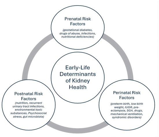
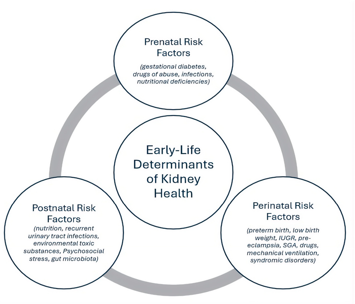
Disclaimer/Publisher’s Note: The statements, opinions and data contained in all publications are solely those of the individual author(s) and contributor(s) and not of MDPI and/or the editor(s). MDPI and/or the editor(s) disclaim responsibility for any injury to people or property resulting from any ideas, methods, instructions or products referred to in the content. |
© 2026 by the authors. Licensee MDPI, Basel, Switzerland. This article is an open access article distributed under the terms and conditions of the Creative Commons Attribution (CC BY) license.