


Abstract
Background: Hospitalised exacerbations of non-cystic fibrosis bronchiectasis are clinically heterogeneous, yet inpatient microbiology and management remain incompletely characterised. Methods: We retrospectively analysed 53 consecutive hospitalisations in 47 adults (2023–2024), describing respiratory severity, microbiological findings, antimicrobial exposure, corticosteroid use and length of stay (LOS), without a comparator group. Results: Acute respiratory failure was documented in 12/53 episodes (22.6%), and Pseudomonas aeruginosa was isolated in 19/53 (35.8%). An exploratory four-domain classification (bacteria, nontuberculous mycobacteria, fungi and viruses), incorporating historical and acute findings, identified ≥2 domains in 24/53 episodes (45.3%); median LOS was 18.5 versus 15 days for ≥2 versus 0–1 domains. Intravenous antibacterials were recorded in 51/53 episodes (96.2%) and inhaled amikacin in 28/53 (52.8%). Median systemic antibacterial treatment duration, calculated from recorded concurrent antibiotic courses, was 11 days during hospitalisation and 11.5 days including documented pretreatment. Systemic corticosteroids were recorded in 28/53 episodes (52.8%), including 15/30 without documented COPD or asthma. Overall median LOS was 16 days (IQR 12–20), with one in-hospital death (1.9%). Conclusions: Hospitalised exacerbations involved heterogeneous microbiological findings and frequent acute inhaled antibiotic and systemic corticosteroid use. The overlap–LOS contrast is descriptive, not prognostic, and reconstructed durations cannot establish treatment appropriateness. Prospective studies should assess prescribing indications, microbiological context and clinical response.
1. Introduction
Non-cystic fibrosis bronchiectasis is a heterogeneous chronic airway disease characterised by irreversible bronchial dilatation, impaired mucociliary clearance, and recurrent airway infection and inflammation [1]. Exacerbations contribute substantially to morbidity, impaired quality of life, and healthcare utilisation, with episodes requiring hospitalisation representing a particularly burdensome aspect of disease management [2,3,4]. According to the international consensus definition, a bronchiectasis exacerbation involves deterioration in at least three key respiratory or constitutional symptoms for a minimum of 48 h, prompting a clinician-determined change in treatment [5]. However, this symptom-based definition should be distinguished from exacerbation severity, as hospital admission may reflect not only acute respiratory deterioration but also underlying disease burden, comorbidities, and healthcare-related factors.
The 2025 European Respiratory Society (ERS) guidelines define severe exacerbations as episodes requiring hospitalisation or intravenous antibiotic treatment, potentially accompanied by respiratory failure, worsening oxygenation, haemoptysis, or haemodynamic instability [6]. Antibiotics remain central to exacerbation management, with treatment selection guided by previous microbiological findings, antimicrobial susceptibility, and clinical severity. Although a 14-day course remains the standard reference, particularly for severe or Pseudomonas aeruginosa-associated exacerbations, shorter regimens may be appropriate in selected patients [6,7]. Nevertheless, the optimal duration and individualisation of antibiotic treatment remain incompletely established. Bronchiectasis-specific studies have investigated treatment response, factors associated with antibiotic duration, and bacterial-load-guided strategies for shortening intravenous therapy, without establishing a universally applicable treatment approach [8,9,10,11]. Importantly, the ERS 2025 recommendations provide contemporary context for interpreting clinical practice during our 2023–2024 observation period.
Beyond treatment duration, several aspects of inpatient therapeutic management remain insufficiently characterised. The role of inhaled antibiotics during acute exacerbations is particularly uncertain, since recommendations supporting their long-term use in selected patients with chronic airway infection cannot be directly extrapolated to adjunctive treatment during hospitalisation [6]. Similarly, systemic corticosteroid prescribing requires careful consideration of coexisting asthma, chronic obstructive pulmonary disease, bronchospasm, and other potentially steroid-responsive conditions, while their benefit and safety during infective bronchiectasis exacerbations remain inadequately established [2,7]. These therapeutic uncertainties are compounded by the microbiological heterogeneity of bronchiectasis, where historical bacterial colonisation, acute pathogen isolation, nontuberculous mycobacteria, fungal organisms, and respiratory viruses may coexist or be detected through different diagnostic pathways. Distinguishing clinically relevant infection from historical colonisation or incidental microbial detection is therefore essential when interpreting microbiological findings and treatment decisions.
Recent hospital-based cohorts have improved our understanding of the clinical characteristics and outcomes of bronchiectasis exacerbations requiring admission [3,4]. However, detailed episode-level descriptions integrating baseline disease severity, acute respiratory impairment, microbiological findings, antimicrobial exposure before and during hospitalisation, and corticosteroid prescribing remain comparatively limited. Accordingly, the present study aimed to characterise the clinical and microbiological profiles and inpatient management of consecutive bronchiectasis exacerbations requiring hospitalisation at a tertiary respiratory centre. Particular attention was given to systemic and inhaled antibiotic use, treatment duration, corticosteroid exposure, and length of hospital stay. We additionally explored the overlap of predefined microbiological domains as a descriptive measure of microbiological heterogeneity, with the objective of identifying clinically relevant questions for future prospective investigation rather than establishing causal or independent prognostic relationships.
2. Methods
2.1. Study Design and Population
We conducted a single-centre retrospective observational study at the Respiratory Diseases Unit of IRCCS Policlinico San Matteo, Pavia, Italy, a tertiary referral centre with a dedicated bronchiectasis outpatient service. Consecutive hospitalisations for acute exacerbations of non-cystic fibrosis bronchiectasis between September 2023 and September 2024 were identified from ward admission records and verified through clinical-record review. Eligible patients were adults aged ≥ 18 years with radiologically confirmed bronchiectasis who required admission primarily for an acute exacerbation. Patients with cystic fibrosis and those hospitalised for unrelated conditions were excluded. All included patients were already under specialist follow-up at our centre, and no interhospital or interdepartmental transfers were included.
Exacerbations were clinically identified by the treating physicians, using the international consensus definition as a reference [5]. Because individual symptom trajectories were not consistently documented, retrospective verification of every consensus criterion was not feasible. Hospitalisation was therefore adopted as the operational indicator of a severe exacerbation, consistent with the framework subsequently outlined in the 2025 European Respiratory Society (ERS) guidelines [6]. Acute respiratory failure and respiratory-support requirements were assessed separately to distinguish the need for hospitalisation from objectively documented respiratory impairment.
This study included 53 hospitalisations involving 47 unique patients: 42 contributed one admission, four contributed two, and one contributed three. Hospitalisation was the primary unit of analysis, and all eligible episodes within the study period were retained. Cohort composition is illustrated in Supplementary Figure S1. This study was conducted in accordance with the Declaration of Helsinki. Ethical approval and informed consent arrangements are reported in the corresponding institutional statements.
2.2. Data Collection and Clinical Definitions
Clinical data were extracted from hospital records and reconciled with the exacerbation database. Additional baseline information was obtained from existing outpatient bronchiectasis records through verified patient-level linkage. Data were distinguished according to whether they described the pre-existing disease or the index exacerbation, and discrepancies were resolved through clinical-record review and investigator verification.
Collected baseline variables included demographic characteristics, smoking history, comorbidities, bronchiectasis aetiology, historical microbiological findings, pulmonary function, disease severity, and maintenance respiratory treatments. Aetiology was assigned according to documented clinical diagnoses rather than inferred from associated conditions. Baseline disease severity was characterised using pre-admission Bronchiectasis Severity Index (BSI) categories, without recalculation during hospitalisation. Historical spirometry was considered representative of baseline lung function only when performed before the patient’s first study admission; subsequent or undated measurements were excluded from this assessment.
Acute respiratory failure was determined from the documented clinical diagnosis and available supporting information on oxygenation and respiratory assistance. Respiratory-support requirements were classified according to the documented modality during hospitalisation, distinguishing room air, conventional oxygen therapy, high-flow nasal oxygen (HFNC), non-invasive ventilation (NIV), and helmet continuous positive airway pressure (CPAP). Helmet CPAP was reported separately from NIV. Clinical-record discrepancies were reviewed and resolved by the treating investigators.
Room air represented a documented absence of supplemental oxygen or ventilatory support rather than missing information. However, respiratory-support categories were not considered interchangeable with physiological measures of gas exchange. Because arterial blood gas and oxygen saturation measurements were not uniformly available, a standardised retrospective classification of respiratory-failure severity could not be applied to all admissions.
Historical respiratory colonisation was identified from documented microbiological history, with individual organisms recorded whenever available. As serial respiratory culture dates were not consistently retrievable, historical colonisation was distinguished from microbiologically confirmed chronic infection according to standardised culture-based criteria.
Length of hospital stay (LOS) was defined as the recorded duration of hospitalisation in days. Discharge generally followed clinical stabilisation and completion of the planned inpatient antimicrobial regimen. Outpatient parenteral antimicrobial therapy and home intravenous antibiotic treatment were unavailable during the study period, an organisational feature considered relevant to the interpretation of LOS. Further operational definitions are provided in Supplementary Table S1. Completeness of key clinical variables and the extent of diagnostic testing are summarised in Supplementary Table S5.
2.3. Microbiological Assessment
Respiratory microbiology was characterised using sputum cultures, bronchoalveolar lavage (BAL) findings, and respiratory viral testing obtained during the index admission. Results from different specimen types were recorded separately, while individual microorganisms were counted only once per hospitalisation.
BAL was performed according to clinical indications, primarily when sputum could not be obtained, in immunocompromised patients, or in the presence of haemoptysis. Respiratory viral PCR testing was guided by clinical and epidemiological suspicion, including seasonal viral circulation, fever, bronchospasm, and associated systemic symptoms. Neither investigation was routinely performed in all patients, and the timing of specimen collection relative to antimicrobial initiation was not uniformly available. Microbiological findings were therefore interpreted in the context of selective diagnostic sampling.
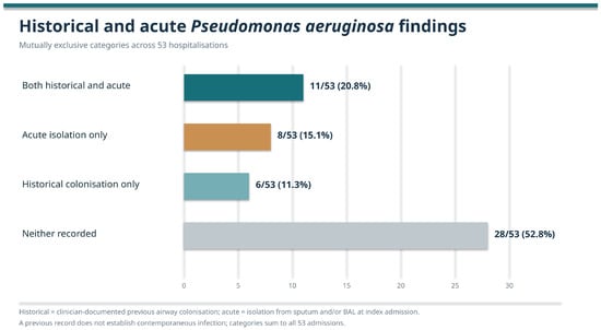
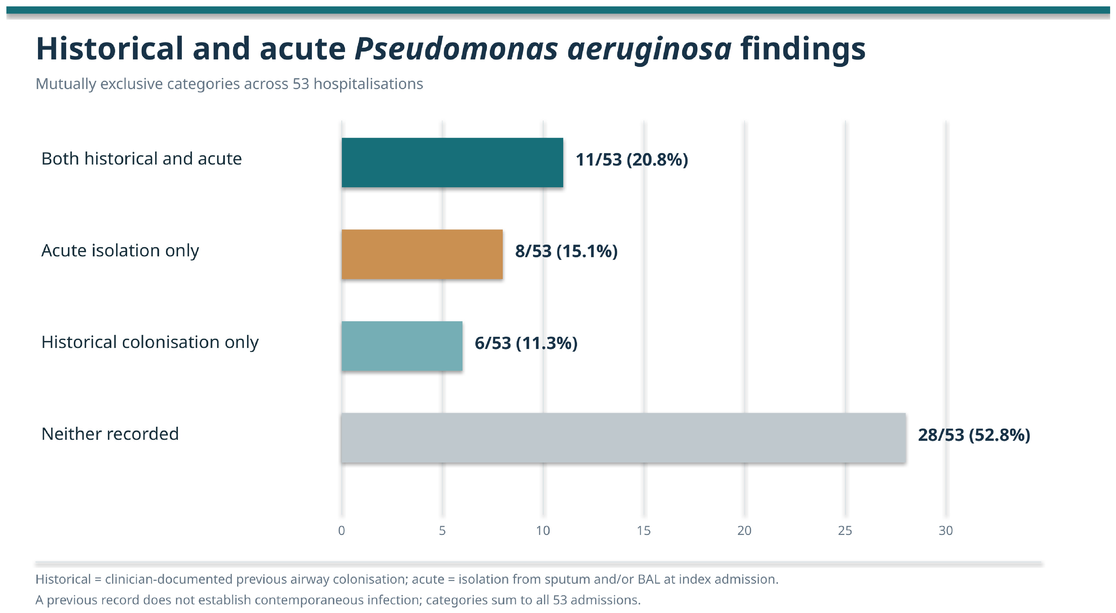
Acute Pseudomonas aeruginosa isolation was defined as recovery from sputum and/or BAL during the index hospitalisation and was analysed separately from documented historical colonisation. Their overlap is illustrated in Figure 1. Other bacterial species were reported individually, without assuming that isolation necessarily established the aetiology of the acute exacerbation.
Figure 1.
Historical colonisation and acute isolation of Pseudomonas aeruginosa (n = 53).
Fungal findings were classified by taxon, distinguishing yeasts from filamentous fungi. Unspecified fungal findings and positive galactomannan results without a corresponding identified organism were not counted as additional named fungal isolates. Respiratory fungal detection was not considered equivalent to clinically established fungal disease or allergic bronchopulmonary aspergillosis.
Previously diagnosed nontuberculous mycobacterial (NTM) pulmonary disease was distinguished from respiratory NTM isolation during hospitalisation. NTM recovery was not automatically interpreted as evidence of active disease or as the cause of the index exacerbation. Antimicrobial susceptibility information was reviewed when available; incomplete susceptibility data precluded systematic assessment of resistance profiles or treatment–susceptibility concordance.
To describe microbiological heterogeneity, we evaluated the overlap of four predefined, non-mutually exclusive domains: (1) bacterial findings, comprising historical respiratory bacterial colonisation and/or acute bacterial isolation; (2) NTM involvement, comprising previously documented pulmonary disease and/or acute respiratory isolation; (3) named fungal organisms recovered from respiratory specimens during admission; and (4) respiratory viruses detected by dedicated PCR testing.
Each domain contributed a maximum of one observation per hospitalisation, irrespective of the number of microorganisms identified. Exploratory microbiological overlap was defined as the presence of findings in at least two domains. This operational classification was used to describe recorded microbiological coexistence, without equating historical and acute findings with simultaneous active infection or treating the resulting categories as validated clinical phenotypes.
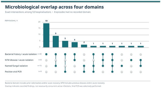
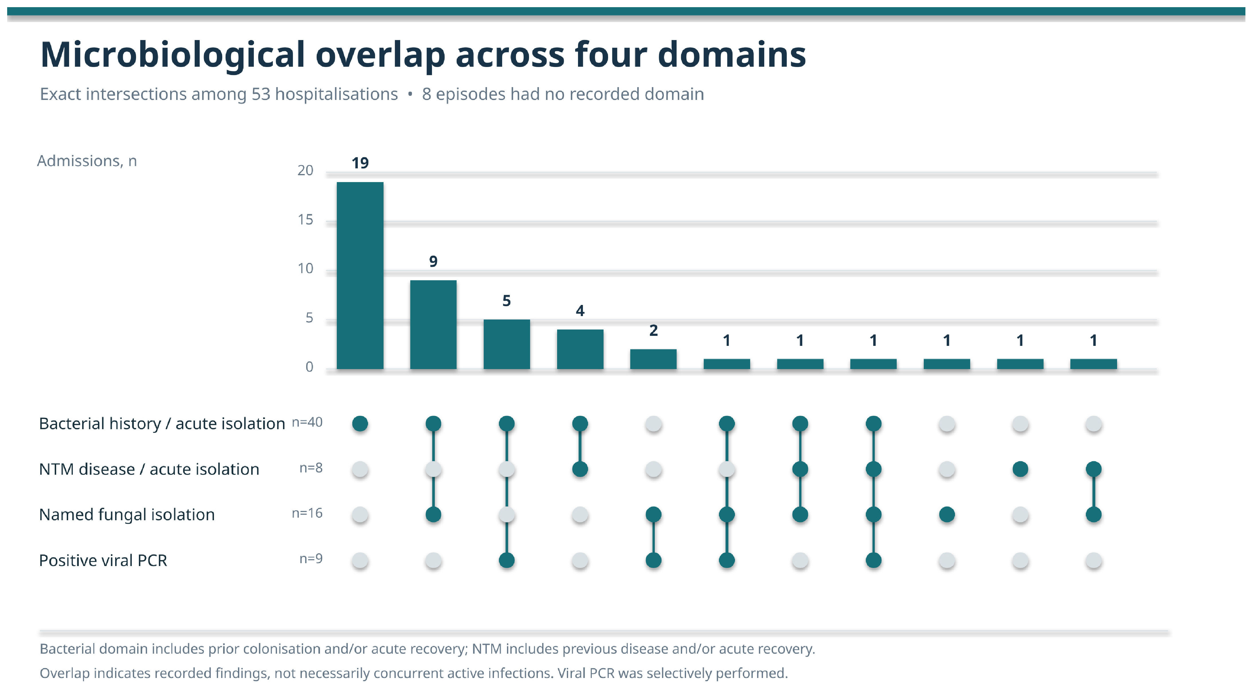
Domain distributions and intersections are displayed using an UpSet plot (Figure 2), with detailed combinations provided in Supplementary Table S4.
Figure 2.
Exact overlap of four exploratory microbiological domains (n = 53). Legend: Intersections represent bacterial history and/or acute detection (B), historical NTM disease and/or acute isolation (N), named fungal respiratory isolation (F), and positive dedicated viral PCR (V). Each domain was counted once per hospitalisation. Eleven non-empty intersections account for 45 episodes; eight additional admissions had no recorded domain. A total of 24/53 episodes included ≥2 domains.
2.4. Antimicrobial and Corticosteroid Treatment
Inpatient antimicrobial treatment was characterised according to individual agents, administration routes, and recorded treatment durations. Intravenous, oral, and inhaled antibacterial treatments were assessed separately. Both route-specific frequencies and mutually exclusive treatment combinations were reported, recognising that different administration routes could be used within the same hospitalisation. In the absence of complete treatment chronology, combined intravenous and oral exposure was not assumed to indicate a sequential switch between routes.
Inhaled amikacin was evaluated separately from systemic antibacterial therapy. According to the treating investigators, it was administered as part of acute exacerbation management in selected circumstances, including first isolation or recurrence of P. aeruginosa, rather than systematically representing continuation of maintenance inhaled treatment. Individual prescribing indications and drug doses were not consistently documented.
Systemic antibacterial treatment duration was calculated from the recorded durations of courses administered for the index exacerbation. As multiple antibiotic courses were confirmed to have been administered concurrently, the longest relevant systemic antibacterial course was used to represent inpatient treatment duration, avoiding the inappropriate summation of overlapping drug-specific courses. Inhaled antibiotics, antifungal agents, and identifiable NTM-directed treatments were excluded from this calculation.
One hospitalisation involving a complex NTM-active regimen was excluded from treatment-duration analyses because the available records did not permit reliable attribution of systemic antibiotic exposure specifically to the acute exacerbation. This episode was retained in the remaining descriptive analyses.
Pre-admission antibiotic exposure was assessed using documented treatment for the same exacerbation episode. Following investigator verification, absent pretreatment entries were interpreted as no pre-admission antibiotic therapy, and discordant records were reconciled. Recorded pretreatment days were added to the inpatient systemic antibacterial duration to estimate overall exposure during the episode.
Treatment-duration estimates were derived from the recorded antibiotic courses and verified information regarding concurrent administration. Because complete medication administration timestamps were unavailable, they represent calculated episode-level durations rather than independently verified daily administration histories.
Systemic corticosteroid exposure was characterised according to recorded agents, available treatment duration, and the presence of COPD and/or asthma. This variable included newly prescribed treatment and documented continuation of maintenance corticosteroids. Although bronchospasm was among the reported clinical indications, individual prescribing rationales, cumulative doses, and adverse events were not systematically available.
Admission C-reactive protein (CRP), procalcitonin, and peripheral blood eosinophil measurements were examined in relation to systemic corticosteroid exposure. As sampling times relative to corticosteroid administration and prior maintenance treatment were not consistently documented, these measurements were considered exploratory and were not interpreted as markers of corticosteroid response.
2.5. Statistical Analysis
Continuous variables were summarised as mean ± standard deviation (SD) or median and interquartile range (IQR), as appropriate. Categorical variables were expressed as frequencies and percentages, with variable-specific denominators reflecting available data. Missing or uninterpretable observations were not imputed.
Hospitalisation was the primary analytical unit (n = 53). To account descriptively for repeated admissions, a sensitivity analysis was performed using only the first recorded hospitalisation of each patient (n = 47). The primary outcome was LOS, analysed as a continuous variable. A secondary descriptive classification distinguished hospitalisations lasting ≥16 days from those lasting <16 days, using the cohort median as the threshold. This grouping was not considered a validated definition of prolonged hospitalisation. LOS was additionally summarised according to the presence of at least two versus zero or one microbiological domains. Given the exploratory nature of the classification, selective diagnostic testing, and potential confounding, these comparisons were presented descriptively without adjusted prognostic estimates. Exploratory comparisons of admission CRP, procalcitonin, and peripheral blood eosinophils according to systemic corticosteroid exposure were restricted to the first-admission cohort. Two-sided Mann–Whitney U tests were used, with Holm adjustment for multiple comparisons. Procalcitonin values reported below 0.02 ng/mL were assigned a value of 0.01 ng/mL for analysis; one uninterpretable observation was excluded. No multivariable regression analyses were included because the small sample size, repeated observations, and heterogeneity of clinical and microbiological characteristics would limit the reliability of adjusted estimates. In-hospital mortality was reported descriptively after clinical-record verification. No mortality modelling was undertaken because only one death occurred. Post-discharge mortality and exacerbation recurrence were excluded from outcome analyses owing to non-standardised follow-up and incomplete ascertainment.
3. Results
3.1. Study Population and Acute Clinical Severity
This study included 53 hospitalisations involving 47 patients with non-cystic fibrosis bronchiectasis. Women accounted for 37/53 hospitalisations (69.8%), and the mean age was 68.6 ± 10.6 years. Mean body mass index (BMI) was 23.5 ± 4.5 kg/m2 (n = 51). The cohort was characterised by a substantial burden of comorbidities, including hypertension in 34/53 admissions (64.2%), rheumatological disease and COPD in 13/53 each (24.5%), asthma in 10/53 (18.9%), and diabetes mellitus in 12/53 (22.6%) (Table 1).
Table 1.
Baseline characteristics, bronchiectasis aetiology and acute respiratory severity.
Bronchiectasis aetiology was available for all 47 patients. Idiopathic bronchiectasis was the most frequent category (26/47, 55.3%), followed by post-infective disease (11/47, 23.4%) and rheumatological disease (6/47, 12.8%). Less frequent aetiologies included alpha-1 antitrypsin deficiency (2/47, 4.3%), immunodeficiency (1/47, 2.1%), and asthma-associated bronchiectasis (1/47, 2.1%).
Pre-admission Bronchiectasis Severity Index (BSI) categories were available for 49/53 hospitalisations. Severe baseline disease predominated (34/49, 69.4%), whereas ten admissions (20.4%) involved patients with moderate BSI and five (10.2%) involved patients with mild BSI. Dated pre-admission spirometry was available for 13/47 patients (27.7%), with a median FEV1 of 54% predicted (IQR 45–79). These examinations were performed between 2018 and 2023 and therefore did not uniformly reflect lung function immediately preceding admission.
Acute respiratory failure was documented in 12/53 hospitalisations (22.6%). Respiratory-support assessment identified 41/53 episodes (77.4%) managed in room air, seven (13.2%) requiring conventional oxygen therapy via nasal cannulae, three (5.7%) receiving high-flow nasal oxygen (HFNC), one (1.9%) requiring non-invasive ventilation (NIV), and one (1.9%) treated with helmet continuous positive airway pressure (CPAP). The latter corresponded to the fatal hospitalisation complicated by acute respiratory distress syndrome (ARDS). Complete arterial blood gas and oxygen saturation measurements were not uniformly available for further physiological severity stratification.
Median admission C-reactive protein (CRP) was 1.78 mg/dL (IQR 0.42–3.88; n = 52). The availability of other key clinical and diagnostic variables is detailed in Supplementary Table S5.
3.2. Respiratory Microbiological Findings
Historical respiratory colonisation was documented in 26/53 hospitalisations (49.1%), with Pseudomonas aeruginosa being the most frequently recorded organism (17/53, 32.1%). Sputum samples were obtained in 39/53 admissions (73.6%), of which 29/39 (74.4%) yielded a recorded positive microbiological result. Bronchoalveolar lavage (BAL) was performed in 28/53 admissions (52.8%), with positive findings documented in 21/28 (75.0%). These positivity figures encompassed heterogeneous microbiological findings and should not be interpreted exclusively as recovery of causative bacterial pathogens.
Acute P. aeruginosa isolation was documented in 19/53 hospitalisations (35.8%). Historical colonisation and acute isolation overlapped in 11 episodes, whereas eight had acute isolation without documented previous colonisation and six had historical colonisation without acute recovery. The remaining 28 admissions had neither finding (Figure 1). Other microorganisms recorded in the historical respiratory microbiological history included Staphylococcus aureus and Escherichia coli in three admissions each, Achromobacter xylosoxidans in two, and Haemophilus influenzae, Nocardia abscessus, and Aspergillus flavus in one each. Categories were not mutually exclusive.
The distribution of microorganisms identified during hospitalisation is reported in Table 2. Among bacteria other than P. aeruginosa, S. aureus and H. influenzae were identified in four admissions each, Serratia marcescens in three, and A. xylosoxidans in two.
Table 2.
Historical microbiology and organism-level findings during the index hospitalisation.
Named fungal organisms were identified in 16/53 hospitalisations (30.2%), including Aspergillus spp. in eight episodes and Candida spp. in eight, with additional fungal taxa and overlapping detections. One BAL report described unspecified mycetes. Positive galactomannan was documented in two BAL samples, including one associated with Scedosporium isolation; these findings were not counted as additional named fungal isolates. Respiratory fungal detection was reported separately from clinically established fungal disease.
Previously recognised nontuberculous mycobacterial (NTM) pulmonary disease was documented in seven hospitalisation records. An additional episode had acute respiratory NTM isolation without a corresponding previous disease record, resulting in eight hospitalisations (15.1%) with either documented NTM disease or acute NTM detection. Respiratory NTM isolation during the index admission occurred in four episodes. Historical disease and acute isolation were considered distinct findings rather than evidence that NTM caused the acute exacerbation.
Respiratory viral PCR was performed in 26/53 hospitalisations (49.1%) and yielded positive results in 9/26 tested episodes (34.6%). An additional respiratory syncytial virus finding documented in the BAL microbiological record was reported separately and was not added to the dedicated PCR-positive numerator. Given the selective use of viral testing, positivity rates refer exclusively to the tested subgroup.
To further characterise microbiological heterogeneity, findings were classified into four exploratory, non-mutually exclusive domains. A bacterial historical or acute respiratory finding was present in 40/53 hospitalisations (75.5%), NTM involvement in 8/53 (15.1%), named fungal respiratory isolation in 16/53 (30.2%), and positive respiratory viral PCR in 9/53 (17.0%).
Overall, 24/53 hospitalisations (45.3%) exhibited findings in at least two microbiological domains, whereas 21/53 (39.6%) involved a single domain and eight (15.1%) had no recorded domain. The distribution and intersections are illustrated in Figure 2 and detailed in Supplementary Table S4. These categories describe recorded microbiological overlap, including historical findings, rather than confirmed simultaneous active coinfection.
3.3. Inpatient Antimicrobial and Corticosteroid Treatment
All 53 hospitalisations involved antibiotic treatment. Intravenous antibacterial therapy was recorded in 51/53 episodes (96.2%), while oral antibacterials administered for acute exacerbation management were documented in 9/53 (17.0%). Inhaled amikacin was administered during acute management in 28/53 hospitalisations (52.8%). These administration routes were not mutually exclusive; their exclusive combinations are presented in Table 3.
Table 3.
Recorded treatment exposures, antibiotic duration and inpatient outcomes.
Among systemic antibacterial agents, piperacillin–tazobactam was most frequently prescribed (27 admissions), followed by ceftriaxone (11) and ceftazidime without avibactam (nine). The complete distribution of recorded antimicrobial agents is provided in Table 4.
Table 4.
Systemic antibacterial agents recorded during hospitalisation.
Maintenance inhaled antibiotic treatment was documented in only one of 52 admissions with available baseline treatment information. In contrast, inhaled amikacin was frequently used during acute exacerbation management, including selected first or recurrent P. aeruginosa isolations. However, complete individual treatment histories were insufficient to establish initiation status or specific prescribing indications for every episode.
Antibiotic treatment preceding hospitalisation was documented in 18/53 episodes (34.0%), with a median duration of 4 days (IQR 2.25–4). Inpatient systemic antibacterial duration could be calculated from recorded treatment courses in 52/53 admissions (98.1%) and had a median of 11 days (IQR 9–12.25). After incorporating documented pre-admission antibiotic exposure, the median estimated overall episode duration was 11.5 days (IQR 10–14).
These calculations considered concurrent systemic antibacterial courses without summing overlapping treatment durations. Inhaled antibiotics, antifungal agents, and identifiable NTM-directed regimens were excluded. One hospitalisation involving a complex NTM-active regimen was excluded from duration analyses because systemic treatment specifically attributable to the acute exacerbation could not be reliably distinguished.
Systemic corticosteroids were recorded in 28/53 hospitalisations (52.8%). Exposure was documented in 13/23 episodes (56.5%) involving patients with COPD and/or asthma and in 15/30 (50.0%) without either condition. Methylprednisolone was the most frequently recorded systemic corticosteroid (25 admissions), with a median treatment duration of 14 days (IQR 9–17). Two additional records indicated maintenance prednisone treatment, while one did not specify the corticosteroid agent or duration. Thus, the overall exposure variable included both acute prescribing and continuation of maintenance therapy.
In the analysis restricted to first admissions, patients receiving systemic corticosteroids had numerically higher admission CRP than those without corticosteroid exposure (median 2.42 versus 1.57 mg/dL). However, exploratory comparisons of CRP, procalcitonin, and peripheral blood eosinophils did not retain statistical significance after Holm correction (all adjusted p = 0.564; Supplementary Table S2). These findings describe differences between prescribing groups rather than corticosteroid treatment effects.
3.4. Length of Stay, Clinical Outcomes and Sensitivity Analyses
Mean length of hospital stay (LOS) was 16.8 ± 6.5 days, with a median of 16 days (IQR 12–20; range 8–45). Twenty-seven hospitalisations (50.9%) lasted at least 16 days.
In the exploratory microbiological-overlap analysis, episodes involving at least two microbiological domains had a median LOS of 18.5 days (IQR 12–21.25; n = 24), compared with 15 days (IQR 11–18; n = 29) among those involving zero or one domain. This unadjusted comparison was descriptive and was not interpreted as evidence of an independent relationship between microbiological overlap and hospitalisation duration.
One in-hospital death occurred (1/53, 1.9%). The patient was receiving immunosuppressive therapy for rheumatoid arthritis and developed ARDS in the context of severe systemic infections, including extrapulmonary involvement. Helmet CPAP was required during the fatal hospitalisation. No mortality modelling was performed.
Sensitivity analyses were restricted to the first recorded hospitalisation of each patient (n = 47), thereby excluding repeated observations. In this cohort, acute P. aeruginosa isolation was recorded in 16/47 admissions (34.0%), systemic corticosteroids in 24/47 (51.1%), and acute respiratory failure in 10/47 (21.3%). Respiratory-support categories comprised room air in 37/47 (78.7%), conventional oxygen therapy in six (12.8%), HFNC in three (6.4%), and NIV in one (2.1%).
Exploratory overlap of at least two microbiological domains was observed in 22/47 first admissions (46.8%). Median LOS was 19 days (IQR 12–21.75) in this subgroup, compared with 15 days (IQR 11–19) among those with zero or one domain. Overall median LOS remained 16 days (IQR 12–20.5), while median calculated inpatient systemic antibacterial duration was 11 days (IQR 9–12.75; n = 46).
The first-admission analyses yielded descriptive patterns broadly consistent with those observed in the full admission-level cohort. Complete comparative results are presented in Supplementary Table S3.
4. Discussion
This study provides a detailed account of bronchiectasis exacerbations requiring hospitalisation in a tertiary respiratory centre, integrating baseline disease characteristics, acute respiratory impairment, microbiological findings, and inpatient treatment exposure. Beyond describing the clinical profile of hospitalised patients, our findings highlight three clinically relevant aspects of inpatient management: heterogeneous and frequently overlapping microbiological observations, substantial use of inhaled amikacin and systemic corticosteroids during acute exacerbations, and prolonged hospitalisation within a care pathway favouring inpatient completion of antimicrobial treatment. Together, these observations identify specific areas in which routine clinical practice extends beyond the evidence currently available to guide treatment decisions.
The cohort was characterised by older age, multimorbidity, and a substantial burden of severe baseline bronchiectasis, consistent with the recognised complexity of this population [12]. However, despite severe pre-admission BSI classifications in approximately 70% of assessable episodes, acute respiratory failure was documented in only 22.6%, while 77.4% were recorded as breathing room air. This distinction underscores the importance of separating chronic disease severity from acute physiological impairment. In specialist bronchiectasis care, hospitalisation may reflect a combination of clinical deterioration, comorbidity burden, microbiological considerations, and the need for supervised treatment rather than respiratory failure alone. The predominance of idiopathic, post-infective, and rheumatological aetiologies further illustrates the heterogeneity of the population, although the sample size precluded reliable aetiology-specific comparisons.
Pseudomonas aeruginosa was the most frequently recovered acute bacterial organism, reinforcing its importance in the assessment of hospitalised exacerbations. Nevertheless, acute isolation and historical colonisation were only partially concordant, highlighting the limitations of relying exclusively on previous microbiological results to characterise an individual episode. Underlying disease context may also influence respiratory microbiology, as illustrated by the distinct infection profiles described in haematology-associated bronchiectasis [13]. Furthermore, bronchoalveolar lavage was frequently performed in our cohort because of selected clinical indications, including inability to expectorate, immunosuppression, and haemoptysis. Together with antibiotic exposure before sampling, this diagnostic approach may have influenced the observed microbiological distribution. Accordingly, respiratory culture positivity should be interpreted within the clinical and sampling context rather than equated with pathogen-attributable exacerbation.
The exploratory microbiological-overlap analysis extends this description by examining the coexistence of bacterial, mycobacterial, fungal, and viral findings. At least two domains were identified in 24 hospitalisations (45.3%), with a numerically longer median length of stay than episodes involving zero or one domain (18.5 versus 15 days). However, these findings do not support an independent prognostic association. The classification incorporated historical and acute observations, while individual domains were subject to different diagnostic pathways and selective testing. Importantly, the isolation of a yeast, recovery of a filamentous fungus, detection of an NTM isolate, and positive viral PCR have distinct clinical implications and cannot be interpreted as equivalent evidence of active infection. Aspergillus-related disease requires additional clinical and immunological assessment, [14] while NTM pulmonary disease must be distinguished from respiratory isolation alone [15]. The observed domain intersections therefore illustrate diagnostic and microbiological heterogeneity rather than define a validated infectious phenotype. Their principal value lies in highlighting the need for more standardised microbiological assessment in future studies.
A particularly noteworthy finding was the frequent use of inhaled amikacin during acute management, documented in 52.8% of hospitalisations despite maintenance inhaled antibiotic treatment being rarely recorded. According to the treating investigators, this strategy was used in selected first or recurrent P. aeruginosa isolations. This distinction is clinically important because recommendations supporting long-term inhaled antibiotics in patients with chronic P. aeruginosa infection and frequent exacerbations do not establish the effectiveness of adjunctive inhaled treatment during an acute hospitalisation [6]. Moreover, prescribing in the context of attempted eradication or recurrent isolation may represent a different clinical objective from chronic suppressive treatment. Our findings therefore characterise local therapeutic practice rather than provide evidence of benefit. Given the absence of standardised information on dose, indication, susceptibility concordance, tolerability, and treatment-specific outcomes, the effectiveness and safety of this approach remain unresolved. Its frequent use nevertheless identifies a clinically relevant question for prospective investigation.
Antibiotic duration represents another important area of uncertainty. By accounting for concurrent systemic antibacterial courses and documented treatment before admission, we calculated a median inpatient duration of 11 days and an estimated overall episode duration of 11.5 days. This approach provides a more informative description of antibiotic exposure than considering the duration of individual agents in isolation. The 2025 ERS guidelines retain approximately 14 days as the usual treatment reference, particularly for severe or P. aeruginosa-associated exacerbations, while recognising the potential for individualisation according to clinical response [6]. Bronchiectasis-specific investigations have similarly examined treatment duration, inflammatory response, and bacterial-load-guided strategies without establishing a universally optimal course [8,9,10,11]. Nevertheless, our findings cannot determine whether the observed treatment durations were sufficient, inappropriate, or associated with favourable outcomes. Complete administration timelines, susceptibility-guided decisions, time to clinical stability, and standardised post-discharge outcomes were unavailable. Furthermore, the ERS 2025 recommendations postdated the observation period and should be used as contemporary clinical context rather than a retrospective measure of prescribing quality. The practical implication is the need for prospective antibiotic stewardship studies linking accurately documented exposure to clinical recovery and subsequent outcomes.
Systemic corticosteroid exposure was also substantial, occurring in 52.8% of hospitalisations and in half of episodes without documented COPD or asthma. This observation raises questions regarding the clinical determinants of corticosteroid prescribing during infective bronchiectasis exacerbations. Bronchospasm and other clinician-assessed features may have influenced treatment, although individual indications were incompletely documented and the exposure variable also included continuation of maintenance corticosteroids. Current guidance does not support routine systemic corticosteroid use for bronchiectasis exacerbations without another established indication, while robust evidence addressing their effectiveness in this setting remains limited [6,7]. In our exploratory first-admission analysis, corticosteroid-treated patients had numerically higher admission C-reactive protein concentrations, but no biomarker comparison remained statistically significant after adjustment for multiple testing. These observations cannot identify a corticosteroid-responsive phenotype, particularly given the uncertain timing of inflammatory measurements relative to treatment. Nor do they establish inappropriate prescribing or treatment inefficacy. Instead, they highlight the need to prospectively document prescribing indications, cumulative exposure, clinical response, and adverse events, particularly in older patients with infectious and metabolic comorbidities.
The median hospital stay of 16 days deserves consideration in the context of contemporary inpatient cohorts. In a recent Italian multicentre study involving 318 hospitalised patients, Bindo et al. reported a median length of stay of 13 days, systemic corticosteroid use in 38.1%, and in-hospital mortality of 3.1% [3]. A substantially shorter median stay of five days was reported in a large US administrative cohort, although differences in eligibility criteria, data sources, and healthcare organisation limit direct comparison [4]. Our longer hospitalisation duration and higher frequency of corticosteroid exposure may reflect differences in case mix, local therapeutic practices, and discharge pathways rather than differences in care quality or treatment effectiveness. At our centre, discharge generally followed clinical stabilisation and completion of the planned inpatient antimicrobial regimen, while outpatient parenteral antibiotic therapy was unavailable. Consequently, length of stay may reflect organisational and treatment-related factors in addition to disease severity. Because time to clinical stability and readiness for discharge were not independently recorded, hospitalisation duration cannot be interpreted as a specific measure of biological recovery or treatment response.
Several limitations warrant consideration. The retrospective single-centre design, small cohort, and restriction to patients followed at a tertiary bronchiectasis service limit generalisability. Repeated admissions introduced within-patient dependence, although first-admission sensitivity analyses yielded broadly similar descriptive findings. Historical spirometry, radiological extent, previous exacerbation history, and complete physiological measurements were not uniformly available. Respiratory-support categories were documented for all admissions following clinical-record reconciliation, but they could not substitute for standardised assessment of gas exchange. Historical microbiological records lacked consistently dated serial cultures, while selective BAL and viral testing introduced ascertainment bias. Treatment exposure was calculated from documented concurrent antibiotic courses rather than complete administration timestamps, and information on antimicrobial susceptibility, individual prescribing indications, corticosteroid doses, adverse events, and clinical response was incomplete. Finally, the absence of a comparator group, limited number of outcome events, and non-standardised post-discharge follow-up precluded causal, prognostic, or treatment-effect inference.
Despite these limitations, our findings contribute a clinically detailed description of inpatient bronchiectasis management that complements existing studies primarily focused on hospitalisation outcomes. The frequent use of acute inhaled amikacin and systemic corticosteroids, together with heterogeneous microbiological findings and substantial hospitalisation duration, identifies practical priorities for future research. Prospective multicentre studies should integrate standardised microbiological assessment, indication-specific treatment documentation, precise antimicrobial exposure, objective measures of clinical stability, and post-discharge outcomes. Such an approach may help determine which aspects of current inpatient practice can be better individualised and supported by evidence [16,17,18,19,20,21,22,23,24,25,26,27,28,29].
5. Conclusions
In this single-centre cohort of 53 hospitalisations involving 47 patients with non-cystic fibrosis bronchiectasis, exacerbations requiring admission occurred predominantly in individuals with substantial baseline disease burden, although acute respiratory failure was documented in fewer than one-quarter of episodes. Pseudomonas aeruginosa was the most frequent acute bacterial isolate, while the coexistence of bacterial, mycobacterial, fungal, and viral findings highlighted the heterogeneity of inpatient microbiological assessment. These overlapping findings should be interpreted descriptively rather than as evidence of simultaneous active infection or an independent prognostic phenotype.
Inpatient management was characterised by frequent acute inhaled amikacin and systemic corticosteroid exposure, both recorded in 52.8% of hospitalisations, including corticosteroid use in half of episodes without documented COPD or asthma. Median calculated inpatient systemic antibacterial duration was 11 days, whereas median length of stay was 16 days, reflecting a clinical setting in which discharge generally followed completion of inpatient treatment. These observations identify relevant uncertainties in prescribing practices and care pathways and characterise important aspects of real-world management but cannot establish treatment effectiveness, appropriateness, or the determinants of hospitalisation duration.
The central challenge is to determine how clinical severity, microbiological findings, and individual treatment indications can be translated into more precise therapeutic decisions. Prospective multicentre studies integrating standardised microbiological assessment, detailed antimicrobial exposure, indication-specific corticosteroid prescribing, objective measures of clinical stability, and post-discharge outcomes are needed to support more individualised, evidence-based management of hospitalised bronchiectasis exacerbations.
Supplementary Materials
The following supporting information can be downloaded at: https://www.mdpi.com/article/10.3390/diseases14110427/s1, Figure S1: Composition of the study cohort and analytical populations; Table S1: Operational definitions, exposure ascertainment and analytical restrictions; Table S2: Admission biomarkers by systemic corticosteroid exposure; Table S3: Sensitivity analysis restricted to the first recorded hospitalisation per patient; Table S4: Exact intersections of the four exploratory microbiological domains; Table S5: Completeness and selective ascertainment of key study variables.
Author Contributions
Conceptualization: F.R.B., V.C. and G.M.S.; validation: F.R.B., L.P., M.A., L.A., M.T., P.C.R., E.G., A.G.C., A.C., V.C. and G.M.S.; writing—original draft preparation: F.R.B., V.C. and G.M.S.; writing—review and editing: all authors; supervision: A.G.C., V.C. and G.M.S. All authors have read and agreed to the published version of the manuscript.
Funding
Ricerca corrente 5×1000-2020 (cod. 090000X121—progetto 08050122) to G.M. Stella.
Institutional Review Board Statement
This study was submitted as part of a main project that was approved by the local Ethical Commission, and each enrolled patient gave written informed consent before enrolment (Comitato di Bioetica, Fondazione IRCCS Policlinico San Matteo, approval numbers: protocol #20090002344; procedure # 20090019080; date of approval: 3 June 2009). This research was conducted according to the principles of the World Medical Association Declaration of Helsinki.
Informed Consent Statement
Informed consent was obtained from subjects involved in this study according to institutional standard procedures.
Data Availability Statement
The data generated and analysed during the current study are not publicly available due to privacy concerns but are available from the corresponding author on reasonable request.
Conflicts of Interest
The authors declare no conflicts of interest.
References
- Barker, A.F.; Karamooz, E. Non–Cystic Fibrosis Bronchiectasis in Adults: A Review. JAMA 2025, 334, 253–264. [Google Scholar] [CrossRef] [Scilit] [PubMed]
- De Angelis, A.; Johnson, E.D.; Sutharsan, S.; Aliberti, S. Exacerbations of Bronchiectasis. Eur. Respir. Rev. 2024, 33, 240085. [Google Scholar] [CrossRef] [Scilit] [PubMed]
- Richardson, H.; Dicker, A.J.; Barclay, H.; Chalmers, J.D. The Microbiome in Bronchiectasis. Eur. Respir. Rev. Off. J. Eur. Respir. Soc. 2019, 28, 190048. [Google Scholar] [CrossRef] [Scilit] [PubMed]
- Lu, D.; Li, C.; Zhong, Z.; Abudouaini, M.; Amar, A.; Wu, H.; Wei, X. Changes in the Airway Microbiome in Patients with Bronchiectasis. Medicine 2023, 102, e36519. [Google Scholar] [CrossRef] [Scilit] [PubMed]
- Hill, A.T.; Sullivan, A.L.; Chalmers, J.D.; De Soyza, A.; Elborn, J.S.; Floto, R.A.; Grillo, L.; Gruffydd-Jones, K.; Harvey, A.; Haworth, C.S.; et al. British Thoracic Society Guideline for Bronchiectasis in Adults. Thorax 2019, 74, 1–69. [Google Scholar] [CrossRef] [Scilit] [PubMed]
- Chalmers, J.D.; Haworth, C.S.; Flume, P.; Long, M.B.; Burgel, P.R.; Dimakou, K.; Blasi, F.; Herrero-Cortina, B.; Dhar, R.; Chotirmall, S.H.; et al. European Respiratory Society Clinical Practice Guideline for the Management of Adult Bronchiectasis. Eur. Respir. J. 2025, 66, 2501126. [Google Scholar] [CrossRef] [Scilit] [PubMed]
- Vidaillac, C.; Chotirmall, S.H. Pseudomonas Aeruginosa in Bronchiectasis: Infection, Inflammation, and Therapies. Expert Rev. Respir. Med. 2021, 15, 649–662. [Google Scholar] [CrossRef] [Scilit] [PubMed]
- Conceição, M.; Shteinberg, M.; Goeminne, P.; Altenburg, J.; Chalmers, J.D. Eradication Treatment for Pseudomonas Aeruginosa Infection in Adults with Bronchiectasis: A Systematic Review and Meta-Analysis. Eur. Respir. Rev. 2024, 33, 230178. [Google Scholar] [CrossRef] [Scilit] [PubMed]
- Scioscia, G.; Amaro, R.; Alcaraz-Serrano, V.; Gabarrús, A.; Oscanoa, P.; Fernandez, L.; Menendez, R.; Mendez, R.; Foschino Barbaro, M.P.; Torres, A. Clinical Factors Associated with a Shorter or Longer Course of Antibiotic Treatment in Patients with Exacerbations of Bronchiectasis: A Prospective Cohort Study. J. Clin. Med. 2019, 8, 1950. [Google Scholar] [CrossRef] [Scilit] [PubMed]
- Chalmers, J.D.; Keir, H.R. Less Is More? Antibiotic Treatment Duration for Exacerbations of Bronchiectasis. Eur. Respir. J. 2021, 58, 2101416. [Google Scholar] [CrossRef] [Scilit] [PubMed]
- Chalmers, J.D.; Smith, M.P.; McHugh, B.J.; Doherty, C.; Govan, J.R.; Hill, A.T. Short- and Long-Term Antibiotic Treatment Reduces Airway and Systemic Inflammation in Non-Cystic Fibrosis Bronchiectasis. Am. J. Respir. Crit. Care Med. 2012, 186, 657–665. [Google Scholar] [CrossRef] [Scilit] [PubMed]
- Murray, M.P.; Turnbull, K.; Macquarrie, S.; Hill, A.T. Assessing Response to Treatment of Exacerbations of Bronchiectasis in Adults. Eur. Respir. J. 2009, 33, 312–318. [Google Scholar] [CrossRef] [Scilit] [PubMed]
- Pollock, J.; Polverino, E.; Dhar, R.; Dimakou, K.; Traversi, L.; Bossios, A.; Haworth, C.; Loebinger, M.R.; De Soyza, A.; Vendrell, M.; et al. Use of Inhaled Corticosteroids in Bronchiectasis: Data from the European Bronchiectasis Registry (EMBARC). Thorax 2025, 80, 358–368. [Google Scholar] [CrossRef] [Scilit] [PubMed]
- Chalmers, J.D.; Aliberti, S.; Blasi, F. Management of Bronchiectasis in Adults. Eur. Respir. J. 2015, 45, 1446–1462. [Google Scholar] [CrossRef] [Scilit] [PubMed]
- Azoicai, A.; Lupu, A.; Alexoae, M.M.; Starcea, I.M.; Mocanu, A.; Lupu, V.V.; Mitrofan, E.C.; Nedelcu, A.H.; Tepordei, R.T.; Munteanu, D.; et al. Lung Microbiome: New Insights into Bronchiectasis’ Outcome. Front. Cell. Infect. Microbiol. 2024, 14, 1405399. [Google Scholar] [CrossRef] [Scilit] [PubMed]
- Mac Aogáin, M.; Dicker, A.J.; Mertsch, P.; Chotirmall, S.H. Infection and the Microbiome in Bronchiectasis. Eur. Respir. Rev. Off. J. Eur. Respir. Soc. 2024, 33, 240038. [Google Scholar] [CrossRef] [Scilit] [PubMed]
- Marsland, I.; Sobala, R.; De Soyza, A.; Witham, M. Multimorbidity in Bronchiectasis: A Systematic Scoping Review. ERJ Open Res. 2023, 9, 00296-2022. [Google Scholar] [CrossRef] [Scilit] [PubMed]
- Menéndez, R.; Méndez, R.; Polverino, E.; Rosales-mayor, E.; Amara-elori, I.; Reyes, S.; Posadas, T.; Fernández-barat, L.; Torres, A. Factors Associated with Hospitalization in Bronchiectasis Exacerbations: A One-Year Follow-up Study. Respir. Res. 2017, 18, 176. [Google Scholar] [CrossRef] [Scilit] [PubMed]
- Chalmers, J.D.; Mall, M.A.; McShane, P.J.; Nielsen, K.G.; Shteinberg, M.; Sullivan, S.D.; Chotirmall, S.H. A Systematic Literature Review of the Clinical and Socioeconomic Burden of Bronchiectasis. Eur. Respir. Rev. 2024, 33, 240049. [Google Scholar] [CrossRef] [Scilit] [PubMed]
- Freund, O.; Wand, O.; Hadad, Y.; Bergeron, A.; Fried, S.; Pomerantz, G.; Shaffer, A.; Cohen-Hagai, K.; Osadchy, A.; Paz, D.; et al. Respiratory Burden in Hematology-Related Bronchiectasis: Clinical Characteristics and Treatable Traits. Respiration 2025, 104, 791–799. [Google Scholar] [CrossRef] [Scilit] [PubMed]
- Choi, H.; Gao, Y. Phenotypes and Endotypes in Bronchiectasis: A Narrative Review of Progress toward Precision Medicine. J. Thorac. Dis. 2025, 17, 2640–2654. [Google Scholar] [CrossRef] [Scilit] [PubMed]
- Chai, Y.-H.; Xu, J.-F. How Does Pseudomonas Aeruginosa Affect the Progression of Bronchiectasis? Clin. Microbiol. Infect. 2020, 26, 313–318. [Google Scholar] [CrossRef] [Scilit] [PubMed]
- Dicker, A.J.; Lonergan, M.; Keir, H.R.; Smith, A.H.; Pollock, J.; Finch, S.; Cassidy, A.J.; Huang, J.T.J.; Chalmers, J.D. The Sputum Microbiome and Clinical Outcomes in Patients with Bronchiectasis: A Prospective Observational Study. Lancet. Respir. Med. 2021, 9, 885–896. [Google Scholar] [CrossRef] [Scilit] [PubMed]
- Wagner, D.; van Ingen, J.; van der Laan, R.; Obradovic, M. Non-tuberculous Mycobacterial Lung Disease in Patients with Bronchiectasis: Perceived Risk, Severity and Guideline Adherence in a European Physician Survey. BMJ Open Respir. Res. 2020, 7, e000498. [Google Scholar] [CrossRef] [Scilit] [PubMed]
- Daley, C.L.; Iaccarino, J.M.; Lange, C.; Cambau, E.; Wallace, R.J.J.; Andrejak, C.; Böttger, E.C.; Brozek, J.; Griffith, D.E.; Guglielmetti, L.; et al. Treatment of Nontuberculous Mycobacterial Pulmonary Disease: An Official ATS/ERS/ESCMID/IDSA Clinical Practice Guideline. Clin. Infect. Dis. Off. Publ. Infect. Dis. Soc. Am. 2020, 71, e1–e36. [Google Scholar] [CrossRef] [Scilit] [PubMed]
- Pollock, J.; Goeminne, P.C.; Aliberti, S.; Polverino, E.; Crichton, M.L.; Ringshausen, F.C.; Dhar, R.; Vendrell, M.; Burgel, P.-R.; Haworth, C.S.; et al. Aspergillus Serologic Findings and Clinical Outcomes in Patients with Bronchiectasis: Data from the European Bronchiectasis Registry. Chest 2025, 167, 975–992. [Google Scholar] [CrossRef] [Scilit] [PubMed]
- Tiew, P.Y.; Thng, K.X.; Chotirmall, S.H. Clinical Aspergillus Signatures in COPD and Bronchiectasis. J. Fungi 2022, 8, 480. [Google Scholar] [CrossRef] [Scilit] [PubMed]
- Michaud, A.; Jarand, J.; Thornton, C.S. Effect of Transient versus Persistent Aspergillus Colonisation on Clinical Outcomes in Bronchiectasis. ERJ Open Res. 2026, 12, 00732-2025. [Google Scholar] [CrossRef] [Scilit] [PubMed]
- Choi, H.; McShane, P.J.; Aliberti, S.; Chalmers, J.D. Bronchiectasis Management in Adults: State of the Art and Future Directions. Eur. Respir. J. 2024, 63, 2400518. [Google Scholar] [CrossRef] [Scilit] [PubMed]

Disclaimer/Publisher’s Note: The statements, opinions and data contained in all publications are solely those of the individual author(s) and contributor(s) and not of MDPI and/or the editor(s). MDPI and/or the editor(s) disclaim responsibility for any injury to people or property resulting from any ideas, methods, instructions or products referred to in the content. |
© 2026 by the authors. Licensee MDPI, Basel, Switzerland. This article is an open access article distributed under the terms and conditions of the Creative Commons Attribution (CC BY) license.