


Abstract
Background: Macrocephaly may encompass a broad array of clinical conditions, but no decisional algorithms exist for its assessment and management. The clinician–geneticist alliance in the specific context of overgrowth syndromes may be of interest for shaping a proper diagnostic path. At present, the literature regarding a genetic link of macrocephaly with pathogenic SETD2 variants is limited. Case Presentation: We report a young man with macrocephaly first noted in infancy, confirmed in every stage of his life, combined with speech delay and moderate learning disability. He came to our first evaluation due to macrocrania and a history of cognitive and behavioral impairment. Brain magnetic resonance was performed after this first visit, confirming macrocephaly and revealing type I Arnold-Chiari malformation, but neither hydrocephalus nor pituitary abnormalities. Genetic testing evaluating different genes involved in overgrowth syndromes revealed the novel variant c.7039C>T (p.Gln2347*) in the SETD2 gene, leading us to diagnose Luscan–Lumish syndrome, an autosomal dominant intellectual developmental disorder associated with protean overgrowth manifestations, including macrocephaly and dysmorphic features. Case-based Review of the Literature and Discussion: An extremely heterogeneous group of disorders with a wide range of etiologies, neurodevelopmental outcomes, and radiologic characteristics may be associated with individuals with macrocephaly. After initial clinical and neuroimaging assessment, a coherent diagnostic work-up should be customized for each case of macrocephaly to clarify an underlying condition and delineate prognosis. Genetic investigation should be considered mandatory in deciphering the inner architecture of macrocephalies and explaining any associated abnormalities that may cause either growth anomaly in se or its concurrent symptoms. Conclusions: A standard recognized classification system integrating clinical, neuroimaging, developmental biological criteria and molecular/genetic assessment of macrocephaly is not currently available; however, future investigations on patients with macrocephaly will help in understanding SETD2-related disorders and provide accurate genetic counseling for more targeted management strategies.
1. Introduction
Macrocephaly is defined as a head ‘occipital-frontal’ circumference (OFC hereafter), measured from the most prominent part of the glabella to the most prominent posterior area of the occiput, greater than two standard deviations above the mean; it affects up to 5% of individuals and encompasses a large variety of complex entities ranging from benign familial macrocephaly and benign external hydrocephalus to more than 200 genetic disorders, including metabolic diseases, skeletal dysplasias, inherited alterations of endochondral ossification, and multiple hamartoma syndromes, in which macrocephaly may refer to an abnormally large head inclusive of the scalp, cranial bone, and intracranial content [1]. Macrocephaly may also be due to true enlargement of the brain parenchyma (megalencephaly) or to other conditions such as hydrocephalus and cranial hyperostosis. This trait commonly represents an indication for neuroimaging studies with the aim to differentiate benign versus frankly neuropathologic pictures [2]. Indeed, mechanisms involved in the formation of cranial bones are multifaceted, and excessive growth may stem from disruption in the intricate interplay among genetic, epigenetic, or hormonal factors [3], often requiring genetic assessment of different genes potentially involved in many growth pathways. Geneticists may be involved in the assessment of macrocephalies, even if most cases are benign and asymptomatic; conversely, cases of syndromic macrocephaly may pose a significant risk of different linked conditions, such as inadequate neurodevelopment or several peculiar neurologic symptoms. A combination of thorough history, physical examination, radiologic investigation, and genetic testing should give an important contribution to the diagnostic process and correct framework of macrocephaly. Herein we report and discuss a case of SETD2-related macrocephaly caused by a loss-of-function (LoF) SETD2 variant to further delineate the phenotypic outcome of mutations in this gene and highlight the role of genetics in the assessment of macrocephaly when combined with neurodevelopment issues.
2. Case Presentation
We report a 19-year-old man, born from caesarean delivery at 40 weeks of gestation with both normal neonatal length and normal weight, but with slightly greater head circumference (35 cm). An ultrasound-confirmed diastasis recti was also found in the neonatal period. The child was unfortunately abandoned by his natural mother and fully adopted at 17 days of life.
His growth was constantly good (around the 60th percentile), while macrocephaly was always observed during routine pediatric evaluations. The child’s ability to transfer from sitting to upright position was reached at 18 months; the first words were pronounced at 4 years, though audiometry screening gave repeatedly normal results. At 5 years, a first neuropsychological assessment found impairments of cognitive, behavioral and language development. No ataxia, spasticity, dystonia, or seizures were ever reported.
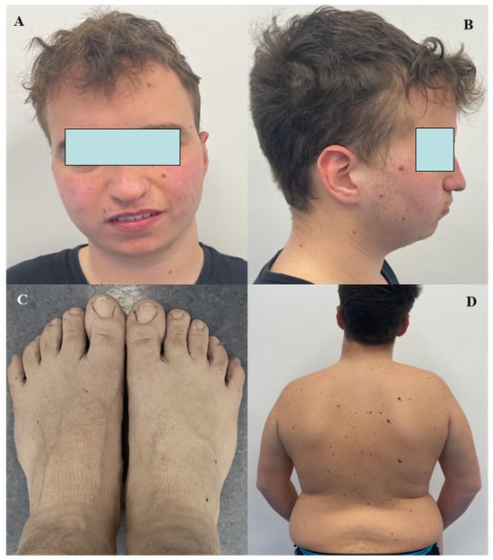
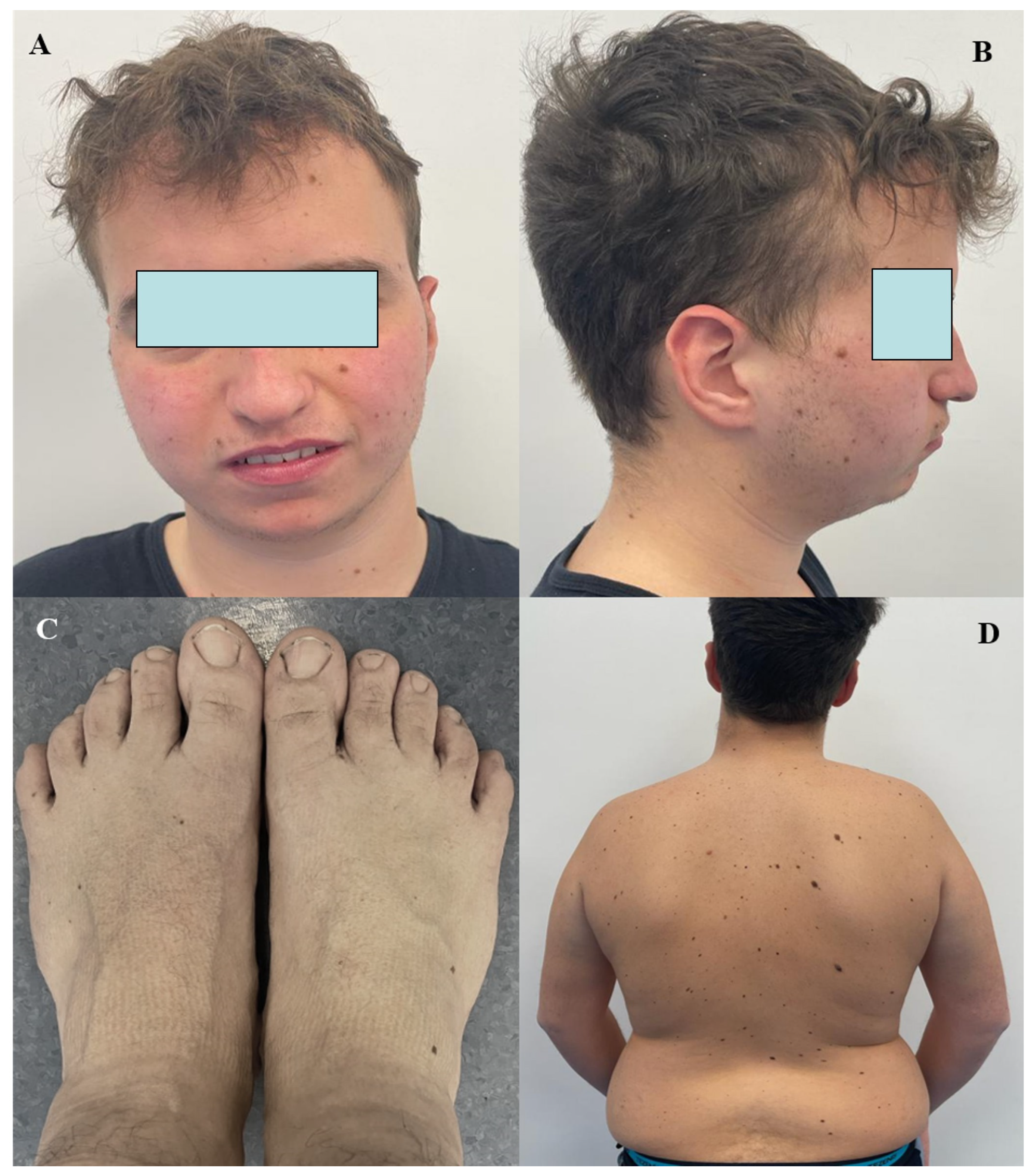
At 19 years, the patient was referred to our outpatient clinic because of macrocrania (head circumference: 61.5 cm, +4.5 SD); his height was 173.2 cm (30th percentile), and his weight was 95.5 kg (95th percentile). He had shown moderate learning disability alongside a speech delay during school years with a combination of cognitive processing and communication challenges, myopia, multiple cutaneous nevi on his chest, valgus knee, plano-valgus foot, and levoconvex scoliosis. His heart, chest and abdomen evaluation was unrevealing; his extremities were well-perfused, while deep-tendon reflexes were symmetrically brisk. The conjunctivae were clear, and fundus oculi were bilaterally normal. He also exhibited acral overgrowth, jaw malocclusion and prognathism (see Figure 1). A mood disorder with cyclothymia had previously been reported, but it did not require any medication. The patient was regularly in both occupational and speech therapy and pursued his study of the violin.
Figure 1.
Frontal (A) and lateral (B) views of the patient; long feet (C) and truncal obesity with multiple nevi on the shoulder (D) were observed. The figures were obtained from the patient’s mother, legal guardian of the patient. The figures were used for phenotypic characterization of the case.
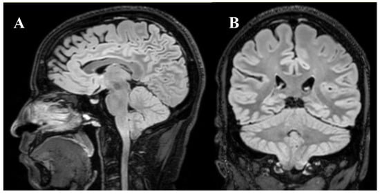
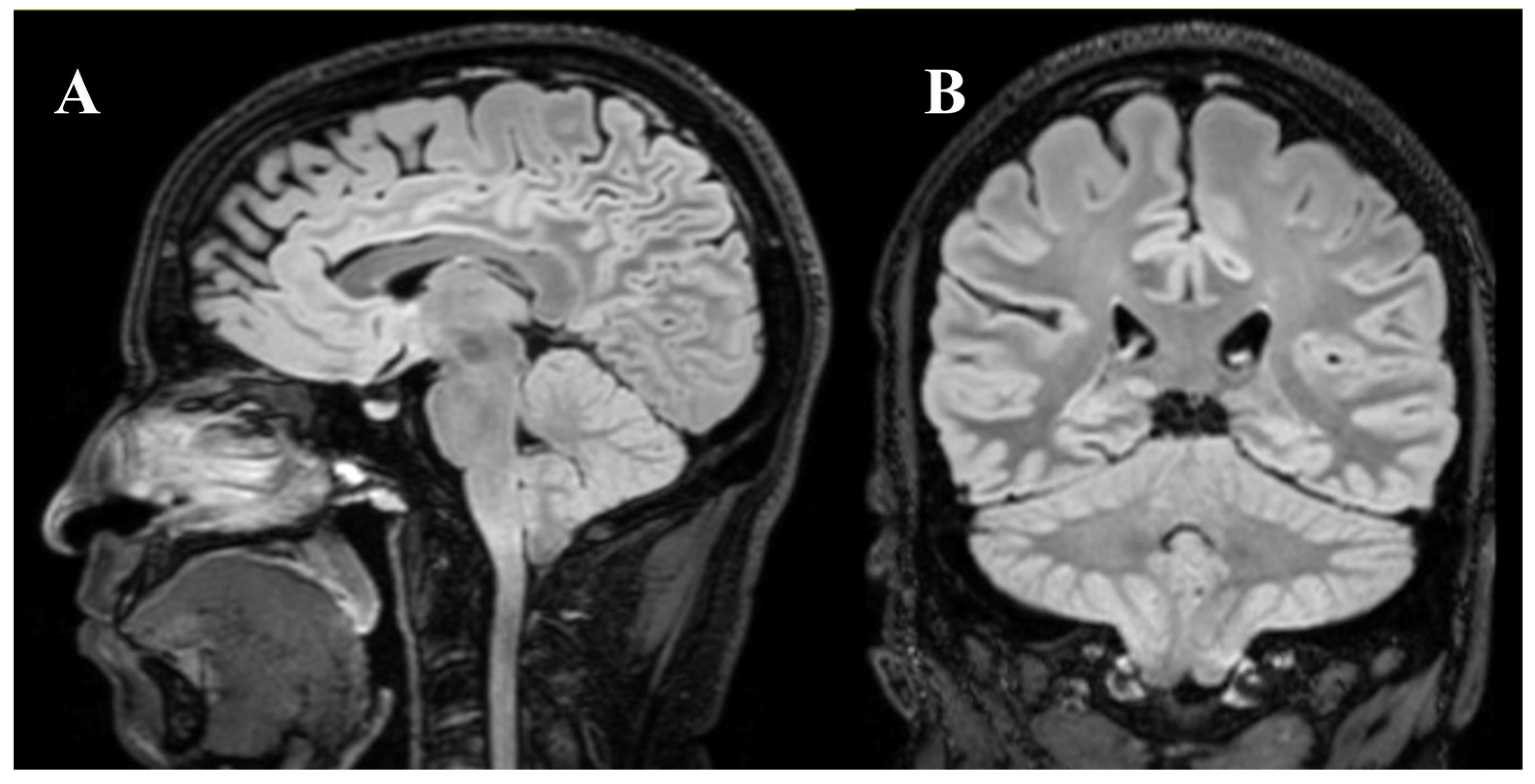
Brain magnetic resonance imaging (MRI) was performed for the first time after our assessment, revealing type I Arnold-Chiari malformation, but no signs of hydrocephalus, pituitary abnormalities, or leukoencephalopathy (Figure 2).
Figure 2.
Sagittal T2-FS (A) and coronal T2-FS (B) FLAIR brain MRI showing caudal ectopia of the cerebellar tonsils (type I Arnold-Chiari malformation) approximately 8–9 mm on the right and 8 mm on the left below McRae’s line, with slightly reduced subarachnoid space at the level of the foramen magnum. The figures were provided by the patient’s mother, and her formal informed consent was obtained. The figures were used for brain feature analysis of the case.
The WAIS-IV test gave an IQ of 68. CGH-array and molecular genetic testing of the FMR1 gene were negative. Parallel sequencing of a panel of genes implicated in macrocephaly and different overgrowth syndromes was performed at 21 years: this test identified a previously unreported truncating variant in the SETD2 gene (MIM: 612778), c.7039C>T (p.Gln2347*), which was classified as likely pathogenic according to the ACMG criteria (PVS1, PM2). A diagnosis of Luscan–Lumish syndrome (LLS, MIM: 616831), generically framed as SETD2-related overgrowth syndrome, was then established; this condition is caused by heterozygous LoF variants in SETD2 and is characterized by macrocephaly, postnatal overgrowth, advanced bone age, and mild intellectual disability with significant speech delay. In 2014, Luscan and colleagues were the first to describe two unrelated obese patients with a Sotos syndrome-like phenotype characterized by macrocephaly, long nose, long hands and feet, and broad mandible [4]. Then, in 2015, Lumish and colleagues delineated a similar phenotype characterized by macrocephaly, intellectual disability, autistic features, behavioral compulsions, and generalized epilepsy. Taken together, these features described above are collected under the eponym LLS [5]. Currently, the patient has a stable brain MRI, normal thyroid function, normal fasting blood glucose, and normal fasting insulin; he is also on diet therapy for a body mass index of 32 kg/m2. In addition, the patient stays active every day, swims twice a week, and plays both the guitar and the violin; he also receives home care services (for people with disability) 5 h per week. Written informed consent was obtained from the patient’s legal guardian for the publication of this report. Ethical review and approval were not required for this report; the case description is coherent with the Declaration of Helsinki.
3. Case-Based Discussion on Macrocephaly
OFC measurement is the customary method for identification of macrocephaly, while measuring cranial height in combination with OFC or the use of brain imaging provides a more accurate determination of intracranial volume, which is most frequently used in research settings. The genetic causes of macrocephaly cover a very broad spectrum of gene disorders, and their gene-related proteins display diverse functions in the overall course of human growth. However, no clear and unique biological pathway has been distinctively associated with brain overgrowth. Mutations in genes encoding chromatin regulators can disrupt chromatin organization, transcriptional and RNA-processing programs or DNA repair, thereby profoundly affecting neuronal development, maturation, and brain connectivity. In particular, post-translational modifications of histones play an active role in regulating gene expression, with histone lysine methylation contributing to the establishment and maintenance of different chromatin states [6]. In more detail, LLS falls under the category of overgrowth–intellectual disability disorders, which mostly encompass intellectual disability of varying degree, autism spectrum disorder, speech acquisition delay, and bodily overgrowth with macrocephaly, all related to the expression of pathogenic SETD2 variants.
The function of histone lysine methyltransferase SET domain-containing protein 2 (SET domain-containing 2, histone lysine methyltransferase) has been specifically linked to SETD2 overgrowth syndrome: this enzyme is responsible for the tri-methylation of lysine 36 on histone H3, controlling the maintenance of genomic stability and both epigenome and cytoskeleton remodeling [7]. In fact, SETD2 works in every stage of development, and its dysregulation may entail growth disorders combined with macrocephaly, though the exact functional consequences of histone modifications remain only partially clarified [8]. As an epigenetic regulator, SETD2 is the only known methyltransferase responsible for tri-methylation of histone H3 on lysine 36, and this chromatin mark is critical for transcriptional activation, mRNA splicing, DNA repair, and DNA methylation [9]. In addition, SETD2 interacts with cell-signaling elements such as transducer proteins and transcription factors, regulating microtubule dynamics through methylation of α-tubulin at lysine 40; in particular, this post-translational modification is important for microtubule stability, mitotic spindle organization, and intracellular transport [10]. The functional consequence of pathogenic SETD2 variants may lead to inappropriate synaptic arborization, resulting in increased dendrite number and size, linking macrocephaly to different neurologic abnormalities and suggesting that SETD2 is crucial during embryonic and postnatal nervous system development, an idea further supported by the widespread expression of this methyltransferase within the developing brain.
The variant found in the reported patient is predicted to result in a truncated protein lacking the WW and SRI domains, which mediate key protein–protein interactions; the absence of these crucial regulatory domains is expected to disrupt interactions essential for recognizing and responding to DNA damage [11,12,13]. Nonsense-mediated mRNA decay with abnormally positioned translation termination codons is a further mechanism leading to dysfunctional SETD2 activity, impacting various cellular tasks and developmental processes, thus having a causal relationship with the core manifestations observed in LLS [14].
More recently, Rabin et al. identified 15 patients with de novo variants in codon 1740 of SETD2, whose features differed from those related to LLS, identifying a specific phenotype called ‘Rabin–Pappas syndrome’. Twelve patients of the group had the heterozygous c.5218C>T (p.Arg1740Trp) variant, with microcephaly and severe intellectual disability, while three had the heterozygous c.5219G>A (p.Arg1740Gln) variant, with moderate-to-severe intellectual disability and low-to-normal OFC. This study showed how diverse pathogenic variants in the same gene may determine a significantly broad phenotypic spectrum [15]. SETD2 has also been found to be mutated in various tumors and is considered one of the most frequently mutated tumor suppressor genes [16,17].
The largest series of SETD2-related patients reported to date was published in 2023 by Parra et al., who were the first to highlight both similarities and differences among possible phenotypes, showing macrocephaly in 67% of all reported cases until then, overgrowth and autism in 50%, speech delay in 44%, developmental delay in 38%, obesity in 32%, and tall stature in 29% [18]. Table 1 lists the most recently reported patients with LLS, revealing that obesity and even neoplastic processes have been described in selected cases [19,20,21,22,23,24,25,26].
Table 1.
An update on the most recent medical literature reporting cases of Luscan–Lumish syndrome with variants disclosed and their referred clinical characteristics.
Overall, the available medical literature on LLS underscores the need for molecular characterization of macrocephaly in larger cohorts of patients stratified for the different neurological co-morbidities. Such studies will be useful to elucidate the pathogenetic mechanisms driven by SETD2, refine genotype–phenotype correlations, and identify actionable targets that may support the development of any potential therapeutic strategies. Prevention of obesity should be another important point of attention for individuals diagnosed with LLS, and follow-up studies are needed to collect data on the specific neoplastic risk in LLS and provide subsequent recommendations.
4. Detailing Clues for a Clinical Characterization of Macrocephaly
The human brain begins to develop during the third week of gestation and continues to evolve into early embryonic life; by the end of the first trimester of gestation, the basics of the central nervous system are established, although the brain structure continues to develop and be refined during pregnancy, changing its size and conformation in response to multiple genetic signaling pathways and even environmental factors [27]. Disruptions in neurogenesis and neuronal migration may be the driving force of both macrocephaly and developmental delay. Of note, the prefrontal cortex of subjects with autism spectrum disorder and macrocephaly may have an increased number of neuronal cells [28]. Up to 15% of individuals with autism spectrum disorder may have macrocephaly [29]. Conversely, children with an OFC > 3 standard deviations typically present with neurogenetic disorders which may be characterized by intellectual disability, autistic features, or other co-morbidities [30]. An appropriate differential diagnosis of macrocephaly requires a multidisciplinary approach involving a specialized team with pediatricians, neurologists, neuroradiologists, and geneticists who can review the patient’s medical history with integrated imaging studies and genetic results, aiming to characterize the phenotype of the individual case and to assess features suggestive of a particular group of specific diseases.
Macrocephaly should be precisely distinguished from megalencephaly, which refers to an oversized and overweight brain, that might have protean possible explanations [31]. Multiple etiologies may be involved in the genesis of macrocephaly. For instance, vitamin D deficiency-related rickets may be heralded by craniotabes with frontal bossing and macrocephaly [32]. Extramedullary ineffective hematopoiesis may cause thinning and distortion of cortical bones in beta-thalassemia, a congenital anemia caused by defective production of the beta-globin chain of hemoglobin, which is often characterized by macrocephaly [33]. In addition, skeletal dysplasias are important causes of familial macrocephaly, reflecting variations in the density, size, and shape of the skull; achondroplasia is the most common cause of short stature worldwide and is constantly associated with disproportionate neurocranium, typical macrocephaly and also megalencephaly, giving rise to highly specific facial features like frontal bossing and midface hypoplasia, combined with thoracic kyphosis, lumbar lordosis, short stature, and brachydactyly [34].
Macrocephaly may also be related to progressive expansion of the cerebrospinal fluid; in fact, benign enlargement of subarachnoid spaces is the most common cause of macrocephaly with a self-limited course occurring in infancy, which can be easily diagnosed by a neuroradiologist through assessments of enlarged frontal interhemispheric fissure and frontal subarachnoid spaces. This form of macrocephaly is simply due to an increase in subarachnoid space volume, especially along the frontal convexity, which becomes frankly evident in the first months of life but resolves spontaneously without subsequent abnormalities and without increased intracranial pressure [35]. At the same time, excessive cerebrospinal fluid buildup may also result in expanded cerebral ventricles and increased intracranial pressure, which are caused by disrupted circulation of the cerebrospinal fluid, giving rise to obstructive or communicating hydrocephalus; brain tumors, idiopathic aqueductal stenosis, X-linked hydrocephalus, Dandy–Walker syndrome, and Arnold-Chiari malformations are the most frequent causes of obstructive hydrocephalus in children, while communicating hydrocephalus is rarer. Of note, macrocephaly may be related to acute hydrocephalus occurring in the first months of life, following acquired or congenital infections and/or intracranial hemorrhagic lesions of various natures. It remains crucial to distinguish between acute and chronic hydrocephalus via neuroimaging, as the former needs urgent neurosurgical application and derivation procedures [36]. Chronic hydrocephalus may refer to cryopyrin-associated periodic syndrome, a monogenic autoinflammatory condition that is pathogenetically defined by NLRP3-inflammasome hyperactivity and oversecretion of interleukin-1 [37,38]. Other forms of hydrocephalus may be part of the phenotypic spectrum of different recognizable syndromes such as RASopathies, caused by germline mutations in genes that encode components or regulators of the Ras/mitogen-activated protein kinase (MAPK) pathway [39]. In a large proportion of cases these disorders may show macrocephaly, which is thought to derive from dysregulated growth processes driven by abnormal Ras/MAPK signaling [40]. Furthermore, abnormal development of synaptic connectivity characterizes fragile X syndrome, which may also display dysmorphic features and macrocephaly in up to 80% of cases [41]. Tuberous sclerosis is an autosomal dominant neurocutaneous disorder due to mutations in the TSC1 or TSC2 gene, encoding hamartin and tuberin, which form a GTPase-activating protein complex that negatively regulates the phosphatidylinositol-3-kinase (PIK3CA)-mTOR pathway, a highly preserved signal transduction network within eukaryotic cells that is essential for cell growth, maturation, proliferation, and metabolism; these patients may display abnormalities of the brain, skin, kidney, and heart with a spectrum of neuroanatomical changes ranging from altered brain size (micro- or macrocephaly) to cortical malformations and neoplasms [42]. Macrocephaly may also appear in different overgrowth syndromes, either generalized or segmental, whose etiopathogenesis has been recently clarified following the discovery of specific mutations within the PI3K/AKT/mTOR pathway. Sometimes these variants appear in a mosaic manner, and genetic diagnosis is not straightforward, requiring them to be distinguished from several non-genetic causes of overgrowth that may include endocrine diseases or even overnutrition. De facto, somatic mutations resulting in an upregulated PI3K/AKT/mTOR pathway underlie PIK3CA-related overgrowth spectrum (or PROS), which refers to a large continuum of conditions associated with megalencephaly, fibroadipose hyperplasia, multiple lipomatosis, epidermal nevi, scoliosis, and macrodactyly [43]. Additionally, Proteus syndrome is a rare condition, distinct from PROS, driven by a mosaic activating somatic mutation in the AKT1 gene and characterized by disproportionate overgrowth of limbs, multiple hamartomas, lipomas, vascular malformations and various types of nevi (both cerebriform connective tissue nevi and epidermal nevi), as well as macrocephaly or dolichocephaly [44].
The presence of developmental regression, coarse face, and white matter abnormalities on brain MRI are suggestive of a possible metabolic disorder involving lysosomal enzymes or suggestive of leukoencephalopathies. Metabolic disorders presenting with psychomotor regression and increased head size as well as megalencephaly may be classically divided into mucopolysaccharidoses, caused by impaired catabolism of glycosaminoglycans, and organic acidurias, caused by impaired catabolism of tryptophan, lysine, and hydroxylysine. Patients with such metabolic conditions usually present dysmorphic features or clinical stigmata involving the skeleton, eye, or heart that could help with characterizing the specific individual diagnosis. Apart from Hunter syndrome, which follows an X-linked inheritance, mucopolysaccharidoses are autosomal recessive lysosomal storage diseases with a complex broad clinical presentation that varies according to enzyme deficiency and the accumulated non-catabolized glycosaminoglycan, including macrocephaly, skeletal dysplasia known as dysostosis multiplex, and heart valve abnormalities [45,46]. Among leukoencephalopathies, Alexander disease is caused by GFAP mutations leading to diffuse demyelination with frontal lobe predominance and macrocephaly, while Canavan disease is caused by ASPA mutations leading to aberrant myelin synthesis, spongiform degeneration of cerebral white matter and macrocephaly [47]. Another leukoencephalopathy largely characterized by progressive macrocephaly starting in second infancy is ‘vanishing white matter disease’, with still poorly understood pathophysiology, which is likely due to genetically based cystic degeneration of the white matter and its replacement by cerebrospinal fluid [48].
A distinctive facial appearance with a broad and prominent forehead with dolichocephaly and a potential risk of hydrocephalus characterizes Sotos syndrome, caused by heterozygous variants or deletions in the NSD1 gene encoding the nuclear receptor binding SET domain protein 1, working as histone methyltransferase. It is commonly related to de novo germline mutations and is marked by macrocephaly, prenatal overgrowth, advanced bone age, developmental delay, and distinctive facial features [49]. This protein reconnects our conversation to SETD2-related overgrowth syndrome, which is described in the case report section herein.
The gene SETD2 encodes a crucial histone methyltransferase that regulates epigenetic transcriptional activation, DNA adjustments, and genome maintenance; it also facilitates mismatch repair and homologous recombination by recruiting MSH6 and RAD51 through H3K36me3 and regulates splicing-coupled elongation. Beyond histones, SETD2 substrates are also non-histone proteins; in fact, they methylate alpha-tubulin and STAT1, linking their catalytic activity to mitosis and interferon responses, mostly in the brain and in the hematopoietic system. SETD2 is also frequently mutated or deleted in a variety of tumors, including hematologic malignancies and many others, altering the epigenetic landscape via depletion of H3K36me3 [50].
Pathogenic variants in SETD2 have been reported in both LLS and Rabin–Pappas syndrome, the first characterized by macrocephaly and the second by microcephaly, though constitutional SETD2 variants have also been anecdotally reported in individuals with autism spectrum disorders, cerebellar hypoplasia/atrophy with epilepsy/global developmental delay, or corpus callosum agenesis, being intragenic LoF variants with a truncating effect (69%) or missense effect (31%) [51]. While LLS is specifically due to LoF of SETD2, the clinical features seen in individuals with distinctive clinical patterns and abnormal somatic growth may suggest alternative mechanisms, such as a gain-of-function effect, which might alter the epigenetic machinery or post-translational modifications of the cytoskeleton. Further research with functional studies should investigate the mechanistic differences underlying various SETD2 variants or the pathogenicity of some missense SETD2 mutations, which will be central for improving our overall knowledge of SETD2-related disorders, clarifying the disease classification or characterization of macrocephaly in clinical practice, and providing genetic counseling with potentially targeted management strategies.
Sharing of the same genetic origin on chromosome 3p21.3 by LLS and Rabin–Pappas syndrome has highlighted the concept of a broader group of diseases known as SETD2-related neurodevelopmental disorders, in which ‘autosomal dominant intellectual developmental disorder 70’ (MRD70, so called as it represents the 70th specific genetic subtype assigned in the medical catalog for autosomal dominant intellectual developmental disorders tracked via MIM) has been included. Specific de novo heterozygous missense substitutions at residue 1740 may lead to either Rabin–Pappas syndrome (p.Arg1740Trp) or MRD70 (p.Arg1740Gln). The first is characterized by severe global developmental delay, speech difficulties, microcephaly (down to −4 SD) or brachycephaly (a skull deformity characterized by symmetrical flattening of the back of the head), and behavioral abnormalities starting during infancy. Other peculiar findings of Rabin–Pappas syndrome may include hypotonia, seizures, hearing loss, ophthalmologic defects, and dysmorphic features like upslanting palpebral fissures, broad straight-lined eyebrows, a high-arched palate, crowded teeth, retrognathia, a broad nasal tip, low-hanging columella, maxillary and mandibular hypoplasia, proximally implanted triphalangeal thumbs and halluces, hypoplastic distal phalanges and nails, fifth finger clinodactyly, and camptodactyly. This phenotype was retrospectively delineated by Rabin et al. through collaborative efforts after the mutation was identified [15]. However, none of these patients had signs of overgrowth. Brain imaging showed structural abnormalities in all patients, i.e., the triad of hypoplasia of the corpus callosum, pons, and cerebellum, associated or not with shallow sulci, ventriculomegaly, and mega cisterna magna. The variable involvement of other organ systems, including skeletal, genitourinary, cardiac, and endocrine glands, confirms the systemic nature of this condition [15].
Conversely, long facial structure and macrocephaly align more properly with the LLS phenotype; some patients may also suffer from frequent respiratory tract infections, ear infections and sinus infections, but no specific immune disorders have ever been reported. Sleep apnea, gastro-esophageal reflux, constipation, heart defects, scoliosis, and joint hypermobility have been anecdotally described [18,19,21,24,25,26]. In addition, subverted epigenetic mechanisms of SETD2 with altered methylation profiling have been proved in a subset of patients with leukemia, leading to SETD2 downregulation, which may contribute to initiation and progression of leukemia after promoting the self-renewal potential of leukemia stem cells [52]. Many intriguing data suggest a specific involvement of SETD2 variants in multiple tumors, inviting further follow-up in individuals with germline variants. Interestingly, SETD2 somatic variants have been found in 15% and 8% of pediatric and adult gliomas, respectively [53], as well as in primary tumors of the central nervous system, like high-grade gliomas, pilocytic astrocytomas, oligodendrogliomas, and medulloblastomas [54]. Moreover, SETD2-related disorders are also characterized by variable degrees of intellectual disability, autism features, speech delay, and behavioral dysregulation. Because pathogenic variants in SETD2 cause distinct phenotypes, the severity and specific neuropsychiatric presentation patterns may vary depending on the underlying genetic variant. Most affected individuals exhibit cognitive impairment, ranging from mild-to-moderate in typical LLS and MRD70 to severe-or-profound global developmental delay, more characteristic of Rabin–Pappas syndrome. Expressive and receptive language delays are hallmark features across the spectrum, with some individuals remaining minimally verbal or nonverbal. Behavioral and psychiatric symptoms are mostly reported in LLS with social communication deficits, repetitive behaviors, rigid adherence to routines, aggressive outbursts and hyperphagia [15,18,26].
Hence, macrocephaly encompasses an extremely heterogeneous group of disorders with a wide range of etiologies, radiologic characteristics, and neurodevelopmental outcomes for which target gene testing, dedicated customized gene panels or exome and genome sequencing might allow the identification of specific disorders, providing insight into the complexity of brain development. Data from a single case, and any conclusions or speculation drawn from it, clearly do not have the weight of findings from other types of research studies; however, our experience with this patient shows that combining neurological, imaging, and genetic assessments can be pivotal for driving the proper framework of a specific macrocephaly class.
5. Conclusions
This review highlights the possibility of diagnosing an SETD2-associated disorder and more specifically LLS following the characterization of a neurodevelopmental disorder with macrocephaly. Defining the severity of intellectual disability or developmental delay and neuroimaging details caused by SETD2 variants is important for the most accurate genetic counseling. Indeed, strong clinician–geneticist partnership in the specific context of overgrowth manifestations may contribute to a proper diagnostic path and to personalized management. Further research is needed to shape the pathogenesis of neurological manifestations caused by SETD2 variants, better understand their phenotypic consequences, or eventually identify distinctive DNA methylation episignatures. Prospective investigations in large cohorts of patients with macrocephaly may consider including ad hoc genetic investigations to exactly understand the link between accelerated head growth and brain development.
Author Contributions
Conceptualization, D.R., C.L., C.V. and G.P.; methodology, C.V. and G.P.; validation, C.L., R.O. and G.Z.; formal analysis, M.T.; investigation, D.R.; data curation, M.T.; writing—original draft preparation, D.R., C.V. and G.P.; writing—review and editing, D.R., G.P. and A.P.; supervision, D.R. and C.V. All authors have read and agreed to the published version of the manuscript.
Funding
Italian Ministry of Health (Current Research Funds, RF-2021-12374963, M.T., Ospedale Pediatrico Bambino Gesù IRCCS, Rome, Italy).
Institutional Review Board Statement
Ethical review and approval were waived for this study in accordance with the institutional policy of the Università Cattolica Sacro Cuore, as this descriptive report involved a single completely de-identified clinical case without any traceable personal or identifying information and did not constitute systematic human subject research. The study was conducted in accordance with the Declaration of Helsinki.
Informed Consent Statement
Written informed consent was specifically obtained from the patient’s legal guardian for the publication of the report and accompanying images.
Data Availability Statement
All data relevant to the presented case are included within the report. Any inquiries can be directed to the corresponding author. Figure 1 and Figure 2 refer to the patient reported within this Case-Based Review. The parent signed an informed consent document to permit the eventual publication of this work. The figures were used for characterization of the case.
Acknowledgments
The authors thank the patient’s family and the paramedical staff in the Rare Disease Unit of the Fondazione Policlinico A. Gemelli IRCCS, Rome: Malagisi MA, Mandalà G, Pace R, Capulli M, Autullo F and Talamoni F.
Conflicts of Interest
The authors declare no conflicts of interest.
Abbreviations
The following abbreviations are used in this manuscript:
| LLS | Luscan–Lumish syndrome |
| SETD2 | SET domain-containing 2, histone lysine methyltransferase |
| IQ | intelligence quotient |
| CGH | comparative genomic hybridization |
| ACMG | American College of Medical Genetics and Genomics |
| MIM | Mendelian Inheritance in Man |
| MRD70 | autosomal dominant intellectual developmental disorder 70 |
References
- Accogli, A.; Geraldo, A.F.; Piccolo, G.; Riva, A.; Scala, M.; Balagura, G.; Salpietro, V.; Madia, F.; Maghnie, M.; Zara, F.; et al. Diagnostic approach to macrocephaly in children. Front. Pediatr. 2022, 9, 794069. [Google Scholar] [CrossRef] [Scilit] [PubMed]
- Cezanne, C.; Freitas, K.; Ferreira, S.L.; Queiroz, A.M.; Monteiro, J.P. Macrocephaly and characteristic MRI findings as early clues to a hereditary overgrowth syndrome. Cureus 2026, 18, e101863. [Google Scholar] [CrossRef] [Scilit] [PubMed]
- Andrei, A.; Saliba, T.; Lubicz, B.; Fricx, C. Diagnostic pitfalls of macrocephaly and intracranial dural arteriovenous fistulas: Connecting the dots with the red flags. Cureus 2024, 16, e55288. [Google Scholar] [CrossRef] [Scilit] [PubMed]
- Luscan, A.; Laurendeau, I.; Malan, V.; Francannet, C.; Odent, S.; Giuliano, F.; Lacombe, D.; Touraine, R.; Vidaud, M.; Pasmant, E.; et al. Mutations in SETD2 cause a novel overgrowth condition. J. Med. Genet. 2014, 51, 512–517. [Google Scholar] [CrossRef] [Scilit] [PubMed]
- Lumish, H.S.; Wynn, J.; Devinsky, O.; Chung, W.K. SEDT2 mutation in a child with autism, intellectual disabilities and epilepsy. J. Autism Dev. Disord. 2015, 45, 3764–3770. [Google Scholar] [CrossRef] [Scilit] [PubMed]
- McDaniel, S.L.; Strahl, B.D. Shaping the cellular landscape with Set2/SETD2 methylation. Cell Mol. Life Sci. 2017, 74, 3317–3334. [Google Scholar] [CrossRef] [Scilit] [PubMed]
- Herz, H.M.; Garruss, A.; Shilatifard, A. SET for life: Biochemical activities and biological functions of SET domain-containing proteins. Trends Biochem. Sci. 2013, 38, 621–639. [Google Scholar] [CrossRef] [Scilit] [PubMed]
- Skucha, A.; Ebner, J.; Grebien, F. Roles of SETD2 in leukemia-transcription, DNA-damage, and beyond. Int. J. Mol. Sci. 2019, 20, 1029. [Google Scholar] [CrossRef] [Scilit] [PubMed]
- Yu, M.; Qian, K.; Wang, G.; Xiao, Y.; Zhu, Y.; Ju, L. Histone methyltransferase SETD2: An epigenetic driver in clear cell renal cell carcinoma. Front. Oncol. 2023, 13, 1114461. [Google Scholar] [CrossRef] [Scilit] [PubMed]
- Michail, C.; Rodrigues Lima, F.; Viguier, M.; Deshayes, F. Structure and function of the lysine methyltransferase SETD2 in cancer: From histones to cytoskeleton. Neoplasia 2025, 59, 101090. [Google Scholar] [CrossRef] [Scilit] [PubMed]
- Karki, M.; Jangid, R.K.; Anish, R.; Seervai, R.N.H.; Bertocchio, J.P.; Hotta, T.; Msaouel, P.; Jung, S.Y.; Grimm, S.L.; Coarfa, C.; et al. A cytoskeletal function for PBRM1 reading methylated microtubules. Sci. Adv. 2021, 7, eabf2866. [Google Scholar] [CrossRef] [Scilit] [PubMed]
- Hacker, K.E.; Fahey, C.C.; Shinsky, S.A.; Chiang, Y.J.; DiFiore, J.V.; Jha, D.K.; Vo, A.H.; Shavit, J.A.; Davis, I.J.; Strahl, B.D.; et al. Structure/function analysis of recurrent mutations in SETD2 protein reveals a critical and conserved role for a SET domain residue in maintaining protein stability and histone H3 Lys-36 trimethylation. J. Biol. Chem. 2016, 291, 21283–21295. [Google Scholar] [CrossRef] [Scilit] [PubMed]
- Li, J.; Duns, G.; Westers, H.; Sijmons, R.; van den Berg, A.; Kok, K. SETD2: An epigenetic modifier with tumor suppressor functionality. Oncotarget 2016, 7, 50719–50734. [Google Scholar] [CrossRef] [Scilit] [PubMed]
- Park, I.Y.; Powell, R.T.; Tripathi, D.N.; Dere, R.; Ho, T.H.; Blasius, T.L.; Chiang, Y.C.; Davis, I.J.; Fahey, C.C.; Hacker, K.E.; et al. Dual chromatin and cytoskeletal remodeling by SETD2. Cell 2016, 166, 950–962. [Google Scholar] [CrossRef] [Scilit] [PubMed]
- Rabin, R.; Radmanesh, A.; Glass, I.A.; Dobyns, W.B.; Aldinger, K.A.; Shieh, J.T.; Romoser, S.; Bombei, H.; Dowsett, L.; Trapane, P.; et al. Genotype-phenotype correlation at codon 1740 of SETD2. Am. J. Med. Genet. A 2020, 182, 2037–2048. [Google Scholar] [CrossRef] [Scilit] [PubMed]
- Paparella, A.T.; Boice, A.G.; Park, I.Y.; Nishad, R.; Tripathi, D.; Nelson, S.A.; Pietryk, E.W.; Jang, H.J.; Frew, I.J.; Rathmell, W.K.; et al. Pharmacologic and oncohistone inhibition of SETD2 converge on genomic instability. Cancers 2026, 18, 819. [Google Scholar] [CrossRef] [Scilit] [PubMed]
- Zhang, Y.L.; Sun, J.W.; Xie, Y.Y.; Zhou, Y.; Liu, P.; Song, J.C.; Xu, C.H.; Wang, L.; Liu, D.; Xu, A.N.; et al. Setd2 deficiency impairs hematopoietic stem cell self-renewal and causes malignant transformation. Cell Res. 2018, 28, 476–490. [Google Scholar] [CrossRef] [Scilit] [PubMed]
- Parra, A.; Rabin, R.; Pappas, J.; Pascual, P.; Cazalla, M.; Arias, P.; Gallego-Zazo, N.; Santana, A.; Arroyo, I.; Artigas, M.; et al. Clinical heterogeneity and different phenotypes in patients with SETD2 variants: 18 new patients and review of the literature. Genes 2023, 14, 1179. [Google Scholar] [CrossRef] [Scilit] [PubMed]
- Ünsel-Bolat, G.; Genç-Akdağ, D.; Bolat, H. Clinical insights into a rare SETD2 disorder: Report of a novel variant. Dev. Neurobiol. 2026, 86, e70002. [Google Scholar] [CrossRef] [Scilit] [PubMed]
- Orozco-Fernández, M.; Peña-Vega, C.P.; Anzola, L.K.; López, J.P. A previously unreported bilateral condylar hyperplasia as a manifestation of Luscan-Lumish syndrome. J. Craniofac. Surg. 2026, 37, e491–e493. [Google Scholar] [CrossRef] [Scilit] [PubMed]
- Wójcik-Niklewska, B.; Filipek, E. Luscan-Lumish syndrome: A case report. World J. Clin. Cases 2025, 13, 101471. [Google Scholar] [CrossRef] [Scilit] [PubMed]
- Lucain, M.; Vitobello, A.; Sadikovic, B.; Albuisson, J.; Gaudillat, L.; Chevarin, M.; Maraval, J.; Thauvin-Robinet, C.; Kerkhof, J.; Philippe, C.; et al. Abnormal DNA methylation profile suggests the extension of the clinical spectrum of the SETD2-related disorders to a syndromic multiple tumor phenotype. Am. J. Med. Genet. A 2025, 197, e64043. [Google Scholar] [CrossRef] [Scilit] [PubMed]
- Wu, Y.; Liu, F.; Wan, R.; Jiao, B. A novel SETD2 variant causing global development delay without overgrowth in a Chinese 3-year-old boy. Front. Genet. 2023, 14, 1153284. [Google Scholar] [CrossRef] [Scilit] [PubMed]
- Zhang, Y.; Zhang, H.; Wu, W.; Wang, D.; Lv, Y.; Zhao, D.; Wang, L.; Liu, Y.; Zhang, K. Clinical and genetic features of Luscan-Lumish syndrome associated with a novel de novo variant of SETD2 gene: Case report and literature review. Front. Genet. 2023, 14, 1081391. [Google Scholar] [CrossRef] [Scilit] [PubMed]
- Suda, K.; Fukuoka, H.; Iguchi, G.; Kanie, K.; Fujita, Y.; Odake, Y.; Matsumoto, R.; Bando, H.; Ito, H.; Takahashi, M.; et al. A case of Luscan-Lumish syndrome: Possible involvement of enhanced GH signaling. J. Clin. Endocrinol. Metab. 2021, 106, 718–723. [Google Scholar] [CrossRef] [Scilit] [PubMed]
- van Rij, M.C.; Hollink, I.H.I.M.; Terhal, P.A.; Kant, S.G.; Ruivenkamp, C.; van Haeringen, A.; Kievit, J.A.; van Belzen, M.J. Two novel cases expanding the phenotype of SETD2-related overgrowth syndrome. Am. J. Med. Genet. A 2018, 176, 1212–1215. [Google Scholar] [CrossRef] [Scilit] [PubMed]
- Stiles, J.; Jernigan, T.L. The basics of brain development. Neuropsychol. Rev. 2010, 20, 327–348. [Google Scholar] [CrossRef] [Scilit] [PubMed]
- Tzila, E.; Panagouli, E.; Tsouka, M.; Shihada, A.; Venieratos, D.; Chrysikos, D.; Troupis, T. Connections between prefrontal cortex anatomy and autism spectrum disorder: A literature review. Acta Med. Acad. 2025, 54, 213–219. [Google Scholar] [CrossRef] [Scilit] [PubMed]
- Klein, S.; Sharifi-Hannauer, P.; Martinez-Agosto, J.A. Macrocephaly as a clinical indicator of genetic subtypes in autism. Autism Res. 2013, 6, 51–56. [Google Scholar] [CrossRef] [Scilit] [PubMed]
- Pirozzi, F.; Nelson, B.; Mirzaa, G. From microcephaly to megalencephaly: Determinants of brain size. Dialogues Clin. Neurosci. 2018, 20, 267–282. [Google Scholar] [CrossRef] [Scilit] [PubMed]
- Currey, L.; Harvey, T.; Pelenyi, A.; Piper, M.; Thor, S. Mechanisms of brain overgrowth in autism spectrum disorder with macrocephaly. Front. Neurosci. 2025, 19, 1586550. [Google Scholar] [CrossRef] [Scilit] [PubMed]
- Arenas, M.A.; Jaimovich, S.; Perez Garrido, N.; Del Pino, M.; Viterbo, G.; Marino, R.; Fano, V. Hereditary hypophosphatemic rickets and craniosynostosis. J. Pediatr. Endocrinol. Metab. 2021, 34, 1105–1113. [Google Scholar] [CrossRef] [Scilit] [PubMed]
- Makis, A.; Saougou, I.; Hatzimichael, E. Inherited microcytic anemias due to disorders of iron and heme metabolism: An updated clinical review. Hemasphere 2026, 10, e70465. [Google Scholar] [CrossRef] [Scilit] [PubMed]
- Merchant, N.; Hoover-Fong, J.; Carroll, R.S. Approach to the patient with achondroplasia-new considerations for diagnosis, management, and treatment. J. Clin. Endocrinol. Metab. 2025, 110, e2309–e2316. [Google Scholar] [CrossRef] [Scilit] [PubMed]
- Karuparti, S.; Lizer, A.; Landwehr, F.; Garcia, F.N.; Johnson, G.; Koller, G.; Aum, D.; Mian, A.; Strahle, J.M. Head growth in infants with benign enlargement of subarachnoid spaces versus macrocephaly. J. Pediatr. 2025, 287, 114743. [Google Scholar] [CrossRef] [Scilit] [PubMed]
- Sampson, M.A.; Berg, A.D.; Huber, J.N.; Olgun, G. Necessity of intracranial imaging in infants and children with macrocephaly. Pediatr. Neurol. 2019, 93, 21–26. [Google Scholar] [CrossRef] [Scilit] [PubMed]
- Cantarini, L.; Lucherini, O.M.; Frediani, B.; Brizi, M.G.; Bartolomei, B.; Cimaz, R.; Galeazzi, M.; Rigante, D. Bridging the gap between the clinician and the patient with cryopyrin-associated periodic syndromes. Int. J. Immunopathol. Pharmacol. 2011, 24, 827–836. [Google Scholar] [CrossRef] [Scilit] [PubMed]
- Rigante, D.; Ansuini, V.; Caldarelli, M.; Bertoni, B.; La Torraca, I.; Stabile, A. Hydrocephalus in CINCA syndrome treated with anakinra. Childs Nerv. Syst. 2006, 22, 334–337. [Google Scholar] [CrossRef] [Scilit] [PubMed]
- Weaver, K.N.; Gripp, K.W. Central nervous system involvement in individuals with RASopathies. Am. J. Med. Genet. C Semin. Med. Genet. 2022, 190, 494–500. [Google Scholar] [CrossRef] [Scilit] [PubMed]
- Nix, J.S.; Blakeley, J.; Rodriguez, F.J. An update on the central nervous system manifestations of neurofibromatosis type 1. Acta Neuropathol. 2020, 139, 625–641. [Google Scholar] [CrossRef] [Scilit] [PubMed]
- Stoll, C. Problems in the diagnosis of fragile X syndrome in young children are still present. Am. J. Med. Genet. 2001, 100, 110–115. [Google Scholar] [CrossRef] [Scilit] [PubMed]
- Feliciano, D.M. The neurodevelopmental pathogenesis of tuberous sclerosis complex (TSC). Front. Neuroanat. 2020, 14, 39. [Google Scholar] [CrossRef] [Scilit] [PubMed]
- Canaud, G.; Hammill, A.M.; Adams, D.; Vikkula, M.; Keppler-Noreuil, K.M. A review of mechanisms of disease across PIK3CA-related disorders with vascular manifestations. Orphanet J. Rare Dis. 2021, 16, 306. [Google Scholar] [CrossRef] [Scilit] [PubMed]
- Chakrabarti, N.; Chattopadhyay, C.; Bhuban, M.; Pal, S.K. Proteus syndrome: A rare cause of gigantic limb. Indian. Dermatol. Online J. 2014, 5, 193–195. [Google Scholar] [CrossRef] [Scilit] [PubMed]
- Ago, Y.; Rintz, E.; Musini, K.S.; Ma, Z.; Tomatsu, S. Molecular mechanisms in pathophysiology of mucopolysaccharidosis and prospects for innovative therapy. Int. J. Mol. Sci. 2024, 25, 1113. [Google Scholar] [CrossRef] [Scilit] [PubMed]
- Rigante, D.; Segni, G. Cardiac structural involvement in mucopolysaccharidoses. Cardiology 2002, 98, 18–20. [Google Scholar] [CrossRef] [Scilit] [PubMed]
- Verkhratsky, A.; Niu, J.; Yi, C.; Butt, A. Neuroglial pathophysiology of leukodystrophies. Adv. Neurobiol. 2025, 43, 257–279. [Google Scholar] [CrossRef] [Scilit] [PubMed]
- Pronk, J.C.; van Kollenburg, B.; Scheper, G.C.; van der Knaap, M.S. Vanishing white matter disease: A review with focus on its genetics. Ment. Retard. Dev. Disabil. Res. Rev. 2006, 12, 123–128. [Google Scholar] [CrossRef] [Scilit] [PubMed]
- Muhsin, E.; Basak, G.; Banu, D.; Alper, G.; Mustafa, S. Neurodevelopment and genetic evaluation of Sotos syndrome cases with a novel mutation: A single-center experience. J. Mol. Neurosci. 2022, 72, 149–157. [Google Scholar] [CrossRef] [Scilit] [PubMed]
- Weng, Y.; Xue, J.; Niu, N. SETD2 in cancer: Functions, molecular mechanisms, and therapeutic regimens. Cancer Biol. Med. 2024, 21, 725–730. [Google Scholar] [CrossRef] [Scilit] [PubMed]
- Marzin, P.; Rondeau, S.; Aldinger, K.A.; Alessandri, J.L.; Isidor, B.; Heron, D.; Keren, B.; Dobyns, W.B.; Cormier-Daire, V. SETD2 related overgrowth syndrome: Presentation of four new patients and review of the literature. Am. J. Med. Genet. C Semin. Med. Genet. 2019, 181, 509–518. [Google Scholar] [CrossRef] [Scilit] [PubMed]
- Zhu, X.; He, F.; Zeng, H.; Ling, S.; Chen, A.; Wang, Y.; Yan, X.; Wei, W.; Pang, Y.; Cheng, H.; et al. Identification of functional cooperative mutations of SETD2 in human acute leukemia. Nat. Genet. 2014, 46, 287–293. [Google Scholar] [CrossRef] [Scilit] [PubMed]
- Fontebasso, A.M.; Schwartzentruber, J.; Khuong-Quang, D.A.; Liu, X.Y.; Sturm, D.; Korshunov, A.; Jones, D.T.; Witt, H.; Kool, M.; Albrecht, S.; et al. Mutations in SETD2 and genes affecting histone H3K36 methylation target hemispheric high-grade gliomas. Acta Neuropathol. 2013, 125, 659–669. [Google Scholar] [CrossRef] [Scilit] [PubMed]
- Viaene, A.N.; Santi, M.; Rosenbaum, J.; Li, M.M.; Surrey, L.F.; Nasrallah, M.P. SETD2 mutations in primary central nervous system tumors. Acta Neuropathol. Comm. 2018, 6, 123. [Google Scholar] [CrossRef] [Scilit] [PubMed]

Disclaimer/Publisher’s Note: The statements, opinions and data contained in all publications are solely those of the individual author(s) and contributor(s) and not of MDPI and/or the editor(s). MDPI and/or the editor(s) disclaim responsibility for any injury to people or property resulting from any ideas, methods, instructions or products referred to in the content. |
© 2026 by the authors. Licensee MDPI, Basel, Switzerland. This article is an open access article distributed under the terms and conditions of the Creative Commons Attribution (CC BY) license.