


Abstract
Background: Metastatic tumors in the oral cavity are exceptionally rare, comprising only 1% to 1.5% of all oral malignancies. These metastases typically indicate widespread disease and are seen in patients with advanced-stage cancer. The primary cancers most commonly metastasizing to the oral cavity include those of the breast, lung, kidney, bone, and colon. Case report: We present the case of a 78-year-old male with advanced prostate adenocarcinoma who developed severe jaw pain, swelling, and increased tooth mobility in the right mandibular region. Despite undergoing various treatments for metastatic prostate cancer, including chemotherapy and zoledronic acid, the patient experienced persistent symptoms for six months. The clinical presentation initially raised suspicion of medication-related osteonecrosis of the jaw (MRONJ). However, the initial panoramic radiograph revealed no apparent pathological changes in the mandibular bone, whereas skeletal scintigraphy demonstrated increased radiotracer uptake in the affected mandibular region, suggesting metastatic involvement. Clinically, the metastasis presented as an exophytic soft-tissue lesion involving the mandibular gingiva. Histopathological examination revealed a poorly differentiated carcinoma, and immunohistochemical staining for NKX3.1 supported its prostatic origin. Palliative radiotherapy alleviated the jaw pain, but the patient died two months after the diagnosis. Conclusions: This case highlights the diagnostic challenge of distinguishing mandibular metastasis from MRONJ in patients receiving long-term antiresorptive therapy. It underscores the importance of histopathological examination of atypical mandibular lesions, particularly when clinical and radiographic findings do not fully support the diagnosis of MRONJ, to avoid diagnostic delay and ensure appropriate patient management.
1. Introduction
Metastatic tumors to the oral cavity are rare, accounting for approximately 1–1.5% of all oral malignancies [1]. Prostate-originating oral and maxillofacial metastases account for approximately 6.2% of all reported oral and maxillofacial metastases from various primary sites [2]. Oral metastases generally indicate advanced or disseminated malignancy and are most frequently associated with primary tumors of the breast, lung, kidney, colon, and bone. The distribution of primary tumors also differs between sexes. In men, the most common primary sites are the lung, prostate, kidney, bone, and adrenal gland, whereas in women, breast, adrenal gland, colon, and thyroid malignancies are more frequently reported. The mandibular molar region represents the most common site of occurrence of oral metastases, particularly within the jawbones [3,4].
Oral metastases are most frequently diagnosed in patients between 50 and 70 years of age, and the reported overall survival is approximately seven months [1,4,5]. Their clinical manifestations are highly variable and may include intraoral swelling, extraoral facial asymmetry, bleeding, soft-tissue friability, cortical bone expansion, ulceration, and exophytic growth, sometimes accompanied by halitosis. Tooth mobility, pain, and paresthesia are among the most frequently reported clinical findings and subjective complaints [6]. Due to their nonspecific presentation, oral metastases may mimic more common oral conditions, including odontogenic infections and medication-related osteonecrosis of the jaw (MRONJ), particularly in patients receiving antiresorptive therapy [7,8].
MRONJ is a potentially serious adverse effect associated with antiresorptive medications, particularly bisphosphonates and denosumab, which are widely used in the management of osteoporosis and malignant bone disease. According to the 2022 position paper of the American Association of Oral and Maxillofacial Surgeons (AAOMS), the diagnosis of MRONJ requires the fulfillment of three criteria: (1) current or previous treatment with antiresorptive agents, alone or in combination with immune modulators or antiangiogenic medications; (2) exposed bone, or bone that can be probed through an intraoral or extraoral fistula in the maxillofacial region, persisting for more than eight weeks; and (3) no history of radiation therapy to the jaws or metastatic disease to the jaws [9].
The clinical presentation of MRONJ varies according to disease stage and may include exposed necrotic bone, pain, soft-tissue inflammation, swelling, purulent discharge, and fistula formation. Importantly, the absence of clinically exposed bone does not necessarily exclude a suspected early presentation. The AAOMS staging system recognizes stage 0 MRONJ, characterized by the absence of clinically evident necrotic bone but the presence of nonspecific symptoms and clinical or radiographic findings. These may include unexplained tooth pain or mobility, dull aching jaw pain, altered neurosensory function, intraoral or extraoral swelling, alveolar bone resorption, altered trabecular patterns, osteosclerosis, and thickening of the lamina dura or periodontal ligament space. However, these findings are nonspecific and should be interpreted in conjunction with the patient’s medical history and clinical examination, as they are insufficient to establish a definitive diagnosis of MRONJ in isolation [9].
The differential diagnosis of MRONJ may be particularly challenging because its clinical and radiographic manifestations can overlap with those of other pathological conditions affecting the jaws, including primary and metastatic malignancies. In patients receiving long-term antiresorptive therapy for cancer-related bone disease, jaw metastases may be particularly difficult to distinguish from MRONJ, as both conditions may present with pain, swelling, tooth mobility, altered bone architecture, and radiographic abnormalities. Nevertheless, a history of antiresorptive treatment should not automatically lead to the attribution of a jaw lesion to MRONJ. Importantly, metastatic disease to the jaws is explicitly excluded from the AAOMS case definition of MRONJ [9]. Therefore, careful differential diagnosis is essential, particularly in patients presenting with atypical clinical or radiographic findings. When malignancy is suspected, histopathological examination may be crucial for establishing the definitive diagnosis and determining appropriate patient management [10].
The treatment of jaw metastases is generally palliative, reflecting the advanced stage of the underlying malignancy and the often poor prognosis. The main objectives are symptom relief, particularly pain control, reduction in tumor burden when feasible, and preservation of oral function and quality of life. Treatment strategies depend on the primary tumor, extent of metastatic disease, general health status, and overall therapeutic goals, and may include systemic anticancer therapy, radiotherapy, and surgical intervention. More aggressive local treatment may be considered in selected patients with good general health and controlled primary disease [1,4].
The present case highlights the diagnostic challenge of distinguishing mandibular metastasis from MRONJ in a patient with a history of long-term zoledronic acid therapy and emphasizes the importance of considering metastatic disease when evaluating atypical jaw lesions in patients receiving antiresorptive agents.
2. Case Report
A 78-year-old man with advanced metastatic prostate adenocarcinoma receiving palliative care presented with a six-month history of severe pain and swelling in the right mandibular region, accompanied by increased mobility of the adjacent teeth. His medical history was significant for prostate adenocarcinoma, initially diagnosed in August 2013, with subsequent multifocal metastases involving the lymph nodes and femur. His oncological treatment included palliative radiotherapy, chemotherapy with cabazitaxel, and hormonal therapy with goserelin. He had also received intravenous zoledronic acid at a dose of 4 mg every four weeks since June 2017, with the dosing interval extended to every six months during the final year of treatment. His other medical condition was hypertension. The patient reported the onset of right mandibular pain in September 2021, followed by persistent symptoms over the subsequent six months. Due to his poor general condition, he initially declined dental evaluation. In early March 2022, during a subsequent home visit, a dentist identified an exophytic intraoral lesion associated with increased tooth mobility and referred the patient for hospital-based dental assessment.
Later that month, the patient was first evaluated by an oncologist, who suspected medication-related osteonecrosis of the jaw (MRONJ) based primarily on his prolonged exposure to intravenous zoledronic acid. Consequently, zoledronic acid therapy was discontinued. Later that same day, the patient underwent a dental examination.
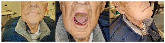
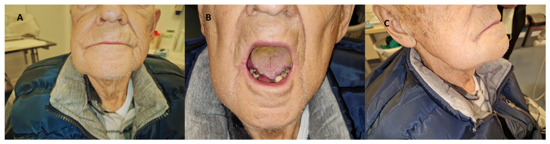
Extraoral examination revealed lower facial asymmetry and swelling of the right mandibular region (Figure 1A–C).
Figure 1.
Clinical presentation of a 78-year-old man with metastatic prostate adenocarcinoma. (A) Frontal view showing facial asymmetry. (B) Frontal view with the mouth open. (C) Lateral view showing swelling of the right mandibular region.
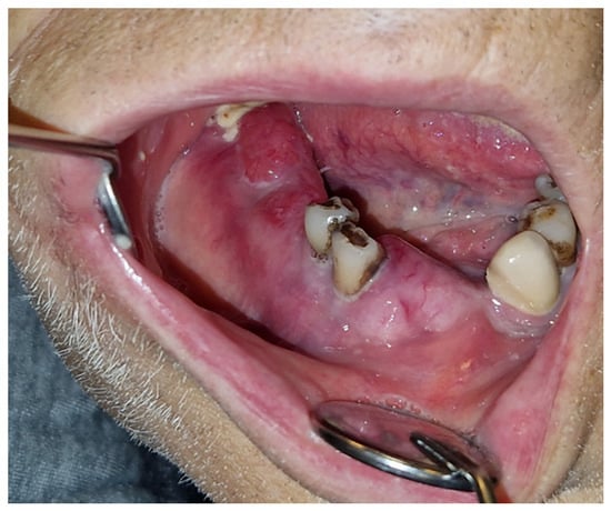
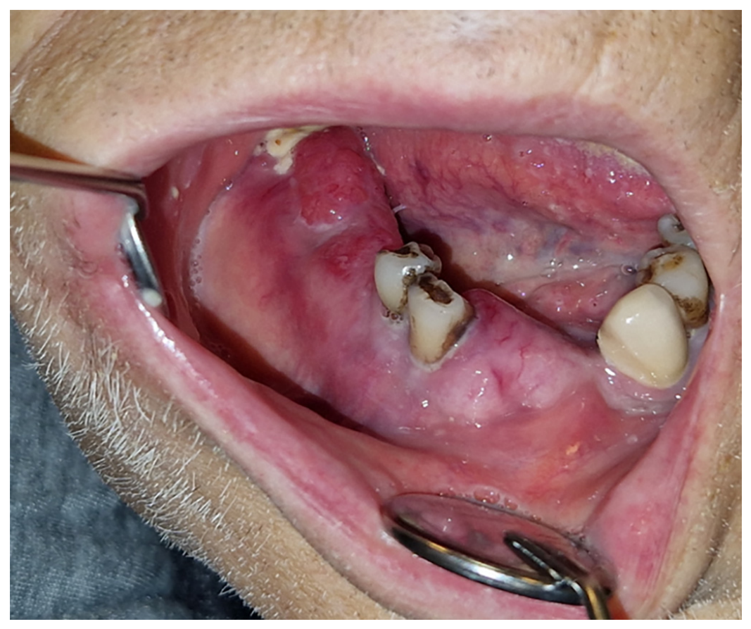
Intraoral examination revealed an approximately 1-cm exophytic, bleeding lesion in the right mandibular molar region, accompanied by significant pain and increased mobility of the adjacent premolars. No exposed bone, fistula formation, or purulent discharge was observed. The patient had no history of recent tooth extraction (Figure 2).
Figure 2.
Intraoral view showing an exophytic, bleeding lesion in the right mandibular molar region.
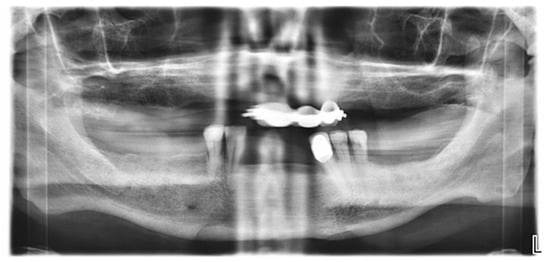
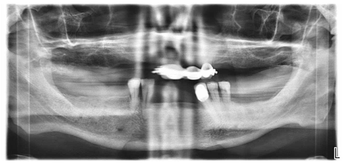
The initial panoramic radiograph (orthopantomogram, OPG) showed no apparent osseous abnormalities in the affected mandibular region, including clearly identifiable osteolytic or osteosclerotic changes (Figure 3).
Figure 3.
OPG showing no radiographic evidence of mandibular bone destruction.
A CT scan of the head and neck region was subsequently performed; however, the original images and corresponding radiological reports were unavailable for retrospective review due to an institutional information technology system failure.
An incisional biopsy of the lesion was performed under local anesthesia in March 2022, and the specimen was obtained from the exophytic soft-tissue lesion in the right mandibular molar region. No bone tissue was included in the biopsy specimen.
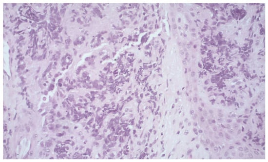
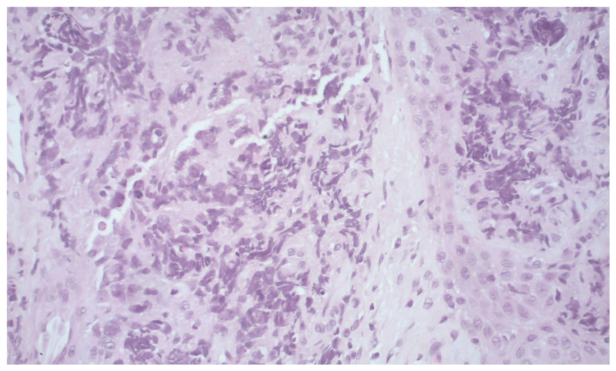
Histopathological examination revealed a poorly differentiated carcinoma composed of small tumor cells with hyperchromatic nuclei and scant cytoplasm, showing crush artifact (Figure 4).
Figure 4.
Histopathological examination showing infiltration by small tumor cells with hyperchromatic nuclei and scant cytoplasm, with areas of crush artifact (H&E, ×20).
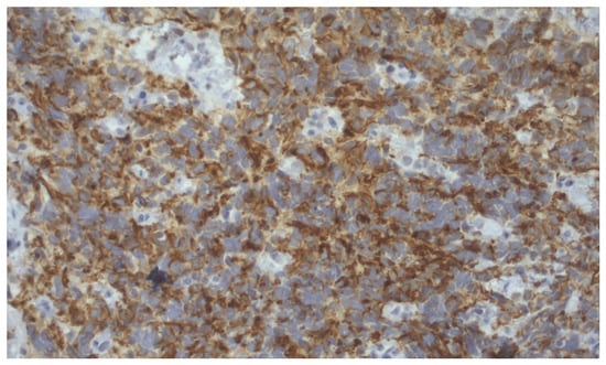
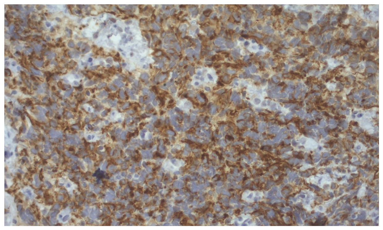
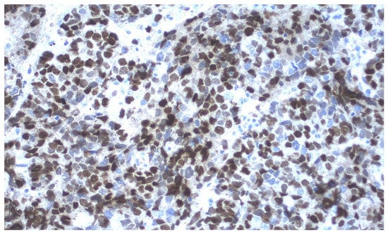
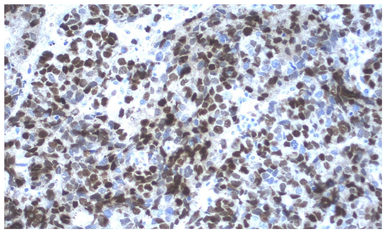
Immunohistochemically, the tumor cells showed marked perinuclear positivity for CK AE1/AE3 (Figure 5), while some cells exhibited weak TTF-1 positivity. The tumor cells were negative for EMA, p40, LCA, CD56, synaptophysin, and chromogranin. Although the initial immunohistochemical findings raised the possibility of pulmonary origin, this interpretation remained inconclusive. Given the patient’s history of prostate adenocarcinoma, additional immunohistochemical analysis was performed. Although PSA expression was negative, strong nuclear positivity for NKX3.1 was observed in the majority of tumor cells (Figure 6), supporting prostatic differentiation and, in conjunction with the clinical history and morphological findings, establishing the diagnosis of metastatic prostate adenocarcinoma.
Figure 5.
Immunohistochemical staining showing positivity of tumor cells for CK AE1/AE3 (×20).
Figure 6.
Immunohistochemical staining showing strong positivity for NKX3.1 in the majority of tumor cells (×20).
Furthermore, skeletal scintigraphy demonstrated multiple foci of increased radiotracer uptake throughout the skeleton, involving the spine, pelvis, bilateral ribs, both femurs, and the right humerus, as well as diffuse pathological uptake in the right mandibular region, raising suspicion of metastatic involvement.
The findings were consistent with progression of the underlying metastatic disease. Zoledronic acid was not reintroduced after the histopathological confirmation of metastatic prostate adenocarcinoma and remained discontinued.
Palliative radiotherapy to the right mandibular region was administered in April 2022, using a single fraction of 8 Gy (3D conformal radiotherapy). Subsequent clinical documentation indicated relief of mandibular pain. However, the exact time of symptom reassessment was not specified, and no standardized pain assessment instrument or changes in analgesic medication or dosage were documented. Unfortunately, the patient died in May 2022, approximately two months after the diagnosis of mandibular metastasis.
The principal clinical events, diagnostic investigations, and therapeutic interventions are summarized chronologically in Table 1.
Table 1.
Timeline of the patient’s clinical course, diagnostic investigations, and therapeutic interventions.
3. Discussion
Oral metastases are uncommon and may present with nonspecific clinical features, making their diagnosis challenging. Although the jawbones are more frequently affected than the oral soft tissues, metastatic lesions may resemble inflammatory, reactive, or treatment-related conditions, including medication-related osteonecrosis of the jaw (MRONJ) [9,10]. The present case illustrates the diagnostic challenge of distinguishing metastatic disease from MRONJ in a patient with advanced prostate adenocarcinoma and a history of long-term intravenous zoledronic acid therapy. Given this history, MRONJ was initially suspected by the treating oncologist, and zoledronic acid was discontinued before specialist dental assessment. However, subsequent examination revealed an exophytic, bleeding lesion in the right mandibular molar region, without clinically exposed or necrotic bone, fistula formation, or purulent discharge. Panoramic radiography also showed no apparent pathological changes. Therefore, the documented clinical findings did not fulfil the established AAOMS diagnostic criteria for MRONJ [9]. Although stage 0 MRONJ may be considered in patients without clinically exposed bone, its diagnosis requires careful interpretation of nonspecific clinical and radiographic findings. In this patient, the atypical soft-tissue presentation and history of advanced malignancy warranted consideration of metastatic disease. Histopathological and immunohistochemical examination subsequently confirmed metastatic prostate adenocarcinoma in the biopsied intraoral soft-tissue lesion.
To contextualize the present case, selected reports of jaw metastases initially suspected as MRONJ or occurring concomitantly with jaw osteonecrosis are summarized in Table 2 [11,12,13,14,15,16,17,18]. These reports represent two distinct clinical scenarios: cases in which metastatic disease mimicked MRONJ without confirmed osteonecrosis, and cases in which metastatic involvement was identified in association with histologically confirmed osteonecrosis. The reported cases also differ in the methods used to establish the diagnosis, ranging from histopathological examination of jaw or soft-tissue specimens to imaging-based assessment. This distinction is important when interpreting the diagnostic evidence and comparing the reported cases.
Table 2.
Selected reports of jaw metastases initially suspected as MRONJ or occurring concomitantly with jaw osteonecrosis [11,12,13,14,15,16,17,18].
Of particular relevance, Bedogni et al. reported malignant cells within or at the margins of necrotic bone in two patients initially diagnosed with bisphosphonate-associated osteonecrosis [11]. Frei et al. described mandibular metastasis from prostate adenocarcinoma associated with bisphosphonate-related osteonecrosis in a patient receiving intravenous zoledronic acid [12]. Although this report shares the primary malignancy, mandibular location, and history of zoledronic acid exposure with the present case, an important difference is the absence of clinically exposed bone and apparent osseous abnormalities on panoramic radiography in our patient. Other reports have similarly demonstrated that metastatic disease, including solid tumors and hematological malignancies, may present with clinical or radiographic findings suggestive of MRONJ [14,15,16,17,18]. Collectively, these observations emphasize that metastatic disease should remain in the differential diagnosis when the clinical presentation is atypical or the diagnosis of MRONJ is uncertain.
Ninety percent of oral metastatic lesions are localized in the bone, while soft tissue metastases are rare [19]. This can be explained with “seed and soil” hypothesis proposed by Stephen Paget in 1889. According to this theory, the metastatic “seed” thrives in organs or environments that provide a favorable “soil” [5], a phenomenon known as metastatic tropism. Among hard tissues, the mandible is the most commonly affected site [1,4], likely due to its higher content of hematopoietic marrow. Although mandibula is the most commonly affected site, cases of oral metastases in the maxilla have also been described in the literature [19].
Radiographic assessment in the present case was limited. Panoramic radiography revealed no apparent mandibular bone destruction; however, an unremarkable panoramic radiograph does not exclude metastatic involvement, particularly in the early stages of disease or when changes are subtle. This is relevant in prostate cancer, in which bone metastases are typically osteoblastic and may not present as clearly defined radiolucent lesions [4]. Although computed tomography of the head and neck was performed, the original images and radiological report were unavailable for retrospective review because of an institutional information technology system failure. Skeletal scintigraphy demonstrated diffuse pathological radiopharmaceutical uptake in the right mandibular region, raising suspicion of metastatic involvement. However, scintigraphy alone could not establish the precise anatomical extent of the lesion or distinguish intraosseous involvement from disease affecting the adjacent soft tissues.
This limitation is particularly important because the incisional biopsy was obtained exclusively from the exophytic soft-tissue lesion, and no bone tissue was included in the specimen. Histopathological and immunohistochemical examination therefore confirmed metastatic prostate adenocarcinoma in the sampled intraoral soft tissue but did not provide direct histological confirmation of mandibular bone infiltration. Accordingly, although the lesion was clinically located in the right mandibular region and scintigraphic findings raised suspicion of metastatic involvement, the precise relationship between the soft-tissue lesion and the underlying mandibular bone could not be definitively established.
The immunohistochemical workup was guided by the morphological features of the tumor and the patient’s oncological history. The initial panel demonstrated marked perinuclear positivity for CK AE1/AE3, while weak TTF-1 positivity was observed in a subset of tumor cells. Although the latter finding raised the possibility of pulmonary origin, it was not conclusive. The tumor cells were negative for EMA, p40, LCA, CD56, synaptophysin, and chromogranin. In view of the patient’s history of prostate adenocarcinoma, additional immunohistochemical analysis was performed. Although PSA expression was negative, strong nuclear NKX3.1 expression in the majority of tumor cells provided important evidence supporting prostatic differentiation. Although NKX3.1 expression has occasionally been reported in other malignancies, including certain sarcomas [20], salivary duct carcinomas, invasive lobular breast carcinomas, tumors of the female urethra, and primary tracheal adenocarcinomas [21,22,23], these alternative primary tumors were not clinically suspected in this patient. The final diagnosis was therefore based on the integrated interpretation of the morphological findings, immunohistochemical profile, and established oncological history. Direct morphological and immunohistochemical comparison with the primary prostate carcinoma was not possible because the original tumor tissue was unavailable for review.
An additional clinically relevant aspect of this case was the discontinuation of zoledronic acid following the initial suspicion of MRONJ. The treating oncologist discontinued the therapy in March 2022, before the patient underwent specialist dental assessment and without a confirmed diagnosis of MRONJ. Although metastatic prostate adenocarcinoma was subsequently confirmed in the biopsied soft-tissue lesion, zoledronic acid was not reintroduced before the patient’s death. The reasons for not resuming treatment could not be established from the available clinical documentation. This sequence highlights the importance of timely communication and multidisciplinary assessment when MRONJ is suspected in patients receiving antiresorptive therapy. Decisions regarding treatment interruption or resumption should be individualized, taking into account the patient’s oncological status, the risk of skeletal-related events, and the oral clinical findings. However, the available information does not allow conclusions to be drawn regarding the effect of zoledronic acid discontinuation on the subsequent course of the patient’s metastatic disease.
The main contribution of this case is not the previously reported association between jaw metastases and antiresorptive therapy, but its illustration of the potential for diagnostic anchoring on MRONJ based primarily on a history of zoledronic acid exposure. The atypical exophytic and bleeding soft-tissue presentation, the absence of apparent changes on panoramic radiography, and the subsequent histopathological diagnosis emphasize the importance of considering malignancy in the differential diagnosis of oral lesions in patients receiving antiresorptive agents. Careful clinical assessment, appropriate imaging, histopathological investigation when indicated, and multidisciplinary decision-making are essential to avoid basing treatment decisions solely on an unconfirmed diagnosis of MRONJ.
This case report has several limitations. First, as a single case, it cannot establish the frequency of misdiagnosis between MRONJ and metastatic disease or allow generalization to a broader patient population. Second, the unavailability of the original CT images and radiological report prevented a comprehensive retrospective assessment of the lesion. Third, because the biopsy specimen contained soft tissue only, direct histological confirmation of mandibular bone involvement was not possible. Additionally, the response to palliative radiotherapy was assessed retrospectively based on the available clinical documentation, which did not specify the timing of pain reassessment, include standardized pain measurements, or provide information on changes in analgesic treatment. Finally, the patient died approximately two months after the histopathological diagnosis, precluding long-term assessment of treatment outcomes.
4. Conclusions
Despite their rarity, oral metastases should be considered in the differential diagnosis of unexplained exophytic lesions and radiographic abnormalities in patients with a history of malignancy. This case highlights the diagnostic challenge of distinguishing metastatic disease from medication-related osteonecrosis of the jaw (MRONJ) in patients receiving antiresorptive therapy. Thorough clinical, radiographic, and histopathological evaluation, together with a multidisciplinary approach, is essential for establishing an accurate diagnosis and enabling appropriate management, with the potential to improve symptom control and quality of life.
Author Contributions
Conceptualization: A.A.-R. and D.V.J.; investigation and resources: A.A.-R., D.V.J., M.V. and S.S.; data curation: A.A.-R., D.V.J., M.V. and S.S.; writing—original draft preparation: A.A.-R. and B.L.B.; writing—review and editing: A.A.-R., D.V.J., B.L.B., M.V., S.S. and F.T.; visualization: A.A.-R., B.L.B. and F.T.; supervision: A.A.-R. and D.V.J. All authors have read and agreed to the published version of the manuscript.
Funding
This research received no external funding.
Institutional Review Board Statement
The case report was conducted in accordance with the Declaration of Helsinki and approved by the Ethical Committee of the School of Dental Medicine, University of Zagreb, Croatia (approval number: 003-01/25-05/06; approval date: 13 June 2025).
Informed Consent Statement
Written informed consent was obtained from the patient during his lifetime for the publication of clinical information and identifiable clinical photographs in this case report.
Data Availability Statement
The data presented in this study are available upon reasonable request from the corresponding author, subject to applicable privacy and ethical restrictions.
Conflicts of Interest
The authors declare no conflicts of interest.
Abbreviations
The following abbreviations are used in this manuscript:
| NKX3 | marker for prostatic adenocarcinoma |
| CK AE1/AE3 | broad spectrum anti pan-keratin antibody cocktail, which differentiates epithelia tumors from non-epithelial tumors |
| TTF-1 | thyroid transcription factor, diagnostic marker and a good prognostic indicator for lung adenocarcinoma |
| EMA | marker of epithelial cells expressed in a variety of normal and neoplastic epithelia |
| p40 | marker for distinguishing lung squamous cell carcinoma from adenocarcinoma |
| LCA | a major membrane glycoprotein restricted to leukocytes |
| CD56 | homophilic binding glycoprotein with a role in cell–cell adhesion |
| MRONJ | medication related osteonecrosis of the jaw |
| CT | computed tomography |
References
- Kirschnick, L.B.; Schuch, L.F.; Cademartori, M.G.; Vasconcelos, A.C.U. Metastasis to the oral and maxillofacial region: A systematic review. Oral Dis. 2022, 28, 23–32. [Google Scholar] [CrossRef] [Scilit] [PubMed]
- Shen, M.L.; Kang, J.; Wen, Y.L.; Ying, W.M.; Yi, J.; Hua, C.G.; Tan, X.F.; Wen, Y.M. Metastatic tumors to the oral and maxillofacial region: A retrospective study of 19 cases in West China and review of the Chinese and English literature. J. Oral Maxillofac. Surg. 2009, 67, 718–737. [Google Scholar] [CrossRef] [Scilit] [PubMed]
- Andabak Rogulj, A.; Tomasovic Loncaric, C.; Muller, D.; Blivajs, I.; Andabak, M.; Vucicevic Boras, V.; Sekerija, M. Solid malignant metastases in the jaw bones. Br. J. Oral Maxillofac. Surg. 2018, 56, 705–708. [Google Scholar] [CrossRef] [Scilit] [PubMed]
- Labrador, A.J.P.; Marin, N.R.G.; Valdez, L.H.M.; Sanchez, K.B.T.; Zabarburu, W.; Ibazetta, K.A.R.; Garcia, A.E.R.; Cabezas, D.M.; Romero, L.; Conte, A.M.; et al. Metastasis to the oral and maxillofacial region. A systematic review. J. Stomatol. Oral Maxillofac. Surg. 2021, S2468-7855, 276–277. [Google Scholar] [CrossRef] [Scilit] [PubMed]
- Hirshberg, A.; Shnaiderman-Shapiro, A.; Kaplan, I.; Berger, R. Metastatic tumours to the oral cavity—Pathogenesis and analysis of 673 cases. Oral Oncol. 2008, 44, 743–752. [Google Scholar] [CrossRef] [Scilit] [PubMed]
- Hirshberg, A.; Berger, R.; Allon, I.; Kaplan, I. Metastatic tumors to the jaws and mouth. Head Neck Pathol. 2014, 8, 463–474. [Google Scholar] [CrossRef] [Scilit] [PubMed]
- Wong, T.; Yap, T.; Wiesenfeld, D. Common causes of ‘swelling’ in the oral cavity. Aust. J. Gen. Pract. 2020, 49, 575–580. [Google Scholar] [CrossRef] [Scilit] [PubMed]
- Kumar, G.; Manjunatha, B. Metastatic tumors to the jaws and oral cavity. J. Oral Maxillofac. Pathol. 2013, 17, 71–75. [Google Scholar] [CrossRef] [Scilit] [PubMed]
- Ruggiero, S.L.; Dodson, T.B.; Aghaloo, T.; Carlson, E.R.; Ward, B.B.; Kademani, D. American Association of Oral and Maxillofacial Surgeons’ Position Paper on Medication-Related Osteonecrosis of the Jaws—2022 Update. J. Oral Maxillofac. Surg. 2022, 80, 920–943. [Google Scholar] [CrossRef] [Scilit] [PubMed]
- He, L.; Sun, X.; Liu, Z.; Qiu, Y.; Niu, Y. Pathogenesis and multidisciplinary management of medication-related osteonecrosis of the jaw. Int. J. Oral Sci. 2020, 12, 30. [Google Scholar] [CrossRef] [Scilit] [PubMed]
- Bedogni, A.; Saia, G.; Ragazzo, M.; Bettini, G.; Capelli, P.; D’Alessandro, E.; Nocini, P.F.; Lo Russo, L.; Lo Muzio, L.; Blandamura, S. Bisphosphonate-associated osteonecrosis can hide jaw metastases. Bone 2007, 41, 942–945. [Google Scholar] [CrossRef] [Scilit] [PubMed]
- Frei, M.; Bornstein, M.M.; Schaller, B.; Reichart, P.A.; Weimann, R.; Iizuka, T. Bisphosphonate-related osteonecrosis of the jaw combined with jaw metastasis of prostate adenocarcinoma: Report of a case. J. Oral Maxillofac. Surg. 2010, 68, 863–867. [Google Scholar] [CrossRef] [Scilit] [PubMed]
- Poulias, E.; Melakopoulos, I.; Tosios, K. Metastatic breast carcinoma in the mandible presenting as a periodontal abscess: A case report. J. Med. Case Rep. 2011, 5, 265. [Google Scholar] [CrossRef] [Scilit] [PubMed]
- Gander, T.; Obwegeser, J.A.; Zemann, W.; Grätz, K.W.; Jacobsen, C. Malignancy mimicking bisphosphonate-associated osteonecrosis of the jaw: A case series and literature review. Oral Surg. Oral Med. Oral Pathol. Oral Radiol. 2014, 117, 32–36. [Google Scholar] [CrossRef] [Scilit] [PubMed]
- Corsi, A.; Ungari, C.; Riminucci, M.; Agrillo, A. Bisphosphonate-Related Osteonecrosis and Metastasis Within the Same Site of the Jaw. J. Oral Maxillofac. Surg. 2017, 75, 1679–1684. [Google Scholar] [CrossRef] [Scilit] [PubMed]
- Lee, C.Y.S.; Rahman, M.; Lee, K.L.; Hirata, K.Y. Thymic Carcinoma Metastatic to the Mandible Mistaken for Medication-Related Osteonecrosis of the Jaw: A Case Report and Review of the Literature. Clin. Surg. 2018, 3, 2060. [Google Scholar]
- Danskin, Y.; Alhajji, D.; Singh, A.; Rota, A.; Huryn, J.M.; Estilo, C.L. Multiple myeloma masquerading as mandibular medication-related osteonecrosis of the jaw: A case report. Aust. Dent. J. 2022, 67, 94–99. [Google Scholar] [CrossRef] [Scilit] [PubMed]
- Gurav, S.; Singh, G.P.; Ostwal, V.; Sharma, M.; Jagtiani, K. Metastatic pancreatic cancer mimicking medication related osteonecrosis of the jaw—A rare clinical presentation. Med. Adv. 2023, 1, 408–412. [Google Scholar] [CrossRef] [Scilit]
- Seoane, J.; Van der Waal, I.; Van der Waal, R.I.; Cameselle-Teijeiro, J.; Antón, I.; Tardio, A.; Alcázar-Otero, J.J.; Varela-Centelles, P.; Diz, P. Metastatic tumours to the oral cavity: A survival study with a special focus on gingival metastases. J. Clin. Periodontol. 2009, 36, 488–492. [Google Scholar] [CrossRef] [Scilit] [PubMed]
- Yoshida, K.I.; Machado, I.; Motoi, T.; Parafioriti, A.; Lacambra, M.; Ichikawa, H.; Kawai, A.; Antonescu, C.R.; Yoshida, A. NKX3-1 Is a Useful Immunohistochemical Marker of EWSR1-NFATC2 Sarcoma and Mesenchymal Chondrosarcoma. Am. J. Surg. Pathol. 2020, 44, 719–728. [Google Scholar] [CrossRef] [Scilit] [PubMed]
- Hawari, R.; Fernandes, L.; Park, K.J.; McCluggage, W.G. Skene’s Gland Derivatives in the Female Genital Tract and Cervical Adenoid Basal Carcinoma are Consistently Positive with Prostatic Marker NKX3.1. Int. J. Gynecol. Pathol. 2021, 40, 400–407. [Google Scholar] [CrossRef] [Scilit] [PubMed]
- Asch-Kendrick, R.J.; Samols, M.A.; Lilo, M.T.; Subhawong, A.P.; Sharma, R.; Illei, P.B.; Argani, P.; Cimino-Mathews, A. NKX3.1 is expressed in ER-positive and AR-positive primary breast carcinomas. J. Clin. Pathol. 2014, 67, 768–771. [Google Scholar] [CrossRef] [Scilit] [PubMed]
- Miura, A.; Mori, T.; Yoshida, A.; Watanabe, Y.; Sunami, K.; Watanabe, S.; Kohno, T.; Tsuta, K. Primary adenocarcinoma of the trachea revealing a mucinous bronchial gland cell origin. Pathol. Res. Pract. 2018, 214, 796–799. [Google Scholar] [CrossRef] [Scilit] [PubMed]

Disclaimer/Publisher’s Note: The statements, opinions and data contained in all publications are solely those of the individual author(s) and contributor(s) and not of MDPI and/or the editor(s). MDPI and/or the editor(s) disclaim responsibility for any injury to people or property resulting from any ideas, methods, instructions or products referred to in the content. |
© 2026 by the authors. Licensee MDPI, Basel, Switzerland. This article is an open access article distributed under the terms and conditions of the Creative Commons Attribution (CC BY) license.