


Abstract
Background/Objectives: This study characterized systemic and mucosal immune responses to an acute, clinically mild Salmonella enterica serovar Typhimurium challenge in standard-fed rats and subsequently examined responses in an independent high-fat diet (HFD)-fed cohort, including exploratory sex-associated differences. Methods: Male and female Wistar Han rats receiving a standard diet were allocated to a non-inoculated group (n = 4) or challenged orally with 105, 107 or 108 colony-forming units of S. Typhimurium (n = 6/group). The highest dose was subsequently applied in an independent cohort fed an HFD for 12 weeks (non-inoculated, n = 6; inoculated, n = 8). Clinical parameters, organ weights, leukocyte profiles, plasma immunoglobulins (Ig) and cytokines, and mucosal immune markers were assessed 72 h post-inoculation. Results: In standard-diet-fed rats, challenge remained clinically mild but induced dose-dependent immunological changes, including granulocyte accumulation in the blood, increased plasma IgM levels, changes in the distribution of IgG isotypes, and elevated cecal Ig-coated bacteria. In HFD-fed rats, inoculation increased circulating leukocyte counts and granulocyte proportions, while plasma Igs remained unchanged and cytokine profiles largely overlapped. Mucosal IgM also decreased, whereas the proportion of Ig-coated bacteria increased. Conclusions: Oral S. Typhimurium inoculation induced acute systemic and mucosal immune changes in both experimental cohorts. This model may support investigations of early host responses to enteric bacterial challenge. Interpretation is limited by the independent cohorts, single 72 h endpoint, and absence of systematic pathogen-specific dissemination and histopathological assessments.
1. Introduction
Infectious diseases remain a major public health concern, causing significant morbidity and mortality worldwide [1]. Increasing evidence suggests that chronic non-communicable conditions such as obesity may modify susceptibility to infection and disease progression [2]. Given the rising global prevalence of obesity, understanding how altered metabolic states influence host–pathogen interactions has become increasingly urgent [3].
Obesity is typically characterized by excessive adipose tissue accumulation accompanied by metabolic disturbances such as insulin resistance and chronic low-grade systemic inflammation driven by immune dysregulation [4]. Excess adiposity promotes accumulation of pro-inflammatory immune cells in adipose tissue, including M1 macrophages and T helper (Th) 1-polarized lymphocytes, accompanied by elevated secretion of cytokines such as tumor necrosis factor (TNF)-α, interleukin (IL)-6, and IL-1β [5,6]. Circulating innate immune cells, including neutrophils and monocytes, are often increased, while adaptive immune responses may be functionally altered [7,8,9].
Obesity also disrupts adipokine balance, with increased leptin and reduced adiponectin levels further sustaining the inflammatory tone [8,10]. These alterations impair the host defense mechanisms, increase infection severity, and reduce vaccine efficacy in both humans and animal models [11,12].
Sex is an additional determinant of immune function. Females generally mount stronger innate and adaptive immune responses, whereas males are more susceptible to many infectious diseases and often display greater systemic inflammation in obesity [13,14,15]. Despite these recognized differences, many preclinical infection studies have historically focused on male animals. In addition, relatively little is known about how obesity and sex interact to shape immune responses to enteric pathogens.
Among enteric pathogens, Salmonella enterica serovar Typhimurium imposes a substantial burden, with an estimated 93–95 million cases and 150,000–155,000 deaths annually, disproportionately affecting children and other vulnerable populations [16]. S. Typhimurium is a facultative intracellular pathogen that, following oral ingestion, colonizes the gastrointestinal tract and disseminates to lymph nodes, spleen, and liver [17,18,19,20,21]. Infection induces rapid recruitment of innate immune cells and the release of a spectrum of pro-inflammatory cytokines including interferon (IFN)-γ, IL-8, IL-12, IL-17A, and TNF-α, followed by activation of adaptive responses [22,23,24].
Although severe infection models have provided important insights into Salmonella pathogenesis, mild models may be valuable for investigating early host immune responses and the immunomodulatory effects of nutritional interventions. Rats are comparatively resistant to S. Typhimurium-induced systemic disease, in contrast to commonly used mouse models in which infection can progress rapidly to systemic disease [25,26,27]. This relative resistance makes the rat a potentially suitable model for investigating clinically mild conditions and their modulation by nutritional factors.
Therefore, the present study first established an acute, clinically mild S. Typhimurium challenge model and characterized the immune response. The selected challenge dose was subsequently applied in an independent cohort of high-fat diet (HFD)-fed rats to characterize the response in this dietary pattern, including exploratory assessment of sex-associated patterns.
2. Materials and Methods
2.1. S. Typhimurium Culture and Inoculum Preparation
Salmonella enterica serovar Typhimurium (strain CDC 6516-60, ATCC, Manassas, VA, USA) was cultured in Tryptone Soya broth (TS, CM0129B; Thermo Fisher Scientific, Vienna, Austria) at 37 °C. An overnight culture was sub-cultured the following day in fresh TS broth and grown to an optical density at 600 nm (OD600). Bacterial cells were collected via centrifugation (4000× g, 10 min, 4 °C), washed, and resuspended in sterile phosphate-buffered saline (PBS). The bacterial concentration used for infection (105, 107 and 108 CFU/mL) was estimated based on the OD600-colony-forming unit (CFU) calibration curve and verified by plating serial dilutions on agar plates.
2.2. Animals and Ethics
All procedures were approved by the Ethical Committee for Animal Experimentation of the University of Barcelona (UB) and the Government of Catalonia (CEEA-UB, Ref. 275/23) and conducted in accordance with European Directive 2010/63/EU. The number of animals was estimated with the Granmo calculator (IMIM, Barcelona, Spain) based on previous plasma IgG levels data of the research group, assuming a two-sided alpha risk of 0.05, a beta risk below 0.20, a minimum detectable difference of 0.65, and a standard deviation of 0.40. Animals were housed under controlled temperature and humidity with a 12 h light–dark cycle at the UB Diagonal campus facility and acclimatized one week prior to experiments.
2.3. Experimental Design
2.3.1. S. Typhimurium Dose–Response Study
Seven-week-old female and male Wistar Han rats (Janvier Labs, Le Genest-Saint-Isle, France) were fed a standard AIN-93M diet (Supplementary Table S1; Teklad Custom Diet, Inotiv, West Lafayette, IN, USA) with ad libitum access to food and water. Animals were randomly assigned to four groups, with one non-inoculated group (NI; n = 4) and three inoculated groups (D1, D2, D3; n = 6/group), with each group balanced by sex. Rats were administered 1 mL of PBS containing 105 (D1), 107 (D2), or 108 CFU (D3) of S. Typhimurium by oral gavage, while NI animals received 1 mL of PBS (Supplementary Figure S1).
2.3.2. S. Typhimurium Challenge in HFD Animals
Eight-week-old male and female Wistar Han rats (Janvier Labs) were fed a HFD (Supplementary Table S1; TD.06415, Inotiv) for 12 weeks (n = 14) with ad libitum access to food and water. Body weight and food and water intake were recorded three times per week, and fecal samples were collected weekly. Oral glucose tolerance tests (OGTT) were performed at week 11 following a 5 h morning fast. Blood glucose was measured from the saphenous vein at baseline and at 15, 30, 60, 90, and 120 min after oral glucose administration (2 g/kg body weight) using a glucometer (Adia Blood Glucose Monitoring System, ForaCare, Morges, Switzerland). Fasting blood samples were stored at –80 °C for further analysis. At week 12, animals were randomly divided into two groups, with females and males equally distributed across the groups: HFD (n = 6) and HFD-inoculated (HFD-I, n = 8). HFD-I rats were orally gavaged with 1 mL of PBS containing 108 CFU/mL of S. Typhimurium. Control animals received 1 mL PBS alone (Supplementary Figure S1).
2.4. Sample Collection and Processing
After the inoculation, rats were monitored daily for general health and clinical signs of disease (weight, appearance and behavior, both unprovoked and in response to handling). Fecal output and characteristics were monitored daily, including the number, weight, and appearance of feces, to identify gastrointestinal alterations such as diarrhea. Fecal samples were collected daily. Body weight and food and water intake were also recorded. No signs of disease were observed in any of the animals during the 72 h post-inoculation period. Seventy-two hours after S. Typhimurium inoculation, animals were anesthetized with ketamine (90 mg/kg, Merial Laboratories S.A., Ripollet, Spain) and xylazine (10 mg/kg, Bayer A.G., Leverkusen, Germany) and euthanized to obtain blood and tissue samples. Blood was collected by cardiac puncture for hematological analysis (Spincell 3, Spinreact, Sant Esteve d’en Bas, Spain), centrifuged for plasma collection (5 min, 10,000× g, 4 °C) and stored at −80 °C. Organs, including mesenteric lymph nodes (MLN), small intestine, thymus, liver, and cecal content, were collected and weighed. For gut wash (GW) collection, a distal ileum segment was weighed, incubated with PBS and centrifuged as described previously [28].
2.5. Fecal Analyses
Fecal water content was determined from the difference between fecal weights before and after drying at 60 °C for 24 h. The pH was measured using a surface electrode (Crison Instruments, S.A., Alella, Spain).
2.6. Plasma Analyses
Immunoglobulin (Ig) isotypes (IgA, IgM, IgG1, IgG2a, IgG2b, IgG2c), cytokines (Granulocyte colony-stimulating factor (G-CSF), granulocyte-macrophage colony-stimulating factor (GM-CSF), IFN-γ, IL-1α, IL-1β, IL-2, IL-4, IL-5, IL-6, IL-10, IL-12p70, IL-13, IL-17A, monocyte chemoattractant protein (MCP)-1/CC chemokine ligand-2 (CCL2), TNF-α), and leptin were measured using the ProcartaPlex™ Rat Antibody Isotyping Panel (6-plex; EPX060-30123-901), the ProcartaPlex™ Rat Th Complete Panel (14-plex; EPX140-30120-901), and the Leptin Rat ProcartaPlex™ Simplex Kit (EPX01A-32039-901; all from Thermo Fisher Scientific, Vienna, Austria), following the manufacturer’s protocols. Plates were analyzed using a MAGPIX® system (Luminex Corporation, Austin, TX, USA), and data were processed with ProcartaPlex Analyst Software 1.0 (MAGPIX® analyzer, Luminex Corporation, Austin, TX, USA) at the Flow Cytometry Unit of the Scientific and Technological Centers of the University of Barcelona (CCiT-UB). The detection limits for each cytokine are provided in Supplementary Table S2. For the calculation of Th1/Th2 ratios, IgG subclasses were grouped according to their association with Th responses, based on previous studies [29]. IgG2b and IgG2c were considered indicative of a Th1-type response, whereas IgG1 and IgG2a were considered representative of a Th2-type response. For cytokines, IFN-γ and IL-4 were used as markers of Th1- and Th2-type responses, respectively.
Plasma adiponectin and lipopolysaccharide-binding protein (LBP) were measured using enzyme-linked immunosorbent assay (ELISA) kits (E-EL-R3012 and E-EL-R0589; Elabscience, Wuhan, China). Insulin was measured in fasting plasma using the Mercodia Rat Insulin ELISA (10-1250-01, Mercodia AB, Uppsala, Sweden).
2.7. Gut Wash Analyses
IgA, IgM, and alpha-1-antitrypsin (A1AT) were quantified using commercially available ELISA kits (IgA: E111-102, Bethyl Laboratories, Montgomery, AL, USA; IgM: 88-50540, Life Technologies, Carlsbad, CA, USA; A1AT: A4782, Antibodies.com, Cambridge, UK) following the manufacturers’ instructions.
2.8. MLN Lymphocyte Phenotyping
Freshly collected MLNs were passed through 40 μm sterile mesh strainers (Thermo Fisher Scientific, Waltham, MA, USA) into cold Roswell Park Memorial Institute 1640 (RPMI) medium (Merck KGaA, Darmstadt, Germany) supplemented with 10% fetal bovine serum (FBS, Sigma–Aldrich, St. Louis, MO, USA), 100 IU/mL penicillin-streptomycin (Merck KGaA), 2 mM L-glutamine (Merck KGaA), and 0.05 mM 2-β-mercaptoethanol (Merck KGaA). Cells were centrifuged (538× g, 10 min, 4 °C) and resuspended in enriched RPMI. Viable cell counts were performed using the Countess™ Automated Cell Counter (Invitrogen™, Thermo Fisher Scientific). A total of 3 × 10⁵ cells were stained for 20 min at 4 °C in the dark with fluorochrome-conjugated monoclonal antibodies (Ab) in PBS + 2% FBS + 0.1% sodium azide (Merck KGaA). Two Ab panels were designed as detailed in Supplementary Table S3. After staining, cells were washed, fixed with 0.5% paraformaldehyde, and stored at 4 °C. Samples were acquired on an Aurora Cytek spectral flow cytometer (Cytek, Fremont, CA, USA) at the CCiT-UB and data were analyzed using FlowJo v10 (Tree Star Inc., Ashland, OR, USA).
2.9. Ig-Coated Bacteria Analyses
Approximately 100–120 mg of cecal content was diluted 1:10 (w/v) in PBS and homogenized using a Minipolytron® (Kinematica AG, Malters, Switzerland). Samples were centrifuged at 300× g for 1 min at 4 °C. The supernatant was diluted in PBS with 1% FBS and centrifuged at 8000× g for 5 min at 4 °C. The pellet was resuspended and incubated with a fluorescein isothiocyanate (FITC)-conjugated antibody against rat IgG, IgM, and IgA (ab102198, Abcam, Waltham, MA, USA) for 30 min at 4 °C in the dark. Following two washes with PBS, propidium iodide (PI, Merck KGaA, Darmstadt, Germany) was added to stain DNA. Samples were analyzed on an Aurora Cytek spectral flow cytometer at the CCiTUB. Total Ig-coated bacteria (IgG + IgM + IgA) were quantified as the percentage of FITC-positive events within PI-positive bacterial populations. An unstained control was included to assess background fluorescence. Data were processed using FlowJo v10 (Tree Star Inc.).
2.10. Statistical and Data Analysis
No inclusion or exclusion criteria were established, and no animals or data points were excluded from the analyses. Blinding was not performed during experimental conduct and data analysis. Statistical analyses were performed using IBM SPSS v27 (IBM Corp., Armonk, NY, USA). Normality was assessed using the Shapiro–Wilk test and homogeneity of variance using Levene’s test. Parametric data were analyzed using one-way analysis of variance (ANOVA) followed by Bonferroni or Dunnett T3 post hoc tests, as appropriate, for comparisons among groups in the dose–response study. Two-way ANOVA was used to assess the effects of inoculation and sex and their interaction in the HFD cohort. For variables measured over time, two-way repeated-measures ANOVA was applied with time as a within-subject factor. Non-parametric data were analyzed using the Kruskal–Wallis test followed by Dunn’s post hoc test with Bonferroni correction or the Mann–Whitney U test. Data are presented as mean ± standard error of the mean (SEM), unless otherwise specified. Cytokine detection frequencies were compared using chi-square tests. Non-metric multidimensional scaling (NMDS) was performed in R (R Foundation for Statistical Computing, Vienna, Austria), and group differences were assessed using analysis of similarities (ANOSIM). A p-value < 0.05 was considered statistically significant.
3. Results
The study comprised two sequential experimental phases. First, a dose–response test in conventionally fed rats was performed to establish a mild challenge model and select the inoculum for subsequent studies. Second, the selected dose was applied in an independent cohort to characterize the immune response to challenge in this dietary context, including exploratory assessment of sex-associated host immune response patterns.
3.1. Characterization of the S. Typhimurium Dose–Response Model
3.1.1. Physiological Responses
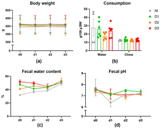
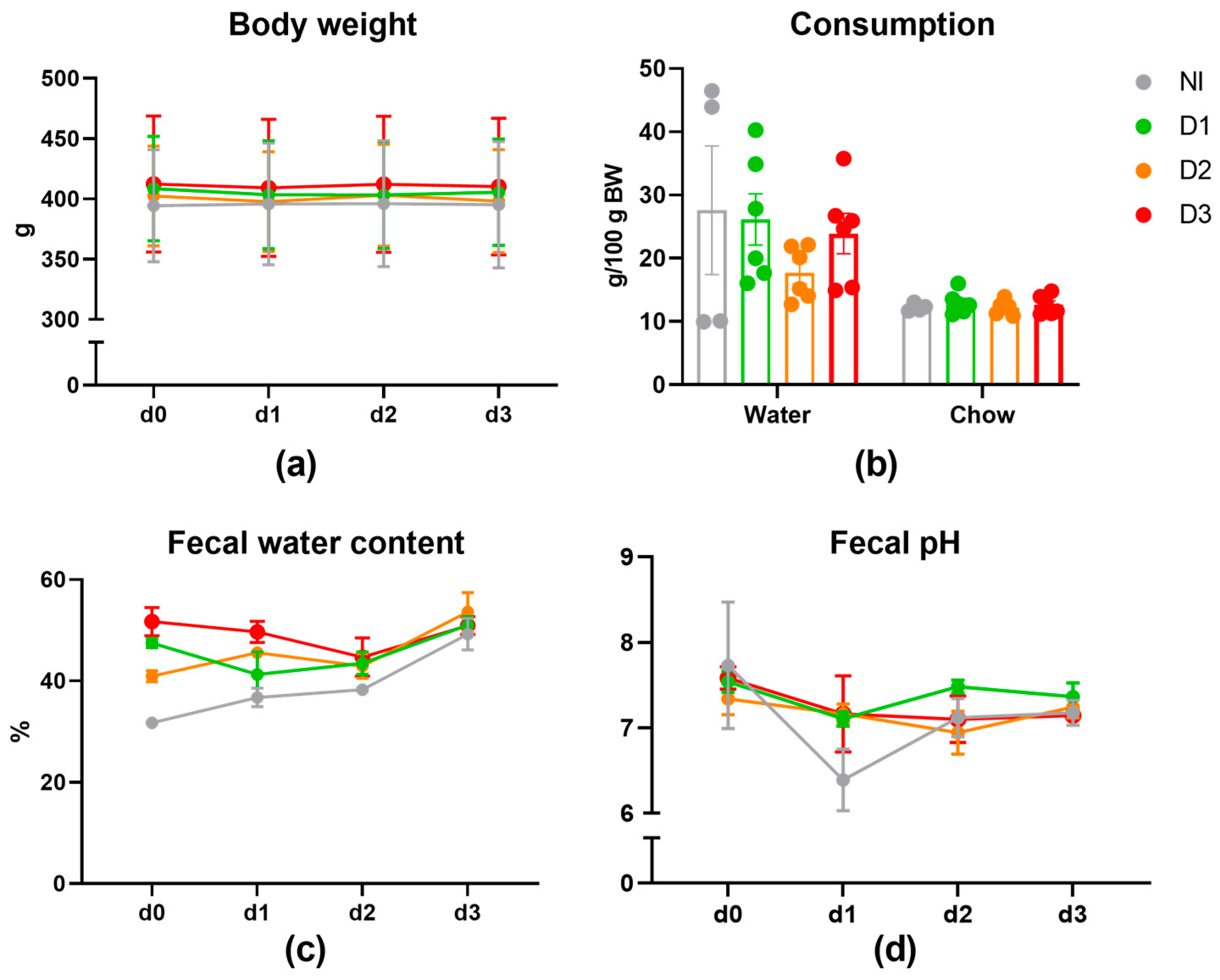
Challenge with S. Typhimurium was clinically well tolerated in all doses tested. No signs of distress were observed, and body weight, chow intake, and water consumption remained stable throughout the three days following inoculation when compared to the non-inoculated group (Figure 1a,b). Fecal water content and pH were unchanged across groups, consistent with the absence of gastrointestinal symptoms such as diarrhea (Figure 1c,d). Morphometric measurements were not affected either (Supplementary Table S4).
Figure 1.
Variables assessed during the three-day course of S. Typhimurium challenge. (a) Body weight. (b) Total food and water intake (d0–d3). (c) Fecal water content. (d) Fecal pH. NI: non-inoculated control; D1: 105 CFU, D2: 107 CFU; D3: 108 CFU. Data are presented as mean ± SEM (n = 4 NI, n = 6 D1–3).
3.1.2. Blood Cell Composition and Mucosal Immune Cell Phenotyping
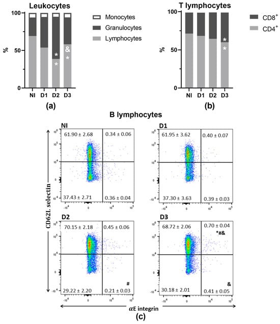
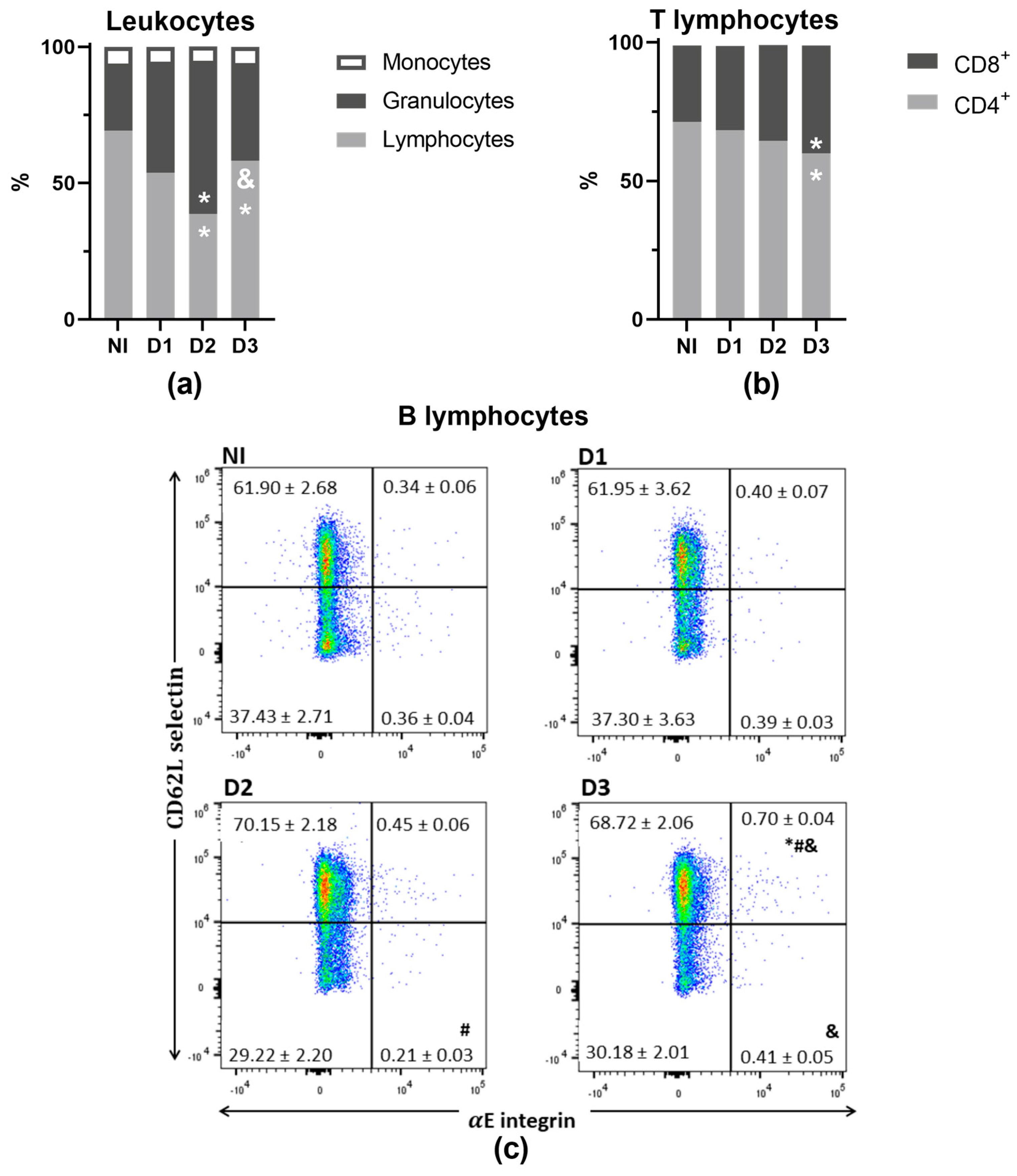
Erythrocyte counts and related parameters, including hemoglobin, hematocrit, mean corpuscular volume (MCV), and mean corpuscular hemoglobin (MCH), were not affected following inoculation. Platelet counts, however, were reduced in the D3 group compared with D2 (Supplementary Table S5). Leukocyte counts remained unchanged, but challenge induced marked shifts in leukocyte distribution (Figure 2a). Animals receiving higher doses of S. Typhimurium (D2 and D3) exhibited higher granulocyte proportions and correspondingly lower lymphocyte proportions than NI controls [22,23].
Figure 2.
Blood leukocyte counts and MLN lymphocyte subsets 72 h following S. Typhimurium inoculation. (a) Proportions of main leukocytes in blood. (b) CD4+ and CD8+ T lymphocytes in MLN. (c) CD62L and CD103 (αE integrin) expression in CD45RA+ B cells in MLNs. NI: non-inoculated control; D1: 105 CFU, D2: 107 CFU; D3: 108 CFU. Data are presented as mean ± SEM (n = 4 NI, n = 6 D1–3). * p < 0.05 vs. NI. # p < 0.05 vs. D1. & p < 0.05 vs. D2.
To further characterize mucosal immune alterations, lymphocyte populations were analyzed in the MLNs. While the overall proportions of B and T cells were not altered (Supplementary Table S6), D3 animals showed an increased proportion of CD8+ T cells against NI animals (Figure 2b).
Furthermore, oral challenge induced only limited changes in the surface expression of the mucosal marker CD103 (αE integrin) and CD62L selectin across MLN lymphocyte populations (Supplementary Table S7). However, animals receiving the highest Salmonella dose showed an increase in the CD62L+αE+ double-positive B lymphocyte population (Figure 2c).
3.1.3. Humoral Systemic Response
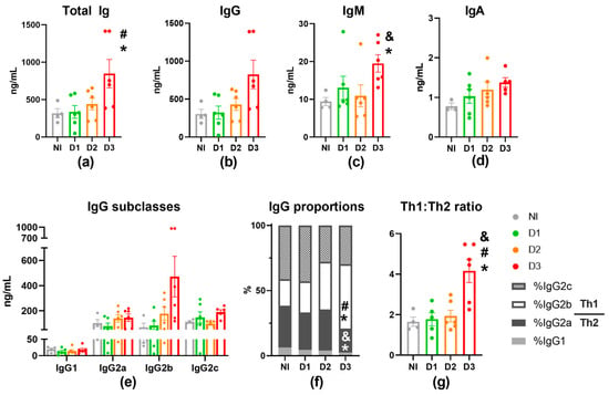
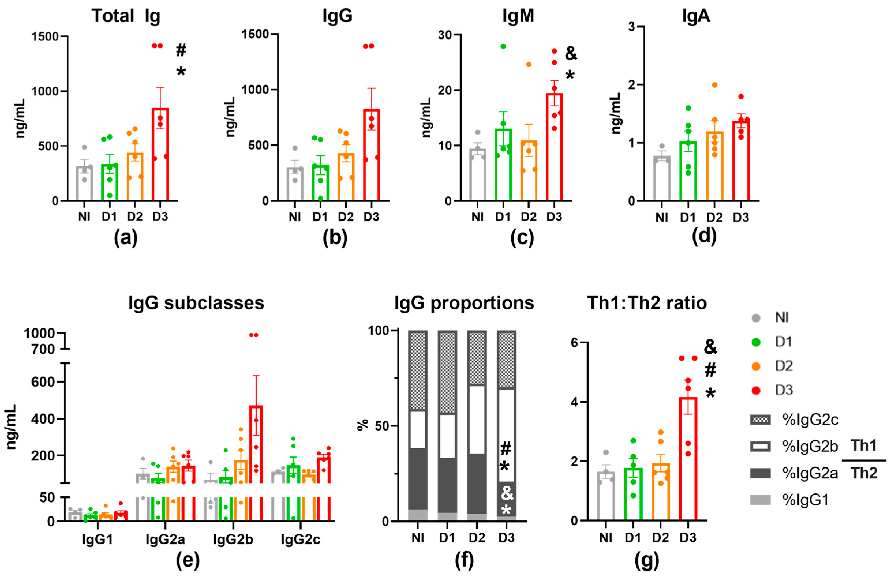
Challenge with the highest inoculation dose (D3) increased total Ig levels compared with NI controls, which was mainly due to increases in IgG and IgM isotypes (Figure 3a–d). While absolute IgG isotype levels remained unchanged (Figure 3e), D3 animals had a higher proportion of IgG2b accompanied by a lower proportion of IgG2a (Figure 3f), leading to a higher Th1/Th2 IgG ratio (Figure 3g).
Figure 3.
Plasma Ig response 72 h following S. Typhimurium inoculation. (a) Total Ig, (b) IgG, (c) IgM, (d) IgA, and (e) IgG isotype levels. (f) IgG isotype proportions. (g) IgG Th1/Th2 ratio (IgG2b and IgG2c as Th1-associated; IgG1 and IgG2a as Th2-associated). NI: non-inoculated control; D1: 105 CFU, D2: 107 CFU; D3: 108 CFU. Data are presented as mean ± SEM (n = 4 NI, n = 6 D1–3). * p < 0.05 vs. N; # p < 0.05 vs. D1. & p < 0.05 vs. D2.
3.1.4. Systemic Cytokine Response
Out of the 15 cytokines analyzed, GM-CSF, IL-1α, IL-4, IL-5, IL-6, and IL-10 were below detection limits in most samples and were therefore excluded from further analysis. Among the detectable cytokines, MCP-1/CCL2 levels decreased in a dose-dependent manner. In contrast, IL-12p70 was more frequently detected in the D3 group (Supplementary Table S8).
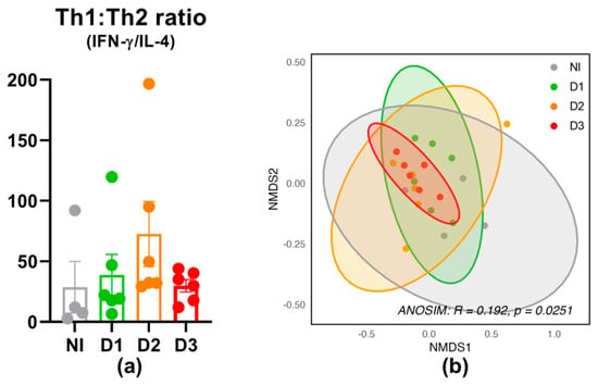
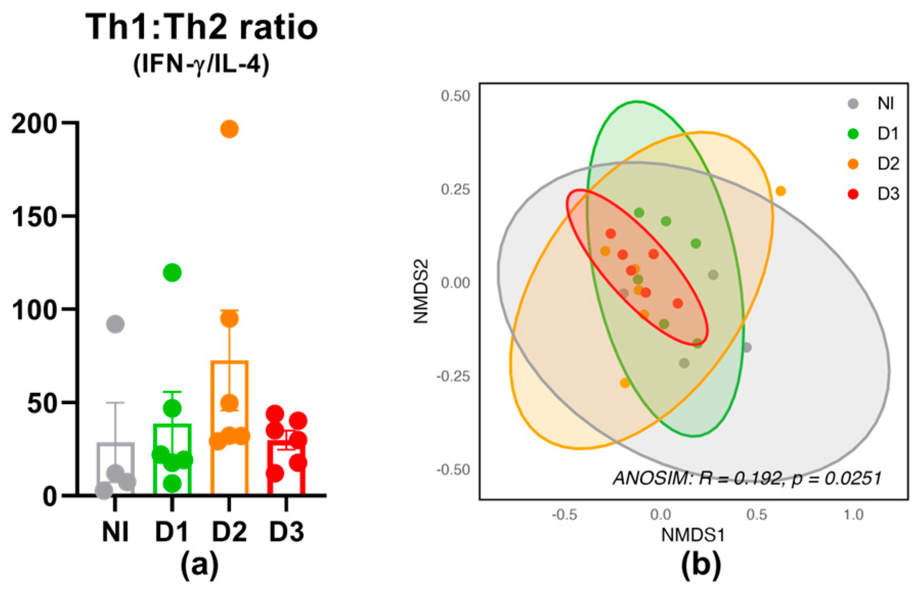
The calculated Th1/Th2 cytokine ratio, based on IFN-γ and IL-4 as representatives of Th cell response, did not differ across the groups (Figure 4a). To assess whether oral Salmonella challenge altered the overall cytokine profile, NMDS analysis was performed based on plasma cytokine concentrations (Figure 4b). The ordination revealed substantial overlaps among the dose groups, although ANOSIM indicated a statistically significant but weak overall group differentiation.
Figure 4.
Plasma cytokines 72 h following S. Typhimurium inoculation. (a) Plasma Th1/Th2-associated cytokine ratio. (b) NMDS ordination based on plasma cytokine concentrations. NI: non-inoculated control; D1: 105 CFU, D2: 107 CFU; D3: 108 CFU. Data are presented as mean ± SEM (n = 4 NI, n = 6 D1–3).
3.1.5. Intestinal Response
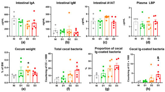
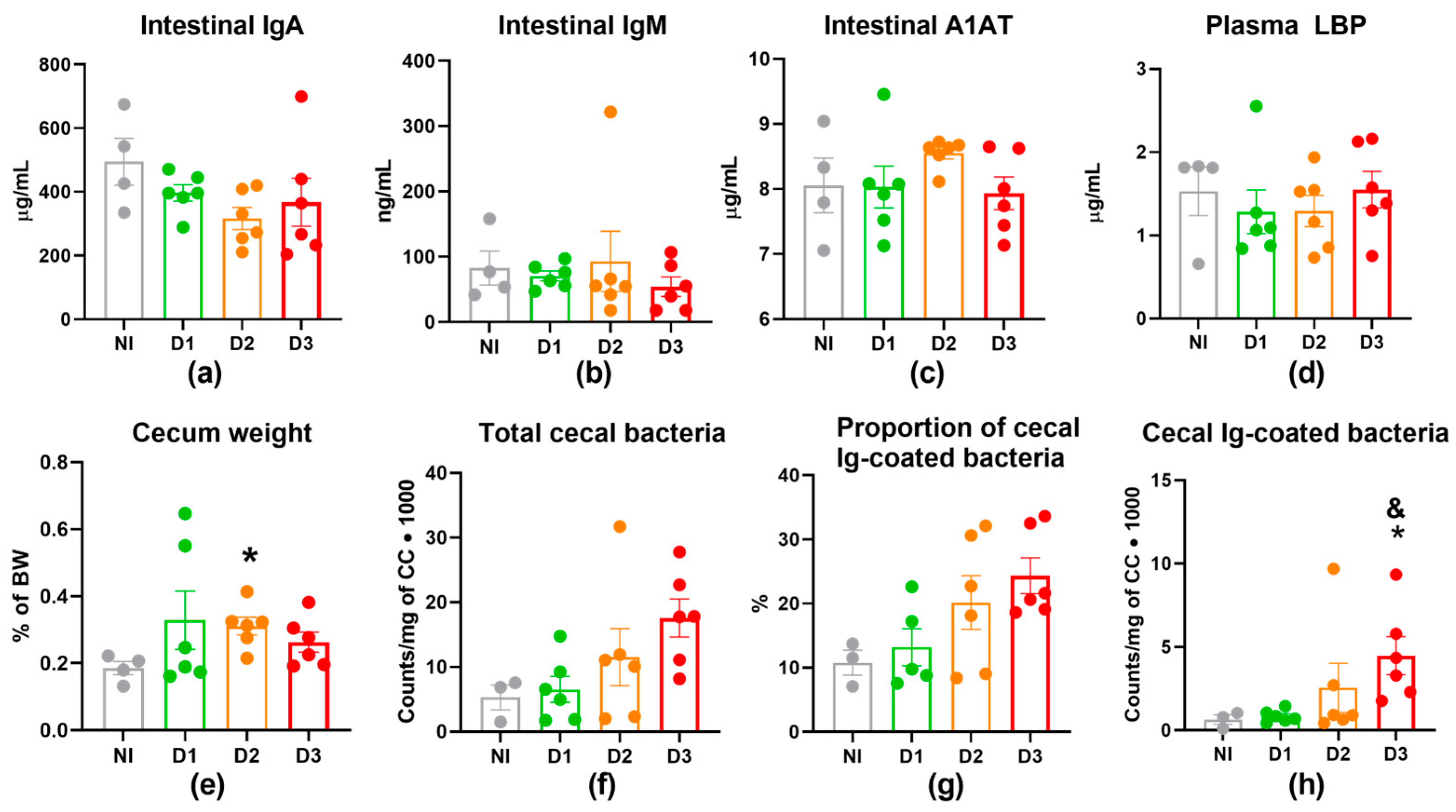
Mucosal IgA and IgM levels in GW, as well as A1AT concentrations in GW and LBP concentrations in plasma, did not change following inoculation (Figure 5a–d). Relative cecum weight was increased in the D2 group (Figure 5e), with total cecal bacterial abundance showing a tendency toward higher levels in D3 animals (p = 0.098; Figure 5f). Interestingly, the number of Ig-coated bacteria in cecal content was increased in D3 animals (Figure 5h).
Figure 5.
Intestinal immune alterations 72 h following S. Typhimurium inoculation. (a) Intestinal IgA, (b) IgM, and (c) A1AT. (d) Plasma LBP. (e) Relative cecum weight. (f) Total cecal bacteria. (g) Proportion of cecal Ig-coated bacteria. (h) Cecal Ig-coated bacteria. NI: non-inoculated control; D1: 105 CFU, D2: 107 CFU; D3: 108 CFU. Data are presented as mean ± SEM (n = 4 NI, n = 6 D1–3). * p < 0.05 vs. NI. & p < 0.05 vs. D1.
3.2. Characterization of S. Typhimurium Challenge Model in HFD-Fed Animals
After characterizing the challenge response in standard-diet-fed reference animals, we next investigated the impact of an oral challenge with the selected inoculation dose (108 CFU) in the context of HFD. Prior to inoculation, all HFD-fed animals assigned to control and inoculated groups were comparable in body weight, glycemic parameters, and morphometric measurements (Supplementary Table S9).
3.2.1. Physiological Response in HFD-Fed Animals
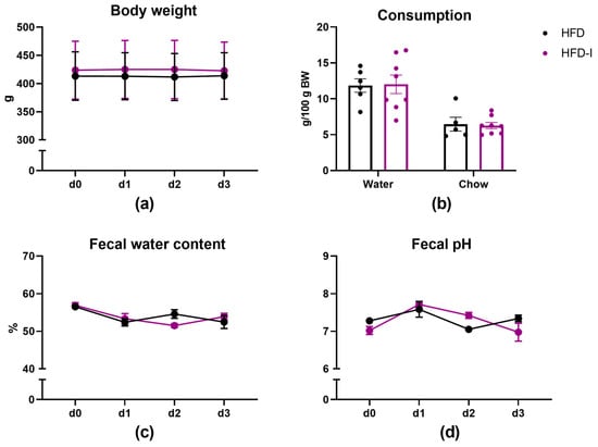
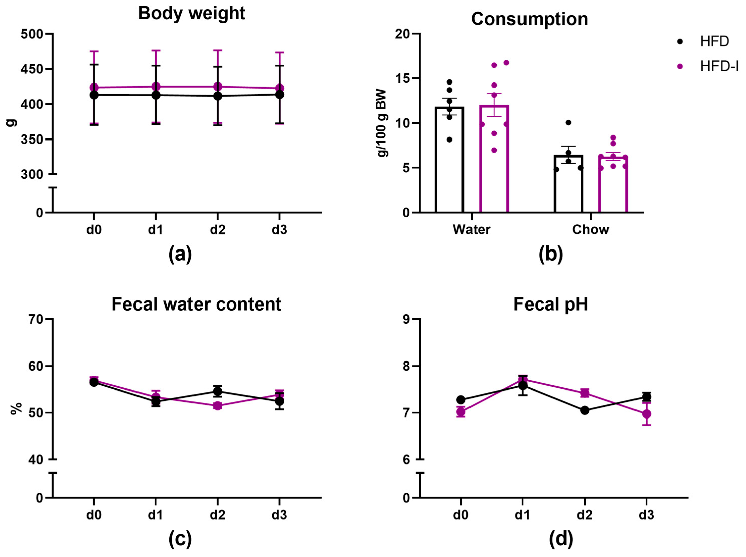
Body weight, food and water intake, and fecal pH and water content were not altered 3 days following inoculation in HFD-fed animals (Figure 6), indicating that the challenge remained clinically mild in the context of obesity and similar to chow-fed rats (Figure 2). Relative organ weights are summarized in Supplementary Table S10. Inoculation was associated with an increase in spleen (inoculation effect: p = 0.02; sex effect: p = 0.45; interaction effect: p = 0.719) and thymus (inoculation effect: p = 0.007; sex effect: p = 0.043; interaction effect: p = 0.300) relative weight.
Figure 6.
Variables assessed during the three-day course of S. Typhimurium challenge in HFD-fed rats. (a) Body weight. (b) Food and water consumption (d2–d3). (c) Fecal water content. (d) Fecal pH. After 12 weeks of HFD feeding, rats were either left non-inoculated (HFD, n = 6) or inoculated with 108 CFU on day 1 (HFD-I, n = 8) and monitored for 72 h. Data are presented as mean ± SEM.
3.2.2. Blood Cell Composition in HFD-Fed Animals
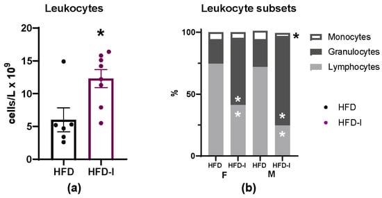
In HFD-fed rats, S. Typhimurium challenge increased total leukocyte counts (Figure 7a; inoculation effect: p = 0.04; sex effect: p = 0.08; interaction effect: p = 0.72). This increase was accompanied by a shift in the proportions of granulocytes (inoculation effect: p < 0.001; sex effect: p = 0.003; interaction effect: p = 0.017), lymphocytes (inoculation effect: p < 0.001; sex effect: p = 0.008; interaction effect: p = 0.039), and monocytes (inoculation effect: p = 0.001; sex effect: p = 0.294; interaction effect: p = 0.013; Figure 7b). The reduction in lymphocyte proportion following inoculation was 33.38 percentage points in females (95% CI: 25.14–41.63) and 47.10 percentage points in males (95% CI: 37.25–56.96); in granulocyte proportion, the increase was 34.40 percentage points in females (95% CI: 26.50–42.30) and 49.83 percentage points in males (95% CI: 40.87–58.78). The remaining hemogram variables are provided in Supplementary Table S11.
Figure 7.
Blood leukocyte counts and subset distribution in HFD-fed rats following S. Typhimurium inoculation. (a) Total leukocyte counts. (b) Relative proportions of leukocyte subsets. After 12 weeks of HFD feeding, rats were either left non-inoculated (HFD, n = 6) or inoculated with 108 CFU on day 1 (HFD-I, n = 8) and analyzed 72 h post-inoculation. Data are presented as mean ± SEM. * p < 0.05 vs. HFD.
3.2.3. Plasma Ig Levels in HFD-Fed Animals
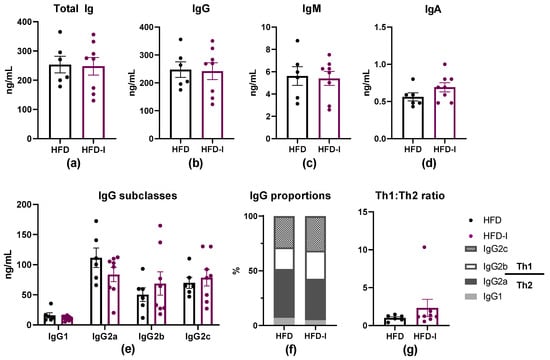
In contrast to the increase in certain plasmatic Ig isotypes due to oral challenge observed in standard chow-fed animals, plasma Ig levels were not significantly affected by sex or inoculation in HFD-fed rats (Figure 8). In particular, levels of total Ig and Ig isotypes (IgG, IgM and IgA) remained the same three days following inoculation (Figure 8a,b). Likewise, there were no changes detected in IgG isotypes, either in absolute levels or in proportions, and correspondingly, the Th1/Th2 IgG ratio did not change 72 h post-inoculation (Figure 8c–e).
Figure 8.
Plasma Ig levels in HFD-fed rats following S. Typhimurium inoculation. (a) Total Ig, (b) IgG, (c) IgM, (d) IgA, and (e) IgG isotype levels. (f) IgG subclass proportions. (g) IgG Th1/Th2 ratio (IgG2b and IgG2c as Th1-associated; IgG1 and IgG2a as Th2-associated). After 12 weeks of HFD feeding, rats were either left non-inoculated (HFD, n = 6) or inoculated with 108 CFU on day 1 (HFD-I, n = 8) and analyzed 72 h post-infection. Data are presented as mean ± SEM.
3.2.4. Systemic Cytokine Response in HFD-Fed Animals
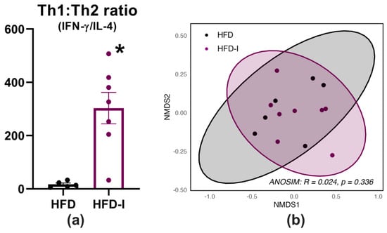
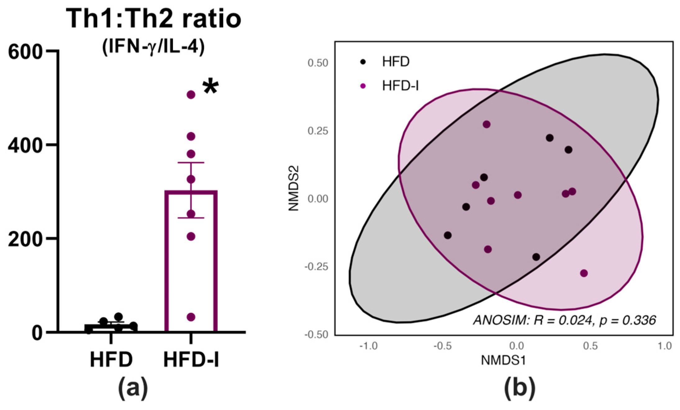
Plasma cytokine concentrations are detailed in Supplementary Table S12. Out of the 15 cytokines analyzed, GM-CSF, IL-1α, IL-5, and IL-6 were below detection limits in most samples and were excluded from further analysis. While there was no observed effect of the inoculation for any individual cytokine, the Th1/Th2 cytokine ratio, calculated as the IFN-γ/IL-4 ratio, was elevated in inoculated rats (inoculation effect: p = 0.025; sex effect: p = 0.942; interaction effect: p = 0.321; Figure 9a). Nevertheless, NMDS ordination revealed overlapping clusters between plasma cytokine profiles of HFD and HFD-inoculated animals, indicating that oral challenge did not induce a clearly distinct systemic cytokine signature 72 h post-inoculation.
Figure 9.
Plasma cytokines in HFD-fed rats following S. Typhimurium inoculation. (a) Plasma Th1/Th2-associated cytokine ratio. (b) NMDS ordination based on plasma cytokine concentrations. After 12 weeks of HFD feeding, rats were either left non-inoculated (HFD, n = 6) or inoculated with 108 CFU on day 1 (HFD-I, n = 8) and analyzed 72 h post-inoculation. Data are presented as mean ± SEM. * p < 0.05 vs. HFD.
3.2.5. Intestinal Response in HFD-Fed Animals
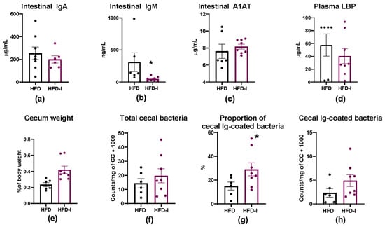
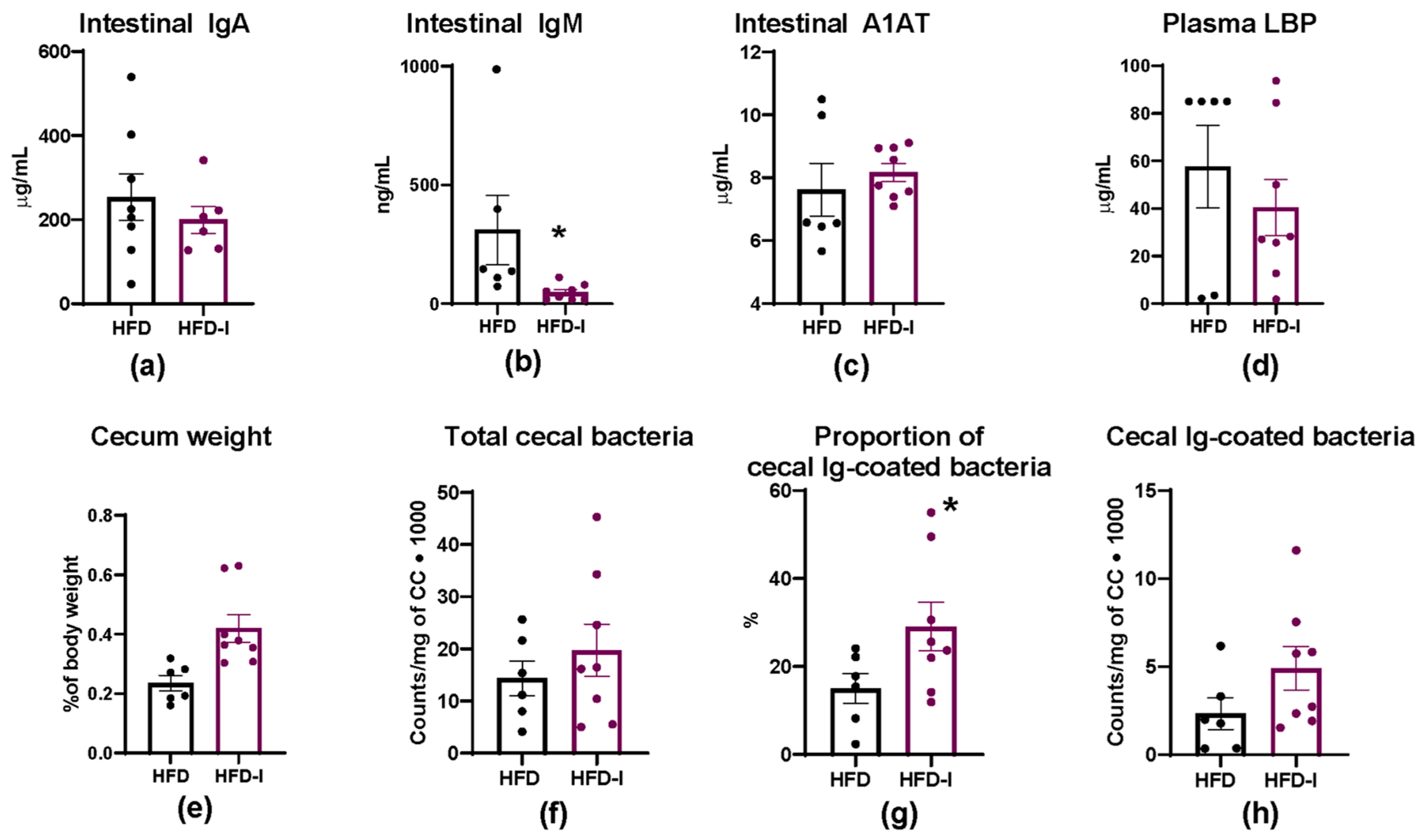
In HFD-fed animals, intestinal IgA levels measured in GW revealed no differences upon oral challenge (Figure 10a). In contrast, intestinal IgM levels were reduced in HFD-inoculated animals (inoculation effect: p = 0.036; sex effect: p = 0.090; interaction: p = 0.096; Figure 10b). A1AT concentrations in GW and plasma LBP concentrations were not affected by oral challenge, regardless of sex (Figure 10c,d).
Figure 10.
Intestinal immune alterations in HFD-fed rats following S. Typhimurium infection. (a) Intestinal IgA, (b) IgM, and (c) A1AT. (d) Plasma LBP. (e) Relative cecum weight. (f) Total cecal bacterial load. (g) Proportion of cecal Ig-coated bacteria. (h) Cecal Ig-coated bacteria. After 12 weeks of HFD feeding, rats were either left non-inoculated (HFD, n = 6) or inoculated with 108 CFU on day 1 (HFD-I, n = 8) and analyzed 72 h post-inoculation. Data are presented as mean ± SEM. * p < 0.05 vs. HFD.
There was no significant effect on cecum weight (Figure 10e), and total cecal bacterial load as well as the absolute number of Ig-coated bacteria did not differ between groups (Figure 10f,h). However, the proportion of bacteria bound to Ig was increased in inoculated animals (inoculation effect: p = 0.024; sex effect: p = 0.017; interaction effect: p = 0.213; Figure 10g).
4. Discussion
The present study established an acute, clinically mild Salmonella challenge model in standard-diet-fed animals that induced immunological alterations in the absence of clinical disease. The selected challenge dose was subsequently applied in an independent cohort of HFD-fed rats to characterize early systemic and mucosal immune responses under this dietary condition, including exploratory assessment of sex-associated response patterns. In both experimental settings, the challenge was clinically mild but was associated with changes in several immune parameters, demonstrating that measurable immunological response can occur in the absence of overt clinical disease.
In standard-diet-fed animals, the absence of clinical signs or diarrhea upon challenge with three selected doses of S. Typhimurium indicates that immune activation can be evaluated without extensive pathology, which makes the model particularly suitable to study subtle immunomodulatory effects of, for instance, future preventive dietary strategies. This is consistent with previous work showing that oral Salmonella exposure can induce immunological and histopathological changes in the absence of manifest disease, particularly at moderate bacterial doses [25]. At the intestinal level, A1AT and LBP concentrations remained unchanged following challenge, supporting the non-destructive nature of the model; however, inoculation induced intestinal and systemic immune activation. Increased relative cecum weight observed after oral inoculation likely reflects localized intestinal adaptation to Salmonella challenge. The cecum is a major site of Salmonella interaction in enteric infection, and experimental models have shown that infection can induce localized edema and structural remodeling in this tissue even without apparent disease [30,31]. In the present model, the increased cecum weight may therefore represent an early local response to the challenge.
In parallel, the tendency toward higher total cecal bacterial abundance and the increase in cecal Ig-coated bacteria suggest changes in host–microbiota immune interactions following challenge. Although this parameter has rarely been evaluated in Salmonella models, studies in other intestinal inflammatory settings have similarly associated increased bacterial Ig coating with active mucosal immune engagement, supporting its relevance as a marker of mucosal immune adaptation [32,33,34].
At the systemic level, the challenge induced a shift toward granulocyte predominance, reflecting innate immune activation just three days post-inoculation. Neutrophil recruitment represents a central host defense mechanism that contributes to limiting pathogen dissemination from the intestinal mucosa [22,23,35]. Similar neutrophil-dominated responses have been described in experimental Salmonella infection rodent models, even under clinically mild conditions [25]. In the present study, Salmonella challenge increased CD8+ T cell proportions in MLNs, consistent with the established role of cytotoxic lymphocyte populations in early anti-Salmonella immune defense [36]. Moreover, the challenge was associated with an increase in the proportion of the CD62L+αE+ B cell subset. In gut-associated immune responses, αE is linked to mucosal localization, whereas CD62L supports trafficking through secondary lymphoid organs such as MLNs [37,38]. Although the functional significance of CD62L+ αE+ B cells is not well defined, their expansion may reflect recently activated B cells acquiring mucosal homing characteristics.
The humoral response in standard-diet-fed animals was characterized by increased total Ig and IgM levels in plasma, together with a higher proportion of Th1-associated IgG isotypes. Given the 72 h time point and the measurement of total as opposed to Salmonella-specific Igs, these changes should not be interpreted as evidence of de novo pathogen-specific class switching or a fully developed Th1-polarized adaptive response. Instead, they reflect early changes in the systemic humoral immune profile following challenge. Natural IgM is known to contribute to early Salmonella control by promoting polyreactive bacterial binding and limiting dissemination [39,40]. In contrast, systemic cytokine alterations remained modest. Several cytokines were below the assay detection limit in most samples; however, circulating cytokines are inherently difficult to quantify due to their low concentrations, transient secretion, and short half-lives, and non-detection therefore does not imply biological absence [41]. This is in line with previous observations that mild enteric Salmonella infection often induces predominantly localized immune activation without systemic cytokine increase [42].
Together, these findings indicate that standard-diet-fed animals mounted an innate, cellular, and humoral response while maintaining a clinically mild phenotype, validating the highest inoculation dose as an appropriate model for investigating host immune regulation 72 h post-inoculation. The subsequent question was how a cohort of HFD-fed animals would respond to this selected dose.
Although Salmonella challenge remained clinically mild in HFD-fed animals, unlike in severe infection models where obesity increases mortality and bacterial burden [43], changes in circulating leukocytes were evident: Total leukocyte counts increased following challenge, accompanied by a higher proportion of granulocytes and a corresponding lower proportion of lymphocytes. This pattern differed from that observed in the standard-diet-fed cohort, in which a comparable increase in total leukocyte counts was not produced. Previous mouse studies have similarly reported that HFD-induced obesity altered early immune responses during oral Salmonella infection without increasing bacterial burden, indicating that obesity primarily reshapes immune regulation rather than worsening infection severity [42]. In both humans and animal models, circulating neutrophil counts are elevated in obesity and correlate with markers of metabolic dysfunction and systemic inflammation [44,45,46,47]. In the context of bacterial challenge, such pre-existing alterations in the inflammatory environment may influence the coordination of early host immune responses.
By contrast, the observed changes in leukocyte populations following challenge in HFD-fed animals were not accompanied by changes in plasma Ig levels and were instead associated with a reduction in intestinal IgM as well as an increase in the proportion of Ig-coated bacteria. Previous studies have shown that HFD can enhance inflammatory activation while impairing antimicrobial immune function, indicating that increased immune cell abundance does not necessarily translate into effective host defense [43]. Experimental evidence further suggests that HFDs compromise mucosal humoral immunity. In particular, HFD has been associated with reduced numbers of Ig-producing cells in the intestinal lamina propria, accompanied by decreased IgA, IgM, and IgG1 immunoreactivity and altered spatial distribution of these cells along the villi [48]. Given the central role of secreted Igs in bacterial coating and immune exclusion, these findings suggest altered mucosal immune regulation during bacterial challenge in HFD-fed animals; however, the functional consequences for bacterial control remain to be determined.
The Th1/Th2 cytokine ratio was elevated in HFD-fed animals, which was not observed in the standard chow-fed cohort. Importantly, the Th1/Th2 ratio should be interpreted with caution, as IL-4 concentrations were below the assay detection limit in several samples and substituted values were required for its calculation. Moreover, these changes were not reflected in a distinct systemic cytokine signature in HFD-fed animals 72 h post-inoculation. Comparable mixed or poorly coordinated cytokine responses have been described in obese infection models, where chronic low-grade inflammation may blunt the capacity to mount well-regulated acute responses [42,43]. Nevertheless, the contrasting patterns observed in this study raise the possibility that the HFD-fed condition contributed to this response, but this cannot be established from the present independent cohorts.
Notably, these HFD-induced immune alterations were further modified by sex of the animals, with males showing higher total circulating leukocytes, a geater proportion of granulocytes, and a lower proportion of lymphocytes. Males are generally more susceptible to metabolic disturbances under HFD conditions, whereas females often show partial protection [15,49,50]. The observations from the present study are consistent with previous reports showing that HFD induces sex-dependent differences in intestinal barrier function, inflammatory responses, and microbiota composition [13,51], supporting the concept that immune regulation is differentially affected by metabolic stress in males and females. A few limitations should be considered when interpreting the findings. As the model was intentionally designed to remain clinically mild and animals were evaluated at an early time point (72 h post-inoculation), the findings primarily reflect early immune responses to the challenge. Consequently, although the observed alterations demonstrate that HFD feeding and biological sex modulate the early immune response to S. Typhimurium, the present study was not designed to determine whether these changes ultimately translate into enhanced host protection, impaired bacterial clearance, or increased disease susceptibility. The inclusion of intestinal histopathological analyses and additional functional outcomes, such as bacterial dissemination or longer-term disease progression, would provide complementary information on the biological significance of the observed immune alterations. In addition, the relatively modest sample size may have limited the statistical power to detect subtle differences in some immune parameters. At the same time, the number of immune endpoints assessed increases the potential for multiple-comparison effects; therefore, particularly sex-stratified findings should be considered exploratory. Furthermore, as the standard-diet and HFD experiments were conducted in independent cohorts, direct comparisons between conditions were not possible. Finally, the AIN-93M (REF) and TD.06415 (HFD) diets differ in several nutritional components beyond fat content, which should be considered when interpreting the findings.
5. Conclusions
An acute, clinically mild Salmonella Typhimurium challenge model was established in rats, enabling the investigation of early host immune responses under conditions in which clinical disease is not manifest. The challenge was associated with systemic and mucosal immune changes in standard-diet-fed rats, while application of the selected dose in an independent HFD-fed cohort revealed changes in circulating leukocyte populations and mucosal humoral parameters. Exploratory analyses further suggest sex-associated variation in circulating leukocyte subsets. These findings should be interpreted in the context of the independent experimental cohorts, modest sample sizes, a single 72 h endpoint, and the absence of systematic Salmonella-specific dissemination, histopathological, and pathogen-specific serological assessments. Overall, this model provides a basis for further studies investigating how nutritional and host-related factors influence early responses to enteric bacterial challenge.
Supplementary Materials
The following supporting information can be downloaded at https://www.mdpi.com/article/10.3390/diseases14110422/s1. Figure S1. Experimental design. Created in Biorender, Ruth Forsten, 2026. Table S1. Composition of experimental diets; Table S2. Upper limit of detection (ULOD) and lower limit of detection (LLOD) of the ProcartaPlexTM Rat Th Complete Panel; Table S3. Antibody (Ab) panels for mesenteric lymph node (MLN) lymphocyte staining; Table S4. Morphometric variables and relative organ weight 72 h after inoculation with S. Typhimurium; Table S5. Hematological variables 72 h after inoculation with S. Typhimurium; Table S6. Mesenteric lymph node (MLN) lymphocyte populations 72 h after inoculation with S. Typhimurium; Table S7. Mesenteric lymph node (MLN) homing marker expression 72 h after inoculation with S. Typhimurium; Table S8. Plasma cytokine concentrations and detection frequencies 72 h after inoculation with S. Typhimurium; Table S9. Baseline metabolic and anthropometric characteristics of HFD-fed rats prior to inoculation; Table S10. Relative organ weights and morphometric parameters in HFD-fed rats 72 h after inoculation with S. Typhimurium; Table S11. Hematological parameters in HFD-fed rats 72 h after inoculation with S. Typhimurium; Table S12. Plasma cytokine concentrations and detection frequencies in HFD-fed rats 72 h after inoculation with S. Typhimurium.
Author Contributions
Conceptualization, F.J.P.-C., M.J.R.-L., S.M. and A.C.; formal analysis, R.F. and S.G.-L.H.; investigation, R.F., S.G.-L.H. and K.R.-A.; data curation, R.F., S.G.-L.H. and K.R.-A.; writing—original draft preparation, R.F.; writing—review and editing, S.G.-L.H., K.R.-A., S.M., A.C., M.C., M.M.-C., M.J.R.-L. and F.J.P.-C.; visualization, R.F.; supervision, M.J.R.-L. and F.J.P.-C.; project administration, M.J.R.-L.; funding acquisition, M.J.R.-L. All authors have read and agreed to the published version of the manuscript.
Funding
This research was funded by the Spanish State Research Agency (AEI), grant number PCI2022-135038-2, under the Joint Programming Initiative “A Healthy Diet for a Healthy Life” (JPI HDHL) call and within the framework of the European Union NextGenerationEU/PRTR program. R.F. received the support of a fellowship from the “la Caixa” Foundation (ID 100010434), fellowship code LCF/BQ/DI23/11990051. S.M. acknowledges funding from Science Foundation Ireland (SFI) (grant number SFI/12/RC/2273_P2).
Institutional Review Board Statement
This animal study protocol was approved by the Ethics Committee for Animal Experimentation of the University of Barcelona and the Government of Catalonia (CEEA-UB, Ref. 275/23, approved on 21 November 2023).
Informed Consent Statement
Not applicable.
Data Availability Statement
The datasets generated and/or analyzed during the current study are available from the corresponding author upon reasonable request. The data are not publicly available due to ongoing research.
Acknowledgments
The authors thank the INSA-UB Maria de Maeztu Unit of Excellence (Grant CEX2021-001234-M). The authors acknowledge the members of the “Autoimmunity, Immunonutrition and Tolerance” research group, particularly Jone Ortuño and Joan Buxeda, for their technical assistance.
Conflicts of Interest
The authors declare no conflicts of interest. The funders had no role in the design of the study; in the collection, analyses, or interpretation of data; in the writing of the manuscript; or in the decision to publish the results.
Abbreviations
The following abbreviations are used in this manuscript:
| A1AT | Alpha-1-antitrypsin |
| Ab | Antibody |
| ANOSIM | Analysis of similarities |
| ANOVA | Analysis of variance |
| CCiT-UB | Scientific and Technological Centers of the University of Barcelona |
| CFU | Colony-forming unit |
| ELISA | Enzyme-linked immunosorbent assay |
| FBS | Fetal bovine serum |
| FITC | Fluorescein isothiocyanate |
| G-CSF | Granulocyte colony-stimulating factor |
| GM-CSF | Granulocyte-macrophage colony-stimulating factor |
| GW | Gut wash |
| HFD | High-fat diet |
| IFN | Interferon |
| Ig | Immunoglobulin |
| IL | Interleukin |
| LBP | Lipopolysaccharide-binding protein |
| MCP | Monocyte chemoattractant protein |
| MCH | Mean corpuscular hemoglobin |
| MCV | Mean corpuscular volume |
| MLN | Mesenteric lymph nodes |
| NMDS | Non-metric multidimensional scaling |
| OGTT | Oral glucose tolerance test |
| PBS | Phosphate-buffered saline |
| PI | Propidium iodine |
| RPMI | Roswell Park Memorial Institute |
| SEM | Standard error of the mean |
| Th | T helper |
| TNF | Tumor necrosis factor |
| TS | Tryptone Soya |
| UB | University of Barcelona |
References
- Alberts, B.; Johnson, A.; Lewis, J.; Raff, M.; Roberts, K.; Walter, P. Introduction to Pathogens. In Molecular Biology of the Cell, 4th ed.; Garland Science: New York, NY, USA, 2002. [Google Scholar]
- Muscogiuri, G.; Pugliese, G.; Laudisio, D.; Castellucci, B.; Barrea, L.; Savastano, S.; Colao, A. The Impact of Obesity on Immune Response to Infection: Plausible Mechanisms and Outcomes. Obes. Rev. 2021, 22, e13216. [Google Scholar] [CrossRef] [Scilit] [PubMed]
- World Health Organization Obesity and Overweight. Available online: https://www.who.int/news-room/fact-sheets/detail/obesity-and-overweight (accessed on 11 May 2026).
- Khanna, D.; Khanna, S.; Khanna, P.; Kahar, P.; Patel, B.M. Obesity: A Chronic Low-Grade Inflammation and Its Markers. Cureus 2022, 14, e22711. [Google Scholar] [CrossRef] [Scilit] [PubMed]
- Kolb, H. Obese Visceral Fat Tissue Inflammation: From Protective to Detrimental? BMC Med. 2022, 20, 494. [Google Scholar] [CrossRef] [Scilit] [PubMed]
- Zatterale, F.; Longo, M.; Naderi, J.; Raciti, G.A.; Desiderio, A.; Miele, C.; Beguinot, F. Chronic Adipose Tissue Inflammation Linking Obesity to Insulin Resistance and Type 2 Diabetes. Front. Physiol. 2020, 10, 1607. [Google Scholar] [CrossRef] [Scilit] [PubMed]
- McLaughlin, T.; Ackerman, S.E.; Shen, L.; Engleman, E. Role of Innate and Adaptive Immunity in Obesity-Associated Metabolic Disease. J. Clin. Investig. 2017, 5, 5–13. [Google Scholar] [CrossRef] [Scilit] [PubMed]
- Blaszczak, A.M.; Jalilvand, A.; Hsueh, W.A. Adipocytes, Innate Immunity and Obesity: A Mini-Review. Front. Immunol. 2021, 12, 650768. [Google Scholar] [CrossRef] [Scilit] [PubMed]
- Michailidou, Z.; Gomez-Salazar, M.; Alexaki, V.I. Innate Immune Cells in the Adipose Tissue in Health and Metabolic Disease. J. Innate Immun. 2022, 14, 4–30. [Google Scholar] [CrossRef] [Scilit] [PubMed]
- Taylor, E.B. The Complex Role of Adipokines in Obesity, Inflammation, and Autoimmunity. Clin. Sci. 2021, 135, 731–752. [Google Scholar] [CrossRef] [Scilit] [PubMed]
- Tagliabue, C.; Principi, N.; Giavoli, C.; Esposito, S. Obesity: Impact of Infections and Response to Vaccines. Eur. J. Clin. Microbiol. Infect. Dis. 2016, 35, 325–331. [Google Scholar] [CrossRef] [Scilit] [PubMed]
- Karlsson, E.A.; Sheridan, P.A.; Beck, M.A. Diet-Induced Obesity Impairs the T Cell Memory Response to Influenza Virus Infection. J. Immunol. 2010, 184, 3127–3133. [Google Scholar] [CrossRef] [Scilit] [PubMed]
- Braga Tibaes, J.R.; Azarcoya-Barrera, J.; Wollin, B.; Veida-Silva, H.; Makarowski, A.; Vine, D.; Tsai, S.; Jacobs, R.; Richard, C. Sex Differences Distinctly Impact High-Fat Diet-Induced Immune Dysfunction in Wistar Rats. J. Nutr. 2022, 152, 1347–1357. [Google Scholar] [CrossRef] [Scilit] [PubMed]
- Fischer, J.; Jung, N.; Robinson, N.; Lehmann, C. Sex Differences in Immune Responses to Infectious Diseases. Infection 2015, 43, 399–403. [Google Scholar] [CrossRef] [Scilit] [PubMed]
- Braga Tibaes, J.R.; Barreto Silva, M.I.; Wollin, B.; Vine, D.; Tsai, S.; Richard, C. Sex Differences in Systemic Inflammation and Immune Function in Diet-Induced Obesity Rodent Models: A Systematic Review. Obes. Rev. 2023, 25, e13665. [Google Scholar] [CrossRef] [Scilit] [PubMed]
- Nazir, J.; Manzoor, T.; Saleem, A.; Gani, U.; Bhat, S.S.; Khan, S.; Haq, Z.; Jha, P.; Ahmad, S.M. Combatting Salmonella: A Focus on Antimicrobial Resistance and the Need for Effective Vaccination. BMC Infect. Dis. 2025, 25, 84. [Google Scholar] [CrossRef] [Scilit] [PubMed]
- Fàbrega, A.; Vila, J. Salmonella enterica Serovar Typhimurium Skills to Succeed in the Host: Virulence and Regulation. Clin. Microbiol. Rev. 2013, 26, 308–341. [Google Scholar] [CrossRef] [Scilit] [PubMed]
- Jiang, L.; Wang, P.; Song, X.; Zhang, H.; Ma, S.; Wang, J.; Li, W.; Lv, R.; Liu, X.; Ma, S.; et al. Salmonella typhimurium Reprograms Macrophage Metabolism via T3SS Effector SopE2 to Promote Intracellular Replication and Virulence. Nat. Commun. 2021, 12, 879. [Google Scholar] [CrossRef] [Scilit] [PubMed]
- Wang, Y.; Wu, C.; Gao, J.; Du, X.; Chen, X.; Zhang, M. Host Metabolic Shift during Systemic Salmonella Infection Revealed by Comparative Proteomics. Emerg. Microbes Infect. 2021, 10, 1849–1861. [Google Scholar] [CrossRef] [Scilit] [PubMed]
- Zuo, L.; Zhou, L.; Wu, C.; Wang, Y.; Li, Y.; Huang, R.; Wu, S. Salmonella spvC Gene Inhibits Pyroptosis and Intestinal Inflammation to Aggravate Systemic Infection in Mice. Front. Microbiol. 2020, 11, 562491. [Google Scholar] [CrossRef] [Scilit] [PubMed]
- Salazar, G.A.; Peñaloza, H.F.; Pardo-Roa, C.; Schultz, B.M.; Muñoz-Durango, N.; Gómez, R.S.; Salazar, F.J.; Pizarro, D.P.; Riedel, C.A.; González, P.A.; et al. Interleukin-10 Production by T and B Cells Is a Key Factor to Promote Systemic Salmonella enterica Serovar Typhimurium Infection in Mice. Front. Immunol. 2017, 8, 889. [Google Scholar] [CrossRef] [Scilit] [PubMed]
- Broz, P.; Ohlson, M.B.; Monack, D.M. Innate Immune Response to Salmonella typhimurium, a Model Enteric Pathogen. Gut Microbes 2012, 3, 62–70. [Google Scholar] [CrossRef] [Scilit] [PubMed]
- Patel, S.; McCormick, B.A. Mucosal Inflammatory Response to Salmonella typhimurium Infection. Front. Immunol. 2014, 5, 87909. [Google Scholar] [CrossRef] [Scilit] [PubMed]
- Huang, F.C. The Interleukins Orchestrate Mucosal Immune Responses to Salmonella Infection in the Intestine. Cells 2021, 10, 3492. [Google Scholar] [CrossRef] [Scilit] [PubMed]
- Havelaar, A.H.; Garssen, J.; Takumi, K.; Koedam, M.A.; Dufrenne, J.B.; Van Leusden, F.M.; De La Fonteyne, L.; Bousema, J.T.; Vos, J.G. A Rat Model for Dose–Response Relationships of Salmonella enteritidis Infection. J. Appl. Microbiol. 2001, 91, 442–452. [Google Scholar] [CrossRef] [Scilit] [PubMed]
- Walker, G.T.; Gerner, R.R.; Nuccio, S.P.; Raffatellu, M. Murine Models of Salmonella Infection. Curr. Protoc. 2023, 3, e824. [Google Scholar] [CrossRef] [Scilit] [PubMed]
- Ten Bruggencate, S.J.M.; Snel, J.; Schoterman, M.H.C.; Ertmann, E.; Van Der Meulen, E.; Schonewille, A.; Bovee-Oudenhoven, I.M.J. Efficacy of Various Dietary Calcium Salts to Improve Intestinal Resistance to Salmonella Infection in Rats. Br. J. Nutr. 2011, 105, 489–495. [Google Scholar] [CrossRef] [Scilit] [PubMed]
- Ramiro-Puig, E.; Pérez-Cano, F.J.; Ramos-Romero, S.; Pérez-Berezo, T.; Castellote, C.; Permanyer, J.; Franch, À.; Izquierdo-Pulido, M.; Castell, M. Intestinal Immune System of Young Rats Influenced by Cocoa-Enriched Diet. J. Nutr. Biochem. 2008, 19, 555–565. [Google Scholar] [CrossRef] [Scilit] [PubMed]
- Cétre, C.; Pierrot, C.; Cocude, C.; Lafitte, S.; Capron, A.; Capron, M.; Khalife, J. Profiles of Th1 and Th2 Cytokines after Primary and Secondary Infection by Schistosoma Mansoni in the Semipermissive Rat Host. Infect. Immun. 1999, 67, 2713. [Google Scholar] [CrossRef] [Scilit] [PubMed]
- Lunnemann, H.M.; Shealy, N.G.; Reyzer, M.L.; Shupe, J.A.; Green, E.H.; Siddiqi, U.; Borden Lacy, D.; Byndloss, M.X.; Markham, N.O. Cecum Axis (CecAx) Preservation Reveals Physiological and Pathological Gradients in Mouse Gastrointestinal Epithelium. Gut Microbes 2023, 15, 2185029. [Google Scholar] [CrossRef] [Scilit] [PubMed]
- Cruz-Cruz, A.; Schreeg, M.E.; Gunn, J.S. A Temporary Cholesterol-Rich Diet and Bacterial Extracellular Matrix Factors Favor Salmonella spp. Biofilm Formation in the Cecum. mBio 2025, 16, e03242-24. [Google Scholar] [CrossRef] [Scilit] [PubMed]
- Lin, R.; Chen, H.; Shu, W.; Sun, M.; Fang, L.; Shi, Y.; Pang, Z.; Wu, W.; Liu, Z. Clinical Significance of Soluble Immunoglobulins A and G and Their Coated Bacteria in Feces of Patients with Inflammatory Bowel Disease. J. Transl. Med. 2018, 16, 359. [Google Scholar] [CrossRef] [Scilit] [PubMed]
- Maccio-Maretto, L.; Piqueras, V.; Barrios, B.E.; Romagnoli, P.A.; Denning, T.L.; Correa, S.G. Luminal Bacteria Coated with IgA and IgG during Intestinal Inflammation as a New and Abundant Stimulus for Colonic Macrophages. Immunology 2022, 167, 64–76. [Google Scholar] [CrossRef] [Scilit] [PubMed]
- Masu, Y.; Kanazawa, Y.; Kakuta, Y.; Shimoyama, Y.; Onodera, M.; Naito, T.; Moroi, R.; Kuroha, M.; Kimura, T.; Shiga, H.; et al. Immunoglobulin Subtype-Coated Bacteria Are Correlated with the Disease Activity of Inflammatory Bowel Disease. Sci. Rep. 2021, 11, 16672. [Google Scholar] [CrossRef] [Scilit] [PubMed]
- Wick, M.J. Innate Immune Control of Salmonella enterica Serovar Typhimurium: Mechanisms Contributing to Combating Systemic Salmonella Infection. J. Innate Immun. 2011, 3, 543–549. [Google Scholar] [CrossRef] [Scilit] [PubMed]
- Mittrücker, H.W.; Köhler, A.; Kaufmann, S.H.E. Characterization of the Murine T-Lymphocyte Response to Salmonella enterica Serovar Typhimurium Infection. Infect. Immun. 2002, 70, 199–203. [Google Scholar] [CrossRef] [Scilit] [PubMed]
- Schulz, O.; Jaensson, E.; Persson, E.K.; Liu, X.; Worbs, T.; Agace, W.W.; Pabst, O. Intestinal CD103+, but Not CX3CR1+, Antigen Sampling Cells Migrate in Lymph and Serve Classical Dendritic Cell Functions. J. Exp. Med. 2009, 206, 3101–3114. [Google Scholar] [CrossRef] [Scilit] [PubMed]
- von Andrian, U.H.; Mackay, C.R. T-Cell Function and Migration—Two Sides of the Same Coin. N. Engl. J. Med. 2000, 343, 1020–1034. [Google Scholar] [CrossRef] [Scilit] [PubMed]
- Alugupalli, A.S.; Cravens, M.P.; Walker, J.A.; Gulandijany, D.; Dickinson, G.S.; Debes, G.F.; Schifferli, D.M.; Bäumler, A.J.; Alugupalli, K.R. The Lack of Natural IgM Increases Susceptibility and Impairs Anti-Vi Polysaccharide IgG Responses in a Mouse Model of Typhoid. Immunohorizons 2023, 6, 807–816. [Google Scholar] [CrossRef] [Scilit] [PubMed]
- Bioley, G.; Monnerat, J.; Lötscher, M.; Vonarburg, C.; Zuercher, A.; Corthésy, B. Plasma-Derived Polyreactive Secretory-like Iga and igM Opsonizing Salmonella enterica Typhimurium Reduces Invasion and Gut Tissue Inflammation through Agglutination. Front. Immunol. 2017, 8, 272003. [Google Scholar] [CrossRef] [Scilit] [PubMed]
- Liu, C.; Chu, D.; Kalantar-Zadeh, K.; George, J.; Young, H.A.; Liu, G. Cytokines: From Clinical Significance to Quantification. Adv. Sci. 2021, 8, 2004433. [Google Scholar] [CrossRef] [Scilit] [PubMed]
- Ramírez-Orozco, R.E.; Franco Robles, E.; Pérez Vázquez, V.; Ramírez Emiliano, J.; Hernández Luna, M.A.; López Briones, S. Diet-Induced Obese Mice Exhibit Altered Immune Responses to Early Salmonella typhimurium Oral Infection. J. Microbiol. 2018, 56, 673–682. [Google Scholar] [CrossRef] [Scilit] [PubMed]
- Strandberg, L.; Verdrengh, M.; Enge, M.; Andersson, N.; Amu, S.; Önnheim, K.; Benrick, A.; Brisslert, M.; Bylund, J.; Bokarewa, M.; et al. Mice Chronically Fed High-Fat Diet Have Increased Mortality and Disturbed Immune Response in Sepsis. PLoS ONE 2009, 4, e7605. [Google Scholar] [CrossRef] [Scilit] [PubMed]
- Feješ, A.; Šebeková, K.; Borbélyová, V. Pathophysiological Role of Neutrophil Extracellular Traps in Diet-Induced Obesity and Metabolic Syndrome in Animal Models. Nutrients 2025, 17, 241. [Google Scholar] [CrossRef] [Scilit] [PubMed]
- Gomez-Casado, G.; Jimenez-Gonzalez, A.; Rodriguez-Muñoz, A.; Tinahones, F.J.; González-Mesa, E.; Murri, M.; Ortega-Gomez, A. Neutrophils as Indicators of Obesity-Associated Inflammation: A Systematic Review and Meta-Analysis. Obes. Rev. 2025, 26, e13868. [Google Scholar] [CrossRef] [Scilit] [PubMed]
- Zaldivar, F.; McMurray, R.G.; Nemet, D.; Galassetti, P.; Mills, P.J.; Cooper, D.M. Body Fat and Circulating Leukocytes in Children. Int. J. Obes. 2006, 30, 906–911. [Google Scholar] [CrossRef] [Scilit] [PubMed]
- Ryder, E.; Diez-Ewald, M.; Mosquera, J.; Fernández, E.; Pedreañez, A.; Vargas, R.; Peña, C.; Fernández, N. Association of Obesity with Leukocyte Count in Obese Individuals without Metabolic Syndrome. Diabetes Metab. Syndr. Clin. Res. Rev. 2014, 8, 197–204. [Google Scholar] [CrossRef] [Scilit] [PubMed]
- Sakamoto, Y.; Niwa, M.; Muramatsu, K.; Shimo, S. High-Fat Diet and Age-Dependent Effects of IgA-Bearing Cell Populations in the Small Intestinal Lamina Propria in Mice. Int. J. Mol. Sci. 2021, 22, 1165. [Google Scholar] [CrossRef] [Scilit] [PubMed]
- Mo, Q.; Deng, X.; Zhou, Z.; Yin, L. High-Fat Diet and Metabolic Diseases: A Comparative Analysis of Sex-Dependent Responses and Mechanisms. Int. J. Mol. Sci. 2025, 26, 4777. [Google Scholar] [CrossRef] [Scilit] [PubMed]
- Sankaran-Walters, S.; Macal, M.; Grishina, I.; Nagy, L.; Goulart, L.; Coolidge, K.; Li, J.; Fenton, A.; Williams, T.; Miller, M.K.; et al. Sex Differences Matter in the Gut: Effect on Mucosal Immune Activation and Inflammation. Biol. Sex. Differ. 2013, 4, 10. [Google Scholar] [CrossRef] [Scilit] [PubMed]
- Lefebvre, C.; Tiffay, A.; Breemeersch, C.E.; Dreux, V.; Bôle-Feysot, C.; Guérin, C.; Breton, J.; Maximin, E.; Monnoye, M.; Déchelotte, P.; et al. Sex-Dependent Effects of a High Fat Diet on Metabolic Disorders, Intestinal Barrier Function and Gut Microbiota in Mouse. Sci. Rep. 2024, 14, 19835. [Google Scholar] [CrossRef] [Scilit] [PubMed]
Disclaimer/Publisher’s Note: The statements, opinions and data contained in all publications are solely those of the individual author(s) and contributor(s) and not of MDPI and/or the editor(s). MDPI and/or the editor(s) disclaim responsibility for any injury to people or property resulting from any ideas, methods, instructions or products referred to in the content. |
© 2026 by the authors. Licensee MDPI, Basel, Switzerland. This article is an open access article distributed under the terms and conditions of the Creative Commons Attribution (CC BY) license.