


Abstract
To address the application requirements of Visual Simultaneous Localization and Mapping (VSLAM) in intracavitary environments and the scarcity of gold-standard datasets for deep learning methods, this study proposes a hybrid attention mechanism combined with U-Net for vascular branch point extraction in endoluminal images (SuperVessel). The network is initialized via transfer learning with pre-trained SuperRetina model parameters and integrated with a vascular feature detection and matching method based on dual branch fusion and structure enhancement, generating a pseudo-gold-standard vascular branch point dataset. The framework employs a dual-decoder architecture, incorporates a dynamic up-sampling module (CBAM-Dysample) to refine local vessel features through hybrid attention mechanisms, designs a Dice-Det loss function weighted by branching features to prioritize vessel junctions, and introduces a dynamically weighted Triplet-Des loss function optimized for descriptor discrimination. Experiments on the Vivo test set demonstrate that the proposed method achieves an average Area Under Curve (AUC) of 0.760, with mean feature points, accuracy, and repeatability scores of 42,795, 0.5294, and 0.46, respectively. Compared to SuperRetina, the method maintains matching stability while exhibiting superior repeatability, feature point density, and robustness in low-texture/deformation scenarios. Ablation studies confirm the CBAM-Dysample module’s efficacy in enhancing feature expression and convergence speed, offering a robust solution for intracavitary SLAM systems.
1. Introduction
In the domain of medical image analysis, the extraction of vascular branch points from intracavitary images constitutes a pivotal step in comprehending the topological relationships of intricate tubular structures [1]. As branch points are the key nodes of the vascular network, their accurate extraction is important for disease diagnosis, surgical planning and navigation reconstruction [2]. Years of research have revealed that improvements in traditional feature extraction methods cannot guarantee accuracy and rate improvement in parallel. In the endocavity scenario, vascular branch points function as natural feature points for the Simultaneous Localization and Mapping (SLAM) system, and their spatial stability is superior to that of traditional texture features. This results in a synergistic optimization of the position estimation and map consistency with the deep-learning-driven depth estimation module.
The Oriented FAST and Rotated BRIEF (ORB) algorithm has been shown to account for two-thirds of the computational time in a conventional ORB-SLAM system running at 30 ms/frame [3]. In the context of feature-based monocularity Visual Odometry (VO), the extraction of salient and repeatable features necessitates robust feature tracking of image frames [4]. The absence of special stabilizing features for the texture of the internal cavity environment, in conjunction with the vast quantity of pixels comprising the endoscopic field of view (i.e., hundreds of thousands), renders traditional methods ineffective in extracting a sufficient number of feature points for robust camera motion computation.
Researchers have adopted deep learning methodologies and sophisticated three-dimensional (3D) reconstruction techniques within SLAM systems. This has culminated in the attainment of high-fidelity 3D reconstruction, which is more appropriate for the internal cavity environment. For instance, a novel methodology founded upon deep learning techniques has been demonstrated to exhibit performance that exceeds that of conventional techniques in the domain of computer vision [5]. Researchers are currently exploring the potential of employing deep learning methodologies to address the issue of feature extraction. SuperPoint [6] represents a significant advancement in this field by leveraging convolutional neural networks to detect points of interest in an image and generate descriptors, thereby demonstrating resilience to lighting and viewpoint variations to a certain extent. Deng et al. proposed a visual SLAM approach that utilized the K-Means algorithm in conjunction with the SuperPoint algorithm to extract feature information from images in the colonoscopy environment [7]. This approach is intended for endoscopic localization and reconstruction of surgical areas in minimally invasive surgical environments. Barbed et al. explored the application of SuperPoint for colonoscopy image features, demonstrating an enhancement in matching accuracy when compared to traditional methods [8]. However, SuperGlue [9] requires corresponding labels for training and cannot be used for Minimally Invasive Surgery (MIS) images where labels are not available. Therefore, Barbed et al. utilized a bi-directional brute force matching approach in conjunction with Random Sample Consensus (RANSAC) to validate the matching process under the assumption of local rigidity, in the absence of temporal information. Nevertheless, it proved impossible to match frames that were too distant, given the consideration of soft tissue deformation of the internal cavity.
Current mainstream methods, such as SuperPoint, demonstrate a lack of focus on texture features for internal cavities. Although pre-training the underlying feature detector on synthetic shape data (e.g., MagicPoint [10]) has shown surprisingly good performance, it still suffers from domain adaptation difficulties. It has been demonstrated that the integration of deep learning methodologies within SLAM systems can compromise the tracking accuracy, particularly in scenarios involving internal cavity environments.
The identification of the structure and the extraction of the topological features of the vascular branching points have become a key research direction for the quantitative analysis of the vascular systems, especially in the case of retinal vascular research, with remarkable results. Azzopardi et al. proposed trainable COSFIRE filters, which improved the sensitivity to the minute vascular branching [11]. Abbasi-Sureshjani et al. introduced directional score transformation, which significantly reduced false detections by analyzing the continuity differences in vessel orientation to identify bifurcations and intersections, significantly reducing false detections [12]. Uslu developed a multitasking network (MTL-Net) capable of generating probability maps of vessel interiors, centerlines, edges, and junctions for generalized detection [13]. Zhao et al. proposed a cascading depth network to locate potential branch points directly from color fundus images, achieving a 4.5% improvement over traditional methods [14]. Lin et al. fused the FAST algorithm and U-Net network to construct features suitable for urethral tracking, achieving an accuracy improvement of 4.34% over the SIFT algorithm [15].
Gold-standard data [16] are imperative for the training of prediction networks and algorithm evaluation. As such supervised algorithms necessitate a substantial number of features with gold-standard features for training, the complexity of gold-standard branching features of MIS images renders large-scale manual annotation arduous and time-consuming. It is important to note that transfer learning has the capacity to modify the parameters of a trained model in order to facilitate the training of a new model. This process has been shown to accelerate and optimize the learning efficiency of the model, thereby enhancing its generalization ability [17].
This study investigates a method for vascular feature detection and matching method based on dual branch fusion and structure enhancement [18], applied to vascular branch points in intracavitary environment. The proposed framework resolves the pseudo-gold-standard vascular branching feature problem through theoretical and empirical contributions. While deep learning enhances precision and efficiency, adaptation to intracavitary features remains challenging.
The key contribution of this study is summarized as follows:
- Addressing the limitations of general-purpose detectors in low-texture environments, we propose SuperVessel, a lightweight network optimized via transfer learning. A pseudo-gold-standard dataset is generated to provide targeted supervision, enabling the end-to-end joint detection of vascular branch points and descriptors specifically adapted for intracavitary navigation.
- To overcome the checkerboard artifacts and topological loss common in standard upsampling (e.g., transposed convolution), we design the CBAM-DySample module. By integrating dynamic point sampling with attention mechanisms, this module explicitly preserves thin vascular structures and suppresses endoscopic noise, significantly enhancing feature expression efficiency.
- We propose two targeted loss functions to solve the issue of equal pixel treatment in standard losses. The Dice-Det loss prioritizes critical vascular junctions to ensure stable tracking, while the Triplet-Des loss uses dynamic weighting to accelerate the learning of discriminative descriptors in anatomically significant regions.
- Experimental results on the Vivo dataset demonstrate that SuperVessel outperforms state-of-the-art methods (e.g., SuperPoint) with significantly higher feature repeatability and accuracy. This confirms its effectiveness in solving the specific challenges of feature loss and drift in vascular SLAM.
2. U-Net-Based Vascular Branching Feature Network
2.1. The Proposed Model Architecture
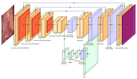
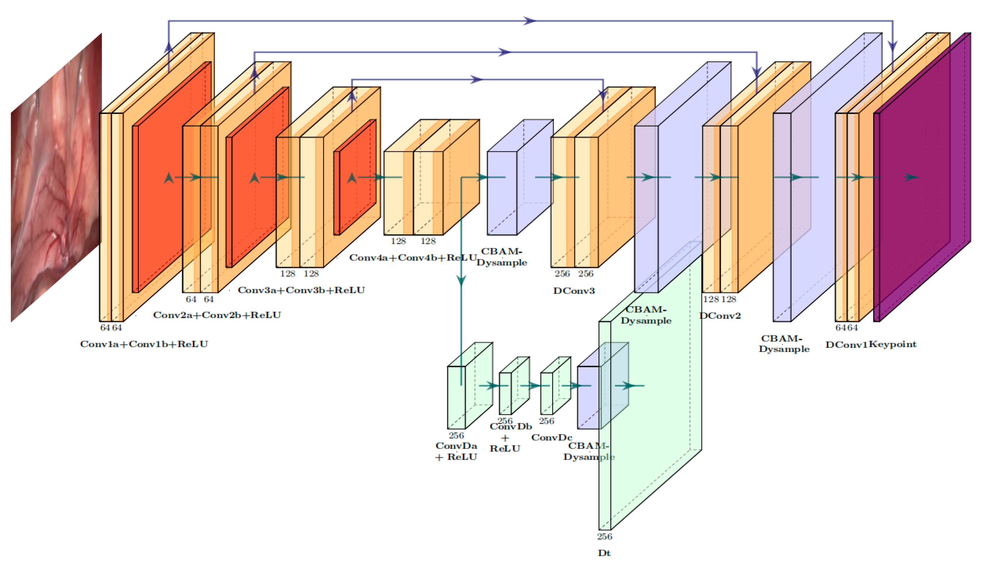
The overall structure of the vascular branching feature extraction network (SuperVessel) is specifically optimized for weakly supervised detection of vascular branch points within lumens. Its overall architecture, as shown in Figure 1, comprises a detection network (Det) and a descriptor network (Des), sharing three core modules: an encoder, a Det detection decoder, and a Des descriptor decoder. Inputting an intraluminal image I of resolution H × W, the network employs multi-scale feature extraction and attention-based dynamic upsampling to achieve branch point detection and descriptor generation within a single forward pass. This approach ultimately delivers high repeatability and reliability in vascular branch point detection.
Figure 1.
Network structure of SuperVessel.
2.2. Det and Des Networks with Shared Encoder
The core of SuperVessel addresses two major tasks in the field of vascular branch point localization (i.e., detection) and characterization (i.e., description) in endoluminal images. Consequently, the branch point probability map and descriptor tensor are synchronously output in a single forward propagation by sharing encoder features through a dual-network cooperative architecture (Det + Des). The deep network employs U-Net [19] as the network architecture, with the initial convolution of the encoder generating the full-size feature maps. The subsequent three convolutional modules comprise two convolutional layers, a 2 × 2 maximal pooling layer, and a Relu nonlinear activation function. The decoder employs four convolutional modules. The first three convolutional blocks comprise two convolutional layers, while the final convolutional module consists of three convolutional layers and a sigmoid activation function. These components collectively generate the full-size feature point probability map, denoted P. Within this map, Pi,j represents the probability that a specific pixel is a branch point (i = 1, …, h, j = 1, …, w). In the present study, the design of a dynamic up-sampling module based on a hybrid attention mechanism for bilinear up-sampling is examined. It is demonstrated that this module is capable of dynamically adjusting the focus area of up-sampling, thereby avoiding unnecessary blurring or distortion while enhancing the features. In the context of the weakly supervised learning framework, the Des network employs the Det network encoder to generate a d-dimensional descriptor for each pixel. This intensive output is achieved by interpolation techniques, which suggests the existence of a gradient correlation between each keypoint and its neighborhood during backpropagation. This correlation can be further enhanced by expanding the size of the neighborhood, thereby demonstrating the positive contribution of a larger sensory field to the training process [20]. The decoder contains two convolutional layers, the purpose of which is to reduce the feature map to a more compact h/16 × w/16 × d format. Thereafter, it employs a dynamic up-sampling module based on a hybrid attention mechanism to generate a full-size descriptor tensor D of h × w × d. The descriptors are then L2-normalised, with Di,j denoting a d-dimensional descriptor. Non-maximum suppression (NMS) is applied to P in order to obtain a binary mask as the ultimate detection result, thus removing duplicates while preserving keypoints.
Training vascular branch-point detection models faces two major challenges: first, models are prone to becoming stuck in locally suboptimal solutions; second, the cost of fully annotating lumen images is extremely high. To address these challenges, a weakly supervised training strategy is adopted, using weakly annotated images as supervisory signals and combining them with a large volume of unannotated images for training. We propose a Dice-Det loss function based on branching feature weights and concurrently introduce the Progressive Key Point Expansion (PKE) method [21] to address the issue of incomplete pseudo gold standards. First, we construct the geometric mapping I′ = H(I) of image I, where H is a specific homothetic transformation. We input I′ into the network to obtain its probability map P′. Thus, we obtain the branch point map PH = H−1(P′) of I through backprojection. Then, we apply NMS to the probability map P of I to obtain the candidate point set PN. For each point (x, y) in PN, if (PH)x, y > 0.5, it is included in the current round’s detection point set Dett. The annotation set is expanded by progressively adding new detection points. The initial annotation set is B0. The tth training round uses Bt = B0∪Dett as the supervision signal.
To address the issue of Dett potentially containing erroneous detection points during initial training, we enhance the geometric matching strategy of GLAMpoints [22] by introducing a dual-matching mechanism (geometric matching + feature matching) to improve detection point accuracy. For each point (x, y) in Dett, we sample descriptors from the Des decoder output to form the descriptor set Dest. Correspondingly, we extract the descriptor set Dest for the transformation point set H(Dett) in I′. For each descriptor d in Dest, we perform a nearest neighbor search in Dest. Moreover, (x, y) is only retained in Dett if the descriptor match between point (x, y) and its spatially corresponding point (x′, y′) passes the Lowe ratio test. For example, if point A in I is erroneously detected in a non-vascular region, its transformed counterpart C in I′ may exhibit similar descriptors d(A) and d(C) due to the lack of specificity in non-vascular textures. However, the distance ratio between d(A) and its second-best match may fail in the Lowe’s ratio test, thereby filtering out the erroneous point A. This dual-matching mechanism effectively enhances Dett’s accuracy, ensuring the quality of progressively expanded annotated sets.
The pixel-based descriptor loss in networks [23,24,25,26] is computationally challenging when attempting to achieve a full-size descriptor tensor. Consequently, we propose a Triplet-Des loss function based on branch points and dynamic weighting.
2.3. CBAM-DySample Module
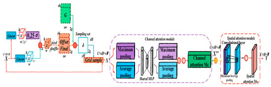
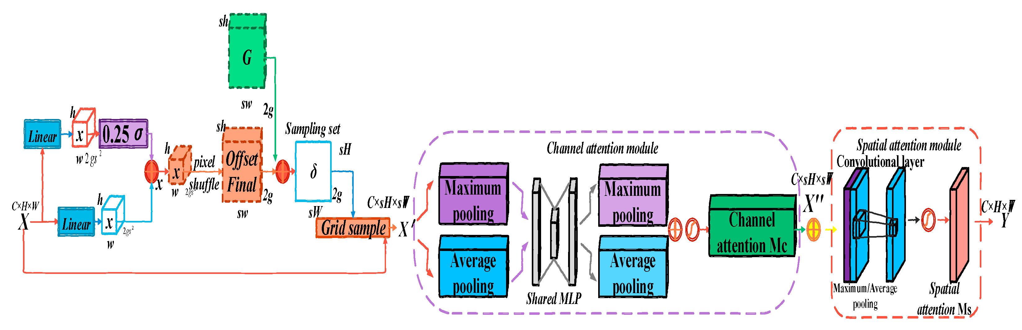
It is evident that U-Net networks universally employ the Upsample module to facilitate the upsampling function. In addition, the researchers have proposed dynamic upsamplers [27,28], which have been shown to achieve substantial performance enhancements. However, the traditional upsampling methods (e.g., bilinear interpolation, transposed convolution) tend to rely only on geometric operations and are prone to blurring or distortion. This is particularly evident in the context of complex images or high-level abstract features, where the recovery of key details is often incomplete due to the inability to fully utilize contextual information. Conversely, the endovascular branching features are typically localized and are significantly influenced by noise and distortion. In order to solve the aforementioned problems, reduce the model size and increase its operation speed, this section designs a dynamic upsampling module based on a hybrid attention mechanism Convolutional Block Attention Module (CBAM) [29] is introduced in Dynamic Sample (DySample) [30], called as CBAM-DySample, as illustrated in Figure 2. Unlike transposed convolution, which applies a fixed kernel and may introduce checkerboard artifacts, the point sampling strategy in DySample dynamically selects relevant spatial information. This approach is particularly advantageous for vascular feature extraction as it effectively preserves thin topological structures and ensures the continuity of vessel branches. The incorporation of CBAM facilitates the automatic focusing of important regions and features during the processes of recovery and amplification. CBAM-DySample has been demonstrated to enhance the feature expression capability and efficiency of the intracavitary vascular branch-point detection network by making more effective use of the spatial and channel features and reducing redundant information and noise.
Figure 2.
Structure of the CBAM-Dysample module.
DySample is an ultra-lightweight and efficient dynamic upsampler that employs point sampling techniques to avoid the use of time-consuming dynamic convolution and additional sub-networks in generating the dynamic kernel. This effectively reduces the overall computational effort and enhances the model’s ability to learn from a small number of feature samples of a few classes. In contradistinction to conventional dynamic upsampling methodologies that are predicated on convolutional kernels, DySample is conceptualised from the vantage point of point sampling, whereby a solitary point is divided into multiple points to achieve more pronounced edges. The input feature map is processed by a sampling point generator to produce a sampling set δ size of 2 × H2 × W2. Specifically, the fixed offsets are first generated by Linear, a combination of linear layers and pixel shuffling techniques [27]:
where the output channel size of 2 gs2 is the linear layer, the offset comprises both horizontal and vertical offsets. Subsequently, the position InitPos of the offset is initialized:
The quantity s is known as the upsampling scale factor. In addition to the static basis, the dynamic range factor is further optimized by introducing changeable parameters in the linear layer and pixel shuffling techniques. First, a dynamic range factor of 0.25σ is generated using a Sigmoid function for adjusting the offset Scope:
Subsequently, the object is subjected to a 2 g × sh × sw offset offsetFinal using the pixel shuffle method [31]:
The sum of the offsets offsetFinal and the original sampling grid G is then normalized in order to obtain a 2 g × sh × sw sampling set δ:
It is imperative to note that N = [W, H] is the normalization factor, and 2 g denotes the x and y coordinates. The feature map X is then subjected to up-sampling by the grid_sample function, with the position in the sampling set δ being utilised to derive the feature map X′:
where considering the characteristics of the lumen vessel feature dataset, simple oversampling methods may lead to data duplication, resulting in insufficient diversity of the minority vessel branch features learned by the model, thereby affecting the model’s generalization ability. The hybrid attention mechanism, through adaptive spatial and channel attention mechanisms, can help the model automatically identify and enhance those identifiable and representative vascular features in the oversampled minority of vascular feature samples. CBAM infers attention maps (channels and spaces) in two dimensions sequentially and then multiplies these attention maps with the input feature map for adaptive feature refinement, enabling the model to learn more recognizable and representative vascular branch features. Therefore, the sampled feature map X′ is first weighted by the channel attention module, and then weighted by the spatial attention module to obtain the final feature map Y.
The Channel Attention Module (CAM) is responsible for generating a channel attention graph by modelling the relationships between feature channels. The mechanism first compresses the spatial dimensions of the input feature maps, while two operations, average pooling and maximum pooling, are used to extract different spatial context information. Subsequently, these spatial descriptors are fed into a shared multilayer perceptron (MLP) for fusion processing, which finally generates the channel attention graph. Its expression is as follows:
The global average pooling and maximum pooling are demonstrated in Equations (8) and (9), respectively:
Then the pooling result is:
Implement the pooling result via two-layer convolution:
The pooling results are summed and activated by Sigmoid:
Finally, the channel attention weights are applied to the feature map:
The pooling results are summed and activated by Sigmoid. In a similar manner, the spatial attention mechanism utilizes the spatial relationships between features to generate a spatial attention graph. In order to compute the spatial attention, it is first necessary to apply the average and maximum pooling operations along the channel axis. These are then concatenated in order to generate an efficient feature descriptor. The application of a convolutional layer to this descriptor results in the generation of a spatial attention map. The expression for this is:
Average and maximum pooling of X″ by channel dimension:
Then, the pooling result is:
The pooling results are spliced and generated by convolution to generate spatial attention weights:
Finally, the spatial attention weights are applied to the feature map:
2.4. Dice-Det Loss Function Based on Branching Feature Weights
As previously mentioned, accurately detecting vascular branch points depends heavily on the model’s ability to capture localized fine features. While the standard Dice loss function performs well on the regional overlap metric, it could be improved upon to better emphasize specific fine targets, such as branch points. The Dice Similarity Coefficient is a common measure of similarity between two sample sets, primarily employed to quantify the degree of overlap between two sets (i.e., the prediction and the target). It is especially effective in segmentation tasks [32]. The Dice loss function estimates the value of loss by the degree of overlap between the predicted value and the true value after the normalized measure:
The dice coefficient value domain is specified as [0, 1]. In the event of two sets being entirely overlapping, the value is assigned as 1. Conversely, if there is no overlap, the value is assigned as 0. It can thus be concluded that the smaller the loss value, the higher the overlap:
In the context of vascular branch point detection, the objective is typically to identify minute vascular branches, endpoints, and other diminutive structures. Conversely, standard Dice Loss is predominantly employed in the context of larger structures, such as entire organs or regions.
where gi and pi are the true and predicted values of the ith pixel point, respectively.
In the context of the pixel-level prediction task, it is imperative to acquire effective local features. It is evident that in order to enhance the capacity of the loss function to detect vascular branching features, the Dice-Det loss function, which is based on branching feature weights, has been devised in this section. This is with a view to ensuring that the network is able to focus more acutely on the vascular branching region. In order to assign higher weights at the vascular branching points and concurrently enhance the capacity to manage unbalanced data, a weighted term weight tensor, designated weight map, is introduced. This is formulated on the basis of the predicted branching point locations. The Predicted Probability Map, generated by the feature point detection network, represents the probability that each pixel point is a feature point, with a probability value ranging from 0 to 1. This map contains the learned Ground Truth Points labels, i.e., the known locations of the vascular branch points. The probability map output from the model is used to calculate the probability of each pixel, and these probabilities are assigned a corresponding weight.
In instances where the probability of a location being a branch point is high (close to 1), it is possible to assign a higher weight. In instances where the probability is low, a smaller weight is assigned. Consequently, during the training process, the model can allocate greater attention to the location of the real feature points and assign the corresponding weights according to the predicted probability. This enables the model to focus on difficult regions (e.g., edges, low probability regions). The weight map is passed to the loss function as the weighting coefficients. By weighting the branch point regions, the pixel points around the branch points are assigned higher weights. This results in the model paying more attention to the branch point regions during training. Consequently, these small regions have a greater impact on the loss function. Since network training usually processes data in batches, the loss function for sample i in N samples is:
where gij is the true value of the ith pixel in the jth sample, pij is the predicted value of the ith pixel in the jth sample, wij is the weight value corresponding to the weight map, and defaults to 1 if the weight map is None. To prevent the denominator from becoming zero, a smoothing factor ϵ is introduced, typically set to a small value such as 1 × 10⁻³.
To stabilize gradient updates during training, we normalize losses by averaging across all samples, preventing instability from batch size variations or extreme sample losses [33].
2.5. Triplet-Des Loss Function Based on Branching Points and Dynamic Weight Weighting
Thanks to the branching point information automatically provided by the branching point decoder, the descriptor decoder’s training can rely entirely on the self-supervised paradigm, eliminating the need for additional point-level labeling. This paradigm greatly reduces reliance on labeled data. However, a key challenge of self-supervised learning is that the model may output the same constant descriptor for all points, which is a mundane and meaningless solution for distinguishing between different points. To overcome this problem and ensure that the learned descriptors are discriminative, Triplet Loss [34], which is commonly used in self-supervised learning, is introduced.
Triplet Loss optimizes the feature representation by means of a triad (Anchor, Positive, Negative), especially in diverse vascular branch point data. This process ensures that features are more similar between the same categories (positive samples) and more different between different categories (negative samples). A triad is designed for the vessel branch points as follows: the current vessel branch point is designated as the ‘Anchor’; other points that are similar to this branch point or are in the same vessel are designated as the ‘Positive’; and branch points or non-branch points that are different from this branch point are designated as the ‘Negative’.
In this context, d(x, y) denotes the distance between x and y, with margin representing a hyperparameter. The optimization is more effective in the feature embedding space, as it can explicitly push similar vascular branching points closer together and dissimilar points farther away. It also better deals with the spatial and topological properties of vascular network data, and it can obtain robust feature representations in the context of self-supervised learning.
In an ideal scenario, for each keypoint (x, y) detected in I, the descriptor decoder output would be equivalent in respect to the corresponding position of I′, and Triplet Loss would ensure the distance between paired keypoints is less than the distance between unpaired keypoints. The full-size descriptor tensor D and D′ are obtained by means of feeding I and I′ into the network, respectively. For each element (x, y) in the PN, let Dx,y be its descriptor. It is hypothesized that (x, y) and (x′, y′) are paired. Therefore, the distance between their descriptors, dx,y must be reduced. The distance metric function d(x1, x2) is to be transformed into a distance based on similarity using cosine distance:
Following the exclusion of (x′, y′), a challenging negative sample mining strategy is introduced, as the direct utilization of all negative samples may result in the inefficient optimization of the model. For both negative samples, the descriptor distance between (x, y) and a randomly selected point in H(PN) is to be denoted, as is the descriptor distance between (x, y) and the most difficult-to-pair point in H(PN):
The requirement of is relatively easy to fulfil, and using alone does not result in well-differentiated descriptors. At the same time, using only makes it difficult to train the network due to its lack of ability to produce good descriptors in the early training stages. Therefore using only drand or dhard as the negative term in the ternary loss is problematic, and to solve this problem, the mean of and is used as the negative term. Descriptor loss LDes is defined as:
It is assumed that m > 0, representing a predefined spacing between positive and negative samples. It is evident that the ‘hard negative samples’ present a more substantial challenge during the training process [35]. In order to focus on the precise differentiation of vascular branch points and the dynamic adjustment of hard negative samples, a Triplet-Des loss function based on branch points and dynamic weighting is designed. The weights are introduced to dynamically adjust the contribution of each triad so that the model can learn the key branch features more quickly. The loss function is defined as follows:
It is imperative to ascertain the location of the weights wi of each triad, which are dynamically adjusted on the basis of the size of the current loss. Concurrently, to further optimize the feature space structure, encourage orthogonality between negative samples, and prevent descriptors from being too similar, a GOR regularization term is introduced:
It can be demonstrated that minimizing the square of the dot product of negative samples and paired points results in a Graph Orthogonality Regularization (GOR) regularization loss of Lgor. Among these, the weight coefficient is β. Combining the two parts of the loss, the final Des loss function is expressed as follows:
3. Experimental Results and Analysis
All experiments were implemented in an Ubuntu 20.04.6 LTS environment using Python 3.8, with GPU acceleration for network training. The hardware configuration consisted of a 13th Gen Intel® Core™ i5-13600KF 14-core CPU, 64 GB RAM, and an NVIDIA GeForce RTX 4090 graphics card. The encoder-decoder network architecture parameters are detailed in Table 1. The model was developed using the PyTorch 1.13.0 framework within an Anaconda3-24.11.3 and PyCharm-2024.2.3 environment, initialized with the Adam optimizer.
Table 1.
The parameters of the Det and Des Network model.
Hyperparameters, including learning rate, NMS threshold, and NMS neighborhood size, were systematically optimized via Bayesian optimization, targeting the maximization of keypoint matching success rate. Training stability was further enhanced through gradient clipping and a StepLR scheduler. Our sensitivity analysis revealed that while the method exhibits moderate sensitivity to parameter variations, it remains robust to reasonable perturbations, with NMS threshold primarily affecting keypoint density and NMS size having minimal impact within tested ranges. The training process spanned 300 epochs, the learning rate showed the highest impact on convergence speed and final performance, with 1 × 10⁻⁴ providing optimal balance between training stability and convergence rate.
To ensure the robustness and reproducibility of our Triplet-Des loss function, we conducted an extensive Bayesian Optimization hyperparameter search. This approach efficiently explored a broad parameter space for margin (0.05 to 1.0) and beta (0.1 to 5.0). The optimization process involved 30 evaluations, statistically supported by a total of 4400 samples, and successfully identified the global optimum, margin = 0.1478 and β = 0.1000.
3.1. Pseudo Gold Standard and Dataset Construction of Vascular Branching Features Based on Transfer Learning
In this section, we detail our strategy for generating a robust pseudo-gold standard, critically addressing potential domain shift effects and pseudo-label noise. A TL strategy is employed, introducing the SuperRetina pre-trained model [21]. This model represents the first network model capable of automatically detecting unlabeled, highly reliable vascular branch points in under-labelled or unlabeled retinal images by means of an asymptotically expanding weakly supervised learning strategy. The weights of the network model are initialized and globally fine-tuned using model parameters that have been pre-trained on the retinal image dataset. The source domain retinal dataset DR and learning task TR, and the target domain intracavitary dataset DV and learning task TV, which are the focus of this section, are trained to obtain the knowledge in DR and TR to enhance the prediction function fV(·). During the training process, the parameters in the model are noted as DetR and DesR before transfer learning, and the parameters in the model after transfer learning to the target domain are noted as DetV and DesV:
The primary benefit of transfer learning is that it enables the utilization of the feature extraction capabilities already acquired in SuperRetina’s pre-trained models. This facilitates the acquisition of optimal initial weights, enhances the detection and description performance for the designated task, achieves efficient and high-quality keypoint detection, and circumvents the time and data overheads associated with training models from the beginning. Crucially, while the imaging modalities of retinal and intracavitary environments differ, both share the fundamental topological characteristic of tubular vascular structures. This morphological similarity serves as the physical basis for effective transfer learning, mitigating the impact of domain shift.
Simultaneously inspired by SuperPoint’s methodology for establishing large-scale pseudo-ground-truth interest point locations in real images, we employ Method [18] to annotate datasets and generate robust, reliable, and accurate branching features. Method [18] is a state-of-the-art method for vascular feature detection, achieving a high repeatability of 0.5612 and matching accuracy of 0.7149 on representative vascular datasets. This high performance ensures the reliability of the generated pseudo-labels, which is crucial for providing the necessary supervisory signals for our learning-based approach. To minimize bias propagation from the retinal domain and effectively reduce initial pseudo-label noise, our pseudo-gold standard generation process leverages the inherent capabilities of Method [18], which specifically incorporates a “Dual-Branch Fusion and Structure Enhancement” step. This crucial mechanism, as an integral component of Method [18]’s inherent high performance, acts as a filter, enhancing valid vessel structures while suppressing potential artifacts or noise that might arise from domain differences. The data sources comprise five manually annotated public Vivo datasets [36] (each containing images of small, flat tissue surfaces on the abdominal wall and anterior pelvis captured during endoscopic colon surgery, with every image featuring 20 well-distributed manually labeled feature points), and the unlabeled Hamlyn Centre Endoscopic Video Dataset [37], which provides challenging sequential in vivo imaging characterized by weak textures, tissue deformations, specular reflections, surgical instruments, and occlusions, specifically consisting of 21 videos with heterogeneous resolutions and calibration parameters.
These resources are designated as Dataset A and Dataset B for constructing the pseudo-ground-truth branching feature dataset for MIS images. Following comprehensive analysis of both datasets’ imaging characteristics, Dataset A serves as the annotated test set while Dataset B undergoes filtration to retain images exhibiting discernible vascular structures with minimal occlusion. The available annotated subsets include Rectified01, Rectified06, Rectified08, and Rectified09. Images containing surgical instruments or motion-blurred frames were excluded from Rectified08 and Rectified09. Given the repetitive surgical scenes across these subsets, limited qualified images per anatomical context were retained, yielding 6 and 14 occlusion-free frames with prominent vasculature from Rectified08 and Rectified09 respectively. To mitigate overfitting caused by highly similar consecutive frames in continuous video sequences, systematic subsampling at 50-frame intervals was implemented for Rectified01 and Rectified06. This curation process ultimately yielded a final dataset comprising 140 training images, 66 validation images (approximating a 7:3 training–validation ratio), 933 unlabeled images, and 242 test images.
It is acknowledged that images of intracavitary vessels may exhibit significant variations when subjected to diverse conditions, including variations in patients, devices, and camera angles. In order to simulate these variations and enhance the model’s adaptability to a more extensive range of scenarios, as well as augment its robustness, the dataset undergoes processing using data augmentation techniques. This process provides a more substantial set of training samples, thereby empowering the transfer learning model to extract more representative features within a more intricate data context. This is imperative to enhance the performance on the intracavitary dataset. The specific data enhancement approach involves extending the training dataset by applying random affine transformations (e.g., rotation, translation, scaling, etc.) to the images, thereby enabling the model to perceive images with diverse deformations. The capacity of the model to adapt to unseen data is enhanced by the incorporation of these alterations during the training process.
3.2. Ablation Experiments
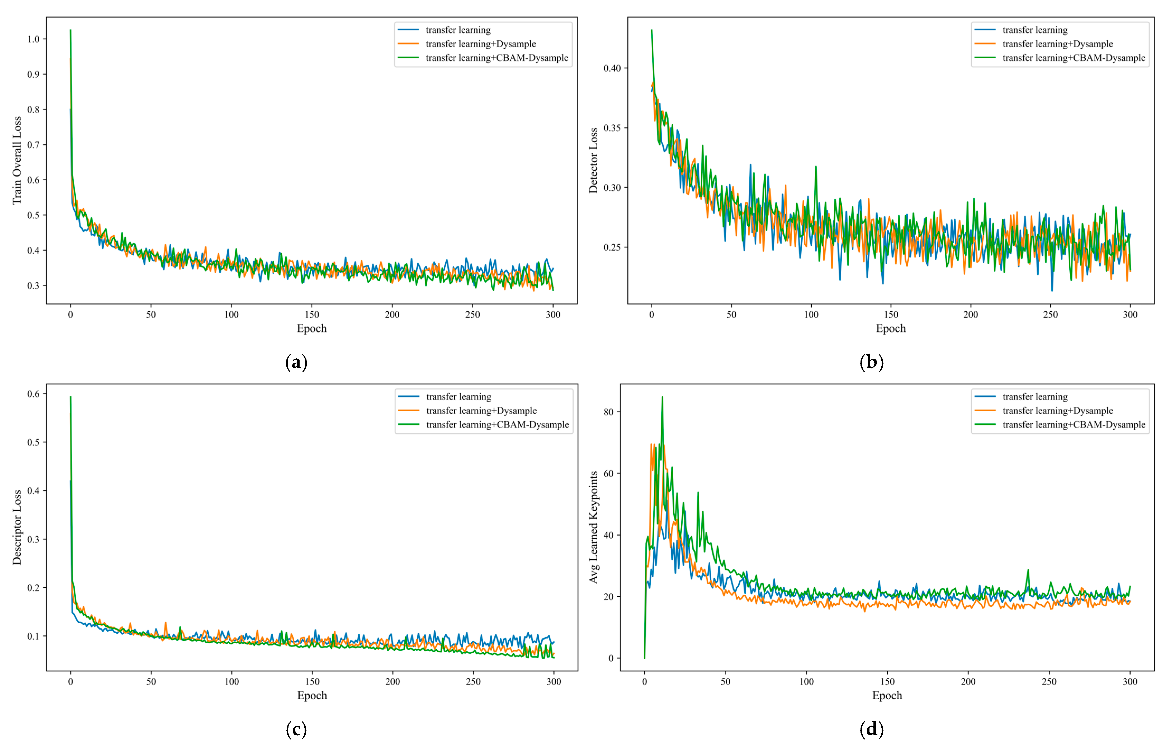
In order to verify the effect of different modules paired with transfer learning strategies on model performance, ablation experiments are designed for training tests. The test is divided into transfer learning strategy-based model, transfer learning strategy-based model combined with Dysample module, and transfer learning strategy-based model combined with dynamic upsampling module based on hybrid attention mechanism. The experimental comparison of total training loss, feature extraction loss, descriptor loss and average number of learned features for each module is shown in Figure 3a–d, and the Dysample module has a significant effect on improving the convergence speed and generalization ability of the model. On this basis, the combined descriptor loss and total loss of the dynamic upsampling module based on hybrid attention mechanism is significantly reduced. Combined with the comparison of the average number of learned features, the design enhances the vascular feature selection capability, helps retain more effective features, and effectively improves the descriptor quality. This optimization makes the model more suitable for the vascular feature matching task that requires high-precision descriptor capability.
Figure 3.
Comparison of training loss curves for transfer learning strategies with different module combinations; (a) Training Loss per Epoch; (b) Detector Loss per Epoch; (c) Descriptor Loss per Epoch; (d) Avg Learned Keypoints per Epoch.
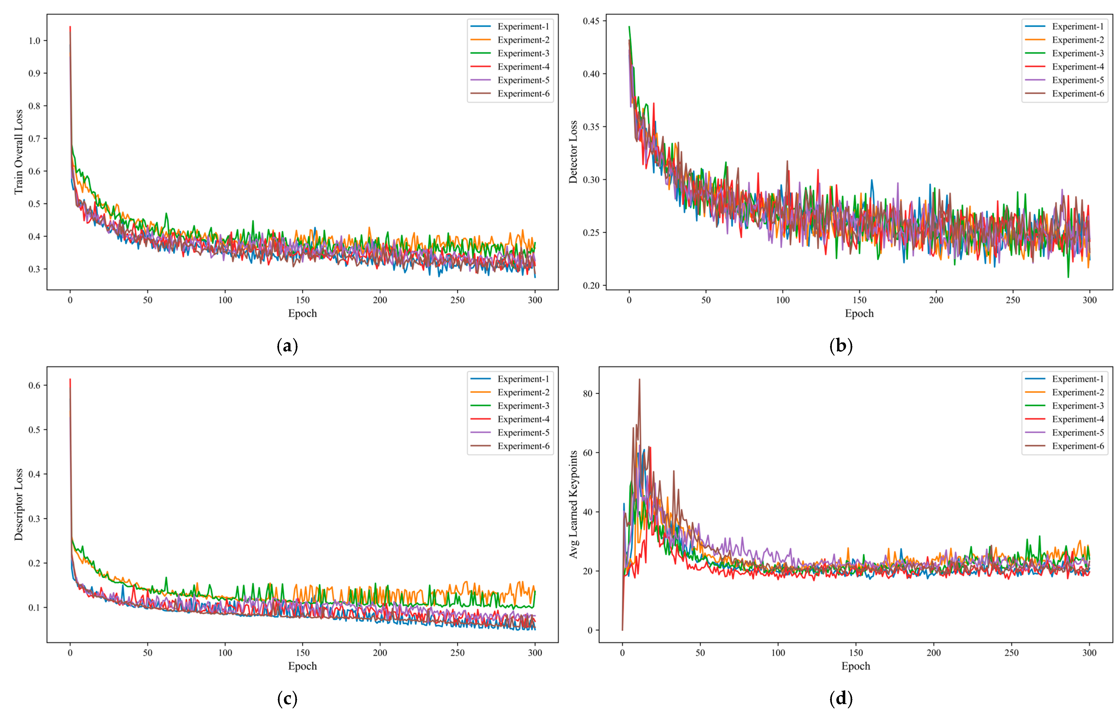
In particular, when designing the dynamic up-sampling module based on hybrid attention mechanism, the same ablation experiment training test was conducted to verify the effectiveness of different design schemes and their impact on the model, as shown in Figure 4a–d Six scenarios were designed for comparison experiments:
Figure 4.
Comparison chart of training loss curves for various experimental schemes; (a) Training Loss per Epoch; (b) Detector Loss per Epoch; (c) Descriptor Loss per Epoch; (d) Avg Learned Keypoints per Epoch.
- CBAM-DySample module in LP mode (linear projection + pixel shuffling) for both Det and Des networks;
- CBAM-DySample module in LP mode for the Det network and PL mode (pixel shuffling + linear projection) for the Des network;
- Both Det and Des networks CBAM-DySample module in PL mode;
- CBAM-DySample module in PL mode for Det network and in LP mode for Des network;
- CBAM-DySample module is introduced only in Det network;
- CBAM-DySample module is introduced only in Des network.
The results show that although Experiment 1 does not perform significantly in feature extraction loss, it performs optimally in descriptor loss and overall loss, has the fastest convergence speed and the lowest final loss, and the trade-off according to the task requirements indicates that this scheme effectively optimizes the global features suitable for scenarios pursuing the efficiency of global convergence and the task of highly discriminative features.
3.3. Feature Extraction and Matching Experiments
In order to test the feature extraction and matching ability of the model, a test set is used to compare our model with the SuperRetina model. The matching algorithm adopts the feature point matching strategy in ORB-SLAM3, which firstly performs K-Nearest Neighbors (KNN) matching and ratio test, and then performs double geometric validation, and simultaneously performs the computation of uni-responsive matrix and basis matrix to select the optimal geometric model for matching [38]. Lowe’s ratio threshold is set at 0.65, the RANSAC threshold was set at 3.0, the base matrix RANSAC threshold was set at 1.0, and the RANSAC confidence level was 0.9.
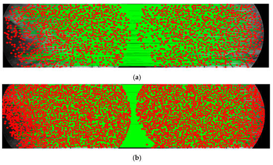
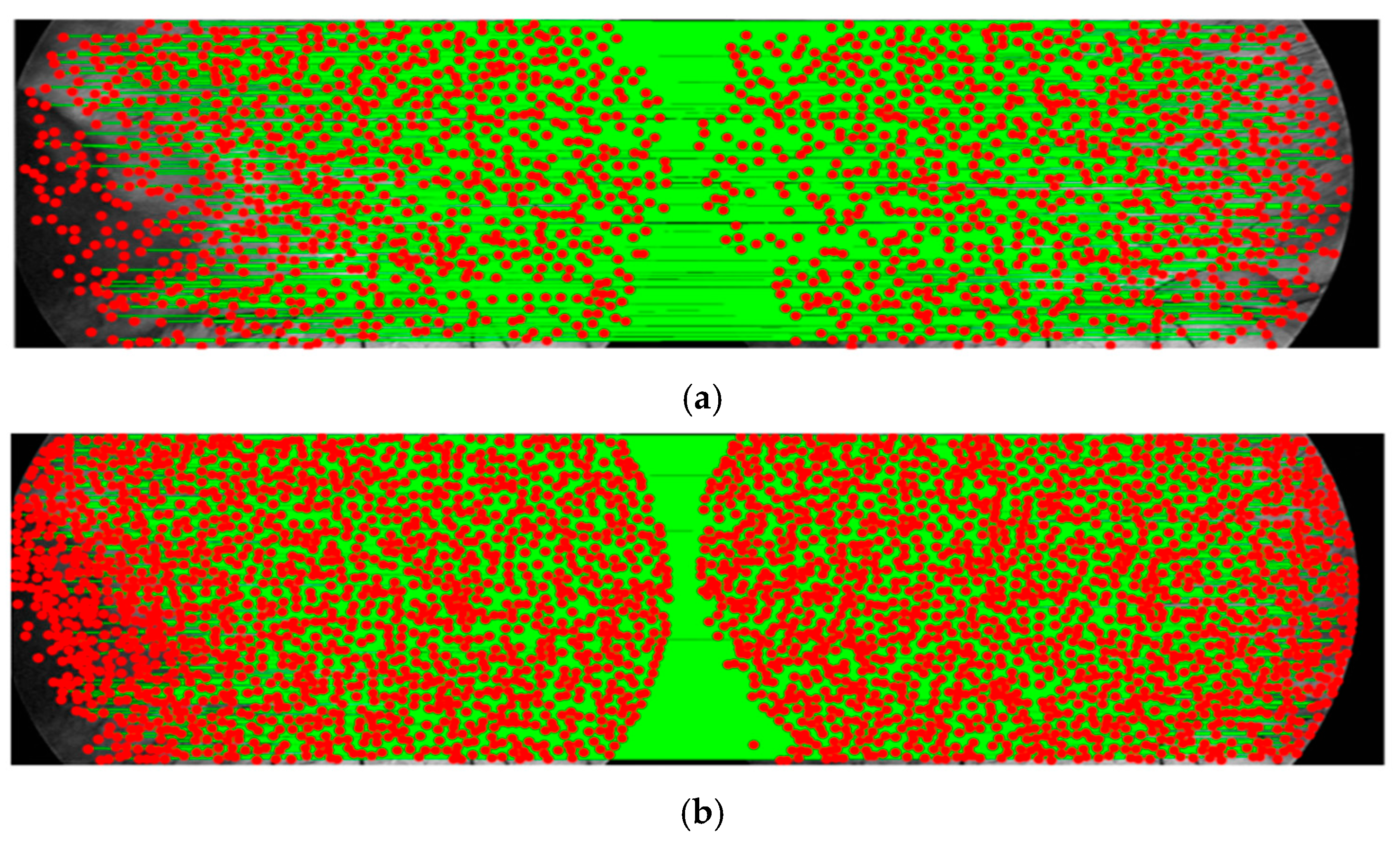
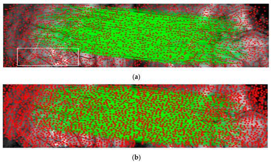
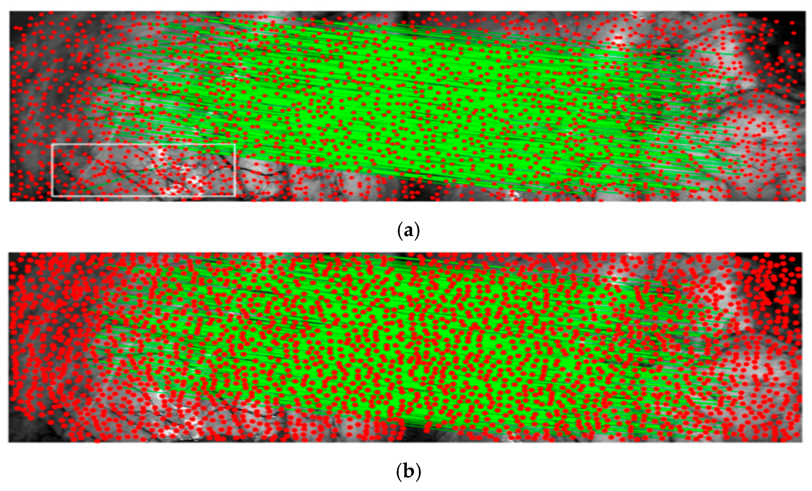
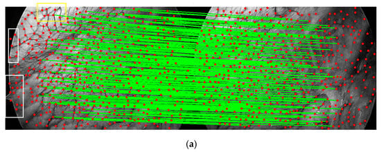
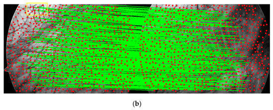
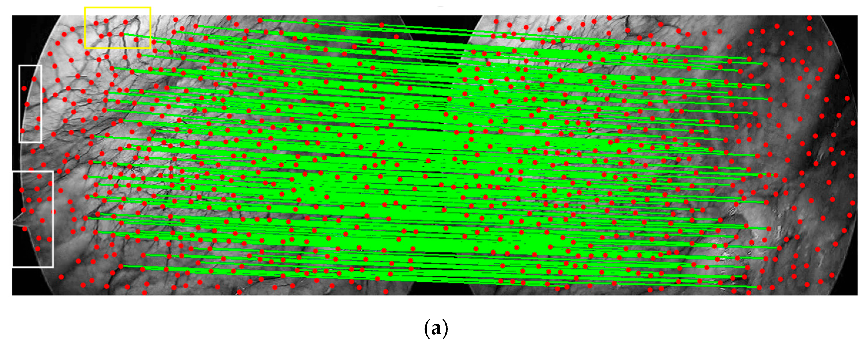
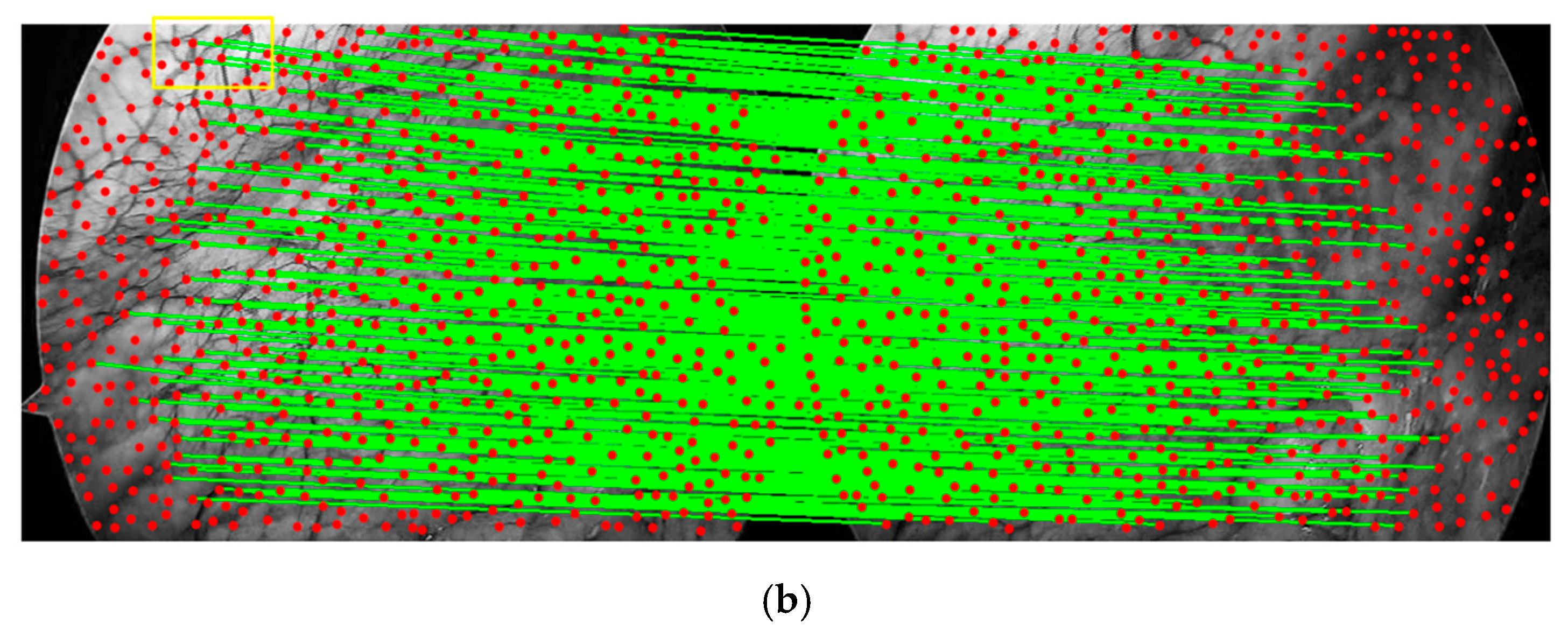
Subjectively, as shown in Figure 5, Figure 6 and Figure 7, which demonstrate the matching effect of the two models in different datasets of base image and a random image, respectively, the SuperRetina model matches a relatively small number of successful and there are obvious mismatches and non-content regions of the feature mis-extracted (such as the region labelled by the white box). In contrast, our model is able to extract more and more stable blood vessel branching points, as seen in the comparison of the regions labelled with yellow boxes.
Figure 5.
Visual comparison chart of matching results for clip2 dataset; (a) SuperRetina model result (goodMatch: 681); (b) SuperVessel model result (goodMatch: 1264).
Figure 6.
Visual comparison chart of matching results for clip4 dataset; (a) SuperRetina model result (goodMatch: 433); (b) SuperVessel model result (goodMatch: 512).
Figure 7.
Visual comparison chart of matching results for clip5 dataset; (a) SuperRetina model result (goodMatch: 245); (b) SuperVessel model result (goodMatch: 364).
Objectively, as shown in Table 2, the total number of matches, the number of successful matches, and the matching accuracy rate of the extracted vessel features of the two models are compared, and it can be seen that our model is better in both the number and the accuracy rate. Meanwhile, we refers to FIRE (Fundus Image Registration Evaluation) [39] to construct the Area Under Curve (AUC) evaluation dataset, and according to the degree of difficulty of matching, the five test datasets are divided into three groups, 99 Easy, 70 Mod, 21 Hard, and the results of the three groups are averaged to serve as the overall index. AUC for the error set ek estimates the cumulative correct rate under different thresholds δ for all samples N. The overall performance is quantified by the integral area, which reflects the better overall performance of our model:
Table 2.
Comparison of feature points extraction.
To evaluate the feature extraction and matching capabilities of the model, the proposed model was compared with traditional methods using a test dataset. Classic algorithms selected include SIFT, FAST, and ORB. Deep learning model selected: Superpoint. Table 3 presents all metric results. Repeatability (Rep) is a common metric for evaluating feature point detector performance, defined as the percentage of points detected across images with different viewpoints. For accuracy (Acc), traditional methods focus more on the quantity of matches. Therefore, the results emphasize match quality. Results show that the latency of 0.130 s/frame is not optimal, but it meets practical requirements and is better suited for SLAM embedding applications under slow, small-angle movements.
Table 3.
Comparison of feature points extraction for SLAM.
4. Discussion
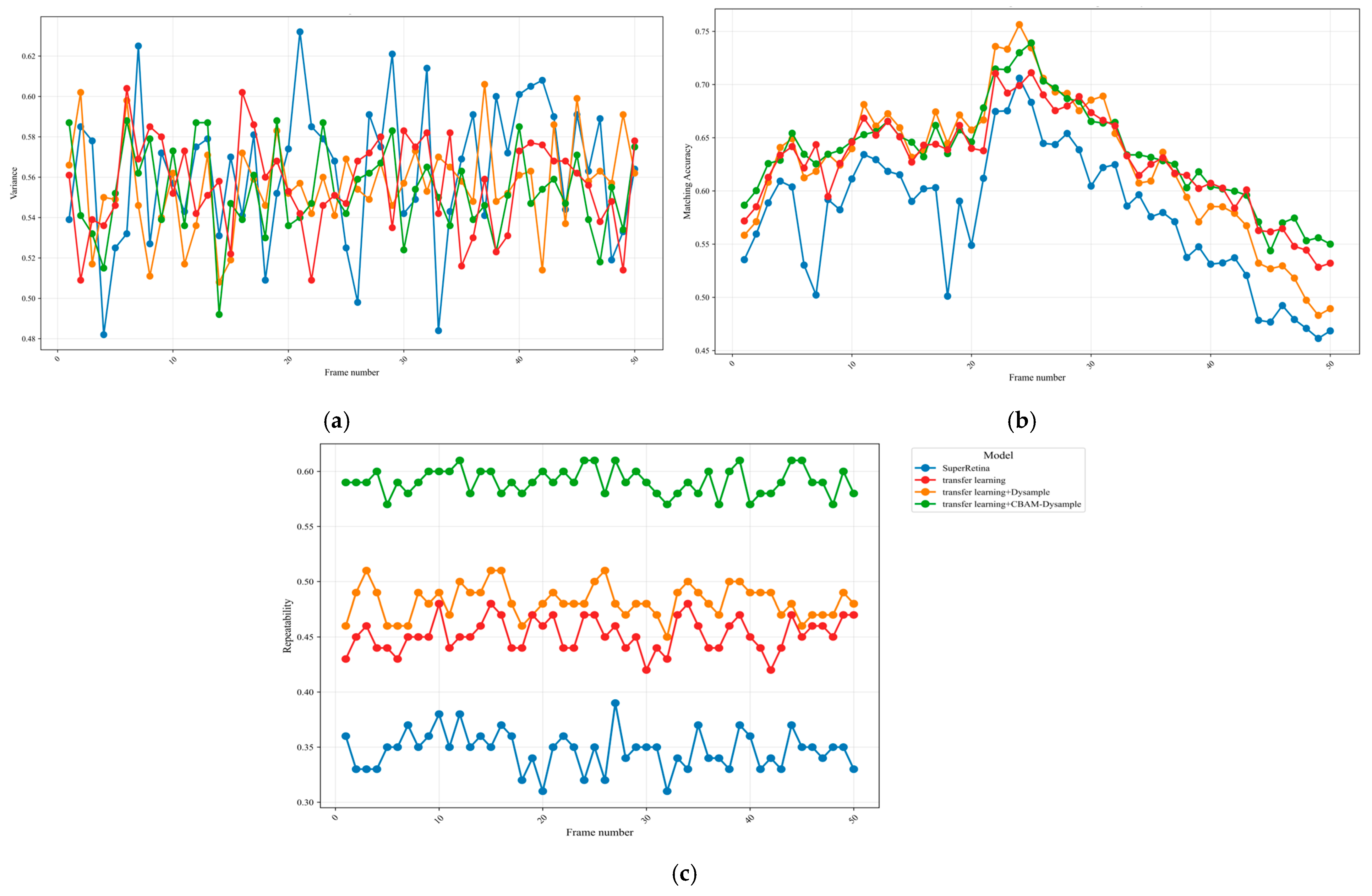
The SuperRetina model and our model are compared in conjunction with the ablation experiment, which contains the inter-frame analysis and overall analysis of the dataset. As shown in Figure 8a–c, we count the variance, matching the accuracy and repeatability of the four models for all frames of the dataset clip1. Compared to the SuperRetina model, comparing all the experimental results shows that our model frame is significantly better. Combined with the comparison of ablation experiments, although the advantages of single-frame matching accuracy and variance of our model are not obvious, the repeatability is significantly better on the basis of stable matching accuracy and variance, indicating that the design of the CBAM-Dysample can focus on the vascular features more effectively, enhance the expression of the vascular branching features, and enhance the robustness of the model.
Figure 8.
Single frame metric analysis of clip1 dataset; (a) Comparison of variance; (b) Comparison of matching accuracy; (c) Comparison of repeatability.
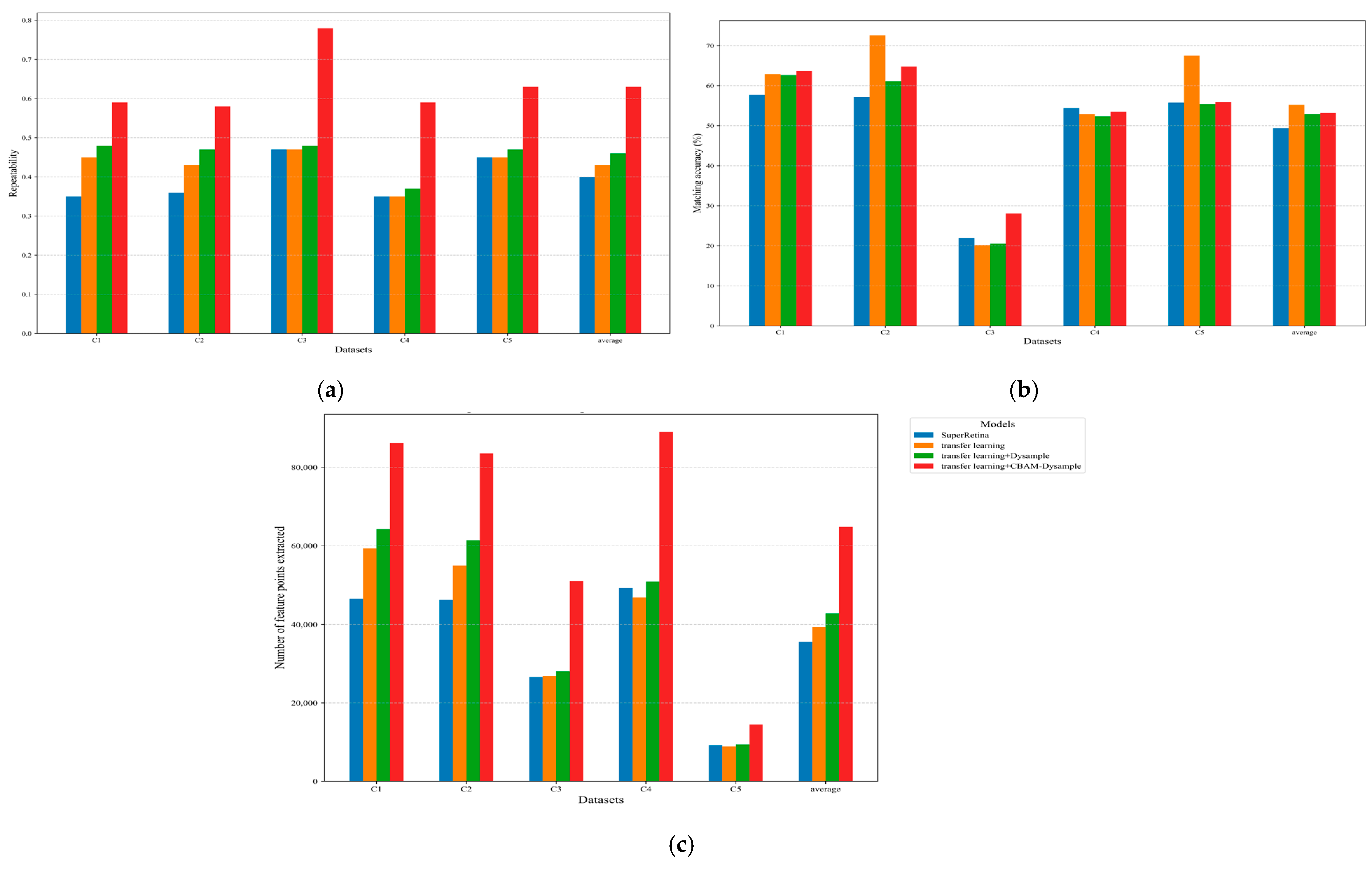
As part of the ablation experiment analysis, Table 4 and Figure 9 shows that (a), (b), and (c) count the repeatability, matching accuracy, and the number of feature points extracted for all datasets, respectively. Our method improves the repeatability, matching accuracy, and number of branch point detection by 23%, 3.77%, and 45.17% compared to the SuperRetina model. The designed CBAM-Dysample module has obvious advantages in repeatability and the number of feature points while maintaining stable matching accuracy. Comparing with the SuperRetina model, it shows that our model has the triple synergy of attention-guided feature selection, Dysample and the generalization ability of transfer learning on the basis of targeted design of the loss function, which is both robust and practical in the task of vascular branch feature detection and matching.
Table 4.
Comparison of ablation experiments.
Figure 9.
Comparison of datasets matching results; (a) Comparison of repeatability across datasets; (b) Comparison of matching accuracy across datasets; (c) Comparison of feature points number across datasets.
This robustness is particularly significant in the broader context of intelligent healthcare. As highlighted in [40], the accurate identification of vascular points is a prerequisite for precise diagnosis and navigation. By ensuring high-quality feature extraction in complex physiological structures, our method aligns with the emerging trend of utilizing Artificial Intelligence (AI) as a standard tool for cardiovascular disease analysis and intervention.
Specifically, the dynamic point sampling of DySample proves instrumental in preserving the crucial thin topological structures of vascular networks, a common failure point for methods relying on standard transposed convolutions, especially in low-texture or highly deformable regions. Concurrently, the integrated CBAM mechanism effectively filters out the pervasive noise and irrelevant tissue features characteristic of endoscopic images, allowing for a concentrated focus on salient vascular landmarks. This synergistic design not only translates into superior numerical performance but also demonstrates a consistent trend of maintaining robust feature detection and matching across varied challenging scenarios, including those with significant illumination changes and tissue deformation, where general-purpose methods often exhibit considerable degradation. While these advancements significantly push the boundaries of vascular SLAM, further exploration into handling extreme occlusions and highly specular reflections remains a critical avenue for future research.
5. Conclusions
This research presents a hybrid attention mechanism combined with U-Net for extracting vascular branching points in intracavitary images. Experimental results demonstrate substantial improvements over the SuperRetina model, with 23% enhancement in repeatability, 3.77% increase in matching accuracy, and 45.17% growth in detected branch points. The proposed method exhibits significant advantages in repeatability and feature point density while maintaining stable matching accuracy, particularly in scenarios characterized by weak texture and deformation, providing a robust vascular branch feature extraction solution for intracavitary SLAM systems. With a latency of 0.130 s/f, it meets real-time requirements for endoscopic procedures.
Despite demonstrating robustness in vascular branching feature detection and matching, the observed low matching accuracy on the C3 dataset indicates limitations in processing complex and dense vascular structures. Future research will focus on laparoscopic visual 3D reconstruction applications, exploring of deep learning-based feature matching integration to enhance matching accuracy and generalization capabilities.
Author Contributions
Conceptualization, K.X., H.W., L.Y. and X.H.; methodology, K.X. and, H.W.; software, K.X.; validation K.X. and H.W.; writing—review and editing K.X., H.W., L.Y. and X.H.; All authors have read and agreed to the published version of the manuscript.
Funding
This work was funded by the Natural Science Foundation of Heilongjiang Province (LH2023F034).
Data Availability Statement
The original data presented in the study are openly available at https://rpal.cse.usf.edu/project1/Vessel_Dataset_Share.zip (accessed on 31 July 2014).
Conflicts of Interest
The authors declare no conflicts of interest.
Abbreviations
The following abbreviations are used in this manuscript:
| VSLAM | Visual Simultaneous Localization and Mapping |
| AUC | Area Under Curve |
| SLAM | Simultaneous Localization and Mapping |
| ORB | Oriented FAST and Rotated BRIEF |
| VO | Visual Odometry |
| 3D | three-dimensional |
| MIS | Minimally Invasive Surgery |
| RANSAC | Random Sample Consensus |
| TL | transfer learning |
| PKE | Progressive Key Point Expansion |
| NMS | non-maximum suppression |
| CBAM | Convolutional Block Attention Module |
| DySample | Dynamic Sample |
| CAM | Channel Attention Module |
| MLP | multilayer perceptron |
| GOR | Graph Orthogonality Regularization |
| KNN | K-Nearest Neighbors |
| FIRE | Fundus Image Registration Evaluation |
References
- Prinet, V.; Monga, O.; Ma, S. Extraction of vascular network in 3D images. Proc. 3rd IEEE Int. Conf. Image Process. 1996, 3, 307–310. [Google Scholar] [CrossRef] [Scilit]
- Prinet, V.; Monga, O.; Ge, C.; Xie, S.L.; Ma, S.D. Thin network extraction in 3D images: Application to medical angiograms. In Proceedings of the 13th International Conference on Pattern Recognition, Vienna, Austria, 25–29 August 1996; Volume 3, pp. 386–390. [Google Scholar] [CrossRef] [Scilit]
- Lin, L.X.; Ye, Y.; Yao, J.M.; Guo, T. Embedded Implementation and Optimization of ORB-SLAM for Mobile Robots. Microcomput. Appl. 2017, 36, 50–53. [Google Scholar] [CrossRef]
- Xie, C.; Liu, Q.; Chen, B.; Hao, Z. Evaluation and analysis of feature point detection methods based on vSLAM Systems. Image Vis. Comput. 2024, 146, 105015. [Google Scholar] [CrossRef] [Scilit]
- Bono, F.M.; Radicioni, L.; Cinquemani, S.; Conese, C.; Tarabini, M. Development of soft sensors based on neural networks for detection of anomaly working condition in automated machinery. In Proceedings of the NDE 4.0, Predictive Maintenance, and Communication and Energy Systems in a Globally Net-Worked World, Long Beach, CA, USA, 4–10 April 2022; Volume 12049, pp. 45–55. [Google Scholar] [CrossRef] [Scilit]
- DeTone, D.; Malisiewicz, T.; Rabinovich, A. Superpoint: Self-supervised interest point detection and description. In Proceedings of the IEEE Conference on Computer Vision and Pattern Recognition Workshops, Salt Lake City, UT, USA, 18–22 June 2018; pp. 224–236. [Google Scholar] [CrossRef] [Scilit]
- Deng, L.; Liu, Z.; Zhang, T.; Yan, Z. Study of visual SLAM methods in minimally invasive surgery. Math. Biosci. Eng. 2023, 20, 4388–4402. [Google Scholar] [CrossRef] [Scilit]
- Barbed, O.L.; Chadebecq, F.; Morlana, J.; Montiel, J.M.M.; Murillo, A.C. Superpoint features in endoscopy. In Proceedings of the MICCAI Workshop on Imaging Systems for GI Endoscopy, Singapore, 18 September 2022; Springer Nature: Cham, Switzerland, 2022; Volume 13754, pp. 45–55. [Google Scholar] [CrossRef] [Scilit]
- Qin, W.; Yang, X.; Liu, C.; Zheng, W. A deep learning method based on YOLOv5 and SuperPoint-SuperGlue for digestive disease warning and cage location backtracking in stacked cage laying hen systems. Comput. Electron. Agric. 2024, 222, 108999. [Google Scholar] [CrossRef] [Scilit]
- Wang, J.; Gao, F. Improved visual inertial odometry based on deep learning. J. Phys. Conf. Ser. 2021, 2078, 012016. [Google Scholar] [CrossRef] [Scilit]
- Azzopardi, G.; Petkov, N. Automatic detection of vascular bifurcations in segmented retinal images using trainable COSFIRE filters. Pattern Recognit. Lett. 2013, 34, 922–933. [Google Scholar] [CrossRef] [Scilit]
- Abbasi-Sureshjani, S.; Smit-Ockeloen, I.; Bekkers, E.; Dashtbozorg, B.; ter Haar Romeny, B. Automatic detection of vascular bifurcations and crossings in retinal images using orientation scores. In Proceedings of the 2016 IEEE 13th International Symposium on Biomedical Imaging (ISBI), Prague, Czech Republic, 13–16 April 2016; IEEE: New York, NY, USA, 2016; pp. 189–192. [Google Scholar] [CrossRef] [Scilit]
- Uslu, F.; Bharath, A.A. A multi-task network to detect junctions in retinal vasculature. In Proceedings of the Medical Image Computing and Computer Assisted Intervention–MICCAI 2018: 21st International Conference, Granada, Spain, 16–20 September 2018; Proceedings, Part II 11. Springer: Berlin/Heidelberg, Germany, 2018; pp. 92–100. [Google Scholar] [CrossRef] [Scilit]
- Zhao, H.; Sun, Y.; Li, H. Retinal vascular junction detection and classification via deep neural networks. Comput. Methods Programs Biomed. 2020, 183, 105096. [Google Scholar] [CrossRef] [Scilit] [PubMed]
- Lin, J.; Zeng, X.; Pan, Y.; Ren, S.; Bao, Y. Intelligent Inspection Guidance of Urethral Endoscopy Based on SLAM with Blood Vessel Attentional Features. Cogn. Comput. 2024, 16, 1161–1175. [Google Scholar] [CrossRef] [Scilit]
- Bashiri, F.S.; Caskey, J.R.; Mayampurath, A.; Dussault, N.; Dumanian, J.; Bhavani, S.V.; Carey, K.A.; Gilbert, E.R.; Winslow, C.J.; Shah, N.S.; et al. Identifying infected patients using semi-supervised and transfer learning. J. Am. Med. Inform. Assoc. 2022, 29, 1696–1704. [Google Scholar] [CrossRef] [Scilit] [PubMed]
- Entezari, R.; Wortsman, M.; Saukh, O.; Shariatnia, M.M.; Sedghi, H.; Schmidt, L. The Role of Pre-training Data in Transfer Learning (Version 2). arXiv 2023, arXiv:2302.13602. [Google Scholar] [CrossRef] [Scilit]
- Xu, K.; Wu, H.; Iwahori, Y.; Yu, X.; Hu, Z.; Wang, A. A Vascular Feature Detection and Matching Method Based on Dual-Branch Fusion and Structure Enhancement. Sensors 2024, 24, 1880. [Google Scholar] [CrossRef] [Scilit]
- Siddique, N.; Paheding, S.; Elkin, C.P.; Devabhaktuni, V. U-Net and Its Variants for Medical Image Segmentation: A Review of Theory and Applications. IEEE Access 2021, 9, 82031–82057. [Google Scholar] [CrossRef] [Scilit]
- Chen, L.C.; Papandreou, G.; Kokkinos, I.; Murphy, K.; Yuille, A.L. DeepLab: Semantic Image Segmentation with Deep Convolutional Nets, Atrous Convolution, and Fully Connected CRFs. IEEE Trans. Pattern Anal. Mach. Intell. 2018, 40, 834–848. [Google Scholar] [CrossRef] [Scilit]
- Liu, J.; Li, X.; Wei, Q.; Xu, J.; Ding, D. Semi-supervised keypoint detector and descriptor for retinal image matching. European Conference on Computer Vision. arXiv 2022, arXiv:2207.07932. [Google Scholar] [CrossRef] [Scilit]
- Camara, J.; Silva, B.; Gouveia, A.; Pires, I.M.; Coelho, P.; Cunha, A. Detection and mosaicing techniques for low-quality retinal videos. Sensors 2022, 22, 2059. [Google Scholar] [CrossRef] [Scilit]
- Guedria, S.; De Palma, N.; Renard, F.; Vuillerme, N. R2D2: A scalable deep learning toolkit for medical imaging Segmentation. Softw. Pract. Exp. 2022, 50, 1966–1985. [Google Scholar] [CrossRef] [Scilit]
- Yang, N.; Han, Y.; Fang, J.; Zhong, W.; Xu, A. UP-Net: Unique keyPoint description and detection net. Mach. Vis. Appl. 2022, 33, 13. [Google Scholar] [CrossRef] [Scilit]
- Rocco, I.; Cimpoi, M.; Arandjelovic, R.; Torii, A.; Pajdla, T.; Sivic, J. NCNet: Neighbourhood consensus networks for estimating image correspondences. IEEE Trans. Pattern Anal. Mach. Intell. 2022, 44, 1020–1034. [Google Scholar] [CrossRef] [Scilit]
- Zhu, X.; Zhang, W.; Li, H. LF-UNet: An Attention-Based U-Net for Retinal Vessel Segmentation. In Proceedings of the 2024 IEEE 19th Conference on Industrial Electronics and Applications, ICIEA 2024, Kristiansand, Norway, 5–8 August 2024; Institute of Electrical and Electronics Engineers Inc.: Piscataway, NJ, USA, 2024. [Google Scholar] [CrossRef] [Scilit]
- Wang, Y.; Yin, T.; Chen, X.; Hauwa, A.S.; Deng, B.; Zhu, Y.; Gao, S.; Zang, H.; Zhao, H. A steel defect detection method based on edge feature extraction via the Sobel operator. Sci. Rep. 2024, 14, 27694. [Google Scholar] [CrossRef] [Scilit]
- Lu, H.; Liu, W.; Fu, H.; Cao, Z. FADE: A Task-Agnostic Upsampling Operator for Encoder–Decoder Architectures. Int. J. Comput. Vis. 2025, 133, 151–172. [Google Scholar] [CrossRef] [Scilit]
- Park, J.; Woo, S.; Lee, J.-Y.; Kweon, I.S. A Simple and Light-Weight Attention Module for Convolutional Neural Networks. Int. J. Comput. Vis. 2020, 128, 783–798. [Google Scholar] [CrossRef] [Scilit]
- Liu, W.; Lu, H.; Fu, H.; Cao, Z. Learning to Upsample by Learning to Sample. In Proceedings of the 2023 IEEE/CVF International Conference on Computer Vision (ICCV), Paris, France, 1–6 October 2023; pp. 6004–6014. [Google Scholar] [CrossRef] [Scilit]
- Zhang, D.; Zhou, J.; Zhang, W.; Lin, Z.; Yao, J.; Polat, K.; Alenezi, F.; Alhudhaif, A. ReX-Net: A reflectance-guided underwater image enhancement network for extreme scenarios. Expert Syst. Appl. 2023, 231, 120842. [Google Scholar] [CrossRef] [Scilit]
- Ding, Z.; Zhang, Y.; Zhu, C.; Zhang, G.; Li, X.; Jiang, N.; Que, Y.; Peng, Y.; Guan, X. CAT-Unet: An enhanced U-Net architecture with coordinate attention and skip-neighborhood attention transformer for medical image segmentation. Inf. Sci. 2024, 670, 120578. [Google Scholar] [CrossRef] [Scilit]
- Liu, Y.; Cai, L.; Chen, Y.; Ma, P.; Zhong, Q. Variable separated physics-informed neural networks based on adaptive weighted loss functions for blood flow model. Comput. Math. Appl. 2024, 153, 108–122. [Google Scholar] [CrossRef] [Scilit]
- Lu, Z.; Lu, Y. A balanced triplet loss for person re-identification. Int. J. Pattern Recognit. Artif. Intell. 2023, 37, 2256022. [Google Scholar] [CrossRef] [Scilit]
- Yu, J.; Zhu, C.; Zhang, J.; Huang, Q.; Tao, D. Spatial Pyramid-Enhanced NetVLAD with Weighted Triplet Loss for Place Recognition. IEEE Trans. Neural Netw. Learn. Syst. 2020, 31, 661–674. [Google Scholar] [CrossRef] [Scilit] [PubMed]
- Lin, B.; Sun, Y.; Sanchez, J.; Qian, X. Vesselness based feature extraction for endoscopic image analysis. In Proceedings of the 2014 IEEE 11th International Symposium on Biomedical Imaging (ISBI), Beijing, China, 29 April–2 May 2014; IEEE: New York, NY, USA, 2014; pp. 1295–1298. [Google Scholar] [CrossRef] [Scilit]
- Recasens, D.; Lamarca, J.; Fácil, J.M.; Montiel, J.M.M.; Civera, J. Endo-Depth-and-Motion: Reconstruction and tracking in endoscopic videos using depth networks and photometric constraints. IEEE Robot. Autom. Lett. 2021, 6, 7225–7232. [Google Scholar] [CrossRef] [Scilit]
- Liu, Y.; Wang, Y. A new ORB feature matching image algorithm based on non-maximum suppression method and retinal sampling model. In Proceedings of the 2021 International Conference on Control, Automation and Information Sciences (ICCAIS), Xi’an, China, 14–17 October 2021; IEEE: New York, NY, USA, 2021; pp. 729–734. [Google Scholar]
- Hernandez-Matas, C.; Zabulis, X.; Triantafyllou, A.; Anyfanti, P.; Douma, S.; Argyros, A.A. FIRE: Fundus Image Registration dataset. Model. Artif. Intell. Ophthalmol. 2017, 1, 16–28. [Google Scholar] [CrossRef] [Scilit]
- Pati, A.; Addula, S.R.; Panigrahi, A.; Sahu, B.; Nayak, D.S.K.; Dash, M. Artificial intelligence in improving disease diagnosis: A case study of cardiovascular disease prediction. In Artificial Intelligence in Medicine and Healthcare; CRC Press: Boca Raton, FL, USA, 2025; pp. 24–49. [Google Scholar]

Disclaimer/Publisher’s Note: The statements, opinions and data contained in all publications are solely those of the individual author(s) and contributor(s) and not of MDPI and/or the editor(s). MDPI and/or the editor(s) disclaim responsibility for any injury to people or property resulting from any ideas, methods, instructions or products referred to in the content. |
© 2026 by the authors. Licensee MDPI, Basel, Switzerland. This article is an open access article distributed under the terms and conditions of the Creative Commons Attribution (CC BY) license.