


Abstract
In 3D segmentation with lesion-only annotations, lesion supervision defines voxel-wise foreground membership, whereas medical knowledge characterizes the relations between lesion states and relevant anatomical structures. How to integrate these two forms of information within a learnable target-state formulation is a key problem in medical knowledge-guided lesion segmentation. To address this problem, we propose KG-Seg, a medical knowledge-guided target-state learning framework. KG-Seg compiles symbolic medical relations, anatomical anchors, and empirical target-distribution priors into spatial–relational representations and calibrates their valid ranges and reliability strengths through knowledge–target consistency. On this basis, target-state learning is formulated as the joint structure of a knowledge-conditioned target learning distribution and a reliability-aware medical knowledge direction field. The former reconstructs the probability mass and relative contribution of different target states, aligning the learning distribution with lesion states supported by medical knowledge. The latter guides the optimization direction, contribution strength, and supervision validity associated with each target state through support, risk, and neutral directions. In this way, medical knowledge is transformed from an external prior into an intrinsic variable that organizes target-state learning. On MSD Lung and MSD Colon, KG-Seg increases Dice by 4.2 and 6.3 percentage points, respectively, relative to the nnUNetv2 baseline. These results indicate that the joint reconstruction of the target-state distribution and optimization direction establishes a learnable connection between the representation of medical relations and model optimization, providing a new paradigm for medical knowledge-guided learning in 3D lesion segmentation with lesion-only annotations.
1. Introduction
In three-dimensional lesion segmentation with lesion-only annotations, lesion masks define voxel-wise foreground membership but do not explicitly represent the medical relations between lesion states and relevant anatomical structures. Even within the same lesion category, lesions may vary substantially in morphology, scale, multiplicity, contrast, and boundary definition. Their medically plausible locations, spatial extents, and relations to surrounding organs, tissue interfaces, and adjacent structures, however, are supported by relatively stable medical knowledge [1]. Accurate three-dimensional lesion segmentation therefore requires both the identification of heterogeneous lesion appearances and the alignment of learned lesion states with the corresponding medical relations. Under the absence of task-specific anatomical annotations, enabling such relational information to participate in lesion learning remains an important problem in three-dimensional segmentation with lesion-only annotations.
Recent advances in convolutional neural networks, Transformers, state-space models, and medical image foundation models have substantially improved representation learning for three-dimensional medical images [2,3]. U-Net, 3D U-Net, V-Net, and nnU-Net have established mature encoder–decoder paradigms for volumetric segmentation [4,5,6,7,8]. Transformer-based and hybrid architectures have further enhanced long-range contextual modeling [9,10,11,12,13], while medical image foundation models have extended general-purpose segmentation and cross-task transfer capabilities [14,15,16,17,18]. These methods can learn increasingly rich visual features and contextual associations. However, existing segmentation objectives remain organized primarily around image appearance and voxel-wise annotations, while medical relations generally remain implicit contextual information and have not been further transformed into explicit structures for target-state learning. Consequently, models may inadequately learn lesion states that are visually challenging but consistent with medical relations and may also produce high lesion responses at locations lacking medical-relation support.
This capability boundary reflects a deeper issue in the organization of the model’s learning objects: target-state learning remains primarily driven by image appearance and voxel-wise supervision, while medical relations have yet to be transformed into an intrinsic structure of the learning process. A voxel label alone represents local foreground membership; its complete learning meaning also depends on lesion-entity membership, local imaging context, anatomical location, and network representation level. These factors jointly constitute a target state in lesion learning. Conventional data-driven training induces a learning distribution and corresponding update effects over target states from image observations and lesion annotations, whereas medical knowledge further characterizes their relations to anatomical structures and associated medical properties. The problem addressed in this work is: how can heterogeneous medical knowledge be transformed into an intrinsic variable of target-state learning so that data evidence and medical relations jointly shape the distributional organization and optimization direction of target states?
Establishing a transformation from medical knowledge representation to target-state learning requires knowledge at different levels to be unified into computable spatial–relational representations. Symbolic medical relations describe containment, proximity, attachment, and other relational semantics between lesions and relevant organs, tissue interfaces, or anatomical regions. Anatomical anchors instantiate these category-level relations within individual image spaces [19]. Empirical target-distribution priors further characterize lesion–anchor distances, coverage ranges, spatial scales, local imaging phenotypes, and target visibility across different network representation levels. These three forms of knowledge respectively provide relational semantics, spatial instantiation, and target-distribution priors, jointly forming a computable representation of lesion-related medical relations. Before target-state learning, knowledge–target consistency calibration coordinates the knowledge-induced space with the empirical target distribution and obtains the corresponding valid range and reliability. On this basis, medical knowledge can shape target-state learning along two dimensions: the distributional dimension reconstructs the probability mass and relative contribution of different target states during learning, whereas the directional dimension uses medical relations to guide their optimization direction and contribution strength and to control the validity of the corresponding auxiliary supervision.
Based on this formulation, we propose KG-Seg, a medical knowledge-guided target-state learning framework. KG-Seg compiles symbolic medical relations, anatomical anchors, and empirical target-distribution priors into spatial–relational representations, followed by knowledge–target consistency calibration before target-state learning. Using this shared knowledge representation, target-state learning is constructed as the joint structure of a knowledge-conditioned target learning distribution and a reliability-aware medical knowledge direction field. The knowledge-conditioned target learning distribution reconstructs the probability mass, contextual form, and relative contribution of different target states, aligning the model’s learning distribution with lesion states supported by medical knowledge. The reliability-aware medical knowledge direction field establishes support, risk, and neutral effects over the same target-state space, guiding how different target states participate in model optimization in terms of direction, contribution strength, and supervision validity. The former establishes the distributional structure of target states, whereas the latter establishes their optimization effects. Both are derived from the same spatial–relational knowledge representation and jointly interact with the basic segmentation objective to form a unified learning process. Through this structure, medical knowledge is transformed from an external prior into an intrinsic organizing variable that connects medical-relation representation with model optimization. The overall framework of KG-Seg is illustrated in Figure 1.
Figure 1.
Overview of KG-Seg. Multi-source medical knowledge is compiled into target-related anatomical support, signed spatial–relational fields, support/risk potentials, and target-distribution guidance, and is then calibrated to obtain reliable support and knowledge reliability. These outputs jointly construct the knowledge-conditioned target learning distribution and the medical knowledge direction field. During training, the target learning distribution guides global, foreground, and knowledge-focused sampling and transformation; the calibrated fields condition multi-scale network prediction; and the distributional and directional information jointly guides the basic segmentation, spatial refinement, and support/risk directional losses. The ellipsis denotes intermediate deep-supervision output levels. Superscript asterisks denote the calibrated or knowledge-conditioned quantities defined in the text.
The main contributions of this work are summarized as follows:
- Medical knowledge-guided target-state learning. We formulate three-dimensional lesion segmentation as a learning problem over structured target states. Medical knowledge is introduced into target-state learning through the joint organization of the distributional and directional dimensions, establishing a learnable connection between medical-relation representation and model optimization.
- Knowledge-conditioned target learning distribution. We compile symbolic medical relations, anatomical anchors, and empirical target-distribution priors into spatial–relational representations, and we coordinate the knowledge-induced space with the empirical target distribution before target-state learning. Based on this knowledge representation, KG-Seg reconstructs the probability mass, contextual form, and relative contribution of different lesion target states while preserving basic discriminative learning over the complete image space.
- Reliability-aware medical knowledge direction field. We construct a medical knowledge direction field comprising support, risk, and neutral effects over the knowledge-conditioned target learning distribution. Medical relations and knowledge reliability guide the optimization direction, contribution strength, and supervision validity of different target states, complementing the target learning distribution within a unified target-state learning structure.
Experiments on MSD Lung and MSD Colon show that KG-Seg increases Dice by 4.2 and 6.3 percentage points, respectively, relative to the nnUNetv2 baseline. Control experiments further show that using medical knowledge only as auxiliary input yields relatively limited gains, whereas its participation in the distributional organization and directional guidance of target states enables it to contribute more effectively to lesion learning. These results support the transformation of medical knowledge from a supplementary representation into an organizing variable of target-state learning, providing a new medical knowledge-guided learning paradigm for three-dimensional lesion segmentation with lesion-only annotations.
2. Related Work
2.1. CNN-Based Medical Image Segmentation
Convolutional neural network (CNN)-based architectures remain a mainstream paradigm for three-dimensional medical image segmentation [20]. U-Net and its three-dimensional variants, including 3D U-Net and V-Net, established encoder–decoder architectures for biomedical image segmentation and enabled dense high-resolution spatial prediction [4,5,6,21,22]. nnU-Net further systematizes image preprocessing, network configuration, data augmentation, and optimization, providing strong and highly reproducible baselines across a wide range of medical image segmentation tasks [7,8]. However, these methods primarily learn target boundaries from image appearance and voxel-level annotations, while symbolic medical relations, anatomical context, and empirical medical knowledge are rarely used explicitly to organize target learning distributions and model optimization directions.
2.2. Transformer and Hybrid Segmentation Models
Transformer and hybrid architectures improve global representation learning in medical image segmentation by enhancing long-range contextual modeling. TransUNet combines convolutional encoders with Transformer-based global reasoning [9], while Swin-UNETR extends hierarchical shifted-window attention to three-dimensional medical image segmentation [10]. Related architectures, including TransBTS and UNETR++, further explore Transformer-based volumetric segmentation [23,24]. Complementing these architectures, SegMamba uses state-space modeling to capture long-range dependencies in three-dimensional medical images [3]. These methods achieve strong segmentation performance through improved feature representation and contextual interaction. Nevertheless, enhanced visual context modeling alone does not explicitly transform lesion–anatomy relations and knowledge reliability into target-specific learning distributions and directional optimization.
2.3. Promptable Segmentation Foundation Models
Promptable segmentation foundation models can flexibly generate target masks from spatial or text prompts. SAM and MedSAM demonstrate the potential of prompt-driven segmentation in natural and medical images [14,15], while three-dimensional medical segmentation models and SAT-style systems further extend this paradigm to volumetric data and multi-task segmentation [16,17,18,25]. These models provide strong general-purpose visual representations, interactive target specification, and cross-task transferability. However, their prompt responses primarily reflect learned visual semantics and do not systematically integrate category-level medical relations, image-specific anatomical anchors, and empirical medical knowledge, nor do they explicitly model the reliable strength with which different knowledge sources should influence lesion learning.
2.4. Medical Knowledge-, Anatomy-, and Structure-Aware Segmentation
External medical knowledge, anatomical priors, and structural regularization have been widely investigated to improve the spatial plausibility and structural consistency of medical image segmentation [26,27,28,29,30,31,32,33]. Existing methods commonly incorporate organ masks, anatomical atlases, or spatial priors as auxiliary inputs, attention guidance, regions of interest, or differentiable constraints, while topology-aware methods further exploit connectivity, boundary, or homological properties to improve the geometric consistency of complex targets. Anatomical priors can influence segmentation through auxiliary representations, attention guidance, anatomical regularization, regions of interest, or differentiable constraints. The distinction of KG-Seg is not the use of anatomical information alone but the joint organization and optimization of the complete target-state space throughout training. Based on the same calibrated medical knowledge representation, the knowledge-conditioned target learning distribution determines which target states participate in learning, their contextual forms, and their relative contributions, whereas the medical knowledge direction field determines their state-dependent support, risk, or neutral optimization effects. Medical knowledge therefore participates jointly in the distributional organization and directional optimization of the target-state space rather than being used only as an auxiliary input or an independently applied constraint.
3. Materials and Methods
KG-Seg uses structured target states as a unified interface between medical knowledge and lesion segmentation learning. First, we define the target-state space on top of conventional voxel-wise segmentation and characterize the participation of medical knowledge in model learning from two dimensions: the target learning distribution and the optimization direction. Symbolic medical relations, anatomical anchors, and empirical target-distribution priors are then compiled into spatial–relational representations and subjected to knowledge–target consistency calibration before target-state learning. Based on the same knowledge representation, the knowledge-conditioned target learning distribution organizes the probability mass, contextual form, and relative contribution of different target states, whereas the reliability-aware medical knowledge direction field further guides their optimization direction, contribution strength, and supervision validity. The two components are finally coupled with the basic segmentation objective to form a unified training process. The overall structure of KG-Seg is illustrated in Figure 1.
3.1. Problem Formulation and Target-State Learning Framework
Let the i-th three-dimensional medical image be denoted by
with voxel domain . For lesion category c, the ground-truth annotation and model-predicted probability are respectively defined as
A voxel label indicates whether spatial location x belongs to a lesion. Lesion learning, however, also depends on the source image, lesion-entity membership, local context, and network representation level associated with that voxel. We therefore define a target state , where denotes the target-state space of category c. Each target state jointly characterizes the source image, lesion-entity membership, local context, deep-supervision output level, and the corresponding spatial location. This formulation extends lesion learning from isolated voxel classification to a unified description of lesion entities, contextual conditions, and network representation states.
Conventional segmentation training induces a target learning distribution over . The corresponding empirical risk is expressed as
where is jointly induced by generic sample selection, foreground sampling, data transformation, deep supervision, and loss aggregation. This distribution primarily assigns probability mass and learning contributions according to image appearance and voxel-wise annotations, while medical differences among lesion entities, local contexts, and anatomical locations are not explicitly represented as a basis for organizing target states.
To provide a unified description of how medical knowledge affects the distribution and optimization effects of target states, KG-Seg defines the medical knowledge-conditioned target optimization space as
where
- denotes the knowledge-conditioned target learning distribution, which characterizes the probability mass and relative contribution of different target states;
- denotes the reliability-aware medical knowledge direction field, which characterizes the optimization direction, contribution strength, and supervision validity associated with different target states.
In this work, the target optimization space specifically refers to the joint structure of distributional organization and directional guidance in target-state learning. The distribution describes which target states participate in learning and with what probability and relative contribution, whereas describes how these states affect model optimization in terms of direction and strength. Together, they constitute the medical knowledge-guided target-state learning framework.
3.2. Spatial–Relational Compilation of Multi-Source Medical Knowledge
Target-state learning requires medical knowledge to be represented in a unified, continuous, and computable spatial form. KG-Seg first represents three complementary forms of knowledge and then compiles them into anatomical supports, continuous relational fields, support–risk potentials, and target learning distribution guidance. Knowledge–target consistency calibration is performed between knowledge compilation and target-state learning to obtain the applicable ranges and reliability strengths of these representations.
3.2.1. Multi-Source Medical Knowledge Representation
For image and lesion category c, multi-source medical knowledge is represented as
where
- denotes symbolic medical knowledge describing containment, proximity, attachment, and other medical relations between the lesion and relevant anatomical structures;
- denotes the anatomical-anchor mask that provides the anatomical location and structural morphology within the current image;
- denotes empirical target-distribution prior knowledge describing lesion–anchor spatial relations, lesion-entity attributes, local imaging phenotypes, and target visibility across different network representation levels.
Symbolic medical knowledge is further represented as
where is the normalized anatomical anchor associated with lesion category c, is the medical relation between the lesion and the anchor, and is the spatial generation operator corresponding to relation .
Let denote a frozen pretrained anatomical segmentation model. Given the set of lesion-related anatomical structures , the anatomical-anchor mask is defined as
where ∨ denotes the spatial union of multiple anatomical masks. The anatomical anchor maps category-level medical relations into the coordinate system of the current image, thereby providing an anatomical reference for the subsequent spatial–relational representation.
The empirical prior is estimated from the lesion annotations and anatomical anchors in the corresponding training fold. It includes lesion–anchor spatial relations, lesion extent, local imaging attributes, and target visibility at different network representation levels.
3.2.2. Spatial–Relational Knowledge Compilation
The knowledge compiler transforms multi-source medical knowledge into continuous spatial representations, target learning distribution guidance, and optimization-direction priors:
where
- is the lesion-related anatomical support induced by the medical relation;
- is the signed anatomical-space field;
- is the signed medical-relation field;
- is the medical support potential;
- is the medical risk potential;
- is the target learning distribution guidance;
- is the physical range associated with the medical relation.
The lesion-related anatomical support is generated by applying the spatial generation operator to the anatomical anchor:
The spatial relations are implemented by taking the union of the selected anatomical masks and applying a physical-distance expansion:
All distances are measured in millimeters in the physical image coordinate system.
This support transforms the symbolic relation into a spatial range within the current image. To continuously represent the position of each voxel relative to the anatomical support, the physical distances inside and outside the support, denoted by and , are computed. The signed anatomical-space field is then defined as
A positive value indicates that the voxel lies inside the anatomical support, whereas a negative value indicates that it lies outside the support. Its magnitude represents the normalized physical distance from the voxel to the support boundary.
Symbolic medical relations, anatomical anchors, and empirical target-distribution prior knowledge are further compiled into continuous support and risk potentials:
For the physical range , let and let denote the distance outside the anatomical support. The support and risk potentials are computed as
The support potential decreases with the distance from the anatomical anchor, whereas the risk potential increases with the distance outside the calibrated support.
The support potential characterizes the degree to which location x is consistent with the medical relation between the lesion and its anatomical anchor, whereas the risk potential characterizes the degree to which the location deviates from a medically plausible spatial pattern. Together, they form the signed medical-relation field:
The signed anatomical-space field encodes voxel positions relative to the anatomical support, whereas the signed medical-relation field provides a continuous representation of medical support–risk relations. The support and risk potentials are retained as the base weights for directional optimization.
3.2.3. Knowledge–Target Consistency Calibration and Knowledge-Conditioned Representation
The spatial support produced through knowledge compilation is coordinated with the empirical target distribution. KG-Seg obtains the reliable physical range and knowledge strength through knowledge–target consistency calibration:
where is the set of candidate physical ranges specified by medical knowledge, denotes the empirical target distribution, and denotes knowledge reliability.
The calibration jointly evaluates lesion-voxel coverage, lesion-instance coverage, anatomical-support occupancy, and anchor validity. Subject to target completeness, it identifies a spatial support with high anatomical specificity and estimates the knowledge strength according to the consistency between the knowledge-induced space and the empirical target distribution. When medical knowledge specifies a single physical range, contains only that range, in which case calibration is mainly used to estimate .
Within each training fold, candidate ranges of 0, 10, 20, 30, and 40 mm are evaluated. The smallest range with lesion-voxel coverage greater than 90% is selected. This criterion yields for Lung and for Colon.
Knowledge reliability is then determined from per-case lesion-voxel inclusion, support-space occupancy, and anchor validity. It is set to 1 when the mean and 10th-percentile inclusion are at least 0.98 and 0.95, the mean and 95th-percentile occupancy are at most 0.20 and 0.35, and the empty-anchor rate is at most 0.05. Under the relaxed thresholds of 0.90, 0.85, 0.30, 0.50, and 0.10, respectively, is set to 0.5; otherwise, it is set to 0. The calibrated anatomical support is defined as
The corresponding signed anatomical-space field and signed medical-relation field are denoted by
KG-Seg employs a three-channel knowledge-conditioned input:
The original image provides visual evidence, whereas the two continuous knowledge fields provide spatial encoding associated with anatomical support and medical relations. Knowledge reliability jointly modulates the spatial–relational representations, target learning distribution guidance, and directional optimization, allowing the same knowledge basis to operate across the distributional and directional dimensions of target-state learning.
3.3. Knowledge-Conditioned Target Learning Distribution
Spatial–relational knowledge compilation provides representations of the medical properties associated with target states. The target learning distribution further transforms these properties into the probability mass, contextual form, and relative contribution of different states during training. KG-Seg reconstructs the conventional target learning distribution as
subject to
The three distributions have complementary functions:
- characterizes background and non-target structures over the complete image and preserves global discrimination;
- characterizes conventional lesion foreground and maintains basic learning coverage of target regions;
- characterizes target states defined by medical knowledge, including lesion entities consistent with medical relations, lesion–anchor relational interfaces, and medically meaningful challenging local contexts.
Global sampling selects image regions without a foreground-center constraint, whereas conventional foreground sampling selects regions containing annotated lesion voxels. Knowledge-focused sampling prioritizes relation-supported lesion-boundary neighborhoods. When these candidates are unavailable, sampling falls back to lesion boundaries and then to conventional lesion foreground.
The target learning distribution guidance and knowledge reliability jointly determine the mixture coefficients and the composition of , assigning corresponding probability mass in to target states supported by reliable medical knowledge. The distributions and preserve complete-image and conventional lesion-foreground learning, whereas introduces medical relations into the distributional organization of target states. The resulting distribution therefore retains global discrimination and conventional foreground supervision while incorporating a medical knowledge-guided target-state structure.
For a target state selected from , the medical attribute-conditioned local-context transformation is expressed as
The local-context transformation is applied with a probability of 0.20 and uses a contrast-compression factor sampled from 0.80 to 0.95, while the lesion annotation remains unchanged.
Under the constraint of preserving the semantic consistency of the lesion annotation, the transformation adjusts the effective proportions of low-contrast, poorly defined, and other challenging medical phenotypes in the target learning distribution. Because the local-context transformation is conditioned on target state u, its operation remains consistent with the medical states represented in the target learning distribution.
The target learning distribution also regulates the relative contribution of positive samples through loss aggregation. Batch Dice maintains voxel-overlap optimization over the complete batch. Sample-wise equal-weight Dice independently computes the overlap error for each positive sample and aggregates these errors with equal weights, thereby reducing the dominance of variations in lesion burden over the overall objective.
Sampling, local-context transformation, and loss aggregation consequently constitute the trainable implementation of . They operate on the occurrence probability, observed context, and loss contribution of target states, respectively, but they serve the same distributional objective: preserving global-background and conventional-foreground learning while assigning learning quality consistent with the medical properties of knowledge-supported lesion states.
3.4. Reliability-Aware Medical Knowledge Direction Field
The knowledge-conditioned target learning distribution describes how different target states enter the learning process and form their relative contributions. On this basis, the reliability-aware medical knowledge direction field further guides the direction and strength with which these states affect model optimization. Directional semantics characterize the support, risk, or neutral effects of medical knowledge on target responses, whereas supervision validity characterizes whether the corresponding auxiliary supervision remains reliable at the current representation level.
3.4.1. Support, Risk, and Neutral Directions
For target state u, the medical knowledge direction is defined as
A medical support state corresponds to valid lesion evidence consistent with the lesion–anchor relation. A medical risk state corresponds to a true-background location that deviates from a medically plausible spatial relation and exhibits a high lesion response. A neutral state retains the effect of the basic segmentation objective without introducing an additional support or risk direction. The three directions provide differential medical knowledge guidance for prediction responses over the same target-state space.
3.4.2. Resolution-Consistent Supervision Validity
Deep-supervision outputs are generated at different spatial resolutions. After label downsampling, a small lesion or a small lesion region may be converted from foreground to background, producing auxiliary supervision that is inconsistent with lesion presence at the highest resolution. To control this inconsistency, let the downsampled annotation corresponding to the l-th deep-supervision output level be
where denotes the label-downsampling operator. A target-occupancy reference is constructed by max-pooling the highest-resolution annotation:
For auxiliary-output grid cell , supervision validity is defined as
When the corresponding highest-resolution region contains a lesion but the auxiliary annotation becomes background after resolution reduction, the grid cell is assigned invalid supervision and its negative auxiliary contribution is masked. The highest-resolution lesion annotation and its boundary remain unchanged.
Directional semantics and supervision validity jointly control auxiliary optimization. When a target state has a neutral direction and valid supervision, it is optimized only through the basic segmentation objective. When its supervision is invalid, the corresponding auxiliary supervision is excluded from model optimization. The direction field therefore guides the optimization effects of different target states, whereas supervision validity prevents resolution reduction from converting a true lesion state into unreliable negative supervision.
3.4.3. Reliable Target-Space Refinement and Directional Optimization
A target-space refinement loss is defined within the calibrated anatomical support :
This loss refines target-overlap optimization within the medically relevant space and is jointly optimized with the basic full-image segmentation objective. It preserves global discrimination while providing a more concentrated optimization effect for prediction errors within the lesion-related space.
The support direction is applied to true lesion voxels, whereas the risk direction is applied to true-background voxels. They are jointly formulated as
where denotes the set of high-confidence hard locations selected from medical-risk regions in the true background, is the risk-probability margin, and .
Candidate locations are restricted to true-background voxels in medical-risk regions and are ranked according to their current lesion probabilities. The number of selected locations is one quarter of the positive-voxel count, bounded between 2048 and 8192 and capped by the number of available candidates. A value of 2048 is used when no positive voxel is present. The risk-probability margin is .
The first term uses the medical support potential to modulate true lesion responses consistent with the medical relation, whereas the second term uses the medical risk potential to modulate high-confidence background responses in medically high-risk regions. Ground-truth annotations delimit the domains of the support and risk directions, thereby transforming medical knowledge potentials into direction-dependent optimization strengths for target responses.
3.5. Unified Optimization Objective and Model Inference
The target learning distribution and the medical knowledge direction field organize the distributional structure and optimization effects of target states, respectively. KG-Seg connects both components to basic segmentation learning through a unified objective, such that medical knowledge guidance is established on top of conventional full-image discrimination.
3.5.1. Basic Segmentation Objective
The basic segmentation objective is defined as
where denotes the auxiliary loss at the l-th deep-supervision output level, and supervision validity controls the contribution of each auxiliary grid cell to the optimization objective.
Cross-entropy and Batch Dice preserve conventional voxel discrimination and batch-level overlap learning. Sample Dice regulates the relative contributions of different positive samples, whereas the deep-supervision terms provide auxiliary learning signals at different network representation levels. Resolution-consistent supervision validity is directly applied to the corresponding auxiliary losses.
3.5.2. Joint Optimization Objective
The final optimization objective of KG-Seg is
where
- is the knowledge reliability obtained through knowledge–target consistency calibration;
- is the progressive weighting coefficient for the medical knowledge terms;
- and control the strengths of reliable target-space refinement and medical knowledge directional optimization, respectively.
The objective preserves basic segmentation learning over the complete image, refines overlap optimization within the reliable target space, and transforms the support and risk potentials into direction-dependent target responses. Knowledge reliability modulates the overall contribution of the medical knowledge terms, whereas the progressive coefficient controls their strength during model training.
3.5.3. Training and Inference Procedures
Multi-source medical knowledge compilation and knowledge–target consistency calibration are completed before model optimization. During training, KG-Seg selects and organizes target states according to , obtains segmentation predictions from the knowledge-conditioned input , and updates the network parameters using the final optimization objective. The target learning distribution operates through target-state sampling, local-context transformation, and loss aggregation, whereas the medical knowledge direction field participates in optimization through target-space refinement, directional loss, and supervision-validity control.
During inference, the frozen pretrained anatomical segmentation model first generates the anatomical-anchor mask. The calibrated signed anatomical-space field and signed medical-relation field are then constructed. The segmentation network uses the same knowledge-conditioned input as that used during model learning:
and produces the lesion prediction over the complete three-dimensional field of view. Target learning distribution sampling, local-context transformation, target-space refinement loss, directional loss, and resolution-consistent supervision are used only during training and are not computed during lesion-segmentation inference.
3.6. Experimental Setup
3.6.1. Datasets and Evaluation Protocol
We evaluate KG-Seg on two public three-dimensional lesion segmentation tasks from the Medical Segmentation Decathlon, namely MSD06-Lung and MSD10-Colon [34]. MSD06-Lung contains 63 three-dimensional CT volumes with lung tumors as the segmentation target, whereas MSD10-Colon contains 126 three-dimensional CT volumes with colon tumors as the target. The two tasks differ substantially in lesion morphology, target scale, and anatomical context, enabling the evaluation of KG-Seg under different lesion distributions and lesion–anatomy relations.
The retrained nnUNetv2, KG-Seg, and all ablation variants use the same five-fold cross-validation splits and evaluation procedures. Each model is trained on the corresponding training subset and evaluated case-wise on the validation subset. Empirical target-distribution priors and knowledge–target consistency calibration are independently estimated within the corresponding training folds without using validation annotations to determine medical knowledge parameters.
3.6.2. Preprocessing and Medical Knowledge Preparation
Except for medical knowledge compilation, the knowledge-conditioned target learning distribution, and the medical knowledge direction field, KG-Seg and its ablation variants follow the preprocessing and network-planning procedures of nnUNet v2 [8]. The training patch size, target spacing, batch size, and basic data augmentation are determined by the corresponding nnUNet v2 plans.
Anatomical anchors are generated using TotalSegmentator version 1.5.7 with the official v1.5.6-weights release and the 1.5 mm configuration [35]. Task251 is used to obtain the five pulmonary-lobe masks, and Task253 is used to obtain the colon mask. TotalSegmentator is not fine-tuned on the MSD tasks and is not involved in gradient updates of the lesion segmentation network.
According to the training-data provenance reported in the official documentation, the TotalSegmentator V1 training data do not overlap with MSD Lung or MSD Colon. In this study, “lesion-only annotations” means that no additional manual organ annotations are required in the target datasets; it does not exclude external anatomical supervision.
According to the anatomical structures and medical relations associated with each lesion category, the anatomical support, signed anatomical-space field, signed medical-relation field, medical support potential, and medical risk potential are subsequently constructed. The anatomical-anchor masks used during model training are precomputed and cached. TotalSegmentator provides only anatomical anchor masks and does not produce lesion predictions.
Because the two MSD tasks do not provide corresponding organ annotations, anchor applicability is assessed using lesion coverage within the anatomical support. The anatomical anchors, medical relations, and support configurations are summarized in Table 1.
Table 1.
Anatomical anchors, medical relations, and support configurations.
3.6.3. Comparison Methods
The comparison includes the retrained nnUNetv2, TransUNet, SwinUNETR, SAT-Ft, SAT-Pro, SAT-Nano, and KG-Seg.
nnUNetv2 and KG-Seg are evaluated using the same five-fold cross-validation protocol, and their results are reported as the mean and sample standard deviation of the five fold-level case-wise macro averages. TransUNet, SwinUNETR, and the SAT variants use published reference values reported in the cited studies [16,36]. Unreported Precision and Recall values are denoted by “–”. Performance improvement is calculated from the comparison between the retrained nnUNetv2 and KG-Seg. The published results of TransUNet, SwinUNETR, and the SAT variants are provided as literature references and are not used for a controlled cross-method ranking.
3.6.4. Evaluation Metrics
Dice, Precision, and Recall are used to evaluate lesion segmentation performance. Dice measures the overall overlap between the predicted mask and the ground-truth lesion annotation. Precision denotes the proportion of true-positive voxels among all predicted lesion voxels. Recall denotes the proportion of correctly predicted voxels among all ground-truth lesion voxels and describes lesion-voxel coverage.
Each metric is first computed independently for every case and then macro-averaged within each validation fold. The final result is the mean of the five fold-level macro averages, with “±” denoting the sample standard deviation of the five fold-level results.
Additional analyses evaluate boundary distance and small-lesion recovery. HD95 is defined as the 95th percentile of the pooled bidirectional nearest-surface distances between the prediction and ground truth, measured in millimeters.
Small lesions are grouped according to the maximum within-slice physical diameter of each ground-truth lesion component in the native image space: less than 30 mm for Colon and from 3 mm to less than 10 mm for Lung. A ground-truth component is considered detected when it has nonzero overlap with the predicted lesion mask. Small-lesion coverage is the proportion of the component’s ground-truth voxels covered by the prediction. Sensitivity and coverage are first calculated within each validation fold and then averaged equally across the five folds.
3.6.5. Training Configuration and Implementation Details
The retrained nnUNetv2, KG-Seg, and all ablation variants are implemented in PyTorch 2.5.1+cu118 using the nnUNet v2 codebase. All experiments are conducted on a single NVIDIA A800 80 GB PCIe GPU (NVIDIA Corporation, Santa Clara, CA, USA) with automatic mixed-precision training.
KG-Seg adopts the nnUNet v2 3d_fullres configuration and a PlainConvUNet backbone. The lesion segmentation network is randomly initialized, and all trainable parameters are jointly optimized. Each model is trained for 400 epochs with 250 parameter updates per epoch and an initial learning rate of . We use SGD with a momentum of 0.99, Nesterov acceleration, and a weight decay of . The learning rate follows a polynomial decay schedule with an exponent of 0.9. The batch size, training patch size, target spacing, and basic data augmentation are determined by the corresponding nnUNet v2 plans. The method-specific implementation parameters are summarized in Table 2.
Table 2.
Method-specific implementation parameters of KG-Seg.
4. Results
4.1. Main Results
Table 3 summarizes the main segmentation results on MSD Lung and MSD Colon. On MSD Lung, KG-Seg achieves Dice, Precision, and Recall values of , , and , corresponding to increases of 4.2, 3.6, and 2.7 percentage points over nnUNetv2, respectively. The simultaneous improvements across all three metrics show that KG-Seg improves overall lesion overlap and lesion-voxel coverage while maintaining accurate predictions.
Table 3.
Main segmentation results on MSD Lung and MSD Colon. The nnUNetv2 and KG-Seg results are reported as the mean ± sample standard deviation of five-fold case-wise macro averages. TransUNet, SwinUNETR, and the SAT variants use published reference values reported in the cited studies. Unreported Precision and Recall values are denoted by “–”. Upward arrows (↑) indicate that higher values are better for all metrics. The row reports the percentage-point change from nnUNetv2 to KG-Seg. Performance improvement is reported only for the comparison between the retrained nnUNetv2 and KG-Seg. Published reference results are not used for controlled cross-method ranking.
On MSD Colon, KG-Seg improves Dice from to , corresponding to an increase of 6.3 percentage points, and increases Recall from to , corresponding to an increase of 17.1 percentage points. Precision changes from to , corresponding to a decrease of 0.4 percentage points. Because missed lesion voxels are a major concern in lesion segmentation, the substantial Recall improvement is particularly important. KG-Seg markedly improves lesion-voxel coverage while keeping Precision essentially stable, resulting in higher overall overlap accuracy.
Compared with MSD Lung, KG-Seg yields larger Dice and Recall gains on MSD Colon, indicating that differences in lesion morphology, scale distribution, and lesion–anchor spatial relations affect the contribution of medical knowledge-guided target-state learning. The mean Dice increase across the two tasks is 5.3 percentage points.
4.2. Ablation and Control Studies
Table 4 summarizes the configurations and five-fold results of the control and ablation variants. We evaluate Knowledge-Input Only and three ablation variants that remove knowledge–target consistency calibration, target learning distribution reconstruction, or the medical knowledge direction field.
Table 4.
Five-fold ablation and control results of KG-Seg. Panel (a) summarizes the enabled components, and Panel (b) reports the corresponding Dice, Precision, and Recall. Knowledge-Input Only uses the signed anatomical-space field and signed medical-relation field without consistency calibration, distribution reconstruction, reliable spatial refinement, or the three-state direction field. Upward arrows indicate that higher values are better.
- Knowledge-Input Only. Knowledge-Input Only retains the signed anatomical-space field and signed medical-relation field and concatenates them with the original image as input to the same segmentation backbone. It excludes knowledge–target consistency calibration, target learning distribution reconstruction, reliable spatial refinement, and the three-state medical knowledge direction field.
On MSD Lung, Knowledge-Input Only increases Dice and Recall over nnUNetv2 by 0.30 percentage points each. On MSD Colon, the corresponding increases are 1.2 and 2.0 percentage points. These results show that spatial–relational knowledge input provides useful anatomical context but yields relatively limited gains. The higher Dice and Recall achieved by the complete KG-Seg framework indicate that medical knowledge contributes more effectively when it further participates in the distributional organization and directional guidance of target states.
- Without Knowledge–Target Consistency Calibration. This variant directly uses uncalibrated medical knowledge spaces and knowledge inputs while retaining target learning distribution reconstruction, reliable spatial refinement, and the medical knowledge direction field.
On MSD Lung, removing calibration produces a Dice of , which is close to the complete model’s , while Recall decreases from to . This result indicates that the pulmonary-lobe anchors already provide a relatively stable spatial reference and that calibration has a limited effect on overall overlap in this task.
On MSD Colon, removing calibration reduces Dice from to , while Recall decreases slightly from to . Compared with Lung, Colon is more sensitive to the coordination between the knowledge range and the empirical target distribution.
- Without Target Learning Distribution Reconstruction. This variant restores conventional target sampling, local-context processing, and loss aggregation while retaining knowledge input, knowledge–target consistency calibration, reliable spatial refinement, and the medical knowledge direction field.
On MSD Lung, removing target learning distribution reconstruction reduces Dice from to and Recall from to .
On MSD Colon, Dice and Recall decrease to and , corresponding to reductions of 3.8 and 8.9 percentage points relative to the complete model. This variant produces the largest Recall reduction among the three core Colon ablations, indicating that the distributional organization of target states is a key contributor to lesion-voxel coverage in this task.
- Without the Medical Knowledge Direction Field. This variant retains knowledge input, knowledge–target consistency calibration, target learning distribution reconstruction, and reliable spatial refinement while removing support, risk, and neutral directional guidance.
On MSD Lung, Dice and Recall decrease to and , corresponding to reductions of 1.8 and 1.1 percentage points relative to the complete model. On MSD Colon, Dice and Recall decrease to and , corresponding to reductions of 2.3 and 3.3 percentage points. The consistent reductions across both tasks show that the direction field further guides how different target states participate in model optimization on top of the target learning distribution. To isolate the contribution of each internal component, six leave-one-component-out variants are evaluated on MSD Colon. Each variant removes only one component while retaining all other settings. Deep supervision remains enabled when supervision-validity control is removed. The corresponding results are reported in Table 5.
Table 5.
Fine-grained five-fold ablation results on MSD Colon. Upward arrows indicate that higher values are better.
Among the single-component ablations, removing knowledge-guided sampling caused the largest performance reduction, decreasing Dice and Recall by 3.2 and 7.5 percentage points, respectively. Removing sample-wise Dice aggregation reduced Dice and Recall by 1.3 and 3.9 percentage points, while removing support loss reduced Dice and Precision by 1.9 and 2.1 percentage points. Removing the local-context transformation reduced Dice, Precision, and Recall by 0.7, 0.7, and 1.9 percentage points, respectively, whereas removing risk loss caused reductions of 0.6, 0.8, and 1.3 percentage points. Removing supervision-validity control reduced Dice by 1.0 percentage point and Recall by 2.5 percentage points. These results show that knowledge-guided sampling contributes most strongly to lesion coverage, while the remaining components provide complementary improvements of different magnitudes.
- Summary of the Ablation Results. The complete KG-Seg model achieves the highest Dice and Recall on both MSD Lung and MSD Colon. Knowledge-Input Only provides only limited improvements, showing that the role of medical knowledge should extend beyond auxiliary input. Removing target learning distribution reconstruction causes the largest Recall reduction on Colon, whereas removing the medical knowledge direction field decreases Dice and Recall on both tasks. These results support the complementary roles of distributional organization and directional guidance in target-state learning.
The effect of calibration differs between the two tasks. Lung is relatively insensitive to calibration, whereas Colon exhibits a clearer Dice reduction after calibration is removed. Overall, the main benefit of KG-Seg lies in improved lesion-voxel coverage and overall overlap. Precision improves over the baseline on Lung and remains close to the baseline on Colon, indicating that the Recall gains are not accompanied by a substantial loss of overall prediction accuracy.
4.3. Knowledge–Target Consistency Analysis
We analyze the relation between the knowledge-induced space and the empirical target distribution under five medical knowledge ranges in Figure 2:
Figure 2.
Knowledge–target consistency under different medical knowledge ranges. The left and right panels show MSD Lung and MSD Colon, respectively. The blue, orange, and green curves denote lesion voxel coverage, lesion instance coverage, and support-space occupancy. The vertical dashed lines mark the selected range for Lung and range for Colon.
On MSD Lung, lesion voxel and instance coverage at are approximately 80% and 75%, respectively. Expanding the support to brings both coverage measures close to saturation, while support-space occupancy remains approximately 16%. Larger ranges provide only limited coverage gains while increasing the support space. Together with the 0.10-percentage-point Dice difference between the calibrated and uncalibrated variants, these results indicate that the pulmonary-lobe anchors already provide a relatively stable spatial reference and that knowledge-range selection is not the principal performance constraint on Lung.
On MSD Colon, lesion voxel and instance coverage at are approximately 63% and 42%, indicating insufficient coverage of the empirical lesion distribution by the original anatomical support. Expanding the range to increases voxel and instance coverage to approximately 94% and 96%, respectively, while maintaining support-space occupancy below 10%. Together with the Dice reduction observed after removing calibration, these results indicate that Colon requires stronger coordination between lesion coverage and the anatomical support range.
At the selected ranges, lesion-voxel coverage was close to saturation for Lung at and approximately for Colon at . No empty anatomical anchors were observed in any of the five training folds for either task, corresponding to an empty-anchor rate of .
4.4. Computational Efficiency
The parameter scale remains close to that of the corresponding nnUNetv2 backbone. The two additional knowledge input channels modify only the first convolutional layer, while the encoder–decoder topology, feature dimensions, and prediction head remain unchanged. The lesion-network parameter counts are 30.7035 M for nnUNetv2 and 30.7041 M for KG-Seg.
Anatomical anchors are generated by the pretrained TotalSegmentator model. The anatomical-anchor masks used during model training are precomputed and cached. Target learning distribution reconstruction, resolution-consistent supervision, reliable spatial refinement, and directional losses operate only during training and introduce no additional trainable encoder or decoder modules.
Computational efficiency was evaluated using the 1.5 mm single anatomical-submodel configuration on the Colon validation case colon_008. Each setting was repeated three times under identical hardware and inference settings, and the median case-level computation time was reported with the models resident in memory. Because image dimensions and the number of sliding windows vary across cases, the comparison primarily characterizes the relative computational overhead of KG-Seg against nnUNetv2 on the same case.
The computation times of the three evaluated configurations are reported in Table 6. The storage requirements are summarized in Table 7.
Table 6.
Computational efficiency on Colon case colon_008.
Table 7.
Storage requirements for the evaluated case.
The anatomical model is stored once and shared across cases, whereas the anatomical-anchor mask is stored for each processed image.
4.5. Boundary and Small-Lesion Analysis
Table 8 summarizes the boundary and small-lesion results. On MSD Colon, KG-Seg reduced HD95 from 191.90 to 112.84 mm. Small-lesion sensitivity increased from 11.67% to 30.00%, and small-lesion voxel coverage increased from 4.68% to 15.85%, corresponding to improvements of 18.33 and 11.17 percentage points, respectively. On MSD Lung, HD95 increased slightly from 59.45 to 61.69 mm. Small-lesion sensitivity remained unchanged at 8.00%, while small-lesion voxel coverage increased slightly from 7.00% to 7.67%.
Table 8.
Boundary and small-lesion results under the matched five-fold evaluation protocol. Downward arrows indicate that lower values are better, whereas upward arrows indicate that higher values are better.
4.6. Multidimensional Visualization Analysis
Representative cases in Figure 3 show that KG-Seg recovers portions of small lesions that are incompletely identified by the baseline and reduces abnormal prediction responses at some locations lacking anatomical-relation support. For lesions with poorly defined boundaries or low local contrast, KG-Seg predictions exhibit more complete spatial overlap with the ground-truth annotations.
Figure 3.
Qualitative results of KG-Seg on representative MSD Colon and MSD Lung cases. Each group shows the ground truth, nnUNetv2 baseline prediction, and KG-Seg prediction. The enlarged regions highlight small lesions, lesion boundaries, and lesion–anchor spatial relations. The superimposed insets provide magnified views of the corresponding regions. Yellow denotes the ground-truth lesion or contour, light blue denotes the baseline mask, magenta denotes the KG-Seg mask, green denotes the calibrated knowledge-support region, and cyan denotes the anatomical-anchor boundary.
The calibrated medical knowledge support and anatomical anchors provide continuous spatial–relational references for lesion prediction. The improved lesion coverage observed in the qualitative results is consistent with the Dice and Recall trends in the quantitative experiments and with the intended roles of the target learning distribution and medical knowledge direction field.
5. Discussion
KG-Seg transforms medical relations into organizing variables of target-state learning. Compared with the nnUNetv2 baseline, the complete model improves Dice and Recall on both MSD Lung and MSD Colon. Knowledge-Input Only provides limited improvements, whereas removing either the target learning distribution or the medical knowledge direction field reduces segmentation performance. These results show that medical knowledge contributes more effectively when it participates in the distributional organization and directional guidance of target states.
5.1. Role of Medical Knowledge in Target-State Learning
Lesion-only annotations describe the spatial membership of foreground voxels but do not explicitly encode the relations between lesion entities and relevant anatomical structures. KG-Seg compiles symbolic medical relations, anatomical anchors, and empirical target-distribution priors into spatial–relational representations. This representation assigns medical properties to lesion entities, local contexts, anatomical locations, and network representation levels within a unified target-state space.
The knowledge-conditioned target learning distribution and the medical knowledge direction field organize two complementary dimensions of target-state learning. The former adjusts the probability mass, contextual form, and relative contribution of different states, allowing lesion states supported by medical knowledge to receive corresponding learning contributions. The latter introduces support, risk, and neutral effects to guide the optimization direction, contribution strength, and supervision validity of these states. Both components share the same spatial–relational knowledge representation and jointly connect medical-relation representation with model optimization.
KG-Seg also retains the global distribution, conventional foreground distribution, and basic segmentation objective to preserve discrimination over the complete image space. We do not use medical knowledge as a hard constraint that excludes predictions outside the support region; instead, it serves as a conditional variable that adjusts the organization of target-state learning. This design preserves image-driven lesion discovery while allowing medical relations to participate in model learning.
5.2. Ablation Results and Task Differences
Knowledge-Input Only increases Dice by 0.30 percentage points on Lung and 1.2 percentage points on Colon, whereas the complete KG-Seg model achieves increases of 4.2 and 6.3 percentage points, respectively. Spatial–relational knowledge input therefore provides useful anatomical information, while the more important contribution lies in transforming medical knowledge from a supplementary representation into an organizing variable of target-state learning.
Removing target learning distribution reconstruction decreases Dice and Recall on Colon by 3.8 and 8.9 percentage points, respectively. Removing the medical knowledge direction field decreases Dice by 1.8 percentage points on Lung and 2.3 percentage points on Colon with corresponding Recall reductions of 1.1 and 3.3 percentage points. The target learning distribution organizes the probability mass and relative contribution of lesion states during training, whereas the direction field further guides how these states affect model optimization. Their combined use establishes the distributional and directional structures of target-state learning.
The fine-grained Colon ablations further identify the internal sources of the module-level gains. Knowledge-guided sampling produces the largest Dice and Recall reductions when removed, while sample-wise Dice aggregation and support loss provide clear complementary improvements. The local-context transformation and risk loss have smaller but directionally consistent effects, and supervision-validity control mainly preserves lesion recall. The distributional and directional modules therefore derive their overall benefits from components with different roles and contribution magnitudes.
Calibration has different effects across the two tasks. Removing calibration changes Lung Dice from to , whereas Colon Dice decreases from to . The knowledge–target consistency analysis shows that Lung already achieves high lesion coverage within a relatively small support range and approaches saturation at . In contrast, Colon has low lesion-instance coverage at and requires expansion to to obtain sufficiently complete target support. The effect of calibration therefore depends on the degree of discrepancy between the anatomical anchors and the empirical target distribution.
On Colon, Precision decreases by 0.4 percentage points, whereas Recall increases by 17.1 percentage points. This result shows that KG-Seg substantially improves lesion-voxel coverage while maintaining a similar level of overall prediction accuracy.
5.3. Applicable Conditions
KG-Seg is applicable to segmentation tasks in which lesions exhibit relatively stable spatial relations with one or more identifiable anatomical structures. Medical relations such as containment, proximity, and attachment must be expressible through anatomical anchors and spatial generation operators. The pretrained anatomical model must also provide sufficiently reliable anchor estimates, and the training data must support the estimation of empirical target-distribution priors, including lesion–anchor distance, coverage range, and spatial scale.
The discriminative value of the knowledge representation may decrease when lesions are widely dispersed, lack stable anatomical references, or exhibit relations that cannot be represented by the current spatial operators. Systematic errors in anatomical-anchor estimation and shifts in lesion distributions across institutions or imaging protocols may also affect the accuracy of the knowledge range and directional guidance.
5.4. Limitations and Future Directions
The current experiments include only MSD Lung and MSD Colon, both of which are CT lesion segmentation tasks. Although these datasets differ in lesion distribution and anatomical relations, they do not cover the broader diversity of lesion types, imaging modalities, and clinical data distributions. Future work will evaluate KG-Seg on multi-center, multi-modality, and external datasets to assess its stability across scanners, imaging protocols, and patient populations.
The current calibration estimates category-level knowledge ranges and reliability from the empirical target distribution of the training data. It does not explicitly model image-specific anchor quality or inter-case variation. Future extensions may incorporate uncertainty-aware anatomical-anchor estimation, instance-adaptive knowledge reliability, and dynamic knowledge ranges, allowing the spatial–relational representation to adapt to the anatomical evidence and target distribution of each image.
6. Conclusions
This work addresses the challenge of incorporating medical relations into lesion learning under lesion-only annotations and proposes KG-Seg, a medical knowledge-guided target-state learning framework. KG-Seg compiles symbolic medical relations, anatomical anchors, and empirical target-distribution priors into spatial–relational representations. Through a knowledge-conditioned target learning distribution and a reliability-aware medical knowledge direction field, it jointly organizes the distributional structure and optimization effects of target states, thereby transforming medical knowledge from an external prior into an intrinsic organizing variable of lesion learning.
Results on MSD Lung and MSD Colon demonstrate that KG-Seg improves overall segmentation performance relative to the nnUNetv2 baseline. The control and ablation studies further show that medical knowledge is more effective when it participates in the distributional organization and directional guidance of target states than when it is used only as auxiliary input. KG-Seg is applicable to tasks in which lesions exhibit stable and spatially representable relations with identifiable anatomical structures, while its performance depends on the quality of anatomical anchors and the representativeness of the empirical target distribution. Future work will investigate instance-adaptive knowledge reliability and dynamic knowledge ranges, together with multi-center and multi-modality validation.
Author Contributions
Conceptualization, J.G., J.L. and Q.W.; methodology, J.G., J.L. and Q.W.; software, J.G.; validation, J.G.; formal analysis, J.G.; investigation, J.G.; data curation, J.G.; visualization, J.G.; writing—original draft preparation, J.G.; writing—review and editing, J.L.; supervision, J.L.; project administration, J.L. All authors have read and agreed to the published version of the manuscript.
Funding
This research was funded by the National Natural Science Foundation of China through the Science Fund for Creative Research Groups, grant number 62421002.
Data Availability Statement
The datasets analyzed in this study are publicly available through the Medical Segmentation Decathlon, including the MSD06-Lung and MSD10-Colon datasets [34]. No new primary data were collected for this study.
Conflicts of Interest
The authors declare no conflicts of interest.
Abbreviations
The following abbreviations are used in this manuscript:
| 3D | Three-Dimensional |
| CE | Cross-Entropy |
| CNN | Convolutional Neural Network |
| CT | Computed Tomography |
| FM | Foundation Model |
| GT | Ground Truth |
| KG-Seg | Medical Knowledge-Guided Target-State Learning Framework |
| K–T | Knowledge–Target |
| Anatomical Support with a Physical Tolerance of mm | |
| MSD | Medical Segmentation Decathlon |
| MSD06-Lung | Lung Tumor Task of the Medical Segmentation Decathlon |
| MSD10-Colon | Colon Tumor Task of the Medical Segmentation Decathlon |
| nnU-Net | Self-Configuring U-Net Framework for Medical Image Segmentation |
| nnUNetv2 | Version 2 of nnU-Net |
| SAT | Segment Anything in 3D Medical Images with Text Prompts |
| SAT-Ft | Fine-Tuned Variant of SAT-Pro |
| SAT-Nano | Compact-Capacity SAT Model Variant |
| SAT-Pro | Large-Capacity SAT Model Variant |
| TransUNet | Transformer U-Net |
References
- Oktay, O.; Ferrante, E.; Kamnitsas, K.; Heinrich, M.; Bai, W.; Caballero, J.; Cook, S.A.; de Marvao, A.; Dawes, T.; O’Regan, D.P.; et al. Anatomically Constrained Neural Networks (ACNNs): Application to Cardiac Image Enhancement and Segmentation. IEEE Trans. Med. Imaging 2018, 37, 384–395. [Google Scholar] [CrossRef] [Scilit] [PubMed]
- Roy, S.; Koehler, G.; Ulrich, C.; Baumgartner, M.; Petersen, J.; Isensee, F.; Jäger, P.F.; Maier-Hein, K.H. MedNeXt: Transformer-Driven Scaling of ConvNets for Medical Image Segmentation. In Proceedings of the Medical Image Computing and Computer-Assisted Intervention–MICCAI 2023; Lecture Notes in Computer Science; Springer: Cham, Switzerland, 2023; Volume 14223, pp. 405–415. [Google Scholar] [CrossRef] [Scilit]
- Xing, Z.; Ye, T.; Yang, Y.; Liu, G.; Zhu, L. SegMamba: Long-Range Sequential Modeling Mamba for 3D Medical Image Segmentation. In Proceedings of the Medical Image Computing and Computer-Assisted Intervention–MICCAI 2024; Lecture Notes in Computer Science; Springer: Cham, Switzerland, 2024; Volume 15008, pp. 578–588. [Google Scholar] [CrossRef] [Scilit]
- Ronneberger, O.; Fischer, P.; Brox, T. U-Net: Convolutional Networks for Biomedical Image Segmentation. In Proceedings of the Medical Image Computing and Computer-Assisted Intervention–MICCAI 2015; Lecture Notes in Computer Science; Springer: Cham, Switzerland, 2015; Volume 9351, pp. 234–241. [Google Scholar] [CrossRef] [Scilit]
- Çiçek, Ö.; Abdulkadir, A.; Lienkamp, S.S.; Brox, T.; Ronneberger, O. 3D U-Net: Learning Dense Volumetric Segmentation from Sparse Annotation. In Proceedings of the Medical Image Computing and Computer-Assisted Intervention–MICCAI 2016; Lecture Notes in Computer Science; Springer: Cham, Switzerland, 2016; Volume 9901, pp. 424–432. [Google Scholar] [CrossRef] [Scilit]
- Milletari, F.; Navab, N.; Ahmadi, S.A. V-Net: Fully Convolutional Neural Networks for Volumetric Medical Image Segmentation. In Proceedings of the 2016 Fourth International Conference on 3D Vision (3DV), Stanford, CA, USA, 25–28 October 2016; pp. 565–571. [Google Scholar] [CrossRef] [Scilit]
- Isensee, F.; Jaeger, P.F.; Kohl, S.A.A.; Petersen, J.; Maier-Hein, K.H. nnU-Net: A Self-Configuring Method for Deep Learning-Based Biomedical Image Segmentation. Nat. Methods 2021, 18, 203–211. [Google Scholar] [CrossRef] [Scilit] [PubMed]
- Isensee, F.; Wald, T.; Ulrich, C.; Baumgartner, M.; Roy, S.; Maier-Hein, K.H.; Jäger, P.F. nnU-Net Revisited: A Call for Rigorous Validation in 3D Medical Image Segmentation. In Proceedings of the Medical Image Computing and Computer-Assisted Intervention–MICCAI 2024; Lecture Notes in Computer Science; Springer: Cham, Switzerland, 2024; Volume 15009, pp. 488–498. [Google Scholar] [CrossRef] [Scilit]
- Chen, J.; Mei, J.; Li, X.; Lu, Y.; Yu, Q.; Wei, Q.; Luo, X.; Xie, Y.; Adeli, E.; Wang, Y.; et al. TransUNet: Rethinking the U-Net architecture design for medical image segmentation through the lens of transformers. Med. Image Anal. 2024, 97, 103280. [Google Scholar] [CrossRef] [Scilit] [PubMed]
- Hatamizadeh, A.; Nath, V.; Tang, Y.; Yang, D.; Roth, H.R.; Xu, D. Swin UNETR: Swin Transformers for Semantic Segmentation of Brain Tumors in MRI Images. In Proceedings of the Brainlesion: Glioma, Multiple Sclerosis, Stroke and Traumatic Brain Injuries; Lecture Notes in Computer Science; Springer: Cham, Switzerland, 2022; Volume 12962, pp. 272–284. [Google Scholar] [CrossRef] [Scilit]
- Hatamizadeh, A.; Tang, Y.; Nath, V.; Yang, D.; Myronenko, A.; Landman, B.; Roth, H.R.; Xu, D. UNETR: Transformers for 3D Medical Image Segmentation. In Proceedings of the IEEE/CVF Winter Conference on Applications of Computer Vision, Waikoloa, HI, USA, 3–8 January 2022; pp. 1748–1758. [Google Scholar] [CrossRef] [Scilit]
- Zhou, H.Y.; Guo, J.; Zhang, Y.; Han, X.; Yu, L.; Wang, L.; Yu, Y. nnFormer: Volumetric Medical Image Segmentation via a 3D Transformer. IEEE Trans. Image Process. 2023, 32, 4036–4045. [Google Scholar] [CrossRef] [Scilit] [PubMed]
- Xie, Y.; Zhang, J.; Shen, C.; Xia, Y. CoTr: Efficiently Bridging CNN and Transformer for 3D Medical Image Segmentation. In Proceedings of the Medical Image Computing and Computer-Assisted Intervention–MICCAI 2021; Lecture Notes in Computer Science; Springer: Cham, Switzerland, 2021; Volume 12903, pp. 171–180. [Google Scholar] [CrossRef] [Scilit]
- Kirillov, A.; Mintun, E.; Ravi, N.; Mao, H.; Rolland, C.; Gustafson, L.; Xiao, T.; Whitehead, S.; Berg, A.C.; Lo, W.Y.; et al. Segment Anything. In Proceedings of the IEEE/CVF International Conference on Computer Vision, Paris, France, 1–6 October 2023; pp. 4015–4026. [Google Scholar] [CrossRef] [Scilit]
- Ma, J.; He, Y.; Li, F.; Han, L.; You, C.; Wang, B. Segment Anything in Medical Images. Nat. Commun. 2024, 15, 654. [Google Scholar] [CrossRef] [Scilit] [PubMed]
- Zhao, Z.; Zhang, Y.; Wu, C.; Zhang, X.; Zhou, X.; Zhang, Y.; Wang, Y.; Xie, W. Large-Vocabulary Segmentation for Medical Images with Text Prompts. npj Digit. Med. 2025, 8, 566. [Google Scholar] [CrossRef] [Scilit] [PubMed]
- Wang, H.; Guo, S.; Ye, J.; Deng, Z.; Cheng, J.; Li, T.; Chen, J.; Su, Y.; Huang, Z.; Shen, Y.; et al. SAM-Med3D: A Vision Foundation Model for General-Purpose Segmentation on Volumetric Medical Images. IEEE Trans. Neural Netw. Learn. Syst. 2025, 36, 17599–17612. [Google Scholar] [CrossRef] [Scilit] [PubMed]
- Du, Y.; Bai, F.; Huang, T.; Zhao, B. SegVol: Universal and Interactive Volumetric Medical Image Segmentation. In Proceedings of the Advances in Neural Information Processing Systems; Curran Associates, Inc.: Red Hook, NY, USA, 2024; Volume 37, pp. 110746–110783. [Google Scholar] [CrossRef] [Scilit]
- Zhu, W.; Huang, Y.; Zeng, L.; Chen, X.; Liu, Y.; Qian, Z.; Du, N.; Fan, W.; Xie, X. AnatomyNet: Deep Learning for Fast and Fully Automated Whole-Volume Segmentation of Head and Neck Anatomy. Med. Phys. 2019, 46, 576–589. [Google Scholar] [CrossRef] [Scilit] [PubMed]
- Kamnitsas, K.; Ledig, C.; Newcombe, V.F.J.; Simpson, J.P.; Kane, A.D.; Menon, D.K.; Rueckert, D.; Glocker, B. Efficient Multi-Scale 3D CNN with Fully Connected CRF for Accurate Brain Lesion Segmentation. Med. Image Anal. 2017, 36, 61–78. [Google Scholar] [CrossRef] [Scilit] [PubMed]
- Zhou, Z.; Siddiquee, M.M.R.; Tajbakhsh, N.; Liang, J. UNet++: A Nested U-Net Architecture for Medical Image Segmentation. In Proceedings of the Deep Learning in Medical Image Analysis and Multimodal Learning for Clinical Decision Support; Lecture Notes in Computer Science; Springer: Cham, Switzerland, 2018; Volume 11045, pp. 3–11. [Google Scholar] [CrossRef] [Scilit] [PubMed]
- Huang, H.; Lin, L.; Tong, R.; Hu, H.; Zhang, Q.; Iwamoto, Y.; Han, X.H.; Chen, Y.W.; Wu, J. UNet 3+: A Full-Scale Connected UNet for Medical Image Segmentation. In Proceedings of the ICASSP 2020—2020 IEEE International Conference on Acoustics, Speech and Signal Processing, Barcelona, Spain, 4–8 May 2020; pp. 1055–1059. [Google Scholar] [CrossRef] [Scilit]
- Wang, W.; Chen, C.; Ding, M.; Yu, H.; Zha, S.; Li, J. TransBTS: Multimodal Brain Tumor Segmentation Using Transformer. In Proceedings of the Medical Image Computing and Computer-Assisted Intervention–MICCAI 2021; Lecture Notes in Computer Science; Springer: Cham, Switzerland, 2021; Volume 12901, pp. 109–119. [Google Scholar] [CrossRef] [Scilit] [PubMed]
- Shaker, A.; Maaz, M.; Rasheed, H.; Khan, S.; Yang, M.H.; Khan, F.S. UNETR++: Delving into Efficient and Accurate 3D Medical Image Segmentation. IEEE Trans. Med. Imaging 2024, 43, 3377–3390. [Google Scholar] [CrossRef] [Scilit] [PubMed]
- Gong, S.; Zhong, Y.; Ma, W.; Li, J.; Wang, Z.; Zhang, J.; Heng, P.A.; Dou, Q. 3DSAM-adapter: Holistic Adaptation of SAM from 2D to 3D for Promptable Tumor Segmentation. Med. Image Anal. 2024, 98, 103324. [Google Scholar] [CrossRef] [Scilit] [PubMed]
- Hogan, A.; Blomqvist, E.; Cochez, M.; D’Amato, C.; de Melo, G.; Gutierrez, C.; Kirrane, S.; Labra Gayo, J.E.; Navigli, R.; Neumaier, S.; et al. Knowledge Graphs. ACM Comput. Surv. 2021, 54, 1–37. [Google Scholar] [CrossRef] [Scilit]
- Mosinska, A.; Márquez-Neila, P.; Koziński, M.; Fua, P. Beyond the Pixel-Wise Loss for Topology-Aware Delineation. In Proceedings of the IEEE Conference on Computer Vision and Pattern Recognition, Salt Lake City, UT, USA, 18–23 June 2018; pp. 3136–3145. [Google Scholar] [CrossRef] [Scilit]
- Kervadec, H.; Dolz, J.; Tang, M.; Granger, E.; Boykov, Y.; Ben Ayed, I. Constrained-CNN Losses for Weakly Supervised Segmentation. Med. Image Anal. 2019, 54, 88–99. [Google Scholar] [CrossRef] [Scilit] [PubMed]
- Hu, X.; Li, F.; Samaras, D.; Chen, C. Topology-Preserving Deep Image Segmentation. In Proceedings of the Advances in Neural Information Processing Systems; Curran Associates, Inc.: Red Hook, NY, USA, 2019; Volume 32, pp. 5657–5668. [Google Scholar]
- Shit, S.; Paetzold, J.C.; Sekuboyina, A.; Ezhov, I.; Unger, A.; Zhylka, A.; Pluim, J.P.W.; Bauer, U.; Menze, B.H. clDice—A Novel Topology-Preserving Loss Function for Tubular Structure Segmentation. In Proceedings of the IEEE/CVF Conference on Computer Vision and Pattern Recognition, Nashville, TN, USA, 20–25 June 2021; pp. 16555–16564. [Google Scholar] [CrossRef] [Scilit]
- Clough, J.R.; Byrne, N.; Oksuz, I.; Zimmer, V.A.; Schnabel, J.A.; King, A.P. A Topological Loss Function for Deep-Learning Based Image Segmentation Using Persistent Homology. IEEE Trans. Pattern Anal. Mach. Intell. 2022, 44, 8766–8778. [Google Scholar] [CrossRef] [Scilit] [PubMed]
- Kervadec, H.; Bouchtiba, J.; Desrosiers, C.; Granger, E.; Dolz, J.; Ben Ayed, I. Boundary Loss for Highly Unbalanced Segmentation. Med. Image Anal. 2021, 67, 101851. [Google Scholar] [CrossRef] [Scilit] [PubMed]
- Karimi, D.; Salcudean, S.E. Reducing the Hausdorff Distance in Medical Image Segmentation with Convolutional Neural Networks. IEEE Trans. Med. Imaging 2020, 39, 499–513. [Google Scholar] [CrossRef] [Scilit] [PubMed]
- Antonelli, M.; Reinke, A.; Bakas, S.; Farahani, K.; Kopp-Schneider, A.; Landman, B.A.; Litjens, G.; Menze, B.; Ronneberger, O.; Summers, R.M.; et al. The Medical Segmentation Decathlon. Nat. Commun. 2022, 13, 4128. [Google Scholar] [CrossRef] [Scilit] [PubMed]
- Wasserthal, J.; Breit, H.C.; Meyer, M.T.; Pradella, M.; Hinck, D.; Sauter, A.W.; Heye, T.; Boll, D.T.; Cyriac, J.; Yang, S.; et al. TotalSegmentator: Robust Segmentation of 104 Anatomic Structures in CT Images. Radiol. Artif. Intell. 2023, 5, e230024. [Google Scholar] [CrossRef] [Scilit] [PubMed]
- Jiang, Y.; Lei, W.; Zhang, X.; Zhang, S. Unleashing the Potential of Vision-Language Pre-Training for 3D Zero-Shot Lesion Segmentation via Mask-Attribute Alignment. In Proceedings of the International Conference on Learning Representations; Yue, Y., Garg, A., Peng, N., Sha, F., Yu, R., Eds.; Curran Associates, Inc.: Red Hook, NY, USA, 2025; Volume 2025, pp. 38926–38951. [Google Scholar]
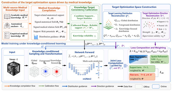
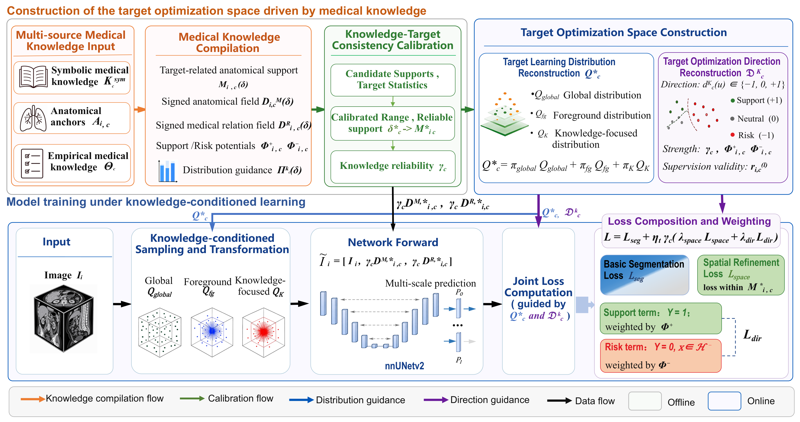
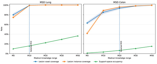
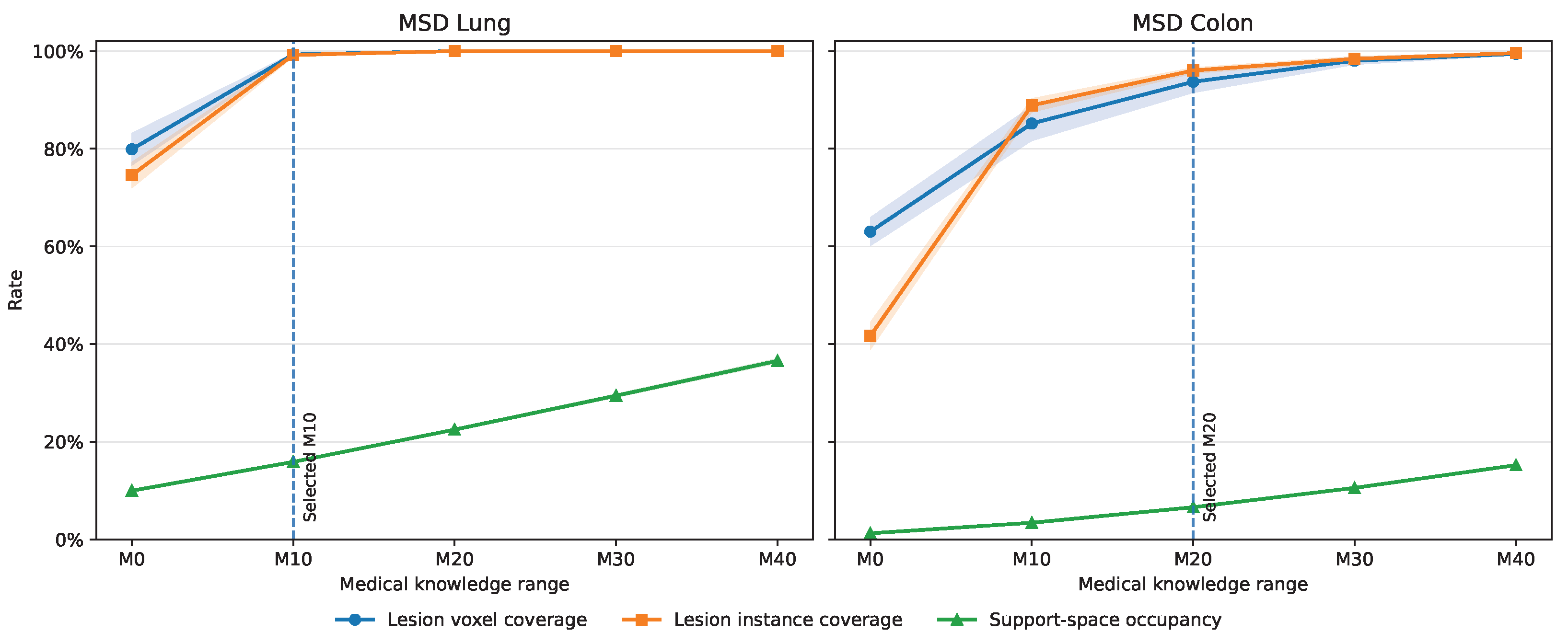
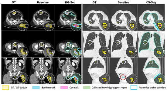
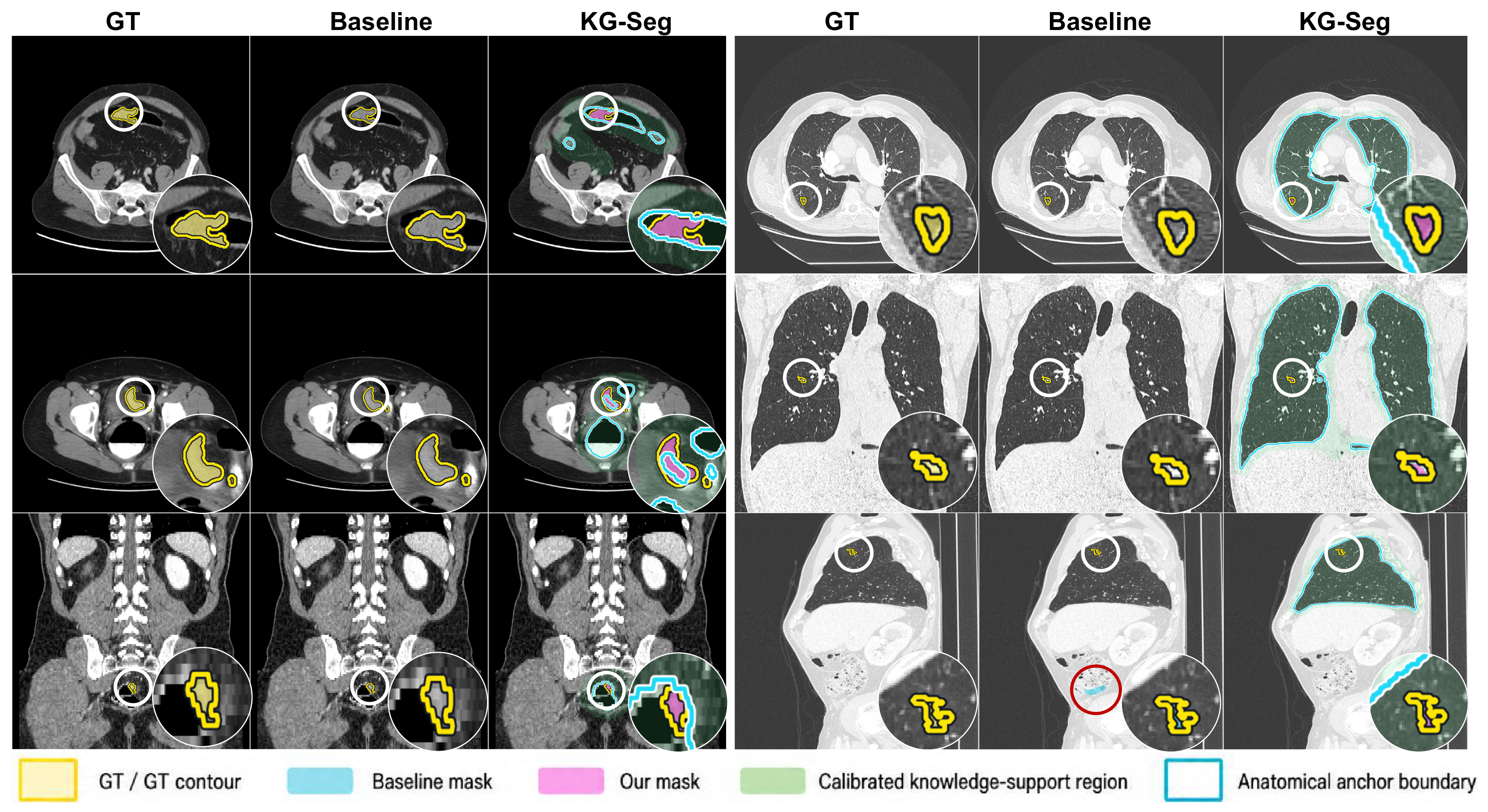

Disclaimer/Publisher’s Note: The statements, opinions and data contained in all publications are solely those of the individual author(s) and contributor(s) and not of MDPI and/or the editor(s). MDPI and/or the editor(s) disclaim responsibility for any injury to people or property resulting from any ideas, methods, instructions or products referred to in the content. |
© 2026 by the authors. Licensee MDPI, Basel, Switzerland. This article is an open access article distributed under the terms and conditions of the Creative Commons Attribution (CC BY) license.