


Abstract
Skin hyperpigmentation and ultraviolet (UV)-induced photodamage represent prevalent cosmetic concerns that demand safe and effective topical intervention strategies. This study evaluated the skin-whitening, pigmentation-improving, and post-UV repair efficacy of a novel dual-compartment sheet mask (DCSM) formulated with eight complementary depigmenting ingredients, namely niacinamide, α-arbutin, glabridin, hexylresorcinol, ascorbyl tetraisopalmitate (VC-IP), paeonol, Camellia japonica flower extract and Ormosia henryi bark extract, through two independent human efficacy studies and in vitro cell experiments. In a 28-day, self-controlled human efficacy study (Cohort 1; n = 32), regular DCSM use was associated with significant increases in the individual typology angle (ITA), significant decreases in the b* value, significant reductions in H-channel standard deviation (H-SD), significant decreases in the melanin index (MI), and significant diminishment of pigment spot area and optical density, suggesting progressive and sustained improvements in skin tone, uniformity, and pigmentation. In a separate UV-induced skin erythema model (Cohort 2; n = 30), seven days of daily DCSM application significantly attenuated UV-induced increases in MI, erythema index (EI), and a* value while promoting recovery of skin lightness (ITA) relative to untreated control sites, indicating effective post-sun erythema alleviation and pigmentation suppression. In vitro, treatment of α-MSH-stimulated A-875 human melanoma cells with 5% (v/v) DCSM emulsion significantly suppressed intracellular melanin synthesis at a non-cytotoxic concentration, providing supportive evidence consistent with the human efficacy findings observed in vivo. Collectively, these multidimensional findings indicate that DCSM use was associated with improvements in skin hyperpigmentation, an even skin tone, and facilitated recovery from UV-induced photodamage, supporting its potential as a cosmetic intervention for pigmentation regulation and post-sun skin repair.
1. Introduction
Skin hyperpigmentation, encompassing conditions such as melasma, post-inflammatory hyperpigmentation (PIH), and ultraviolet (UV)-induced pigmentation, represents one of the most prevalent aesthetic concerns worldwide [1,2], with a particularly high burden among Asian populations [3,4]. These conditions are associated with visible changes in skin tone, pigmentation heterogeneity, and uneven complexion, and are frequently linked to psychosocial distress and diminished quality of life [2,5]. Against the backdrop of intensifying global demand for effective skin-whitening solutions, driven by growing awareness of UV radiation and environmental stressors as primary triggers of skin hyperpigmentation, the cosmetic industry has responded with a broad range of topical skin-whitening products [6,7,8]. However, rigorous evidence supporting their efficacy through validated instrumental assessment remains limited. Among the available topical delivery formats, facial masks have emerged as a dominant product category owing to their ability to provide an occlusive, prolonged-contact delivery environment that enhances the penetration and bioavailability of active ingredients [9].
UV radiation is the primary environmental trigger of acquired hyperpigmentation. Beyond inducing delayed tanning, UV exposure simultaneously provokes erythema and persistent pigmentation, which compound visible skin damage [10,11,12]. Effective skin whitening and post-sun repair formulations must therefore address both UV-induced erythema and pigmentation simultaneously, a dual requirement that underscores the need for multi-active formulation strategies combining ingredients with both erythema-reducing and melanin-inhibiting properties [13,14].
To meet this demand, the cosmetic industry has developed a broad repertoire of topical whitening agents with complementary functional profiles. Niacinamide, α-arbutin, glabridin, and hexylresorcinol are among the most well-validated depigmenting actives [14,15]. Ascorbyl tetraisopalmitate (VC-IP), a lipophilic vitamin C ester, offers enhanced stratum corneum penetration and antioxidant activity well-suited for emulsion-based systems [16,17,18,19]. Paeonol, derived from Paeonia suffruticosa, has been reported to suppress melanin synthesis and attenuate UV-induced skin responses [20]. In addition, botanical extracts such as Camellia japonica flower extract [21,22] and Ormosia henryi Prain, which are enriched in flavonoids with notable antioxidant properties [23], have demonstrated complementary bioactivities that further broaden the functional scope of multi-active formulations. Combining these agents within a single formulation represents a rational multi-active strategy for broader depigmenting efficacy [14,15]. Sheet masks serve as an efficacious delivery vehicle for such combinations, as their semi-occlusive nature enhances skin hydration, promotes transdermal penetration, and maximizes contact time with the stratum corneum [24,25]. However, the simultaneous incorporation of lipophilic and hydrophilic actives into a single formulation poses a significant challenge to chemical stability, as interactions between oil-soluble and water-soluble components may compromise ingredient integrity prior to application. This challenge remains insufficiently addressed in multi-active whitening formulations. To address this challenge, a dual-compartment design was adopted, physically separating the oil-phase and water-phase components prior to use, with the aim of preserving the chemical stability of both lipophilic and hydrophilic ingredients.
In the present study, we conducted a comprehensive human efficacy and in vitro evaluation of a novel dual-compartment sheet mask (DCSM) formulated with eight complementary depigmenting ingredients: niacinamide, α-arbutin, glabridin, hexylresorcinol, VC-IP, paeonol, Camellia japonica flower extract, and Ormosia henryi bark extract. Two independent human efficacy studies were conducted in Chinese subjects: Cohort 1 assessed long-term improvements in skin color, melanin content, and pigmentation status over 28 days of regular use, employing melanin index (MI), individual typology angle (ITA), b* value, H-channel standard deviation (H-SD), and VISIA-based pigment spot analysis; Cohort 2 evaluated post-UV irradiation repair efficacy using a validated minimal erythema dose (MED)-based model, with MI, erythema index (EI), a* value, and ITA assessed as key efficacy indicators. In addition, the anti-melanogenic activity of the formulation was examined in vitro using α-MSH-stimulated A-875 human melanoma cells, with intracellular melanin content and cell viability as endpoints. Together, these studies provide a multidimensional evidence base for the whitening and post-sun repair efficacy of this multi-active facial mask formulation.
2. Materials and Methods
2.1. Composition and Application of the Mask
The dual-compartment sheet mask (DCSM) comprised two physically separated compartments: the oil-phase compartment contained glabridin, Ormosia henryi bark extract, and VC-IP, selected based on their lipid-soluble properties. The water-phase compartment contained a patented composite ingredient comprising niacinamide, hexylresorcinol, paeonol, Camellia japonica flower extract, and α-arbutin. The precise concentrations of individual active ingredients and the oil-to-water ratio of the DCSM are proprietary and cannot be disclosed. All ingredients are incorporated at concentrations compliant with applicable cosmetic regulatory guidelines.
Prior to each application, the oil-phase compartment contents were dispensed into the water-phase compartment containing the pre-immersed mask sheet. The two phases were combined and thoroughly emulsified by manual shaking for 30 s followed by gentle inversion mixing for an additional 30 s, yielding a transient homogeneous milky-white oil-in-water dispersion that simultaneously saturated the mask sheet. No specialized emulsification apparatus was required, as the formulation was designed for consumer self-preparation. The saturated mask sheet was then removed and applied to the designated test area for 15 min.
2.2. Ethical Statement
This study was conducted in accordance with the Declaration of Helsinki. Both human efficacy studies were approved by the Ethics Committee of Fudan University (Approval No. FE222411; approval on 2 November 2024). All volunteers were fully informed of the study’s purpose, procedures, potential risks, benefits, rights, and obligations associated with study participation, and provided written informed consent prior to enrollment.
2.3. Human Efficacy Studies
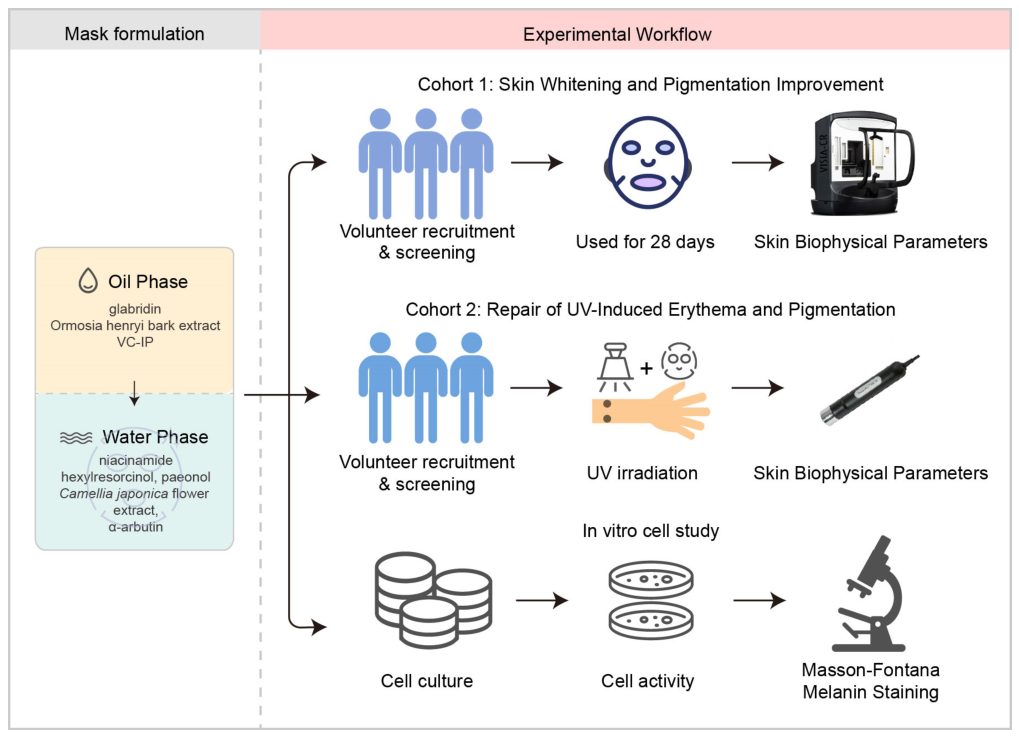
Stability, compatibility, and safety assessments were conducted in accordance with applicable regulatory requirements prior to human efficacy testing; however, detailed data from these assessments are proprietary and are not presented in this manuscript. The selection of subjects for both cohorts was carried out strictly according to the methods outlined in Chapter 8 of the Cosmetic Safety and Technical Standards (2015 Edition) for human efficacy evaluation. The human study portion of this research consisted of two independent cohorts. The first cohort primarily investigated the effects of the DCSM on skin color tone, melanin content, and pigmentation status. The second cohort focused on evaluating its skin repair efficacy following ultraviolet irradiation. The two human efficacy cohorts were conducted as independent studies with separate subject populations, distinct study periods, and non-overlapping objectives. The overall study design and experimental workflow, including mask formulation composition and the two human efficacy cohorts, are schematically summarized in Figure 1.
Figure 1.
Schematic overview of the study design and experimental workflow, including mask formulation composition, Cohort 1 (skin whitening and pigmentation improvement), Cohort 2 (repair of UV-induced erythema and pigmentation), and in vitro cell study. VC-IP—ascorbyl tetraisopalmitate; UV—ultraviolet; Downward arrows—Prior to each application, the oil-phase compartment contents were dispensed into the water-phase compartment containing the pre-immersed mask sheet.
2.3.1. Cohort 1: Skin Whitening and Pigmentation Improvement
Inclusion Criteria
Eligible participants were healthy Chinese females aged 18–45 years who were willing to participate and provide written informed consent. They were required to have sensitive skin with a score of ≥18 on the Huaxi Sensitive Skin Questionnaire (HSSSQ); facial acne scars assessed by investigators, accompanied by facial pigmentation with a cheek pigmentation grade ≥ 2 (excluding freckles, pigmented nevi and other types of pigmentation that are difficult to improve with topical preparations); and self-reported dull, sallow and lackluster facial skin. Participants also needed to be able to cooperate well with the study and maintain a regular lifestyle during the test period and to read and understand all of the contents of the informed consent form and portrait use authorization form and sign both voluntarily. They were not permitted to use any cosmetics, drugs or health products that could affect the test results during the study period.
Exclusion Criteria
We excluded subjects with obvious scars, birthmarks or other conditions in the test area that had the potential to affect outcome evaluation; those with a history of severe allergic reactions; pregnant or lactating females, or those planning to become pregnant during the study period; those with severe cardiac, hepatic or renal dysfunction and severe immunodeficiency; those with mental illness, severe endocrine and metabolic diseases, or those taking oral contraceptives; those who have long-term reliance on hormones, antihistamines or other medications to maintain their physical condition; those who had participated in other clinical drug trials or related trials within 60 days prior to the study, or used drugs that may have affected test results within the last 2 weeks; those who had received facial medical cosmetic care, beauty salon care or treatment within 90 days prior to the study; those who started using cosmetics or health products with similar efficacy that could affect test results within 30 days prior to the study; and those judged by investigators as inappropriate for participation in this study.
Protocol Restrictions
To minimize potential confounding factors and ensure the validity of the study outcomes, all participants were required to comply with the following restrictions throughout the study period: participants were prohibited from using any therapeutic agents, cosmetics, topical pharmaceutical preparations, or dietary supplements intended to improve skin condition that could potentially interfere with the study results. Medical procedures, massages, and professional aesthetic salon treatments were strictly forbidden during the study period; a 7-day washout period was required prior to study initiation, during which participants discontinued the use of any facial skincare products of the same category or that were claimed to have similar efficacy to the test samples. Participants were instructed to maintain their habitual daily routines consistent with those prior to study enrollment. Intense outdoor sun exposure was to be minimized, and travel, vacations, or substantially irregular lifestyle patterns were not permitted during the study period; the use of all other skincare steps and products was required to remain consistent with pre-study habits. Participants were neither permitted to discontinue any skincare products that were in use prior to enrollment, nor to introduce any new facial skincare products during the study period; on each assessment visit day, participants were instructed to refrain from applying any skincare products to the designated test areas prior to instrumental measurements.
Study Protocol
This was a single-center, prospective, self-controlled human efficacy study. All subjects applied one sheet of DCSM once every other day for 28 consecutive days. Skin parameter measurements were performed at baseline (D0), Day 10 (D10) and Day 28 (D28). Quantitative evaluation of skin biophysical parameters was conducted using multiple professional skin-detection instruments. The specific efficacy evaluation indicators, test instruments, and corresponding interpretations are summarized in Table 1. All skin measurements were performed in a standardized environment at 21 ± 1 °C and 50 ± 10% relative humidity (RH). To minimize measurement variability, all instrumental assessments were performed under standardized conditions. Subjects were required to acclimatize in the controlled measurement room (21 ± 1 °C, 50 ± 10% RH) for at least 20 min prior to measurement. Subjects were instructed to refrain from applying any skincare products to the test sites on the day of each assessment visit. Probe-based measurements were performed by the same trained operator throughout the study, with standardized probe pressure applied according to the manufacturer’s instructions.
Table 1.
Skin color and pigmentation evaluation indicators.
2.3.2. Cohort 2: Repair of UV-Induced Erythema and Pigmentation
Inclusion Criteria
Eligible participants were healthy Chinese females aged 18–55 years (excluding pregnant or lactating women) who were willing to participate and provide written informed consent; they had a skin ITA value of 28–50° at the test site; no abnormal manifestations such as pigmentation, inflammation, scars, pigmented nevi, or hypertrichosis on the test site; no history of photosensitivity diseases and no recent use of drugs affecting photosensitivity; willingness to accept artificial light irradiation on the test site; no history of allergic diseases or allergies to cosmetics or other topical preparations; regular skincare habits and consistent use of skincare products; the ability to strictly cooperate with the study protocol and maintain a regular lifestyle during the study period.
Exclusion Criteria
We excluded subjects who used antihistamines within the past week or immunosuppressants within the past month; those with a history of psoriasis, eczema, atopic dermatitis, severe acne, or other conditions that could have affected outcome evaluation; those with a history of allergic diseases or allergies to cosmetics or other topical preparations; those who used anti-inflammatory drugs on the test site within the past 2 months; subjects with insulin-dependent diabetes mellitus; asthma or other chronic respiratory diseases under treatment; subjects involved on ongoing dermatological therapy; subjects who had undergone anticancer chemotherapy within the past 6 months; subjects with immunodeficiency or autoimmune diseases; pregnant or lactating women, or those planning to conceive in the near future; subjects who had participated in other interventional studies within the past 2 months; subjects with highly sensitive constitution; people who were unable to complete the study protocol; and those deemed unsuitable by the investigator.
Protocol Restrictions
To control for potential confounding factors and ensure the integrity of the study outcomes, all participants were required to adhere to the following restrictions: for one month prior to and throughout the entire study period, participants were prohibited from using any topical pharmaceutical preparations or dietary supplements intended to improve skin condition that could potentially interfere with the study results. Massages and medical procedures were also strictly forbidden; within 2 days prior to each assessment visit, participants were instructed to refrain from applying any functional skincare products (including cosmetics and skincare preparations) or topical medications to the designated test areas; during the study period, participants were advised to minimize intense outdoor sun exposure and to avoid travel or vacations. When outdoor activities were unavoidable, appropriate photoprotective measures were required, including wearing hats, using umbrellas, and wearing sun-protective clothing; during the study period, the test product replaced participants’ habitual skincare products of the same category, while all other skincare routines and products remained unchanged; throughout the study period, participants were instructed to maintain a regular daily routine and to avoid excessive sleep deprivation.
Study Protocol
A split-site self-controlled experimental design was implemented in this study. To ensure subject safety, a stable ultraviolet-induced skin erythema model was established by artificial UV irradiation on the medial forearms of each subject. DCSM-treated and negative control sites were designated on the contralateral forearms; assignment to the left or right forearm was determined on an individual basis following assessment of site eligibility. Sites were evenly distributed within calibrated test regions to ensure comparable skin conditions between sites.
UV irradiation was performed using a Solar Light solar simulator (UVA+B mode; Model 601–300 W V2.5, Multiport, Solar Light Company, Inc., Glenside, PA, USA) equipped with a 300 W xenon arc lamp. Each test site was irradiated through a 0.64 cm2 (0.8 cm × 0.8 cm) aperture placed directly on the skin, delivering a dose of 2× the individual minimal erythema dose (MED) [26]. Following UV irradiation, the test sites were divided into two groups: an untreated area serving as the negative control, and a treatment area to which the DCSM was applied. The DCSM was cut to a size of no less than 6 cm2 and applied to each designated test site in accordance with Chapter 8 of the Cosmetic Safety and Technical Standards (2015 Edition). The mask was applied once daily and left on for 15 min, followed by gentle massage until the remaining essence was fully absorbed.
Skin biophysical parameters were assessed by professional instrumental detection, along with on-site instrumental and investigator evaluation of skin status by trained investigators, at baseline (D0) (24 h after UV irradiation and prior to the first DCSM application) and Day 7 after treatment (D7). All efficacy evaluation indicators and corresponding testing instruments are summarized in Table 2. All skin measurements were performed in a standardized environment at 21 ± 1 °C and 50 ± 10% relative humidity. To minimize measurement variability, subjects were required to acclimatize in the controlled measurement room (21 ± 1 °C, 50 ± 10% RH) for at least 20 min prior to each measurement. Subjects were instructed to refrain from applying any products to the test sites on the day of each assessment visit. Probe-based measurements were performed by the same trained operator throughout the study. VISIA CR image analysis was performed using standardized software-based quantification (Image-Pro Plus 7.0, IPP; Canfield Scientific, Inc., Parsippany, NJ, USA) to minimize subjective evaluator bias.
Table 2.
Skin erythema and pigmentation evaluation indicators.
2.4. In Vitro Cell Experiments
2.4.1. Cell Culture and Treatment
The A-875 human melanoma cell line was purchased from Sunncell (#SNL-232, Sunncell, Shanghai, China) and cryopreserved at passage 1 (P1) with a cell density of 1 × 10⁶ cells per vial. A-875 cells were cultured in high-glucose DMEM (#E600003-05005, Sangon Biotech, Shanghai, China) supplemented with 10% fetal bovine serum (#E600051-0500, Sangon Biotech, Shanghai, China) and 1% penicillin–streptomycin solution (#E607011-0100, Sangon Biotech, Shanghai, China) in a humidified incubator with 5% CO2 at 37 °C, and cells at passages 3 to 5 (P3–P5) were used for all in vitro experiments. The emulsion obtained from the DCSM (hereafter referred to as DCSM emulsion) was used for all in vitro cell experiments. For cytotoxicity screening, the DCSM emulsion was serially diluted with complete culture medium to obtain five concentration gradients of 20%, 10%, 5%, 2.5%, and 1.25%. The MTT assay with a working concentration of 0.5 mg/mL (#ST1537, Beyotime, Shanghai, China) was utilized to detect and evaluate cell viability after treatment. Briefly, MTT solution was added to each well and incubated at 37 °C with 5% CO2 for 2 h, after which the formazan crystals were dissolved in DMSO and absorbance was measured. A predefined cell viability threshold of ≥80% relative to the untreated control was used to define non-cytotoxic concentrations, consistent with Cosmetic Safety and Technical Standards (2015 Edition). Absorbance values were measured at 570 nm with reference wavelength correction at 630 nm. The blank control (culture medium without cells) was used for instrument zeroing. Cell viability was calculated as the percentage relative to the untreated control group (cells cultured in medium only, set as 100%):
2.4.2. Fontana–Masson Melanin Staining
Intracellular melanin deposition was visualized and detected using a commercial Fontana–Masson melanin staining kit (#G2032, Solarbio Life Sciences, Shanghai, China). A-875 cells were seeded into 12-well plates pre-placed with cell slides and divided into four experimental groups, including the blank control group, the model group induced by 100 nM α-melanocyte-stimulating hormone (α-MSH, #A1025, APExBIO, Houston, TX, USA), the positive control group treated with 100 nM α-MSH combined with 500 μg/mL kojic acid (#K3125, Sigma-Aldrich, St. Louis, MO, USA), and the experimental group treated with α-MSH and the DCSM emulsion. After continuous incubation for 48 h, the cell culture medium was discarded, and the cells were rinsed thoroughly with DPBS (#E607009-0500, Sangon Biotech, Shanghai, China). Cells were fixed with 4% paraformaldehyde (#BL539A, Biosharp, Shanghai, China) at room temperature for 15 min and washed again with DPBS. Subsequently, cells were incubated with ammoniacal silver solution at 56 °C in the dark for 30 min, fully rinsed with DPBS, treated with hypo-solution for 3 min, and finally washed with DPBS to complete the staining procedure. Microscopic observation and image acquisition were performed after staining. Images were acquired under identical microscopic settings (magnification and white balance) across all groups to ensure consistency. ImageJ software (Version 1.54f, National Institutes of Health, Bethesda, MD, USA) was used to analyze the integrated optical density (IOD) of the stained images to quantitatively reflect intracellular melanin content, with values normalized to the α-MSH-treated model group (set as 100%) to enable direct comparison across treatment groups.
2.5. Statistical Analysis
Human efficacy study data were statistically analyzed using GraphPad Prism 10.6.1. The Shapiro–Wilk test was applied to assess data normality. Data with a Shapiro–Wilk p-value > 0.05 were considered normally distributed and analyzed using the paired t-test; data with a p-value ≤ 0.05 were regarded as non-normally distributed and analyzed using the Wilcoxon signed-rank test. All statistical tests were two-tailed, and p < 0.05 was considered statistically significant. Cell experiment data were analyzed by one-way ANOVA followed by Tukey’s multiple comparisons test. * p < 0.05, ** p < 0.01, *** p < 0.001, **** p < 0.0001.
3. Results
3.1. Human Efficacy of the DCSM on Skin Whitening and Pigmentation Improvement
To evaluate the skin-whitening and depigmenting efficacy of the DCSM, a 28-day self-controlled human efficacy study (Cohort 1) was conducted from 10 October to 7 November 2025. A total of 32 healthy Chinese female subjects were recruited; all completed the full study and were included in the final analysis. The mean age was 39.19 ± 5.11 years (range: 21–45 years). The age distribution was slightly skewed toward older participants, consistent with the target population of subjects presenting with facial pigmentation concerns. All participants fulfilled the predefined inclusion criteria. Informed consent was obtained from all participants. Skin colorimetric and pigmentation parameters were evaluated at D0, D10, and D28.
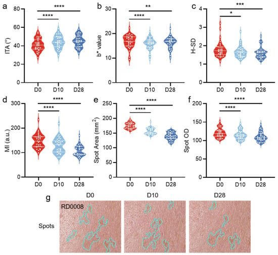
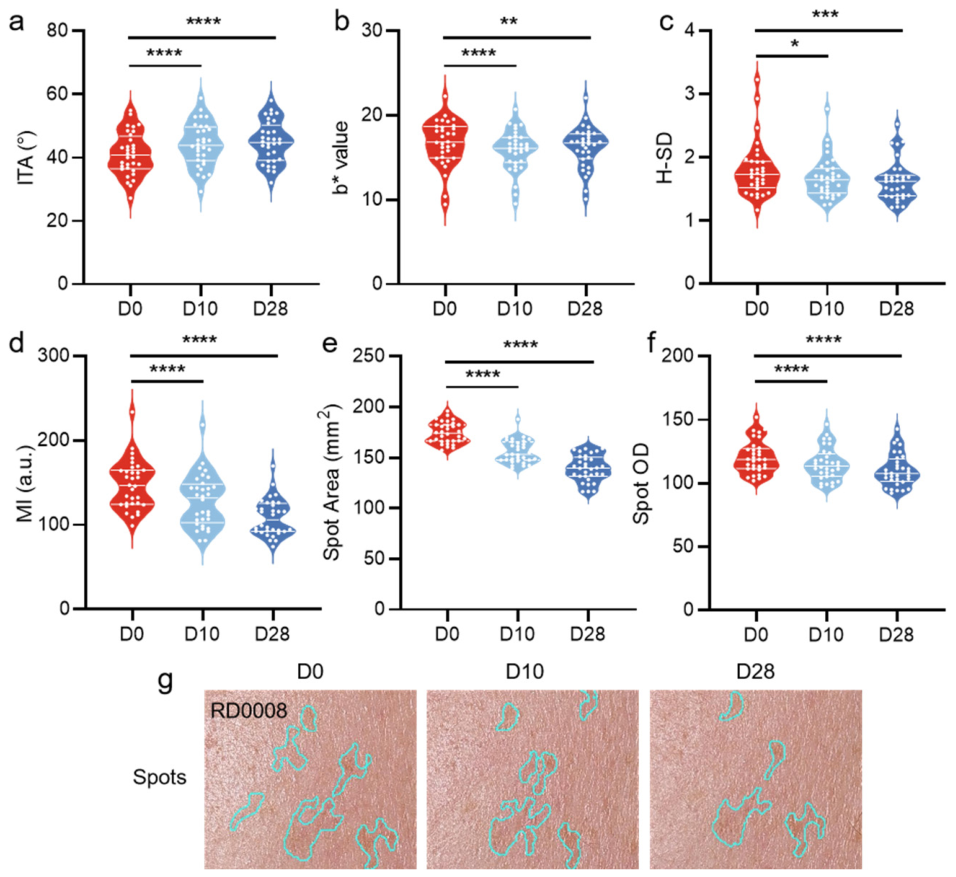
Compared with baseline, significant improvements across multiple instrumental skin parameters were observed at both D10 and D28 following DCSM application (Figure 2, Table S1). Specifically, the ITA (Figure 2a), a colorimetric indicator of skin lightness, was significantly elevated, while the b* value (Figure 2b), reflecting the degree of skin yellowness, was markedly decreased. Furthermore, the H-SD (Figure 2c), an index of skin color homogeneity, was significantly reduced, and the MI (Figure 2d), reflecting cutaneous melanin content, was markedly decreased. With respect to pigmentation amelioration, both the average spot area (Figure 2e) and optical density of pigment spots (Figure 2f) declined significantly over the treatment period. Representative visual imaging further corroborated these findings, showing progressive lightening of pigment spots and a continuous reduction in spot area from D0 to D28 (Figure 2g), consistent with the instrumental data. These results collectively indicate that DCSM use was associated with reductions in melanin deposition, improvements in skin tone uniformity, and alleviation of facial hyperpigmentation in a time-dependent manner.
Figure 2.
Changes in instrumental evaluation parameters at different time points following DCSM application. (a) ITA, (b) b* value, (c) H-SD, (d) MI, (e) spot area, (f) spot OD and (g) representative VISIA images of a subject at D0, D10, and D28. Data are presented as violin plots with individual data points. The middle line represents the median. The lower and upper lines represent the 25th and 75th percentiles, respectively. Data normality was assessed using the Shapiro–Wilk test. As all outcome measures met the assumption of normality, paired Student’s t-tests were applied for all comparisons. Raw p-values were adjusted using the Benjamini–Hochberg false discovery rate (FDR) correction to account for multiple comparisons across all 12 endpoints (6 outcome measures × 2 time points). n = 32. * p < 0.05, ** p < 0.01, *** p < 0.001, **** p < 0.0001. ITA—individual typology angle. H-SD—standard deviation of channel H. MI—melanin index. Spot OD—Spot Optical Density.
3.2. Human Efficacy of the DCSM in Repairing UV-Induced Skin Erythema and Pigmentation
To evaluate the post-sun repair efficacy of DCSM, a UV-induced skin erythema model was established in 30 healthy female subjects (mean age: 42.6 ± 9.5 years; range: 19–54 years) by irradiation at twice the minimal erythema dose from July 23 to August 1, 2025. All subjects completed the study and were included in the final analysis. Informed consent was obtained from all participants.
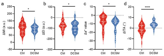
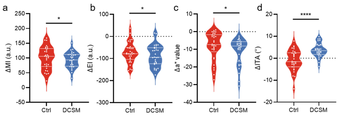
Seven days after UV irradiation (D7), changes (Δ = D7 − D0) in skin physiological parameters were compared between DCSM-treated and untreated control sites (Figure 3, Table S2). Compared with the control sites, DCSM-treated sites exhibited a significantly attenuated increase in MI (Figure 3a), indicating more effective suppression of UV-induced melanogenesis. Both groups showed negative ΔEI (Figure 3b) and Δa* (Figure 3c) values, reflecting spontaneous post-UV resolution of erythema and skin redness, respectively; however, the DCSM-treated sites exhibited significantly more marked reductions in both ΔEI and Δa*, indicating greater attenuation of UV-induced erythema. Strikingly, DCSM-treated sites maintained a positive ΔITA in contrast to the negative ΔITA observed at control sites (Figure 3d), suggesting that DCSM may have contributed to the preservation of skin lightness following UV exposure. These findings highlight the potential of DCSM in post-UV skin repair, encompassing both erythema alleviation and mitigation of UV-induced pigmentation.
Figure 3.
Comparison of changes in skin physiological parameters between DCSM-treated and untreated control sites following UV irradiation. (a) ΔMI, (b) ΔEI, (c) Δa* value, and (d) ΔITA represent the changes from D0 to D7 at untreated Ctrl sites and DCSM sites. Data are presented as violin plots with individual data points. The middle line represents the median; the lower and upper lines represent the 25th and 75th percentiles, respectively. Data normality was assessed using the Shapiro–Wilk test. Statistical significance was determined by a paired t-test for normally distributed data or a Wilcoxon signed-rank test for non-normally distributed data. Raw p-values were adjusted using the Benjamini–Hochberg false discovery rate (FDR) correction to account for multiple comparisons across all 4 endpoints; n = 30. * p < 0.05, **** p < 0.0001. DCSM—dual-compartment sheet mask; MI—melanin index; EI—erythema index; ITA—individual typology angle; Ctrl—control.
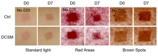
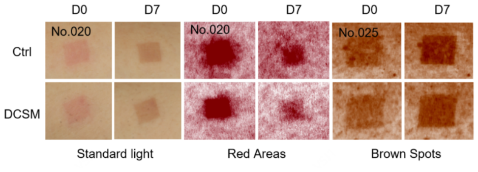
Representative subjects were selected to illustrate skin changes at D0 and on D7, based on a comprehensive assessment of skin phenotype and observable skin changes (Figure 4). Visual imaging revealed that, compared with baseline, control sites exhibited notable skin darkening and intensified brown spots at D7, with erythema persisting in the UV-irradiated area. In contrast, DCSM-treated sites demonstrated more pronounced resolution of erythema and visible lightening of brown spots relative to the control sites at D7. These visual observations were consistent with the instrumental measurement results, further supporting the efficacy of DCSM in alleviating UV-induced erythema and reducing skin pigmentation.
Figure 4.
Representative images of UV-induced skin changes at D0 and D7. Ctrl vs. DCSM. DCSM—dual-compartment sheet mask; Ctrl—control.
3.3. Inhibitory Effect of the DCSM on Melanin Synthesis in A-875 Melanoma Cells
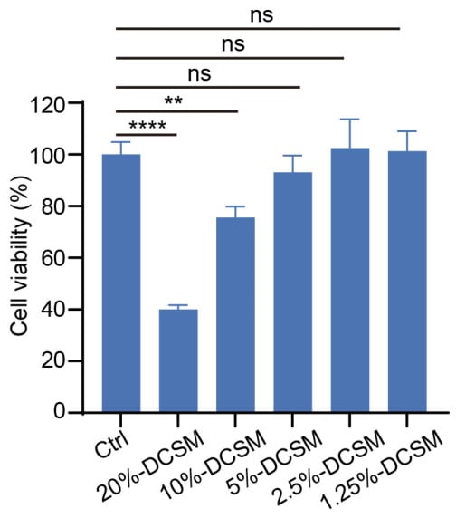
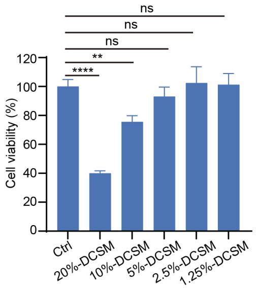
To investigate the potential skin-lightening mechanism of DCSM at the cellular level, its effect on intracellular melanin biosynthesis was evaluated in A-875 human melanoma cells. Prior to melanin induction experiments, cell viability was assessed across serially diluted DCSM concentrations (Figure 5). While 20% and 10% DCSM markedly reduced cell viability, concentrations of 5%, 2.5%, and 1.25% exhibited no significant cytotoxicity. Given that 5% DCSM showed no significant difference relative to the untreated control, this concentration was selected for subsequent melanin induction experiments.
Figure 5.
Effect of the DCSM at different concentrations on the viability of A-875 melanoma cells. A-875 cells were treated with varying concentrations of DCSM (1.25%, 2.5%, 5%, 10%, and 20%, v/v). Data are presented as mean ± SD. Statistical differences among groups were determined by one-way ANOVA followed by Tukey’s multiple comparisons test. Experiments were performed in triplicate and repeated independently at least twice with similar results. ** p < 0.01, **** p < 0.0001; ns—not significant. DCSM—dual-compartment sheet mask emulsion; Ctrl—control.
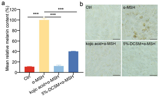
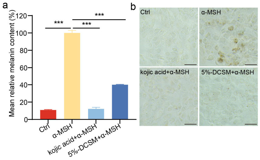
To further investigate the inhibitory effect of DCSM on melanin synthesis, intracellular melanin content was quantified by Fontana–Masson silver staining (Figure 6). Compared with the untreated control, α-MSH treatment significantly elevated melanin levels, indicating successful establishment of the melanogenesis induction model. The positive control (kojic acid + α-MSH) markedly reduced melanin content relative to the α-MSH group, supporting the reliability of the experimental system. Similarly, treatment with 5% (v/v) DCSM + α-MSH significantly decreased intracellular melanin content (Figure 6a). Representative staining micrographs (Figure 6b) further corroborated these observations, showing a marked increase in brown melanin granules in the α-MSH group, which were substantially reduced in both the kojic acid + α-MSH and DCSM + α-MSH groups. Overall, these results suggest that DCSM can inhibit melanin biosynthesis in A-875 melanoma cells at non-cytotoxic concentrations, highlighting its potential as a skin-lightening agent, possibly through the suppression of melanin synthesis.
Figure 6.
Effects of DCSM on intracellular melanin synthesis in A-875 melanoma cells. Intracellular melanin content was assessed by Fontana–Masson silver staining following 48 h treatment with α-MSH (melanin inducer), kojic acid + α-MSH (positive control), or 5% (v/v) DCSM + α-MSH. (a) Quantification of mean relative melanin content (%), with the α-MSH-treated group set as 100% reference. (b) Representative silver staining micrographs of each treatment group; melanin deposits appear as brown granules. Scale bars = 200 μm. Data are presented as mean ± SD. Statistical differences were determined by one-way ANOVA followed by Tukey’s multiple comparisons test. Experiments were performed in triplicate and repeated independently at least twice with similar results. *** p < 0.001. DCSM—dual-compartment sheet mask emulsion. α-MSH—α-melanocyte-stimulating hormone; Ctrl—control.
4. Discussion
The present study systematically evaluated the skin-whitening, pigmentation reduction, and post-UV repair efficacy of a DCSM formulated with multiple functional whitening ingredients, through a combination of human efficacy studies and in vitro cellular experiments. The DCSM significantly reduced melanin content, enhanced skin tone uniformity, lightened facial pigment spots, attenuated UV-induced erythema, and suppressed intracellular melanin synthesis in A-875 melanoma cells.
4.1. Rationale for Multi-Ingredient Formulation Design
The dual-compartment design of the mask was intended to preserve the stability and bioactivity of chemically incompatible ingredients by separating the oil-phase and water-phase components prior to use. This formulation strategy was designed to ensure that labile active ingredients, particularly the lipophilic VC-IP, retained their antioxidant and anti-melanogenic potency until the point of application [14]. VC-IP is a stable, lipophilic form of vitamin C that readily penetrates the stratum corneum and is subsequently converted to free ascorbic acid within the skin, where it exerts tyrosinase inhibitory activity and reduces melanin intermediates [8,27]. Beyond its anti-melanogenic properties, ascorbic acid and its derivatives also stimulate collagen synthesis, suggesting additional anti-aging benefits of VC-IP in the present formulation [18]. Clinical evidence has confirmed that topical formulations containing ascorbyl tetraisopalmitate can effectively improve skin hyperpigmentation and photoaging [27].
The oil-phase compartment also contained glabridin, a principal bioactive isoflavan derived from licorice root (Glycyrrhiza glabra). Glabridin has been extensively documented as a potent reversible inhibitor of tyrosinase, the rate-limiting enzyme in the melanogenesis pathway, and has demonstrated skin-lightening activity both in vitro and in clinical settings [8,28]. In addition, Ormosia henryi extract, a botanical ingredient whose species is characterized by rich flavonoid content and notable antioxidant capacity [23], was incorporated to further scavenge UV-induced ROS and attenuate oxidative stress-driven melanocyte activation [14]. The water-phase compartment incorporated niacinamide, hexylresorcinol, paeonol, Camellia japonica flower extract, and α-arbutin, each contributing to the overall anti-pigmentation effect through distinct and complementary mechanisms. Niacinamide is well established to inhibit the transfer of melanosomes from melanocytes to keratinocytes, thereby reducing visible pigmentation without directly inhibiting tyrosinase [8,28]. Hexylresorcinol, a resorcinol derivative, has been shown to possess superior tyrosinase inhibitory activity compared with kojic acid and arbutin, and exhibits potent anti-melanogenic effects at low concentrations [29,30]. α-Arbutin inhibits tyrosinase activity through competitive inhibition, reducing melanin biosynthesis in a dose-dependent manner [31,32]. The clinical application of hypopigmenting agents such as arbutin and niacinamide has been well-documented in the management of melasma and other pigmentary disorders [33]. The synergistic combination of these mechanistically diverse ingredients likely underlies the robust and multidimensional depigmenting efficacy observed in this study.
4.2. Human Efficacy of the DCSM on Skin Whitening and Pigmentation Improvement (Cohort 1)
In Cohort 1, significant improvements across all instrumental skin parameters were observed at D10 and D28 relative to baseline. Specifically, the significant increase in ITA and concurrent decrease in b* value indicate a colorimetric shift toward a fairer and less yellowish skin tone, which represents a meaningful outcome for Asian skin types characterized by a tendency toward sallow complexion. The reduction in H-SD reflects enhanced skin tone uniformity, which is particularly relevant for subjects presenting with uneven pigmentation, as stipulated by the inclusion criteria of this cohort. The concurrent reductions in both the area and optical density of pigment spots provide objective, quantitative evidence of the formulation’s capacity to attenuate existing hyperpigmented lesions, consistent with findings from prior human efficacy studies evaluating multi-ingredient depigmenting formulations [8,34]. Visual imaging results further corroborated the instrumental findings, suggesting progressive lightening of pigment spots with continuous use.
It should be acknowledged that Cohort 1 employed a self-controlled design without a parallel untreated control group, which precludes definitive exclusion of time-dependent spontaneous skin changes or confounding effects attributable to concomitant skincare practices [35]. Nevertheless, the multi-parametric, time-cumulative significant improvements observed are consistent with the established pharmacological profiles of the active ingredients employed, including niacinamide, which requires continuous application over several weeks to yield clinically detectable reductions in melanin index and pigmentation [36,37], supporting the attribution of the observed effects to the active ingredients’ cumulative pharmacological activity rather than spontaneous temporal variation.
4.3. Human Efficacy of the DCSM in Repairing UV-Induced Skin Erythema and Pigmentation (Cohort 2)
In Cohort 2, a standardized UV-induced erythema model was established via 2× individual MED irradiation, and a split-site, self-controlled design was employed to minimize inter-individual variability. Following 7 days of daily application, DCSM-treated sites exhibited significantly attenuated increases in ΔMI, ΔEI, and Δa*, alongside a significantly greater ΔITA, relative to untreated control sites, collectively indicating effective suppression of UV-induced melanogenesis and accelerated erythema resolution. The occlusive properties inherent to the sheet mask format may further enhance percutaneous penetration and prolong skin contact time, potentially facilitating the delivery of active ingredients to target cutaneous layers [25].
The anti-erythema effect is likely mediated by the combined anti-inflammatory and antioxidant activities of the formulation’s ingredients. UV irradiation triggers a cascade of inflammatory responses in the skin, including the release of prostaglandins, cytokines, and ROS, which collectively stimulate melanocyte activation and upregulate tyrosinase expression [12,14,38]. Paeonol, a phenolic compound derived from Paeonia suffruticosa, has been reported to possess anti-inflammatory properties that may contribute to the attenuation of UV-induced erythema [20,39]. In addition, bioactive components derived from Camellia japonica, including exosomes from associated microbiota, have demonstrated whitening and anti-inflammatory activities through modulation of melanogenesis and inflammatory signaling pathways [21]. The suppression of UV-induced melanin accumulation observed in this study is consistent with the known mechanisms of VC-IP and glabridin in inhibiting UV-stimulated tyrosinase activity and reducing the formation of melanin precursors [16,27]. These results support the potential application of the DCSM as a post-sun soothing and pigmentation-lightening intervention, which is particularly relevant in the context of increasing consumer demand for cosmetic products that address photodamage.
4.4. In Vitro Mechanistic Evidence
The in vitro cell experiments using A-875 human melanoma cells provided mechanistic support for the human efficacy findings. The α-MSH-induced melanogenesis model is a well-validated system for studying melanin synthesis, as α-MSH activates the MC1R/cAMP/MITF signaling pathway, leading to upregulation of tyrosinase and downstream melanogenic enzymes [40,41,42]. The significant reduction in intracellular melanin content at the non-cytotoxic concentration of 5% DCSM, as quantified by Fontana–Masson staining and ImageJ-based IOD analysis, suggests that the melanin-inhibitory effects of the formulation are mediated, at least in part, through direct inhibition of the melanogenesis pathway. The use of kojic acid as a positive control, a well-established competitive inhibitor of tyrosinase [43], provides a meaningful benchmark for interpreting the magnitude of the observed inhibitory effect. Notably, the DCSM demonstrated marked suppression of intracellular melanin content, suggesting considerable anti-melanogenic activity at the cellular level. The concordance between the in vitro mechanistic data and the human efficacy outcomes strengthens the overall evidence base for the DCSM’s efficacy and provides a plausible biological rationale for the observed skin-brightening effects documented in human subjects.
4.5. Limitations and Future Directions
Several limitations of the present study warrant acknowledgment. First, the self-controlled design of Cohort 1 precludes blinding and cannot fully exclude time-dependent confounders; future randomized, double-blind, placebo-controlled trials are required to provide stronger causal evidence of efficacy. Second, the relatively small sample sizes (32 and 30 subjects, respectively) and single-center design limit the generalizability of the findings to broader populations. Third, the 28-day observation period is insufficient to capture long-term treatment effects or the durability of improvements following cessation of use. Fourth, the in vitro experiments were conducted using a single cell line, and the molecular mechanisms underlying melanin inhibition were not fully characterized; future studies should incorporate quantification of key melanogenic regulators including tyrosinase activity, TRP-1, TRP-2, and MITF at both the protein and transcriptional levels. Fifth, the relative contributions of individual formulation ingredients to the overall efficacy were not delineated, and ingredient-level mechanistic investigations would be valuable for rational formulation optimization. Sixth, the precise concentrations of individual active ingredients are proprietary and cannot be disclosed, which limits the full reproducibility of the formulation. Seventh, in Cohort 2, the untreated negative control site does not account for the non-specific physical effects of mask application, including hydration, occlusion, cooling, and 15 min skin contact. Future studies should incorporate a vehicle-soaked mask as the control condition to enable isolation of the pharmacological effects of the active ingredients from the physical effects of the mask format itself. Eighth, formal evaluator blinding was not implemented in either cohort; future studies should incorporate blinded outcome assessment to minimize potential observer bias. Furthermore, measurements were not restricted to a fixed time of day, which may have introduced diurnal variation in skin parameters such as hydration and TEWL. Finally, the enrolled population was exclusively female, which may limit the generalizability of findings across sexes; future studies should recruit more balanced sex distributions to evaluate potential sex-related differences in treatment response.
With respect to potential improvements to the DCSM formulation, several directions warrant further investigation, including systematic dose–response optimization of individual ingredient concentrations, incorporation of advanced nanocarrier delivery systems to enhance transdermal penetration, and evaluation of alternative mask substrate materials with enhanced occlusive properties. Furthermore, the present study did not include a direct comparison between the dual-compartment format and a conventional single-compartment formulation; therefore, the independent contribution of the dual-compartment design to formulation stability and efficacy outcomes cannot be quantitatively isolated. Future comparative studies incorporating single-compartment controls would be valuable to directly demonstrate the added benefit of the dual-compartment format.
5. Conclusions
In summary, this study provides converging human efficacy and in vitro evidence suggesting the skin-whitening, pigmentation-reducing, and post-UV repair potential of the DCSM. In a 28-day human efficacy study involving 32 female subjects with facial pigmentation and sensitive skin, regular DCSM use was associated with significant reductions in the melanin index, b* value, H-channel standard deviation, and pigment spot area and optical density, alongside significant increases in ITA, suggesting both immediate and sustained improvements in skin tone, uniformity, and pigmentation. In a UV-induced erythema model involving 30 female subjects, daily application of the DCSM for 7 days was associated with significantly attenuated UV-induced increases in the melanin index, erythema index, and a* value, and it promoted recovery of skin lightness (ITA), indicating potential post-sun soothing and pigmentation-lightening activity. These human efficacy findings were corroborated by in vitro experiments demonstrating that the DCSM significantly suppressed α-MSH-induced intracellular melanin synthesis in A-875 melanoma cells at non-cytotoxic concentrations. Collectively, these findings support the potential of this multi-ingredient DCSM as a cosmetic intervention for skin whitening, pigmentation improvement, and post-UV skin repair. Future randomized controlled trials with larger sample sizes and longer follow-up periods, as well as molecular mechanistic studies, are warranted to further substantiate and extend these findings.
Supplementary Materials
The following supporting information can be downloaded at https://www.mdpi.com/article/10.3390/cosmetics13060313/s1, Table S1: Summary of Statistical Results for Cohort 1; Table S2: Summary of Statistical Results for Cohort 2.
Author Contributions
R.H.: Conceptualization, methodology, software, validation, formal analysis, investigation, writing—original draft preparation, visualization; D.C.: project administration, visualization; X.L.: data curation, visualization, methodology; G.Y.: data curation; X.K.: data curation; formal analysis; investigation; methodology; project administration; resources; software; visualization; writing—review and editing; funding acquisition; supervision; X.B.: data curation; formal analysis; investigation; methodology; project administration; resources; software; visualization; writing—review and editing; funding acquisition; supervision. All authors have read and agreed to the published version of the manuscript.
Funding
This research received no external funding.
Institutional Review Board Statement
This study was conducted in accordance with the Declaration of Helsinki and approved by the Ethics Committee of Fudan University (Approval No. FE222411; approval on 2 November 2024).
Informed Consent Statement
Informed consent for participation was obtained from all subjects involved in the study.
Data Availability Statement
The data presented in this study are available upon request from the corresponding author.
Acknowledgments
Thank you to all the participants in this study. Moreover, we are thankful for the support of the Human Phenome Data Center of Fudan University. Artificial Intelligence (AI) Tools Declaration: During the preparation of this manuscript, the authors used Monica (claude-sonnet-4-6) for the purposes of language editing and manuscript polishing. The authors have reviewed and edited the output and take full responsibility for the content of this publication.
Conflicts of Interest
The authors R.H., D.C., X.L., G.Y., and X.B. are employees of Shanghai Forest Cabin Cosmetics Group Co., Ltd. X.K. is affiliated with the Scleroderma Research Laboratory, Department of Rheumatology, Shanghai Integrated Traditional Chinese and Western Medicine Hospital Affiliated to Shanghai University of Traditional Chinese Medicine, and the School of Life Sciences, Fudan University. The authors declare that this research was conducted in the absence of any commercial or financial relationships that could be construed as a potential conflict of interest.
Abbreviations
The following abbreviations are used in this manuscript:
| MI | Melanin Index |
| EI | Erythema Index |
| ITA | Individual Typology Angle |
| H-SD | Standard Deviation of Channel H (Skin Tone Evenness Index) |
| b* | Yellowness Value (CIE L*a*b* Color Space) |
| a* | Redness Value (CIE L*a*b* Color Space) |
| IOD | Integrated Optical Density |
References
- Passeron, T.; Desai, S.R.; Abdallah, M.; Al-Niaimi, F.; Dlova, N.; Grimes, P.E.; Ocampo-Candiani, J.; Sarkar, R.; Xiang, L.F.; Miot, H.A. Global consensus on the management of melanin hyperpigmentation disorders. J. Eur. Acad. Dermatol. Venereol. 2026, 40, 760–772. [Google Scholar] [CrossRef] [Scilit] [PubMed]
- Passeron, T.; Liu, W.; Morita, A.; Goh, C.L.; Alexis, A.; Dreno, B.; Kang, H.Y.; Ly, F.; Puig, S.; Schalka, S.; et al. Pigmentary Disorders around the World: Self-Reported Prevalence and Impact on QOL and Social Stigmatization. J. Investig. Dermatol. 2026, 146, 1661–1668. [Google Scholar] [CrossRef] [Scilit] [PubMed]
- Krutmann, J.; Piquero-Casals, J.; Morgado-Carrasco, D.; Granger, C.; Trullàs, C.; Passeron, T.; Lim, H.W. Photoprotection for people with skin of colour: Needs and strategies. Br. J. Dermatol. 2023, 188, 168–175. [Google Scholar] [CrossRef] [Scilit] [PubMed]
- Handel, A.C.; Lima, P.B.; Tonolli, V.M.; Miot, L.D.B.; Miot, H.A. Risk factors for facial melasma in women: A case–control study. Br. J. Dermatol. 2014, 171, 588–594. [Google Scholar] [CrossRef] [Scilit] [PubMed]
- Dabas, G.; Vinay, K.; Parsad, D.; Kumar, A.; Kumaran, M.S. Psychological disturbances in patients with pigmentary disorders: A cross-sectional study. J. Eur. Acad. Dermatol. Venereol. 2020, 34, 392–399. [Google Scholar] [CrossRef] [Scilit] [PubMed]
- Lim, H.W.; Schalka, S.; Guitera, P.; Xiang, L.F. Our World Is Changing-Global Epidemiologic and Etiologic Perspectives in Dermatology. JEADV Clin. Pract. 2025, 4, S5–S14. [Google Scholar] [CrossRef] [Scilit]
- Zieneldien, T.; Ma, S.; Tan, I.J.; Kim, J.; Busot, D.; Cohen, B.A. The Environmental Determinants of Skin Health: Linking Climate Change, Air Pollution, and the Dermatologic Disease Burden. Int. J. Environ. Res. Public Health 2025, 22, 1820. [Google Scholar] [CrossRef] [Scilit] [PubMed]
- Hollinger, J.C.; Angra, K.; Halder, R.M. Are Natural Ingredients Effective in the Management of Hyperpigmentation? A Systematic Review. J. Clin. Aesthet. Dermatol. 2018, 11, 28–37. [Google Scholar] [PubMed]
- Janssens-Böcker, C.; Wiesweg, K.; Doberenz, C. Native collagen sheet mask improves skin health and appearance: A comprehensive clinical evaluation. J. Cosmet. Dermatol. 2024, 23, 1685–1702. [Google Scholar] [CrossRef] [Scilit] [PubMed]
- Schlessinger, D.I.; Rahimi, N.; Schlessinger, J. Biochemistry, Melanin. In StatPearls; StatPearls Publishing: Treasure Island, FL, USA, 2025. [Google Scholar]
- Hong, S.; Lim, H.; Kim, D.-H. Biological Roles of Melanin and Natural Product-Derived Approaches for Its Modulation. Int. J. Mol. Sci. 2026, 27, 653. [Google Scholar] [CrossRef] [Scilit] [PubMed]
- de Gruijl, F.R. UV adaptation: Pigmentation and protection against overexposure. Exp. Dermatol. 2017, 26, 557–562. [Google Scholar] [CrossRef] [Scilit] [PubMed]
- Jafry, M.; Guan, L.L.; Mohammad, T.F. A practical guide to over-the-counter treatments for hyperpigmentation. JEADV Clin. Pract. 2024, 3, 433–447. [Google Scholar] [CrossRef] [Scilit]
- Mawazi, S.M.; Ghani, N.A.A.B.A.; Shaikh, F.A. Exploring the Role of Antioxidants in Skin Whitening and Brightening Products: A Comprehensive Updated Review of Ingredients, Mechanisms, Benefits, and Potential Risks. Cosmetics 2026, 13, 206. [Google Scholar] [CrossRef] [Scilit]
- Mahrous, M.H.; Abdel-dayem, S.I.A.; Adel, I.M.; El-Dessouki, A.M.; El-Shiekh, R.A. Efficacy of Natural Products as Tyrosinase Inhibitors in Hyperpigmentation Therapy: Anti-Melanogenic or Anti-Browning Effects. Chem. Biodivers. 2025, 22, e202403324. [Google Scholar] [CrossRef] [Scilit] [PubMed]
- Enescu, C.D.; Bedford, L.M.; Potts, G.; Fahs, F. A review of topical vitamin C derivatives and their efficacy. J. Cosmet. Dermatol. 2022, 21, 2349–2359. [Google Scholar] [CrossRef] [Scilit] [PubMed]
- Han, X.; Yan, Q.; Gao, N.; Kang, Y.; Li, N.; Zou, A. Biocompatible Lyotropic Nanocarriers for Improved Delivery of Ascorbyl Tetraisopalmitate in Skincare. Langmuir 2025, 41, 3278–3290. [Google Scholar] [CrossRef] [Scilit] [PubMed]
- Boo, Y.C. Ascorbic Acid (Vitamin C) as a Cosmeceutical to Increase Dermal Collagen for Skin Antiaging Purposes: Emerging Combination Therapies. Antioxidants 2022, 11, 1663. [Google Scholar] [CrossRef] [Scilit]
- Jesus, A.; Mota, S.; Torres, A.; Cruz, M.T.; Sousa, E.; Almeida, I.F.; Cidade, H. Antioxidants in Sunscreens: Which and What For? Antioxidants 2023, 12, 138. [Google Scholar] [CrossRef] [Scilit] [PubMed]
- Li, Y.; Han, J.; Gong, R.; Liu, Y.; Zhou, Y.; Gong, T.; Wang, B.; Zhang, L.; Li, S.; Chen, J. Protective effects of Paeonia suffruticosa callus extract in skin through anti-inflammation and repair UVB-induced damage. Int. J. Cosmet. Sci. 2025, 47, 652–664. [Google Scholar] [CrossRef] [Scilit] [PubMed]
- Choi, B.-M.; Lee, G.; Hong, H.; Park, C.-M.; Yeom, A.; Chi, W.-J.; Kim, S.-Y. Whitening and Anti-Inflammatory Activities of Exosomes Derived from Leuconostoc mesenteroides subsp. DB-21 Strain Isolated from Camellia japonica Flower. Molecules 2025, 30, 1124. [Google Scholar] [CrossRef] [Scilit] [PubMed]
- Kim, M.; Son, D.; Shin, S.; Park, D.; Byun, S.; Jung, E. Protective effects of Camellia japonica flower extract against urban air pollutants. BMC Complement. Altern. Med. 2019, 19, 30. [Google Scholar] [CrossRef] [Scilit] [PubMed]
- Zhang, R.; Su, L.; Shang, N.; Wei, X. Ontogenetic and seasonal variation in leaf traits regulates flavonoid accumulation and antioxidant capacity in Ormosia henryi prain. Ind. Crops Prod. 2026, 250, 123976. [Google Scholar] [CrossRef] [Scilit]
- Taesotikul, T.; Pengnam, S.; Charoenying, T.; Pamornpathomkul, B.; Chaksmithanont, P.; Patrojanasophon, P.; Pornpitchanarong, C. Dissolvable Face Mask with Liposomal Licorice Extract and Kojic Acid: An Innovative Approach for Skin Brightening. Cosmetics 2026, 13, 21. [Google Scholar] [CrossRef] [Scilit]
- Anurukvorakun, O.; Jarupinthusophon, S. Hemp Seed Extract-Enriched Oxygenating Facial Mask: Effects on Skin Hydration, Sebum Control, and Erythema Reduction. Cosmetics 2025, 12, 286. [Google Scholar] [CrossRef] [Scilit]
- Tian, Y.; Hoshino, T.; Chen, C.J.; E, Y.; Yabe, S.; Liu, W. The evaluation of whitening efficacy of cosmetic products using a human skin pigmentation spot model. Skin Res. Technol. 2009, 15, 218–223. [Google Scholar] [CrossRef] [Scilit] [PubMed]
- Fossa Shirata, M.M.; Maia Campos, P.M.B.G. Sunscreens and Cosmetic Formulations Containing Ascorbyl Tetraisopalmitate and Rice Peptides for the Improvement of Skin Photoaging: A Double-blind, Randomized Placebo-controlled Clinical Study. Photochem. Photobiol. 2021, 97, 805–815. [Google Scholar] [CrossRef] [Scilit] [PubMed]
- Kanlayavattanakul, M.; Lourith, N. Skin hyperpigmentation treatment using herbs: A review of clinical evidences. J. Cosmet. Laser Ther. 2017, 20, 123–131. [Google Scholar] [CrossRef] [Scilit] [PubMed]
- Aceto, G.; Di Muzio, L.; Di Lorenzo, R.; Laneri, S.; Cairone, F.; Cesa, S.; Petralito, S.; Paolicelli, P.; Casadei, M.A. Dual delivery of ginger oil and hexylresorcinol with lipid nanoparticles for the effective treatment of cutaneous hyperpigmentation. J. Drug Deliv. Sci. Technol. 2023, 87, 104790. [Google Scholar] [CrossRef] [Scilit]
- Panzella, L.; Napolitano, A. Natural and Bioinspired Phenolic Compounds as Tyrosinase Inhibitors for the Treatment of Skin Hyperpigmentation: Recent Advances. Cosmetics 2019, 6, 57. [Google Scholar] [CrossRef] [Scilit]
- Ghofrani, N.S.; Sheikhi, M.; Amirzakaria, J.Z.; Hassani, S.; Aminzadeh, S.; Haghbeen, K. New Insight into the Interactions of Arbutin with Mushroom Tyrosinase. Protein J. 2021, 40, 689–698. [Google Scholar] [CrossRef] [Scilit] [PubMed]
- Sawarthia, S.; Sardesai, V.R.; Lunge, S.B.; Chandwani, B.H. Newer botanicals in melasma: A review. Indian J. Drugs Dermatol. 2023, 9, 5–12. [Google Scholar] [CrossRef] [Scilit] [PubMed]
- Sardana, K.; Ghunawat, S. Rationale of using hypopigmenting drugs and their clinical application in melasma. Expert Rev. Clin. Pharmacol. 2015, 8, 123–134. [Google Scholar] [CrossRef] [Scilit] [PubMed]
- D’Angelo Costa, G.M.; Maia Campos, P.M.B.G. Efficacy of topical antioxidants in the skin hyperpigmentation control: A clinical study by reflectance confocal microscopy. J. Cosmet. Dermatol. 2021, 20, 538–545. [Google Scholar] [CrossRef] [Scilit] [PubMed]
- Wu, W.; Liu, X.; Wang, G.; Li, Y.; Jiang, X. Moisturizing Performance and Barrier-Related Biophysical Effects of an Elastic Liposome-Based Facial Mask: An Open-Label Clinical Study. Dermatol. Ther. 2026, 2671125. [Google Scholar] [CrossRef] [Scilit]
- Hakozaki, T.; Minwalla, L.; Zhuang, J.; Chhoa, M.; Matsubara, A.; Miyamoto, K.; Greatens, A.; Hillebrand, G.G.; Bissett, D.L.; Boissy, R.E. The effect of niacinamide on reducing cutaneous pigmentation and suppression of melanosome transfer. Br. J. Dermatol. 2002, 147, 20–31. [Google Scholar] [CrossRef] [Scilit] [PubMed]
- Boo, Y.C. Mechanistic Basis and Clinical Evidence for the Applications of Nicotinamide (Niacinamide) to Control Skin Aging and Pigmentation. Antioxidants 2021, 10, 1315. [Google Scholar] [CrossRef] [Scilit] [PubMed]
- Parać, E.; Bukvić Mokos, Z. Unmasking Melasma: Confronting the Treatment Challenges. Cosmetics 2024, 11, 143. [Google Scholar] [CrossRef] [Scilit]
- Jin, X.; Wang, J.; Xia, Z.-M.; Shang, C.-H.; Chao, Q.-L.; Liu, Y.-R.; Fan, H.-Y.; Chen, D.-Q.; Qiu, F.; Zhao, F. Anti-inflammatory and Anti-oxidative Activities of Paeonol and Its Metabolites Through Blocking MAPK/ERK/p38 Signaling Pathway. Inflammation 2015, 39, 434–446. [Google Scholar] [CrossRef] [Scilit] [PubMed]
- Li, C.; Kuai, L.; Cui, R.; Miao, X. Melanogenesis and the Targeted Therapy of Melanoma. Biomolecules 2022, 12, 1874. [Google Scholar] [CrossRef] [Scilit] [PubMed]
- Wolf Horrell, E.M.; Boulanger, M.C.; D’Orazio, J.A. Melanocortin 1 Receptor: Structure, Function, and Regulation. Front. Genet. 2016, 7, 95. [Google Scholar] [CrossRef] [Scilit] [PubMed]
- Pillaiyar, T.; Namasivayam, V.; Manickam, M.; Jung, S.-H. Inhibitors of Melanogenesis: An Updated Review. J. Med. Chem. 2018, 61, 7395–7418. [Google Scholar] [CrossRef] [Scilit] [PubMed]
- Pillaiyar, T.; Manickam, M.; Namasivayam, V. Skin whitening agents: Medicinal chemistry perspective of tyrosinase inhibitors. J. Enzym. Inhib. Med. Chem. 2017, 32, 403–425. [Google Scholar] [CrossRef] [Scilit] [PubMed]

Disclaimer/Publisher’s Note: The statements, opinions and data contained in all publications are solely those of the individual author(s) and contributor(s) and not of MDPI and/or the editor(s). MDPI and/or the editor(s) disclaim responsibility for any injury to people or property resulting from any ideas, methods, instructions or products referred to in the content. |
© 2026 by the authors. Licensee MDPI, Basel, Switzerland. This article is an open access article distributed under the terms and conditions of the Creative Commons Attribution (CC BY) license.