


Abstract
Background: Combined high-intensity focused ultrasound (HIFU) and dynamic quadripolar radiofrequency (DQRF) represent non-invasive energy-based technologies increasingly used in facial and neck rejuvenation. These approaches aim to improve skin laxity, wrinkles, and overall skin quality by stimulating dermal remodeling and neocollagenesis, while minimizing downtime and adverse effects. Given the multifactorial nature of facial aging, the need for standardized clinical evaluation of combined regenerative approaches remains relevant. Materials and Methods: This cohort study included 20 of the 28 patients initially enrolled, who successfully completed the combined HIFU and DQRF treatments according to a predefined protocol with multiple sessions and follow-up assessments over one year. Participants were stratified into three age groups (25–40, 41–55, 56–70 years). Outcomes were evaluated using validated patient-reported measures (FACE-Q, FLO-11, FLSQ, SGA, FTS) and standardized photographic analysis. Objective wrinkle and photoaging assessment was performed using validated scales including FWS, Fitzpatrick Wrinkle Classification, WAS, and Griffiths Photonumeric Scale. Safety and tolerability were also monitored throughout the study. Results: Overall, both subjective and objective outcomes showed consistent improvement across all age groups. The most pronounced clinical and patient-reported benefits were observed in the 41–55-year group, which showed the highest response across FACE-Q improvements and photographic score reductions. The 25–40-year group showed mild but consistent improvements, while the 56–70-year group exhibited moderate but less marked changes, despite higher baseline severity. Photographic analysis confirmed reductions in wrinkle depth and photoaging indices across all cohorts. Patient-reported outcomes indicated increased satisfaction, improved perceived facial freshness, and reduced wrinkle-related concerns. No clinically relevant adverse events were reported. Conclusions: The combined use of HIFU and DQRF appears to be a safe and potentially effective non-invasive approach for facial and neck rejuvenation, with an age-dependent response pattern favoring mid-age patients. Results support the role of energy-based regenerative technologies in aesthetic medicine, although further controlled studies with larger samples are required to confirm efficacy, optimize protocols, and refine patient selection.
1. Introduction
The increasing demand for non-invasive aesthetic procedures has driven significant advancements in regenerative medicine technologies, particularly for facial and neck rejuvenation [1].
Among these technologies, high-intensity ultrasound (HIFU) [2,3,4,5,6,7] and dynamic quadripolar radiofrequency or diathermy (DQRF) [8,9,10,11,12,13] have gained considerable attention due to their efficacy in improving skin laxity, wrinkles, and overall skin quality. These methods offer a non-surgical alternative to traditional aesthetic procedures, providing patients with effective results while minimizing downtime and potential complications such as scarring, edema, hematoma, pain, swelling, and redness [2,14]. The ability of these treatments to stimulate collagen and elastin production has positioned them as valuable tools in aesthetic medicine, particularly for individuals seeking gradual yet long-lasting improvements in skin texture, firmness, and overall appearance [3,4,8,9,11,13].
Facial aging is a multifactorial process influenced by intrinsic and extrinsic factors, including genetic predisposition, lifestyle habits, environmental exposure, and hormonal changes. Over time, the progressive decline in collagen and elastin fibers leads to the loss of skin elasticity, volume depletion, and the formation of wrinkles [15,16].
In response to this challenge, non-invasive technological approaches have emerged as an effective solution to counteract the visible signs of aging without resorting to surgical interventions. Unlike injectables or surgical facelifts, which provide immediate and often dramatic changes, non-invasive treatments work by stimulating the body’s natural regenerative mechanisms. This approach not only enhances aesthetic outcomes but also aligns with the growing preference for procedures that support skin health and function over time [17,18].
Ultrasound (US) and radiofrequency (RF) technologies operate through distinct yet complementary mechanisms. US works by delivering focused US waves at precise depths within the skin, inducing thermal coagulation and triggering neocollagenesis. This process results in tissue tightening and a lifting effect, making it particularly beneficial for improving skin laxity in the face and neck [2,3,4,5,6,7,12]. Conversely, RF generates controlled electromagnetic waves that heat the dermis, stimulating fibroblast activity and promoting collagen remodeling [8,9,10].
The complementary use of these two technologies has been shown to enhance overall treatment efficacy, as US can target deeper layers while RF optimizes superficial tissue tightening. The combination of these modalities represents a significant advancement in non-invasive aesthetic medicine, offering a comprehensive approach to facial rejuvenation [11,13,14].
Despite the increasing popularity of non-invasive aesthetic treatments, their clinical efficacy requires rigorous scientific evaluation. Various studies have reported positive outcomes with US and RF treatments [1,2,3,4,5,6,7,8,9,10,11,12,13,14,19]; however, discrepancies in study methodologies, patient selection criteria, and evaluation techniques necessitate further research to establish standardized protocols. The assessment of treatment efficacy often relies on subjective measures, such as patient satisfaction and physician evaluation, as well as objective tools, including digital imaging, 3D skin analysis, and histological examinations. Standardized evaluation methods are crucial for ensuring reproducibility and reliability in clinical practice, facilitating the optimization of treatment protocols for diverse patient needs.
In addition to efficacy, patient safety remains a fundamental concern in aesthetic medicine. While US and RF treatments are generally well-tolerated, they are not entirely devoid of risks [20]. Adverse effects, although rare, can include transient erythema, swelling, discomfort, and, in some cases, minor burns or subcutaneous fat atrophy. Understanding the contraindications and potential complications associated with these technologies is essential for optimizing patient outcomes and minimizing risks. A thorough pre-treatment assessment, including a comprehensive medical history and skin evaluation, is imperative to identify potential contraindications and tailor treatments to individual patient profiles. Moreover, post-treatment care plays a crucial role in ensuring optimal healing and maximizing therapeutic benefits.
2. Study Objective
Given the growing adoption of non-invasive aesthetic technologies, there is a pressing need for high-quality clinical studies to validate their effectiveness.
This study aims to assess the effects of HIFU combined with DQRF for facial and neck rejuvenation in age-matched patients, using standardized assessment tools and objective evaluation criteria. The goal is to inform clinical guidelines, optimize treatment protocols, and enhance patient satisfaction by providing reliable data on the long-term efficacy and safety of these innovative therapies.
3. Materials and Method
This cohort study investigated the aesthetic effects of HIFU combined with DQRF on both photoaging and chronoaging in photo-exposed areas of the head and neck region.
The device utilized (Dual Hi and Artemis, Med&Tech, Occhiobello, Italy) holds CE certification for dermatological indications and is routinely employed in clinical settings for skin rejuvenation. The investigation was strictly observational, lacking a control group, randomization, or any experimental interventions, with all procedures adhering to established standard clinical protocols.
Ethical review and approval were waived by the scientific ethics committee, as the study involved the use of a CE-certified medical device applied exclusively in line with its designated clinical purpose. Patients received comprehensive information regarding the treatment’s mechanism of action, procedure details, application, expected benefits, and contraindications. Treatments were conducted solely by experienced clinicians, and all participants provided written informed consent for the intervention, protection of privacy, and utilization of anonymized clinical data and photographic documentation for scientific publication. No deviations from standard practice or additional risks to participants were introduced.
The study adhered to national and institutional regulations governing non-interventional observational research, in full compliance with the principles of the Declaration of Helsinki.
Only patients fulfilling all predefined inclusion and exclusion criteria were considered eligible and included in the study.
3.1. Inclusion and Exclusion Criteria
Before the experiment began, an initial consultation was conducted with each patient to assess their recent and past clinical history. During this consultation, any suspicious or potentially malignant lesions were documented using a digital dermatoscope connected to a mobile device. These images were subsequently sent to a referring dermatologist for evaluation, where their nature was assessed, and their absolute harmlessness was confirmed. The SED protocol [20] was implemented between the clinician and dermatologist. At the conclusion of the study, photographs of the lesions were taken again to demonstrate that no correlation existed between the treatment and the skin lesions.
3.1.1. Inclusion Criteria
Inclusion criteria for the study required participants to be healthy adults (>18 years old), with or without atopic diathesis, who apply sunscreen with a Sun Protection Factor (SPF) of 50+ and UVA Protection Factor (UVA-PF) 5 daily to sun-exposed areas. Participants were also required to sign an informed consent form.
3.1.2. Exclusion Criteria
Exclusion criteria included patients with chronic inflammatory or autoimmune diseases that are not in remission or in remission for less than six months (e.g., psoriasis), multiple chemical sensitivity, addictions (excluding smoking) or body dysmorphic disorder, acute or chronic infectious diseases (e.g., hepatitis B), vaccines administered within the past three weeks, active or non-induced dermatosis from wearing a face mask, injectable aesthetic treatments within the last 6–8 months (i.e., hyaluronic acid, botulinum toxin or others infiltration), implantable medical devices (e.g., pacemakers, insulin pumps), metal implants (e.g., coronary stents), sensitive organs (e.g., thyroid), pregnancy and breastfeeding, skin cancers (e.g., melanoma), whether active or in remission, endocrine tumors, whether active or in remission, blood coagulation disorders or anticoagulant therapy, uncontrolled diabetes, cardiovascular diseases, allergies or sensitivities to the gel used during treatment, important damaged skin (e.g., open wounds, recent scars, abrasions, burns), active skin diseases or infections (e.g., herpes), active skin inflammation (e.g., acne, rosacea, psoriasis, dermatitis), ongoing infections in the face–neck region (e.g., conjunctivitis), predisposition to keloid formation, and sensitivity to thermal stimulation.
3.2. Participants
The study cohort included 28 participants in total.
A total of 8 patients discontinued the study (dropouts) after initiating 1–2 treatment sessions: 1 patient due to pregnancy, 2 due to oncologic conditions, 1 due to initiation of alternative aesthetic treatments and 4 due to loss of interest.
In addition, some patients were deemed not eligible or were excluded prior to the initiation of treatment due to poor motivation and limited willingness to adhere to the scheduled visits required by the 1-year treatment protocol, as well as for clinical and behavioral factors such as the presence of rosacea, recent aesthetic treatments (within the previous 6 months), failure to comply with study requirements (including avoidance of sun exposure during warmer months and consistent use of sunscreen), and smoking habits. The treatments were provided free of charge as part of the study, conditional upon participants’ agreement to adhere to the predefined treatment protocol and attend all scheduled study visits. Eight participants discontinued treatment after 1–2 sessions, formally withdrew from the study, and did not complete the predefined protocol. Consequently, their data were excluded from the final study dataset and statistical analyses. The final study population therefore consisted of 20 participants who completed the treatment protocol. A complete-case analysis was performed including only these 20 participants. Accordingly, the efficacy and safety analyses, including adverse-event assessments, refer exclusively to the 20 study completers and their scheduled follow-up assessments.
As part of the multidisciplinary approach and aesthetic dermatological screening (ADS), all participants underwent dermatological assessment before and after treatment, in collaboration with a dermatologist. Clinical and dermoscopic examinations were performed using a HEINE DELTA 30 dermatoscope to document and evaluate pre-existing or suspicious skin lesions and any changes observed throughout the study. The dermatologist reviewed the findings and assessed the identified lesions as benign-appearing and non-pathological. This procedure also contributed to the monitoring of potential skin-related adverse events associated with the treatments.
All patients in line with inclusion and exclusion criteria received combined treatment with both technologies. All enrolled participants met the study’s predefined inclusion and exclusion criteria. The final study population consisted of 20 participants (1 male and 19 females), who were stratified into three age groups according to their age at baseline: Group 1 (25–40 years), comprising 5 patients; Group 2 (41–55 years), comprising 9 patients; and Group 3 (56–70 years), comprising 6 patients. All subgroup analyses were based on these 20 study completers.
3.3. Treatment Protocol and Study Design
Each participant followed a predefined treatment schedule, with HIFU and DQRF sessions performed at designated intervals. All treatments were performed by a single experienced clinician (O.R.), who had more than 5 years of experience in the use of HIFU and DQRF devices. Clinical and photographic outcome assessments were independently performed by an experienced evaluator (G.P.), who was blinded to treatment allocation and timepoint. Clinical and photographic outcome assessments were independently performed by two experienced evaluators (G.P. and O.R), who were blinded to the timing of photographic acquisition and to the chronological order of the images. The evaluators were not informed whether the photographs had been taken at baseline (T0) or at the end of the follow-up period (T10), thereby minimizing potential assessment bias.
When HIFU and DQRF were administered during the same treatment visit, DQRF was always performed first, followed by HIFU. This sequence was based on the working hypothesis that prior radiofrequency-induced tissue heating and remodeling might increase tissue permeability and thereby facilitate the subsequent effects of focused ultrasound. However, this rationale was theoretical and was not directly investigated in the present study.
At baseline (T0), photographs, facial profiling, patient history, and dermatoscopic images were taken. The study includes a total of 12 timepoints: one baseline assessment before treatment (T0), ten timepoints corresponding to the treatment phase (T1–T10), and the follow-up evaluation (T11) to monitor post-treatment effects.
T0: Patients meeting the inclusion criteria (see inclusion and exclusion criteria) will undergo instrumental assessments, including standard facial photography and a 3D facial scan using a facial scanner. They will then complete a questionnaire designed to collect relevant clinical and demographic data, as well as patient-reported outcomes. Additionally, an aesthetic anamnesis will be conducted, covering health status and contraindications.
Subsequently, digital dermatoscopic images with Dermatoscopio Delta 30 Heine (HEINE Optotechnik GmbH & Co. KG, Herrsching, Germany )will be taken and sent to a dermatologist designated for the study to confirm the patient’s eligibility. Upon the dermatologist’s approval, a second appointment will be scheduled for the patient to initiate the treatment.
T0: At baseline, photographs, facial profiling, patient history, and dermatoscopic images were taken.
T1: Both the first HIFU and the first diathermy will be performed.
T2: (after 30 days from T1): The second diathermy is performed.
T3 (after 60 days from T1): The third diathermy is performed.
T4 (after 90 days from T1): The second HIFU and the fourth diathermy are performed.
T5 (30 days after T4): The fifth diathermy is performed.
T6 (after 60 days from T4): The sixth diathermy is performed.
T7 (after 90 days from T4): The third HIFU and the seventh diathermy are performed.
T8 (after 30 days from T7): The eighth diathermy is performed.
T9 (after 60 days from T7): The ninth diathermy is performed.
T10 (after 90 days from T7): The fourth HIFU and the tenth diathermy is performed.
Follow-up:
T11 (after 60 days from T10): The potential effects of the techniques are evaluated clinically and instrumentally (photos post-treatment questionnaire).
Treatments and evaluations were performed by professionals qualified in aesthetic medicine who have been practicing for at least 5 years.
3.4. Devices and Settings
The devices used for the treatment were Artemis (Med & Tech, Occhiobello, Italy) for DQRF, and Dual Hi (Med & Tech, Occhiobello, Italy) for HIFU. The energy parameters for each device are detailed below.
3.4.1. DQRF
DQRF was delivered using the Lift handpiece of the Artemis device, with each session lasting 15 min. Energy levels ranged from 20 to 30 Joules, adjusted according to individual tolerance, skin sensitivity, and the anatomical area being treated.
The handpiece was applied across the entire face using gentle, controlled pressure. In the neck, cheek, and temporal regions, the treatment was performed using upward, antigravitational movements, following the natural contours of the face. In the periocular, perioral, and zygomatic regions, the handpiece was moved using gentle circular motions to ensure uniform treatment coverage while adapting the application technique to the specific anatomical characteristics of each area.
Particular attention was paid to more sensitive facial regions, especially the periorbital area, whereas areas such as the cheeks and forehead generally allowed greater treatment tolerance. Treatment intensity was individualized throughout each session to ensure patient comfort and procedural safety.
3.4.2. HIFU
For HIFU, the total number of shots is approximately around 600–700 shots (no more than 700 shots), with energy levels per shot ranging from 0.20 to 0.50 joule, adjusted based on individual tolerance, skin sensitivity, and the anatomical area being treated.
Younger patients and more sensitive facial regions, particularly the periorbital area, generally required greater attention to energy adjustment.
Three handpieces were used sequentially, each targeting a specific tissue layer:
- 4.5 mm: SMAS (Superficial Musculoaponeurotic System)
- 3.0 mm: Deep connective tissue
- 1.5 mm: Superficial connective tissue
The number of shots per handpiece varied depending on skin characteristics and facial morphology:
- 4.5 mm: 200–300 shots
- 3.0 mm: 300–400 shots
- 1.5 mm: 100–200 shots
A pitch of 1.5 mm was consistently maintained. A higher allocation of shots at 4.5 mm was used in patients presenting with greater subcutaneous adiposity or marked skin laxity, whereas the 1.5 mm transducer was used more extensively in patients with hollow facial features. The distribution of shots was therefore individualized according to tissue thickness, facial anatomy, and treatment objectives.
3.5. Aesthetic Evaluation Methods
Aesthetic assessment was conducted through 2 primary methods: Patient-Reported Outcomes and Photographic Analysis [14].
3.5.1. Patient-Reported Outcomes
Patients completed a structured digital questionnaire on mobile phone (Outgrow.co, New York, NY, USA) designed to assess their subjective perception of aesthetic improvements, satisfaction, and overall treatment effectiveness. The questionnaire included validated scales:
- Table 1. FACE-Q: Facial Evaluation, Psychological Well-being, and Aging. Rate from 1 to 4 (1 = very dissatisfied/strongly disagree, 2 = slightly dissatisfied/slightly disagree, 3 = quite satisfied/agree, 4 = very satisfied/strongly agree) and rate on a VAS from −15 (I look 15 years younger) to +15 (I look 15 years older) passed to 0 (I show my age).
- Table 2. FLO-11: Rate your wrinkles 0 = not at all, 5 = somewhat, 10 = very much.
- Table 3. FACE- Q SFAOS: assessment of facial appearance, psychological well-being, and aging. Rate each item on a 1 to 4 scale (1 = very dissatisfied/strongly disagree, 2 = somewhat dissatisfied/somewhat disagree, 3 = somewhat satisfied/somewhat agree, 4 = very satisfied/strongly agree).
- Table 4. FLS-Q: Evaluation of aesthetic expectation and satisfaction, psychological impact, and quality of life of the patient at baseline and post-treatment (follow-up). Rate each item from 1 to 10.
- SGA (Subject Global Assessment)
- Table 5. FTS: Treatment satisfaction (7-point scale: 1 = very dissatisfied, 2 = dissatisfied, 3 = slightly dissatisfied, 4 = neutral (neither satisfied nor dissatisfied), 5 = quite satisfied, 6 = satisfied, 7 = very satisfied) and importance evaluation (5-point scale: 1 = not important, 2 = slightly important, 3 = important, 4 = very important, 5 = extremely important).
The original FACE-Q questionnaire was used. For each scale, the overall score was calculated as the arithmetic mean of the corresponding item scores. All items were rated on a 4-point response scale (1–4), with higher scores indicating greater satisfaction or better psychological well-being. No Rasch-based score conversion was applied. The resulting values are reported as mean scores. The resulting values are reported as mean item ratings rather than standardized FACE-Q scale scores.
3.5.2. Photographic Analysis
Standardized high-resolution facial photographs were taken under controlled lighting conditions at predetermined time points with Reflex Nikon 7000 camera.
To objectively evaluate changes in facial skin condition and wrinkle severity, a panel of validated clinical scales was employed, which provided a semi-quantitative evaluation of skin texture, discoloration, and wrinkle depth:
- Facial Wrinkle scale (FWS) [30,31] (Table 6) is used to assess dynamic wrinkles in the upper third of the face, including glabellar lines, crow’s feet, and horizontal forehead lines. Each area was evaluated at rest and during maximal facial expression.Table 6. FWS: Wrinkle scale for the upper face at baseline. Assign a score at rest and during maximum effort of frowning, elevation, or forced smile: 0 = none; 1 = mild; 2 = moderate; 3 = severe.
- Fitzpatrick’s Classification of Facial Wrinkling [32] (Table 7) is applied to perioral and periorbital regions; this scale allowed categorization of wrinkle severity based on depth and degree of elastosis.Table 7. Fitzpatrick’s Classification of Facial Wrinkling. Evaluation of perioral and periorbital wrinkles. Assign a score of 1–9, classified as follows: Class I (score 1–3) MILD: Fine wrinkles. Minor texture alterations with subtly accentuated skin lines; Class II (score 4–6) MODERATE: Fine to moderate depth wrinkles with moderate number of lines. Distinct popular elastosis, individual papules with yellow translucency and dyschromia; Class III (score 7–9) SEVERE: Fine to deep wrinkles, numerous lines with or without redundant skin. Multipapular and confluent elastosis, thickened yellow and pallid cutis rhomboidalis.
- Wrinkle Assessment Scale-Lemperle G (WAS) [33] (Table 8) is utilized for a comprehensive evaluation of static wrinkles across 11 facial regions, assigning a numerical score from 1 (no wrinkles) to 6 (very deep wrinkles and redundant folds).Table 8. Wrinkle Assessment Scale (Lemperle G). Evaluation of wrinkle depth in 11 facial regions. Assign a score from 0 to 6: 0 (No wrinkle); 1 (Just perceptible wrinkle); 2 (Shallow wrinkles); 3 (Moderately deep wrinkles); 4 (Deep wrinkles with well-defined edges); 5 (Very deep wrinkles); 6 (Redundant folds).
- Glogau Classification [15] (Table 9) is used to classify overall photoaging and chronological skin aging, based on the presence of wrinkles, pigmentary changes, and skin texture alterations. Patients were categorized into four aging stages and the original four aging categories were retained: Type I (mild), Type II (moderate), Type III (advanced), and Type IV (severe). To allow a more detailed quantitative assessment, Type I was assigned a score of 0, whereas Types II, III, and IV were each subdivided into three severity levels (low, moderate, and high), corresponding to scores of 1–3, 4–6, and 7–9, respectively. The resulting modified scoring system ranged from 0 to 9, with higher scores indicating greater clinical severity. Clinical grading was independently performed by blinded evaluators based on standardized photographic documentation. The reported mean score (4.70 ± SD) represents the average value obtained using this modified 0–9 scale and should therefore be interpreted as a quantitative measure of clinical severity rather than as an original Glogau category.Table 9. Glogau Classification. Chrono-aging assessment based on wrinkle presence and severity. Assign a score of 0–9: Type I: MILD (no wrinkles). Early photoaging, mild pigmentary changes, no keratoses, minimal wrinkles. Patient age: twenties or thirties. Minimal or no makeup (score 0). Type II: MODERATE (wrinkles in motion). Early to moderate photoaging, early senile lentigines visible, keratoses palpable but not visible, parallel smile lines beginning to appear. Patient age: late thirties or forties. Usually wears some foundation (score 1–3). Type III: ADVANCED (wrinkles at rest). Advanced photoaging, obvious dyschromia, telangiectasia, visible keratoses, wrinkles even when not moving. Patient age: fifties or older. Always wears heavy foundation (score 4–6). Type IV: SEVERE (only wrinkles). Severe photoaging, yellow-gray color of skin, prior skin malignancies, wrinkled throughout, no normal skin. Patient age: sixties or seventies. Cannot wear makeup “cakes & cracks” (score 7–9).
- Griffiths Photonumeric Scale [34] (Table 10) is employed for the global assessment of photoaging signs, including wrinkle depth, loss of elasticity, irregular pigmentation, and telangiectasias. This scale combines photographic reference images with numerical grading to ensure reproducible scoring.Table 10. Griffiths Photonumeric Scale. Assessment of photodamage and repair after treatment. Assign a score of 0–9: 0 = None, 1–3 = Mild, 4–6 = Moderate, 7–9 = Severe. FINE WRINKLE (visual assessment of the number and depth of superficial wrinkles, i.e., shallow indentations or lines. Fine wrinkles typically appear in periorbital and perioral regions and are usually found further from the eyes and mouth than are coarse wrinkles). COARSE WRINKLES (visual assessment of the number and depth of coarse wrinkles, i.e., deep lines, furrows, or creases. Coarse wrinkles appear on the forehead, glabella, chin, and nasolabial and periorbital areas, and they tend to be located closer to the eyes and mouth than fine wrinkles). MOTTLED HYPERPIGMENTATION (visual assessment of light, patchy, mottled hyperpigmentation and solar freckling including melasma based on quantitative and qualitative criteria such as the area/density of pigment, color intensity—dark vs. light—and uniformity of distribution, i.e., the more uneven or blotchy, the greater the score.) (Lentigines, nevi, and other pigmented lesions are not to be included in this assessment.) YELLOWING, i.e., sallow complexion (visual assessment of color tone from very pink or rosy to very sallow or pale). Each parameter is assessed and the overall severity is rated as follows.
Photographs were taken prior to the start of the treatment (T0), six months after treatment initiation (T7), and two months following the last HIFU session (T11).
All assessments were performed by an independent evaluator (G.P) blinded to the treatment phase. Standardized photographs were taken at each timepoint under consistent lighting and positioning conditions to support scale-based evaluation.
These multi-dimensional assessment tools were chosen to ensure a thorough analysis of both localized and global skin changes, accounting for dynamic and static wrinkles, dermal quality, and the degree of photoaging. Their combined use was deemed appropriate for capturing the gradual, tissue-level improvements typically associated with regenerative technologies.
3.6. Statistical Analysis
Data were analyzed using a descriptive statistical approach. Continuous variables were expressed as mean ± standard deviation (SD), while categorical variables were reported as frequencies and percentages. No formal tests for normality were performed, nor were inferential statistical analyses applied, given the exploratory nature of the study and the observational cohort design. Changes in clinical and patient-reported outcomes were evaluated by comparing baseline (T0) and post-treatment follow-up (T11) values, as well as intermediate timepoints when available, using descriptive trend analysis. Results were further stratified according to age groups (25–40, 41–55, and 56–70 years) to explore potential differences in treatment response across cohorts. Given the repeated-measures structure of the dataset, outcomes were interpreted in terms of intra-individual and intra-group temporal trends rather than formal hypothesis testing. All analyses were performed using IBM SPSS Statistics version 29.0 software (IBM Corp., Armonk, NY, USA) or equivalent tools.
4. Results
The analysis of data collected through pre- and post-treatment questionnaires indicated a generalized improvement in subjective parameters related to facial aesthetics and psychological perception across the study population. Summary data from clinical and questionnaire-based assessments are reported in the corresponding tables. Six representative cases (two per age group) were randomly selected and presented for reader-based clinical photographic evaluation to illustrate treatment outcomes across different baseline conditions and age strata. Further detailed datasets, as well as complete patient-level photographic documentation, may be obtained from the corresponding authors upon formal and reasonable request.
4.1. Questionnaire
At baseline, FACE-Q scores [Table 1] indicated moderate satisfaction with facial appearance (mean 2.57 ± 0.41), with slightly lower scores for freshness and fatigue-related items. Psychological domains were moderately preserved (2.67 ± 0.38). Patients tended to perceive themselves as slightly younger than their chronological age (−3.1 ± 5.2 years). FLO-11 [Table 2] baseline scores (3,22 ± 2.90) confirmed a moderate perceived impact of wrinkles, mainly related to skin texture and tired/stressed appearance. FACE-Q SFAOS [Table 3] scores increased after treatment (2.34 to 2.65), with improvements mainly in facial freshness, perceived restfulness, and overall appearance, while structural parameters (e.g., proportions) showed minimal change. FLSQ [Table 4] scores showed moderate-to-high expectations and post-treatment satisfaction, with mean perceived improvement in facial appearance (5.95), satisfaction with treatment (7.05), and natural results (5.75). Acceptance was high, particularly regarding willingness to recommend the treatment (7.80). FTS results [Table 5] indicated moderate-to-high satisfaction (mean 4.65–5.45), especially for natural appearance and procedural comfort. Importance ratings remained in the low-to-moderate range (2.45–2.90).
4.1.1. Questionnaire Results by Age Group: Age Group: 25–40 Years (N = 5)
Patients in this group demonstrated a baseline profile characterized by mild to moderate. This group showed low baseline concern for structural aging and relatively preserved aesthetic perception (FACE-Q 2.31 ± 0.51) [Table 1]. Wrinkle burden was low (FLO-11: 2.64 ± 2.42) and expectations were moderate [Table 2].
Post-treatment results showed only modest changes. FACE-Q SFAOS remained stable (2.16 ± 0.51) [Table 3], and SGA confirmed minimal perceived improvement (0.4 ± 0.55). FLSQ [Table 4] and FTS [Table 5] indicated moderate satisfaction, with heterogeneous individual responses and limited perceived transformation.
4.1.2. Questionnaire Results by Age Group: Age Group: 41–55 Years (N = 9)
This group showed the highest baseline aging concern, with moderate FACE-Q scores (2.59 ± 0.39) [Table 1] and higher FLO-11 burden (3.52 ± 2.98) [Table 2]. Expectations were high (FLSQ 5.91 ± 1.86) [Table 4].
Post-treatment outcomes showed the greatest improvement among groups. FACE-Q SFAOS increased from 2.59 ± 0.49 to 3.16 ± 0.53 [Table 3]. SGA confirmed moderate perceived wrinkle improvement (2.67 ± 1.15). FLSQ and FTS [Table 5] scores indicated high satisfaction with outcomes and recommendation intent, although variability in perceived benefit remained.
4.1.3. Questionnaire Results by Age Group: Age Group: 56–70 Years (N = 6)
This cohort showed moderate baseline concern (FACE-Q 2.41 ± 0.34) [Table 1] and moderate FLO-11 burden (3.25 ± 2.57), with a more acceptance-oriented perception of aging [Table 2]. Expectations were moderate-to-high (FLSQ 5.84 ± 2.11) [Table 4].
Post-treatment results showed consistent but moderate improvements. FACE-Q SFAOS increased from 2.22 ± 0.40 to 2.86 ± 0.42 [Table 3]. SGA indicated moderate perceived improvement (2.50 ± 0.84). FLSQ and FTS [Table 5] scores confirmed good satisfaction, mainly related to natural and subtle aesthetic outcomes.
4.1.4. Questionnaire Results by Age Group: Cross-Group Observations
Across all age groups, FACE-Q scores improved after treatment, with the largest effect observed in the 35–50-year group. Improvements were mainly driven by perceived facial freshness, restfulness, and overall facial harmony, while structural parameters changed minimally.
FLO-11 confirmed a higher emotional burden of aging in the 35–50-year group, whereas younger and older groups showed more heterogeneous responses.
FLSQ and FTS scores indicated consistent moderate-to-high satisfaction across cohorts, with strong acceptance of natural results and good willingness to recommend the treatment.
Overall, the intervention was well tolerated in all groups and produced consistent subjective improvements, with variability in magnitude of perceived benefit according to age.
4.2. Facial Photos
Photographic assessment using the FWS, Fitzpatrick Wrinkle Classification System, WAS, and Griffiths Photoaging Scale [Table 6, Table 7, Table 8, Table 9 and Table 10] showed measurable improvements in facial appearance following treatment. Baseline scores reflected the expected age-related increase in wrinkle severity and photoaging, with mean FWS values ranging from approximately 1.4 in the youngest group to 3.0 in the oldest group, while mean Fitzpatrick scores increased from 3.1 to 6.9 across age categories. Similarly, mean WAS scores ranged from 1.8 to 5.6 and Griffiths scores from 1.9 to 5.8, indicating progressively greater wrinkle severity and cumulative photoaging with advancing age. Post-treatment assessments showed reductions across all photographic scales. The most pronounced changes were observed in the 41–55-years group, where mean FWS scores decreased from 2.5 to 0.5, Fitzpatrick scores from 4.9 to 3.5, WAS scores from 3.5 to 1.8, and Griffiths scores from 3.8 to 1.8. In the 56–70-years group, improvements were also documented, with mean FWS scores decreasing from 2.94 to 2.64, Fitzpatrick scores from 6.9 to 5.8, WAS scores from 5.6 to 4.4, and Griffiths scores from 5.8 to 4.8. The 25–40-years group exhibited smaller but consistent changes, primarily involving reductions in fine wrinkles and improvements in skin texture, with FWS scores decreasing from 1.4 to 0.6, Fitzpatrick scores from 3.1 to 2.1, WAS scores from 1.8 to 1.1, and Griffiths scores from 1.9 to 1.5. Overall, photographic evaluation confirmed improvements in wrinkle appearance, skin quality, tissue firmness, and facial contour definition across all age groups following combined HIFU and DQRF treatment.
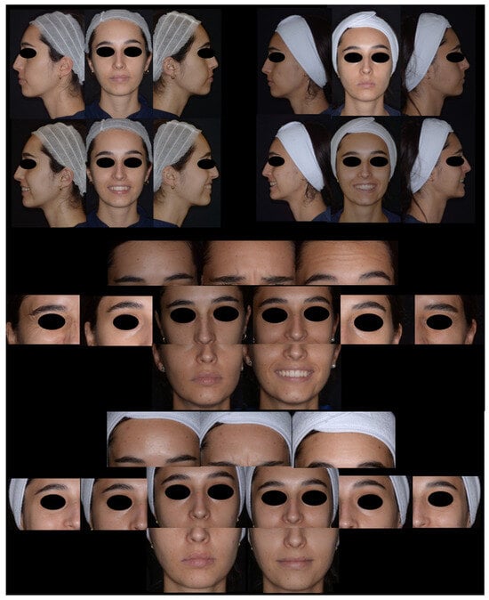
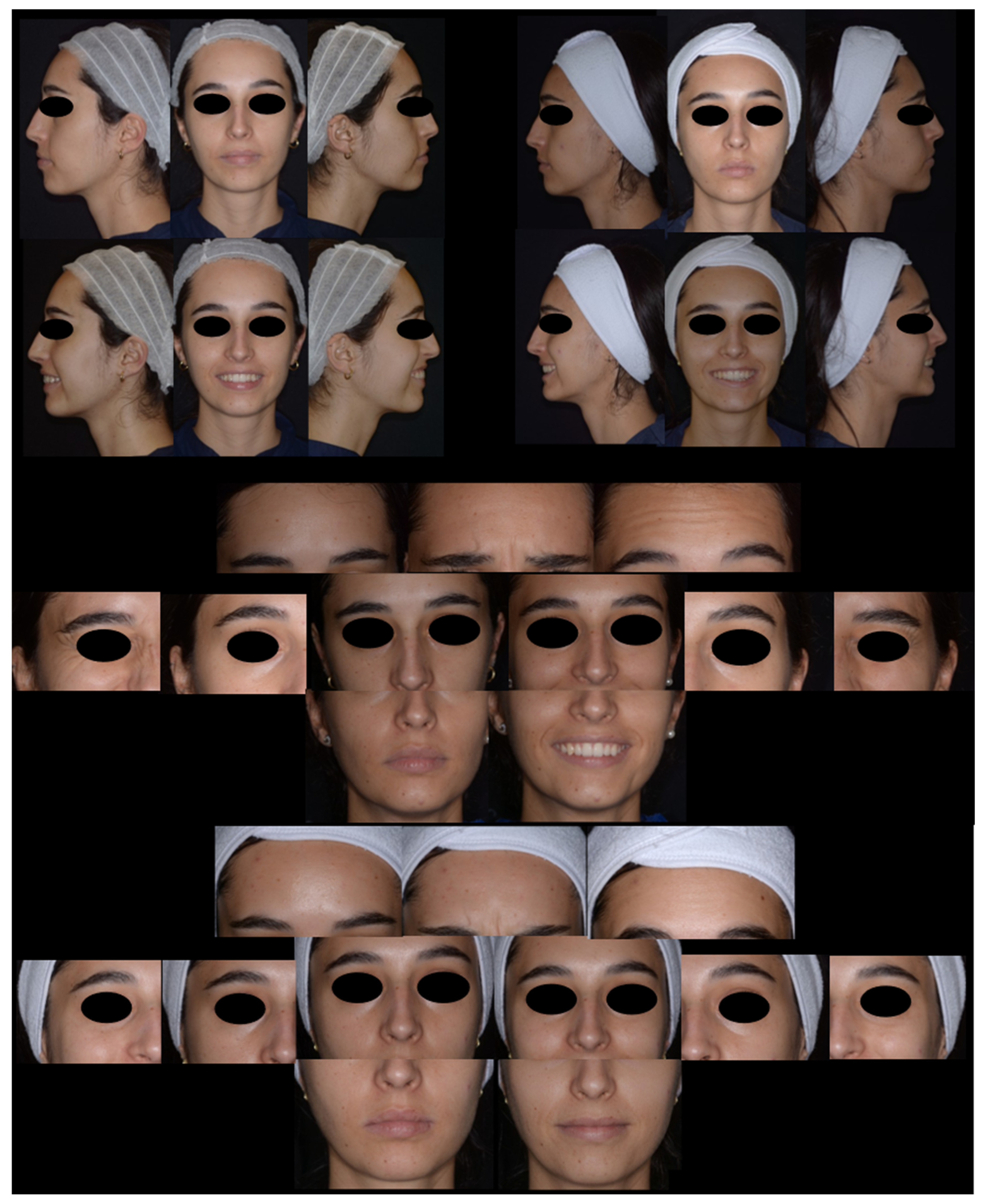
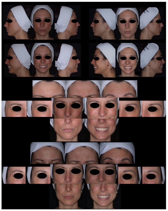
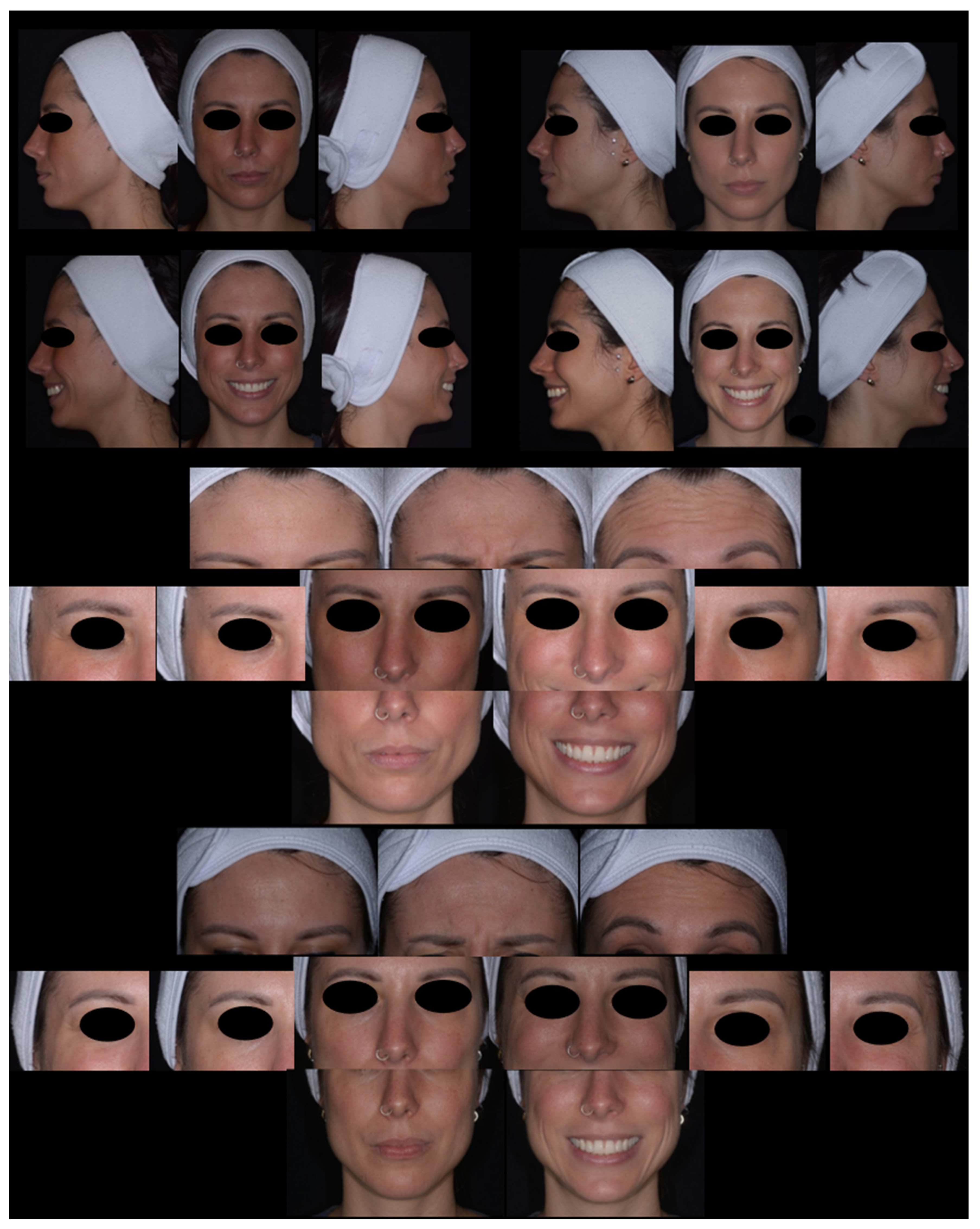
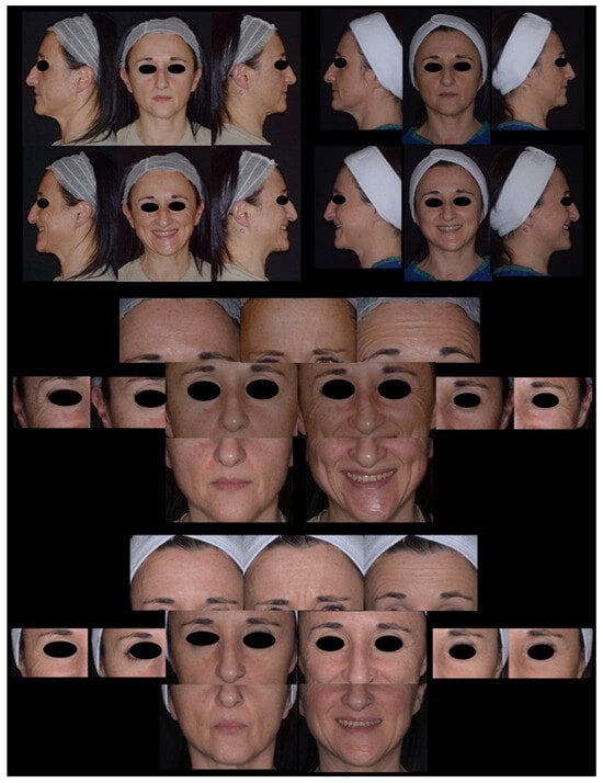
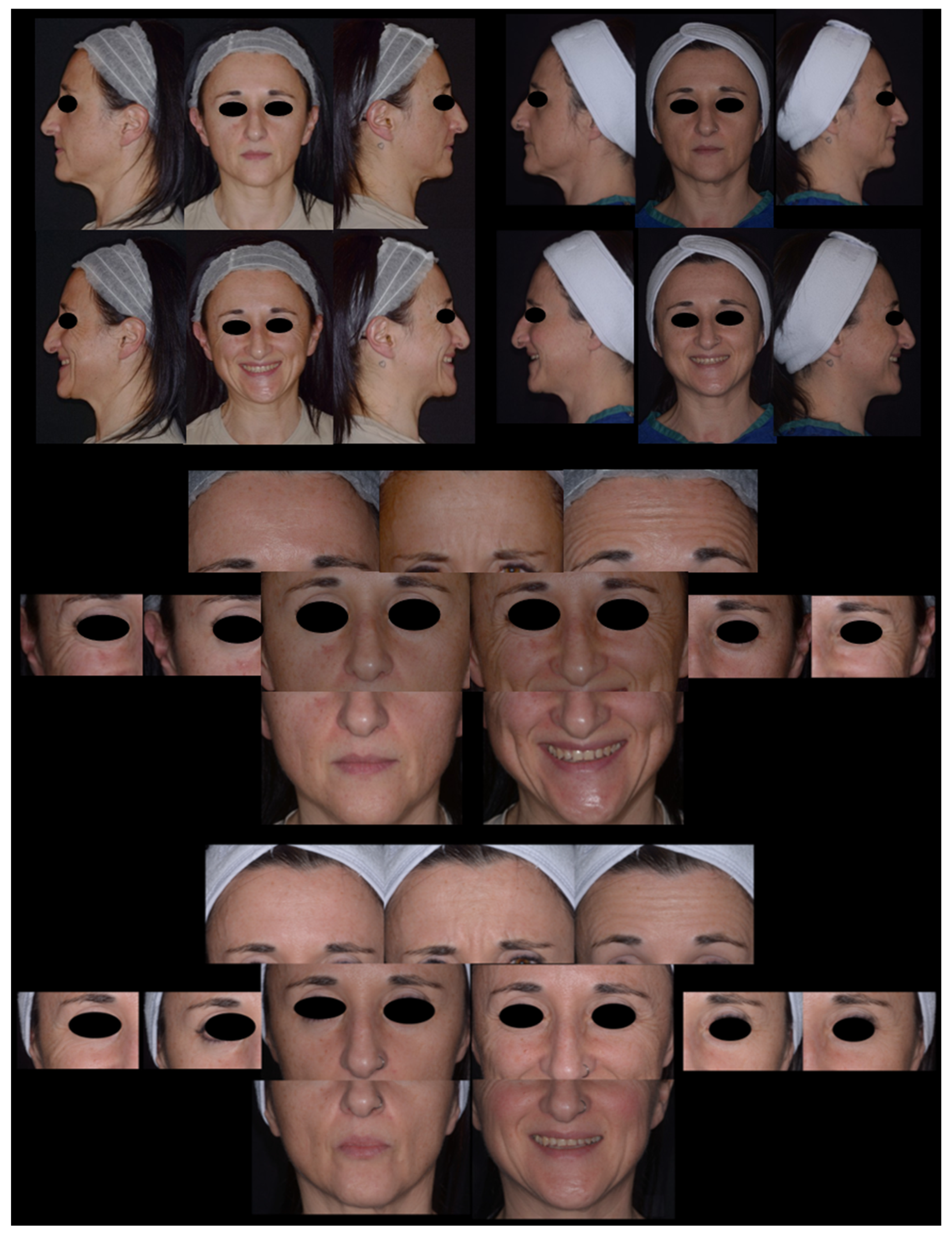
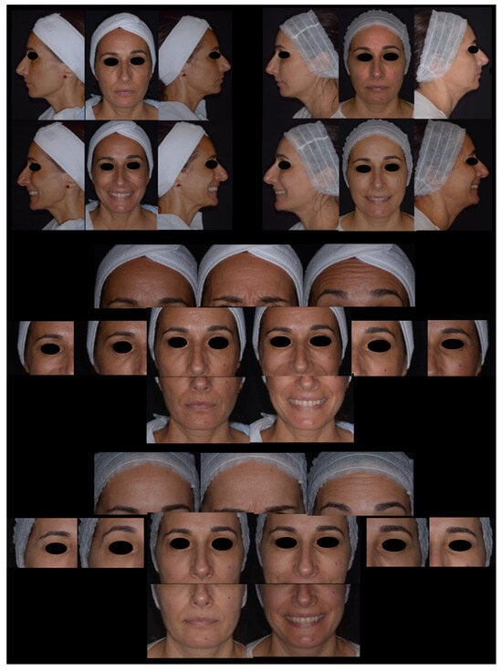
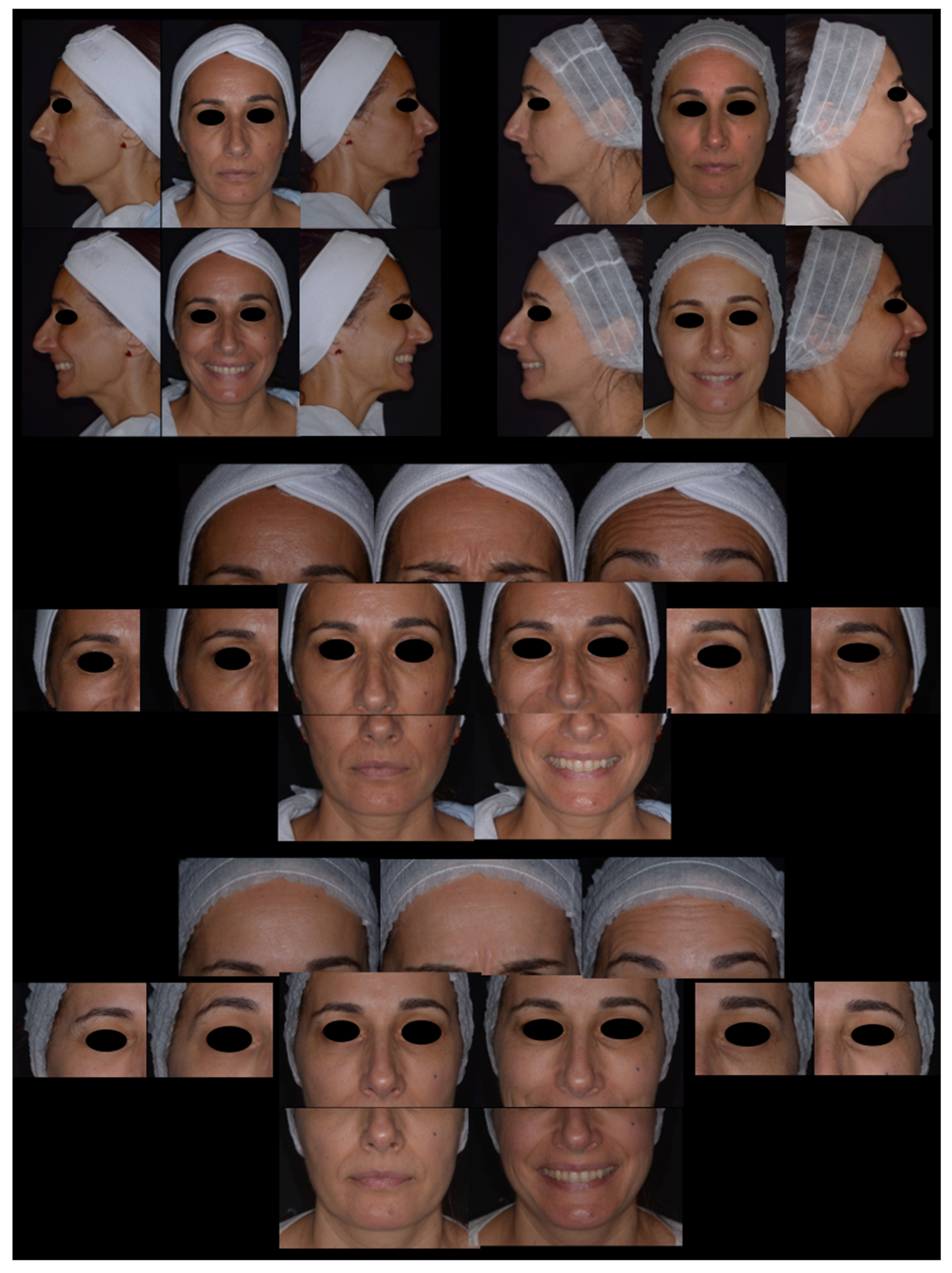
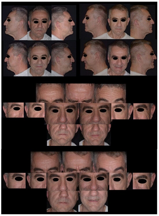
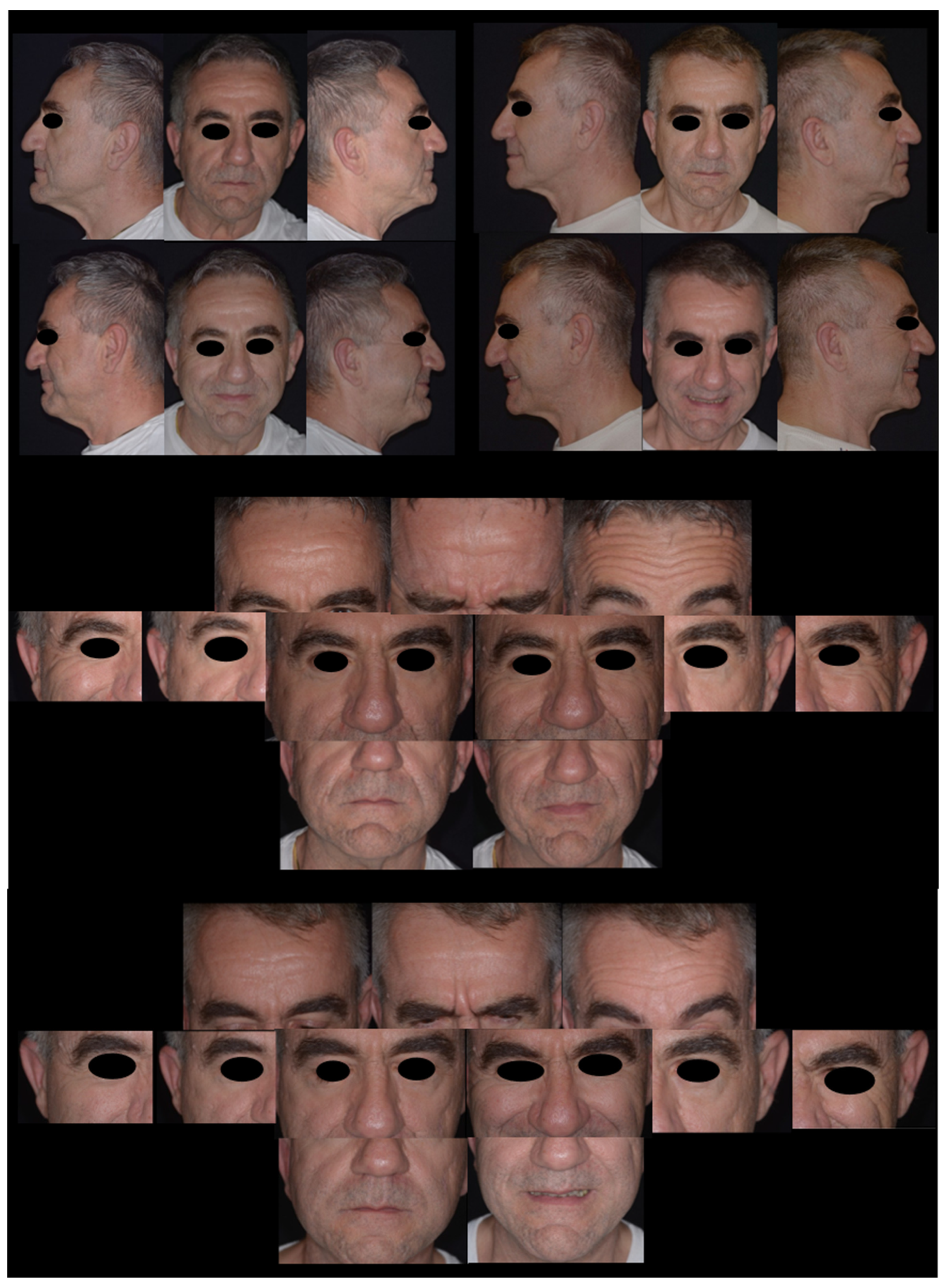
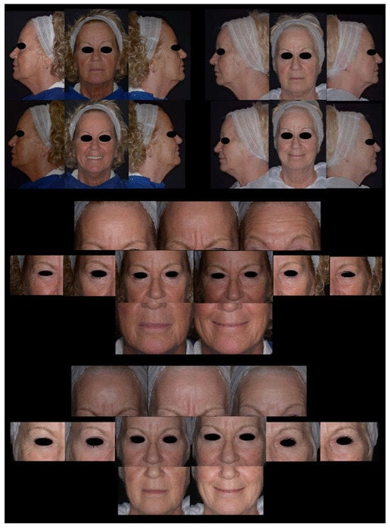
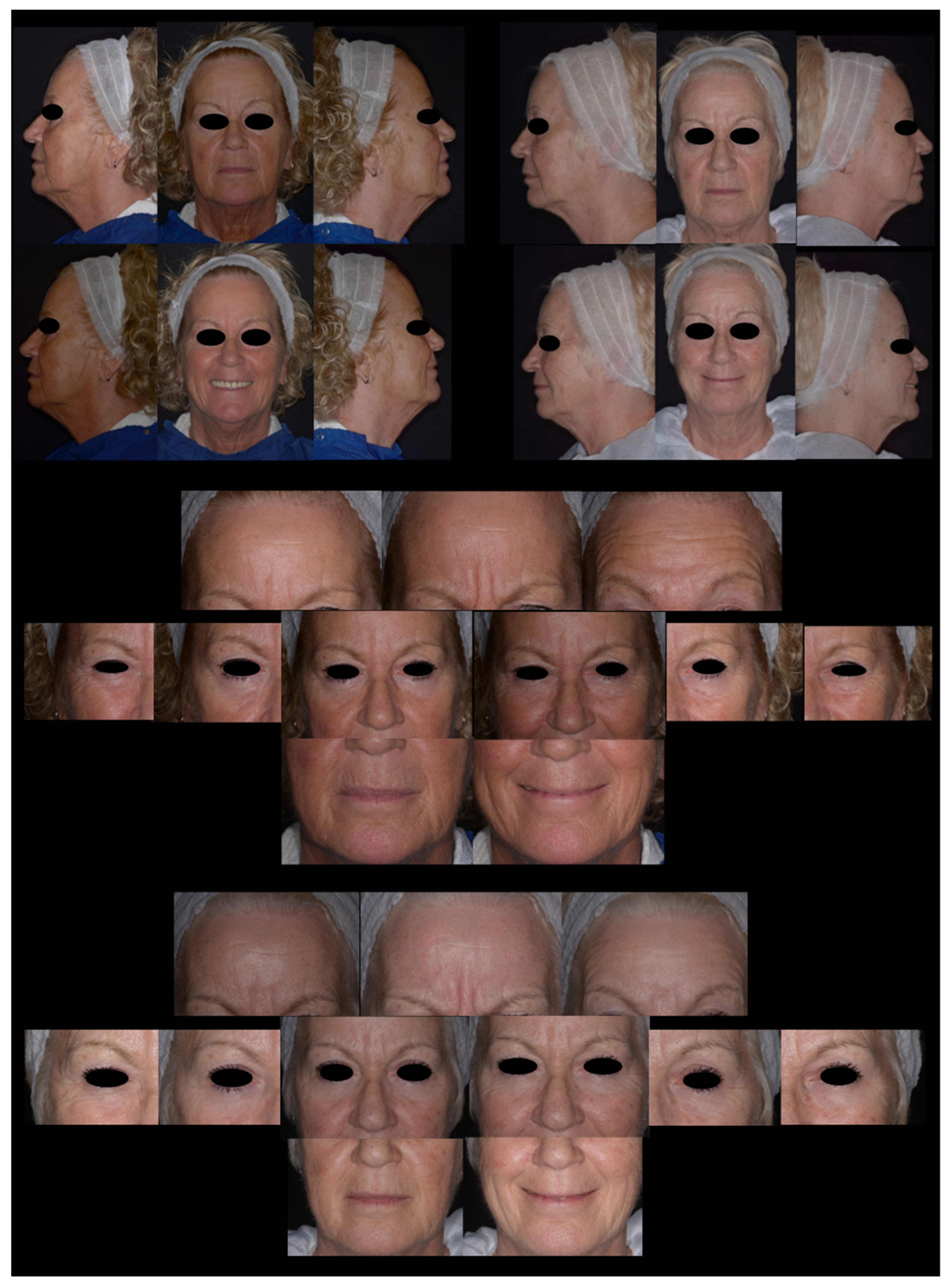
Figure 1, Figure 2, Figure 3, Figure 4, Figure 5 and Figure 6 present composite photographic documentation of the patient at baseline (left) and at the end-of-treatment follow-up (right). The upper panel shows full-face views in frontal and right and left lateral projections. The central panel shows close-up views acquired at baseline, including the forehead at rest, during frowning and eyebrow elevation; the right eye at rest and tightly closed; and the middle third of the face at rest and during a pronounced smile. The lower panel shows the corresponding close-up views acquired at follow-up, including the forehead, right eye, and middle third of the face, as well as the left eye at rest and tightly closed and the lower third of the face at rest and during smiling.
Figure 1.
Patient BI (25–41 years old). A young patient (25 years old) presenting with minimal superficial wrinkles and some expression lines. At the end of the treatment, a mild-to-moderate improvement of forehead, periorbital, and perioral wrinkles is observed, together with an overall improvement in skin texture.
Figure 2.
Patient TV (25–41 years old). The patient (33 years old) presented with good baseline skin quality, with mild superficial wrinkles and expression lines on the forehead and cheeks. Following treatment, an overall improvement in skin quality and texture was observed, together with a mild-to-moderate reduction in expression lines in the forehead and cheek areas.
Figure 3.
Patient MO (41–55 years age group). At baseline, the 54-year-old patient exhibited marked chronoaging, static and dynamic wrinkles, significant skin laxity and tissue redundancy and prominent periorbital rhytides. Following treatment, a more even skin texture and a mild-to-moderate reduction in wrinkle depth were observed. Improvements were noted in the cheeks, periorbital area, and forehead, with wrinkles appearing less pronounced and less numerous compared with baseline.
Figure 4.
Patient NB (41–55 years age group). At baseline, the 52-year-old patient exhibited moderate photoaging and chronoaging, pronounced tear trough deformities and nasolabial folds, superficial perioral and periorbital rhytides, and mildly marked static and dynamic forehead wrinkles. Following treatment, a moderate reduction in tear trough prominence was observed, together with a mild improvement in cheek and periorbital wrinkles.
Figure 5.
Patient CL (56–70 years age group). At baseline, the 59-year-old male patient exhibited acne scar sequelae, thick seborrheic skin, pronounced dynamic facial wrinkles, and diffuse superficial rhytides involving the perioral, preauricular, and periorbital regions, as well as the cheeks, chin, and neck. Following treatment, an overall improvement in skin quality and surface texture was observed, together with a moderate reduction in wrinkle severity in the preauricular and periorbital regions, forehead, eyelids, nasolabial folds, chin, and neck.
Figure 6.
Patient (56–70 years age group). A 70-year-old female patient at baseline presented with superficial rhytides of the forehead, fine wrinkling of the cheeks, and evident periorbital lines, with mild tissue laxity of the upper third of the face. Following treatment, a visible improvement in forehead wrinkles and superficial cheek rhytides was observed. A reduction in periorbital wrinkles is also evident, together with an apparent lifting of the eyebrow arch and a more elevated appearance of the upper eyelid, contributing to an overall improvement in upper facial harmony and skin quality.
4.2.1. Results by Age Group: Age Group 25–40 Years (N = 5)
In the 25–40 years age group (N = 5), baseline photographic assessment indicated a relatively low degree of facial aging. Mean FWS, Fitzpatrick, WAS, and Griffiths scores were 1.4, 3.1, 1.8, and 1.9, respectively, reflecting mild wrinkle severity, limited photoaging, and predominantly superficial wrinkle formation. These scores were consistent with preserved facial contours and overall good skin quality.
At follow-up, minimal to moderate improvements were observed across all photographic scales. Mean FWS scores decreased from 1.4 to 0.6, corresponding to a reduction in the visible facial wrinkles. Mean Fitzpatrick scores improved from 3.1 to 2.1, indicating a lower degree of clinically detectable wrinkling and photoaging. Mean WAS scores decreased from 1.8 to 1.1, reflecting a reduction in wrinkle depth and prominence, while Griffiths scores improved from 1.9 to 1.5, suggesting an overall enhancement in skin appearance and aging-related skin characteristics. The magnitude of change was moderate, reflecting the low baseline severity observed in this age group. The consistent reduction across all photographic scales documented measurable improvements in wrinkle appearance and skin quality following treatment. Representative clinical photographs (Figure 1 and Figure 2) of two patients at baseline (T0) and follow-up (T11) were randomly selected for photographic clinical evaluation.
4.2.2. Results by Age Group: Age Group 41–55 Years (N = 9)
In the 41–55 years age group (N = 9), pre-treatment facial photographs documented moderate signs of facial aging. At baseline, the mean FWS score was 2.5, reflecting the presence of clinically relevant dynamic and static wrinkles involving the forehead, glabellar region, periorbital area, and perioral district. Mean Fitzpatrick photoaging classification was 4.94, indicating a moderate-to-advanced degree of facial aging. Mean WAS score was 3.73, consistent with moderately deep wrinkles across multiple facial subunits, while the mean Griffiths photoaging score was 3.75, reflecting moderate photodamage with involvement of both fine and coarse wrinkle components. Baseline photographic assessment showed that wrinkle distribution was predominantly located in the upper and lower thirds of the face, with higher scores recorded in the periorbital, nasolabial, and perioral regions. Skin surface characteristics appeared heterogenous, with variable degrees of textural irregularity and reduced uniformity across facial areas.
Post-treatment facial photographs showed a measurable improvement in all assessed facial regions. The mean FWS score decreased from 2.5 to 0.5, indicating reduction in wrinkle visibility and severity. The Fitzpatrick score improved from 4.94 to a mean of 3.5, while the WAS score decreased from 3.73 to 1.94, reflecting a shift toward mild-to-superficial wrinkle patterns. The Griffiths score decreased from 3.75 to 1.75, indicating an overall reduction in photodamage-related features. Collectively, the photographic analysis documented measurable reductions in wrinkle severity and visible improvements in overall skin quality within this age group.
From a regional perspective, post-treatment images showed lower recorded severity of wrinkles in the forehead, crow’s feet, nasolabial folds, and marionette lines. Skin surface appeared more homogeneous, with improved regularity of texture and more consistent light reflection across facial areas. Facial contours, particularly along the mandibular line and mid-face region, showed reduced laxity scores compared with baseline assessments.
These clinical photographic observations were supported by the quantitative scale measurements, which showed reductions across all four validated photographic scoring systems (FWS, Fitzpatrick, WAS, and Griffiths), confirming a global improvement in measured wrinkle severity and photoaging indices within this age group.
4.2.3. Results by Age Group: Age Group 56–70 Years (N = 6)
In the 56–70 years age group (N = 6), baseline assessment using the FWS showed severe wrinkle involvement across all facial subunits, with mean scores at rest of 3.00 for the glabellar region, 2.83 for crow’s feet, and 2.83 for forehead lines. At maximum contraction, glabellar and forehead scores remained high (mean 3.00). Post-treatment, a slight reduction was observed in dynamic components, with mean scores decreasing to 2.33 for crow’s feet and forehead at rest, while glabellar scores remained largely unchanged. Fitzpatrick scores indicated severe photoaging at baseline, with mean perioral score of 6.83 and periorbital score of 6.50, decreasing at follow-up to 5.50 and 5.67, respectively. WAS scores showed a consistent reduction across all facial regions, with baseline mean values ranging from 4.50 to 5.67 and post-treatment values ranging from 4.17 to 5.17, with the most relevant decreases observed in glabellar (5.33 to 4.50), periorbital (5.33 to 4.50), preauricular (5.50 to 4.50), and marionette lines (5.17 to 4.50). Glogau Classification scores remained within the advanced photoaging range, with a mean score of 6.83 at baseline and 6.83 at follow-up. Griffiths Photonumeric Scale scores showed a mild overall improvement, with fine and coarse wrinkle scores and mottled hyperpigmentation decreasing from 6.00 to 5.00, and yellowing decreasing from 5.00 to 4.00. Overall, quantitative assessment showed a modest reduction in wrinkle severity across all validated scales, with changes predominantly involving periorbital, perioral, and forehead regions, while subjects remained within higher severity categories across all indices at follow-up.
4.2.4. Cross-Group Observations
Photographic analysis showed consistent treatment-related improvements across all age groups, with variability in magnitude according to baseline severity and tissue aging status. In the 25–40 years group, baseline values indicated minimal-to-mild photoaging, with relatively low scores across all scales (FWS 1.37, Fitzpatrick 3.10, WAS 1.76, Griffiths 1.90). Following treatment, a consistent but modest improvement was observed across all indices, with reductions to 0.57 (FWS), 2.10 (Fitzpatrick), 1.11 (WAS), and 1.45 (Griffiths). The pattern of change primarily reflected attenuation of fine wrinkles and improvement in skin texture and surface homogeneity, in line with the low baseline severity of this cohort.
The 41–55 years group showed the most pronounced response to treatment. Baseline photographic assessment showed moderate photoaging (FWS 2.50, Fitzpatrick 4.94, WAS 3.52, Griffiths 3.75), with clinically relevant involvement of both dynamic and static wrinkles across multiple facial regions. Post-treatment, all scales showed marked reductions, with FWS decreasing to 0.50, Fitzpatrick to 3.50, WAS to 1.75, and Griffiths to 1.75. These changes indicate a substantial improvement in wrinkle severity, skin quality, and global photoaging appearance, with a shift toward mild or superficial wrinkle patterns and improved facial uniformity. In the 56–70 years group, baseline values reflected advanced photoaging and higher wrinkle burden (FWS 3.00, Fitzpatrick 6.92, WAS 5.58, Griffiths 5.75). Post-treatment assessments showed more limited but still consistent improvements across all scales, with FWS decreasing to 2.31, Fitzpatrick to 5.83, WAS to 4.39, and Griffiths to 4.75. Although the reduction in scores was less pronounced compared with younger cohorts, improvements were observed particularly in superficial wrinkle components and skin texture, while deeper static wrinkles and global photoaging features remained partially evident.
Across the entire study population, photographic assessment using the FWS, Fitzpatrick, WAS, and Griffiths scales consistently showed reductions in wrinkle severity and cumulative photoaging. The magnitude of response was inversely related to baseline severity, with the most substantial absolute improvements observed in the middle-aged group (41–55 years), followed by the youngest cohort, while the oldest group showed more limited but still measurable benefits.
Overall, combined HIFU and DQRF treatment produced consistent improvements in facial appearance, skin quality, and wrinkle severity across all age categories, with differential responsiveness according to baseline tissue aging characteristics.
5. Discussion
This cohort study observed clinical facial rejuvenation improvements following combined HIFU and DQRF, as assessed through both validated patient-reported outcome measures and objective photographic scales across different age strata. These findings reinforce the evolving role of non-invasive energy-based technologies in contemporary aesthetic dermatology, where treatment success is increasingly defined by dermal remodeling, tissue quality restoration, and patient-perceived naturalness rather than volumetric correction alone [1,2,3].
The biological plausibility of the observed outcomes is supported by the complementary mechanisms of action of the two technologies. HIFU induces controlled thermal coagulation at targeted dermal and subdermal depths, including the SMAS layer, triggering neocollagenesis and structural tightening through collagen denaturation and remodeling [4,5,12]. In contrast, DQRF delivers volumetric dermal heating that enhances fibroblast activity and promotes superficial dermal restructuring [8,9,10]. The combination of these modalities likely produces a synergistic multi-layered regenerative effect, consistent with histometric evidence describing differential depth targeting of ultrasound and radiofrequency-based systems [11,13].
A central finding of this study is the presence of differences observed across age groups, with the mid-age cohort showing the most pronounced and consistent improvement across both objective and subjective endpoints. This phenomenon may be explained by an optimal balance between residual regenerative capacity and baseline structural aging. In this population, dermal fibroblasts remain functionally responsive to thermal stimulation, while clinically relevant signs of aging are sufficiently present to allow measurable improvement [15,16]. In contrast, younger individuals exhibit limited baseline impairment, whereas older patients show reduced regenerative capacity due to intrinsic dermal and extracellular matrix alterations [15,16]. Although age-related differences in skin structure and regenerative capacity may contribute to variations in treatment response, these findings are exploratory and should be interpreted cautiously, particularly because of the small number of participants within each subgroup.
Importantly, patient-reported outcomes mirrored this objective pattern, with the mid-age group demonstrating the strongest alignment between clinical improvement and perceived aesthetic benefit. This concordance underscores the importance of psychological and perceptual factors in defining treatment satisfaction and aligns with evidence linking aesthetic improvement with quality-of-life enhancement in cosmetic dermatology populations [21,22,23,27].
Baseline psychosocial analysis provides further insight into these dynamics. Pre-treatment FACE-Q scores indicated moderate dissatisfaction with facial appearance, driven predominantly by context-dependent perceptions of facial fatigue and reduced freshness rather than fixed structural defects [21,22,23]. Psychological well-being remained relatively preserved, suggesting that aesthetic concern is primarily appearance-driven rather than associated with global psychological distress.
Perceived age discrepancy analysis revealed that patients considered themselves, on average, older than their chronological age, with substantial inter-individual variability. This dissociation between objective and subjective age perception may represent a key psychological driver influencing aesthetic motivation and expectation formation, potentially contributing to heterogeneity in subjective treatment response [21,22,23,27].
Within younger participants (25–40 years), wrinkle-specific concern was relatively low at baseline, as reflected by FLO-11 scores. However, item-level interpretation suggests that dissatisfaction is more strongly associated with perceived fatigue and skin texture irregularities than with structural rhytides, consistent with previous validation studies of FLO-11 domains [23].
Across the entire cohort, PROMs (FACE-Q, FLO-11, FLSQ, and FTS) were associated with consistent post-treatment improvements, confirming that structural dermal changes translated into meaningful psychosocial benefit. These findings are consistent with prior validation studies demonstrating strong correlations between aesthetic improvement and quality-of-life enhancement [21,22,23,27].
Photographic analysis documented reductions in wrinkle severity and photoaging indices across validated scales (FWS, Fitzpatrick classification, WAS, and Griffiths scale), which are widely used in aesthetic dermatology to quantify dermal elastosis and wrinkle depth [32,33,34]. The consistency across scoring systems suggests that improvements reflect true dermal remodeling rather than superficial textural changes. Nevertheless, these findings should not be interpreted as direct evidence of dermal regeneration, collagen synthesis, or histological remodeling, as no histological or molecular analyses were performed. Such biological mechanisms remain plausible explanations based on previously published evidence but were not directly investigated in this cohort.
Safety outcomes were highly favorable, with no clinically relevant adverse events reported. This aligns with previously published data confirming the excellent safety profile of both HIFU and RF technologies when applied within standardized clinical parameters [7,10,19].
Methodologically, the study benefits from longitudinal design and the integration of multiple validated outcome measures, including blinded photographic assessment. However, limitations include the absence of a control group, limited sample size, and lack of inferential statistical analysis, restricting causal interpretation [18,19].
Inter-individual variability remains a relevant consideration and is likely influenced by baseline skin quality, genetic factors, and environmental exposure, as previously reported in energy-based aesthetic treatments [13,17].
Overall, these findings support the role of combined HIFU and DQRF as an effective non-invasive strategy for facial rejuvenation, capable of inducing measurable improvements in dermal quality, wrinkle severity, and patient satisfaction. Importantly, results reinforce the concept of regenerative aesthetic medicine, which prioritizes tissue remodeling and functional restoration over volumetric correction [17,18].
Future studies should incorporate randomized controlled designs and multivariate predictive models to better characterize response determinants and optimize patient selection.
6. Conclusions
This cohort study suggests that the combined use of HIFU and DQRF may represent a promising non-invasive approach for facial and cervical rejuvenation. Improvements in skin appearance, wrinkle severity, and global aesthetic perception were observed across all evaluated age groups. However, these findings should be interpreted cautiously, given the small sample size, uncontrolled study design, and descriptive nature of the analysis.
Differences in clinical and patient-reported outcomes were observed across age groups, with the most consistent improvements recorded in the mid-age cohort (41–55 years), a finding that may reflect balance between residual dermal regenerative capacity and the presence of clinically relevant signs of aging. Younger patients, despite generally favorable baseline skin quality, showed more limited perceived improvement, which may be related to lower initial severity and higher expectation-driven variability in subjective assessment. In contrast, older patients exhibited more modest but generally stable changes, consistent with reduced regenerative potential in more advanced stages of skin aging. These observations are exploratory and do not establish an age-dependent treatment response, which would require confirmation in larger, adequately powered studies.
Across the cohort, changes in photographic assessments were generally consistent with patient-reported outcomes, suggesting a degree of concordance between clinician-assessed changes in facial appearance and perceived aesthetic benefit, including facial appearance, psychological well-being, and treatment satisfaction. Nevertheless, photographic improvements should be interpreted as changes in clinical appearance and not as direct evidence of dermal regeneration or underlying biological remodeling. These findings also highlight the usefulness of combining validated PROMs with standardized photographic evaluation tools for a more comprehensive assessment of outcomes in non-invasive aesthetic procedures.
The safety profile of the combined protocol appeared favorable under the conditions of this study, with no clinically relevant adverse events observed throughout treatment and follow-up.
Overall, these preliminary findings contribute to the ongoing investigation of the potential role of combined energy-based technologies within the conceptual framework of regenerative aesthetic medicine, which emphasizes tissue remodeling and functional improvement rather than volumetric correction. However, further controlled studies involving larger sample sizes, standardized protocols, objective assessment methods, and longer follow-up periods are warranted to clarify their respective and combined contributions, better define treatment indications, optimize patient selection, and assess the durability of clinical outcomes.
Author Contributions
Conceptualization, O.R., G.P. and T.T.; Methodology, O.R., G.P., T.T. and M.D.F.; Validation, T.T. and M.D.F.; Formal analysis, O.R. and M.D.F.; Investigation, O.R. and M.D.F.; Resources, O.R., G.P. and T.T.; Data curation, O.R., G.P. and M.D.F.; Writing—original draft, O.R.; Supervision, G.P. and T.T.; Project administration, G.P. and T.T.; Funding acquisition M.D.F. All authors have read and agreed to the published version of the manuscript.
Funding
This research received no external funding.
Institutional Review Board Statement
The present study evaluated the clinical use of HIFU and DQRF for antiaging treatment in routine practice. The CE-certified devices (Dual Hi and Artemis, Med&Tech) were used strictly according to their intended clinical purpose, with Dual Hi employed for HIFU treatment and Artemis for DQRF treatment. No control group, randomization, or experimental interventions were involved. Ethical review and approval were waived in accordance with national and institutional regulations for observational studies. All procedures were performed by experienced clinicians, and written informed consent for treatment, privacy, and publication of anonymized data and images was obtained from all participants, in compliance with the Declaration of Helsinki.
Informed Consent Statement
Informed consent was obtained from all subjects involved in the study. Each participant was thoroughly informed about the mechanism of action of the device, potential contraindications, expected benefits, and the objectives of the treatment. Written consent regarding privacy and the use of photographic documentation, strictly for scientific purposes, was collected, signed, and archived for every patient. Written informed consent has also been obtained from the patients for the publication of images included in this paper.
Data Availability Statement
The raw data supporting the conclusions of this article will be made available by the authors on request. Additional datasets and full clinical photographic records may be obtained from the corresponding author upon reasonable and justified request.
Acknowledgments
The authors acknowledge that no additional financial support, administrative or technical assistance, or donations in kind were received for this study. During the preparation of this manuscript, ChatGPT (OpenAI, GPT-6, November 2026 version) was used exclusively for minor language editing, linguistic refinement, and bibliographic formatting. No AI tools were used for data generation, data collection, statistical analysis, interpretation of results, or scientific content development. The authors reviewed and revised all AI-assisted output and take full responsibility for the content of this publication.
Conflicts of Interest
The authors declare that no financial or commercial relationships influenced this study. No external funding was received, and all aspects of the study—including its design, data collection, analysis, interpretation, manuscript writing, and the decision to publish—were conducted independently of any commercial entity. All the authors declare that the research was conducted in the absence of any commercial or financial relationships that could be construed as potential conflicts of interest. The author Giovanna Perrotti, affiliated with the Lake Como Institute (Private Practice), declares that the research was conducted in the absence of any commercial or financial relationships that could be construed as potential conflicts of interest.
References
- Trovato, F.; Ceccarelli, S.; Michelini, S.; Vespasiani, G.; Guida, S.; Galadari, H.I.; Nisticò, S.P.; Colonna, L.; Pellacani, G. Advancements in Regenerative Medicine for Aesthetic Dermatology: A Comprehensive Review and Future Trends. Cosmetics 2024, 11, 49. [Google Scholar] [CrossRef] [Scilit]
- National Council on Radiation Protection and Measurements. Biological Effects of Ultrasound: Mechanisms and Clinical Implications; NCRP Report No. 74; National Council on Radiation Protection and Measurements: Bethesda, MD, USA, 1983. [Google Scholar]
- Miller, M.W.; Miller, D.L.; Brayman, A.A. A review of in vitro bioeffects of inertial ultrasonic cavitation from a mechanistic perspective. Ultrasound Med. Biol. 1996, 22, 1131–1154. [Google Scholar] [CrossRef] [Scilit] [PubMed]
- White, W.M.; Makin, I.R.S.; Barthe, P.G.; Slayton, M.H.; Gliklich, R.E. Selective creation of thermal injury zones in the superficial musculoaponeurotic system using intense focused ultrasound: A new target for noninvasive facial rejuvenation. Arch. Facial Plast. Surg. 2007, 9, 22–29. [Google Scholar] [PubMed]
- Miller, D.; Smith, N.; Bailey, M.; Czarnota, G.; Hynynen, K.; Makin, I.J. Overview of therapeutic ultrasound applications and safety considerations. Ultrasound Med. 2012, 31, 623–634. [Google Scholar] [CrossRef] [Scilit] [PubMed]
- De Lucas, B.; Pérez, L.M.; Bernal, A.; Gálvez, B.G. Ultrasound therapy: Experiences and perspectives for regenerative medicine. Genes 2020, 11, 1086. [Google Scholar] [CrossRef] [Scilit] [PubMed]
- Bader, K.B.; Makin, I.R.S.; Abramowicz, J.S. Ultrasound for aesthetic applications: A review of biophysical mechanisms and safety. J. Ultrasound Med. 2022, 41, 1597–1607. [Google Scholar] [PubMed]
- Delgado, A.R.; Chapas, A. Introduction and overview of radiofrequency treatments in aesthetic dermatology. J. Cosmet. Dermatol. 2022, 21, S1–S10. [Google Scholar] [CrossRef] [Scilit] [PubMed]
- Elsaie, M.L. Cutaneous remodeling and photo-rejuvenation using radiofrequency devices. Indian J. Dermatol. 2009, 54, 201–205. [Google Scholar] [CrossRef] [Scilit] [PubMed]
- Austin, G.K.; Struble, S.L.; Quatela, V.C. Evaluating the effectiveness and safety of radiofrequency for face and neck rejuvenation: A systemic review. Lasers Surg. Med. 2022, 54, 27–45. [Google Scholar] [CrossRef] [Scilit] [PubMed]
- Suh, D.H.; Choi, J.H.; Lee, S.J.; Jeong, K.H.; Song, K.Y.; Shin, M.K. Comparative histometric analysis of the effects of high-intensity focused ultrasound and radiofrequency on skin. J. Cosmet. Laser Ther. 2015, 17, 230–236. [Google Scholar] [CrossRef] [Scilit] [PubMed]
- Serdar, Z.A.; Karabay, E.A.; Tatlıparmak, A.; Aksoy, B. Efficacy of high-intensity focused ultrasound in facial and neck rejuvenation. J. Cosmet. Dermatol. 2020, 19, 353–358. [Google Scholar] [CrossRef] [Scilit] [PubMed]
- Gayana, S.K.; Khong, S.-J.M.L.; Ismail, A.H.; Adibah, N.S.K.D.; Sujani, S.; Clinic, J.Z.; Devindaran, N.; Medveon, S.K.; Rashid, M.F.A.; Zaman, U.M.S.M. Safety and efficacy of high-intensity focused ultrasound and monopolar radiofrequency combination therapy for skin tightening: A retrospective study in Malaysia. Malays. J. Med. Sci. 2024, 31, 114–123. [Google Scholar] [CrossRef] [Scilit] [PubMed]
- Rossi, O.; Perrotti, G.; Scaini, R.; Del Fabbro, M.; Damiani, G.; Testori, T. How to establish the baseline for non-invasive technological regenerative esthetic medicine in the face and neck region: A literature review. J. Pers. Med. 2025, 15, 283. [Google Scholar] [CrossRef] [Scilit] [PubMed]
- Glogau, R.G. Aesthetic and anatomic analysis of the aging skin. Semin Cutan. Med. Surg. 1996, 15, 134–138. [Google Scholar] [CrossRef] [Scilit] [PubMed]
- Nam, K.I.; Shin, S.; Song, J. Anatomical importance of fat compartments in facial aging: A review. Anat. Biol. Anthropol. 2025, 38, 145–152. [Google Scholar]
- Goldie, K. The evolving field of regenerative aesthetics. J. Cosmet. Derm. 2023, 22, 1–7. [Google Scholar] [CrossRef] [Scilit] [PubMed]
- Zarbafian, M.; Fabi, S.G.; Dayan, S.; Goldie, K. The Emerging Field of Regenerative Aesthetics—Where We Are Now. Dermatol. Surg. 2022, 48, 101–108. [Google Scholar] [CrossRef] [Scilit] [PubMed]
- Ayatollahi, A.; Gholami, J.; Saberi, M.; Hosseini, H.; Firooz, A. Systematic review and meta-analysis of safety and efficacy of high-intensity focused ultrasound (HIFU) for face and neck rejuvenation. Lasers Med. Sci. 2020, 35, 1007–1024. [Google Scholar] [CrossRef] [Scilit] [PubMed]
- Rossi, O.; Perrotti, G.; Scaini, R.; Del Fabbro, M.; Damiani, G.; Testori, T. Medicina estetica rigenerativa tecnologica non invasiva del distretto testa e collo: Controindicazioni e screening estetico-dermatologico (SED). Parte 2. Quintessenza Int. 2025, 1, 54–62. [Google Scholar]
- Klassen, A.F.; Cano, S.J.; Scott, A.; Snell, L.; Pusic, A.L. Measuring patient-reported outcomes in facial aesthetic patients: Development of the FACE-Q. Facial Plast. Surg. 2010, 26, 303–309. [Google Scholar] [CrossRef] [Scilit] [PubMed]
- Pusic, A.L.; Klassen, A.F.; Cano, S.J. Development and psychometric evaluation of the FACE-Q satisfaction with appearance scale: A new patient-reported outcome instrument for facial aesthetics patients. Clin. Plast. Surg. 2013, 40, 307–312. [Google Scholar]
- Klassen, A.F.; Cano, S.J.; Schwitzer, J.A.; Scott, A.M.; Pusic, A.L. FACE-Q scales for health-related quality of life, early life impact, satisfaction with outcomes, and decision to have treatment: Development and validation. Plast. Reconstr. Surg. 2015, 135, 375–382. [Google Scholar] [CrossRef] [Scilit] [PubMed]
- Carruthers, J.D.; Carruthers, A. Facial sculpting and tissue augmentation. Dermatol. Surg. 2006, 32, 312–318. [Google Scholar] [CrossRef] [Scilit] [PubMed]
- Yaworsky, A.; Daniels, S.; Tully, S.; Beddingfield, F., III; Kowalski, J.; Fitzgerald, K.; Somogyi, C.; Burgess, S.M. The impact of upper facial lines and psychological impact of crow’s feet lines: Content validation of the Facial Line Outcomes (FLO-11) Questionnaire. J. Cosmet. Dermatol. 2014, 13, 297–306. [Google Scholar] [CrossRef] [Scilit] [PubMed]
- Dayan, S.H.; Bacos, J.T.; Ho, T.V.; Gandhi, N.D.; Gutierrez-Borst, S.; Kalbag, A. Topical skin therapies in subjects undergoing full facial rejuvenation. J. Cosmet. Dermatol. 2019, 18, 323–329. [Google Scholar] [CrossRef] [Scilit] [PubMed]
- Gama, J.T.; Rossetto, L.A.; Brito, N.B.; Veiga, D.F.; Ferreira, L.M. Cross-cultural validation of the FACE-Q Satisfaction with Facial Appearance Overall Scale (FACE-Q SFAOS) in Brazilian rhytidoplasty patients. Clinics 2020, 75, e1568. [Google Scholar] [CrossRef] [PubMed]
- Pompilus, F.; Burgess, S.; Hudgens, S.; Banderas, B.; Daniels, S. Development and validation of a novel patient-reported treatment satisfaction measure for hyperfunctional facial lines: Facial line satisfaction questionnaire. J. Cosmet. Dermatol. 2014, 14, 274–285. [Google Scholar] [CrossRef] [Scilit] [PubMed]
- Cox, S.E.; Finn, J.C.; Stetler, L.; Mackowiak, J.; Kowalski, J.W. Development of the Facial Lines Treatment Satisfaction Questionnaire and initial results for botulinum toxin type A-treated patients. Dermatol. Surg. 2003, 29, 444–449. [Google Scholar] [CrossRef] [Scilit]
- Kerscher, M.; Fabi, S.; Fischer, T.; Gold, M.; Joseph, J.; Prager, W.; Rzany, B.; Yoelin, S.; Roll, S.; Klein, G.; et al. IncobotulinumtoxinA demonstrates safety and prolonged duration of effect in a dose-ranging study for glabellar lines. J. Drugs Dermatol. 2021, 20, 1052. [Google Scholar]
- Carruthers, J.; Carruthers, A. Botulinum toxin type A treatment of multiple upper facial sites: Patient-reported outcomes. Dermatol. Surg. 2007, 33, S10–S17. [Google Scholar] [CrossRef] [Scilit] [PubMed]
- Fitzpatrick, R.E.; Goldman, M.P.; Satur, N.M.; Tope, W.D. Pulsed carbon dioxide laser resurfacing of photo-aged facial skin. Arch. Dermatol. 1996, 132, 395–402. [Google Scholar] [CrossRef] [Scilit]
- Lemperle, G.; Holmes, R.E.; Cohen, S.R.; Lemperle, S.M. A classification of facial wrinkles. Plast. Reconstr. Surg. 2001, 108, 1735–1745. [Google Scholar] [CrossRef] [Scilit] [PubMed]
- Griffiths, C.E.; Wang, T.S.; Hamilton, T.A.; Voorhees, J.J.; Ellis, C.N. A photonumeric scale for the assessment of cutaneous photodamage. Arch. Dermatol. 1992, 128, 347–351. [Google Scholar] [CrossRef] [Scilit]

Disclaimer/Publisher’s Note: The statements, opinions and data contained in all publications are solely those of the individual author(s) and contributor(s) and not of MDPI and/or the editor(s). MDPI and/or the editor(s) disclaim responsibility for any injury to people or property resulting from any ideas, methods, instructions or products referred to in the content. |
© 2026 by the authors. Licensee MDPI, Basel, Switzerland. This article is an open access article distributed under the terms and conditions of the Creative Commons Attribution (CC BY) license.