


Abstract
Age-related changes to the chin region and neck are among the most common reasons for consulting a plastic surgeon, and the outcome of rejuvenation surgery largely depends on the accuracy of preoperative planning. Digital imaging methods allow for an objective assessment of individual anatomical features, personalization of the surgical strategy, and improved predictability of the result. In this narrative review, based on a structured, PRISMA-informed search (PubMed, Scopus, Google Scholar, and eLibrary; 65 publications included), we systematize contemporary digital and radiological imaging methods for the chin region and neck—two-dimensional photography, three-dimensional photogrammetry, computed tomography and magnetic resonance imaging, cone-beam computed tomography (CBCT), CAD/CAM technologies, virtual and mixed reality, and artificial intelligence tools—and assess their diagnostic value for planning rejuvenation surgery. Available preliminary, predominantly single-centre evidence suggests that CBCT performed in the upright patient position may be the most informative modality for this task: by capturing bony landmarks together with the position and outer contours of the relevant soft-tissue structures under gravitational load, it supports an objective assessment of the contribution of individual structures to age-related deformities and well-founded, individualized surgical planning. Its intrinsic soft-tissue contrast resolution is nevertheless limited compared with MRI. The integration of digital and radiological imaging methods is a vital part of modern planning of aesthetic surgery for the chin region and neck.
1. Introduction
Age-related changes to the chin region and neck are among the most common reasons for patients seeking help from a plastic surgeon. With aging, soft-tissue atrophy, platysma ptosis, the formation of a “double chin,” a decrease in the cervicomental angle, and a loss of definition of the mandibular contour all occur. These changes affect not only appearance but also the perception of a person’s age, health, and social attractiveness [1,2,3]. A detailed understanding of the anatomy of the chin region and neck—the ligamentous apparatus of the cervicomental angle [4], the fat compartments of the chin and midface [5,6], and the age-related dynamics of these structures—provides the basis for rational planning of an intervention. Against the background of a steady increase in demand for facial and neck rejuvenation procedures [7], the need for objective methods of assessing these changes is growing.
Despite the variety of surgical correction techniques—from platysmaplasty and liposuction to facelift with mentoplasty—preoperative planning still largely relies on the surgeon’s subjective assessment. The lack of objective tools complicates the choice of surgical technique and the representation accuracy of the planned result; it may also reduce satisfaction with the surgical outcome [8,9,10]. The modern armamentarium includes various modifications of platysmaplasty and deep-plane lifting of the face and neck [11,12,13,14], ultrasound-assisted lifting of the deep layers [15], approaches adapted to the anatomical features of patients of various ethnic groups [16], and thread lifting [17]; a number of reviews are devoted to the selection of indications and a personalized surgical strategy for rejuvenation of the chin region and neck [18]. Such a wide range of techniques only reinforces the need for objective preoperative imaging.
In this context, the introduction of digital imaging—in particular, modeling based on cone-beam computed tomography (CBCT) and computed tomography (CT) data, as well as photogrammetry and virtual surgical planning—opens new possibilities for precise anatomical analysis and individualized selection of the surgical strategy [19,20,21]. Digital technologies make it possible to increase the reproducibility of procedures and improve interdisciplinary interaction. Thus, studying the role of digital imaging in the surgical correction of age-related changes to the chin region and neck appears timely and necessary for improving the efficiency and predictability of aesthetic operations [19,22,23].
The aim of this study was to analyze and systematize contemporary digital and radiological imaging methods used in planning rejuvenation surgery of the chin region and neck, and to determine the most clinically justified and rational method regarding diagnostic value, safety, and practical applicability.
2. Materials and Methods
This literature review was conducted in accordance with the principles and recommendations of PRISMA 2020 (Preferred Reporting Items for Systematic Reviews and Meta-Analyses) [24], ensuring the transparency and reproducibility of the study selection procedure. At the same time, the present work is a narrative literature review: the PRISMA 2020 methodology was applied as a reporting standard for the search procedure only. The review protocol was not registered, duplicate independent screening and formal risk-of-bias assessment were not performed, and no meta-analysis was conducted.
2.1. Review Design and Time Frame
This review included publications published from January 2020 to June 2026. A systematic search was performed in four electronic databases: PubMed, Scopus, Google Scholar (English-language sources), and eLibrary (Russian-language sources). The final search was carried out on 18 June 2026. Publications in Russian and English were considered.
2.2. Search Strategy
The search was based on a combined query of two conceptual blocks (area of intervention AND imaging/planning method) using the logical operators AND/OR. The search strategy was adapted to the syntax of each database and, for reproducibility, is presented below in the form actually used.
PubMed (title/abstract field, [tiab]):
(“neck lift*”[tiab] OR “neck rejuvenation”[tiab] OR “facial rejuvenation”[tiab] OR facelift[tiab] OR rhytidectomy[tiab] OR cervicofacial[tiab] OR “lower face”[tiab]) AND (“cone-beam computed tomography”[tiab] OR “computed tomography”[tiab] OR “magnetic resonance imaging”[tiab] OR “3D imaging”[tiab] OR photogrammetry[tiab] OR ultrasound[tiab] OR “surgical planning”[tiab]) AND 2020:2025[pdat].
Scopus (TITLE-ABS-KEY field):
TITLE-ABS-KEY((“neck lift” OR “neck rejuvenation” OR “facial rejuvenation” OR facelift OR rhytidectomy OR cervicofacial OR “lower face”) AND (“cone-beam computed tomography” OR “computed tomography” OR “magnetic resonance imaging” OR “3D imaging” OR photogrammetry OR ultrasound OR “surgical planning”)) AND PUBYEAR > 2019 AND PUBYEAR < 2026.
Google Scholar (title search, intitle; 2020–2025; excluding patents and citations):
intitle:facelift OR intitle:rhytidectomy OR intitle:“neck rejuvenation” tomography OR imaging OR ultrasound.
eLibrary (title + keywords field; 2020–2025; document type “articles”):
(facelift OR “neck rejuvenation” OR rhytidectomy) AND (tomography OR ultrasonography OR “magnetic resonance imaging” OR CBCT).
Google Scholar was used as an auxiliary source; owing to technical export limitations and the simplified query syntax of this database, the first 200 most relevant results were included in the analysis. The number of records identified in each database on the search date is given in Table 1.
Table 1.
Numbers of records identified by the combined query (period 2020–2025; search date 18 June 2026).
2.3. Eligibility Criteria
Inclusion criteria: This review included works that met all of the following conditions: (1) publication type—original studies (prospective and retrospective) and narrative and systematic reviews; individual case reports and small series were allowed when they had illustrative or methodological value for demonstrating an imaging method; (2) population—adult patients (≥18 years) with involutional (age-related) changes to the chin region and neck; (3) topic—methods of digital and radiological imaging of the soft tissues and/or bony structures of the chin region and neck; (4) clinical focus—use of imaging for preoperative planning of rejuvenation (aesthetic) interventions; anatomical (including cadaveric) and phantom/technical studies were allowed, provided the results were clinically applicable; (5) language—Russian or English; and (6) publication period—2020–2025.
Exclusion criteria: The following were excluded from the analysis: single-case reports without methodological or illustrative value; conference abstracts and concept notes; book chapters; comments and editorial materials; publications without full-text access; works devoted to childhood conditions, congenital and syndromic deformities, and post-traumatic conditions; reconstructive interventions on the bony structures of the upper and lower jaws after treatment of malignant tumors; reconstructive operations on the soft tissues of the chin region and neck after oncological diseases; articles on orthognathic surgery not related to aesthetic planning; and duplicate publications.
2.4. Study Selection (Screening)
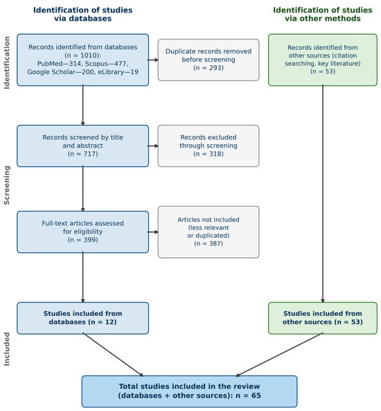
Selection was carried out stepwise according to the PRISMA scheme: identification → removal of duplicates → screening by titles and abstracts → assessment of full-text versions for eligibility → inclusion in the final analysis. The number of publications included in the final sample is reflected in the flow diagram (Figure 1). In addition to the database search, further relevant publications were identified by analyzing the reference lists of the included works (citation searching) and by drawing on key sources on anatomy and methodology; such sources are reflected in the flow diagram as a separate “other sources” branch. Because studies devoted specifically to imaging-based planning of chin-and-neck rejuvenation are relatively scarce, most included works (53 of 65) were identified through this branch rather than through the database search; accordingly, the present work is best regarded as a narrative review with a structured, PRISMA-informed search rather than a full systematic review, and the flow diagram (Figure 1) is provided for transparency of the search process.
Figure 1.
Flow diagram of the structured, PRISMA-informed literature search.
2.5. Data Extraction and Synthesis
From the included works, information was extracted on the imaging method, the anatomical structures studied, clinical tasks, and the advantages and limitations of each method. Owing to the methodological heterogeneity of the studies, a qualitative (narrative) synthesis of the data was performed without meta-analysis.
3. Results and Discussion
An analysis of the contemporary digital and radiological imaging methods used in planning facial rejuvenation surgery showed that each of the existing approaches has its own features of application and diagnostic value depending on the clinical task. All methods can be conditionally divided into 2D imaging, 3D imaging, CAD/CAM technologies, virtual and mixed realities, and artificial-intelligence-based tools (Table 2).
Table 2.
Classification of digital and radiological imaging methods in plastic surgery. This table classifies the method groups and their principal role; a detailed appraisal of advantages and limitations is given at the end of Section 3.
Studies devoted specifically to imaging of the chin region and neck for rejuvenation planning proved to be few. Only a handful address this topic directly—chiefly, our group’s cone-beam CT work [19,20,21] and several ultrasound reports [25,26,27,28,29,30,31]. To cover the subject fully, we therefore drew on adjacent fields where facial soft-tissue and skeletal imaging is better studied: the midface, orthodontic and orthognathic populations, and general facial-aging cohorts. Their imaging methodology transfers to our task, even though the studies themselves do not always concern the chin-and-neck region directly.
In total, 65 works were included (2006–2026; 56 of them within the 2020–June 2026 window and 9 earlier foundational sources). By modality, these were 5 cone-beam CT, 6 CT/MSCT, 2 MRI, 7 ultrasound, 8 three-dimensional photogrammetry and surface-scanning, 1 two-dimensional photography, 7 artificial-intelligence and digital-twin, 2 virtual- and mixed-reality, 3 CAD/CAM and virtual-planning, and 2 dosimetric studies, together with 22 background works on surgical technique, anatomy, epidemiological trends and methodology (5 + 6 + 2 + 7 + 8 + 1 + 7 + 2 + 3 + 2 + 22 = 65). Two additional dosimetric references [32,33], cited to substantiate the effective-dose figures, were added during revision and fall outside this search-derived corpus. Complete per-study evidence table is provided in Supplementary Table S1.
Because these works were heterogeneous and few studied the chin-and-neck region directly, we describe and compare their findings narratively rather than pooling them statistically. This shortage of dedicated, prospective imaging studies is one of the main reasons for the present review. It also explains why our central conclusion is stated cautiously: the available evidence points to the potential of upright cone-beam CT but does not yet establish its advantage, which will require further, preferably multicentre, studies.
In clinical practice, digital imaging offers several clear benefits: it makes the surgical result more accurate and predictable; it allows the operative plan to be tailored to each patient’s anatomy and age-related changes; and it improves visual communication with the patient, helping to set realistic expectations [18,19].
3.1. Two-Dimensional (2D) Imaging
Two-dimensional imaging is the simplest method for visualizing the proportions and shape of the face. In the context of facial plastic surgery, this method involves acquiring and analyzing two-dimensional images (photographs) of the patient’s face to assess the soft-tissue anatomy, plan surgical interventions, and document results. This approach uses standardized photographs in various projections (frontal, profile, oblique) with adherence to specific acquisition parameters such as lighting, distance, and head position to ensure the reproducibility and accuracy of subsequent analysis [34,35]. Ultrasound examination also displays the soft tissues of the face and neck in a two-dimensional format and has been used to assess the thickness of the skin, the supra- and subplatysmal fat, and the platysma, including in the upright patient position [25,26,27,28,29,30]. However, ultrasound provides only a local, layer-by-layer image in the area where the transducer is placed, depends substantially on the operator and the scanning angle, does not visualize bony landmarks, and does not form an integral three-dimensional picture of the relationship between the soft-tissue and bony structures of the chin region and neck; the combination of ultrasound with three-dimensional imaging is used mainly to monitor the results of device-based procedures [31]. Thus, neither ultrasound nor conventional 2D photography—which provides only a subjective assessment of proportions without information on tissue volume and depth [34,35]—provides a complete and objective picture of age-related changes; they serve as auxiliary methods requiring supplementation with three-dimensional imaging. Despite its availability and low cost, 2D imaging is limited by the absence of spatial information. In aesthetic surgery of the face and neck, it is used as a basic method complemented by 3D imaging.
3.2. Three-Dimensional (3D) Imaging
Three-dimensional imaging of the face for plastic surgery is performed using specialized three-dimensional scanning devices that make it possible to accurately reproduce the anatomy of the soft tissues and bones in digital format. The methods of 3D imaging used in planning facial rejuvenation surgery are as follows:
- Cone-beam computed tomography (CBCT): The main principle of this method is 3D X-ray imaging with a low radiation dose. It provides detailed assessment of bony structures and visualizes the position and outer contours of facial soft tissues, and is performed with the patient in the upright body position [20,21].
- Computed tomography (CT) of the maxillofacial region: The contrast resolution of CT is higher than that of CBCT, indicating better display of soft tissues. CT is used for an objective quantitative assessment of age-related changes in the structures of the chin region and neck: the thickness of the superficial musculoaponeurotic system (SMAS) [36], the volume and ptosis of the submandibular salivary glands [37], the anatomy of the paramedian platysmal bands [38], and the remodeling of the bony contour of the mandible and calvarium [39,40]. Three-dimensional CT modeling is also used for the repositioning of osteocutaneous structures [41].
Magnetic resonance imaging (MRI): The main principle of soft-tissue imaging is the response of hydrogen atoms in a magnetic field. This method can be informative for assessing the facial fat pads, the spaces of the face and neck, the platysma, and fascial structures (especially in the cervical zone) [42].
- Three-dimensional photogrammetric systems are now widely used: They make it possible to obtain a high-precision digital model of the patient’s face. The scanning equipment ranges from budget options, such as smartphones with LiDAR systems, to high-precision devices such as the VECTRA® M3/H1/XT (Canfield Scientific, Parsippany, NJ, USA) [43], Crisalix® 3D (Crisalix SA, Lausanne, Switzerland), and Canfield IntelliStudio 3D. According to a systematic review by Petrides et al., among the three-dimensional scanning methods, stereophotogrammetric systems most consistently demonstrate high accuracy and reproducibility in assessing facial soft tissues [44]; modern portable structured-light scanners provide comparable reliability at a lower cost and with greater accessibility [45]. Comparative studies of various 3D scanning systems [46,47,48] confirm their clinical applicability, and the validation of budget solutions based on LiDAR-enabled smartphones has demonstrated that their measurement accuracy is acceptable for clinical tasks [49]. Stereophotogrammetry is also used for the quantitative assessment of age-related changes in facial morphology across different age groups [50]. However, all methods of three-dimensional surface scanning (stereophotogrammetry, structured light) record only the external relief of the soft tissues—the surface shape and the light-and-shadow pattern—and do not display the deep soft-tissue and bony structures; therefore, they do not allow for objective determination of the causes of age-related deformities and require supplementation with radiological imaging methods.
- Hybrid 3D systems combine 3D photography, CT, and MRI analyses: This method has practical applications not only in planning rejuvenation surgery of the chin region and neck but also in combined interventions involving simultaneous platysmaplasty and bony genioplasty [19].
Three-dimensional imaging is the most promising direction in the digital preoperative planning of rejuvenation surgery for the chin region and neck. Among these methods, cone-beam computed tomography appears the most promising for the combined assessment of bony landmarks and soft-tissue contours and is discussed in detail below [19,20,21].
3.3. CAD/CAM Technologies
CAD/CAM systems are computer-aided design and computer-aided manufacturing technologies used to create and accurately implement three-dimensional objects. In medicine and, in particular, in facial plastic surgery, CAD/CAM makes it possible to design surgical guides and implants and to plan operations with a high degree of individualization. A 3D-printed implant model serves as a mock-up, while the actual implant—for example, a titanium plate or a synthetic implant for modeling the mandible and chin—is manufactured via milling or casting [51].
By themselves, CAD/CAM technologies do not represent an imaging method: they use data obtained from three-dimensional radiological imaging (CBCT or CT) and convert them into surgical guides and individual implants. The accuracy of such virtual planning and prediction of soft-tissue positions has been confirmed in systematic reviews and clinical studies [52,53], making CAD/CAM a valuable addition to CBCT and CT in individualized modeling; however, it does not replace radiological imaging as the source of the initial data.
3.4. Virtual and Mixed Realities (VR/AR)
Virtual and mixed reality (VR/AR) technologies in facial aesthetic plastic surgery are used for preoperative modeling, simulation of the expected result, and intraoperative navigation. VR allows the surgeon and the patient to “immerse” themselves in a three-dimensional model of the face to assess the extent of changes. AR overlays digital anatomy onto the real face in real time, increasing surgical accuracy and personalization. Both technologies improve planning and visualization of the result [54]. In a systematic review based on the screening of 1731 records, Stevanie et al. concluded that the practical application of high-technology virtual- and mixed-reality methods in planning aesthetic and orthognathic operations demonstrates a positive result. This is especially evident in the reduction in operative time, since virtual-reality software allows for individualized modeling of the patient’s clinical condition and visualization of the anticipated operative stages, making it possible to rehearse the surgical strategy in advance and thereby increase the likelihood of a successful outcome [55]. The use of virtual-reality technologies facilitates demonstration of the anticipated surgical result and improves communication with the patient. However, the high cost and the need for specialized training still limit the widespread introduction of these technologies into the routine practice of facial aesthetic surgery.
3.5. Artificial Intelligence (AI)
Artificial intelligence (AI) technologies in facial aesthetic surgery are used for automated image analysis, the detection of anatomical landmarks, morphometric assessments of proportions, and prediction of the surgical result. Systematic reviews of the application of artificial intelligence and machine learning in facial plastic and reconstructive surgery confirm the increasing accuracy of diagnosis, planning, and patient counseling; however, most solutions are still at the clinical validation stage [56,57,58,59].
One of the actively developing directions is the creation of patients’ digital twins—virtual models that make it possible to predict the surgical result while considering harmonious proportions of the face and neck consistent with the principles of the golden ratio and modern aesthetic concepts of a proportional face. A systematic review by Seth et al. (2024) demonstrated high postoperative patient satisfaction with the use of this technology; its main limitation remains its high costs of implementation and use [60].
Artificial intelligence is also used to objectify age-related changes: automated analysis of the mandibular shape reveals subtle, sex-specific changes in the bony contour that are relevant to rejuvenation planning [40], and AI-based morphometric analysis platforms make it possible to objectively assess facial proportions [61].
Despite their promise, AI and AI-based digital tools remain auxiliary, and their accuracy directly depends on the quality of the input imaging data. The high cost of specialized systems also remains a substantial barrier; as a more accessible alternative, low-cost solutions for three-dimensional facial imaging, including LiDAR-enabled smartphone applications, are being considered [49]. At the same time, objective assessments of patient satisfaction remain a key indicator of the effectiveness of such technologies.
Despite the variety of digital methods, radiological diagnostics (CT, CBCT, and MRI) currently remain the standard for imaging soft and bony tissues in preoperative planning (Table 3).
Table 3.
Comparative analysis of existing tomographic imaging methods.
Magnetic resonance imaging provides high soft-tissue contrast resolution and makes it possible to objectively assess the position of the soft-tissue structures of the neck, including the submandibular salivary glands and fascial elements, at the preoperative planning stage and during postoperative follow-up [42,62]. The age-related dynamics of the facial fat compartments are also objectively documented by CT and MRI [42,63], which is important for planning volumetric correction and lipofilling.
Despite the high diagnostic value of soft-tissue imaging methods in planning aesthetic operations in older patients, computed tomography and magnetic resonance imaging have certain limitations. One significant drawback is that these examinations are most often performed with the patient in the supine position. In this position, objective assessment of the gravitational ptosis of the soft tissues of the face and neck is difficult, which may lead to a distorted representation of the degree of age-related changes. In this regard, the use of radiological methods performed in the upright patient position becomes especially relevant, as it allows for a more accurate assessment of the severity of soft-tissue ptosis, the features of its distribution, and the participation of various anatomical structures in shaping the contours of the chin region and neck. This, in turn, contributes to more well-founded preoperative planning and selection of the optimal surgical strategy [20,21,25,26].
Among the available and cost-effective methods of preoperative planning, CBCT has demonstrated high diagnostic value in assessing age-related changes to the chin region and neck. The diagnostic value of CBCT in displaying soft-tissue structures and bony landmarks has also been confirmed in related areas of maxillofacial diagnostics [64,65], while the radiation dose from this method is substantially lower than that from multislice CT and can be further optimized by adjusting the acquisition parameters [66,67]. A significant advantage of this method is the ability to perform the examination in the upright patient position, which makes it possible to account for the effect of gravity on the soft tissues and to more objectively assess the severity of their ptosis. The use of CBCT makes it possible to determine in detail the position, outer contour, and ptosis of the platysma; the position, thickness, and localization of adipose tissue (above and/or below the platysma); the presence of ptosis of the submandibular salivary glands [62]; the state of the anterior bellies of the digastric muscles and the degree of their participation in shaping the contours of the cervicomental angle; and the position of the hyoid bone. In addition, the use of CBCT makes it possible to clearly demonstrate to the patient the existing contour deformities of the face and neck and to discuss possible methods of their correction based on a clear and objective visual image. Cone-beam computed tomography performed in the upright patient position provides an objective assessment of the state of each soft-tissue structure in age-related deformities of the chin region and neck. The data obtained make it possible to more accurately plan the targeted impact on specific anatomical structures during rejuvenation surgery and to predict the expected aesthetic results [20,21]. It should be emphasized, however, that CBCT visualizes these structures mainly through bone–soft-tissue–air interfaces and their outer contours; its intrinsic soft-tissue contrast resolution is limited and inferior to that of MRI, and quantitative reproducibility data (contrast-to-noise ratio, inter-observer agreement) for such measurements are still lacking.
Because CBCT involves ionizing radiation, its use for a purely aesthetic indication must comply with the justification and ALARA (as low as reasonably achievable) principles. The effective dose of a large field-of-view facial CBCT protocol should be reported and weighed against the clinical benefit, particularly when repeat imaging is anticipated; the dose figures in refs [66,67] refer to dentomaxillofacial protocols and are not directly transferable to a facial acquisition. The effective dose of the facial CBCT protocol on our unit (GENORAY Volux 550, 16 × 14 cm field of view) was 0.135 mSv—consistent with published effective doses for large field-of-view maxillofacial CBCT (e.g., 135 µSv for the large field-of-view i-CAT unit (Imaging Sciences International, Hatfield, PA, USA) in [32]) and an order of magnitude lower than that of conventional or multislice CT [32,33]. Where ionizing radiation is not justified, upright ultrasound [25,26,27,28,29,30] offers a radiation-free alternative that satisfies the same gravitational-loading rationale, although it does not assess bony structures and is operator-dependent.
Throughout this review, “upright” denotes imaging performed with the patient in a natural head position, since even small changes in cervical flexion markedly alter the cervicomental angle and affect reproducibility. In our practice, CBCT was acquired with the patient standing, gazing horizontally straight ahead, with the face oriented vertically and without a chin rest or any head restraint, so as not to distort the gravitational position of the soft tissues. This positioning follows the standardized protocol previously described by our group [20]: a large field-of-view facial acquisition (16 × 14 cm) with the scanning planes oriented to anatomical landmarks (horizontal—the lip-closure line; vertical—the projection of the tragus). During acquisition, patients were asked to keep the mimic and cervical muscles relaxed and to avoid facial expressions, speech, and swallowing, so that the true gravitational ptosis of the soft tissues could be assessed.
A summary comparison of digital and radiological imaging methods regarding diagnostic value relative to safety is presented in Table 4 [20,21].
Table 4.
Advantages and disadvantages of digital and radiological imaging methods in planning rejuvenation surgery. This table provides a detailed advantages/limitations appraisal, complementing the classification in Table 2.
4. Limitations
This review has several limitations. First, it is a narrative review: the protocol was not registered, study selection and full-text screening were performed by a single reviewer without duplicate independent assessment, and a formal risk-of-bias appraisal of the included studies was not carried out. Second, most of the included works (53 of 65) were identified through citation searching rather than the database search, and the search was restricted to publications in Russian and English (January 2020–June 2026), which may have introduced selection and language bias. Third, and most importantly, the central conclusion that upright CBCT is the most informative modality rests largely on a small number of single-centre studies from the authors’ own group [20,21]; independent, multicentre data with quantitative comparison of contrast-to-noise ratio, measurement reproducibility, and clinical outcomes are lacking, and the conclusion should therefore be regarded as a well-supported hypothesis rather than a definitively established finding. Fourth, the comparison of imaging modalities is qualitative; effective doses, contrast resolution, and reproducibility were not meta-analytically pooled.
5. Conclusions
Modern planning of surgery to correct age-related changes to the chin region and neck benefits greatly from the integration of digital and radiological imaging methods, which enable the surgeon to assess the patient’s anatomical features in detail, personalize the surgical strategy, and increase the predictability of the intervention outcome. Among the many existing radiological methods, on the basis of preliminary, predominantly single-centre evidence, cone-beam computed tomography appears to offer greater diagnostic value owing to the possibility of performing the examination with the patient in the upright body position, thereby providing an accurate assessment of the bony landmarks and soft-tissue structures of the chin region and neck. Within preoperative planning, radiological imaging methods contribute to increasing the accuracy of interventions, improving communication with the patient, and facilitating an individualized approach, which defines these methods as an integral part of modern facial aesthetic surgery.
Supplementary Materials
The following supporting information can be downloaded at: https://www.mdpi.com/article/10.3390/cosmetics13050274/s1. Table S1: Evidence table of the included studies. References [1,2,3,4,5,6,7,8,9,10,11,12,13,14,15,16,17,18,19,20,21,22,23,24,25,26,27,28,29,30,31,32,33,34,35,36,37,38,39,40,41,42,43,44,45,46,47,48,49,50,51,52,53,54,55,56,57,58,59,60,61,62,63,64,65,66,67] are cited in the Supplementary Materials.
Author Contributions
Conceptualization, A.S.B. and V.I.S.; methodology, A.S.B. and S.Y.G.; literature search and analysis, S.Y.G. and A.E.A.; software, N.S.B.; writing—original draft preparation, S.Y.G. and N.S.B.; writing—review and editing, A.S.B., V.I.S. and A.E.A.; supervision, A.S.B. and N.S.B. All authors have read and agreed to the published version of the manuscript.
Funding
This research received no external funding.
Institutional Review Board Statement
Not applicable.
Informed Consent Statement
Not applicable.
Data Availability Statement
This review analyzed only previously published data, which are available in the cited publications. The single primary value reported—the effective dose of the facial CBCT protocol on the authors’ unit (0.135 mSv)—was obtained from the scanner’s dose report and is available from the corresponding author on reasonable request.
Conflicts of Interest
The authors declare no conflicts of interest.
Abbreviations
The following abbreviations are used in this manuscript:
| 2D | Two-dimensional |
| 3D | Three-dimensional |
| AI | Artificial intelligence |
| CAD/CAM | Computer-aided design/computer-aided manufacturing |
| CBCT | Cone-beam computed tomography |
| CT | Computed tomography |
| LiDAR | Light detection and ranging |
| MRI | Magnetic resonance imaging |
| MSCT | Multislice computed tomography |
| PRISMA | Preferred Reporting Items for Systematic Reviews and Meta-Analyses |
| SMAS | Superficial musculoaponeurotic system |
| VR | Virtual reality |
| ENT | Ear, nose, and throat |
References
- Bravo, F.G. Neck Contouring and Rejuvenation in Male Patients Through Dual-Plane Reduction Neck Lift. Clin. Plast. Surg. 2022, 49, 257–273. [Google Scholar] [CrossRef] [Scilit]
- Borab, Z.M.; Fisher, S.; Rohrich, R.J. The 7-Step Neck-Lift Z-Plasty. Plast. Reconstr. Surg. 2025, 155, 316e–318e. [Google Scholar] [CrossRef] [Scilit]
- Iranmanesh, B.; Khalili, M.; Mohammadi, S.; Amiri, R.; Aflatoonian, M. Employing Microbotox Technique for Facial Rejuvenation and Face-Lift. J. Cosmet. Dermatol. 2022, 21, 4160–4170. [Google Scholar] [CrossRef] [Scilit]
- Labbé, D.; Rocha, C.S.M.; De Souza Rocha, F. Cervico-Mental Angle Suspensory Ligament: The Keystone to Understand the Cervico-Mental Angle and the Ageing Process of the Neck. Aesthet. Plast. Surg. 2017, 41, 832–836. [Google Scholar] [CrossRef] [Scilit]
- Kenkel, J.M.; Jones, D.H.; Fagien, S.; Glaser, D.A.; Monheit, G.D.; Stauffer, K.; Sykes, J.M. Anatomy of the Cervicomental Region: Insights from an Anatomy Laboratory and Roundtable Discussion. Dermatol. Surg. 2016, 42, S282–S287. [Google Scholar] [CrossRef] [Scilit]
- Pilsl, U.; Anderhuber, F. The Chin and Adjacent Fat Compartments. Dermatol. Surg. 2010, 36, 214–218. [Google Scholar] [CrossRef] [Scilit]
- Liu, Y.; Mao, R.; Xiao, M.; Zhu, W.; Liu, Y.; Xiao, H. Facial Rejuvenation: A Global Trend of Dermatological Procedures in the Last Decade. Plast. Reconstr. Surg. Glob. Open 2024, 12, e5801. [Google Scholar] [CrossRef] [Scilit]
- Dibbs, R.P.; Chamata, E.; Ferry, A.M.; Friedman, J.D. Revision Facelift and Neck Lift. Semin. Plast. Surg. 2021, 35, 088–097. [Google Scholar] [CrossRef] [Scilit]
- Gu, J.T.; Wang, T.D.; Loyo, M. Direct Neck Lift for Men. Facial Plast. Surg. Clin. N. Am. 2024, 32, 353–360. [Google Scholar] [CrossRef] [Scilit]
- Edward James Whitehead, D.; Çakmak, Ö. Face and Neck Lift Options in Patients of Ethnic Descent. Facial Plast. Surg. Clin. N. Am. 2022, 30, 489–498. [Google Scholar] [CrossRef] [Scilit]
- Jacono, A.A.; Alemi, A.S.; Harmon, J.J.; Ahmedli, N. The Effect of a Novel Platysma Hammock Flap During Extended Deep Plane Facelift on the Signs of Aging in the Neck. Aesthet. Surg. J. 2022, 42, 845–857. [Google Scholar] [CrossRef] [Scilit]
- Timberlake, A.T.; Cameron Brawley, C.; Paul, B.C.; Rosenberg, D.B. Complete Platysma Muscle Suspension in Deep-Plane Face-Lift Surgery. Plast. Reconstr. Surg. 2025, 155, 699e–703e. [Google Scholar] [CrossRef] [Scilit]
- Talei, B.; Shauly, O.; Marxen, T.; Menon, A.; Gould, D.J. The Mastoid Crevasse and 3-Dimensional Considerations in Deep Plane Neck Lifting. Aesthet. Surg. J. 2024, 44, NP132–NP148. [Google Scholar] [CrossRef] [Scilit]
- Van Pham, T.; Truong, K.H.T.N. Short or Minimum Incision Deep Plane/Extended Deep Plane Facelift/Rhytidectomy. Facial Plast. Surg. 2020, 36, 376–385. [Google Scholar] [CrossRef] [Scilit]
- Gentile, R.D. Ultrasonic Deep-Plane Neck Lift. Facial Plast. Surg. 2025, 41, 54–69. [Google Scholar] [CrossRef] [Scilit]
- Han, C.H. Rotation of a Part of the Composite Flap for Simultaneous Gonion Reduction and Malar Augmentation During Rhytidectomy in Asians. Ann. Plast. Surg. 2024, 92, 267–273. [Google Scholar] [CrossRef] [Scilit]
- Le Louarn, C. Face Lift with U Threads. Ann. Chir. Plast. Esthet. 2021, 66, 62–68. [Google Scholar] [CrossRef] [Scilit]
- Borisenko, A.S.; Sharobaro, V.I.; Burkhonova, N.S.; Avdeev, A.E.; Ahmed Alsheikh, Y.M. Advanced Surgical Approaches for the Rejuvenation of the Submental and Cervicofacial Regions: A Literature Review for a Personalized Approach. Cosmetics 2025, 12, 26. [Google Scholar] [CrossRef] [Scilit]
- Borisenko, A.S.; Sharobaro, V.I.; Avdeev, A.E.; Burkhonova, N.S.; Fisun, A.O. Using 3D Imaging to Preoperatively Plan Facelift Procedures for the Lower Third of the Face and Neck. Cosmetics 2024, 11, 28. [Google Scholar] [CrossRef] [Scilit]
- Alimova, S.M.; Sharobaro, V.I.; Avdeev, A.E.; Sidorenkov, D.A.; Guseva, T.S. Cone-Beam Computed Tomography for Objective Diagnosis of Age-Related Soft Tissue Changes in Lower Face and Neck. Aesthet. Plast. Surg. 2023, 47, 2370–2377. [Google Scholar] [CrossRef] [Scilit]
- Alimova, S.M.; Truten, V.P.; Shamanaeva, L.S.; Guseva, T.S. A New Approach to Surgical Rejuvenation of the Face and Neck: The Influence of Cone Beam Computed Tomography on Choice of Treatment Strategy in Patients with Involutional Changes. Aesthet. Plast. Surg. 2025, 49, 5327–5334. [Google Scholar] [CrossRef] [Scilit]
- Borisenko, A.S.; Sharobaro, V.I.; Avdeev, A.E.; Ahmed Alsheikh, Y.M. Docking Methods for Robot-Assisted Rhytidectomy and Platysmaplasty. Plast. Reconstr. Surg. Glob. Open 2025, 13, e6733. [Google Scholar] [CrossRef] [Scilit]
- Borisenko, A.S.; Sharobaro, V.I.; Burkhonova, N.S.; Avdeev, A.E.; Ermolaev, A.A.; Ahmed Alsheikh, Y.M. Isolated Medial Platysmaplasty and Harmonization of the Lower Third of Face with Local Fat Autograft: A Case Report. Plast. Reconstr. Surg. Glob. Open 2024, 12, e5897. [Google Scholar] [CrossRef] [Scilit]
- Page, M.J.; McKenzie, J.E.; Bossuyt, P.M.; Boutron, I.; Hoffmann, T.C.; Mulrow, C.D.; Shamseer, L.; Tetzlaff, J.M.; Akl, E.A.; Brennan, S.E.; et al. The PRISMA 2020 Statement: An Updated Guideline for Reporting Systematic Reviews. BMJ 2021, 372, n71. [Google Scholar] [CrossRef] [Scilit]
- Alimova, S.; Sharobaro, V.; Yukhno, A.; Bondarenko, E. Possibilities of Ultrasound Examination in the Assessment of Age-Related Changes in the Soft Tissues of the Face and Neck: A Review. Appl. Sci. 2023, 13, 1128. [Google Scholar] [CrossRef] [Scilit]
- Sharobaro, V.; Alimova, S.; Telnova, A.; Shamanaeva, L. Ultrasound Diagnosis of Age-Related Involutional Changes in the Lower Third of Face and Neck to Determine Treatment Techniques. Pak. J. Med. Sci. 2021, 37, 272–276. [Google Scholar] [CrossRef] [Scilit]
- Nasreddine, H.; Tlaiss, Y.; Hassan, F.; Ibrahim, R. Ultrasound Assessment of Facial and Neck Aging: A Noninvasive Approach to a Minimally Invasive Treatment. J. Med. Ultrasound 2024, 32, 244–248. [Google Scholar] [CrossRef] [Scilit]
- Zhang, M.; Yang, Y.; Shi, Y.; Dong, R.; Chong, Y.; Li, Z.; Jin, L.; Huang, J.; Yu, N.; Long, X. A Comprehensive Ultrasound Investigation of Lower Facial and Neck Structure. Aesthet. Plast. Surg. 2025, 49, 1–12. [Google Scholar] [CrossRef] [Scilit]
- Kim, S.; Lee, I.; Kim, J.; Lee, Y. Preoperative Ultrasound Measurement of Subplatysmal Fat Thickness as a Predictor of Excision Volume in Neck Contouring Surgery: A Quantitative Analysis. J. Craniofac. Surg. 2026, 37, 1612–1615. [Google Scholar] [CrossRef] [Scilit]
- Alimova, S.M.; Sharobaro, V.I.; Telnova, A.V.; Stepanyan, E.E. Planning of Methods of Surgical Correction of Soft Tissues of the Face and Neck. Med. Vis. 2021, 25, 47–52. [Google Scholar] [CrossRef] [Scilit]
- Zhu, J.; Han, Y.; Liu, Y.; Chang, R.; Gao, W.; Gong, X.; Zhu, Y.; Shang, Y.; Shen, L.; Yu, W.; et al. Evaluation of a Novel Microfocused Ultrasound with Three-Dimensional Digital Imaging for Facial Tightening: A Prospective, Randomized, Controlled Trial. Dermatol. Ther. 2024, 14, 233–249. [Google Scholar] [CrossRef] [Scilit]
- Ludlow, J.; Davies-Ludlow, L.; Brooks, S.; Howerton, W. Dosimetry of 3 CBCT Devices for Oral and Maxillofacial Radiology: CB Mercuray, NewTom 3G and i-CAT. Dentomaxillofac. Radiol. 2006, 35, 219–226. [Google Scholar] [CrossRef] [Scilit]
- Ludlow, J.B.; Ivanovic, M. Comparative Dosimetry of Dental CBCT Devices and 64-Slice CT for Oral and Maxillofacial Radiology. Oral Surg. Oral Med. Oral Pathol. Oral Radiol. Endod. 2008, 106, 106–114. [Google Scholar] [CrossRef] [Scilit]
- Zogheib, T.; Jacobs, R.; Bornstein, M.M.; Agbaje, J.O.; Anumendem, D.; Klazen, Y.; Politis, C. Comparison of 3D Scanning Versus 2D Photography for the Identification of Facial Soft-Tissue Landmarks. Open Dent. J. 2018, 12, 61–71. [Google Scholar] [CrossRef] [Scilit]
- Tian, W.M.; Porras Fimbres, D.C.; Tran, M.M.; Zeng, S.L.; Gnaedinger, A.G.; Kaplan, S.; Marcus, J.R.; Patel, A. Quality and Reliability of 2D and 3D Clinical Photographs in Plastic Surgery: A Scoping Review. Aesthet. Plast. Surg. 2025, 49, 4447–4457. [Google Scholar]
- Okuda, I.; Abe, K.; Yoshioka, N.; Komemushi, T.; Jinzaki, M.; Ohjimi, H. Objective Analysis of Age-Related Changes in the Superficial Musculoaponeurotic System in Japanese Females Using Computed Tomography. Aesthet. Surg. J. Open Forum 2023, 5, ojad043. [Google Scholar] [CrossRef] [Scilit]
- Sawan, T.; Tower, J.I.; Gordon, N.A.; Paskhover, B. The Submandibular Gland and The Aging Neck: A Longitudinal Volumetric Study. Aesthet. Plast. Surg. 2021, 45, 987–991. [Google Scholar] [CrossRef] [Scilit]
- Davidovic, K.; Frank, K.; Schenck, T.L.; Cohen, S.R.; Dayan, S.; Gotkin, R.H.; Sykes, J.M.; Liew, S.; Nikolis, A.; Cotofana, S. Anatomy behind the Paramedian Platysmal Band: A Combined Cadaveric and Computed Tomographic Study. Plast. Reconstr. Surg. 2021, 148, 979–988. [Google Scholar] [CrossRef] [Scilit]
- Nur Kuzan, B.; Yusuf Kuzan, T. Evaluation of Facial Aging in Different Age and Gender Groups with Computed Tomography–Based Calvarium and Face Measurements. Dermatol. Surg. 2024, 50, 636–642. [Google Scholar] [CrossRef] [Scilit]
- Hussein, S.M.; Rames, J.D.; Shehab, A.A.; Pazelli, A.M.; Sears, V.A.; Wentworth, A.J.; Morris, J.M.; Sharaf, B.A. How Does the Mandible Age? Comprehensive Artificial Intelligence–Assisted Shape Analysis in the White Population. Plast. Reconstr. Surg. Glob. Open 2025, 13, e6650. [Google Scholar] [CrossRef] [Scilit]
- Tilaveridis, I.; Pantazi, G.; Venetis, G.; Ditsios, K.; Angelopoulos, C.; Antoniades, K. The Value of Three-Dimensional Modeling on Repositioning and Soft Tissue Filling in Microsurgical Reconstruction of Secondary Mandibular Defects: A Retrospective Study. Cureus 2022, 14, e22336. [Google Scholar] [CrossRef] [Scilit]
- Cevik Cenkeri, H.; Sarigul Guduk, S.; Derin Cicek, E. Aging Changes of the Superficial Fat Compartments of the Midface over Time: A Magnetic Resonance Imaging Study. Dermatol. Surg. 2020, 46, 1600–1605. [Google Scholar] [CrossRef] [Scilit]
- De Stefani, A.; Barone, M.; Hatami Alamdari, S.; Barjami, A.; Baciliero, U.; Apolloni, F.; Gracco, A.; Bruno, G. Validation of Vectra 3D Imaging Systems: A Review. Int. J. Environ. Res. Public Health 2022, 19, 8820. [Google Scholar] [CrossRef] [Scilit]
- Petrides, G.; Clark, J.R.; Low, H.; Lovell, N.; Eviston, T.J. Three-Dimensional Scanners for Soft-Tissue Facial Assessment in Clinical Practice. J. Plast. Reconstr. Aesthet. Surg. 2021, 74, 605–614. [Google Scholar] [CrossRef] [Scilit]
- Schipper, J.A.M.; Merema, B.J.; Hollander, M.H.J.; Spijkervet, F.K.L.; Dijkstra, P.U.; Jansma, J.; Schepers, R.H.; Kraeima, J. Reliability and Validity of Handheld Structured Light Scanners and a Static Stereophotogrammetry System in Facial Three-Dimensional Surface Imaging. Sci. Rep. 2024, 14, 8172. [Google Scholar] [CrossRef] [Scilit]
- Gašparović, B.; Morelato, L.; Lenac, K.; Mauša, G.; Zhurov, A.; Katić, V. Comparing Direct Measurements and Three-Dimensional (3D) Scans for Evaluating Facial Soft Tissue. Sensors 2023, 23, 2412. [Google Scholar] [CrossRef] [Scilit]
- Jindanil, T.; Ponbuddhichai, R.; Massant, C.; Xu, L.; Fontenele, R.C.; Llano-Pérula, M.C.D.; Jacobs, R. Three-Dimensional Facial Imaging: A Comparative Assessment of the Clinical Applicability of State-of-the-Art Technologies for Three-Dimensional Facial Imaging. Int. J. Dent. 2025, 2025, 8822293. [Google Scholar] [CrossRef] [Scilit]
- Knoops, P.G.M.; Beaumont, C.A.; Borghi, A.; Rodriguez-Florez, N.; Breakey, R.W.; Rodgers, W.; Angullia, F.; Jeelani, N.U.O.; Schievano, S.; Dunaway, D.J. Comparison of Three-Dimensional Scanner Systems for Craniomaxillofacial Imaging. J. Plast. Reconstr. Aesthet. Surg. 2017, 70, 441–449. [Google Scholar] [CrossRef] [Scilit]
- Loy, R.C.H.; Liew, M.K.M.; Yong, C.W.; Wong, R.C.W. Validation of Low-Cost Mobile Phone Applications and Comparison with Professional Imaging Systems for Three-Dimensional Facial Imaging: A Pilot Study. J. Dent. 2023, 137, 104676. [Google Scholar] [CrossRef] [Scilit]
- Mengoa, M.G.R.; Garcia, A.A.M.N.; Fioravanti, K.S.; Neppelenbroek, K.H.; Oliveira, T.M.; Sforza, C.; Soares, S. Facial Morphology Analysis of Caucasian Brazilian Adult Women Using Stereophotogrammetry. Braz. Oral Res. 2024, 38, e105. [Google Scholar]
- Baba, N.Z.; Goodacre, B.J.; Goodacre, C.J.; Müller, F.; Wagner, S. CAD/CAM Complete Denture Systems and Physical Properties: A Review of the Literature. J. Prosthodont. 2021, 30, 113–124. [Google Scholar] [CrossRef] [Scilit]
- Awad, D.; Reinert, S.; Kluba, S. Accuracy of Three-Dimensional Soft-Tissue Prediction Considering the Facial Aesthetic Units Using a Virtual Planning System in Orthognathic Surgery. J. Pers. Med. 2022, 12, 1379. [Google Scholar] [CrossRef] [Scilit]
- Starch-Jensen, T.; Hernández-Alfaro, F.; Kesmez, Ö.; Gorgis, R.; Valls-Ontañón, A. Accuracy of Orthognathic Surgical Planning Using Three-Dimensional Virtual Techniques Compared with Conventional Two-Dimensional Techniques: A Systematic Review. J. Oral Maxillofac. Res. 2023, 14, e1. [Google Scholar] [CrossRef] [Scilit]
- Chou, D.W.; Annadata, V.; Willson, G.; Gray, M.; Rosenberg, J. Augmented and Virtual Reality Applications in Facial Plastic Surgery: A Scoping Review. Laryngoscope 2024, 134, 2568–2577. [Google Scholar] [CrossRef] [Scilit]
- Stevanie, C.; Ariestiana, Y.Y.; Hendra, F.N.; Anshar, M.; Boffano, P.; Forouzanfar, T.; Sukotjo, C.; Kurniawan, S.H.; Ruslin, M. Advanced Outcomes of Mixed Reality Usage in Orthognathic Surgery: A Systematic Review. Maxillofac. Plast. Reconstr. Surg. 2024, 46, 29. [Google Scholar] [CrossRef] [Scilit]
- Stephanian, B.; Karki, S.; Debnath, K.; Saltychev, M.; Rossi-Meyer, M.; Kandathil, C.K.; Most, S.P. Role of Artificial Intelligence and Machine Learning in Facial Aesthetic Surgery: A Systematic Review. Facial Plast. Surg. Aesthet. Med. 2024, 26, 679–705. [Google Scholar] [CrossRef] [Scilit]
- Espinosa Reyes, J.A.; Puerta Romero, M.; Cobo, R.; Heredia, N.; Solís Ruiz, L.A.; Corredor Zuluaga, D.A. Artificial Intelligence in Facial Plastic and Reconstructive Surgery: A Systematic Review. Facial Plast. Surg. 2024, 40, 615–622. [Google Scholar] [CrossRef] [Scilit]
- Adegboye, F.O.; Peterson, A.A.; Sharma, R.K.; Stephan, S.J.; Patel, P.N.; Yang, S.F. Applications of Artificial Intelligence in Facial Plastic and Reconstructive Surgery: A Narrative Review. Facial Plast. Surg. Aesthet. Med. 2025, 27, 275–281. [Google Scholar] [CrossRef] [Scilit]
- Souza, S.; Bhethanabotla, R.M.; Mohan, S. Applications of Artificial Intelligence in Facial Plastic and Reconstructive Surgery: A Systematic Review. Curr. Opin. Otolaryngol. Head Neck Surg. 2024, 32, 222–233. [Google Scholar] [CrossRef] [Scilit]
- Seth, I.; Lim, B.; Lu, P.Y.J.; Xie, Y.; Cuomo, R.; Ng, S.K.-H.; Rozen, W.M.; Sofiadellis, F. Digital Twins Use in Plastic Surgery: A Systematic Review. J. Clin. Med. 2024, 13, 7861. [Google Scholar] [CrossRef] [Scilit]
- Salinas, C.A.; Liu, A.; Sharaf, B.A. Facial Morphometrics in Black Celebrities: Contemporary Facial Analysis Using an Artificial Intelligence Platform. J. Clin. Med. 2023, 12, 4499. [Google Scholar] [CrossRef] [Scilit]
- Lee, M.K.; Sepahdari, A.; Cohen, M. Radiologic Measurement of Submandibular Gland Ptosis. Facial Plast. Surg. 2013, 29, 422–428. [Google Scholar] [CrossRef] [Scilit]
- Gierloff, M.; Stöhring, C.; Buder, T.; Gassling, V.; Açil, Y.; Wiltfang, J. Aging Changes of the Midfacial Fat Compartments: A Computed Tomographic Study. Plast. Reconstr. Surg. 2012, 129, 263–273. [Google Scholar]
- Alhazmi, N.; Alrasheed, F.; Alshayea, K.; Almubarak, T.; Alzeer, B.; Alorf, M.S.; Alshanqiti, A.; Albalawi, M. Facial Soft Tissue Characteristics Among Sagittal and Vertical Skeletal Patterns: A Cone-Beam Computed Tomography Study. Cureus 2023, 15, e44428. [Google Scholar] [CrossRef] [Scilit]
- Setvaji, N.R.; Muthuswamy Pandian, S. Evaluation of Soft Tissue Compensations in Subjects with Facial Asymmetry Using Cone Beam Computed Tomography (CBCT): A Retrospective Study. Cureus 2024, 16, e52601. [Google Scholar] [CrossRef] [Scilit]
- Reidelbach, C.S.; Neubauer, J.; Russe, M.F.; Kusterer, J.; Semper-Hogg, W. Evaluation of Skin Doses for Cone-Beam Computed Tomography in Dentomaxillofacial Imaging: A Preclinical Study. PLoS ONE 2021, 16, e0254510. [Google Scholar] [CrossRef] [Scilit]
- Shina, L.O.; Hajee, S.I. Dose Optimization of Cone Beam Computed Tomography: Measurement of Parotid Absorbed Dose and Image Quality Assessment. J. Med. Life 2025, 18, 214–222. [Google Scholar] [CrossRef] [Scilit]

Disclaimer/Publisher’s Note: The statements, opinions and data contained in all publications are solely those of the individual author(s) and contributor(s) and not of MDPI and/or the editor(s). MDPI and/or the editor(s) disclaim responsibility for any injury to people or property resulting from any ideas, methods, instructions or products referred to in the content. |
© 2026 by the authors. Licensee MDPI, Basel, Switzerland. This article is an open access article distributed under the terms and conditions of the Creative Commons Attribution (CC BY) license.