


Abstract
Oxidative stress contributes to hair follicle degeneration by inducing inflammation and apoptosis in dermal papilla cells. This study investigated whether Saccharomyces cerevisiae fermentation enhances the anti-hair loss potential of Laminaria japonica (kelp) by-products by improving their antioxidant, anti-inflammatory, and cytoprotective activities. Fermentation significantly increased the superoxide dismutase (SOD) activity of kelp. In H2O2-stimulated human hair follicle dermal papilla cells, fermented kelp culture (KF) dose-dependently protected against oxidative-stress-induced cytotoxicity, reduced IL-6 and IL-8 secretion, inhibited NFκB phosphorylation, restored insulin-like growth factor-1 (IGF-1) expression, enhanced AKT phosphorylation, reduced Bax expression, and restored total caspase-3 expression. These findings indicate that KF attenuates oxidative-stress-induced inflammation and apoptosis while preserving dermal papilla cell function. A four-week application study further showed that a shampoo containing KF reduced hair loss and improved hair gloss, scalp sebum, and scalp scaling. Together, these findings suggest that S. cerevisiae fermentation enhances the biological activity of kelp by-products and supports the potential of KF as a functional cosmetic ingredient for anti-hair loss applications.
1. Introduction
Hair loss is a multifactorial disorder associated with ultraviolet (UV) exposure, aging, chronic stress, nutritional imbalance, hormonal changes, and chronic inflammation [1,2,3,4]. Among these factors, oxidative stress is considered one of the major pathogenic factors responsible for hair follicle dysfunction, in which excessive accumulation of reactive oxygen species (ROS) impairs the biological function of human hair follicle dermal papilla cells (HFDPCs) [5,6,7]. Excessive intracellular reactive ROS accumulation induces oxidative stress, resulting in activation of nuclear factor kappa-light-chain-enhancer of activated B cell (NFκB)-dependent inflammatory signaling and increased production of pro-inflammatory cytokines, including IL-6 and IL-8, which contribute to dermal papilla cell dysfunction and hair follicle regression [8,9]. Persistent oxidative stress further induces apoptosis through Bax and caspase-3 associated pathways while suppressing AKT and insulin like growth factor-1 (IGF-1)-mediated cell survival signaling, thereby disrupting the normal hair cycle and accelerating the transition from the anagen to the catagen phase [10]. Several studies have also demonstrated that regulation of oxidative stress, inflammation, and apoptosis is considered an important therapeutic strategy for preserving HFDPC function and preventing hair loss [2]. Current therapeutic agents, including minoxidil, finasteride, and dutasteride, can promote hair growth and delay hair follicle regression, but their long-term use is often limited by adverse effects [11,12,13,14,15,16]. This limitation has increased interest in safe, naturally derived alternatives that protect dermal papilla cells and preserve hair follicle homeostasis [7,17].
Laminaria japonica (kelp) is an edible brown seaweed rich in bioactive constituents, including polysaccharides, polyphenols, carotenoids, and minerals, which exhibit antioxidant, anti-inflammatory, and immunomodulatory activities [18,19,20,21,22]. The diverse biological activities of kelp have supported its application in the food, pharmaceutical, and cosmetic industries [23,24,25,26,27]. Large quantities of kelp by-products are generated during industrial processing and are often discarded despite retaining substantial amounts of bioactive constituents, making their effective utilization a major challenge for the sustainable valorization of marine biomass [28,29]. Microbial fermentation is widely used as a bioconversion process to improve the biological functionality of natural resources by converting complex macromolecules into readily bioavailable metabolites through microbial enzymatic activity [30,31,32]. Previous studies have reported that fermentation of kelp using Bacillus subtilis enhanced antioxidant and anti-inflammatory activities, whereas Enterococcus faecium fermentation increased antioxidant capacity by improving the availability of functional metabolites [33,34,35]. Among food-grade microorganisms, S. cerevisiae is widely used because of its safety, industrial applicability, and efficient bioconversion capacity. During fermentation, enzymes produced by S. cerevisiae degrade structural macromolecules and promote the release of low-molecular-weight bioactive compounds with enhanced bioavailability. Previous studies have reported that fermentation of Lycium barbarum polysaccharides using S. cerevisiae enhances biological activity and delays aging in Caenorhabditis elegans [36]. Similarly, S. cerevisiae-fermented kelp has been reported to enhance its anti-inflammatory activity [37]. In our previous study, we developed KF, a S. cerevisiae-fermented kelp by-product-based organic agricultural material, and demonstrated that its application significantly increased the accumulation of bioactive constituents in hydroponically cultivated ginseng sprouts [37,38,39]. Collectively, available evidence indicates that microbial fermentation can substantially improve the utilization and functional value of kelp residues. Therefore, S. cerevisiae was selected in the present study as the fermentation microorganism to enhance the biological activity of kelp by-products and to evaluate their potential as functional ingredients for anti-hair loss application.
In the present study, the protective effects of KF against oxidative-stress-induced damage were investigated using H2O2-stimulated HFDPCs. The underlying mechanism was examined by evaluating antioxidant activity together with inflammatory, apoptosis-related, and hair-growth-associated signaling pathways. Furthermore, the efficacy of a shampoo containing KF was evaluated in a human application test to determine whether the protective effects observed in HFDPCs could be translated into improvements in hair and scalp condition.
2. Materials and Methods
2.1. Seaweed and Reagents
Kelp was purchased from Badapum Co., Ltd. (Wando, Republic of Korea). Molasses was provided by Mosco International Commodities Pvt., Ltd. (Gujarat, India). Cell Counting kit-8 (CCK-8) was obtained from Dojindo Laboratories (Kumamoto, Japan). Dulbecco’s Modified Eagle Medium (DMEM), phosphate-buffered saline (PBS), and penicillin–streptomycin were acquired from Hyclone (Logan, UT, USA). Human anti-Akt (pan) (11E7, 4685), human anti-Bax (D3R2M), human anti-βactin (13E5, 4970), human anti-Caspase-3 (D3R6Y, 14220), human anti-IGF (E6B70, 73034), human anti-NFκB (D14E12, 242), human anti-Phospho-Akt (Ser473) (D9E, 4060), human anti-Phospho-NFκB (Ser536) (93H1,3033), and horseradish-peroxidase-conjugated secondary antibody (7074S) were purchased from Cell Signaling Technology (Danvers, MA, USA). ECL reagent, 5% (w/v) skim milk in Tris-buffered saline (TBS; 50 mM Tris–HCl (pH 7.5), and 150 mM NaCl, 2.5 mM KCl) were acquired from Bio-Red (Hercules, CA, USA). Dimethyl sulfoxide (DMSO, D2650) and Minoxidil (M6699, ≥98% (HPLC)) were purchased from Sigma Aldrich (St. Louis, MO, USA). Hydrogen Peroxide (H2O2, 084-07441) was obtained from Fujifilm Wako Pure Chemical Corporation (Osaka, Japan).
2.2. Microbial Fermentations
To prepare the kelp by-product fermentation (KF), a fermentation protocol was adapted from a previously established method [37,38]. KF was prepared using a patented Saccharomyces cerevisiae fermentation process (Korean patent application number.10-2023-0122734). Briefly, kelp by-product was thoroughly washed with high-pressure tap water and boiled for 1 min to remove foreign substances and reduce salinity. Kelp powder was prepared by crushing kelp using a screw-type crusher (Sunwon System Co., Ansan, Republic of Korea) and drying it in a drying oven (Jeio Tech Co., Daejeon, Republic of Korea) at 50 °C for 20 h. Next, a nutrient mixture containing 3% (w/v) defatted soybean extract (Hokyoung-Tech Co., Anseong, Republic of Korea), 2% (w/v) molasses, and 3% (w/v) desalted kelp powder was homogenized and heat-treated at 121 °C for 15 min. The mixture was then inoculated with Saccharomyces cerevisiae (KCTC 17299, Korea Collection for Type Cultures, Jeongup, Republic of Korea) at a final concentration of 0.01% (v/v) and incubated at 30 °C for 72 h in a shaking incubator (200 rpm, KoBiotech Co., Ltd., Incheon, Republic of Korea). To terminate fermentation, the fermenters were pasteurized at 90 °C for 30 min, filtered through a sterile 0.45 μm membrane filter, and stored at 4 °C. The resulting solution was then concentrated using a rotary vacuum evaporator (N-1200-B, Eyela, Tokyo, Japan) and stored at −20 °C prior to experiments.
2.3. Superoxide Dismutase (SOD) Activity Assay
The superoxide dismutase (SOD) activity of KF was determined using a Superoxide Dismutase Assay Kit (Dojindo, Kumamoto, Japan) according to the manufacturer’s instructions. Briefly, KF samples at various concentrations were added to a 96-well plate. WST working solution, dilution buffer, and enzyme working solution were then added to each well according to the manufacturer’s protocol. After incubation at 37 °C for 20 min, the absorbance was measured at 450 nm using a microplate ELISA reader (EPOCH, Biotek, Winooski, VT, USA).
2.4. Cell Culture
Human Follicle Dermal Papilla Cells (HFDPCs, C-26501) sourced from PromoCell (Heidelberg, Germany) were maintained in follicle dermal papilla cell growth medium (C-26502, PromoCell, Heidelberg, Germany) containing supplement mix and 1% penicillin–streptomycin at 37 °C in a humidified 5% CO2. Cell cultivation was using a Detach kit (PromoCell, Heidelberg, Germany) and transferred to 75 mm tissue culture flasks once they reached 80–90% confluence, typically every 4 days. Cell within 10 passages were used for the experiments.
2.5. Cell Viability Assay
HFDPCs (5 × 10³ cells/well) were seeded in 96-well plates and incubated overnight at 37 °C. The cells were pretreated with 300 μM H2O2 for 30 min and subsequently treated with Minoxidil (10 μM), Kelp extract (KE), or KF for 48 h. Cell viability was determined using a Cell-Counting Kit-8 (CCK-8) assay in accordance with the manufacturer’s instructions. The absorbance was measured using a microplate ELISA reader (EPOCH, Biotek, Winooski, VT, USA) at 450 nm, and the percentage of cell viability was computed according to Equation. Images were acquired using a microscope (CKX53, Olympus, Tokyo, Japan) with a ×10 objective. Serum-free DMEM was exploited as the negative control, Minoxidil was used the positive control [35,40].
Cell viability = Absorbance of sample/Absorbance of negative control.
2.6. Enzyme-Linked Immunosorbent Assay (ELISA)
Concentrations in culture supernatants of the cytokine levels IL-6 and IL-8 were measured using a BD OptEIA™ ELISA kit II (BD Biosciences, San Jose, CA, USA) in accordance with the manufacturer’s instructions. Briefly, the supernatants or standard were added to a 96well plate coated with monoclonal antibody specific for IL-6 or IL-8. The wells were washed, and streptavidin-horseradish peroxidase conjugated with biotinylated anti-Human IL-6 or IL-8 antibody was added. The wells were washed, and TMB substrate solution was added, which produced a blue color in direct proportion to the amount of IL-6 or IL-8 present in the supernatants. The absorbance was measured using a microplate ELISA reader (EPOCH, Biotek, Winooski, VT, USA) at a wavelength range of 450 nm. The percentage values were calculated relative to the H2O2-treated group, which was set at 100%.
2.7. Western Blot Analysis
Cells were washed twice with PBS and lysed on ice for 30 min in RIPA lysis buffer (Thermo, Waltham, MA, USA) supplemented with phosphatase and protease inhibitor cocktail (Thermo, Waltham, MA, USA). The protein concentration was measured using a BCA protein Assay kit (Pierce Biotechnology, Rockford, IL, USA). Subsequently, the 10% Tris-Glycine plus gels- separated proteins (Bio-Red, Hercules, CA, USA) were transferred to PVDF membranes (Bio-Red, Hercules, CA, USA) that were then separately incubated with indicated antibodies. Protein bands were quantified of ChemiDoc imaging system (Bio-Red, Hercules, CA, USA). Band intensities were quantified using Image Lab Software ver 6.0 (Bio-Red, Hercules, CA, USA). Following detection of the target protein, the membrane was stripped using stripping buffer and reproved with the subsequent antibody. The levels of p-NFκB and p-AKT were normalized to their corresponding total protein levels detected on the same membrane, while β-actin detected on the same membrane was used as an internal loading control.
2.8. Human Application Test
A Human application test was conducted to evaluate the anti-hair loss efficacy of the test shampoo. Standardized fermented KF [33,34] were used as active ingredients for hair care cosmetic products (No. 2025-14112, they are classified as raw materials for cosmetics by the International Nomenclature of Cosmetic Ingredient (INCI)). The complete shampoo formulation consisted of purified water, KF (25 ppm), sodium C14-16 olefin sulfonate, cocamidopropyl betaine, coco-betaine, coco-glucoside, sodium cocoyl isethionate, caffeine, 1,2-hexanediol, sodium chloride, glycerin, polyquaternium-10, glyceryl caprylate, lemongrass leaf oil, tartaric acid, butylene glycol, trehalose, propanediol, Ecklonia cava extract (2 ppm), KE (2 ppm), Camellia japonica flower extract, Undaria pinnatifida extract (2 ppm), dipotassium glycyrrhizate, Centella asiatica extract, persimmon extract, C. japonica leaf extract, carob fruit extract, green tea extract, Polygonum cuspidatum root extract, Scutellaria baicalensis extract, chamomile flower extract, and rosemary leaf extract. Twenty-one healthy female volunteers aged 20–60 years were enrolled and instructed to use the test shampoo once daily for 4 weeks (Table 1). The clinical study was performed at Prederma Institute of Dermatological Clinical Research (Korea) in accordance with the ethical principles of the Declaration of Helsinki and was approved by the Institutional Review Board (IRB No. PDCR-IRB-2026-N-052). Before each clinical assessment, the hair and scalp were cleansed, and participants were acclimatized for 20–30 min in a controlled environment maintained at 22 ± 2 °C and 50 ± 5% relative humidity. For product application, the hair and scalp were thoroughly wetted with lukewarm water, and an appropriate amount of the test shampoo was evenly applied to the hair and scalp. After 2–3 min, the shampoo was thoroughly rinsed off with lukewarm water.
Table 1.
Demographic data.
2.9. Assessment of Hair Shedding and Hair Gloss
Hair shedding was assessed before and after product use by collecting shed hairs during hair washing using a Drain Net (BL Korea, Hanam-si, Republic of Korea). The collected hairs were counted and photographed using a Nikon D7500 digital camera (Nikon, Tokyo, Japan). A lower number of shed hairs was considered indicative of reduced hair shedding.
Hair gloss was measured before and after product use using a Skin-Glossymeter GL200 (Courage & Khazaka Electronic GmbH, Cologne, Germany). Measurements were performed three times within 5 cm of the vertex area, and the mean value was used for analysis. The Glossymeter measures hair gloss based on the principle of light reflection, in which light emitted from a light-emitting diode (LED) is incident at an angle of 60°. One sensor detects the directly reflected light via a mirror, while the other detects the light reflected vertically from the hair surface. Hair gloss was expressed in Glossymeter Units (GU), with higher GU values indicating greater hair gloss.
2.10. Assessment of Scalp Sebum and Dandruff
Scalp sebum was measured at the vertex area before and after product use using a Sebumeter® SM815 (Courage & Khazaka Electronic GmbH, Cologne, Germany). Sebum was assessed once by applying the Sebumeter cassette, a translucent lipid-absorbing tape, to the scalp surface for approximately 30 s. Sebum content was quantified as sebum level (μg/cm2) based on the light transmission principle, with lower values indicating reduced scalp sebum.
Scalp dandruff was evaluated before and after product use using a D-Squame® Standard Sampling Disc (D100; Clinical and Derm, Dallas, TX, USA) and a D-Squame® Pressure Instrument (D500; Clinical and Derm, Dallas, TX, USA). Samples were collected once from the vertex area under standardized pressure and analyzed using a Visioscan® VC20 (Courage & Khazaka Electronic GmbH, Cologne, Germany), a skin surface imaging system equipped with a UVA light source and a high-resolution video sensor. The Desquamation Index (D.I., %) was used to quantify scalp scaling, with lower D.I. values indicating reduced scalp dandruff.
2.11. Statistical Analysis
Statistical analysis was performed by Paired t-test or Wilcoxon signed ranks test for comparison between 2 groups or 1-way analysis of variance with Tukey’s multiple evaluations for comparison between more than 2 groups. For the in vitro experiments, each condition was tested in triplicate using three wells. All values are expressed as means ± standard deviation (SD) except Figures 6 and 7 (means ± standard error of the mean (SEM)).
3. Results
3.1. KF Exhibits Antioxidant Activity
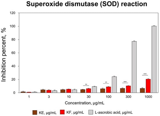
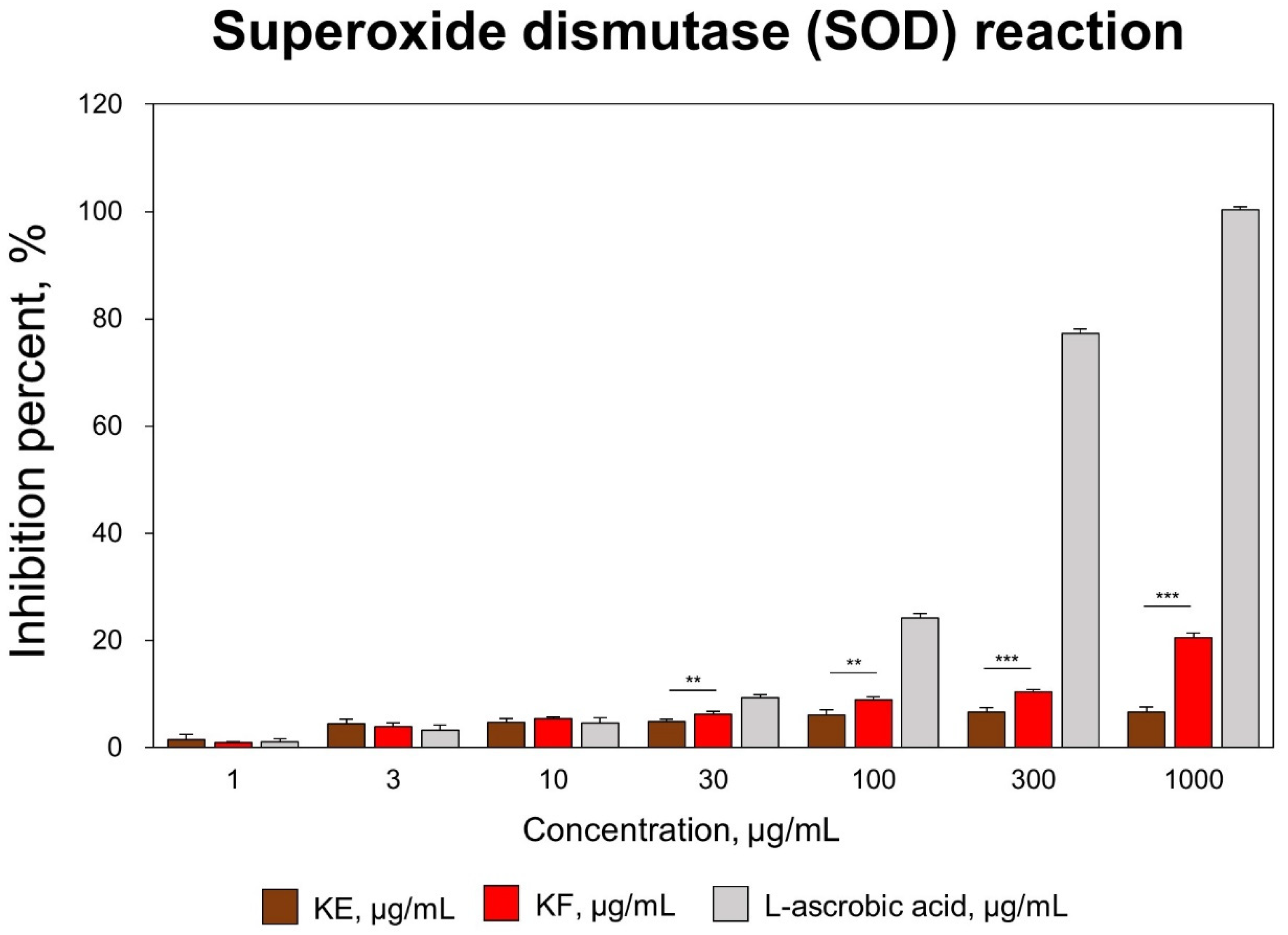
Superoxide dismutase (SOD) is a major antioxidant enzyme that catalyzes the conversion of superoxide radicals into molecular oxygen and hydrogen peroxide, thereby protecting cells from oxidative-stress-induced damage and maintaining cellular redox homeostasis [31,41,42]. The antioxidant activity of kelp extract (KE) and KF was evaluated using the SOD radical-scavenging assay. As shown in Figure 1, KF exhibited concentration-dependent SOD radical-scavenging activity and reached 20.5% at 1000 μg/mL, whereas KE exhibited only 6.7% activity under the same conditions, demonstrating that microbial fermentation markedly enhanced the antioxidant capacity of kelp.
Figure 1.
Anti-oxidant effect of kelp-fermenters (KFs) determined using superoxide dismutase (SOD) activity. The fermentation conditions were a temperature of 30 °C for 70 h. Data are displayed as mean ± SD (n = 3, KE vs. KF, ** p < 0.01 and *** p < 0.001). L-ascorbic acid was used as a positive control. KE; Kelp extract, KF; Kelp fermenters.
3.2. Protection of HFDPCs Against Oxidative Damage by KF
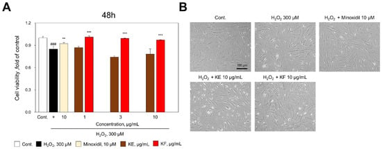
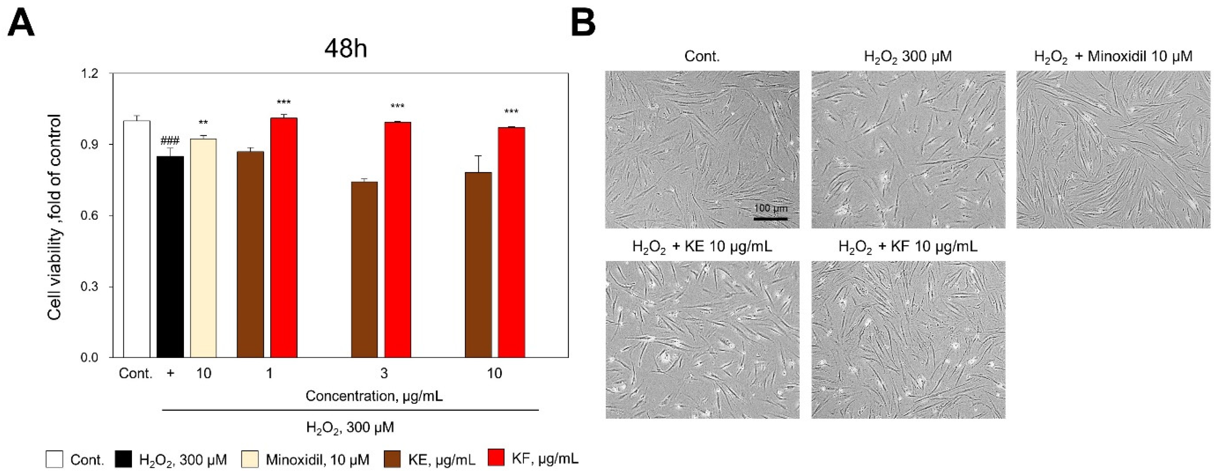
To determine the protective effects of KE and KF against oxidative-stress-induced cell damage, HFDPCs were stimulated with H2O2 and treated with KE or KF at concentrations of 1, 3, and 10 μg/mL. H2O2 stimulation significantly reduced cell viability by approximately 15% compared with the untreated control. Minoxidil (10 μM) restored cell viability to approximately 61%, whereas KF restored cell viability to a level comparable to that of the untreated control at all tested concentrations, whereas KE exhibited a relatively weaker protective effect (Figure 2A). Consistent with the cell viability results, microscopic observation demonstrated that treatment with 10 μg/mL KF effectively protected HFDPC morphology against H2O2-induced cell damage (Figure 2B). Based on these results, the subsequent experiments were performed using 10 μg/mL KF.
Figure 2.
KF effect on reducing H2O2-damaged oxidative stress in HFDPCs. (A) Cell viability was evaluated using the CCK-8 assay following treatment with 300 μM H2O2 in the presence or absence of KE, KF, or Minoxidil. Data are presented as mean ± SD (n = 3). Statistical significance was determined by one-way ANOVA followed by Tukey’s multiple comparison test. ### p < 0.001 versus the control group; ** p < 0.01 and *** p < 0.001 versus the H2O2-treated group. (B) Representative phase-contrast images of HFDPCs treated with 300 μM H2O2 alone or together with Minoxidil (10 μM), KE (10 μg/mL), or KF (10 μg/mL). Scale bar = 100 μm. Minoxidil was used as a positive control. KE; Kelp extract, KF; Kelp fermenters.
3.3. Attenuation of Oxidative-Stress-Induced Inflammatory Responses
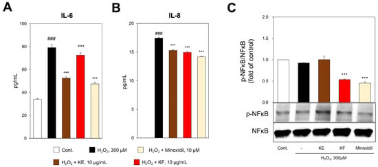
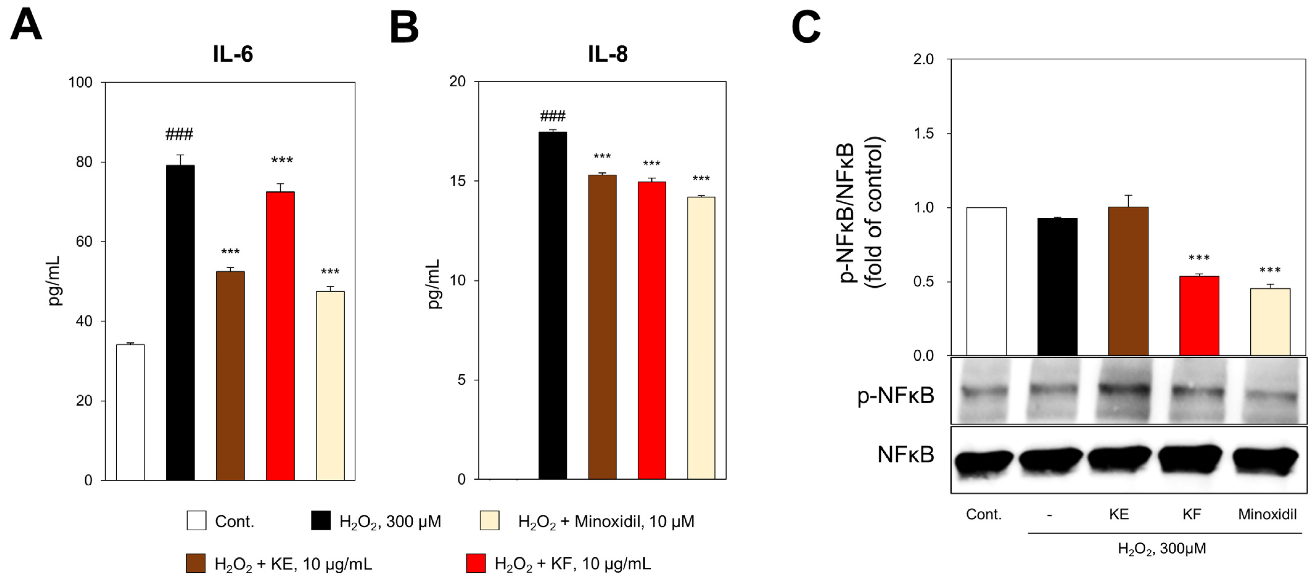
Inflammatory cytokines, including IL-6 and IL-8, are key mediators of oxidative-stress-induced hair follicle dysfunction and contribute to the progression of hair loss by disrupting the normal physiological function of dermal papilla cells [8,43]. To determine whether the cytoprotective effects of KF were accompanied by attenuation of inflammatory signaling, we evaluated the production of IL-6 and IL-8 in H2O2-damaged HFDPCs. The secretion of IL-6, which was markedly increased by H2O2 stimulation, was significantly reduced to 59.1% and 14.7% following treatment with KE and KF, respectively (Figure 3A). The secretion of IL-8, which was also increased by H2O2 stimulation, was significantly reduced to 12.3% and 14.3% following treatment with KE and KF, respectively. Notably, the inhibitory effect of KF on IL-8 secretion was comparable to that of the positive control, minoxidil (18.6%) (Figure 3B). Furthermore, the phosphorylation of NFκB, which was increased by H2O2 stimulation, was significantly inhibited by 41.9% following KF treatment, and this inhibitory effect was comparable to that of the positive control, minoxidil (50.9%) (Figure 3C). These results indicate that KF effectively suppressed oxidative-stress-induced inflammatory responses in HFDPCs.
Figure 3.
Anti-Inflammatory Effects of KF in H2O2-Induced HFDPCs. Levels of IL-6 (A) and IL-8 (B) in the culture supernatants of H2O2-stimulated HFDPCs treated with KE, KF, or minoxidil were determined using ELISA. (C) Representative Western blot images and quantitative analysis of p-NFκB expression in H2O2-stimulated HFDPCs treated with KE, KF, or minoxidil. p-NFκB expression was normalized to total NFκB. Data are presented as the mean ± SD (n = 3). Statistical significance was determined by one-way analysis of variance (ANOVA) followed by Tukey’s multiple comparison test. ### p < 0.001 versus the control group; *** p < 0.001 versus the H2O2-treated group. Minoxidil was used as a positive control. KE; Kelp extract, KF; Kelp fermenters.
3.4. KF Suppresses Apoptosis-Related Signaling in H2O2-Damaged HFDPCs
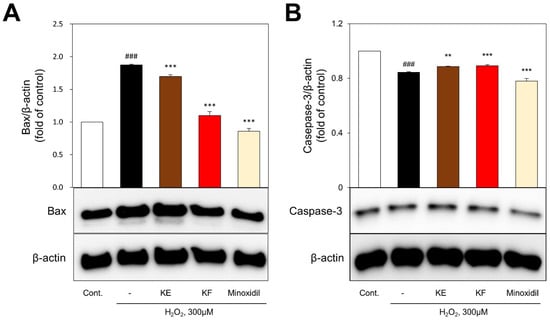
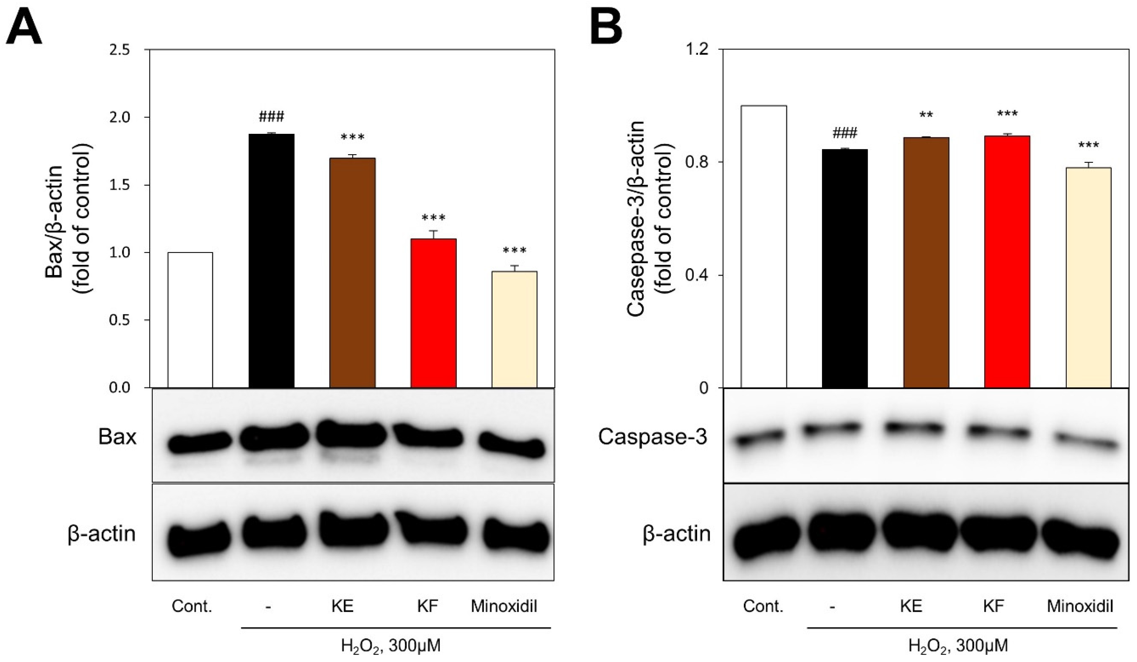
To evaluate the expression of apoptosis-related signaling proteins, we obtained total protein after the cells were pretreated with 300 μM H2O2 for 30 min and subsequently treated with 10 μM minoxidil, KE, or KF for 48 h. We then analyzed the expression of Bax, caspase-3, IGF-1, and AKT by Western blotting. Bax expression, which was markedly increased by H2O2-induced damage, was significantly suppressed by 19.5% and 88.5% following treatment with KE and KF, respectively (Figure 4A). The expression of total caspase-3, which was reduced in H2O2-treated HFDPCs, was restored by 27.6% and 30.4% following treatment with KE and KF, respectively (Figure 4B). These results indicate that treatment with KF effectively attenuated oxidative-stress-induced apoptosis in HFDPCs and may contribute to the protection of dermal papilla cells against oxidative stress.
Figure 4.
Effects of KF on protein expression of hair growth factor in H2O2-damaged HFDPCs. Cells were pretreated with 300 μM H2O2 for 30 min, followed by treatment with 10 μg/mL KF or KE for 48 h. The expression of Bax (A) and Caspase-3 (B) were analyzed by western blots. The results were normalized to β-actin expression and are presented as fold changes relative to the H2O2-treated group. Minoxidil (10 μM) was used as a positive control. Data are expressed as the mean ± SD (n = 3, ### p < 0.001 vs. control; ** p < 0.01 and *** p < 0.001 compared with H2O2).
3.5. KF Restores Hair-Growth-Associated Signaling in H2O2-Damaged HFDPCs
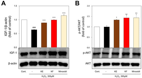
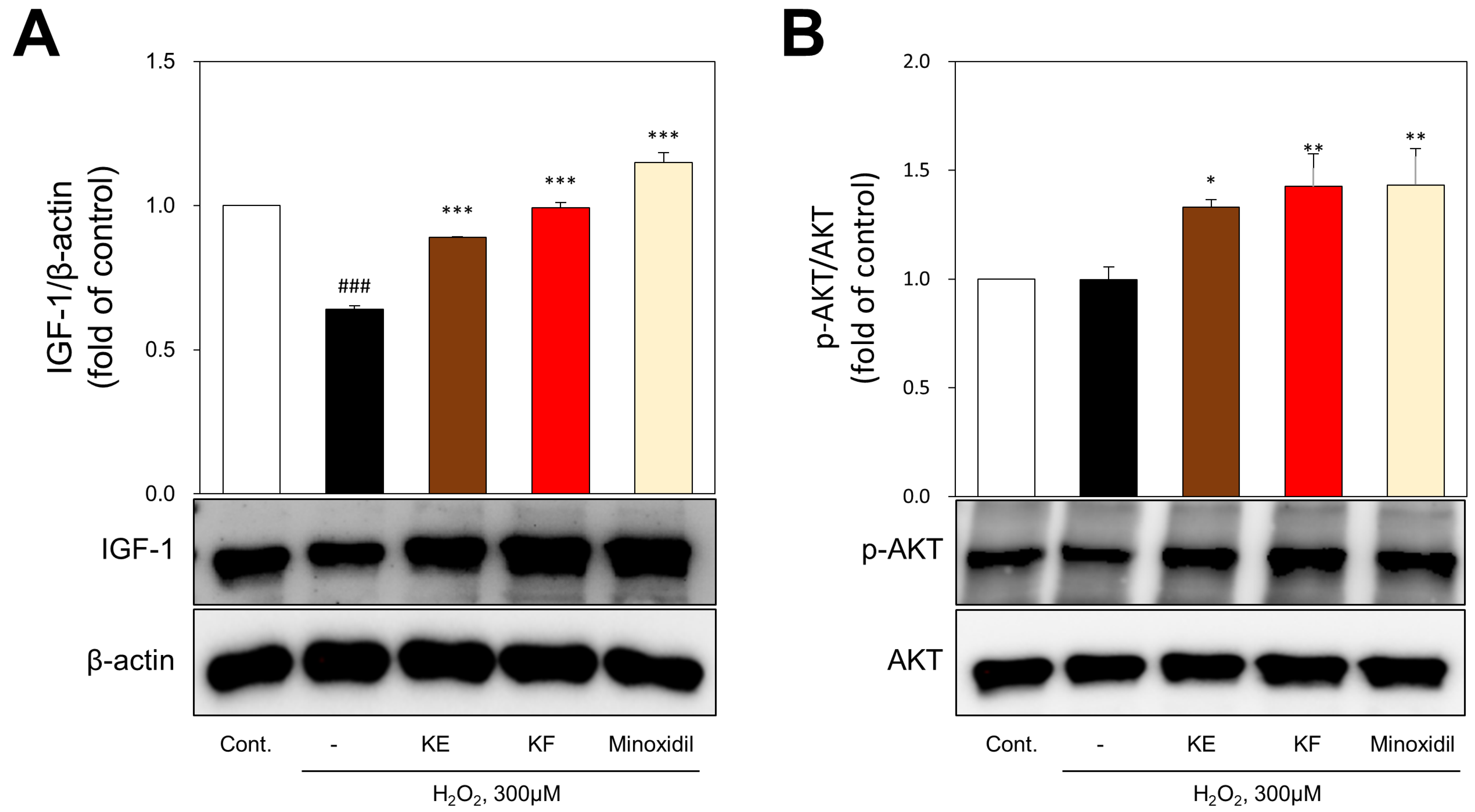
To determine the effects of KE and KF on hair-growth-related signaling, the expression of IGF-1 and phosphorylation of AKT were evaluated. The expression of IGF-1, which was markedly reduced by H2O2 stimulation, was significantly restored to 69.5% and 97.9% following treatment with KE and KF, respectively (Figure 5A). In particular, IGF-1 expression in the KF-treated group recovered to a level comparable to that of the untreated control. Next, as shown in Figure 5B, the phosphorylation of AKT was significantly increased by 1.33- and 1.43-fold following treatment with KE and KF, respectively, while minoxidil increased AKT phosphorylation by 1.44-fold. These results suggest that KF effectively restored hair-growth-related signaling to a level comparable to that of the positive control, indicating its potential as a functional material for improving hair loss.
Figure 5.
KF decreased apoptosis of H2O2-damaged oxidative stress in HFDPCs. Representative Western blot images and densitometric quantification of IGF-1 (A) and phosphorylated AKT (p-AKT) expression (B) in H2O2-stimulated HFDPCs treated with KE, KF, or minoxidil. IGF-1 expression was normalized to β-actin. p-AKT expression was normalized to total AKT. Statistical significance was determined by one-way analysis of variance (ANOVA) followed by Tukey’s multiple comparison test. ### p < 0.001 versus the untreated control group; * p < 0.05, ** p < 0.01, and *** p < 0.001 versus the H2O2-treated group. Minoxidil was used as a positive control. KE; Kelp extract, KF; Kelp fermenters.
3.6. Improvement in Hair Loss and Hair Luster Following Treatment with KF-Containing Shampoo
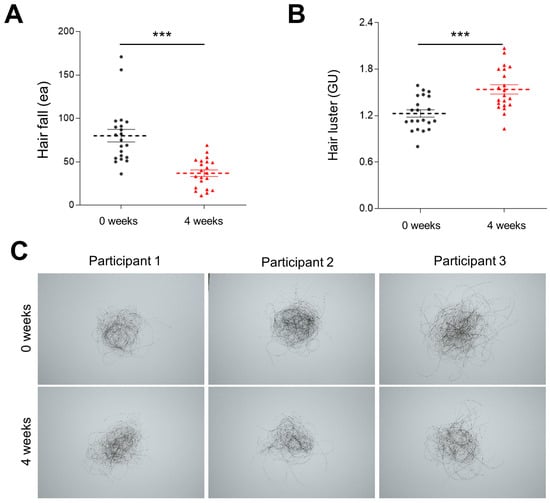
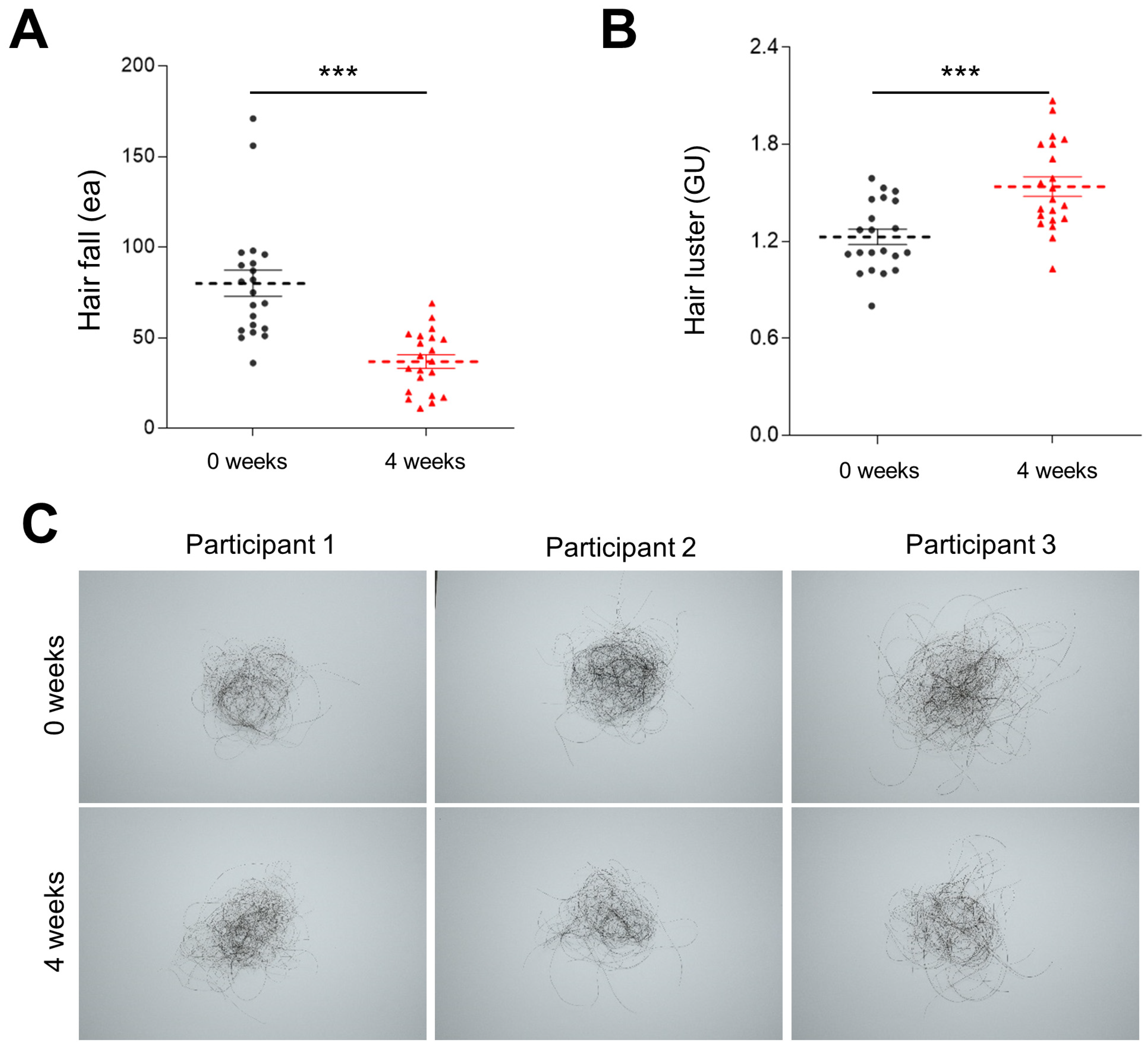
Following the protective effects of KF observed in H2O2-stimulated HFDPCs, we further evaluated the efficacy of a KF-containing shampoo in a 4-week human application test. Hair loss and hair luster were assessed after 4 weeks of shampoo application. Compared with before application, hair loss was significantly reduced by 53.9% after 4 weeks of shampoo application (Figure 6A). As shown in Figure 6C, representative clinical images also demonstrated a visible reduction in hair loss after 4 weeks of shampoo application. In addition, hair luster was significantly increased by 25.3% after 4 weeks of shampoo application compared with before application (Figure 6B). These results indicate that the KF-containing shampoo effectively improved hair condition.
Figure 6.
Efficacy of KF-containing shampoo on hair loss and hair luster after 4 weeks of treatment. (A) Quantitative analysis of hair loss before and after 4 weeks of treatment. (B) Quantitative analysis of hair luster before and after 4 weeks of treatment. (C) Representative images of shed hairs collected during the hair wash test (upper panel) before and after 4 weeks of KF shampoo use. Data are presented as the mean ± SD (n = 21). Statistical significance was analyzed using a paired t-test or Wilcoxon signed-rank test. *** p < 0.001 versus baseline (before treatment).
3.7. Improved Scalp Condition Through KF-Containing Shampoo
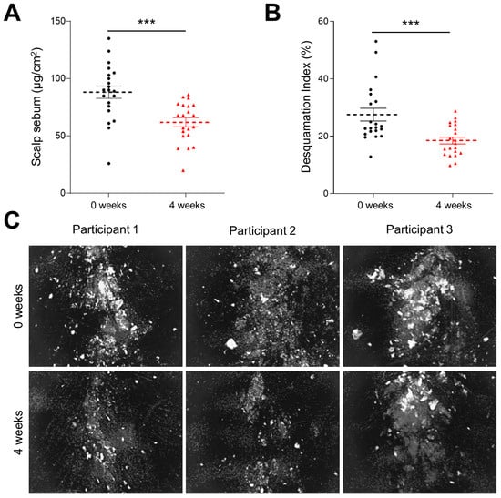
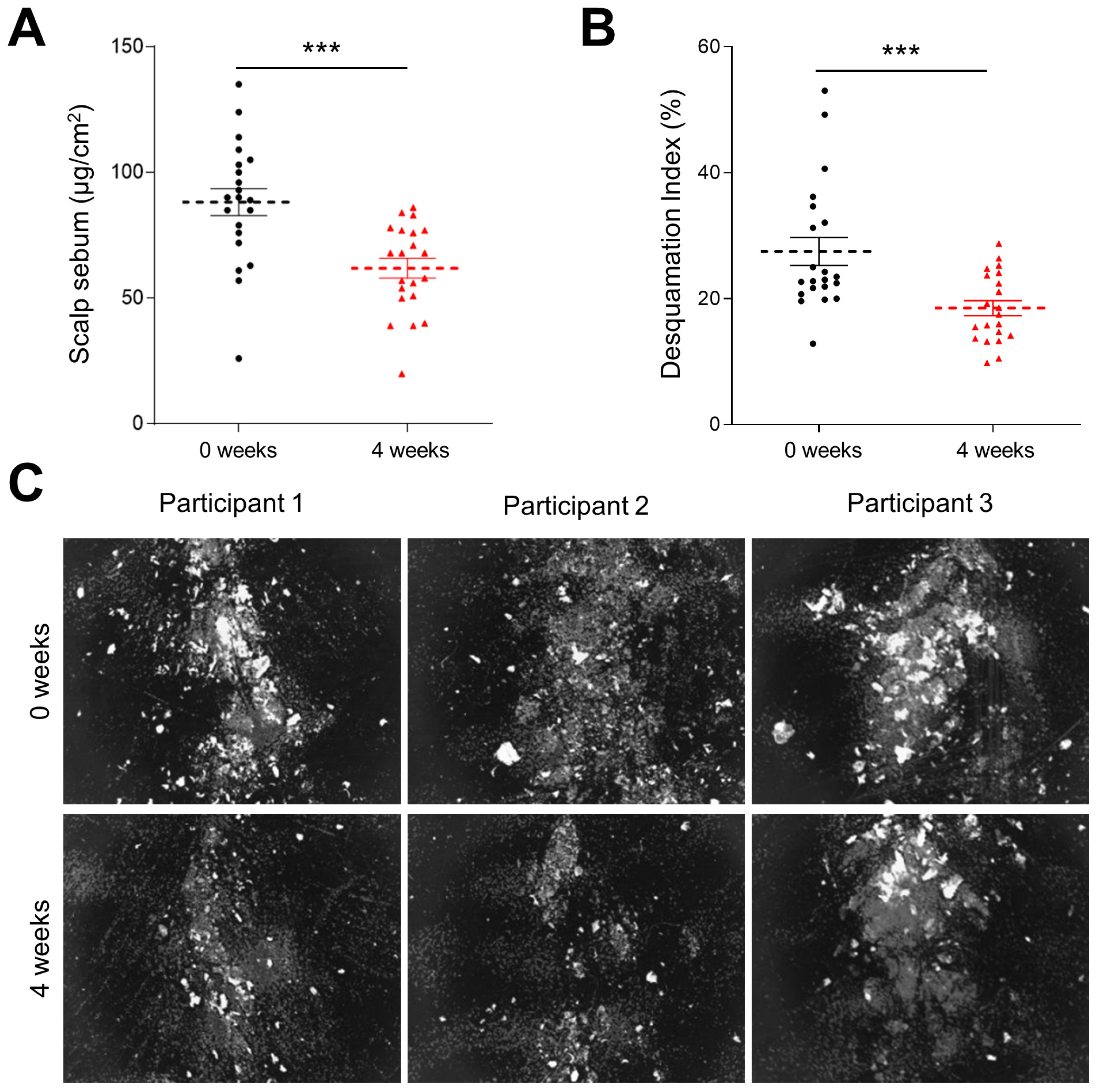
To further evaluate the effects of KF-containing shampoo on scalp condition, we assessed scalp sebum and scalp scaling after 4 weeks of application. Scalp sebum was measured using a Sebumeter®, and scalp scaling was evaluated using the D-Squame® sampling system and Visioscan® analysis. Compared with before application, scalp sebum was significantly reduced, by 29.8%, after 4 weeks of shampoo use (Figure 7A). Also, scalp scaling was significantly decreased, by 32.7%, compared with before application (Figure 7B,C). These results indicate that the KF-containing shampoo effectively improved scalp condition by reducing excessive sebum production and scalp scaling.
Figure 7.
Effects of KF-containing shampoo on scalp sebum and desquamation after 4 weeks of treatment. (A) Changes in scalp sebum (μg/cm2) after 4 weeks of treatment. (B) Changes in desquamation index (%) after 4 weeks of treatment. (C) Representative images of scalp keratin assessed using a scalp imaging system before and after 4 weeks of KF shampoo use. Data are presented as the mean ± SD (n = 21). Statistical significance was analyzed using a paired t-test or Wilcoxon signed-rank test. *** p < 0.001 versus baseline (before treatment).
4. Discussion
Approximately 20–40% of harvested kelp is utilized for food production, whereas the remaining 60–80% becomes processing residue [44]. Although these by-products retain substantial amounts of bioactive constituents, they are primarily used in industrial applications regarding abalone and fish aquaculture [45]. Therefore, the valorization of kelp-derived materials is attracting increasing attention from both environmental and industrial perspectives. Previous studies have reported that kelp by-products can serve as suitable substrates for microbial fermentation, enabling their conversion into value-added functional materials [28,29]. Moreover, fermentation of kelp has been reported to enhance antioxidant and anti-inflammatory activities through microbial bioconversion of functional constituents [33,34,35]. In our previous studies, we developed a KF and demonstrated that its application as a nutrient solution increased the accumulation of bioactive constituents in hydroponically cultivated ginseng sprouts [37,38]. Therefore, the present study employed S. cerevisiae fermentation to enhance the biological functionality of kelp by-products and to evaluate their potential as a functional cosmetic ingredient for anti-hair-loss applications.
Lin et al. [30] reported that fermentation of kelp using Bacillus subtilis enhanced antioxidant and anti-inflammatory activities through microbial bioconversion of functional constituents, whereas Zhao et al. [33] demonstrated that Enterococcus faecium fermentation improved antioxidant activity by increasing the availability of bioactive metabolites. Sun et al. [36] further showed that S. cerevisiae-fermented kelp exhibited greater anti-oxidant and anti-inflammatory activities than the non-fermented material. Our findings are in line with previous studies: KF exhibited approximately 3.1-fold higher SOD radical-scavenging activity than the non-fermented kelp extract. KF exhibited significantly improved antioxidant capacity, which was associated with enhanced HFDPC viability under H2O2-induced oxidative stress, suggesting that KF protected dermal papilla cells from oxidative-stress-induced cellular damage.
Tran et al. [8] reported that suppression of NFκB signaling reduced inflammatory cytokine production and protected HFDPCs from oxidative-stress-induced cellular damage, highlighting the importance of inflammatory regulation in maintaining dermal papilla cell survival. Our results confirm that KF inhibited H2O2-induced NFκB phosphorylation to a level comparable to that of minoxidil and significantly reduced IL-6 and IL-8 production. These changes were accompanied by a marked reduction in Bax expression, which was approximately 4.5-fold greater than that observed with KE, together with restoration of total caspase-3 expression. The concurrent suppression of inflammatory signaling and apoptosis suggests that the cytoprotective effect of KF is associated with attenuation of oxidative-stress-mediated inflammatory responses. Jang et al. [46] demonstrated that activation of the IGF-1/AKT signaling pathway is essential for maintaining dermal papilla cell survival and promoting hair follicle growth. Hyun et al. [47] showed that Morus alba root extract promoted hair growth through activation of AKT-associated signaling in HFDPCs. Lee et al. [48] likewise reported that veratric acid stimulated hair growth through a similar mechanism. KF restored IGF-1 expression to 97.9% of that of the untreated control and increased AKT phosphorylation 1.43-fold. Recovery of the IGF-1/AKT pathway, together with suppression of inflammatory and apoptotic signaling, suggests that KF not only protected HFDPCs from oxidative-stress-induced damage but also preserved signaling pathways required for dermal papilla cell survival and hair follicle maintenance.
Current therapeutic agents for hair loss, including minoxidil and finasteride, have demonstrated clinical efficacy by promoting hair growth and delaying hair follicle regression [11,12,13,14]. Nevertheless, their long-term use is often limited by adverse effects such as scalp irritation, contact dermatitis, cardiovascular complications, and sexual dysfunction [15,16], emphasizing the need for safer naturally derived alternatives. Mayer et al. [49] demonstrated that topical application of fermented tropical-fruit-derived formulations improved hair follicle condition by enhancing cellular energy metabolism and reducing oxidative damage, supporting the potential of fermented natural products for hair care. Application of a KF-containing shampoo for four weeks resulted in a 53.9% reduction in hair loss, together with improvements in hair luster (25.3%), scalp sebum (29.8%), and scalp scaling (32.7%). The observed improvements paralleled the in vitro effects of KF on HFDPCs, including enhanced resistance to oxidative stress and suppression of inflammatory and apoptotic signaling. However, the limitations of this human application test include the absence of a comparative control group and the small sample size. Although favorable safety and efficacy outcomes were observed in this study population, further placebo-controlled studies in larger and more diverse populations are needed to confirm these findings and clarify the specific contribution of KF.
In summary, KF exhibited enhanced biological functionality, increasing antioxidant capacity, suppressing oxidative-stress-induced inflammatory and apoptotic signaling, restoring hair-growth-associated signaling in HFDPCs. The protective effects observed in vitro were further supported by favorable changes in hair and scalp parameters following application of a KF-containing shampoo. Microbial fermentation therefore provides an effective approach to converting underutilized kelp by-products into high-value functional materials and facilitating the sustainable use of seaweed processing residues. Consequently, fermented kelp-derived materials may serve as sustainable functional ingredients for hair care formulations while increasing the value of seaweed-derived biomass.
5. Conclusions
KF exhibited enhanced biological functionality, including increased antioxidant capacity, suppression of oxidative-stress-induced inflammatory and apoptotic responses, restoration of hair-growth-associated signaling in dermal papilla cells, and improvements in hair and scalp condition following application of a KF-containing shampoo. Fermented kelp-derived materials therefore represent a sustainable source of functional cosmetic ingredients for hair care while providing an effective approach to increasing the industrial value of seaweed processing residues.
6. Patents
A patent for a composition for preventing or improving hair loss containing fermented kelp culture broth as an active ingredient was filed [Patent application number: 10-2026-0119276].
Author Contributions
Conceptualization, J.-M.S., K.-Y.K. and K.-W.P.; data curation, S.-K.M., J.-Y.C., S.-E.L., Y.-S.O., J.-H.K., J.-K.P., H.-Y.J., B.-G.J. and M.-R.J.; formal analysis, S.-K.M., J.-Y.C., Y.-S.O. and M.-R.J.; investigation, S.-K.M., J.-Y.C., Y.-S.O., J.-H.K., J.-K.P., H.-Y.J., B.-G.J. and M.-R.J.; methodology, S.-K.M., J.-H.K., J.-M.S., K.-Y.K. and K.-W.P.; validation, J.-H.K., J.-K.P., H.-Y.J. and B.-G.J.; visualization, S.-K.M., J.-Y.C., S.-E.L. and Y.-S.O.; software, S.-K.M., J.-Y.C. and S.-E.L.; project administration, J.-M.S. and K.-W.P.; funding acquisition, resources, and supervision, K.-W.P.; writing—original draft preparation, S.-K.M., J.-Y.C. and S.-E.L.; writing—review and editing, S.-K.M., J.-M.S., K.-W.P. and K.-Y.K. All authors have read and agreed to the published version of the manuscript.
Funding
This paper has been written with the support of Jeollannam-do (‘2025 R&D supporting program’ operated by Jeonnam Technopark; Project No. J25034-05).
Institutional Review Board Statement
The study was conducted in accordance with the Declaration of Helsinki and approved by the Institutional Review Board of the Prederma Institute of Dermatological Clinical Research, Korea (IRB No. PDCR-IRB-2026-N-052, approved on 24 April 2026).
Informed Consent Statement
Informed consent was obtained from all subjects involved in the study. Written informed consent to publish this paper has not been obtained from the patient(s) since their images and individual data did not appear in the paper.
Data Availability Statement
The data supporting the findings of this study are available from the corresponding authors upon reasonable request.
Conflicts of Interest
S.-E.L., M.-R.J., and J.-M.S. were employed by Prederma Institute of Dermatological Clinical Research Co. The remaining authors declare that the research was conducted in the absence of any commercial or financial relationships that could be construed as a potential conflict of interest.
Abbreviations
The following abbreviations are used in this manuscript:
| AKT | Protein kinase B |
| ANOVA | One-way analysis of variance |
| Bax | Bcl-2-associated X protein |
| CCK-8 | Cell Counting Kit-8 |
| D.I. | Desquamation Index |
| ELISA | Enzyme-linked immunosorbent assay |
| GU | Glossymeter Units |
| HFDPCs | hair follicle dermal papilla cells |
| IGF-1 | Insulin-like growth factor-1 |
| IL-6 | Interleukin-6 |
| IL-8 | Interleukin-8 |
| KF | Saccharomyces cerevisiae-fermented kelp by-products |
| L. japonica | Laminaria japonica |
| LED | Light-emitting diode |
| NFκB | Nuclear factor kappa-light-chain-enhancer of activated B cells |
| ROS | Reactive oxygen species |
| S. cerevisiae | Saccharomyces cerevisiae |
| SOD | Superoxide dismutase |
| UV | Ultraviolet |
References
- Trüeb, R.M.; Henry, J.P.; Davis, M.G.; Schwartz, J.R. Scalp Condition Impacts Hair Growth and Retention via Oxidative Stress. Int. J. Trichol. 2018, 10, 262–270. [Google Scholar] [CrossRef] [Scilit] [PubMed]
- Du, F.; Li, J.; Zhang, S.; Zeng, X.; Nie, J.; Li, Z. Oxidative stress in hair follicle development and hair growth: Signalling pathways, intervening mechanisms and potential of natural antioxidants. J. Cell. Mol. Med. 2024, 28, e18486. [Google Scholar] [CrossRef] [Scilit] [PubMed]
- Trüeb, R.M. The impact of oxidative stress on hair. Int. J. Cosmet. Sci. 2015, 37, 25–30. [Google Scholar] [CrossRef] [Scilit] [PubMed]
- Zhang, Y.; Yin, S.; Xu, R.; Xiao, J.; Yi, R.; Mao, J.; Duan, Z.; Fan, D. Recombinant Type XVII Collagen Promotes Hair Growth by Activating the Wnt/β-Catenin and SHH/GLI Signaling Pathways. Cosmetics 2025, 12, 156. [Google Scholar] [CrossRef] [Scilit]
- Kim, M.; Woo, J.; Kim, J.; Choi, M.; Shin, H.J.; Kim, Y.; Kim, J.; Shin, D.W. Iris germanica L. Rhizome-Derived Exosomes Ameliorated Dihydrotestosterone-Damaged Human Follicle Dermal Papilla Cells Through the Activation of Wnt/β-Catenin Pathway. Int. J. Mol. Sci. 2025, 26, 4070. [Google Scholar] [CrossRef] [Scilit] [PubMed]
- Bejaoui, M.; Taarji, N.; Saito, M.; Nakajima, M.; Isoda, H. Argan (Argania spinosa) press cake extract enhances cell proliferation and prevents oxidative stress and inflammation of human dermal papilla cells. J. Dermatol. Sci. 2021, 103, 33–40. [Google Scholar] [CrossRef] [Scilit] [PubMed]
- Jeon, C.Y.; Lee, J.Y.; Min, J.; Park, J.-Y.; Kim, M.; Yoon, W.; Choi, S.-I.; Shin, D.W. Upcycled Postbiotic Cell-Free Supernatants from Limosilactobacillus fermentum MG901 and MG4237 Alleviated Oxidative Stress-Induced Dysfunction in Human Follicle Dermal Pa-pilla Cells. Cosmetics 2026, 13, 46. [Google Scholar] [CrossRef] [Scilit]
- Tran, H.G.; Shuayprom, A.; Ruchusatsawat, A.; Meesilpavikkai, K.; Dalm, V.A.S.H.; Ruchusatsawat, K.; Wongpiyabovorn, J. Oxyresveratrol enhances hair regeneration in human dermal papilla cell and androgenetic alopecia mouse model. Sci. Rep. 2025, 15, 17551. [Google Scholar] [CrossRef] [Scilit] [PubMed]
- You, J.; Jang, Y.; Sim, J.; Ryu, D.; Cho, E.; Park, D.; Jung, E. Anti-Hair Loss Effect of Veratric Acid on Dermal Papilla Cells. Int. J. Mol. Sci. 2025, 26, 2240. [Google Scholar] [CrossRef] [Scilit] [PubMed]
- Choi, J.Y.; Boo, M.Y.; Boo, Y.C. Can Plant Extracts Help Prevent Hair Loss or Promote Hair Growth? A Review Comparing Their Therapeutic Efficacies, Phytochemical Components, and Modulatory Targets. Molecules 2024, 29, 2288. [Google Scholar] [CrossRef] [Scilit] [PubMed]
- Messenger, A.G.; Rundegren, J. Minoxidil: Mechanisms of Action on Hair Growth. Br. J. Dermatol. 2004, 150, 186–194. [Google Scholar] [CrossRef] [Scilit] [PubMed]
- Randolph, M.; Tosti, A. Oral Minoxidil Treatment for Hair Loss: A Review of Efficacy and Safety. J. Am. Acad. Dermatol. 2021, 84, 737–746. [Google Scholar] [PubMed]
- Estill, M.C.; Ford, A.; Omeira, R.; Rodman, M. Finasteride and Dutasteride for the Treatment of Male Androgenetic Alopecia: A Review of Efficacy and Reproductive Adverse Effects. Georget. Med. Rev. 2023, 7, 1–19. [Google Scholar] [CrossRef] [Scilit]
- Blume-Peytavi, U.; Kunte, C.; Krisp, A.; Bartels, N.G.; Ellwanger, U.; Hoffmann, R. Comparison of the Efficacy and Safety of Topical Minoxidil and Topical Alfatradiol in the Treatment of Androgenetic Alopecia in Women. J. Dtsch. Dermatol. Ges. 2007, 5, 391–395. [Google Scholar] [CrossRef] [Scilit] [PubMed]
- Rossi, A.; Cantisani, C.; Melis, L.; Iorio, A.; Scali, E.; Calvieri, S. Minoxidil Use in Dermatology, Side Effects and Recent Patents. Recent Pat. Inflamm. Allergy Drug Discov. 2012, 6, 130–136. [Google Scholar] [CrossRef] [Scilit] [PubMed]
- Kim, J.; Song, S.Y.; Sung, J.H. Recent Advances in Drug Development for Hair Loss. Int. J. Mol. Sci. 2025, 26, 3461. [Google Scholar] [CrossRef] [Scilit] [PubMed]
- Jeon, C.Y.; Go, M.Y.; Kim, I.-K.; Park, M.-R.; Lee, H.W.; Kim, Y.-K.; Shin, D.W. Hair Growth-Promoting Effects of Astragalus sinicus Extracts in Human Follicle Dermal Papilla Cells. Cosmetics 2025, 12, 6. [Google Scholar] [CrossRef] [Scilit]
- Islam, M.N.; Ishita, I.J.; Jin, S.E.; Choi, R.J.; Lee, C.M.; Kim, Y.S.; Jung, H.A.; Choi, J.S. Anti-Inflammatory Activity of Edible Brown Alga Saccharina japonica and Its Constituents Pheophorbide a and Pheophytin a in LPS-Stimulated RAW264.7 Macrophage Cells. Food Chem. Toxicol. 2013, 55, 541–548. [Google Scholar] [CrossRef] [Scilit] [PubMed]
- Wang, J.; Jin, W.; Zhang, W.; Hou, Y.; Zhang, H.; Zhang, Q. Hypoglycemic Property of Acidic Polysaccharide Extracted from Saccharina japonica and Its Potential Mechanism. Carbohydr. Polym. 2013, 95, 143–147. [Google Scholar] [CrossRef] [Scilit] [PubMed]
- Vishchuk, O.S.; Ermakova, S.P.; Zvyagintseva, T.N. Sulfated Polysaccharides from Brown Seaweeds Saccharina japonica and Undaria pinnatifida: Isolation, Structural Characteristics, and Antitumor Activity. Carbohydr. Res. 2011, 346, 2769–2776. [Google Scholar] [CrossRef] [Scilit] [PubMed]
- Wang, J.; Zhang, Q.; Zhang, Z.; Li, Z. Antioxidant Activity of Sulfated Polysaccharide Fractions Extracted from Laminaria japonica. Int. J. Biol. Macromol. 2008, 42, 127–132. [Google Scholar] [CrossRef] [Scilit] [PubMed]
- Huang, L.; Wen, K.; Gao, X.; Liu, Y. Hypolipidemic Effect of Fucoidan from Laminaria japonica in Hyperlipidemic Rats. Pharm. Biol. 2010, 48, 422–426. [Google Scholar] [CrossRef] [Scilit] [PubMed]
- Patra, J.K.; Das, G.; Baek, K.H. Chemical Composition and Antioxidant and Antibacterial Activities of an Essential Oil Extracted from an Edible Seaweed, Laminaria japonica L. Molecules 2015, 20, 12093–12113. [Google Scholar] [CrossRef] [Scilit] [PubMed]
- Kim, S.K.; Bhatnagar, I. Physical, Chemical, and Biological Properties of Wonder Kelp—Laminaria. Adv. Food Nutr. Res. 2011, 64, 85–96. [Google Scholar] [CrossRef] [Scilit] [PubMed]
- Podkorytova, A.V.; Vafina, L.H.; Kovaleva, E.A.; Mikhailov, V.I. Production of Algal Gels from the Brown Alga Laminaria japonica Aresch. and Their Biotechnological Applications. J. Appl. Phycol. 2007, 19, 827–830. [Google Scholar] [CrossRef] [Scilit]
- Kılınç, B.; Cirik, S.; Turan, G.; Tekogul, H.; Koru, E. Seaweeds for Food and Industrial Applications. In Food Industry; Intech: Rijeka, Croatia, 2013. [Google Scholar]
- Purcell-Meyerink, D.; Packer, M.A.; Wheeler, T.T.; Hayes, M. Aquaculture Production of the Brown Seaweeds Laminaria digitata and Macrocystis pyrifera: Applications in Food and Pharmaceuticals. Molecules 2021, 26, 1306. [Google Scholar] [CrossRef] [Scilit] [PubMed]
- Liu, Z.; Li, X.; Xie, W.; Deng, H. Extraction, Isolation and Characterization of Nanocrystalline Cellulose from Industrial Kelp (Laminaria japonica) Waste. Carbohydr. Polym. 2017, 173, 353–359. [Google Scholar] [CrossRef] [Scilit] [PubMed]
- Liu, C.; Gao, J.; Jiang, H.; Sun, J.; Gao, X.; Mao, X. Value-Added Utilization Technologies for Seaweed Processing Waste in a Circular Economy. Compr. Rev. Food Sci. Food Saf. 2024, 23, e70027. [Google Scholar] [CrossRef] [Scilit] [PubMed]
- Al-Maqtari, Q.A.; Waleed, A.A.; Mahdi, A.A. Microbial Enzymes Produced by Fermentation and Their Applications in the Food Industry: A Review. Int. J. Agric. Innov. Res. 2019, 8, 62–82. [Google Scholar]
- Zhu, W.; Jiang, B.; Zhong, F.; Chen, J.; Zhang, T. Effect of Microbial Fermentation on the Fishy-Odor Compounds in Kelp (Laminaria japonica). Foods 2021, 10, 2532. [Google Scholar] [CrossRef] [Scilit] [PubMed]
- Vittaladevaram, V. Fermentative Production of Microbial Enzymes and Their Applications: Present Status and Future Prospects. J. Appl. Biol. Biotechnol. 2017, 5, 90–94. [Google Scholar] [CrossRef] [Scilit]
- Lin, H.T.V.; Lu, W.J.; Tsai, G.J.; Chou, C.T.; Hsiao, H.I.; Hwang, P.A. Enhanced Anti-Inflammatory Activity of Brown Seaweed Laminaria japonica by Fermentation Using Bacillus subtilis. Process Biochem. 2016, 51, 1945–1953. [Google Scholar] [CrossRef] [Scilit]
- Zhao, Y.; Ra, C.H. Inhibitory and Antioxidant Activities of Saccharina japonica Extracts Processed by Subcritical Water Extraction and Enterococcus faecalis Fermentation. J. Microbiol. Biotechnol. 2025, 35, e2504045. [Google Scholar] [CrossRef] [Scilit] [PubMed]
- Lee, I.S.; Ko, S.J.; Lee, Y.N.; Lee, G.; Rahman, M.H.; Kim, B. The Effect of Laminaria japonica on Metabolic Syndrome: A Systematic Review of Its Efficacy and Mechanism of Action. Nutrients 2022, 14, 3046. [Google Scholar] [CrossRef] [Scilit] [PubMed]
- Wang, Z.; Sun, Q.; Fang, J.; Wang, C.; Wang, D.; Li, M. The Anti-Aging Activity of Lycium Barbarum Polysaccharide Extracted by Yeast Fermentation: In Vivo and in Vitro Studies. Int. J. Biol. Macromol. 2022, 209, 2032–2041. [Google Scholar] [CrossRef] [Scilit] [PubMed]
- Park, K.W.; Kim, J.H.; Jeong, B.G.; Park, J.K.; Jang, H.Y.; Oh, Y.S.; Kang, K.Y. Increased Accumulation of Ginsenosides in Panax ginseng Sprouts Cultivated with Kelp Fermentates. Plants 2024, 13, 463. [Google Scholar] [CrossRef] [Scilit] [PubMed]
- Kim, J.H.; Park, K.W.; Jeong, B.G.; Park, J.K.; Jang, H.Y.; Oh, Y.S.; Choi, J.Y.; Kang, K.Y. Ginsenoside-Enriched Panax ginseng Sprouts Cultivated from Aquaponic System with a Novel Nutrient Solution Regulate LPS-Induced Inflammatory Cytokines and UVB-Induced Photoaging Responses via MAPK/AP-1 Signaling Pathways. Plants 2025, 14, 1712. [Google Scholar] [CrossRef] [Scilit] [PubMed]
- Sun, Q.; Fang, J.; Wang, Z.; Song, Z.; Geng, J.; Wang, D.; Wang, C.; Li, M. Two Laminaria japonica Fermentation Broths Alleviate Oxidative Stress and Inflammatory Response Caused by UVB Damage: Photoprotective and Reparative Effects. Mar. Drugs 2022, 20, 650. [Google Scholar] [CrossRef] [Scilit] [PubMed]
- Han, J.H.; Kwon, O.S.; Chung, J.H.; Cho, K.H.; Eun, H.C.; Kim, K.H. Effect of minoxidil on proliferation and apoptosis in dermal papilla cells of human hair follicle. J. Dermatol. Sci. 2004, 34, 91–98. [Google Scholar] [CrossRef] [Scilit] [PubMed]
- Ighodaro, O.M.; Akinloye, O.A. First Line Defence Antioxidants—Superoxide Dismutase (SOD), Catalase (CAT) and Glutathione Peroxidase (GPX): Their Fundamental Role in the Entire Antioxidant Defence Grid. Alex. J. Med. 2018, 54, 287–293. [Google Scholar] [CrossRef] [Scilit]
- Zheng, M.; Liu, Y.; Zhang, G.; Yang, Z.; Xu, W.; Chen, Q. The Applications and Mechanisms of Superoxide Dismutase in Medicine, Food, and Cosmetics. Antioxidants 2023, 12, 1675. [Google Scholar] [CrossRef] [Scilit] [PubMed]
- Kim, C.; Shin, J.M.; Kim, D.; Park, S.; Hong, D.; Jung, K.E.; Kim, C.D.; Seo, Y.J.; Lee, Y. Role of Substance P in Regulating Micro-Milieu of Inflammation in Alopecia Areata. Ann. Dermatol. 2022, 34, 270–277. [Google Scholar] [CrossRef] [Scilit] [PubMed]
- Huang, J.; Wu, R.; Wu, Y.; Liang, F.; Chen, Y.; Yang, F.; Zheng, H.; Wang, Z.; Xu, H.; Chen, S.; et al. Fermentation Preparation of Umami Sauce and Peptides from Kelp Scraps by Natural Microbial Flora. Foods 2025, 14, 1751. [Google Scholar] [CrossRef] [Scilit] [PubMed]
- Wang, G.; Meng, Z.; Chen, L.; Jiang, J.; Feng, Y.; Zhang, B. Effects of Kelp Residues Fermented with Probiotics on the Culture of Sea Cucumber, Apostichopus japonicus. Aquac. Res. 2020, 51, 1133–1142. [Google Scholar] [CrossRef] [Scilit]
- Jang, H.; Jo, Y.; Lee, J.H.; Choi, S. Aging of Hair Follicle Stem Cells and Their Niches. BMB Rep. 2023, 56, 2–9. [Google Scholar] [CrossRef] [Scilit] [PubMed]
- Hyun, J.; Im, J.; Kim, S.W.; Kim, H.Y.; Seo, I.; Bhang, S.H. Morus alba Root Extract Induces the Anagen Phase in the Human Hair Follicle Dermal Papilla Cells. Pharmaceutics 2021, 13, 1155. [Google Scholar] [CrossRef] [Scilit] [PubMed]
- Lee, K.E.; Park, J.E.; Jung, E.; Ryu, J.; Kim, Y.J.; Youm, J.K.; Kang, S. A Study of Facial Wrinkles Improvement Effect of Veratric Acid from Cauliflower Mushroom through Photo-Protective Mechanisms against UVB Irradiation. Arch. Dermatol. Res. 2016, 308, 183–192. [Google Scholar] [CrossRef] [Scilit] [PubMed]
- Mayer, W.; Weibel, M.; De Luca, C.; Ibragimova, G.; Trakhtman, I.; Kharaeva, Z.; Chandler, D.L.; Korkina, L. Biomolecules of Fermented Tropical Fruits and Fermenting Microbes as Regulators of Human Hair Loss, Hair Quality, and Scalp Microbiota. Biomolecules 2023, 13, 699. [Google Scholar] [CrossRef] [Scilit] [PubMed]

Disclaimer/Publisher’s Note: The statements, opinions and data contained in all publications are solely those of the individual author(s) and contributor(s) and not of MDPI and/or the editor(s). MDPI and/or the editor(s) disclaim responsibility for any injury to people or property resulting from any ideas, methods, instructions or products referred to in the content. |
© 2026 by the authors. Licensee MDPI, Basel, Switzerland. This article is an open access article distributed under the terms and conditions of the Creative Commons Attribution (CC BY) license.