


Abstract
Mesotherapy and related cosmetic microinjection or microneedling procedures are increasingly used for aesthetic indications. However, their safety profile is difficult to define because injectates, devices, injection depths, and treatment protocols vary widely. We report a case of rapid-onset papular granuloma annulare (GA)-like granulomatous reaction after a mesotherapy-like cosmetic procedure. An Asian woman in her forties developed multiple 2–3 mm erythematous papules in a regular grid-like distribution corresponding to facial injection sites. Papules were limited to the left hemiface despite bilateral treatment with the same sodium hyaluronate-containing skincare serum delivered using a suction-assisted 9-pin multi-needle mesogun. Histology showed necrobiotic granulomas with palisading epithelioid histiocytes and increased dermal mucin. After nine sessions of intralesional triamcinolone acetonide, near-complete clinical resolution was documented 14 months after the first intralesional triamcinolone session. This case highlights an unusual rapid-onset, papular, grid-patterned GA-like granulomatous reaction after a mesotherapy-like cosmetic procedure. The temporal and anatomical relationship to injection supports a procedure-associated reaction. Potential contributors include injected materials, use of products not intended for injection, suction-assisted multi-needle delivery, altered injection pressure or depth, repeated passes, mechanical trauma or Koebnerisation, delayed-type hypersensitivity, foreign-body response, contamination, and host susceptibility. This case does not establish a causative agent or reproducible procedure-specific adverse reaction, but documents a distinctive rapid-onset, grid-like papular GA-like reaction after poorly characterized cosmetic interventions.
1. Introduction
Mesotherapy, first introduced by Michel Pistor in 1952, involves delivery of small quantities of substances into the dermis or subcutaneous tissue. In contemporary aesthetic practice, the term is used variably to describe a broad range of microinjection and microneedle-assisted procedures with differing devices, injection depths, protocols, operators, and injectates. This heterogeneity complicates safety assessment and attribution of adverse events [1,2]. Reported complications include infection, panniculitis, necrosis, allergic reactions, foreign-body granulomas, and granuloma annulare (GA)-like reactions [1,3,4,5,6].
GA is a non-infectious granulomatous dermatosis characterized histologically by necrobiotic collagen, palisading or interstitial histiocytes, and mucin deposition. Although GA is classically annular clinically, it may also present in papular, localized, generalized, perforating, subcutaneous, and other variants [7,8,9]. Iatrogenic GA-like reactions have been reported after mesotherapy, injectable collagen, botulinum toxin, and other cosmetic procedures [2,5,10,11].
We describe an Asian patient with rapid-onset, grid-like papular GA-like granulomatous reaction after a mesotherapy-like cosmetic procedure.
2. Case Description
We performed a retrospective chart review of a patient evaluated at our centre for persistent papular facial eruption after a mesotherapy-like cosmetic procedure. The patient provided written consent for publication of clinical and histological images.
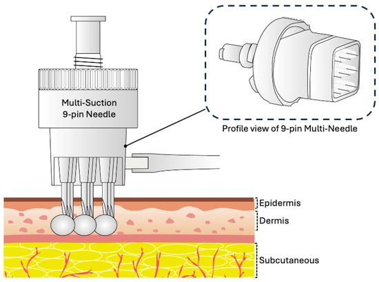
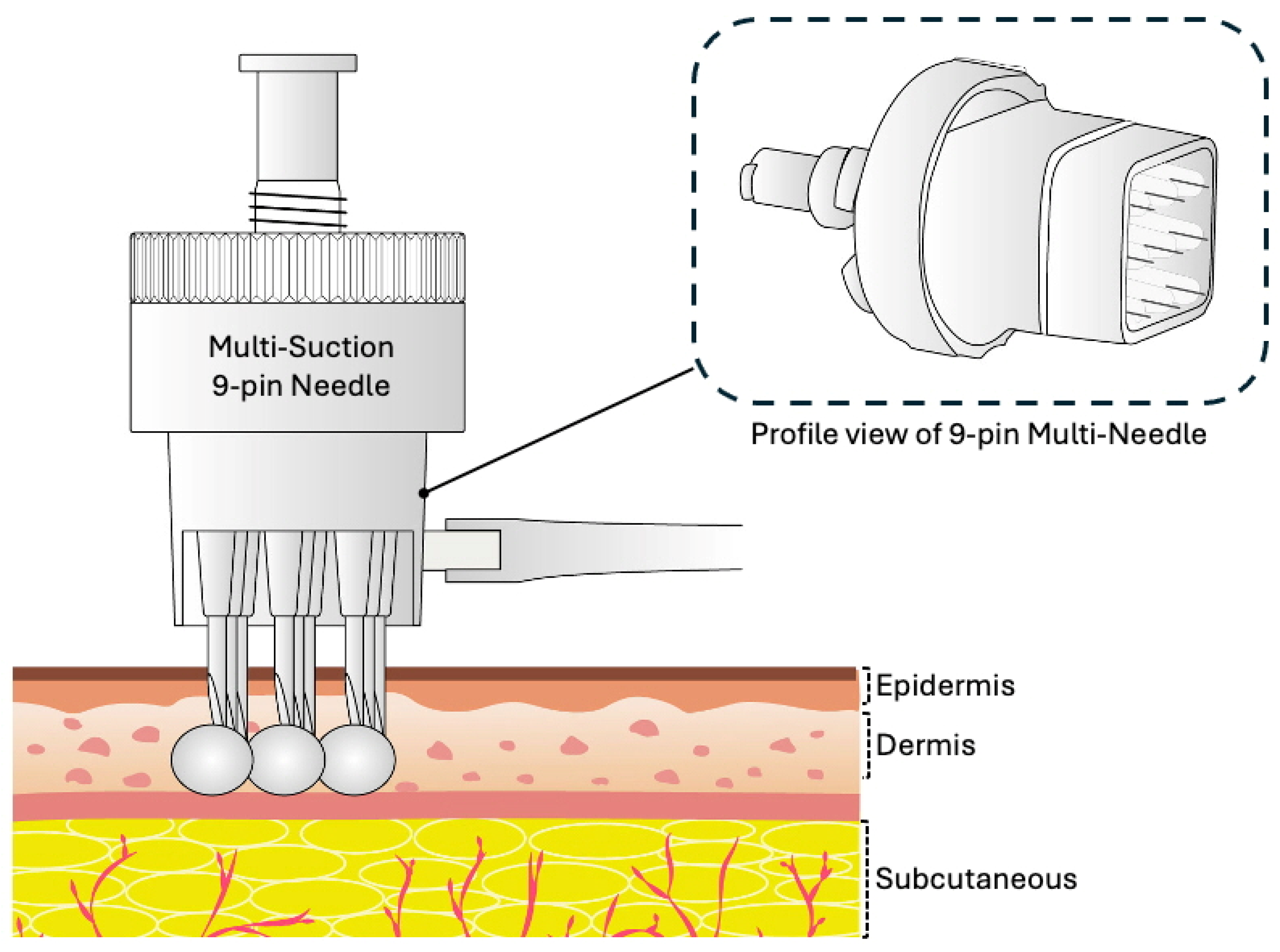
An Asian woman in her forties presented with multiple left hemifacial papules after her fifth facial rejuvenation session over a 12-month period at an aesthetic clinic. She had not experienced complications during the preceding four sessions. According to the patient, the same skincare serum was used for all sessions and was delivered using an automatic mesogun injector with a sterile disposable 9-pin multi-needle stamp device (Figure 1). The device was applied perpendicular to the skin, with negative pressure lifting the skin toward the needle tips.
Figure 1.
Schematic illustration of a suction-assisted 9-pin multi-needle device used for mesotherapy-like cosmetic injection procedures. The device delivers multiple simultaneous needle punctures with suction-assisted contact, potentially allowing delivery of substances into the dermis or superficial subcutaneous tissue.
The product used was identified as KANKOO® skincare essence/serum (Singapore). Its listed ingredients were water, propylene glycol, glycerin, butylene glycol, Glycyrrhiza glabra root extract, phenoxyethanol, PEG-40 hydrogenated castor oil, dipotassium glycyrrhizate, disodium EDTA, hydroxyethylcellulose, sodium hyaluronate, and fragrance. The packaging bore a cosmetics production licence number and cosmetic product standard, but did not indicate that the product was a sterile injectable preparation. Information regarding injection volume, injection depth, device settings, and the treatment protocol remained unavailable.
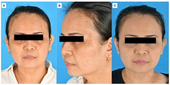
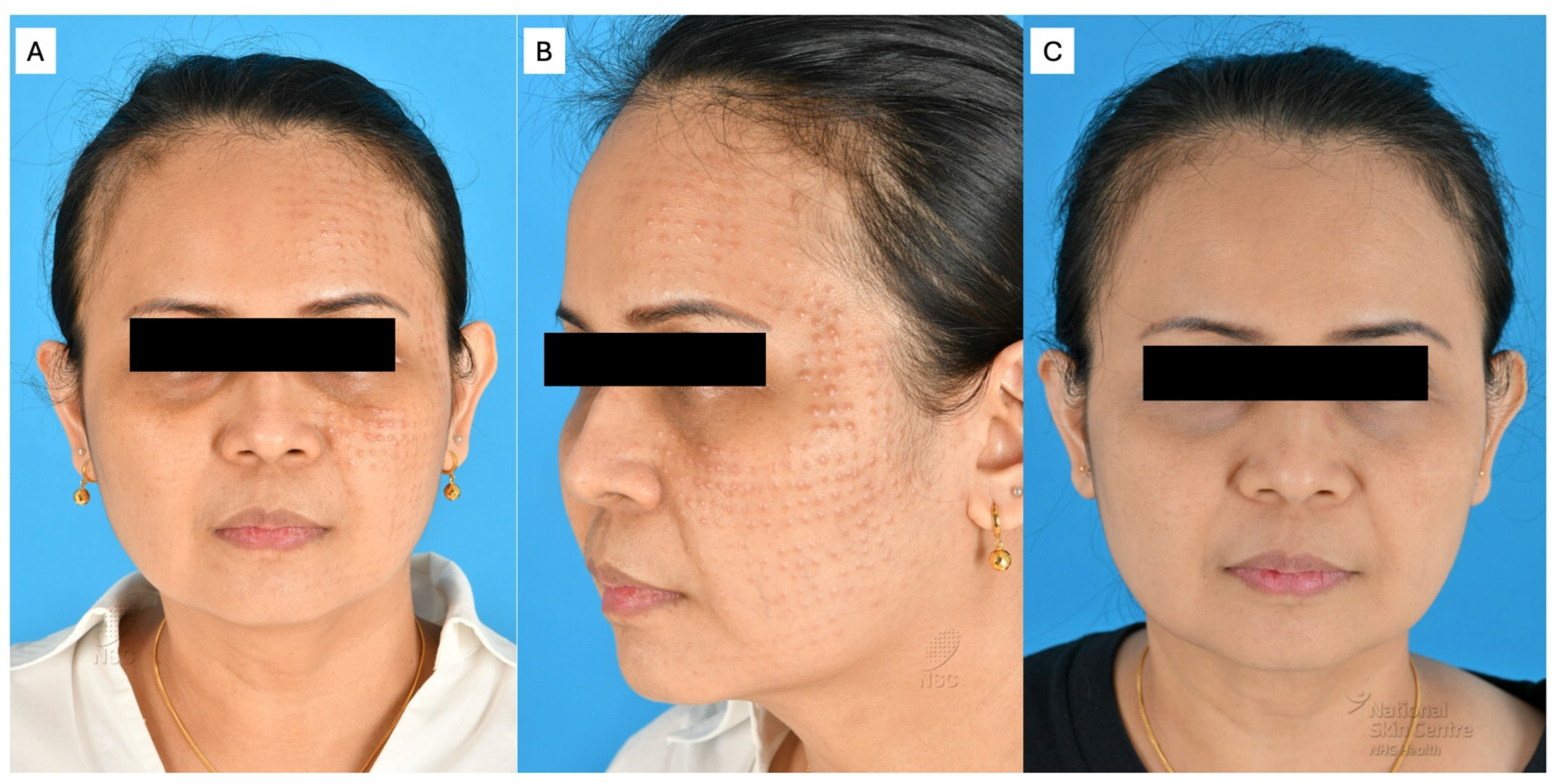
During the fifth session, the right hemiface was first treated uneventfully. When the therapist proceeded to the left hemiface, there was reported resistance to serum flow through the syringe and 9-pin multi-needle tip. Treatment was paused for device “adjustments” before being completed, although the details were not explained to the patient. She reported more pain and discomfort on the left hemiface than during previous treatments. Immediately after the procedure, she developed small “bumps” corresponding to injection sites, limited to the left hemiface. These became more prominent that same night. Examination showed multiple 2–3 mm erythematous papules in a regular grid-like configuration over the left forehead, temple, cheek, and lower face (Figure 2A,B).
Figure 2.
Case 1. Multiple 2–3 mm papules were uniformly spaced in a grid-like configuration and limited to the patient’s left hemiface on frontal view (A) and left lateral view (B). Post-treatment frontal view after nine sessions of intralesional triamcinolone acetonide 5 mg/mL administered approximately every 4 weeks shows near-complete clinical resolution at review 14 months after the first intralesional triamcinolone session (C).
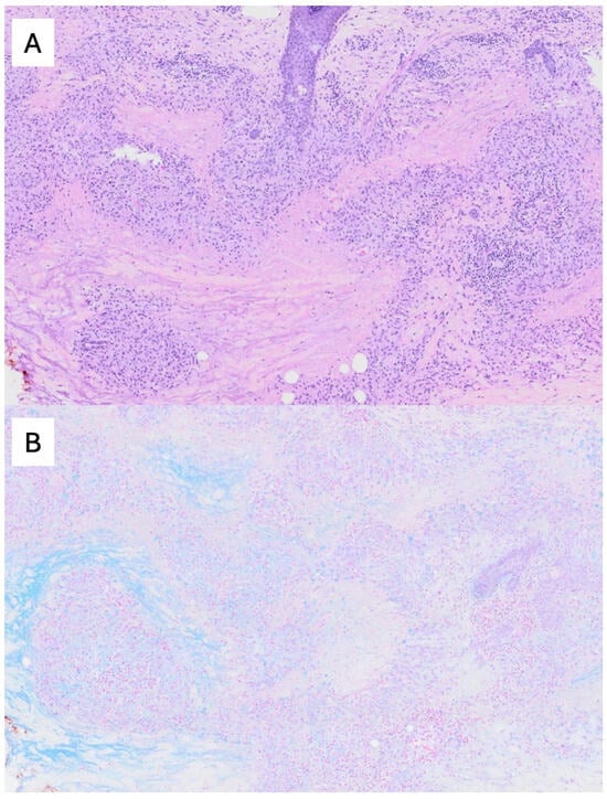
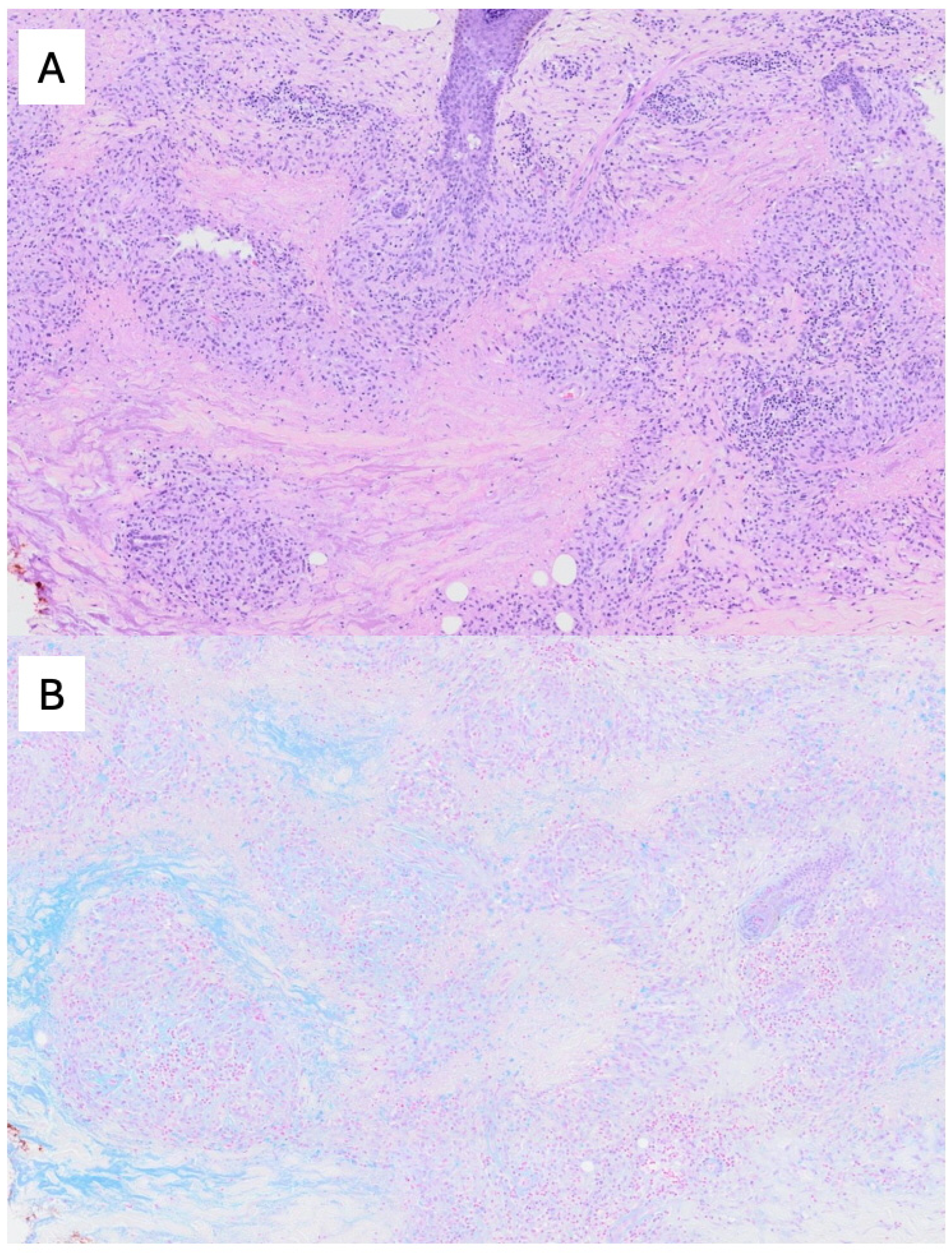
Punch biopsy showed foci of necrobiosis surrounded by palisading epithelioid histiocytes with occasional multinucleated giant cells (Figure 3A). Alcian blue staining demonstrated increased mucin within necrobiotic areas (Figure 3B). No polarizable foreign material was identified. Available microbiological investigations were negative. The clinicopathological interpretation was a papular granuloma annulare-like granulomatous reaction after a mesotherapy-like cosmetic injection procedure.
Figure 3.
Case 1. Skin biopsy showed areas of necrobiosis surrounded by palisading epithelioid histiocytes and lymphocytes within the dermis on haematoxylin and eosin staining (A). Alcian blue staining demonstrated mucin deposition within the necrobiotic areas (B). No polarizable foreign material was identified. Original magnification: ×100.
Initial treatment with co-formulated topical betamethasone dipropionate 0.05%/fusidic acid 2% cream, oral amoxicillin/clavulanate, oral prednisolone, and doxycycline was ineffective. At our centre, she subsequently underwent nine sessions of intralesional triamcinolone acetonide 5 mg/mL approximately every 4 weeks. At review 14 months after the first intralesional triamcinolone session, clinical photography showed near-complete resolution with marked flattening of the papules (Figure 2C). No obvious scarring or steroid-induced atrophy was evident on the available post-treatment photograph.
3. Discussion
This case adds to the spectrum of reported granulomatous reactions after mesotherapy, microneedling, and cosmetic injection procedures. Prior reports have described GA or granulomatous dermatitis after mesotherapy using phosphatidylcholine/deoxycholate, hyaluronic acid, procaine, silica, buflomedil, tranexamic acid-containing mixtures, and other injectates, as well as after microneedling-assisted delivery of topical cosmeceuticals (Table 1). Reported onset is usually delayed, ranging from days to months after the procedure [2,5,6,12]. The same-day onset in our case is therefore unusual, although it should be interpreted cautiously (earlier reports may have recorded the time of clinical presentation rather than the first appearance of subtle lesions).
Although granuloma annulare-like and other granulomatous reactions after mesotherapy, microneedling, fillers, and cosmetic injections have been reported previously, this case is notable for its same-day onset, striking grid-like papular morphology, unilateral distribution despite bilateral treatment, partial product identification, supportive histopathology, and objective photographic follow-up. The report should therefore be interpreted as a descriptive clinicopathological observation rather than evidence of a defined causal association.
The unilateral distribution despite bilateral exposure to the same product suggests that local procedural factors may have contributed. Potential factors include altered injection pressure or depth, repeated passes because of impaired flow, device adjustment, or suction-assisted trauma, especially because the patient reported that treatment of the left hemiface was more painful than usual. However, causality cannot be assigned to a single factor, and mechanical trauma cannot be considered the only or primary trigger. Other possible mechanisms include reaction to the injected material itself, impurities, preservatives, degradation products, and host immune susceptibility. The onset during the fifth session despite four prior uneventful sessions may also be explained by sensitization after repeated exposure, variation in procedural technique, transient device malfunction or altered flow, batch-to-batch product variation, or coincidental host factors at the time of treatment.
Trauma and Koebnerisation are plausible but unproven contributors. Trauma-related papular GA has been reported at sites of repeated direct injury, supporting the concept that cutaneous trauma may contribute to connective tissue injury, collagen degeneration, mucin deposition, and granulomatous inflammation in susceptible individuals [8,13]. Microneedling and mesotherapy devices create multiple epidermal and dermal punctures and can facilitate transepidermal or intradermal delivery of topicals or injectates [14,15,16]. In our patient, resistance to serum flow (experienced as more reported pain), subsequent device adjustment, greater procedural pain, and unilateral involvement raise the hypothesis that altered pressure, depth, repeated passes, or suction-assisted trauma may have interacted with the injectate to produce a localized GA-like reaction. Treatment-refractory granulomatous reactions have been reported after microneedling combined with transepidermal delivery of vitamin C, niacinamide and botanical extracts, supporting the concept that both procedural trauma and introduced topical agents may contribute [17].
The role of injected substances also remains important. Soft tissue fillers and injectable microimplants can produce early or delayed inflammatory, immune-mediated, foreign-body, or granulomatous reactions, and some materials may act as adjuvants in predisposed hosts [18,19,20]. Hyaluronic acid-containing products have been associated with inflammatory nodules, delayed inflammatory reactions, and granulomatous responses, although the precise contribution of hyaluronic acid itself versus impurities, cross-linking agents, degradation products, biofilm, or other additives is often difficult to determine [6,18,19]. In this case, the product was identified from packaging as a sodium hyaluronate-containing cosmetic skincare serum with multiple excipients, including phenoxyethanol, PEG-40 hydrogenated castor oil, hydroxyethylcellulose, and fragrance. However, its sterility, suitability for injection, exact concentrations, lot details, injection volume, injection depth, and device settings could not be verified. Recent literature continues to document mesotherapy-induced facial foreign body-type granulomatous reactions, including reactions after intradermal injection of products not originally intended for injection [6].
Clinically, although the lesions were not annular, GA can present in papular variants, and diagnosis depends on clinicopathological correlation rather than annular morphology alone [7,8,9]. In our case, the regular grid-like distribution reflected the pattern of device contact and injection sites, while the biopsy findings supported GA-like granulomatous dermatitis. Differential diagnoses include foreign-body granuloma, sarcoidosis or sarcoidal granulomatous reaction, infectious granulomatous dermatitis including atypical mycobacterial infection, interstitial granulomatous dermatitis, rheumatoid nodule, and granulomatous contact or hypersensitivity reaction.
The clinicopathological distinction between GA-like granulomatous dermatitis and conventional foreign-body granuloma is relevant. GA is characterized histologically by necrobiotic collagen, palisading or interstitial histiocytes, and mucin deposition [7,8]. Conventional foreign-body granulomas more often show histiocytes and multinucleated giant cells aggregated around identifiable exogenous material; depending on the injected filler or implant, foreign material may show distinctive histological or polarizing features [18,20]. In our case, the absence of polarizable material and the presence of necrobiosis and dermal mucin supported a GA-like pattern, although complete exclusion of all exogenous material is not possible because some substances may be non-polarizable or degraded.
Although the histological findings in this case closely resembled GA, the preceding cosmetic injection procedures make interpretation more complex. The necrobiotic collagen could represent degeneration of the patient’s native dermal collagen in response to local injury, introduced exogenous material, or a combination of both. Similarly, the increased dermal mucin could reflect a true GA-like reaction, injected hyaluronic acid-containing material, or both. Therefore, we have used the term “GA-like reaction” rather than definitive idiopathic GA.
Treatment response remains difficult to interpret. Localized GA is commonly treated with topical or intralesional corticosteroids, but response is variable and spontaneous resolution may occur [7,8]. In this case, topical corticosteroid/antibiotic combination therapy, systemic antibiotics, oral corticosteroid, and doxycycline were not effective. Near-complete resolution was documented after nine sessions of intralesional triamcinolone 5 mg/mL, but the contribution of time and spontaneous improvement cannot be excluded. Published treatments for post-procedural granulomatous reactions include topical and intralesional corticosteroids, tetracycline-class antibiotics, dapsone, methotrexate, and other anti-inflammatory or immunomodulatory therapies, but evidence is limited to case reports and small series [3,6,16,21].
This case also highlights practical safety issues. Mesotherapy is not a single standardized intervention; it may involve variable injectates, devices, depths, operators, and protocols. Systematic reviews have emphasized the lack of standardization and need for better safety evaluation [1,6,14]. Use of topical or inadequately characterized products intradermally is particularly concerning, because needling and mesotherapy-like procedures bypass the epidermal barrier and may introduce antigens, contaminants, or non-sterile substances into the dermis [6,14,15].
Table 1.
Characteristics of Reported Granuloma Annulare-like or Granulomatous Reactions after Mesotherapy, Microneedling, or Cosmetic Injections.
This report has important limitations. It describes a single observational case and cannot establish causality. Although the product was identified from packaging as KANKOO® skincare essence/serum, a sodium hyaluronate-containing cosmetic skincare serum with multiple excipients, the exact ingredient concentrations, lot-specific details, sterility, suitability for injection, injection volume, injection depth, device settings, and treatment protocol could not be verified. It may not have represented conventional mesotherapy because the product may not have been intended for injection. Although post-treatment photography was available, serial standardized photographs at every treatment interval were not available. Finally, because GA-like reactions may improve spontaneously, treatment response should be interpreted cautiously.
4. Conclusions
We report a case of rapid-onset unilateral papular GA-like granulomatous dermatitis after a mesotherapy-like cosmetic procedure involving intradermal use of a cosmetic skincare serum. The grid-like distribution and temporal relationship to injection suggest a procedure-associated reaction, but causality cannot be established. Potential contributors include the injected cosmetic serum, excipients, unverified sterility or injection suitability, device-related factors, altered injection pressure or depth, mechanical trauma or Koebnerisation, delayed-type hypersensitivity, foreign-body response, contamination, and host susceptibility. This case underscores the importance of documenting product identity, sterility, device parameters, injection technique, and objective follow-up in patients presenting with persistent papules after cosmetic needling or injection procedures.
Author Contributions
Conceptualization, W.C.T. and S.W.N.C.; methodology, W.C.T. and S.W.N.C.; investigation, R.T.M.A. and J.S.S.L.; data curation, C.L.G. and S.W.N.C.; writing—original draft preparation, W.C.T. and R.T.M.A.; writing—review and editing, J.S.S.L. and S.W.N.C.; visualization, W.C.T. and S.W.N.C.; supervision, S.W.N.C. All authors have read and agreed to the published version of the manuscript.
Funding
This research received no external funding.
Institutional Review Board Statement
This manuscript is a single patient case report. Under the NHG Health Research Policy (Singapore), single-patient case reports are not classified as formal human subjects research and are therefore exempt from Domain Specific Review Board (DSRB) ethics review and approval, provided they do not constitute systematic investigation or research intended to generate generalizable knowledge.
Informed Consent Statement
The patients did not participate in any research protocol or receive any treatment as part of a study. Rather, they underwent routine clinical care, and the manuscript retrospectively describes their clinical course. As there was no research study for the patients to enroll in, no study participation informed consent form was obtained. At our institution, informed consent for participation is not required for retrospective case reports or case series of this nature. However, written informed consent for publication, including the use of clinical photographs and clinical information, was obtained from the patient.
Data Availability Statement
The raw data supporting the conclusions of this article will be made available by the authors on request.
Conflicts of Interest
The authors declare no conflicts of interest.
References
- Plachouri, K.M.; Georgiou, S. Mesotherapy: Safety profile and management of complications. J. Cosmet. Dermatol. 2019, 18, 1601–1605. [Google Scholar] [CrossRef] [Scilit]
- Strahan, J.E.; Cohen, J.L.; Chorny, J.A. Granuloma annulare as a complication of mesotherapy: A case report. Dermatol. Surg. 2008, 34, 836–838. [Google Scholar] [CrossRef] [Scilit]
- Tan, J.; Rao, B. Mesotherapy-induced panniculitis treated with dapsone: Case report and review of reported adverse effects of mesotherapy. J. Cutan. Med. Surg. 2006, 10, 92–95. [Google Scholar] [CrossRef] [Scilit]
- Gokdemir, G.; Küçükünal, A.; Sakiz, D. Cutaneous granulomatous reaction from mesotherapy. Dermatol. Surg. 2009, 35, 291–293. [Google Scholar] [CrossRef] [Scilit]
- Robati, R.M.; Bahmanjahromi, A.; Bidari-Zerehpoosh, F. Periorbital granuloma annulare following mesotherapy. Dermatol. Ther. 2020, 33, 8–9. [Google Scholar] [CrossRef] [Scilit]
- Zhang, Q.; Yang, L.; Yang, F.; Liu, L.; Jiang, X. Mesotherapy-Induced Cutaneous Foreign Body-Type Granulomatous Reaction in the Face Treated with Minocycline: Case Report and Literature Review. Clin. Cosmet. Investig. Dermatol. 2023, 16, 861–867. [Google Scholar] [CrossRef] [Scilit]
- Joshi, T.P.; Duvic, M. Granuloma Annulare: An Updated Review of Epidemiology, Pathogenesis, and Treatment Options. Am. J. Clin. Dermatol. 2022, 23, 37–50. [Google Scholar] [CrossRef] [Scilit]
- Piette, E.W.; Rosenbach, M. Granuloma annulare: Pathogenesis, disease associations and triggers, and therapeutic options. J. Am. Acad. Dermatol. 2016, 75, 467–479. [Google Scholar] [CrossRef] [Scilit]
- Uzuncakmak, T.K.; Durdu, M.; Karadag, A.S. Granulomatous annular diseases. Clin. Dermatol. 2023, 41, 355–367. [Google Scholar] [CrossRef] [Scilit]
- Rapaport, M.J. Granuloma annulare caused by injectable collagen. Arch. Dermatol. 1984, 120, 837–838. [Google Scholar] [CrossRef] [Scilit]
- Balighi, K.; Kamyab, K.; Azizpour, A. Granuloma annulare after Botulonium toxin A injection: A rare association. J. Cosmet. Dermatol. 2020, 19, 2549–2551. [Google Scholar] [CrossRef] [Scilit]
- Heck, E.; Traboulsi, D. A case of a delayed granulomatous reaction on the face following microneedling: A case report. SAGE Open Med. Case Rep. 2022, 10, 10–12. [Google Scholar] [CrossRef] [Scilit]
- Hu, S.W.; Kaplan, J.; Patel, R.R.; Kamino, H. Trauma-related papular granuloma annular. Dermatol. Online J. 2013, 19, 15. [Google Scholar] [CrossRef] [Scilit]
- Chu, S.; Foulad, D.P.; Mesinkovska, N.A. Safety Profile for Microneedling: A Systematic Review. Dermatol. Surg. 2021, 47, 1249–1254. [Google Scholar] [CrossRef] [Scilit]
- Soltani-Arabshahi, R.; Wong, J.W.; Duffy, K.L.; Powell, D.L. Facial allergic granulomatous reaction and systemic hypersensitivity associated with microneedle therapy for skin rejuvenation. JAMA Dermatol. 2014, 150, 68–72. [Google Scholar] [CrossRef] [Scilit]
- Friedmann, D.P.; Mehta, E.; Verma, K.K.; Harris, R. Granulomatous Reactions From Microneedling: A Systematic Review of the Literature. Dermatol. Surg. 2025, 51, 263–266. [Google Scholar] [CrossRef] [Scilit]
- Friedmann, D.P.; Mehta, E.; Verma, K.K.; Harris, R. Treatment-refractory granulomatous reaction to microneedling with transepidermal drug delivery of vitamin C, niacinamide, and botanical extracts. Int. J. Dermatol. 2025, 64, 1106–1107. [Google Scholar] [CrossRef] [Scilit]
- Alijotas-Reig, J.; Fernández-Figueras, M.T.; Puig, L. Inflammatory, immune-mediated adverse reactions related to soft tissue dermal fillers. Semin. Arthritis Rheum. 2013, 43, 241–258. [Google Scholar] [CrossRef] [Scilit]
- Alijotas-Reig, J.; Fernández-Figueras, M.T.; Puig, L. Late-onset inflammatory adverse reactions related to soft tissue filler injections. Clin. Rev. Allergy Immunol. 2013, 45, 97–108. [Google Scholar] [CrossRef] [Scilit]
- Requena, C.; Izquierdo, M.J.; Navarro, M.; Martínez, A.; Vilata, J.J.; Botella, R.; Amorrortu, J.; Sabater, V.; Aliaga, A.; Requena, L. Adverse Reactions to Injectable Aesthetic Microimplants. Am. J. Dermatopathol. 2001, 23, 197–202. [Google Scholar] [CrossRef] [Scilit]
- Martin, E.D.; Huang, W.W. Resolution of microneedling-associated granulomatous dermatitis with oral methotrexate. JAAD Case Rep. 2021, 10, 96–98. [Google Scholar] [CrossRef] [Scilit]
Disclaimer/Publisher’s Note: The statements, opinions and data contained in all publications are solely those of the individual author(s) and contributor(s) and not of MDPI and/or the editor(s). MDPI and/or the editor(s) disclaim responsibility for any injury to people or property resulting from any ideas, methods, instructions or products referred to in the content. |
© 2026 by the authors. Licensee MDPI, Basel, Switzerland. This article is an open access article distributed under the terms and conditions of the Creative Commons Attribution (CC BY) license.