


Simple Summary
Spaceflight can harm astronauts’ hearts, brains, muscles, bones, eyes, and reproductive systems. To study these risks, scientists need animal models that are easy to observe and share similar biology with humans. Zebrafish are small, transparent, breed quickly, and have many organs that work like ours. This review summarizes how zebrafish have been used to study space conditions, especially radiation and changes in gravity. Radiation mainly damages the heart, brain, muscles, eyes, and reproductive system, while altered gravity affects bones, muscles, and sensory system. Zebrafish studies have helped explore mechanisms of injury and identify candidate protective compounds. Although ground-based simulations cannot fully reproduce spaceflight conditions, zebrafish remain a complementary model for informing astronaut health research.
Abstract
As deep-space exploration advances, the health effects of the space environment have become a critical concern. Zebrafish (Danio rerio) have emerged as a valuable vertebrate model for space biology owing to their genetic tractability, optical transparency, and conserved organ systems. This review summarizes recent advances in using zebrafish to study spaceflight-associated health risks, focusing on ionizing radiation and altered gravity. We examine effects across the cardiovascular, nervous, musculoskeletal, sensory, reproductive, and other systems. Radiation induces cardiac dysfunction, neurobehavioral deficits, skeletal deformation, ocular damage, and reproductive toxicity, often through oxidative stress, apoptosis, and dysregulated signaling pathways. Microgravity and hypergravity primarily affect the musculoskeletal and vestibular systems, with microgravity reducing bone ossification and impairing otolith development. We also discuss current limitations, including the gap between ground-based simulations and actual space conditions, and the lack of combined-stressor studies. Finally, we highlight future directions, such as in-orbit live imaging, CRISPR/Cas9-based validation, and high-throughput countermeasure screening. This review aims to provide a theoretical framework for using zebrafish to assess space-environment-associated health risks and to inform research on health risks relevant to long-duration spaceflight.
1. Introduction
In 1961, Soviet cosmonaut Yuri Gagarin completed the first human orbital flight aboard Vostok-1, marking the beginning of human space exploration. As deep-space exploration expands, concerns regarding cumulative health risks have intensified [1,2]. The landmark Twin Study conducted by the National Aeronautics and Space Administration (NASA) systematically demonstrated that prolonged exposure to the space environment induces multi-system alterations, including epigenetic reprogramming, cardiovascular deconditioning, and cognitive impairment [3]. Consequently, ensuring astronaut health during long-duration spaceflight and elucidating the mechanisms of spaceflight-induced physiological damage have emerged as key priorities in space life sciences research.
The space environment differs substantially from Earth in multiple aspects, including ionizing radiation, microgravity, confinement, and disrupted circadian rhythms. Among these factors, space radiation and microgravity are recognized as two principal risk factors [4]. The most common types of space radiation include solar particle events (SPE) and galactic cosmic rays (GCR), along with secondary particles produced by the interactions of primary particles (SPE/GCR) with shielding materials. These radiations are categorized as high linear energy transfer (LET) and low-LET, which induce distinct patterns of damage to biomolecules and cellular structures [5]. During orbital flight and the cruise phase of an interplanetary mission, spacecraft and their occupants remain in near-continuous free fall and therefore experience microgravity. Microgravity, defined as a gravitational level on the order of 10−4 to 10−6 of Earth gravity, alters fluid dynamics and mechanical signaling, thereby influencing cellular behavior and tissue homeostasis [6,7]. In contrast, hypergravity refers to conditions in which effective acceleration exceeds normal gravity at Earth’s surface. During spaceflight, it occurs mainly for brief periods during launch and atmospheric re-entry [8].
Since direct spaceflight experiments are difficult to perform, ground-based simulation of the space environment is essential. Ground-based radiation studies commonly use accelerator-generated high-energy proton or heavy-ion beams, whereas low-LET radiation, such as γ-rays or X-rays, is often used as a simplified reference exposure for mechanistic investigations [9,10,11]. Compared with simulated hypergravity, simulated microgravity (SMG) has been more extensively investigated. For cells, tissues, embryos, and small organisms, SMG is commonly modeled using rotary culture systems, random positioning machines, and magnetic or acoustic levitation [12,13]. Notably, parabolic flights can provide brief periods of actual microgravity [14]. When increased gravity is investigated, centrifugation can be used to generate hypergravity in cell cultures and animal models [8]. However, some terrestrial radiation studies often involve short-term, high-dose or high-dose-rate exposure, whereas spaceflight involves prolonged, low-dose-rate exposure to a complex mixed radiation field [15]. Moreover, interactions among radiation, altered gravity, and other spaceflight factors may not be captured separately. Therefore, biological responses to ground-based simulations cannot fully represent those expected during actual spaceflight.
As early as 1947–1952, the U.S. V-2 and Aerobee rocket series successfully investigated key physiological parameters and neural vestibular function changes in various animal models during suborbital flights. Subsequently, Sputnik 2 carried the first animal into Earth orbit, and Sputnik 5 achieved the first safe return of animals from orbit [16]. In recent decades, in-orbit biological experiments have continued to evolve. The China Space Station’s Closed Aquatic Ecosystem (CSS-CAES) supported a zebrafish experiment in 2024, followed by the implementation of a mouse study in 2025 [17]. Among available model organisms, zebrafish have emerged as a key system compatible with both mechanistic research and orbital validation, owing to their high genetic and physiological conservation with humans, transparent embryos, and short reproductive cycle [3]. Ground-based simulations of microgravity and radiation using zebrafish have produced extensive data on skeletal metabolism, muscle atrophy, neural development, and cardiovascular function, providing a solid basis for their structured application in space health risk assessment.
This narrative review examines recent advances in zebrafish research under simulated space conditions. It aims to elucidate the underlying physiological and molecular mechanisms, to assess potential health risks in deep space exploration, and to provide insights for future space life science studies and astronaut health protection strategies.
2. Zebrafish as a Versatile Model Organism
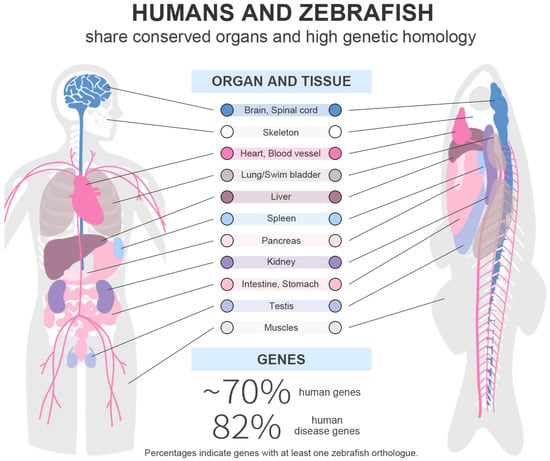
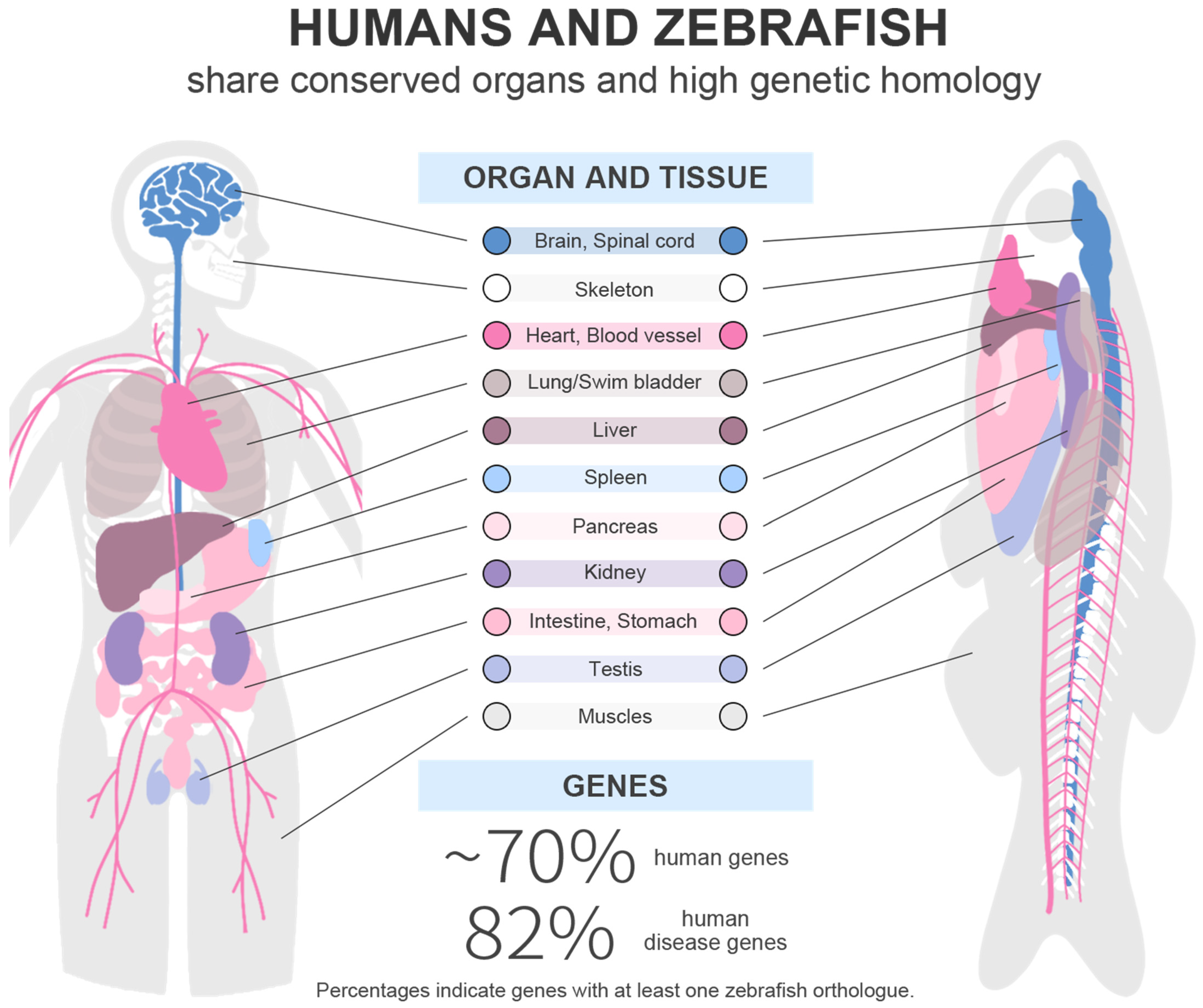
Since George Streisinger introduced zebrafish embryos into developmental research in the 1980s, this model organism has become an essential system for life science research [18]. Comparison to the human reference genome shows that approximately 70% of human genes have at least one obvious zebrafish orthologue [19]. As shown in Figure 1, its major organ systems, including cardiovascular, nervous, musculoskeletal, and other systems, are largely conserved with those of humans, positioning it as an ideal model for studying physiological functions [20]. Compared to mammalian models, zebrafish offer distinct advantages, including small size, high reproductive capacity, and external embryonic development, which facilitates experimental manipulation [21,22].
Figure 1.
Zebrafish share conserved organs and high genetic homology with humans.
Zebrafish exhibit rapid early development, with embryos becoming suitable for experimental use as larvae by 3 days post-fertilization (dpf) and reaching sexual maturity by 90 dpf [23]. Zebrafish eggs are relatively large compared to other fish species (~0.7 mm in diameter at fertilization), and the optical clarity of embryos and larvae enables real-time, non-invasive live imaging. Developmental and functional abnormalities, including pericardial edema, venous thrombosis, spinal curvature, and tail malformation, are directly observable by microscopy [24,25]. When combined with fluorescent labeling techniques, zebrafish also enable real-time tracking of cellular development and apoptosis [26]. Furthermore, zebrafish require lower maintenance costs and less space and have an extended lifespan (3–5 years), making them an economical and efficient model organism. In disease research, 2601/3176 (82%) of human disease-associated genes have at least one zebrafish orthologue [19,27]. According to large-scale forward genetic screens, zebrafish mutants cover nearly all organ systems and cell types identified, most of which exhibit high conservation with mammals [19,28]. Currently, zebrafish have demonstrated significant potential in diverse fields, including cancer biology and in vivo drug screening, highlighting their established value in elucidating disease mechanisms [29,30].
In space biology research, long-term orbital studies utilizing zebrafish and other fish species have already established a solid foundation. The Skylab 3 mission in 1973 marked the first instance of sending mummichogs (Fundulus heteroclitus) into space within a polyethylene bag aquarium. In 1998, the Closed Equilibrated Biological Aquatic System (CEBAS) was developed and successfully sustained swordtail fish for 16 days, further demonstrating the broad feasibility of long-term aquatic experiments in space [31,32]. Recently, CSS-CAES has represented a major advancement in experimental duration, cultivation capacity, and the number of organisms. During the 43-day mission, zebrafish completed growth, development, and reproduction in this closed aquatic ecosystem, demonstrating the feasibility of long-duration vertebrate aquatic experiments in space. It maintains gas cycling through hornwort photosynthesis and zebrafish respiration, while intelligent LED lighting and liquid pumps optimize dissolved oxygen concentration and enhance material exchange [17].This breakthrough provides an essential platform and technical foundation for vertebrate biological research in space.
3. Effects on Major Physiological Systems
To facilitate comparison across the available evidence, the primary studies discussed in this section are summarized in Table 1. The table includes the zebrafish model and developmental stage, exposure conditions, and major physiological outcomes. The following subsections discuss these findings according to the major physiological systems.
3.1. Cardiovascular System
Spaceflight could induce acute cardiovascular alterations in astronauts, including post-flight orthostatic intolerance with impaired heart rate variability and baroreflex function, as well as in-flight arrhythmias such as ventricular ectopy [33]. In addition, exposure to cosmic radiation during space travel might induce late cardiovascular disease and accelerate atherosclerosis [33,34]. Given their unique physiological advantages, zebrafish offer an ideal model for studying cardiovascular diseases and spaceflight-induced cardiotoxicity [35].
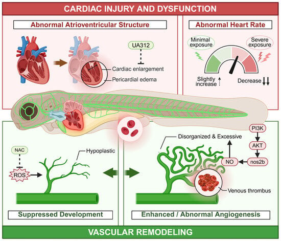
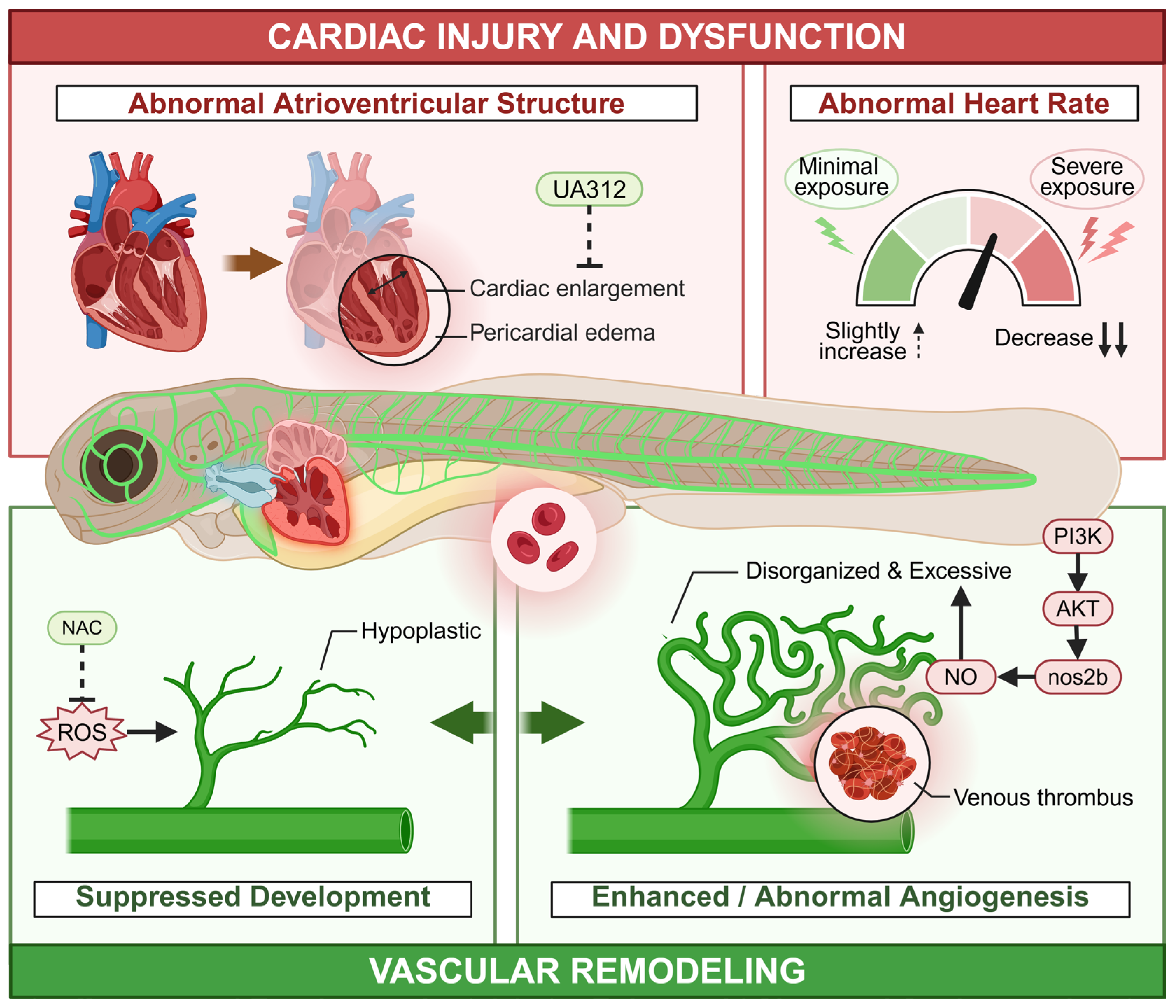
Heart rate can be easily measured in the early zebrafish embryo and serves as a common indicator of radiation-induced cardiotoxicity [36]. For example, studies have reported decreased heart rate in embryos exposed to high-LET radiation, such as 56Fe ion and 12C ion [36,37,38]. Low-LET radiation can also affect the heart rate of zebrafish embryos [39]. Low-dose (1 and 2 Gy) and very low-dose (0.01, 0.05, and 0.1 Gy) γ-irradiation sightly increased in heart rate, while high-dose (10 Gy) γ-irradiation caused a significant decrease in heart rate in zebrafish embryo [40,41]. In addition to functional impairment, radiation also induces overt structural damage, with pericardial edema recognized as a classic morphological hallmark of severe toxicity, particularly at high doses of both high- and low-LET radiation [36,37,39,42]. Pericardial edema is usually accompanied by a decreased heart rate in zebrafish embryos with high-dose radiation treatment. Further anatomical observations by Xu et al. revealed that γ-irradiation induced cardiac enlargement and abnormal atrioventricular structure, which could be ameliorated by UA312 (optimized derivative of Ursolic acid) [43]. Although oxidative stress and apoptosis have been proposed as the primary contributing factors of radiation-induced cardiotoxicity, current insights are primarily derived from whole-embryo rather than cardiac-specific investigations [36,37,39,44]. As a result, the precise mechanisms underlying cardiac susceptibility remain to be fully elucidated.
Radiation also affected vascular formation and function. Jang et al. used a flk-1-GFP transgenic zebrafish, which contained fluorescent endothelial cells in the vasculature, as a module to explore the effect of a proton beam on blood vessel formation in zebrafish embryos. They found that a proton beam inhibited the development of vessels, and the antioxidant NAC reversed this effect [45]. Vala et al. reported that 0.5 Gy X-ray irradiation accelerated subintestinal-vessel sprouting in zebrafish exposed at 3 dpf and increased inter-ray vessel density during caudal-fin regeneration in adults [46]. Together, these findings raise the possibility of a biphasic vascular response to radiation. However, differences in experimental conditions and measured endpoints mean that a threshold effect cannot be excluded. Controlled dose–response studies are needed to test this hypothesis. Additionally, Xu et al. reported that γ-irradiation significantly increased venous thrombus formation in the tails of zebrafish larvae, characterized by the accumulation of red blood cells [43]. Taken together, these studies indicated that radiation can lead to abnormal development of the vascular system. Excessive angiogenesis may enhance the formation of microvessels, thereby increasing the risk of tumor growth and metastasis [47,48]. In adult zebrafish, Marques et al. further showed that low-dose ionizing radiation enhanced angiogenic responses during caudal-fin regeneration, as indicated by increased inter-ray vessel density. However, radiation did not accelerate overall fin regeneration or alter the expression of early regeneration markers, suggesting that its effects on vascular responses may not necessarily translate into enhanced tissue regeneration [49].
Microgravity is another significant factor affecting the cardiovascular system, which is highly sensitive to changes in gravity. Microgravity induces a cephalad fluid shift and unloads the cardiovascular system from gravitational stress, thereby initiating a deconditioning phenotype reminiscent of aging [34]. In 2003, Gillette-Ferguson et al. first used a GFP transgenic zebrafish as a model to investigate the effect of SMG mediated by a NASA-developed bioreactor on the development of zebrafish embryos. They found that GFP fluorescence intensity in the heart exhibited the most significant increase in the whole embryo, indicating that the heart may be more sensitive to SMG than other organs of the zebrafish embryo [50]. This result was further verified by Shimada et al. [51]. As one of the important indexes reflecting heart function, heart rate is also sensitive to microgravity. Li et al. reported that the microgravity environment simulated by acoustic levitation induced an increase in heart rate in zebrafish embryos [52]. This phenomenon was also observed in zebrafish embryos after horizontal rotary culture [53]. Furthermore, Sun et al. found that horizontal rotary culture resulted in downregulation of vascular-related genes, such as flk1, flt4, and ephrinB2, in zebrafish embryos [53]. These results indicate that SMG may also influence vascular development in zebrafish. In a recent study, SMG was found to induce remodeling of the caudal vein plexus network by enhancing angiogenesis in zebrafish embryos. Mechanistic studies showed that SMG induced abnormal development of the cardiovascular system through the PI3K-nos2b signaling pathway [54].
Overall, these studies indicate that radiation and SMG could alter cardiac activity and vascular development in zebrafish (Figure 2). However, most available evidence comes from embryos or larvae exposed to short-term ground-based simulations and primarily reflects developmental or vascular changes. In particular, zebrafish studies have not yet directly assessed human-relevant outcomes such as orthostatic intolerance, reduced cardiac performance, or post-exposure cardiovascular recovery. Therefore, zebrafish offer a valuable model for investigating how spaceflight-related stressors affect cardiovascular development and vascular remodeling, while the relevance of these findings to astronaut cardiovascular deconditioning requires further validation.
Figure 2.
Effect of space environment on cardiovascular system. Created in BioRender. Er, T. (2026) https://BioRender.com/ljrlpjo (accessed on 19 October 2026).
3.2. Nervous System
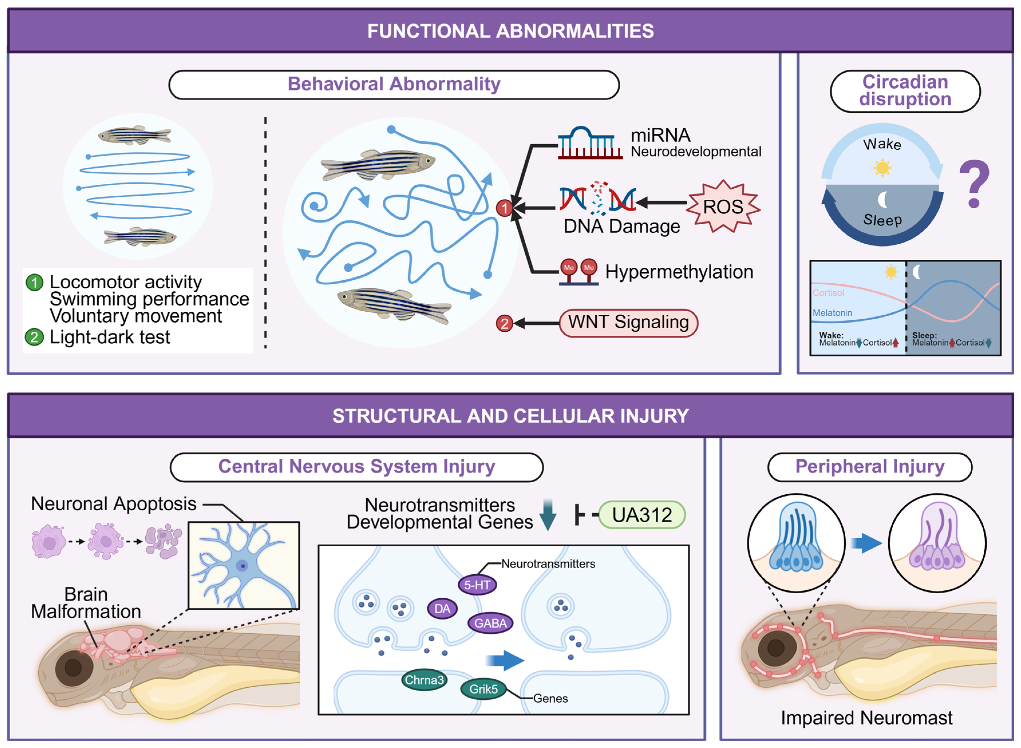
The central nervous system (CNS) is a primary research focus in space biology owing to its vital role in physiological and behavioral regulation [4,55]. To investigate these effects, zebrafish have been widely used as a model for radiation-induced neurotoxicity, with CNS damage as the most extensively examined endpoint [56]. Doses of 0.1 and 1 Gy of γ-irradiation led to abnormal telencephalic development and neuronal apoptosis in this brain region [25]. Zhao et al. reported that even an extremely low dose of 15 mGy γ-irradiation induced brain cell apoptosis and organelle damage in zebrafish embryos, potentially through dysregulation of the Wnt signaling pathway [57]. Structural abnormalities of the nervous system generally reflect underlying functional impairments, which can be characterized by behavioral changes. For example, Houdigui et al. found that prolonged low-dose-rate γ-irradiation (0.5, 5, and 50 mGy/h) altered behavioral responses in the light-dark test in zebrafish larvae [56]. Similarly, He et al. reported that low-dose-rate γ-irradiation (0.1, 0.2, and 0.4 mGy/h) induced dose-dependent behavioral responses in zebrafish embryos: increased swimming speed and morphological alterations were observed at 0.2 and 0.4 mGy/h, whereas a opposite response was noted at 0.1 mGy/h [58]. In addition to affecting locomotor patterns, low-dose γ-irradiation concurrently disrupted circadian rhythms by altering melatonin secretion, as evidenced by dysregulation of the core clock genes CLOCK1A and BMAL1B [25]. Radiation-induced neurobehavioral deficits are not limited to early development; adult zebrafish also exhibit pronounced functional impairments following exposure. Liao et al. reported that radiation exposure increased ROS generation and contributed to DNA damage and cognitive behavioral abnormalities in zebrafish [59]. In an adult zebrafish cranial-irradiation model, radiation-induced brain injury was associated with increased oxidative stress and inflammatory responses, whereas honokiol exerted protective effects [60].
These behavioral phenotypes point to specific underlying molecular regulatory mechanisms. Xu et al. investigated that significant downregulation of multiple neurotransmitters alongside decreased expression of key neurodevelopmental genes [43]. miRNAs have been reported to play an important role in the development of the zebrafish nervous system [61,62]. Furthermore, higher dose rates (0.2 and 0.4 mGy/h) of γ-irradiation upregulated development-associated miRNAs (dre-miR-196a-5p, dre-miR-210-3p, and dre-miR-338), whereas the lowest rate (0.1 mGy/h) suppressed their expression [58]. Except for the CNS, radiation can also influence the peripheral nervous system in zebrafish. It was reported that radiation inhibited cell regeneration in zebrafish lateral line neuromasts, a sensory system for detecting water motion and pressure gradients, by disrupting Wnt signaling [63,64].
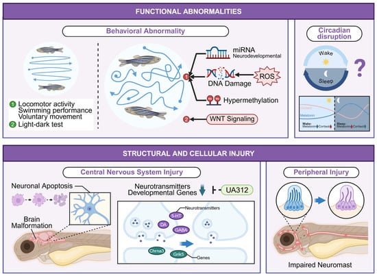
The effects of microgravity on the development of the nervous system have been investigated in mice or even humans [65,66,67]. However, only a few studies have examined the effects of microgravity on zebrafish neurodevelopment. For example, Li et al. examined the development and nervous system function of zebrafish embryo brains exposed to a microgravity environment simulated by acoustic levitation. They found that embryos exhibited a vague brain-beam profile and abnormal brain morphology under microgravity. Moreover, as an indicator of nervous system function, the voluntary movement dependence of the embryo was increased in response to microgravity stimuli [52]. In addition to the brain, neurons in other organs can also be influenced by microgravity. Shimada et al. demonstrated that microgravity altered gene expression in Rohon-Beard neurons of zebrafish embryos [51]. Hypergravity is another factor influencing the health of astronauts during space missions [68]. In a recent study, Salazar et al. reported that hypergravity significantly reduced larval movement frequency and impaired normal swimming behavior, possibly associated with genome-wide hypermethylation [69]. Figure 3 summarizes the reported structural, cellular, and functional changes in the zebrafish nervous system. Thus, zebrafish studies provide a valuable complementary model for linking molecular changes to functional outcomes and identifying pathways that may be relevant to spaceflight-associated neurological responses. Since many studies have been conducted in embryos or larvae under simulated conditions, they may provide mechanistic evidence to guide further validation in mammalian models and human spaceflight studies.
Figure 3.
Effect of space environment on nervous system. Created in BioRender. Er, T. (2026) https://BioRender.com/ljrlpjo (accessed on 19 October 2026).
3.3. Musculoskeletal System
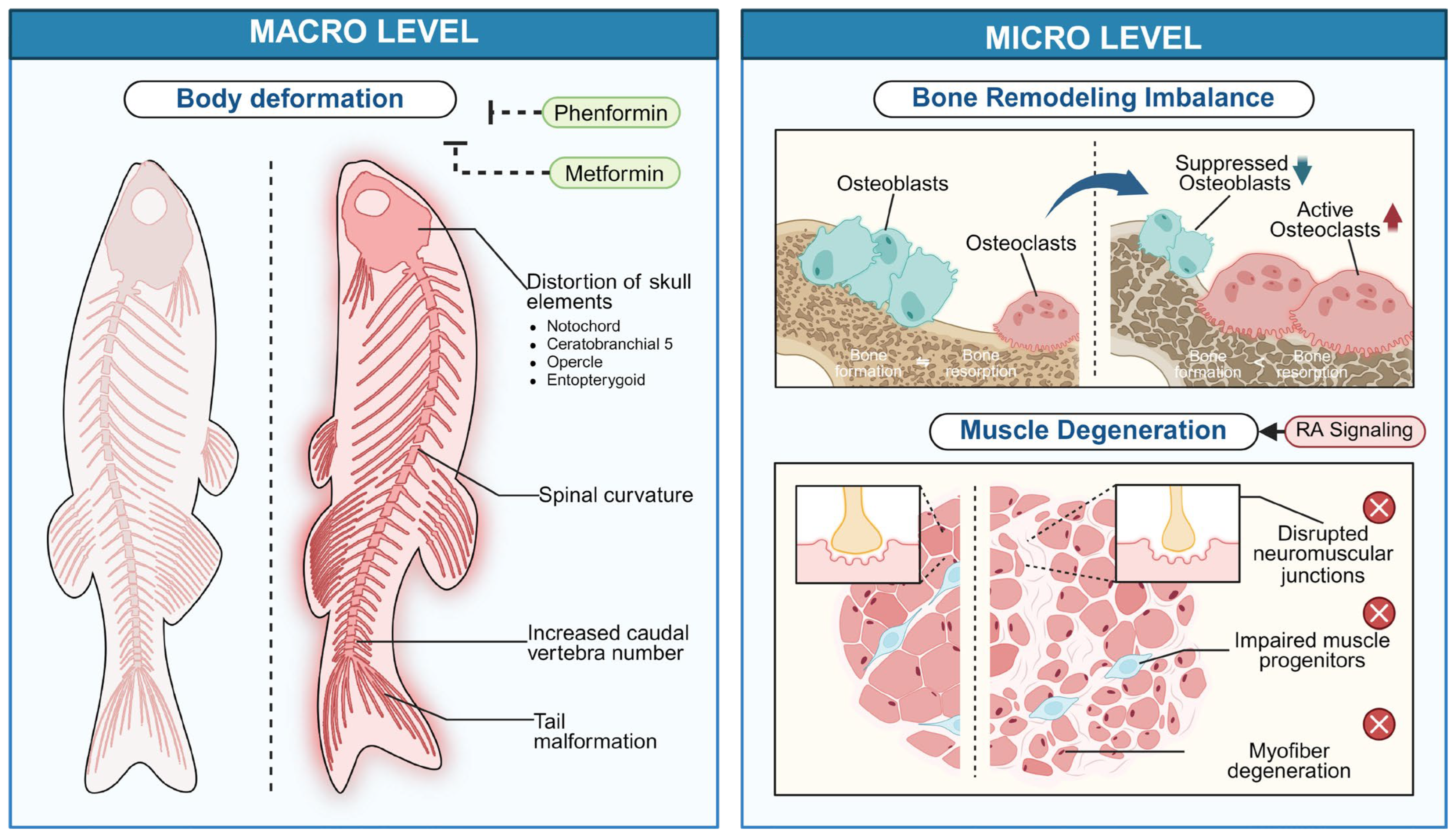
Multiple space environment factors, particularly ionizing radiation, microgravity, and hypergravity, can induce structural alterations in the musculoskeletal system [39,52,69,70]. Commonly observed manifestations include spinal curvature, tail malformation, and overall body deformation, which can all be mitigated by treatment with Phenformin or Metformin [39,52,70]. Among these factors, microgravity conditions have a particularly significant effect on bone development. Simulation of microgravity using a rotating-wall vessel resulted in increased variation in caudal vertebrae number, ranging from 13 to 17, compared with the normal range of 15 to 16 [71]. Carvajal-Agudelo et al. reported that 24 h of 3D-SMG generated using a random positioning machine reduced ossification in multiple developing skeletal elements, potentially due to enhanced bone resorption [72]. Ex vivo-cultured scales from Adult zebrafish scales have been used as a model of SMG-induced bone remodeling. Using this model, the same group attributed SMG-associated osteogenic impairment to increased osteoclast activity and decreased osteoblast activity [73]. In an ex vivo model of regenerating zebrafish scales exposed to SMG, Kabir et al. reported that an antioxidant-loaded zein nanocage formulation increased osteogenic gene expression, calcification, and the calcium-to-phosphorus ratio, consistent with enhanced scale regeneration [74]. In addition, 2D-SMG exhibited similar structural effects on skeleton development. Edsall et al. exposed zebrafish embryos to 2D-SMG using a rotating-wall vessel. At 10 dpf, ossification of the notochord, ceratobranchial 5, opercle, and entopterygoids was delayed, and pharyngeal arch morphology was altered. No corresponding morphological differences were apparent at 35 dpf, whereas adult fish (120 dpf) showed abnormalities in some cranial bones, suggesting both transient and delayed skeletal effects of embryonic SMG exposure [75]. The short- and long-term effects of SMG on zebrafish and its molecular mechanisms still need to be further elucidated. Complementary, actual spaceflight experiments in a related teleost model have also demonstrated musculoskeletal responses. Medaka reared aboard the International Space Station showed reduced mineral density in the pharyngeal bone and tooth region, increased osteoclast activity, and early changes in osteoblast- and osteoclast-related gene expression after exposure to microgravity [76,77]. Although these studies were conducted in medaka, these findings provide an in-orbit reference for interpreting ground-based zebrafish SMG studies and highlight the need to validate simulated-microgravity findings under actual spaceflight conditions.
Direct musculoskeletal effects of hypergravity have been examined in a limited number of zebrafish studies. Aceto et al. reported increased head size and formation of selected cranial bones after 3 G exposure from 5 to 9 dpf, accompanied by differential expression of genes involved in skeletal and muscular system development and function [78]. In a separate study, exposure to 3 or 6 G from 3 to 5 dpf for 48 h did not cause gross changes in cartilage or muscle morphology, but altered cartilage material properties, chondrocyte morphology, extracellular matrix organization, and lower-jaw strain distribution [79]. Taken together, these findings indicate that hypergravity can affect skeletal development and cartilage biomechanics, with the magnitude and nature of the response depending on developmental stage and exposure duration.
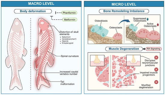
Skeletal muscle atrophy is the most extensively studied adverse effect of microgravity, and has a significant impact on the muscular function of astronauts [80]. Many studies have investigated the effects of microgravity on muscle and its protective mechanisms in mice [81,82]. However, only a few studies have used zebrafish as a model. In 2014, the Japan Aerospace Exploration Agency (JAXA) employed zebrafish as a model to investigate the effects of microgravity on muscle via Aquatic Habitat, an aquarium placed in a microgravity environment of the International Space Station (ISS) [83]. In this mission, the researchers detected deterioration of zebrafish muscle and investigated whether it recovered upon their return to Earth. Furthermore, Houdigui et al. conducted a ground-based simulation of ionizing radiation exposure and demonstrated that γ-irradiation perturbed key muscle development pathways. These effects manifested as myofiber disorganization, structural alterations in neuromuscular junctions, and impaired muscle progenitor cells. Mechanistically, these phenotypes were linked to dysregulated retinoic acid (RA) signaling, though the precise causal pathways and downstream effectors remain to be fully elucidated [56]. Figure 4 summarizes the reported changes in body and skeletal morphology, bone remodeling, and muscle structure. In summary, the spaceflight environment induces profound musculoskeletal deterioration, including structural bone loss and muscle atrophy. Understanding these changes is critical for developing effective countermeasures to protect astronaut health and ensure mission success during long-duration space exploration.
Figure 4.
Effect of space environment on musculoskeletal system. Created in BioRender. Er, T. (2026) https://BioRender.com/ljrlpjo (accessed on 19 October 2026).
3.4. Sensory System
The ocular system enables visual perception, a process highly vulnerable to the complex space environment [84]. Studies have reported that astronauts on long-duration missions (>1 month of spaceflight) exhibit changes in both visual acuity (due to hyperopic shifts) and ocular structure, including optic disc edema, choroidal folds, and globe flattening. Collectively, these manifestations are termed Spaceflight-Associated Neuro-Ocular Syndrome (SANS), which remains one of the highest-priority health risks in human spaceflight programs [85]. In zebrafish embryos, Zhou et al. found that high-LET radiation from 12C6+ ion beams (2 and 4 Gy) induced significant upregulation of proapoptotic genes, leading to increased ocular apoptosis. Moreover, high-dose (8 Gy) radiation also caused microphthalmia, a vision-related developmental defect. The results of histological analysis showed that 12C6+ radiation caused a heterogeneous distribution of cells in the ganglion cell layer and inner nuclear cell layer of the embryo eye. These ocular developmental impairments are mechanistically linked to radiation-triggered apoptosis, potentially mediated through a ROS-independent p53 signaling pathway [86]. In addition to high-LET radiation, low-LET radiation, such as γ-ray and X-ray, has also been reported to influence the development of the zebrafish embryo eye. Geiger et al. reported that γ-rays at a dose of 10 Gy induced multiple pathological alterations, along with increased apoptosis in the eye of the embryo [87]. Similarly, high doses of X-rays were reported to induce pathological changes and decreased whole-eye diameters in zebrafish [88]. Taken together, these studies suggested that both high-LET and low-LET radiation, especially at high doses, can induce ocular damage and disrupt the normal development of the eye in zebrafish embryos. Zebrafish have also been used to investigate the effect of microgravity on ocular development. Using an HSP70-GFP transgenic line with lens-specific expression, Shimada et al. first demonstrated that both microgravity and hypergravity disrupted lens morphogenesis [89].
The vestibular system is equally critical, which mediates balance and spatial orientation [90]. This system comprises the semicircular canals and otoliths, which function as sensors for angular and linear acceleration, respectively [91]. Spaceflight-induced microgravity results in transient vestibular dysfunction in astronauts, manifesting as impaired balance, gaze control, and motion perception [92]. The vestibulo-ocular reflex (VOR) serves as a key metric for vestibular function [93,94]. As a compensatory movement of the eye, VOR stabilizes retinal images by compensating for head movements induced by gravity. Moorman et al. demonstrated that SMG impairs VOR, with the 30–66 hpf period identified as critical for vestibular development and highly sensitive to gravitational alterations [95]. As the major sensor of linear acceleration and gravity, otoliths have been reported to be involved in SMG-induced abnormal behaviors in zebrafish. Moorman et al. found that SMG-exposed larvae exhibited aberrant swimming patterns, including corkscrew motions and vertical loops, accompanied by reduced saccular otolith size or complete otolith loss. The results indicated a significant delay in otolith development, which may contribute to vestibular dysfunction [96]. Mo et al. demonstrated that otoliths are essential for the VOR, as mutant larvae lacking anterior otoliths showed no eye movement response to rotation [97]. However, a recent study reported that delayed otolith development did not have a negative effect on the vestibular circuit development [98]. Hence, the mechanisms of SMG-induced abnormal vestibular development require further investigation.
3.5. Reproductive System
Spaceflight adversely affects reproductive function, leading to suppressed testosterone levels and impaired spermatogenesis in males, as well as disrupted estrous cycles in females [99,100,101,102,103]. Reproductive end point was critical for evaluating the function of the reproductive system. Given the characteristics of embryos, medaka fish have already been used to investigate the effects of spaceflight on reproductive endpoints [104]. Hurem et al. demonstrated that subchronic γ-irradiation caused progressive reproductive impairment in females, with embryo production declining within one month and progressing to gonadal regression and sterility after one year, while testicular morphology remained unaffected at equivalent doses [105]. However, a subsequent study using acute radiation exposure confirmed that both X-ray and carbon ion irradiation reduced testis weight and induced testicular cell apoptosis, identifying the mitochondrial apoptotic pathway as a key mechanism underlying this effect [106]. Zhu et al. further elucidated the sex-specific responses to X-ray radiation: male gonads showed extensive disruption, while females exhibited coordinated protective responses [107]. In contrast to these direct parental effects, Guirandy et al. reported that irradiated adults, while exhibiting no immediate reproductive performance deficits, produced non-irradiated progeny with developmental abnormalities, indicating transgenerational genetic instability [108].The increased developmental abnormalities were also shown to be specifically associated with paternal radiation exposure, which impaired sperm morphology and motility [107]. Furthermore, γ-irradiation could also induce masculinization of offspring, associated with increased DNA methylation of key female sex-differentiation genes in ovarian tissue [109]. Collectively, these studies highlight that radiation effects on reproduction depend critically on dose, sex, and the time window of exposure, encompassing both direct organ damage and transgenerational consequences.
3.6. Other System
During the Apollo missions, over half of the astronauts exhibited immune suppression and bacterial infections [110]. Subsequent research, including the NASA Twin Study, has consistently shown that spaceflight induces alterations in multiple immune-related pathways [3]. SMG is known to affect both innate and adaptive immunity by impairing immune cell development and proliferation [111,112,113]. Zhu et al. reported that SMG exacerbated the toxicity of viral infection in zebrafish embryos by suppressing innate immunity. RNA-seq (GSE145967, corresponding to NASA GeneLab accession GLDS-373) analysis revealed that SMG downregulated genes involved in inflammatory and innate immune responses, including components of the NF-κB, RIG-I-like receptor (RLR), and Toll-like receptor (TLR) signaling pathways. Further investigation demonstrated that SMG inhibited TRIM25, an E3 ubiquitin ligase, thereby impairing RLR pathway activation and disrupting the positive feedback loop essential for antiviral defense [114]. Radiation exerts dual immunomodulatory effects: high doses (>1 Gy) suppress immunity and enhance inflammation, whereas intermediate doses (0.1–1 Gy) mitigate inflammatory responses [115,116]. Traver et al. found that lethal doses of γ-irradiation (20 and 40 Gy) caused specific ablation of hematolymphoid cells and death in adult zebrafish [117]. Other studies also reported that γ-irradiation induced oxidative damage in immune cells and zebrafish embryos, which could be mitigated by natural antioxidants [118,119]. However, unlike the acute high-dose radiation typically studied in terrestrial models, the space environment presents a scenario of chronic, low-dose radiation exposure. This sustained exposure profile may induce a pro-inflammatory state, potentially elevating long-term risks for chronic degenerative diseases.
The digestive system of zebrafish, as in other vertebrates, is derived from the endoderm, including the liver, gallbladder, pancreas and four intestinal domains (intestinal bulb, mid-intestine, hindgut, and proctodeum). Notably, zebrafish are stomachless, while the intestinal bulb is considered their functional analog [120]. Beyond its primary roles in nutrient digestion and absorption, the digestive tract also serves as a critical barrier against environmental toxins [121]. Using two complementary low-dose γ-irradiation models, Cahill et al. characterized radiation-associated responses in the zebrafish digestive system. In a hypothermic liver model, a very low dose of γ-irradiation (16.34 cGy) altered transcriptomic pathways related to lipid metabolism, wound healing, immune responses, and fibrosis [122]. In a separate torpor-like model, low-dose irradiation induced DNA damage and oxidative stress and altered steroid-signaling, metabolic, and cell-cycle-related pathways in the gastrointestinal tract; importantly, the torpor-like state reduced radiation-associated DNA damage [123]. At higher doses, Wu et al. demonstrated that 10 Gy of γ-irradiation induced hepatic fibrosis and hepatocyte apoptosis in adult zebrafish, and that Oligo-Fucoidan (OF) showed protective effects against such damage [124]. Radiation also severely impacted intestinal morphology and function. Jensen et al. demonstrated that a dose of 2 Gy triggered a pathological progression from initial hypertrophy to subsequent atrophy in the intestine, ultimately impairing its function [125]. Furthermore, Park et al. reported that high-dose (45–50 Gy) photon irradiation severely deformed intestinal villi, with mistletoe extract demonstrating protective effects [126]. Thus, these radiation-induced hepatointestinal injuries in zebrafish may provide complementary evidence relevant to hepatic and intestinal risks in space.
Table 1.
Effects on Major Physiological Systems. For in vivo studies, the model indicates the developmental stage at exposure; ex vivo entries identify the isolated tissue used. Embryo (0–72 hpf), Larva (72 hpf–30 dpf), Adult (>90 dpf) [127]. A linear energy transfer (LET) of approximately 10 keV/μm is commonly used as the demarcation between low- and high-LET radiation [128]. * Damage pathway, # Rescue measure.
4. Future Perspectives and Limitation
The announcement of long-term spaceflight missions, such as those to Mars, has brought the multidimensional and cumulative health risks associated with the space environment to the forefront of scientific concern. Zebrafish, with their unique advantages including evolutionary conservation, optical transparency, high fecundity, and genetic tractability, have emerged as an ideal vertebrate model for ground-based studies, providing an indispensable platform for elucidating the molecular mechanisms and physiological pathways underlying space environment-induced damage. This review synthesizes current evidence on the effects of space-relevant environmental factors on zebrafish. Ionizing radiation profoundly affects the cardiovascular, immune, and reproductive systems, with the heart identified as an early and sensitive target. Gravity alterations primarily affect the musculoskeletal system, vestibular apparatus, and swim bladder, with responses varying by developmental stage and exposure conditions. Notably, these responses vary across developmental stages and sexes, indicating the need for further investigation.
Despite these advances, current evidence from zebrafish space biology remains constrained by several limitations.
- (1)
- Limited translational relevance of zebrafish models. Given that many published studies were performed in embryos or larvae, their findings may not fully reflect the physiological responses expected in adult astronauts. Future work should compare different developmental stages and validate key observations in adult zebrafish and complementary vertebrate models, thereby clarifying the specific translational contributions of zebrafish to space-health research.
- (2)
- Limitations of ground-based space-environment simulations and combined stressors. Ground-based models do not fully reproduce mission-relevant radiation or gravity conditions. In particular, many studies summarized here use acute, single-exposure X-ray or γ-ray irradiation, whereas deep-space missions involve prolonged, lower-dose-rate exposure to a mixed radiation field composed mainly of protons, together with less abundant but biologically important high-energy, high-charge ions. SMG devices may also generate residual acceleration and altered fluid dynamics. In addition, direct zebrafish studies combining radiation and SMG remain limited, so single-stressor findings cannot demonstrate whether their joint effects are additive, synergistic, or antagonistic. Future studies should compare the effects of combined radiation and SMG exposure with those of each stressor alone. Other spaceflight-related stressors, including noise, confinement, and circadian rhythm disruption, may also modify biological responses and warrant consideration.
- (3)
- Limited coverage of biological processes and multisystem responses. Zebrafish possess well-established advantages and a strong research foundation for studying tissue regeneration, wound healing, gut microbiota, and host–microbe interactions, owing to their regenerative capacity, optical accessibility, and experimental tractability. However, these biological processes remain insufficiently investigated under spaceflight or simulated space conditions, particularly in adult fish. Moreover, most existing studies focus on individual physiological systems, and the cross-system effects of spaceflight-related stressors remain poorly understood. Future work should directly investigate these processes and their multisystem interactions under integrated space-relevant conditions.
- (4)
- Translating phenotypic observations into molecular mechanisms and protective strategies. The molecular mechanisms of many space-environment-induced phenotypes remain unclear, and evidence for reported protective candidates is still preliminary. These agents have generally been explored in embryonic models under limited dosing conditions and acute irradiation, without sufficient information on pharmacokinetics, adult efficacy, dose dependence, formulation, stability, or tolerability. Future studies should combine zebrafish genetic tools with systematic pharmacological evaluation to establish causality and translational potential.
From CEBAS (Germany) to CAES (China), closed aquatic ecosystem platforms have advanced substantially, with the latter demonstrating the in-orbit cultivation and reproduction of zebrafish during a 43-day mission. Future studies using zebrafish in such systems can advance along several key directions. First, further development of in-orbit live imaging could enable longitudinal monitoring of physiological processes, including cardiovascular function, neuronal activity, and cellular turnover. This approach could provide repeated, in situ observations under actual spaceflight conditions. Second, pre-established CRISPR/Cas9-edited zebrafish strains, together with appropriate controls, could be used to test candidate genes involved in DNA-damage responses and gravity-related signaling during spaceflight. Finally, ground-based high-throughput screening could identify candidate countermeasures for subsequent targeted in-orbit evaluation. Such stepwise studies could strengthen the assessment of proposed mechanisms and protective effects under spaceflight conditions.
5. Conclusions
Taken together, these initiatives would deepen fundamental understanding of organismal adaptation to space, while providing practical knowledge and tools to safeguard crew health during future long-duration, deep-space missions. This progression reflected an essential shift in space life sciences from observational research toward proactive intervention and precision health preservation. In the future, while the physiological responses of zebrafish to the space environment remain to be fully characterized, zebrafish are expected to gain growing prominence as a prime model for space biology research.
Author Contributions
T.E. and C.Z. drafted the manuscript and prepared the figures. Z.W. revised and edited the manuscript. S.R. contributed to literature collection. J.M., H.M. and C.Z. conceptualized and supervised the Review and edited the manuscript. All authors have read and agreed to the published version of the manuscript.
Funding
This work was financially supported by Beijing Life Science Academy (2025300CE0020, 2025300CC0120, 2025300CA0070); National Natural Science Foundation of China (82302117).
Data Availability Statement
No new data were created or analyzed in this study. Data sharing is not applicable to this article.
Conflicts of Interest
All authors declare that they have no competing interests.
References
- Dai, Z.; Lei, X.; Yang, C.; Zhao, L.; Lu, L.; Li, Y. Systematic biomedical research of the NASA Twins Study facilitates the hazard risk assessment of long-term spaceflight missions. Protein Cell 2019, 10, 628–630. [Google Scholar] [CrossRef] [Scilit] [PubMed]
- Zhao, L.; Mi, D.; Sun, Y. Issues and challenges of space radiation risk assessment in manned deep space exploration missions. Chin. Sci. Bull. 2018, 63, 1523–1537. [Google Scholar] [CrossRef] [Scilit]
- Garrett-Bakelman, F.E.; Darshi, M.; Green, S.J.; Gur, R.C.; Lin, L.; Macias, B.R.; McKenna, M.J.; Meydan, C.; Mishra, T.; Nasrini, J.; et al. The NASA Twins Study: A multidimensional analysis of a year-long human spaceflight. Science 2019, 364, eaau8650. [Google Scholar] [CrossRef] [Scilit] [PubMed]
- Wuyts, F.L.; Deblieck, C.; Vandevoorde, C.; Durante, M. Brains in space: Impact of microgravity and cosmic radiation on the CNS during space exploration. Nat. Rev. Neurosci. 2025, 26, 354–371. [Google Scholar] [CrossRef] [Scilit] [PubMed]
- Chancellor, J.C.; Scott, G.B.I.; Sutton, J.P. Space Radiation: The Number One Risk to Astronaut Health beyond Low Earth Orbit. Life 2014, 4, 491–510. [Google Scholar] [CrossRef] [Scilit] [PubMed]
- Unsworth, B.R.; Lelkes, P.I. Growing tissues in microgravity. Nat. Med. 1998, 4, 901–907. [Google Scholar] [CrossRef] [Scilit] [PubMed]
- Goswami, N.; Blaber, A.P.; Valenti, G.; Hinghofer-Szalkay, H.; Evans, J.; Bailey, D.M.; Vernikos, J.; Choukér, A.; Green, D.A.; White, O.; et al. Gravity, microgravity, and artificial gravity: Physiological effects, implementation, and applications. Physiol. Rev. 2025, 106, 751–840. [Google Scholar] [CrossRef] [Scilit] [PubMed]
- Loon, J.; Tanck, E.; van Nieuwenhoven, F.; Snoeckx, L.H.E.H.; Jong, H.; Wubbels, R. A brief overview of animal hypergravity studies. J. Gravitational Physiol. A J. Int. Soc. Gravitational Physiol. 2005, 12, 5–10. [Google Scholar]
- Simonsen, L.C.; Slaba, T.C.; Guida, P.; Rusek, A. NASA’s first ground-based Galactic Cosmic Ray Simulator: Enabling a new era in space radiobiology research. PLoS Biol. 2020, 18, e3000669. [Google Scholar] [CrossRef] [Scilit] [PubMed]
- Durante, M.; Cucinotta, F.A. Heavy ion carcinogenesis and human space exploration. Nat. Rev. Cancer 2008, 8, 465–472. [Google Scholar] [CrossRef] [Scilit] [PubMed]
- Shuryak, I.; Sachs, R.K.; Brenner, D.J. Quantitative modeling of carcinogenesis induced by single beams or mixtures of space radiations using targeted and non-targeted effects. Sci. Rep. 2021, 11, 23467. [Google Scholar] [CrossRef] [Scilit] [PubMed]
- Klaus, D.M. Clinostats and bioreactors. Gravit. Space Biol. Bull. 2001, 14, 55–64. [Google Scholar] [PubMed]
- Poon, C. Factors implicating the validity and interpretation of mechanobiology studies in simulated microgravity environments. Eng. Rep. 2020, 2, e12242. [Google Scholar] [CrossRef] [Scilit]
- Shelhamer, M. Parabolic flight as a spaceflight analog. J. Appl. Physiol. 2016, 120, 1442–1448. [Google Scholar] [CrossRef] [Scilit] [PubMed]
- Durante, M.; Cucinotta, F.A. Physical basis of radiation protection in space travel. Rev. Mod. Phys. 2011, 83, 1245–1281. [Google Scholar] [CrossRef] [Scilit]
- NASA Communications. A Brief History of Animals in Space. 1998. Available online: https://www.nasa.gov/history/a-brief-history-of-animals-in-space/ (accessed on 24 October 2026).
- Xie, M.; Tian, Q.; Wang, G.; Zheng, W.; Wang, S. Breakthrough in enclosed aquatic ecosystems in space: Supporting zebrafish survival for 43 days. Innovation 2024, 5, 100711. [Google Scholar] [CrossRef] [Scilit]
- Streisinger, G.; Walker, C.; Dower, N.; Knauber, D.; Singer, F. Production of clones of homozygous diploid zebra fish (Brachydanio rerio). Nature 1981, 291, 293–296. [Google Scholar] [CrossRef] [Scilit] [PubMed]
- Howe, K.; Clark, M.D.; Torroja, C.F.; Torrance, J.; Berthelot, C.; Muffato, M.; Collins, J.E.; Humphray, S.; McLaren, K.; Matthews, L.; et al. The zebrafish reference genome sequence and its relationship to the human genome. Nature 2013, 496, 498–503, Erratum in Nature 2014, 505, 248. [Google Scholar] [CrossRef] [Scilit] [PubMed]
- Choi, T.-Y.; Choi, T.-I.; Lee, Y.-R.; Choe, S.-K.; Kim, C.-H. Zebrafish as an animal model for biomedical research. Exp. Mol. Med. 2021, 53, 310–317. [Google Scholar] [CrossRef] [Scilit] [PubMed]
- Shin, J.T.; Fishman, M.C. From zebrafish to human: Modular medical models. Annu. Rev. Genomics Hum. Genet. 2002, 3, 311–340. [Google Scholar] [CrossRef] [Scilit] [PubMed]
- Lieschke, G.J.; Currie, P.D. Animal models of human disease: Zebrafish swim into view. Nat. Rev. Genet. 2007, 8, 353–367. [Google Scholar] [CrossRef] [Scilit] [PubMed]
- Adhish, M.; Manjubala, I. Effectiveness of zebrafish models in understanding human diseases—A review of models. Heliyon 2023, 9, e14557. [Google Scholar] [CrossRef] [Scilit] [PubMed]
- Chahardehi, A.M.; Arsad, H.; Lim, V. Zebrafish as a Successful Animal Model for Screening Toxicity of Medicinal Plants. Plants 2020, 9, 1345. [Google Scholar] [CrossRef] [Scilit] [PubMed]
- Zhao, W.; Mao, L.; He, C.; Ding, D.; Hu, N.; Song, X.; Long, D. Effects of low dose radiation on behavior rhythm of zebrafish (Danio rerio). Ecotoxicol. Environ. Saf. 2023, 255, 114779. [Google Scholar] [CrossRef] [Scilit] [PubMed]
- Ko, S.-K.; Chen, X.; Yoon, J.; Shin, I. Zebrafish as a good vertebrate model for molecular imaging using fluorescent probes. Chem. Soc. Rev. 2011, 40, 2120–2130. [Google Scholar] [CrossRef] [Scilit] [PubMed]
- MacRae, C.A.; Peterson, R.T. Zebrafish as tools for drug discovery. Nat. Rev. Drug Discov. 2015, 14, 721–731. [Google Scholar] [CrossRef] [Scilit] [PubMed]
- Goldsmith, J.R.; Jobin, C. Think small: Zebrafish as a model system of human pathology. J. Biomed. Biotechnol. 2012, 2012, 817341. [Google Scholar] [CrossRef] [Scilit] [PubMed]
- Zon, L.I.; Peterson, R.T. In vivo drug discovery in the zebrafish. Nat. Rev. Drug Discov. 2005, 4, 35–44. [Google Scholar] [CrossRef] [Scilit] [PubMed]
- White, R.; Rose, K.; Zon, L. Zebrafish cancer: The state of the art and the path forward. Nat. Rev. Cancer 2013, 13, 624–636. [Google Scholar] [CrossRef] [Scilit] [PubMed]
- Bluem, V.; Andriske, M.; Paris, F.; Voeste, D. The C.E.B.A.S.-minimodule: Behaviour of an artificial aquatic ecological system during spaceflight. Adv. Space Res. 2000, 26, 253–262. [Google Scholar] [CrossRef] [Scilit] [PubMed]
- Przybyla, C. Space aquaculture: Prospects for raising aquatic vertebrates in a bioregenerative life-support system on a lunar base. Front. Astron. Space Sci. 2021, 8, 699097. [Google Scholar] [CrossRef] [Scilit]
- Krittanawong, C.; Isath, A.; Kaplin, S.; Virk, H.U.H.; Fogg, S.; Wang, Z.; Shepanek, M.; Scheuring, R.A.; Lavie, C.J. Cardiovascular disease in space: A systematic review. Prog. Cardiovasc. Dis. 2023, 81, 33–41. [Google Scholar] [CrossRef] [Scilit] [PubMed]
- Hughson, R.L.; Helm, A.; Durante, M. Heart in space: Effect of the extraterrestrial environment on the cardiovascular system. Nat. Rev. Cardiol. 2018, 15, 167–180. [Google Scholar] [CrossRef] [Scilit] [PubMed]
- Wang, R.; Zhang, Q.; Zhang, S.; Wang, Z.; Zhou, Z.; Yuan, T.; Zhang, B. Zebrafish in Cardiovascular Disease Research: From Model to Application. Int. J. Biol. Sci. 2026, 22, 4806–4825. [Google Scholar] [CrossRef] [Scilit] [PubMed]
- Si, J.; Zhou, R.; Song, J.; Gan, L.; Zhou, X.; Di, C.; Liu, Y.; Mao, A.; Zhao, Q.; Wang, Y.; et al. Toxic effects of 56Fe ion radiation on the zebrafish (Danio rerio) embryonic development. Aquat. Toxicol. 2017, 186, 87–95. [Google Scholar] [CrossRef] [Scilit] [PubMed]
- Si, J.; Zhou, R.; Zhao, B.; Xie, Y.; Gan, L.; Zhang, J.; Wang, Y.; Zhou, X.; Ren, X.; Zhang, H. Effects of ionizing radiation and HLY78 on the zebrafish embryonic developmental toxicity. Toxicology 2019, 411, 143–153. [Google Scholar] [CrossRef] [Scilit] [PubMed]
- Li, X.; Zha, X.; Wang, Y.; Jia, R.; Hu, B.; Zhao, B. Toxic effects and foundation of proton radiation on the early-life stage of zebrafish development. Chemosphere 2018, 200, 302–312. [Google Scholar] [CrossRef] [Scilit] [PubMed]
- Gan, L.; Guo, M.; Si, J.; Zhang, J.; Liu, Z.; Zhao, J.; Wang, F.; Yan, J.; Li, H.; Zhang, H. Protective effects of phenformin on zebrafish embryonic neurodevelopmental toxicity induced by X-ray radiation. Artif. Cells Nanomed. Biotechnol. 2019, 47, 4202–4210. [Google Scholar] [CrossRef] [Scilit] [PubMed]
- Freeman, J.L.; Weber, G.J.; Peterson, S.M.; Nie, L.H. Embryonic ionizing radiation exposure results in expression alterations of genes associated with cardiovascular and neurological development, function, and disease and modified cardiovascular function in zebrafish. Front. Genet. 2014, 5, 268. [Google Scholar] [CrossRef] [Scilit] [PubMed]
- Zhao, W.; Hu, N.; Ding, D.; Long, D.; Li, S.; Li, G.; Zhang, H. Developmental toxicity and apoptosis in zebrafish embryos induced by low-dose gamma-ray irradiation. Environ. Sci. Pollut. Res. Int. 2019, 26, 3869–3881. [Google Scholar] [CrossRef] [Scilit] [PubMed]
- Hu, M.; Hu, N.; Ding, D.; Zhao, W.; Feng, Y.; Zhang, H.; Li, G.; Wang, Y. Developmental toxicity and oxidative stress induced by gamma irradiation in zebrafish embryos. Radiat. Environ. Biophys. 2016, 55, 441–450. [Google Scholar] [CrossRef] [Scilit] [PubMed]
- Xu, F.F.; Shang, Y.; Wei, H.Q.; Zhang, W.Y.; Wang, L.X.; Hu, T.; Zhang, S.Q.; Li, Y.L.; Shang, H.H.; Hou, W.B.; et al. Ursolic acid derivative UA312 ameliorates ionizing radiation-induced cardiotoxicity and neurodevelopmental toxicity in zebrafish via targeting chrna3 and grik5. Acta Pharmacol. Sin. 2025, 46, 1–16. [Google Scholar] [CrossRef] [Scilit] [PubMed]
- Taleb, A.; Ahmad, K.A.; Ihsan, A.U.; Qu, J.; Lin, N.; Hezam, K.; Koju, N.; Hui, L.; Qilong, D. Antioxidant effects and mechanism of silymarin in oxidative stress induced cardiovascular diseases. Biomed. Pharmacother. 2018, 102, 689–698. [Google Scholar] [CrossRef] [Scilit] [PubMed]
- Jang, G.H.; Ha, J.H.; Huh, T.L.; Lee, Y.M. Effect of proton beam on blood vessel formation in early developing zebrafish (Danio rerio) embryos. Arch. Pharm. Res. 2008, 31, 779–785. [Google Scholar] [CrossRef] [Scilit] [PubMed]
- Vala, I.S.; Martins, L.R.; Imaizumi, N.; Nunes, R.J.; Rino, J.; Kuonen, F.; Carvalho, L.M.; Ruegg, C.; Grillo, I.M.; Barata, J.T.; et al. Low doses of ionizing radiation promote tumor growth and metastasis by enhancing angiogenesis. PLoS ONE 2010, 5, e11222. [Google Scholar] [CrossRef] [Scilit] [PubMed]
- Semenza, G.L. Intratumoral hypoxia, radiation resistance, and HIF-1. Cancer Cell 2004, 5, 405–406. [Google Scholar] [CrossRef] [Scilit] [PubMed]
- Madani, I.; De Neve, W.; Mareel, M. Does ionizing radiation stimulate cancer invasion and metastasis? Bull. Cancer 2008, 95, 292–300. [Google Scholar] [CrossRef] [Scilit] [PubMed]
- Marques, F.G.; Carvalho, L.; Sousa, J.S.; Rino, J.; Diegues, I.; Poli, E.; Pina, F.; Saúde, L.; Constantino Rosa Santos, S. Low doses of ionizing radiation enhance angiogenesis and consequently accelerate post-embryonic development but not regeneration in zebrafish. Sci. Rep. 2020, 10, 3137. [Google Scholar] [CrossRef] [Scilit] [PubMed]
- Gillette-Ferguson, I.; Ferguson, D.G.; Poss, K.D.; Moorman, S.J. Changes in gravitational force induce alterations in gene expression that can be monitored in the live, developing zebrafish heart. Adv. Space Res. 2003, 32, 1641–1646. [Google Scholar] [CrossRef] [Scilit] [PubMed]
- Shimada, N.; Sokunbi, G.; Moorman, S.J. Changes in gravitational force affect gene expression in developing organ systems at different developmental times. BMC Dev. Biol. 2005, 5, 10. [Google Scholar] [CrossRef] [Scilit] [PubMed]
- Li, L.; Gu, N.; Dong, H.; Li, B.; Kenneth, T.V.G. Analysis of the effects of acoustic levitation to simulate the microgravity environment on the development of early zebrafish embryos. RSC Adv. 2020, 10, 44593–44600, Correction in RSC Adv. 2021, 11, 3891. https://doi.org/10.1039/d1ra90003j. [Google Scholar] [CrossRef] [Scilit] [PubMed]
- Sun, T.; Xie, X.; Zhang, J.Q.; Bao, J.; Tang, C.Z.; Lei, D.X.; Qiu, J.H.; Wang, G.X. Effect of horizontal rotary culture on zebrafish vascular development. Yi Chuan 2013, 35, 502–510. [Google Scholar] [CrossRef] [Scilit] [PubMed]
- Xie, X.; Lei, D.; Zhang, Q.; Wang, Y.; Wen, L.; Ye, Z.; Ud Din, A.; Jia, D.; Apicella, A.; Wang, G. Effect of simulated microgravity induced PI3K-nos2b signalling on zebrafish cardiovascular plexus network formation. J. Biomech. 2019, 87, 83–92. [Google Scholar] [CrossRef] [Scilit] [PubMed]
- Roy-O’Reilly, M.; Mulavara, A.; Williams, T. A review of alterations to the brain during spaceflight and the potential relevance to crew in long-duration space exploration. npj Microgravity 2021, 7, 5. [Google Scholar] [CrossRef] [Scilit] [PubMed]
- Murat El Houdigui, S.; Adam-Guillermin, C.; Loro, G.; Arcanjo, C.; Frelon, S.; Floriani, M.; Dubourg, N.; Baudelet, E.; Audebert, S.; Camoin, L.; et al. A systems biology approach reveals neuronal and muscle developmental defects after chronic exposure to ionising radiation in zebrafish. Sci. Rep. 2019, 9, 20241. [Google Scholar] [CrossRef] [Scilit] [PubMed]
- Zhao, W.; Yuan, P.; Hu, N.; Long, D.; Ding, D.; Wang, H. Effects of Low-Dose Gamma-Ray Radiation on Apoptosis and Development of Zebrafish Embryo Brain. Radiat. Res. 2020, 194, 61–70. [Google Scholar] [CrossRef] [Scilit] [PubMed]
- He, C.Q.; Mao, L.; Yao, J.; Zhao, W.C.; Huang, B.; Hu, N.; Long, D.X. The Threshold Effects of Low-Dose-Rate Radiation on miRNA-Mediated Neurodevelopment of Zebrafish. Radiat. Res. 2021, 196, 633–646. [Google Scholar] [CrossRef] [Scilit] [PubMed]
- Liao, G.; Li, R.; Chen, X.; Zhang, W.; Du, S.; Yuan, Y. Sodium valproate prevents radiation-induced injury in hippocampal neurons via activation of the Nrf2/HO-1 pathway. Neuroscience 2016, 331, 40–51. [Google Scholar] [CrossRef] [Scilit] [PubMed]
- Liao, G.; Zhao, Z.; Yang, H.; Li, X. Honokiol ameliorates radiation-induced brain injury via the activation of SIRT3. J. Int. Med. Res. 2020, 48, 300060520963993. [Google Scholar] [CrossRef] [Scilit] [PubMed]
- Tal, T.L.; Franzosa, J.A.; Tilton, S.C.; Philbrick, K.A.; Iwaniec, U.T.; Turner, R.T.; Waters, K.M.; Tanguay, R.L. MicroRNAs control neurobehavioral development and function in zebrafish. FASEB J. 2012, 26, 1452–1461. [Google Scholar] [CrossRef] [Scilit] [PubMed]
- Delaloy, C.; Gao, F.B. MicroRNA-9 multitasking near organizing centers. Nat. Neurosci. 2008, 11, 625–626. [Google Scholar] [CrossRef] [Scilit] [PubMed]
- Bleckmann, H.; Zelick, R. Lateral line system of fish. Integr. Zool. 2009, 4, 13–25. [Google Scholar] [CrossRef] [Scilit] [PubMed]
- Li, R.; Liao, G.; Yin, G.; Wang, B.; Yan, M.; Lin, X.; Zhang, W.; Chen, X.; Du, S.; Yuan, Y. Ionizing Radiation Blocks Hair Cell Regeneration in Zebrafish Lateral Line Neuromasts by Preventing Wnt Signaling. Mol. Neurobiol. 2018, 55, 1639–1651. [Google Scholar] [CrossRef] [Scilit] [PubMed]
- Chen, H.; Lv, K.; Dai, Z.; Ji, G.; Wang, T.; Wang, Y.; Zhang, Y.; Kan, G.; Li, Y.; Qu, L. Intramuscular injection of mechano growth factor E domain peptide regulated expression of memory-related sod, miR-134 and miR-125b-3p in rat hippocampus under simulated weightlessness. Biotechnol. Lett. 2016, 38, 2071–2080. [Google Scholar] [CrossRef] [Scilit] [PubMed]
- Patel, N.; Pass, A.; Mason, S.; Gibson, C.R.; Otto, C. Optical Coherence Tomography Analysis of the Optic Nerve Head and Surrounding Structures in Long-Duration International Space Station Astronauts. JAMA Ophthalmol. 2018, 136, 193–200. [Google Scholar] [CrossRef] [Scilit] [PubMed]
- Monticone, M.; Liu, Y.; Pujic, N.; Cancedda, R. Activation of nervous system development genes in bone marrow derived mesenchymal stem cells following spaceflight exposure. J. Cell. Biochem. 2010, 111, 442–452. [Google Scholar] [CrossRef] [Scilit] [PubMed]
- Limper, U.; Ahnert, T.; Maegele, M.; Froehlich, M.; Grau, M.; Gauger, P.; Bauerfeind, U.; Gorlinger, K.; Potzsch, B.; Jordan, J. Simulated Hypergravity Activates Hemostasis in Healthy Volunteers. J. Am. Heart Assoc. 2020, 9, e016479. [Google Scholar] [CrossRef] [Scilit] [PubMed]
- Salazar, M.; Joly, S.; Anglada-Escudé, G.; Ribas, L. Epigenetic and physiological alterations in zebrafish subjected to hypergravity. PLoS ONE 2024, 19, e0300310. [Google Scholar] [CrossRef] [Scilit] [PubMed]
- Wang, B.; Dong, J.; Xiao, H.; Li, Y.; Jin, Y.; Cui, M.; Zhang, S.-Q.; Fan, S.-J. Metformin fights against radiation-induced early developmental toxicity. Sci. Total Environ. 2020, 732, 139274. [Google Scholar] [CrossRef] [Scilit] [PubMed]
- Franz-Odendaal, T.A.; Edsall, S.C. Long-term effects of simulated microgravity and vibration exposure on skeletal development in zebrafish. Stem Cells Dev. 2018, 27, 1278–1286. [Google Scholar] [CrossRef] [Scilit] [PubMed]
- Carvajal-Agudelo, J.D.; Eaton, J.; Franz-Odendaal, T.A. Reduced ossification caused by 3D simulated microgravity exposure is short-term in larval zebrafish. Life Sci. Space Res. 2024, 41, 127–135. [Google Scholar] [CrossRef] [Scilit] [PubMed]
- Carvajal-Agudelo, J.D.; McNeil, A.; Franz-Odendaal, T.A. Effects of simulated microgravity and vibration on osteoblast and osteoclast activity in cultured zebrafish scales. Life Sci. Space Res. 2023, 38, 39–45. [Google Scholar] [CrossRef] [Scilit] [PubMed]
- Kabir, A.; Mukilarasi, B.; Manohar, A.; Gadani, M.; Sinha, A.K.; Sharma, P.; Verma, A.; Selvaraj, V.; Sudhakar, S. Protein bioactive complexes promote osteogenesis under microgravity environment. Int. J. Biol. Macromol. 2025, 303, 140483. [Google Scholar] [CrossRef] [Scilit] [PubMed]
- Edsall, S.C.; Franz-Odendaal, T.A. An assessment of the long-term effects of simulated microgravity on cranial neural crest cells in zebrafish embryos with a focus on the adult skeleton. PLoS ONE 2014, 9, e89296. [Google Scholar] [CrossRef] [Scilit] [PubMed]
- Chatani, M.; Mantoku, A.; Takeyama, K.; Abduweli, D.; Sugamori, Y.; Aoki, K.; Ohya, K.; Suzuki, H.; Uchida, S.; Sakimura, T.; et al. Microgravity promotes osteoclast activity in medaka fish reared at the international space station. Sci. Rep. 2015, 5, 14172. [Google Scholar] [CrossRef] [Scilit] [PubMed]
- Chatani, M.; Morimoto, H.; Takeyama, K.; Mantoku, A.; Tanigawa, N.; Kubota, K.; Suzuki, H.; Uchida, S.; Tanigaki, F.; Shirakawa, M.; et al. Acute transcriptional up-regulation specific to osteoblasts/osteoclasts in medaka fish immediately after exposure to microgravity. Sci. Rep. 2016, 6, 39545. [Google Scholar] [CrossRef] [Scilit] [PubMed]
- Aceto, J.; Nourizadeh-Lillabadi, R.; Maree, R.; Dardenne, N.; Jeanray, N.; Wehenkel, L.; Alestrom, P.; van Loon, J.J.; Muller, M. Zebrafish Bone and General Physiology Are Differently Affected by Hormones or Changes in Gravity. PLoS ONE 2015, 10, e0126928. [Google Scholar] [CrossRef] [Scilit] [PubMed]
- Lawrence, E.A.; Aggleton, J.; van Loon, J.; Godivier, J.; Harniman, R.; Pei, J.; Nowlan, N.; Hammond, C. Exposure to hypergravity during zebrafish development alters cartilage material properties and strain distribution. Bone Jt. Res. 2021, 10, 137–148. [Google Scholar] [CrossRef] [Scilit] [PubMed]
- Lee, P.H.U.; Chung, M.; Ren, Z.; Mair, D.B.; Kim, D.H. Factors mediating spaceflight-induced skeletal muscle atrophy. Am. J. Physiol. Cell Physiol. 2022, 322, C567–C580. [Google Scholar] [CrossRef] [Scilit] [PubMed]
- Colaianni, G.; Mongelli, T.; Cuscito, C.; Pignataro, P.; Lippo, L.; Spiro, G.; Notarnicola, A.; Severi, I.; Passeri, G.; Mori, G.; et al. Irisin prevents and restores bone loss and muscle atrophy in hind-limb suspended mice. Sci. Rep. 2017, 7, 2811. [Google Scholar] [CrossRef] [Scilit] [PubMed]
- Deng, C.; Wang, P.; Zhang, X.; Wang, Y. Short-term, daily exposure to cold temperature may be an efficient way to prevent muscle atrophy and bone loss in a microgravity environment. Life Sci. Space Res. 2015, 5, 1–5. [Google Scholar] [CrossRef] [Scilit] [PubMed]
- Sato, F.; Sehara-Fujiwara, A. Zebrafish Muscle—Effects of the Gravity on Maintenance of Muscle Mass in Zebrafish; Experiment Report; Japan Aerospace Exploration Agency: Chofu, Japan, 2014.
- Mader, T.H.; Gibson, C.R.; Pass, A.F.; Kramer, L.A.; Lee, A.G.; Fogarty, J.; Tarver, W.J.; Dervay, J.P.; Hamilton, D.R.; Sargsyan, A.; et al. Optic Disc Edema, Globe Flattening, Choroidal Folds, and Hyperopic Shifts Observed in Astronauts after Long-duration Space Flight. Ophthalmology 2011, 118, 2058–2069. [Google Scholar] [CrossRef] [Scilit] [PubMed]
- Macias, B.R.; Patel, N.B.; Gibson, C.R.; Samuels, B.C.; Laurie, S.S.; Otto, C.; Ferguson, C.R.; Lee, S.M.C.; Ploutz-Snyder, R.; Kramer, L.A.; et al. Association of long-duration spaceflight with anterior and posterior ocular structure changes in astronauts and their recovery. JAMA Ophthalmol. 2020, 138, 553–559. [Google Scholar] [CrossRef] [Scilit] [PubMed]
- Zhou, R.; Zhang, H.; Wang, Z.; Zhou, X.; Si, J.; Gan, L.; Li, J.; Liu, Y. The developmental toxicity and apoptosis in zebrafish eyes induced by carbon-ion irradiation. Life Sci. 2015, 139, 114–122. [Google Scholar] [CrossRef] [Scilit] [PubMed]
- Geiger, G.A.; Parker, S.E.; Beothy, A.P.; Tucker, J.A.; Mullins, M.C.; Kao, G.D. Zebrafish as a biosensor? Effects of ionizing radiation and amifostine on embryonic viability and development. Cancer Res. 2006, 66, 8172–8181. [Google Scholar] [CrossRef] [Scilit] [PubMed]
- Zhou, R.; Si, J.; Zhang, H.; Wang, Z.; Li, J.; Zhou, X.; Gan, L.; Liu, Y. The effects of X-ray radiation on the eye development of zebrafish. Hum. Exp. Toxicol. 2014, 33, 1040–1050. [Google Scholar] [CrossRef] [Scilit] [PubMed]
- Shimada, N.; Moorman, S.J. Changes in gravitational force cause changes in gene expression in the lens of developing zebrafish. Dev. Dyn. 2006, 235, 2686–2694. [Google Scholar] [CrossRef] [Scilit] [PubMed]
- Tanaka, K.; Nishimura, N.; Kawai, Y. Adaptation to microgravity, deconditioning, and countermeasures. J. Physiol. Sci. 2017, 67, 271–281. [Google Scholar] [CrossRef] [Scilit] [PubMed]
- MacNeilage, P.R.; Turner, A.H.; Angelaki, D.E. Canal-otolith interactions and detection thresholds of linear and angular components during curved-path self-motion. J. Neurophysiol. 2010, 104, 765–773. [Google Scholar] [CrossRef] [Scilit] [PubMed]
- Carriot, J.; Mackrous, I.; Cullen, K.E. Challenges to the Vestibular System in Space: How the Brain Responds and Adapts to Microgravity. Front. Neural Circuits 2021, 15, 760313. [Google Scholar] [CrossRef] [Scilit] [PubMed]
- Fetter, M.; Dichgans, J. Three-dimensional human VOR in acute vestibular lesions. Ann. N. Y. Acad. Sci. 1996, 781, 619–621. [Google Scholar] [CrossRef] [Scilit] [PubMed]
- Quinn, K.J.; Rude, S.A.; Brettler, S.C.; Baker, J.F. Chronic recording of the vestibulo-ocular reflex in the restrained rat using a permanently implanted scleral search coil. J. Neurosci. Methods 1998, 80, 201–208. [Google Scholar] [CrossRef] [Scilit] [PubMed]
- Moorman, S.J.; Cordova, R.; Davies, S.A. A critical period for functional vestibular development in zebrafish. Dev. Dyn. 2002, 223, 285–291. [Google Scholar] [CrossRef] [Scilit] [PubMed]
- Moorman, S.J.; Burress, C.; Cordova, R.; Slater, J. Stimulus dependence of the development of the zebrafish (Danio rerio) vestibular system. J. Neurobiol. 1999, 38, 247–258. [Google Scholar] [CrossRef] [Scilit]
- Mo, W.; Chen, F.; Nechiporuk, A.; Nicolson, T. Quantification of vestibular-induced eye movements in zebrafish larvae. BMC Neurosci. 2010, 11, 110. [Google Scholar] [CrossRef] [Scilit] [PubMed]
- Roberts, R.; Elsner, J.; Bagnall, M.W. Delayed Otolith Development Does Not Impair Vestibular Circuit Formation in Zebrafish. J. Assoc. Res. Otolaryngol. 2017, 18, 415–425. [Google Scholar] [CrossRef] [Scilit] [PubMed]
- Strollo, F.; Riondino, G.; Harris, B.; Strollo, G.; Casarosa, E.; Mangrossa, N.; Ferretti, C.; Luisi, M. The effect of microgravity on testicular androgen secretion. Aviat. Space Environ. Med. 1998, 69, 133–136. [Google Scholar] [PubMed]
- Smith, S.M.; Heer, M.; Wang, Z.; Huntoon, C.L.; Zwart, S.R. Long-duration space flight and bed rest effects on testosterone and other steroids. J. Clin. Endocrinol. Metab. 2012, 97, 270–278. [Google Scholar] [CrossRef] [Scilit] [PubMed]
- Mishra, B.; Luderer, U. Reproductive hazards of space travel in women and men. Nat. Rev. Endocrinol. 2019, 15, 713–730, Correction in Nat. Rev. Endocrinol. 2019, 15, 744. [Google Scholar] [CrossRef] [Scilit] [PubMed]
- Ronca, A.E.; Baker, E.S.; Bavendam, T.G.; Beck, K.D.; Miller, V.M.; Tash, J.S.; Jenkins, M. Effects of sex and gender on adaptations to space: Reproductive health. J. Womens Health 2014, 23, 967–974. [Google Scholar] [CrossRef] [Scilit] [PubMed]
- Serova, L.V.; Denisova, L.A. The effect of weightlessness on the reproductive function of mammals. Physiologist 1982, 25, S9–S12. [Google Scholar] [PubMed]
- Ijiri, K. Development of space-fertilized eggs and formation of primordial germ cells in the embryos of Medaka fish. Adv. Space Res. 1998, 21, 1155–1158. [Google Scholar] [CrossRef] [Scilit] [PubMed]
- Hurem, S.; Gomes, T.; Brede, D.A.; Mayer, I.; Lobert, V.H.; Mutoloki, S.; Gutzkow, K.B.; Teien, H.C.; Oughton, D.; Alestrom, P.; et al. Gamma irradiation during gametogenesis in young adult zebrafish causes persistent genotoxicity and adverse reproductive effects. Ecotoxicol. Environ. Saf. 2018, 154, 19–26. [Google Scholar] [CrossRef] [Scilit] [PubMed]
- Li, H.; Zhang, W.; Zhang, H.; Xie, Y.; Sun, C.; Di, C.; Si, J.; Gan, L.; Yan, J. Mitochondrial proteomics reveals the mechanism of spermatogenic cells apoptosis induced by carbon ion radiation in zebrafish. J. Cell. Physiol. 2019, 234, 22439–22449. [Google Scholar] [CrossRef] [Scilit] [PubMed]
- Zhu, Y.; Lv, Q.; Yang, T.; Xiang, S.; Xu, M.; Pan, D.; Ma, S.; Liu, X.; Chen, J. Ionizing radiation (IR) induced sex-specific reproductive and offspring developmental toxicity in zebrafish. Mar. Environ. Res. 2026, 213, 107663. [Google Scholar] [CrossRef] [Scilit] [PubMed]
- Guirandy, N.; Gagnaire, B.; Frelon, S.; Munch, T.; Dubourg, N.; Camilleri, V.; Cavalie, I.; Floriani, M.; Arcanjo, C.; Murat El Houdigui, S.; et al. Adverse effects induced by chronic gamma irradiation in progeny of adult fish not affecting parental reproductive performance. Environ. Toxicol. Chem. 2019, 38, 2556–2567. [Google Scholar] [CrossRef] [Scilit] [PubMed]
- Guirandy, N.; Simon, O.; Geffroy, B.; Daffe, G.; Daramy, F.; Houdelet, C.; Gonzalez, P.; Pierron, F. Gamma irradiation-induced offspring masculinization is associated with epigenetic changes in female zebrafish. Ecotoxicol. Environ. Saf. 2024, 269, 115790. [Google Scholar] [CrossRef] [Scilit] [PubMed]
- Ullrich, O.; Huber, K.; Lang, K. Signal transduction in cells of the immune system in microgravity. Cell Commun. Signal. 2008, 6, 9. [Google Scholar] [CrossRef] [Scilit] [PubMed]
- ElGindi, M.; Sapudom, J.; Ibrahim, I.H.; Al-Sayegh, M.; Chen, W.; Garcia-Sabaté, A.; Teo, J.C.M. May the Force Be with You (Or Not): The Immune System under Microgravity. Cells 2021, 10, 1941. [Google Scholar] [CrossRef] [Scilit] [PubMed]
- Sonnenfeld, G. The immune system in space and microgravity. Med. Sci. Sports Exerc. 2002, 34, 2021–2027. [Google Scholar] [CrossRef] [Scilit] [PubMed]
- Wang, P.; Tian, H.; Zhang, J.; Qian, J.; Li, L.; Shi, L.; Zhao, Y. Spaceflight/microgravity inhibits the proliferation of hematopoietic stem cells by decreasing Kit-Ras/cAMP-CREB pathway networks as evidenced by RNA-Seq assays. FASEB J. 2019, 33, 5903–5913. [Google Scholar] [CrossRef] [Scilit] [PubMed]
- Zhu, L.; Nie, L.; Xie, S.; Li, M.; Zhu, C.; Qiu, X.; Kuang, J.; Liu, C.; Lu, C.; Li, W.; et al. Attenuation of Antiviral Immune Response Caused by Perturbation of TRIM25-Mediated RIG-I Activation under Simulated Microgravity. Cell Rep. 2021, 34, 108600. [Google Scholar] [CrossRef] [Scilit] [PubMed]
- Carvalho, H.A.; Villar, R.C. Radiotherapy and immune response: The systemic effects of a local treatment. Clinics 2018, 73, e557s. [Google Scholar] [CrossRef] [Scilit] [PubMed]
- Lumniczky, K.; Impens, N.; Armengol, G.; Candeias, S.; Georgakilas, A.G.; Hornhardt, S.; Martin, O.A.; Rodel, F.; Schaue, D. Low dose ionizing radiation effects on the immune system. Environ. Int. 2021, 149, 106212. [Google Scholar] [CrossRef] [Scilit] [PubMed]
- Traver, D.; Winzeler, A.; Stern, H.M.; Mayhall, E.A.; Langenau, D.M.; Kutok, J.L.; Look, A.T.; Zon, L.I. Effects of lethal irradiation in zebrafish and rescue by hematopoietic cell transplantation. Blood 2004, 104, 1298–1305. [Google Scholar] [CrossRef] [Scilit] [PubMed]
- Lee, W.; Kim, E.A.; Um, J.H.; Kang, N.; Han, E.J.; Oh, J.Y.; Park, S.Y.; Jeon, Y.J.; Ahn, G. Radio-Protective Effects of Octopus ocellatus Meat Consisted of a Plentiful Taurine Against Damages Caused by Gamma Ray Irradiation. Adv. Exp. Med. Biol. 2017, 975, 955–971. [Google Scholar] [CrossRef] [Scilit] [PubMed]
- Lee, W.; Yang, H.W.; Cha, S.H.; Han, E.J.; Shin, E.J.; Han, H.J.; Jung, K.; Heo, S.J.; Kim, E.A.; Kim, K.N.; et al. Radio-Protective Effects of Loliolus beka Gray Meat Consisted of a Plentiful Taurine Against Damages Caused by Gamma Ray Irradiation. Adv. Exp. Med. Biol. 2019, 1155, 729–738. [Google Scholar] [CrossRef] [Scilit] [PubMed]
- Gao, Y.; Jin, Q.; Gao, C.; Chen, Y.; Sun, Z.; Guo, G.; Peng, J. Unraveling differential transcriptomes and cell types in zebrafish larvae intestine and liver. Cells 2022, 11, 3290. [Google Scholar] [CrossRef] [Scilit] [PubMed]
- Wallace, K.N.; Pack, M. Unique and conserved aspects of gut development in zebrafish. Dev. Biol. 2003, 255, 12–29. [Google Scholar] [CrossRef] [Scilit] [PubMed]
- Cahill, T.; da Silveira, W.A.; Renaud, L.; Wang, H.; Williamson, T.; Chung, D.; Chan, S.; Overton, I.; Hardiman, G. Investigating the effects of chronic low-dose radiation exposure in the liver of a hypothermic zebrafish model. Sci. Rep. 2023, 13, 918. [Google Scholar] [CrossRef] [Scilit] [PubMed]
- Cahill, T.; da Silveira, W.A.; Renaud, L.; Williamson, T.; Wang, H.; Chung, D.; Overton, I.; Chan, S.S.L.; Hardiman, G. Induced Torpor as a Countermeasure for Low Dose Radiation Exposure in a Zebrafish Model. Cells 2021, 10, 906. [Google Scholar] [CrossRef] [Scilit] [PubMed]
- Wu, S.-Y.; Yang, W.-Y.; Cheng, C.-C.; Hsiao, M.-C.; Tsai, S.-L.; Lin, H.-K.; Lin, K.-H.; Yuh, C.-H. Low molecular weight fucoidan prevents radiation-induced fibrosis and secondary tumors in a zebrafish model. Cancers 2020, 12, 1608. [Google Scholar] [CrossRef] [Scilit] [PubMed]
- Jensen, L.E.; Oliveira, J.L.O.B.; Sensulini, L.A.; Martins, R.S.; Giarola, R.S.; Fernandes, M.A.R.; Pellizzon, C.H.; Delicio, H.C. Physiological and morphological intestinal changes by irradiation of a single dose in the intestine of adult zebrafish. In Proceedings of the 22nd National Meeting of Biomedicine (22º Encontro Nacional de Biomedicina), Botucatu, Brazil, 17–19 October 2019. [Google Scholar]
- Park, S.; Kim, S.; Koun, S.; Park, H.-C.; Yoon, W.S.; Rim, C.H. Radioprotective effect of mistletoe extract on intestinal toxicity: In vivo study using adult zebrafish. Int. J. Radiat. Biol. 2023, 99, 845–852. [Google Scholar] [CrossRef] [Scilit] [PubMed]
- Bryson-Richardson, R.; Berger, S.; Currie, P. Atlas of Zebrafish Development; Academic Press: London, UK, 2012. [Google Scholar]
- Rosenberg, I. Radiation Oncology Physics: A Handbook for Teachers and Students. Br. J. Cancer 2008, 98, 1020. [Google Scholar] [CrossRef] [Scilit]
Disclaimer/Publisher’s Note: The statements, opinions and data contained in all publications are solely those of the individual author(s) and contributor(s) and not of MDPI and/or the editor(s). MDPI and/or the editor(s) disclaim responsibility for any injury to people or property resulting from any ideas, methods, instructions or products referred to in the content. |
© 2026 by the authors. Licensee MDPI, Basel, Switzerland. This article is an open access article distributed under the terms and conditions of the Creative Commons Attribution (CC BY) license.