


Abstract
Water pollution from industrial dyes is a critical challenge due to the resistance of these types of compounds to degradation and potentially harmful effects on living organisms and human health. In this study, the electrochemical degradation of methylene blue (MB) was investigated using ink-based copper foam electrodes with reduced graphene oxide (rGO), antimony trioxide (Sb2O3), and rGO/Sb2O3 composites. The materials used to synthesize the electrodes were characterized by X-ray diffraction (XRD), which showed the successful synthesis of GO, rGO, and the Sb2O3-rGO composite. Additionally, the synthesized electrodes were examined using SEM. The MB degradation was studied using kinetic behavior and removal efficiency at pH levels from 3 through 6, monitored using UV-Vis spectroscopy. The electrocatalytic degradation was studied using sodium sulfate as the electrolyte across a pH range of 3 to 8. All electrodes investigated were determined to follow first-order kinetics. The Sb2O3-rGO composite showed the highest rate constants of MB degradation at pH 7 and 8, with rate constants of 0.0160 and 0.0159 min−1, respectively. At the same time, the rGO ink-based electrode worked fastest at pH 3 and pH 4 with rate constants of 0.0178 and 0.0158 min−1, respectively. The Sb2O3 also works best at pH 3 and 4 with rate constants of 0.0151 and 0.0152 min−1. SEM analysis shows the composite electrode was more resilient to degradation than other materials.
1. Introduction
Water pollution is a critical worldwide environmental challenge that affects both human and environmental health. Contamination arises from industrial effluent, agricultural runoff, and household waste, which introduce harmful pollutants into aquatic systems, causing severe ecological imbalances. Among these contaminants, synthetic dyes released by the textile, paper, and plastic industries are particularly hazardous due to their persistent and toxic nature [1,2].
Methylene blue (MB), 3,7-bis(dimethylamino) phenothiazine chloride or tetramethylthionine chloride, is a planar heterocyclic aromatic compound with a broad range of applications, including as a biological staining agent, a dye, and a prescription drug [3,4]. Despite these utilities, it poses significant risks to living systems, including plants, animals, and humans. It has been reported to be teratogenic, carcinogenic, and embryotoxic in angelfish and rats, and plants are likewise affected, exhibiting inhibited growth, pigment loss, and reduced protein content [2,4]. When MB is discharged into aquatic environments, it disrupts the functions of the ecosystem by reducing light penetration, which affects photosynthesis and increases chemical oxygen demand (COD), biological oxygen demand (BOD), and oxygen requirements, ultimately threatening aquatic life [2,4,5].
MB has been documented in clinical and toxicological practice to affect different physiological processes and organs in systems such as gastrointestinal [2,6,7], cardiovascular [8,9,10], central nervous [11,12,13], hematologic [14,15,16], urinary [2,17,18], and dermatologic systems [19,20].
Various strategies have been explored to remove the MB, among other dyes, from wastewater effluents before they are released into the environment. These approaches generally fall into one of three classifications: either physical, chemical, or biological methods [1,4,21,22]. Physical methods are relatively easy to set up and operate. They include filtration, in its various forms [23,24,25,26,27,28,29,30,31], as well as adsorption [32]. Biological methods have been studied, which involve the use of microorganisms, fungi, and plants that produce enzymes capable of degrading dyes through metabolic or enzymatic processes [33,34,35,36,37,38], microbial remediation [39,40], and fungi degradation [41,42,43]. Chemical methods encompass a variety of techniques, which include coagulation-flocculation [44,45,46,47], chemical precipitation [48], ion exchange [49,50], oxidation [33,51,52,53], and electrochemistry [54,55,56,57].
Most conventional wastewater treatment methods are generally either costly or generate hazardous wastes that must be further treated or managed in the long term. In contrast, electrochemical and photochemical treatments rely on the in situ generation of reactive species that degrade the dye molecules into less harmful compounds. Currently, photocatalysis and electrocatalysis are being studied as effective alternatives to current technologies, offering the benefits of using simple equipment and mild operating conditions without producing secondary waste. Recent studies have explored a wide range of options for improving MB degradation.
Metal oxide electrodes have been widely investigated because of their high oxygen evolution potential, chemical stability, and ability to generate reactive oxygen species. Baddouh et al. [58] investigated the electrochemical degradation of MB using Ti/RuO2–IrO2 and SnO2 electrode materials. The efficiency of the electrochemical process was evaluated by examining and optimizing key operational parameters, including current density, sodium chloride concentration, initial pH, and temperature. The Ti/RuO2-IrO2 electrode demonstrated superior degradation performance compared with SnO2, achieving higher removal efficiency and stability under optimized conditions. Gunawan et al. [59] successfully carried out the electrochemical degradation of MB using seawater as the electrolyte and Pb/PbO2 electrodes fabricated from waste batteries. The process achieved an optimal degradation efficiency of 92.68% at an applied potential of 5 V after 60 min of electrolysis. The operation was considered safe, as the release of Pb2+ ions during electrolysis remained well below permissible safety limits, confirming the stability and environmental suitability of the electrode system. Yao et al. [60] prepared PbO2-ZrO2 nanocomposite electrodes by pulse electrodeposition and applied them for the electrocatalytic degradation of MB. Under optimal conditions, 30 mg L−1 MB, 50 mA cm−2 current density, and 0.2 mol L−1 Na2SO4, the PbO2-ZrO2 nanocomposite electrode achieved complete MB removal after 120 min of electrolysis, following pseudo-first-order kinetics. Molla et al. [61] used a Ti/α-PbO2/β PbO2 composite electrode for the electrochemical decolorization of MB, and the operating conditions were systematically optimized. SEM, XRD, XPS, cyclic voltammetry, and electrochemical impedance spectroscopy analyses were used to characterize the morphology, structure, composition, and electrochemical behavior of the electrodes. Under optimal conditions, 0.05 g L−1 Na2SO4 electrolyte, 0.05–0.10 A cm−2 current density, and pH 6, the Ti/α-PbO2/β-PbO2 composite electrode achieved complete removal of MB in less than one hour. Lan et al. [62] developed Ti/SnO2 Sb/PbO2 composite electrodes for the electrochemical degradation of MB in wastewater. The electrodes were fabricated by combining hydrothermal synthesis of a Ti/SnO2 Sb interlayer with an electrodeposited PbO2 outer layer. Comparative electrochemical tests showed that the Ti/SnO2 Sb/PbO2 electrode exhibited significantly higher performance than the Ti/SnO2 Sb electrode due to its enhanced ability to generate reactive species. Under optimal conditions, the Ti/SnO2 Sb/PbO2 electrode achieved 92.73% removal of MB, demonstrating its high catalytic efficiency for dye degradation applications.
Graphene oxide (GO)-based materials have gained attention in electrocatalysis due to their large surface area, high conductivity, and abundant functional groups, which promote both adsorption and catalytic activity. Huynh et al. [63] fabricated a graphene oxide chitosan (GO/CS) granular adsorbent by self-assembly for the removal of MB from aqueous solutions. The material achieved an adsorption capacity of 117.16 mg g−1 under optimal conditions of pH 8 and 303 K with a contact time of four hours. The maximum adsorption capacity of the dye on the granular adsorbent was calculated as 951.35 mg g−1. The adsorption followed a pseudo-second order kinetic model and fit the Freundlich isotherm. The process was spontaneous and exothermic, and most ions were condensed onto the surface within the first four hours. The adsorbent retained its efficiency after six adsorption–desorption cycles, demonstrating its reusability and potential as an effective material for the treatment of dye-contaminated wastewater. Tubon-Usca et al. [64] enhanced the adsorption capacity of natural zeolite by forming a nanocomposite with GO through a simple thermal process. Characterization by UV-Vis and FTIR spectroscopy, SEM, BET analysis, and EDS confirmed the successful incorporation of graphene oxide onto the zeolite surface, resulting in increased surface area, improved stability, and the presence of GO-related functional groups. Batch adsorption experiments were conducted to evaluate the effects of contact time, pH, and temperature on MB removal. The adsorption process followed a pseudo-second order kinetic model and was best described by the Freundlich and Dubinin–Radushkevich isotherms. Optimal conditions were obtained at 273 K and pH 5–9, achieving 90% removal efficiency after 70 min. The material maintained its adsorption efficiency for up to five cycles with minimal capacity loss, demonstrating its potential as a reusable adsorbent. Sharif et al. [65] developed GO tin oxide (GO/SnO2) and GO antimony tin oxide (GO/Sb–SnO2) nanocomposites for the adsorption and electrochemical regeneration of MB. The composites, combining the high conductivity of GO with the catalytic activity of SnO2 and Sb–SnO2, achieved adsorption capacities of at least 35 mg g−1, about 1.4 and 1.75 times higher than bare GO and GO–TiO2, respectively. Both materials reached 100 percent regeneration efficiency in NaCl and Na2SO4 electrolytes. Compared with bare GO, the charge required for complete MB oxidation was reduced by 50% and 30% for GO/SnO2 and GO/Sb–SnO2 in NaCl, and by 35% and 40% in Na2SO4, demonstrating greater energy efficiency and electrocatalytic performance.
The current study investigated the electrocatalytic degradation of MB using reduced graphene oxide (rGO), Sb2O3, and rGO/Sb2O3 composite electrodes, which were prepared by ink-based coating on copper foam substrates. The anode materials were synthesized and characterized using both XRD and SEM. The anodes were fabricated, and their effectiveness for the removal of MB was investigated at a constant current density of 0.1 A/cm2. The kinetics of degradation were studied under varying pHs from 3 to 8, and the percentage of MB removed was also determined as an efficiency indicator.
2. Materials and Methods
2.1. Materials
All chemicals used in this study are reagent grade and employed without further purification unless specified. The materials included natural graphite powder (GP, Fisher Science, Waltham, MA, USA), sodium nitrite (NaNO2, EM Science, Hatfield, PA, USA), sulfuric acid (H2SO4, Fisher Chemical, Waltham, MA, USA), potassium permanganate (KMnO4, Acros Organics, Geel, Belgium), hydrogen peroxide (H2O2, Fisher Scientific, Waltham, MA, USA), acetone (C3H6O, Fisher Chemical, Walthman, MA, USA), copper mesh, hydrochloric acid (HCl, Fisher Chemical, Walthman, MA, USA), antimony (III) Trioxide (Sb2O3, Acros Organics, Geel, Belgium), Nafion TM D520 dispersion (FuelCellStore, Bryan, TX, USA), methylene blue (C16H18CIN3S), sodium sulphate (Na2SO4, Fisher Scientific, Walthman, MA, USA), deionized water with a resistivity of 18.2 MΩ·cm at 25 °C was obtained using a Milli-Q water purification system (Millipore, Burlington, MA, USA).
2.2. Synthesis of Reduced Graphene Oxide
rGO was synthesized according to the method described by Ardestani et al. [66,67]. In this process, 3.0 g (0.25 moles) of graphite powder was mixed with 1.5 g (0.021 moles) of sodium nitrite in 69 mL of concentrated sulfuric acid, thoroughly stirred, and cooled on ice to 4 °C. Potassium permanganate (9.0 g, 0.057 moles) was then added gradually at a rate of about 0.5 g per minute under constant stirring. Subsequent to the addition, the mixture was removed from the ice bath and brought to room temperature. Deionized (DI) water was added dropwise while maintaining the temperature below 60 °C, and the reaction was subsequently heated to 98 °C for 15 min before cooling to room temperature. Finally, 3 mL of 30% hydrogen peroxide and an additional 420 mL of DI water were added. The resulting mixture was vacuum filtered, washed thoroughly with DI water and acetone, and dried in an oven at 60 °C for 24 h. The rGO was synthesized by thermal reduction of GO at 350 °C under a 5% hydrogen/95% N2 gas mixture for 1 h.
2.3. Electrode Fabrication
Copper foam was used as the support material. It was cut into rectangular pieces measuring 2.0 × 1.0 cm (electrode area 2.0 cm2), cleaned by sonication in acetone for 30 min, immersed in 0.1 M HCl for 30 s, rinsed with DI water, and dried using a heat gun. A 14-gauge copper wire was then soldered to each piece to provide an electrical connection for the electrochemical setup.
Four types of anodes were fabricated using the various active materials. All tested electrodes, the base copper foam, rGO, Sb2O3, and a 1:1 (m/m) rGO-Sb2O3, were prepared using the same method. In brief, 10 mg of active material, either rGO, Sb2O3, or rGO-Sb2O3, was suspended in 1.25 mL of DI water with 50 µL of 5% Nafion solution, sonicated for 30 min, coated onto cleaned copper foam, dried overnight, and then heated at 100 °C until dry.
2.4. Material Characterization
The synthesized compounds were dried, finely ground with a mortar and pestle, and subsequently characterized using Powder X-ray diffraction (XRD). Patterns were collected using a Bruker D2 Phaser diffractometer (Bruker, Billerica, MA, USA) equipped with a cobalt source (Kα = 1.789 Å) and an iron filter. Data were acquired over a 2θ range of 10° to 75°, with a step size of 0.05° and a counting time of 5 s per step. The resulting diffraction patterns were processed using the Le Bail fitting method in the FullProf Suite software (Version 5.10), employing crystallographic parameters available in the literature.
2.5. SEM
SEM was performed using a Zeiss LEO LS10 SEM (Zeiss, Jena, Germany). Energy Dispersive Spectroscopy (EDS) spectra were collected to determine elemental composition using an EDAX EDS (EDAX, Mahwah, NJ, USA). The samples were cut to fit on an aluminum post and then imaged. The instrument parameters included a tungsten filament, a working voltage of 8.71 kV, and a working distance of 13 mm.
2.6. Electrocatalytic Degradation of Methylene Blue
The electrocatalytic degradation of MB was evaluated using different anodes, including rGO, Sb2O3, rGO/Sb2O3, and bare copper foam. A titanium mesh (platinized titanium anode, JETS INC, 5.0 × 7.5 cm, with attached plate for electrical connection) served as the counter electrode. Both electrodes were fully immersed in a 20 ppm MB solution and exposed on both sides, with an interelectrode distance of 1.0 cm maintained to ensure consistent testing conditions. The system was powered by an ENGiNDOT (MDC01) DC power supply operating at a constant current of 0.2 A.
Kinetic studies were performed across a pH range of 3–8. For each reaction condition, three samples were collected at time zero and subsequently at 15 min intervals (15, 30, 45, and 60 min). Absorbance was measured at these intervals using a PerkinElmer Lambda 950 UV/Vis/NIR spectrometer (PerkinElmer, Norwalk, CT, USA) over the 200–800 nm range, and the MB peak at 665 nm was used to determine concentration changes. The decrease in absorbance with time provided the basis for calculating both removal efficiency and kinetic parameters. All measurements were performed at room temperature under natural light, and all studies were performed using triplicate experiments for quality assurance, quality control purposes, and reproducibility.
3. Results and Discussion
3.1. X-Ray Diffraction (XRD) Characterization
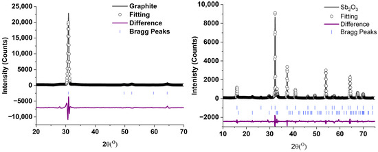
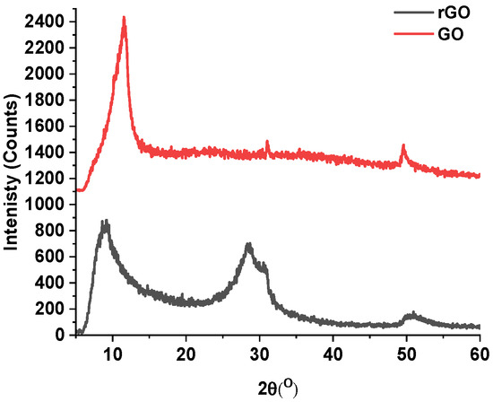
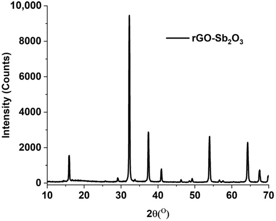
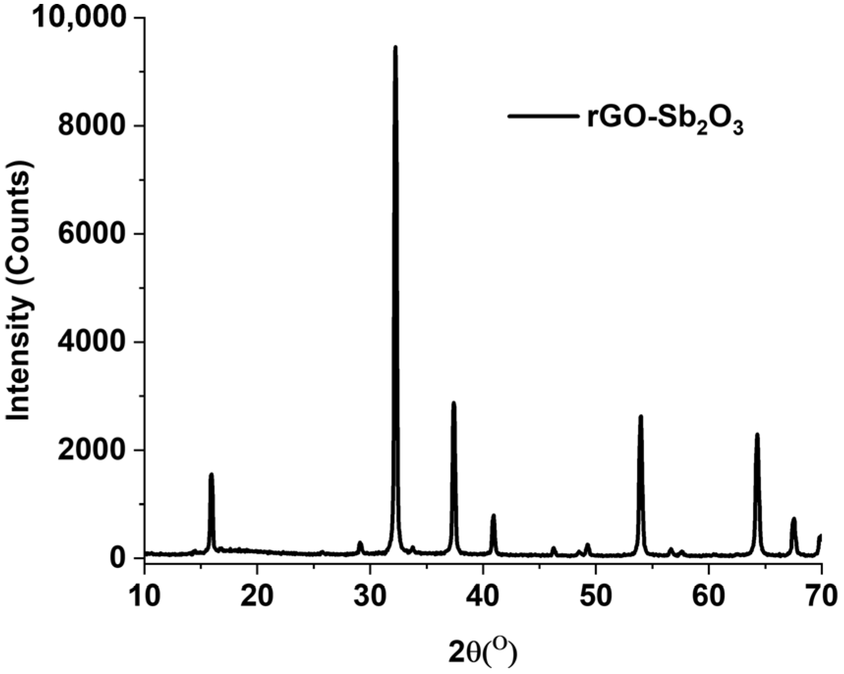
X-ray diffraction (XRD) was used to investigate the crystallographic structure, phase composition, and structural changes in pure graphite, rGO, pure Sb2O3, and the rGO/Sb2O3 1:1 composite. The diffraction patterns are shown in Figure 1, Figure 2 and Figure 3.
Figure 1.
The LeBail fitting of graphite and Sb2O3 starting materials.
Figure 2.
XRD patterns of the synthesized GO and rGO.
Figure 3.
XRD patterns of the rGO-Sb2O3 electrode material.
The pure graphite showed a sharp (002) reflection at 2θ ≈ 31°, consistent with highly crystalline graphite reported in the literature using a cobalt source [68]. The pure Sb2O3 displayed sharp and intense reflections at 2θ ≈ 28.5°, 33.5°, 41.9°, and 51.1°, characteristic of the monoclinic valentinite phase and consistent with standard references [69].
The fitting of the XRD of the Sb2O3 starting material showed the presence of two phases, a cubic and an orthorhombic phase, as can be seen in Table 1. The Cubic phase of the Sb2O3 was the major phase, whereas the orthorhombic phase was the minor phase. The fitting was found to have a reduced χ2 of 3.15, indicating a good fitting of the experimental data. The graphite starting material was observed to be single-phase hexagonal, and the fitting showed a reduced χ2 of 1.85, indicating a good agreement between the XRD pattern and the literature.
Table 1.
LeBail fitting results and crystal lattice parameters of the graphite and the Sb2O3 starting materials, and the corresponding literature crystal lattice.
In contrast, graphene oxide exhibited a broad (001) peak at 2θ ≈ 10–11°, confirming oxidation and exfoliation through the intercalation of oxygen functional groups, in agreement with previous studies [68]. Graphene oxide and reduced graphene oxide do not have crystal lattices or consistent crystal lattices; the lattice varies based on the movement of the c-axis, which in graphite is termed the 002 plane and in GO and rGO is 001. Figure 2 shows the XRD of the synthesized GO and rGO. Based on calculations in the present study, the GO and rGO have a 002 plane corresponding to a c-axis in the crystal of 19.8 and 21.5 Å, respectively. In the graphene oxide diffraction pattern, the 001 diffraction plane is clearly visible, while the 002 diffraction plane is almost completely missing. In the rGO sample, the 001 and 002 diffraction planes are almost the same height; this is due to the thermal reduction in some of the functional groups between the layers, which allows the structure to collapse slightly back to the 6.70(1) lattice parameter of the original graphite.
The rGO/Sb2O3 diffraction pattern only shows the diffraction peaks due to the Sb2O3 were observable due to the peak positioning and the crystallinity of the Sb2O3 phase. The peaks present for the rGO are in the same position as the Sb2O3 material. These results indicate that the structures of the individual synthesized materials are in agreement with those reported in the literature, validating the successful preparation of rGO and potentially the Sb2O3 composite, which was light gray in color.
3.2. SEM Analysis
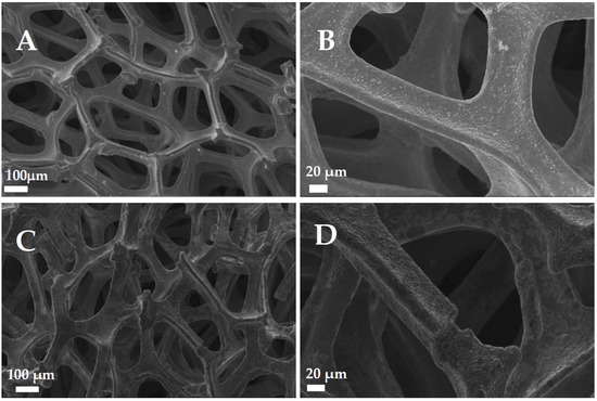
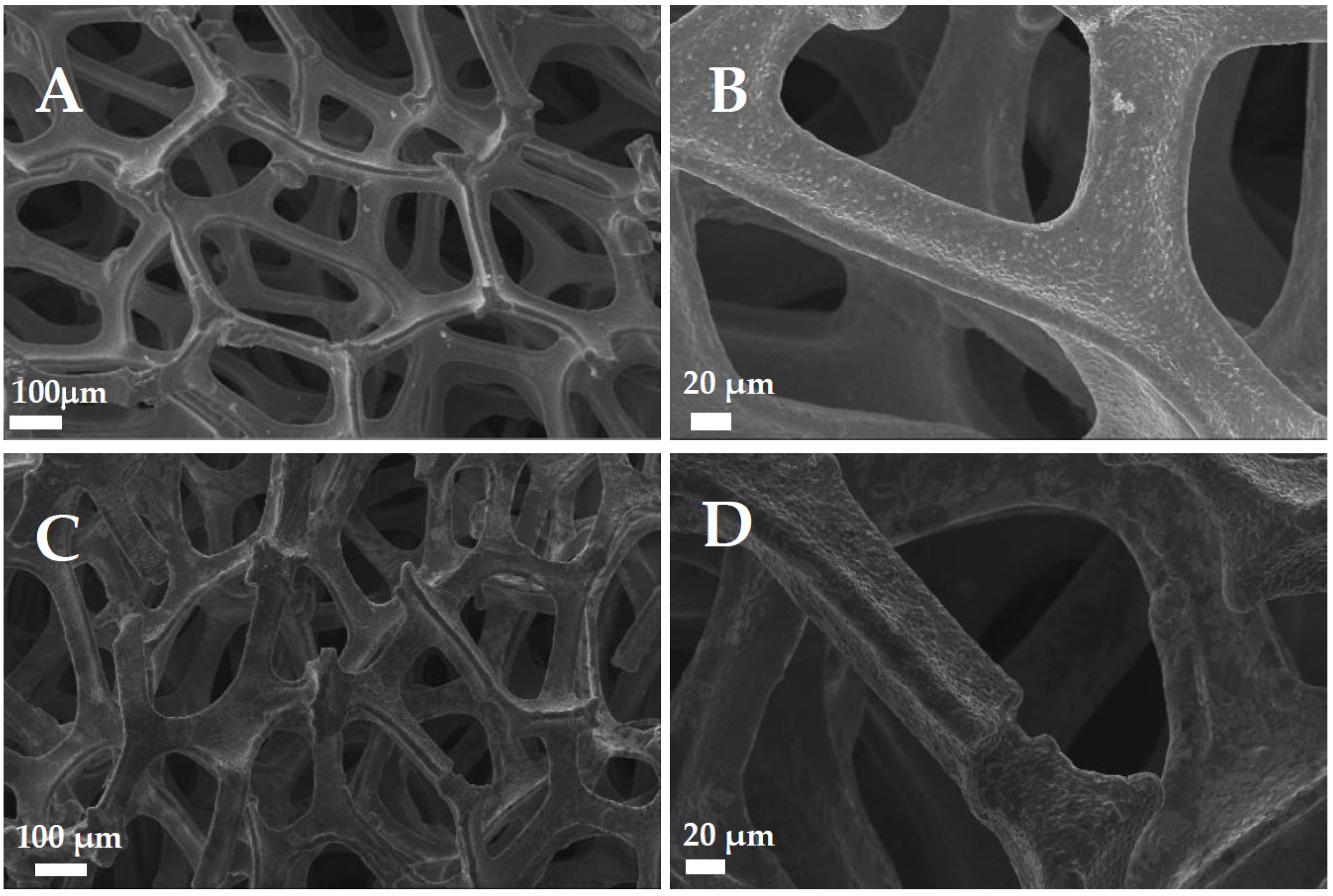
Figure 4 shows the SEM micrographs of bare copper foam before and after the reaction. Figure 4A,C show low-magnification images of an open, three-dimensional porous network with irregular honeycomb patterns. The voids are typically in the range of 150–250 μm. Initially, the struts are well-defined with a relatively smooth surface. After the reaction with MB, the overall architecture is preserved, but clear signs of structural degradation are observed, as shown in Figure 4C. Several parts of the honeycomb structure appear broken or thinned, and the networked openings are larger and less uniform than in the sample before the reaction with MB. The sides making up the structure have an etched and uneven contour, as shown in Figure 4D. The surface appears eroded, suggesting material loss and presenting a rougher surface.
Figure 4.
SEM copper mesh base for electrode before reaction, shown at various magnifications (A,B), and copper mesh base for electrode after reaction, shown at various magnifications (C,D).
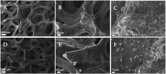
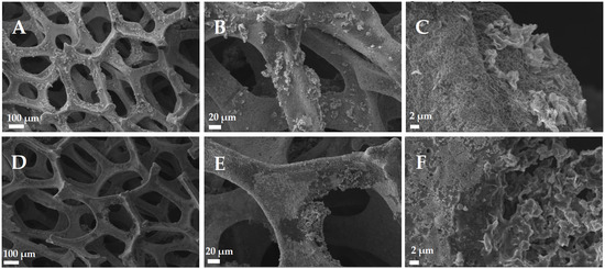
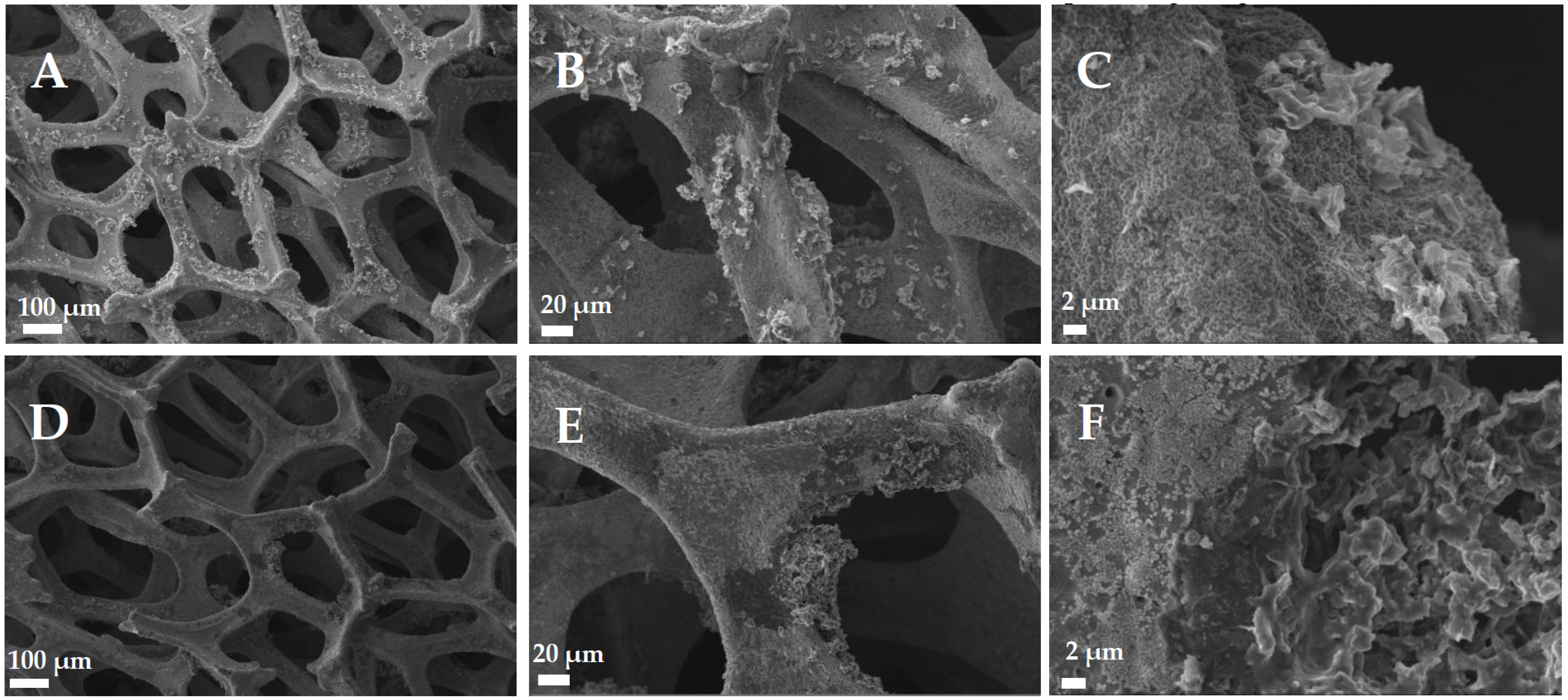
The SEM micrographs in Figure 5A–C show the morphology of the Sb2O3-coated copper before the reaction at different magnifications. In Figure 5A, the material displays a well-preserved three-dimensional porous architecture of the underlying copper foam. From the low-magnification view, a thin coating can be observed over the copper foam, but the fine surface details are not clearly resolved. At intermediate magnification, the presence of the Sb2O3 layer becomes evident: the surface appears coarse, indicating the deposition of ink-oxide material over the metallic framework. At higher magnification, the coating reveals a stratified morphology, where agglomerated Sb2O3 particles have coalesced into a continuous but uneven layer across the copper surface. The oxide appears non-uniform in thickness, forming lump-like deposits distributed irregularly. This morphology is consistent with previous reports on solution-grown Sb2O3 films, where rounded particles progressively merge to form an agglomerated granular coating [73].
Figure 5.
SEM of Sb2O3 ink-based electrode before reaction, shown at various magnifications (A–C), and Sb2O3 composite ink-based electrode after reaction, shown at various magnifications (D–F).
Figure 5D–F shows the SEM micrographs of the Sb2O3-coated copper electrodes after the reaction with MB. At low magnification Figure 5D, the porous network appears clean and smooth, indicating a notable reduction in the oxide coating. The overall three-dimensional architecture of the copper mesh is preserved, but the surface shows substantial loss of the previously observed Sb2O3 material with no evident difference observed between the inner and outer regions of the structure.
At intermediate magnification Figure 5E, the oxide no longer forms a continuous film along the copper foam but instead appears as patchy deposits of fine granular material distributed irregularly across the surface. The exposed copper mesh looks compact, showing no signs of porosity or erosion.
At higher magnification Figure 5F, the remaining Sb2O3 exhibits a particle-based degradation pattern. The former granular coating has broken down into discrete fine particles, suggesting that the oxide was removed gradually in the form of small particles rather than as continuous layers. The resulting surface consists of small, rounded Sb2O3 grains sparsely distributed over the copper framework, consistent with mechanical or chemical removal of the oxide coating throughout the reaction process.
The SEM micrographs in Figure 6A–C show the morphology of the rGO-coated copper before the reaction with MB at different magnifications, while Figure 6D–F presents the corresponding images after the reaction.
Figure 6.
SEM of rGO ink-based electrode before reaction, shown at various magnifications (A–C), and the rGO ink-based electrodes after reaction, shown at various magnifications (D–F).
At low magnification, Figure 6A, the honeycomb-like copper framework is well preserved, showing a regular three-dimensional architecture similar to the bare copper mesh. Compared with the Sb2O3-coated sample, the surface here exhibits a rougher and more textured appearance, suggesting that the rGO coating forms a granular or patchy coverage along the copper struts rather than a continuous film. The coating is visible following the contours of the metallic structure, and its brightness varies locally, indicating non-uniform deposition with localized accumulations of rGO.
At intermediate magnification, Figure 6B, the rGO becomes more distinguishable. The surface appears coarse and irregular, with thin, patchy deposits made of compact granular features, consistent with aggregated rGO domains that settled unevenly during deposition.
In Figure 6C, the granular nature of the coating is more evident. The surface is densely populated with 0.5–1.0 µm clusters, forming a heterogeneous, textured layer of aggregated rGO. The coating does not appear smooth or continuous but rather irregular and compact, consistent with the restacking and aggregation of rGO reported by Tang et al. [74].
After the reaction with MB, the morphology changes markedly. At the highest magnification, Figure 6D, the bare copper substrate appears largely free of coating, showing smooth and continuous portions of the copper foam with no evidence of corrosion or pitting. In several regions, however, residual clusters of rGO remain attached to the surface, visible as bright, granular islands. In Figure 6E, the electrode surface appears to have roughened areas and localized agglomerates. At the highest magnification, Figure 6F, the surface exhibits numerous small, dispersed particulates, together with occasional larger granules concentrated in specific regions, indicating that the rGO layer was partially removed and redistributed during the reaction.
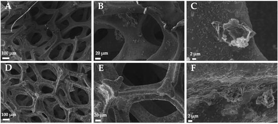
The SEM micrographs in Figure 7A–C show the morphology of the rGO/Sb2O3 composite-coated copper before the reaction with MB, while Figure 7D–F present the same electrode after the reaction at corresponding magnifications.
Figure 7.
SEM of rGO-Sb2O3 composite ink-based electrode before reaction, shown at various magnifications (A–C), and rGO-Sb2O3 composite ink-based electrode after reaction, shown at various magnifications (D–F).
At low magnification, Figure 7A, the copper network retains its open, interconnected three-dimensional architecture. The composite coating is heterogeneously distributed, forming irregular accumulations mainly at the junctions of the metallic network. In Figure 7B, the coating exhibits clusters and fragments adhering to the struts in a discontinuous manner, reflecting an uneven co-deposition of rGO and Sb2O3. At higher magnification, Figure 7C, the composite consists of agglomerated granules and thin flakes, suggesting layered accumulation of Sb2O3 particles interlaced with rGO.
After the reaction with MB, the overall framework remains well preserved. The surface morphology, however, becomes rougher and less compact, as seen in Figure 7E,F. The large composite bundles appear partially degraded, leaving behind a surface of fine dispersed residues distributed over the copper surface. The Cu foam remains relatively smooth and continuous, with no evidence of structural deterioration, indicating that the observed roughness arises from progressive disintegration of the rGO–Sb2O3 layer rather than from substrate erosion. Fine particulates seen in Figure 7F likely correspond to fragmented remnants of the composite, which appear to remain loosely attached after the reaction, suggesting partial disintegration of the coating rather than complete removal.
Table 2 and Table 3 show the results of the EDS analysis of the electrodes before and after reaction with MB and shows the presence of C in all the samples, as would be expected. All the samples contained carbon at the beginning due to the presence of the Nafion polymer component of the dye. In addition, all the samples showed a significant amount of Cu, as was expected due to the copper mesh support. Moreover, the samples should have antimony present. However, there is some loss of Sb after reaction; some of the decrease is due to the dilution of the sample with carbon, as the amount of carbon present in the samples has increased after reaction, which may be due to adsorption of the dye after reaction or adsorption of some of the reaction products. Interestingly, almost 95% of Sb2O3 was lost after reaction using the Sb2O3 electrode. However, the loss was much less significant in the rGO-Sb2O3 electrode, which was approximately 50% (not accounting for dilution of the sample). The data suggest the rGO has a stabilizing effect on the Sb2O3 during the reaction.
Table 2.
EDS results of the synthesized electrodes before application in the electrochemical degradation of MB at a current density of 0.1 A/cm2.
Table 3.
EDS results of the synthesized electrodes after application in the electrochemical degradation of MB at a current density of 0.1 A/cm2.
3.3. MB Removal Efficiency
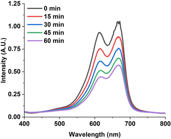
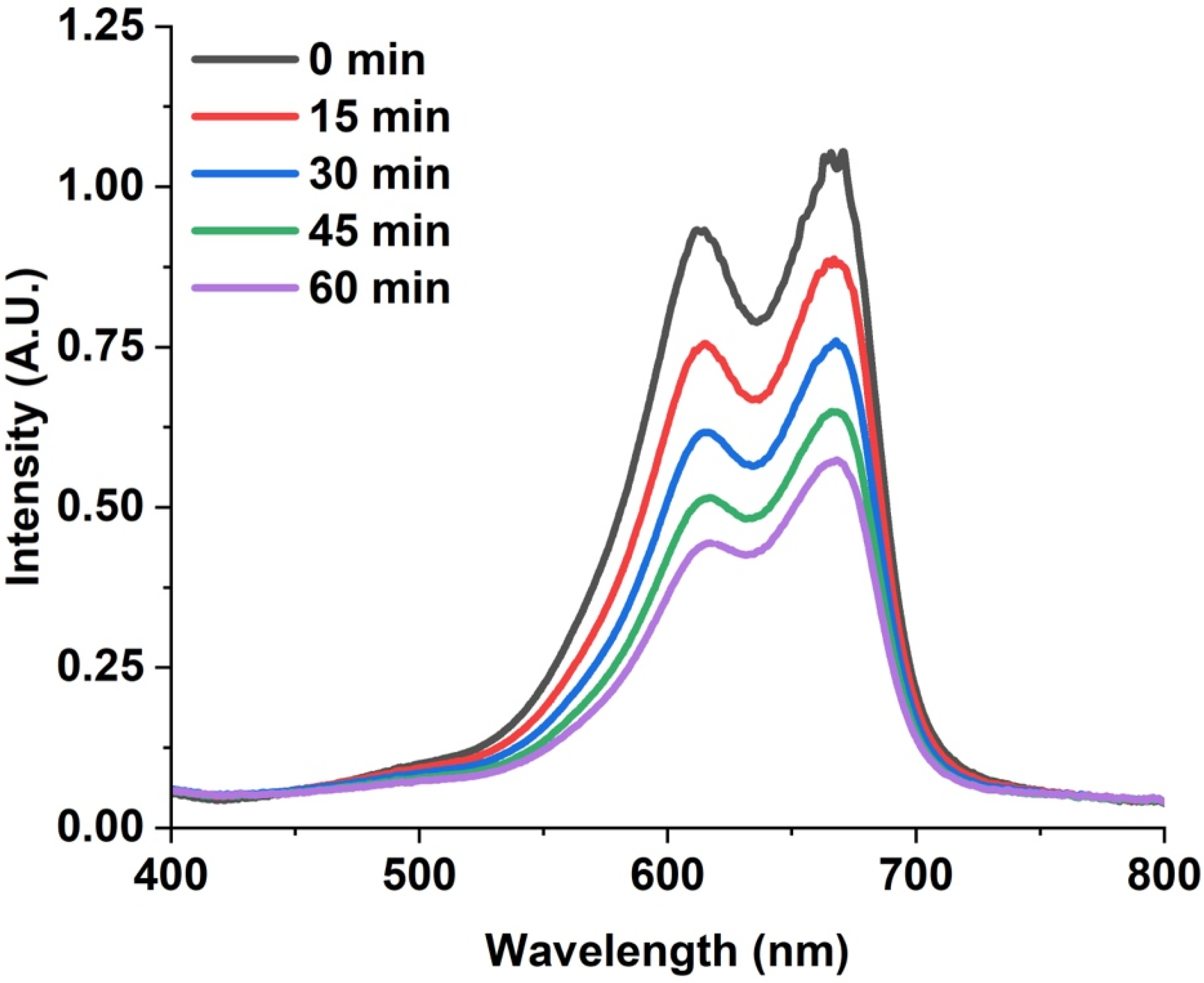
Figure 8 shows a sample of the decrease in the absorption of the MB solution at pH 3 during the electrocatalytic degradation using the rGO-Sb2O3 electrode. As can be seen in the figure, the intensity of the absorption spectra of the MB decreases as the reaction proceeds with time.
Figure 8.
UV-Vis spectrum of MB at pH 3 during the electrocatalytic degradation using the rGO-Sb2O3 electrode.
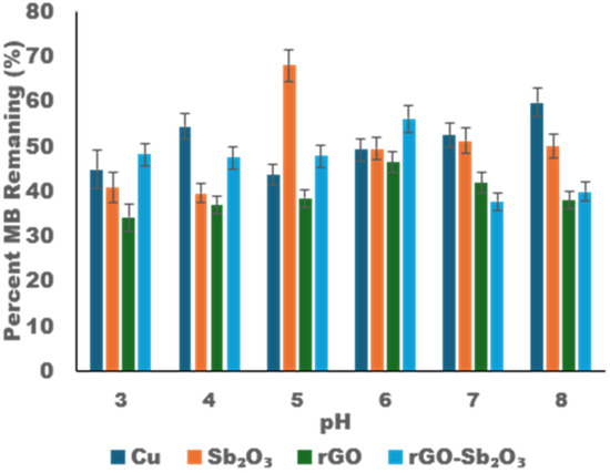
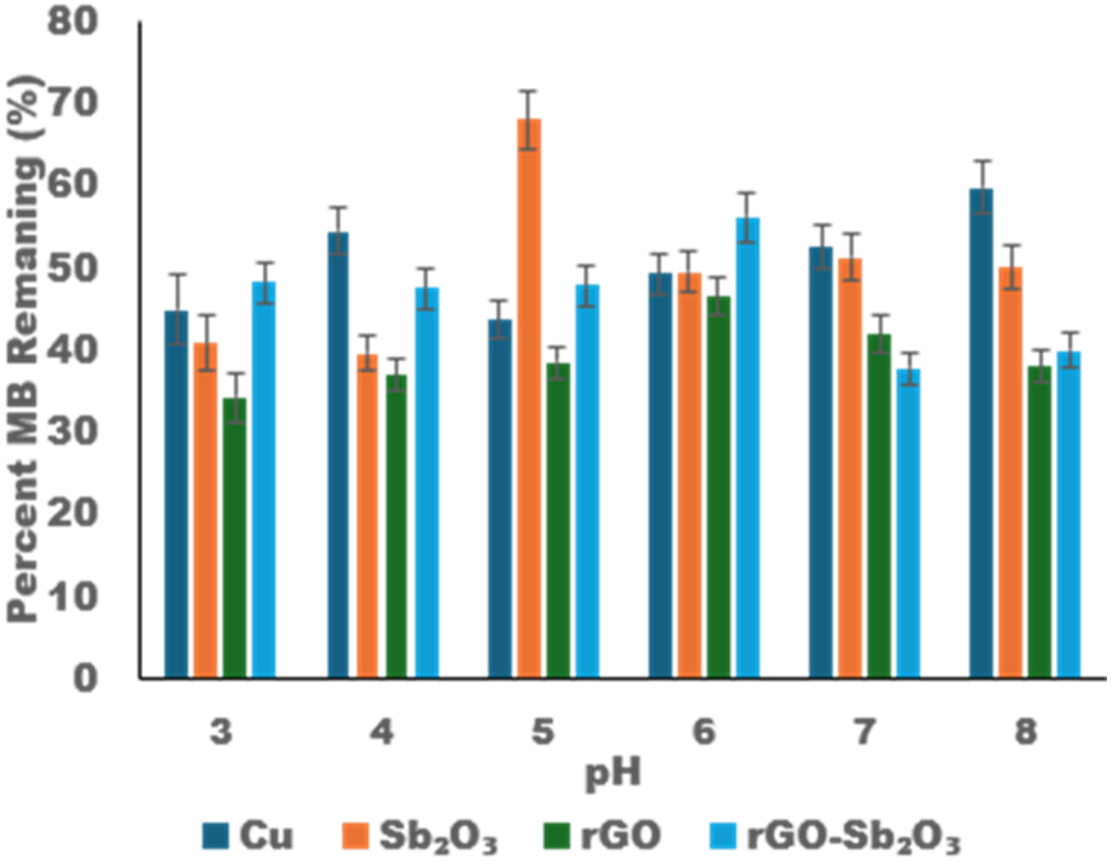
The removal of MB varies with pH, as illustrated in Figure 9, where the amount of MB remaining in solution after one hour of reaction is presented for experiments using a Na2SO4 electrolyte. The studies were performed to avoid the presence of Cl−, which has been shown to generate reactive species that do enhance the reaction rates but are not truly representative of the electrode behavior [75,76]. It should also be noted that pH 2 was avoided due to potential electrode degradation. In addition, dye removal from wastewater is generally higher at pH 7 and above [77]. However, some wastewater treatment processes reduce the pH to around 4–5 [78,79]. The bare copper showed the lowest removal at almost all pH levels for the tested electrodes. The Sb2O3 was observed to work best at lower pH, around 3 and 4, and the performance was lowest at pH 5. Whereas the rGO consistently worked better at lower pHs and became slightly worse at higher pHs. pH 3 showed the lowest amount of MB remaining in solution at approximately 34% remaining, or 66% of the MB was removed. The rGO-Sb2O3 electrode demonstrated the best removal at pH 7 and pH 8, with only 38% of the MB dye remaining in solution, indicating a 62% removal. Of the tested electrodes, the rGO and rGO-Sb2O3 appear to work the best; this may be due to the ability of the rGO phase to interact and initially adsorb the dye, and thus enhance the electrode–dye interaction and result in a higher removal of the MB from solution, as has been indicated in the literature [80,81]. These values are within reason for the reaction time. Luan et al. showed removal of MB ranging between 50 and 70% (C/Co) using carbon nanotubes/carbon fibers at a pH between 1.8 and 8.19 and current densities of 10–100 mA/cm2 after 3 h of reaction [82]. However, within the first hour of reaction, they observed in a 10 ppm solution of MB, approximately 20–50% remaining in solution. Using a Flexabel Graphite electrode, Goren showed a removal percentage of up to 100% at pH 4 and found that the removal percentage decreased as pH increased [83]. Yao et al. showed removal efficiencies of 100% and 73% after 2 h of reaction using a PbO2-ZrO2 electrode in Na2SO2 with current densities of 50 mA/cm2 [60]. Hu et al. used a SnO2 anode, which was promoted with Pb, which had removals of 94% at 10% Pb doping and a Na2SO4 electrolyte, and current densities between 20 and 60 mA/cm2 [84]. Using a graphite rod–iron rod system, Teng et al. showed removals of 25–37% with a Na2SO4 supporting electrolyte [81]. Ti4O7 electrodes showed removal of up to 100% in 40 min in NaCl electrolyte and current densities of 5–15 mA/cm2 [85]. Nleonu et al. showed MB removal of approximately 53% in a solution of 80 ppm of MB and a reaction time of 40 min [75].
Figure 9.
Percent MB remaining in solution after 1 h of reaction at pH 3 through 8.
3.4. MB Degradation Kinetics
The degradation kinetics of MB were evaluated by fitting concentration–time data to a pseudo-first-order model across the pH range of 2 to 8. The reliability of this model is consistent with earlier studies on ZnO photocatalysts [86], graphene oxide composites [87], and UV-driven degradation systems [88].
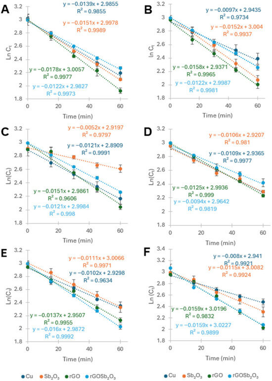
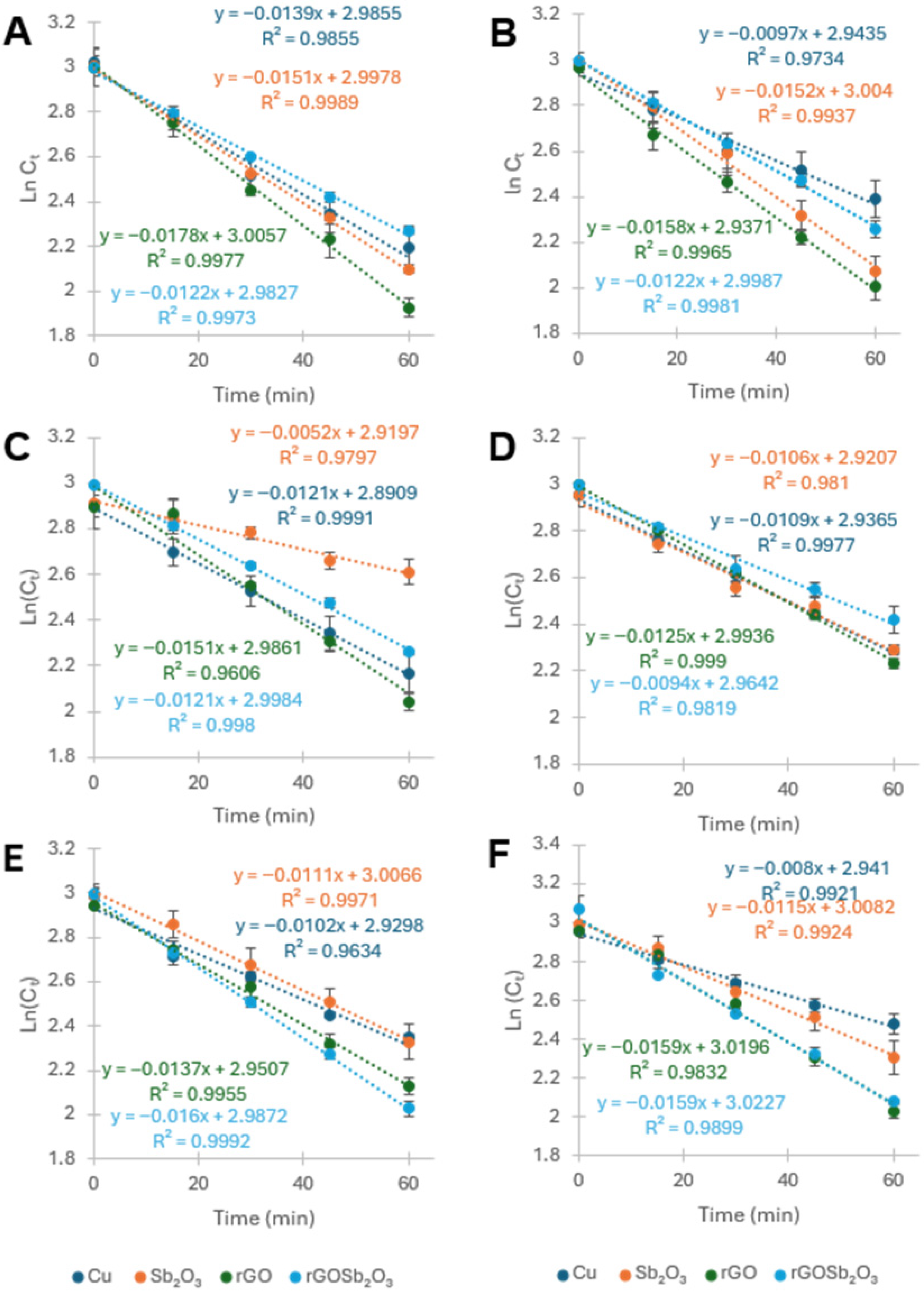
Linearized kinetics plots were generated by plotting ln(Cₜ) versus time. The plots showed strong correlation coefficients, for the most part, 0.99 or greater. A few of the plots show slightly lower correlation coefficients, 0.96–0.98; the few plots below the 0.99 correlation coefficient were not sufficient to change the fitting model. The MB degradation process for all electrodes was observed to follow first-order kinetics. Figure 10A–F shows the linearized plots for the various electrodes, and Table 4 summarizes the kinetic data from the various reactions. The slopes of the lines were the rate constants (k, min−1) for the various electrodes.
Figure 10.
Kinetics plots for the degradation of MB using the copper base electrode, Sb2O3 electrode, rGO electrode, and the rGO-Sb2O3 composite electrode at pH 3 (A), pH 4 (B), pH 5 (C), pH 6 (D) pH 7 (E), and pH 8 (F).
The kinetic results for MB degradation, summarized in Table 5, confirm that all electrodes followed pseudo-first order behavior with high linearity in ln(C0/Cₜ) versus time plots. Among the individual systems, Sb2O3 showed some of the highest catalytic activity under acidic conditions, although performance declined markedly at neutral and basic pH, more than likely due to surface passivation by hydroxide species. rGO performed consistently well across all pH conditions, reflecting its high conductivity and surface area, but lacked the catalytic enhancement provided by metal oxides. Bare copper foam showed the lowest activity overall, with minor activity in acidic media attributed to surface redox processes. The rGo-Sb2O3 composite material showed relatively good rates at all pHs; however, the rGO-Sb2O3 composite material presents higher rate constants at higher pHs. The present materials have a comparable rate to some of the materials in the literature, as can be seen in Table 2. In some cases, the rGo-Sb2O3 composite had a higher rate constant, such as the CNT/CF K 0.00483 min−1 [82]. For the most part, the current materials showed lower rate constants than the literature values; more than likely, this was due to the electrolyte/current density properties. The current electrodes show a rate constant of ½ to 1/10 of those shown in the literature, as can be seen in Table 2. The literature values typically are much higher than those of the present study, which utilized NaCl as the electrolyte, which is known to catalyze the reaction by generating reactive chloride [76,89]. The relationship between rate constant and electrode becomes very relevant with respect to the electrolyte; the SO42− is not known to enhance the reaction by generating secondary reactive species and, thus, should give a better representation of the electrode and not the complete system, i.e., the generation of secondary reactive species involving chloride.
Table 4.
Observed rate constants for the electrocatalytic/electrochemical degradation of MB from aqueous solution [60,75,81,82,83,84,85,90,91].
Table 5.
Summary of the kinetic data collected for the Cu base electrode, Sb2O3, rGO, and rGO-Sb2O3 composite electrode from pH 3 to pH 8.
4. Conclusions
The electrode materials were successfully synthesized and structurally characterized using XRD analysis. The Sb2O3 starting material exhibited both cubic and orthorhombic phases, with the cubic phase being predominant. The graphite precursor was identified as a single-phase hexagonal material, showing high crystallinity and excellent agreement with standard references. As expected, GO presented a 001 diffraction plane with an almost absent 002 plane. The rGO samples present the characteristic 002 plane, and due to thermal reduction occurring between layers, the 001 plane is also present. In the rGO-Sb2O3 composite, the diffraction peaks were mainly associated with the Sb2O3 phase, indicating that the oxide dominates the crystalline framework.
This study demonstrated the efficient electrocatalytic degradation of MB using Sb2O3, rGO, and rGO-Sb2O3 composite electrodes supported on copper foam. Among all systems tested, the rGO-Sb2O3 composite showed the most stable and effective performance across different pH conditions, achieving nearly complete dye removal at low pH. The enhanced activity resulted from the synergistic effect between the high conductivity and surface area of rGO and the catalytic properties of Sb2O3.
These results highlight rGO-Sb2O3 composite electrodes as a promising, scalable, and sustainable option for wastewater treatment. Their high electrochemical performance, strong degradation efficiency, and stability across a wide pH range make them suitable for real-water applications targeting synthetic dyes and other emerging contaminants.
Author Contributions
Conceptualization, J.G.P. and H.M.M.; Methodology, H.M.M.; Formal analysis, M.I.M.A., A.M.F., T.M.E., J.G.P. and H.M.M.; Investigation, M.I.M.A. and T.M.E.; Data curation, H.M.M.; Writing—original draft, M.I.M.A. and A.M.F.; Writing—review and editing, A.M., J.G.P. and H.M.M.; Supervision, H.M.M.; Project administration, J.G.P. and H.M.M.; Funding acquisition, J.G.P. and H.M.M. All authors have read and agreed to the published version of the manuscript.
Funding
This research was funded by Welch Foundation grant number [BX-0048].
Institutional Review Board Statement
Not applicable.
Data Availability Statement
The data presented in this study are available on request from the corresponding author.
Conflicts of Interest
The authors declare no conflicts of interest.
References
- Tripathi, M.; Singh, S.; Pathak, S.; Kasaudhan, J.; Mishra, A.; Bala, S.; Garg, D.; Singh, R.; Singh, P.; Pathak, N.; et al. Recent Strategies for the Remediation of Textile Dyes from Wastewater: A Systematic Review. Toxics 2023, 11, 940. [Google Scholar] [CrossRef] [Scilit]
- Khan, I.; Saeed, K.; Zekker, I.; Zhang, B.; Hendi, A.H.; Ahmad, A.; Ahmad, S.; Zada, N.; Ahmad, H.; Shah, L.A.; et al. Review on Methylene Blue: Its Properties, Uses, Toxicity and Photodegradation. Water 2022, 14, 242. [Google Scholar] [CrossRef] [Scilit]
- Jia, P.; Tan, H.; Liu, K.; Gao, W. Removal of Methylene Blue from Aqueous Solution by Bone Char. Appl. Sci. 2018, 8, 1903. [Google Scholar] [CrossRef] [Scilit]
- Oladoye, P.O.; Ajiboye, T.O.; Omotola, E.O.; Oyewola, O.J. Methylene blue dye: Toxicity and potential elimination technology from wastewater. Results Eng. 2022, 16, 100678. [Google Scholar] [CrossRef] [Scilit]
- Aniagor, C.; Aly, A.; Mohamed, L.A.; Hashem, A. Removal of Methylene blue dye from contaminated wastewater using lignocellulosic biomasses: A comparative study. Waste Manag. Bull. 2024, 2, 213–225. [Google Scholar] [CrossRef] [Scilit]
- Bužga, M.; Machytka, E.; Dvořáčková, E.; Švagera, Z.; Stejskal, D.; Máca, J.; Král, J. Methylene blue: A controversial diagnostic acid and medication? Toxicol. Res. 2022, 11, 711–717. [Google Scholar] [CrossRef] [Scilit] [PubMed]
- Recipi, A.; Di Stefano, A.; Radicioni, M.; Jas, V.; Moro, L.; Danese, S. Methylene blue MMX® tablets for chromoendoscopy. Safety tolerability and bioavailability in healthy volunteers. Contemp. Clin. Trials 2012, 33, 260–267. [Google Scholar] [CrossRef] [Scilit] [PubMed]
- Patel, B.Y.; Patel, H.K. Current approaches toward the removal of methylene blue dye from synthetic textile effluent using bacterial treated agricultural waste absorbent through statistical design. Heliyon 2023, 9, e19857. [Google Scholar] [CrossRef] [Scilit] [PubMed]
- Pasin, L.; Umbrello, M.; Greco, T.; Zambon, M.; Pappalardo, F.; Crivellari, M.; Borghi, G.; Morelli, A.; Zangrillo, A.; Landoni, G. Methylene blue as a vasopressor: A meta-analysis of randomised trials. Crit. Care Resusc. 2013, 15, 42–48. [Google Scholar] [CrossRef] [Scilit]
- Fernandes Junior, H.J.; de Araújo, E.A.; Machado Junior, J.A.; Lutz Motta, F.M.; Guarize, G.F.; Cheng, L.C.; Tantray, J.; Medeiros, J.V.R.; Nicolau, L.A.D.; Taha, M.; et al. Cardiotoxic and Cardioprotective Effects of Methylene Blue in the Animal Model of Cardiac Ischemia and Reperfusion. Biomedicines 2024, 12, 2575. [Google Scholar] [CrossRef] [Scilit]
- Ramsay, R.R.; Dunford, C.; Gillman, P.K. Methylene blue and serotonin toxicity: Inhibition of monoamine oxidase A (MAO A) confirms a theoretical prediction. Br. J. Pharmacol. 2007, 152, 946–951. [Google Scholar] [CrossRef] [Scilit]
- Gillman, P.K. CNS toxicity involving methylene blue: The exemplar for understanding and predicting drug interactions that precipitate serotonin toxicity. J. Psychopharmacol. 2011, 25, 429–436. [Google Scholar] [CrossRef] [Scilit]
- Hazekamp, C.; Schmitz, Z.; Scoccimarro, A. Methylene Blue-Induced Serotonin Toxicity: Case Files of the Medical Toxicology Fellowship at the New York City Poison Control Center. J. Med. Toxicol. 2024, 20, 54–58. [Google Scholar] [CrossRef] [Scilit]
- Rehman, H.U. Methemoglobinemia. West. J. Med. 2001, 175, 193–196. [Google Scholar] [CrossRef] [Scilit] [PubMed]
- Alyahya, B.; Alalshaikh, A.; Sabbahi, G.; Alnowiser, M.; Al-Mohawes, M. Methylene Blue Infusion to Treat Severe Dapsone-Induced Methemoglobinemia in a Pediatric Patient. Cureus 2021, 13, 10. [Google Scholar] [CrossRef] [Scilit]
- Pjetraj, D.; Mechri, M.E.; Bacelli, S.; Fabiani, E.; Caponi, L.; Gatti, S.; Lionetti, E. A blue blood toddler- a case report of methemoglobinemia and literature review. Ital. J. Pediatr. 2025, 51, 53. [Google Scholar] [CrossRef] [Scilit]
- Özgör, F.; Şimşek, A.; Savun, M.; Sarılar, Ö.; Akbulut, M.F.; Gürbüz, Z.G. Green urine color due to methylene blue. New J. Urol. 2017, 12, 63–65. [Google Scholar]
- Lam, C.W.; Wong, S.Y. A case of green urine due to a traditional Chinese medicine containing methylene blue. N. Z. Med. J. 2010, 123, 71–76. [Google Scholar]
- Rosique, M.J.; Rosique, R.G.; Farias, F.M.; Oliveria, C.C.; Farina, J.A.; Évora, P.R. Methylene blue reduces progression of burn and increases skin survival in an experimental rat model. Burns 2017, 43, 1702–1708. [Google Scholar] [CrossRef] [Scilit]
- Salhab, M.; Al Sarakbi, W.; Mokbel, K. Skin and fat necrosis of the breast following methylene blue dye injection for sentinel node biopsy in a patient with breast cancer. Int. Semin. Surg. Oncol. 2005, 2, 26. [Google Scholar] [CrossRef] [Scilit]
- Adesanmi, B.; Hung, Y.; Paul, H.; Huhnke, C. Comparison of dye wastewater treatment methods: A review. GSC Adv. Res. Rev. 2022, 10, 126–137. [Google Scholar] [CrossRef] [Scilit]
- Sardar, M.; Manna, M.; Maharana, M.; Sen, S. Remediation of Dyes from Industrial Wastewater Using Low-Cost Adsorbents. In Green Adsorbents to Remove Metals, Dyes and Boron from Polluted Water; Springer International Publishing: Cham, Switzerland, 2021; Volume 49, p. 277. [Google Scholar]
- Shao, X.; Wang, J.; Liu, Z.; Hu, N.; Liu, M.D.; Zhang, R.; Quan, C. Cellulose based cation-exchange fiber as filtration material for the rapid removal of methylene blue from wastewater. Cellulose 2021, 28, 9355–9367. [Google Scholar] [CrossRef] [Scilit]
- Li, Q.; Li, Y.; Ma, X.; Du, Q.; Sui, K.; Wang, D.; Wang, C.; Li, H.; Xia, Y. Filtration and adsorption properties of porous calcium alginate membrane for methylene blue removal from water. Chem. Eng. J. 2017, 316, 623–630. [Google Scholar] [CrossRef] [Scilit]
- Pham, D.C.; Cao, T.M.; Nguyen, M.C.; Nguyen, T.D.; Nguyen, V.H.; Bui, V.H.; Nguyen, T.T. Integrating Photocatalysis and Microfiltration for Methylene Blue Degradation: Kinetics and Cost Estimation. Chem. Eng. Technol. 2022, 45, 1748–1758. [Google Scholar] [CrossRef] [Scilit]
- Ali, H.; Mansor, S.E.; Taha, G.M. Microfiltration and adsorptive membranes for simultaneous removal of methyl orange and methylene blue using hybrid composites. Polym. Bull. 2022, 79, 7891–7908. [Google Scholar]
- Bielska, M.; Szymanowski, J. Removal of methylene blue from waste water using micellar enhanced ultrafiltration. Water Res. 2006, 40, 1027–1033. [Google Scholar] [CrossRef] [Scilit]
- Zaghbani, N.; Hafiane, A.; Dhahbi, M. Separation of methylene blue from aqueous solution by micellar enhanced ultrafiltration. Sep. Purif. Technol. 2007, 55, 117–124. [Google Scholar] [CrossRef] [Scilit]
- Zheng, L.; Su, Y.; Wang, L.; Jiang, Z. Adsorption and recovery of methylene blue from aqueous solution through ultrafiltration technique. Sep. Purif. Technol. 2009, 68, 244–249. [Google Scholar] [CrossRef] [Scilit]
- Zhang, L.; Zaoui, A.; Sekkal, W. Adsorption efficiency of highly methylene blue dye concentrations with multilayer chitosan-modified clays for a precise nanofiltration performance of polluted water. J. Water Process Eng. 2024, 57, 104651. [Google Scholar]
- Cheng, S.; Oatley, D.L.; Williams, P.M.; Wright, C.J. Characterisation and application of a novel positively charged nanofiltration membrane for the treatment of textile industry wastewaters. Water Res. 2012, 46, 33–42. [Google Scholar] [CrossRef] [Scilit] [PubMed]
- Agarwala, R.; Mulky, L. Adsorption of Dyes from Wastewater: A Comprehensive Review. ChemBioEng Rev. 2023, 10, 326–335. [Google Scholar] [CrossRef] [Scilit]
- Dutta, S.; Adhikary, S.; Bhattacharya, S.; Roy, D.; Chatterjee, S.; Chakraborty, A.; Banerjee, D.; Ganguly, A.; Nanda, S.; Rajak, P. Contamination of textile dyes in aquatic environment: Adverse impacts on aquatic ecosystem and human health, and its management using bioremediation. J. Environ. Manag. 2024, 353, 120103. [Google Scholar] [CrossRef] [Scilit] [PubMed]
- Al-Baldawi, I.A.; Abdullah, S.R.; Anuar, N.; Hasan, H.A. Phytotransformation of methylene blue from water using aquatic plant (Azolla pinnata). Environ. Technol. Innov. 2018, 11, 15–22. [Google Scholar] [CrossRef] [Scilit]
- Imron, M.F.; Kurniawan, S.B.; Soegianto, A.; Wahyudianto, F.E. Phytoremediation of methylene blue using duckweed (Lemna minor). Heliyon 2019, 5, e02206. [Google Scholar] [CrossRef] [Scilit] [PubMed]
- Tan, K.A.; Morad, N.; Ooi, J. Phytoremediation of Methylene Blue and Methyl Orange Using Eichhornia crassipes. Int. J. Environ. Sci. Dev. 2016, 7, 724–728. [Google Scholar] [CrossRef] [Scilit]
- Mansour, A.; Alprol, A.E.; Abualnaja, K.; El-Beltagi, H.S.; Ramadan, K.; Ashour, M. Dried Brown Seaweed’s Phytoremediation Potential for Methylene Blue Dye Removal from Aquatic Environments. Polymers 2022, 14, 1375. [Google Scholar] [CrossRef] [Scilit]
- Urucu, O.A.; Garosi, B.; Musah, R.A. Efficient Phytoremediation of Methyl Red and Methylene Blue Dyes from Aqueous Solutions by Juncus effusus. ACS Omega 2025, 10, 1943–1953. [Google Scholar] [CrossRef] [Scilit]
- Modi, S.; Yadav, V.K.; Amari, A.; Osman, H.; Igwegbe, C.A.; Fulekar, M.H. Nanobioremediation: A bacterial consortium-zinc oxide nanoparticle-based approach for the removal of methylene blue dye from wastewater. Environ. Sci. Pollut. Res. 2023, 30, 72641–72651. [Google Scholar] [CrossRef] [Scilit]
- Sarioglu, O.F.; Keskin, N.O.; Celebioglu, A.; Tekinay, T.; Uyar, T. Bacteria encapsulated electrospun nanofibrous webs for remediation of methylene blue dye in water. Colloids Surf. B Biointerfaces 2017, 152, 245–251. [Google Scholar] [CrossRef] [Scilit]
- Purnomo, A.S.; Fauzany, U.S.; Rizqi, H.D.; Alkas, T.R.; Kamei, I. Biodecolorization and biotransformation of methylene blue using mixed cultures of brown-rot fungus Daedalea dickinsii and filamentous fungus Aspergillus oryzae: Identification of metabolites and degradation pathway. RSC Adv. 2024, 14, 5061–5068. [Google Scholar] [CrossRef] [Scilit] [PubMed]
- Sintakindi, A.; Ankamwar, B. Uptake of Methylene Blue from Aqueous Solution by Naturally Grown Daedalea africana and Phellinus adamantinus Fungi. ACS Omega 2020, 5, 12905–12914. [Google Scholar] [CrossRef] [Scilit]
- Mohammed, Y.M.; Mabrouk, M.E. Optimization of methylene blue degradation by Aspergillus terreus YESM 3 using response surface methodology. Water Sci. Technol. 2020, 82, 2007–2018. [Google Scholar] [CrossRef] [Scilit]
- Szyguła, A.; Guibal, E.; Ruiz, M.; Sastre, A.M. The removal of sulphonated azo-dyes by coagulation with chitosan. Colloids Surf. Physicochem. Eng. Asp. 2008, 330, 219–226. [Google Scholar]
- Hashem, A.H.; Saied, E.; Hasanin, M.S. Green and ecofriendly bio-removal of methylene blue dye from aqueous solution using biologically activated banana peel waste. Sustain. Chem. Pharm. 2020, 18, 100333. [Google Scholar] [CrossRef] [Scilit]
- Chakraborty, T.; Das, K.; Kundu, P.K. Analytical approach to a Jeffrey nanofluid flow towards a Stagnation point coexisting with Magnetic field and Melting heat effects. J. Mol. Liq. 2017, 229, 443–452. [Google Scholar] [CrossRef] [Scilit]
- Ihaddaden, S.; Aberkane, D.; Boukerroui, A.; Robert, D. Removal of methylene blue (basic dye) by coagulation-flocculation with biomaterials (bentonite and Opuntia ficus indica). J. Water Process Eng. 2022, 49, 102952. [Google Scholar] [CrossRef] [Scilit]
- Zhang, D.; Zhang, X.; Zhang, Z.; Zhang, X.; Qui, F.; Lui, Z.; Li, W. Treatment of methylene blue wastewater with nano-PbCrO4 photocatalyst prepared from chromite ore processing residue. J. Clean. Prod. 2022, 379, 134352. [Google Scholar] [CrossRef] [Scilit]
- Khan, M.A.; Alothman, Z.A.; Naushad, M.; Rizwan Khan, M.; Luqman, M. Adsorption of methylene blue on strongly basic anion exchange resin (Zerolit DMF): Kinetic, isotherm, and thermodynamic studies. Desalin. Water Treat. 2015, 53, 515–523. [Google Scholar] [CrossRef] [Scilit]
- Adeyi, A.; Jamil, S.; Abdullah, L.; Choong, T.; Lau, K.; Alias, N. Simultaneous Adsorption of Malachite Green and Methylene Blue Dyes in a Fixed-Bed Column Using Poly(Acrylonitrile-Co-Acrylic Acid) Modified with Thiourea. Molecules 2020, 25, 2650. [Google Scholar] [CrossRef] [Scilit]
- Ikhlaq, A.; Parveen, S.; Raadishid, M.; Massod, Z.; Rizvi, O.S.; Al Johani, T.A.; Ahsan, M.; Amjad, H.; Qi, F. Methylene blue (MB) removal from aqueous solution by alum; catalytic ozonation process. Discov. Chem. Eng. 2024, 4, 8. [Google Scholar] [CrossRef] [Scilit]
- Jaafar, A.; Boussaoud, A. Effects of some Advanced Oxidation Processes and Chlorine on disappearance of Methylene Blue. Int. J. Innov. Appl. Stud. 2015, 10, 509–515. [Google Scholar]
- Sugha, A.; Bhatti, M.S. Degradation of methylene blue dye by UV/H2O2 advanced oxidation process: Reaction kinetics, residual H2O2 and specific energy consumption evaluation. Desalin. Water Treat. 2022, 274, 297–307. [Google Scholar] [CrossRef] [Scilit]
- Ganiyu, S.O.; Zhou, M.; Martínez-Huitle, C.A. Heterogeneous electro-Fenton and photoelectro-Fenton processes: A critical review of fundamental principles and application for water/wastewater treatment. Appl. Catal. B Environ. 2018, 235, 103–129. [Google Scholar] [CrossRef] [Scilit]
- Martínez-Huitle, C.A.; Panizza, M. Electrochemical oxidation of organic pollutants for wastewater treatment. Curr. Opin. Electrochem. 2018, 11, 62–71. [Google Scholar] [CrossRef] [Scilit]
- Sirés, I.; Brillas, E.; Oturan, M.A.; Rogrigo, M.A.; Panizza, M. Electrochemical advanced oxidation processes: Today and tomorrow. A review. Environ. Sci. Pollut. Res. 2014, 21, 8336–8367. [Google Scholar] [CrossRef] [Scilit]
- Rajkumar, D.; Palanivelu, K. Electrochemical treatment of industrial wastewater. J. Hazard. Mater. 2004, 113, 123–129. [Google Scholar] [CrossRef] [Scilit] [PubMed]
- Baddouh, A.; Rguiti, M.M.; El Ibrahimi, B.; Sajjad, H.; Errami, M.; Tkac, V.; Bazzi, L.; Hilali, M. Anodic Oxidation of Methylene Blue Dye from Aqueous Solution Using SnO2 Electrode. Iran. J. Chem. Chem. Eng. 2019, 38, 175–184. [Google Scholar]
- Gunawan, G.; Prasetya, N.B.; Widodo, D.S.; Wijaya, R.A. Electrochemical Degradation of Methylene Blue with Seawater and Pb/Electrochemical Degradation of Methylene Blue with Seawater and Pb/PbO2 Electrodes from Battery Waste PbO2 Electrodes from Battery Waste. Karbala Int. J. Mod. Sci. 2023, 9, 725–741. [Google Scholar] [CrossRef] [Scilit]
- Yao, Y.; Zhao, C.; Zhao, M.; Wang, X. Electrocatalytic degradation of methylene blue on PbO2-ZrO2 nanocomposite electrodes prepared by pulse electrodeposition. J. Hazard. Mater. 2013, 263, 726–734. [Google Scholar] [CrossRef] [Scilit]
- Molla, M.A.; Yanagi, G.; Furukawa, M.; Tateishi, I.; Katsumata, H.; Kaneco, S. Optimization of Operating Conditions for Electrochemical Decolorization of Methylene Blue with Ti/α-PbO2/β-PbO2 Composite Electrode. J. Compos. Sci. 2021, 5, 117. [Google Scholar] [CrossRef] [Scilit]
- Lan, X.; Chu, Z.; Zhou, J.; Yilmaz, M.; Zhu, M. Eficient Electrochemical Oxidation of Methylene Blue in Wastewater Using Ti/SnO2–Sb/PbO2 Anodes: An Engineering Perspective. J. Electrochem. Soc. 2025, 172, 63503. [Google Scholar] [CrossRef] [Scilit]
- Huynh, V.T.; Le, T.P.; Le, T.L.; Dang, H.G.; Tran, T.B. A graphene oxide based composite granule for methylene blue separation from aqueous solution: Adsorption, kinetics and thermodynamic studies. Heliyon 2024, 10, e28648. [Google Scholar] [CrossRef] [Scilit]
- Tubon-Usca, G.; Centeno, C.; Pomasqui, S.; Beneduci, A.; Arias, F.A. Enhanced Adsorption of Methylene Blue in Wastewater Using Natural Zeolite Impregnated with Graphene Oxide. Appl. Sci. 2025, 15, 2824. [Google Scholar] [CrossRef] [Scilit]
- Sharif, F.; Roberts, E.P. Electrochemical Oxidation of an Organic Dye Adsorbed on Tin Oxide and Antimony Doped Tin Oxide Graphene Composites. Catalysts 2020, 10, 263. [Google Scholar] [CrossRef] [Scilit]
- Ardestani, M.M.; Mahpishanian, S.; Rad, B.F.; Janmohammadi, M.; Baghdadi, M. Preparation and characterization of room-temperature chemically expanded graphite: Application for cationic dye removal. Korean J. Chem. Eng. 2022, 39, 1496–1506. [Google Scholar] [CrossRef] [Scilit]
- Flores, K.; Valdes, C.; Ramirez, D.; Eubanks, T.M.; Lopez, J.; Hernandez, C.; Alcoutlabi, M.; Parsons, J.G. The effect of hybrid zinc oxide/graphene oxide (ZnO/GO) nano-catalysts on the photocatalytic degradation of simazine. Chemosphere 2020, 259, 127414. [Google Scholar] [CrossRef] [Scilit]
- Dreyer, D.R.; Park, S.; Bielawski, C.W.; Ruoff, R.S. The chemistry of graphene oxide. Chem. Soc. Rev. 2010, 39, 228–240. [Google Scholar] [CrossRef] [Scilit]
- Vuković, M.; Branković, Z.; Poleti, D.; Rečnik, A.; Branković, G. Novel simple methods for the synthesis of single-phase valentinite Sb2O3. J. Sol-Gel Sci. Technol. 2014, 72, 527–533. [Google Scholar] [CrossRef] [Scilit]
- Wyckoff, R.W. Crystal Structures; Interscience Publishers: New York, NY, USA, 1963; Volume 1. [Google Scholar]
- Svensson, C. The crystal structure of orthorhombic antimony trioxide, Sb2O3. Acta Crystallogr. Sect. 1974, 30, 458–461. [Google Scholar] [CrossRef] [Scilit]
- Svensson, C. Refinment of Crystal-Structure of Cubic Antimony Trioxide, Sb2O3. Acta Crystallogr. Sect. B 1975, 31, 2016–2018. [Google Scholar] [CrossRef] [Scilit]
- Bryngelsson, H.; Eskhult, J.; Nyholm, L.; Herranen, M.; Alm, O.; Edström, K. Electrodeposited Sb and Sb/Sb2O3 Nanoparticle Coatings as Anode Materials for Li-Ion Batteries. Chem. Mater. 2007, 19, 1170–1180. [Google Scholar] [CrossRef] [Scilit]
- Tang, H.; Zhang, S.; Huang, T.; Zhang, J.; Xing, B. Mechanisms of the Aggregation of Graphene Oxide at High pH: Roles of Oxidation Debris and Metal Adsorption. Environ. Sci. Technol. 2021, 55, 14639–14648. [Google Scholar] [CrossRef] [Scilit]
- Nleonu, E.C.; Okeke, C.O.; Onyeulor, C.; Ben Amor, I. Electrochemical Degradation of Methylene Blue using Aluminum Doped Copper Oxide Electrode: Modeling and Optimization. Saudi J. Eng. Technol. 2024, 9, 509–518. [Google Scholar] [CrossRef] [Scilit]
- Samide, A.; Tutunaru, B.; Tigae, C.; Efrem, R.; Moanțǎ, A.; Drǎgoi, M. Removal of Methylene Blue and Methyl Blue From Wastewater By Electrochemical Degradation. Environ. Prot. Eng. 2014, 40, 93–104. [Google Scholar] [CrossRef] [Scilit]
- Das, S.; Cherwoo, L.; Singh, R. Decoding dye degradation: Microbial remediation of textile industry effluents. Biotechnol. Notes 2023, 4, 64–76. [Google Scholar] [CrossRef] [Scilit]
- Ponnusamy, S.K.; Anbalagan, S. 11—Sustainable wastewater treatments in textile sector. In Sustainable Fibres and Textiles; Muthu, S.S., Ed.; Woodhead Publishing: Cambridge, UK, 2017. [Google Scholar]
- Mulushewa, Z.; Dinbore, W.T.; Ayele, Y. Removal of methylene blue from textile waste water using kaolin and zeolite-x synthesized from Ethiopian kaolin. Environ. Anal. Health Toxicol. 2021, 36, e2021007. [Google Scholar]
- Akmal, A.; Tahir, D.; Heryanto, H.; Artasasta, M.; Musa, B. Fundamentals of electrocatalysis and photocatalysis for the remediation of wastewater containing methylene blue. Environ. Sci. Pollut. Res. Int. 2025, 32, 18604–18630. [Google Scholar]
- Teng, X.; Li, J.; Wang, Z.; Wei, Z.; Chen, C.; Du, K.; Zhao, C.; Yang, G.; Li, Y. Performance and mechanism of methylene blue degradation by an electrochemical process. RSC Adv. 2020, 10, 24712–24720. [Google Scholar] [CrossRef] [Scilit]
- Luan, N.; Yang, Y.; Chang, C. Electrochemical degradation of methylene blue accompanied with the reduction of CO2 by using carbon nanotubes grown on carbon fiber electrodes. Sustain. Environ. Res. 2022, 32, 13. [Google Scholar] [CrossRef] [Scilit]
- Goren, A.Y.; Recepoğlu, Y.K.; Edebali, O.; Sahin, C.; Genisoglu, M.; Okten, H.E. Electrochemical Degradation of Methylene Blue by a Flexible Graphite Electrode: Techno-Economic Evaluation. ACS Omega 2022, 7, 32640–32652. [Google Scholar] [CrossRef] [Scilit]
- Hu, Z.; Guo, C.; Wang, P.; Guo, R.; Liu, X.; Tian, Y. Electrochemical degradation of methylene blue by Pb modified porous SnO2 anode. Chemosphere 2022, 305, 135447. [Google Scholar] [CrossRef] [Scilit]
- Xiong, F.; Ye, J.; Liu, Y.; Yuan, T.; Wei, W. High-Performance Electrochemical Degradation of Methylene Blue by a Ti4O7 Anode Prepared via Industrial Tailing Titanium Powder. J. Electron. Mater. 2022, 51, 3560–3568. [Google Scholar] [CrossRef] [Scilit]
- Yang, C.; Wang, X.; Ji, Y.; Ma, T.; Zhang, F.; Wang, Y.; Ci, M.; Chen, D.; Jiang, A.; Wang, W. Photocatalytic degradation of methylene blue with ZnO@C nanocomposites: Kinetics, mechanism, and the inhibition effect on monoamine oxidase A and B. NanoImpact 2019, 15, 100174. [Google Scholar] [CrossRef] [Scilit]
- Jaramillo-Fierro, X.; Cuenca, G. Enhancing Methylene Blue Removal through Adsorption and Photocatalysis-A Study on the GO/ZnTiO3/TiO2 Composite. Int. J. Mol. Sci. 2024, 25, 4367. [Google Scholar] [CrossRef] [Scilit]
- Etay, H.; Kumar, A.; Yadav, O.P. Kinetics of photocatalytic degradation of methylene blue dye in aqueous medium using ZnO nanoparticles under UV radiation. J. Anal. Pharm. Res. 2023, 12, 32–37. [Google Scholar]
- Nwanebu, E.O.; Liu, X.; Pajootan, E.; Yargeau, V.; Omanovic, S. Electrochemical Degradation of Methylene Blue Using a Ni-Co-Oxide Anode. Catalysts 2021, 11, 793. [Google Scholar] [CrossRef] [Scilit]
- Al-Qaim, F.F.; Mussa, Z.H.; Al-Rubaye, S.H.; Abu Kassim, N.S.; Zainal Abidin, N.A. Electrochemical elimination of methylene blue dye using carbon cloth material. Malays. J. Anal. Sci. 2023, 27, 1257–1273. [Google Scholar]
- Jawad, N.H.; Najim, S.T. Removal of Methylene Blue by Direct Electrochemical Oxidation Method Using a Graphite Anode. IOP Conf. Ser. Mater. Sci. Eng. 2018, 454, 12023. [Google Scholar] [CrossRef] [Scilit]

Disclaimer/Publisher’s Note: The statements, opinions and data contained in all publications are solely those of the individual author(s) and contributor(s) and not of MDPI and/or the editor(s). MDPI and/or the editor(s) disclaim responsibility for any injury to people or property resulting from any ideas, methods, instructions or products referred to in the content. |
© 2026 by the authors. Licensee MDPI, Basel, Switzerland. This article is an open access article distributed under the terms and conditions of the Creative Commons Attribution (CC BY) license.