


Highlights
What are the main findings?
- Short fiber-reinforced resin composites (SFRCs) demonstrated superior hydrolytic stability compared to conventional bulk-fill resin composites, maintaining consistent flexural strength and structural reliability after short-term water storage.
What is the implication of the main finding?
- These findings highlight the potential of SFRCs as a predictable and robust biomimetic dentin replacement, particularly in clinical scenarios where the restoration material may be exposed to the oral environment.
Abstract
This study investigated the effects of 7-day water storage as an accelerated aging condition on the mechanical properties of short fiber-reinforced resin composites (SFRCs) and a bulk-fill resin composite (RC). Two SFRCs (everX Flow Bulk, EXB; everX Flow Dentin, EXD) and one bulk-fill RC (SDR) serving as a control were evaluated. Specimens were stored in distilled water at 37 °C for either 1 or 7 days. Flexural strength, flexural modulus, and Vickers hardness were evaluated. Fractured surfaces were observed using scanning electron microscopy (SEM). For statistical analysis, a two-way ANOVA and Tukey’s test were used (α = 0.05). After 7-day water storage, the flexural strength of SDR significantly decreased (p < 0.05), while SFRCs maintained their initial strength (p > 0.05). In contrast, the flexural modulus significantly decreased in all materials (p < 0.05). Vickers hardness remained unaffected by water storage for all groups (p > 0.05). SEM observation revealed fiber pull-out in SFRCs. Although water immersion induced matrix degradation reflected in a reduced flexural modulus, SFRCs demonstrated promising resistance to initial water aging by maintaining flexural strength after water storage. These findings suggest that SFRCs may be a promising option for biomimetic dentin replacement under short-term hydrolytic aging conditions.
1. Introduction
Resin composites (RCs) are widely used in restorative dentistry. In particular, in deep cavities, bulk-fill RCs are often placed as a base or liner to protect the pulp [1,2]. In this context, when restoring teeth with large or deep cavities, the mechanical performance can influence the overall strength of the restored tooth. Previous studies have shown that restored teeth using materials with higher mechanical strength exhibit greater fracture resistance than those using materials with inferior properties [3,4,5]. Therefore, the mechanical properties of restorative materials are critical for achieving favorable long-term clinical outcomes.
To enhance mechanical performance, short fiber-reinforced composites (SFRCs) have been developed. SFRCs have demonstrated superior flexural strength compared with conventional RCs [6]. In addition, they demonstrate improved resistance to crack propagation, reflected in increased fracture toughness, compared with particulate filler composites [7]. This behavior is attributed to mechanisms such as stress redistribution at the crack tip, crack blunting, and fiber bridging. In fact, previous reports demonstrated a higher fracture load in teeth restored by SFRC compared to those restored by conventional or bulk-fill RCs [8,9,10]. Consequently, the use of SFRCs in deep cavities may be advantageous for reinforcing the restored tooth structure.
The reinforcing capability of fibers is governed not only by their inherent properties but also by the stability of the fiber–matrix interface and the extent to which it is shielded from the oral environment. Accordingly, clinical use of SFRCs typically presumes that they are fully overlaid with a conventional RC, thereby preventing direct contact of the fibers with moisture and occlusal forces and wear [7,11]. It has been reported that the exposure of reinforcing fibers negatively influences both mechanical performance and biocompatibility [12].
Hydrolytic degradation of fiber-reinforced composites is primarily associated with water diffusion into the resin matrix and subsequent deterioration of the fiber–matrix interface. Water molecules absorbed by the polymer network may act as plasticizers, reducing intermolecular interactions within the resin matrix [12]. In addition, prolonged exposure to moisture can promote hydrolysis of the silane coupling layer at the fiber surface, weakening the interfacial bond between the fibers and the surrounding resin [13]. Because efficient stress transfer from the matrix to the reinforcing fibers depends on the integrity of this interface, degradation of the fiber–matrix bond may adversely affect the mechanical performance of SFRCs.
Water uptake by the resin matrix may accelerate degradation at the fiber–matrix interface, leading to a gradual decline in flexural strength and fatigue resistance. Furthermore, wear or finishing procedures may partially remove the resin matrix, resulting in fiber exposure. Such exposure can increase surface roughness and promote plaque retention as well as discoloration [14,15,16]. Collectively, these observations indicate that inadequate coverage of SFRCs has the potential to impair long-term clinical performance. However, in clinical situations, base materials such as bulk-fill RCs and SFRCs may become exposed to the oral environment because of wear, marginal breakdown, or loss of the overlying restoration, even though they are intended to remain fully covered, potentially without patient awareness. Such exposure may lead to material degradation.
Therefore, this study aimed to investigate the effects of water storage as an accelerated aging condition on the mechanical properties of SFRCs. The null hypotheses were that (1) water storage would not significantly affect the mechanical properties, and (2) no significant differences would be found between SFRCs and a bulk-fill RC used as a control material.
2. Materials and Methods
2.1. Preparation of Specimens
Table 1 summarizes the RCs used in this study. According to a previous study, the SFRCs used in this study contain randomly distributed E-glass fibers with a diameter of approximately 6 μm and a length of 200–300 μm, corresponding to an aspect ratio of approximately 33–50 [17]. For the bending tests, bar-shaped specimens (25.0 × 2.0 × 2.0 mm) were prepared using stainless-steel molds. The RCs were placed into the molds and covered with a plastic strip to obtain a uniform surface. A glass slide was then positioned over the mold and lightly pressed to remove excess resin. Polymerization was carried out from both the top and bottom surfaces using a laboratory light-curing unit (α Light V, GC, Tokyo, Japan; wavelength range: LED light source with a wavelength range of 400 to 475 nm) for 60 s per side. After curing, any excess resin was removed using abrasive paper. For the indentation tests, disc-shaped specimens (10 mm in diameter and 3 mm in thickness) were prepared, and the specimen surfaces were polished to a mirror finish. The specimens were then stored in distilled water at 37 °C. For the aging condition, specimens were stored for 7 days, whereas for the control condition, the storage period was 24 h (1 day).
Table 1.
Materials used in this study.
2.2. Three-Point Bending Tests
Flexural properties were evaluated by a three-point bending test using bar-shaped specimens (25.0 × 2.0 × 2.0 mm) and a universal testing machine (AGS-X, Shimadzu, Kyoto, Japan), in accordance with ISO 4049 [18]. Each specimen was placed on two supports (with a span length of 20 mm), and load was applied at the center using a stainless-steel loading rod with a diameter of 2.0 mm. The crosshead speed was set at 1.0 mm/min. Flexural strength and flexural modulus were calculated from the load–displacement data using analysis software (Trapezium Lite X version 1.0.3, Shimadzu, Kyoto, Japan).
2.3. Indentation Tests
Surface hardness was evaluated using a micro-Vickers hardness tester (HM-100, Mitutoyo, Kanagawa, Japan) with disc-shaped specimens. Measurements were performed under a load of 0.2 kgf (1.96 N) with a dwell time of 10 s. Three measurements were obtained at different locations on each specimen, and the mean value was used for statistical analysis.
2.4. Scanning Electron Microscopy (SEM) Observation
Following the three-point bending tests, the fractured surfaces of the specimens were examined using a scanning electron microscope (TM4000Plus III, Hitachi, Tokyo, Japan). Observations were performed in backscattered electron (BSE) mode in a vacuum environment, at an accelerating voltage of 15 kV and a magnification of 500×.
2.5. Statistical Analysis
The sample size was set at 10 specimens per group (n = 10). For the statistical analysis, a two-way analysis of variance (ANOVA) was performed, with RC type as Factor A and storage condition (1 or 7 days) as Factor B, followed by Tukey’s post hoc test for multiple comparisons. The level of statistical significance was set at 5% (α = 0.05). Post hoc power analysis confirmed that the statistical power was sufficient (>0.99). Furthermore, to evaluate the structural reliability, a Weibull analysis was conducted for flexural strength, flexural modulus, and Vickers hardness. The Weibull modulus (m) and characteristic values (σ) were determined using median rank regression. All analyses were performed using statistical software (SPSS version 27, IBM, Armonk, NY, USA).
3. Results
3.1. Results of Two-Way ANOVA
Table 2 shows the results of the two-way ANOVA for flexural strength, flexural modulus, and Vickers hardness. The main effects of both RC and storage condition, as well as their interaction (RC × Storage), were statistically significant (p < 0.01 for all factors in flexural strength; p < 0.01 for main effects; and p < 0.01 for the interaction in flexural modulus). This indicates that flexural properties are not only influenced by the material and storage conditions independently but also that the effect of storage conditions is material-dependent. For the Vickers hardness, only the main effect of RC was statistically significant (p < 0.01). Neither the main effect of storage condition (p > 0.05) nor the interaction (p > 0.05) showed a significant difference. Thus, Vickers hardness was solely dependent on the material type and was not affected by the storage conditions in this study.
Table 2.
Results of two-way ANOVA for flexural strength, flexural modulus, and Vickers hardness.
3.2. Flexural Strength
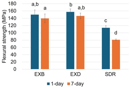
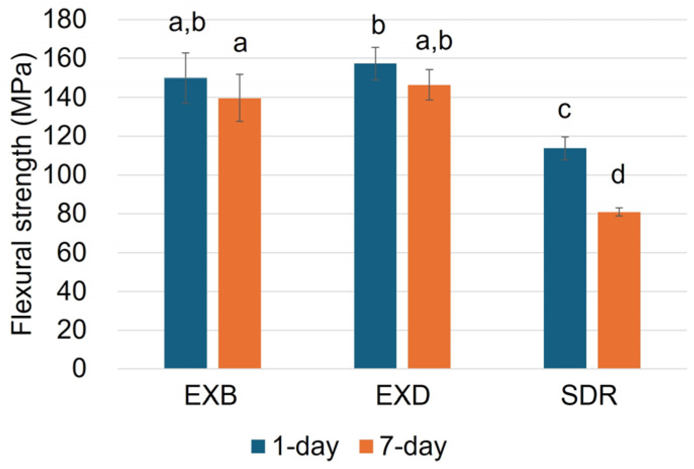
The mean and standard deviation of the flexural strength are presented in Figure 1. SDR showed a significant decrease after 7-day water storage (p < 0.05), whereas there were no significant differences between storage conditions for EXB and EXD (p > 0.05). Among the RCs, SDR showed significantly lower flexural strength compared with EXB and EXD (p < 0.05), with no significant difference between EXB and EXD (p > 0.05).
Figure 1.
Mean values of flexural strength (MPa). Error bars represent standard deviations. Means with the same letter are not significantly different.
3.3. Flexural Modulus
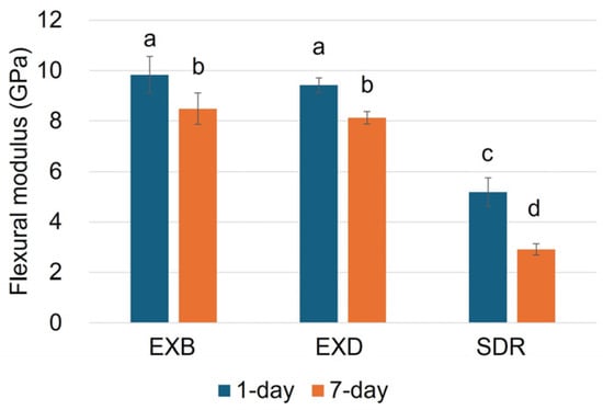
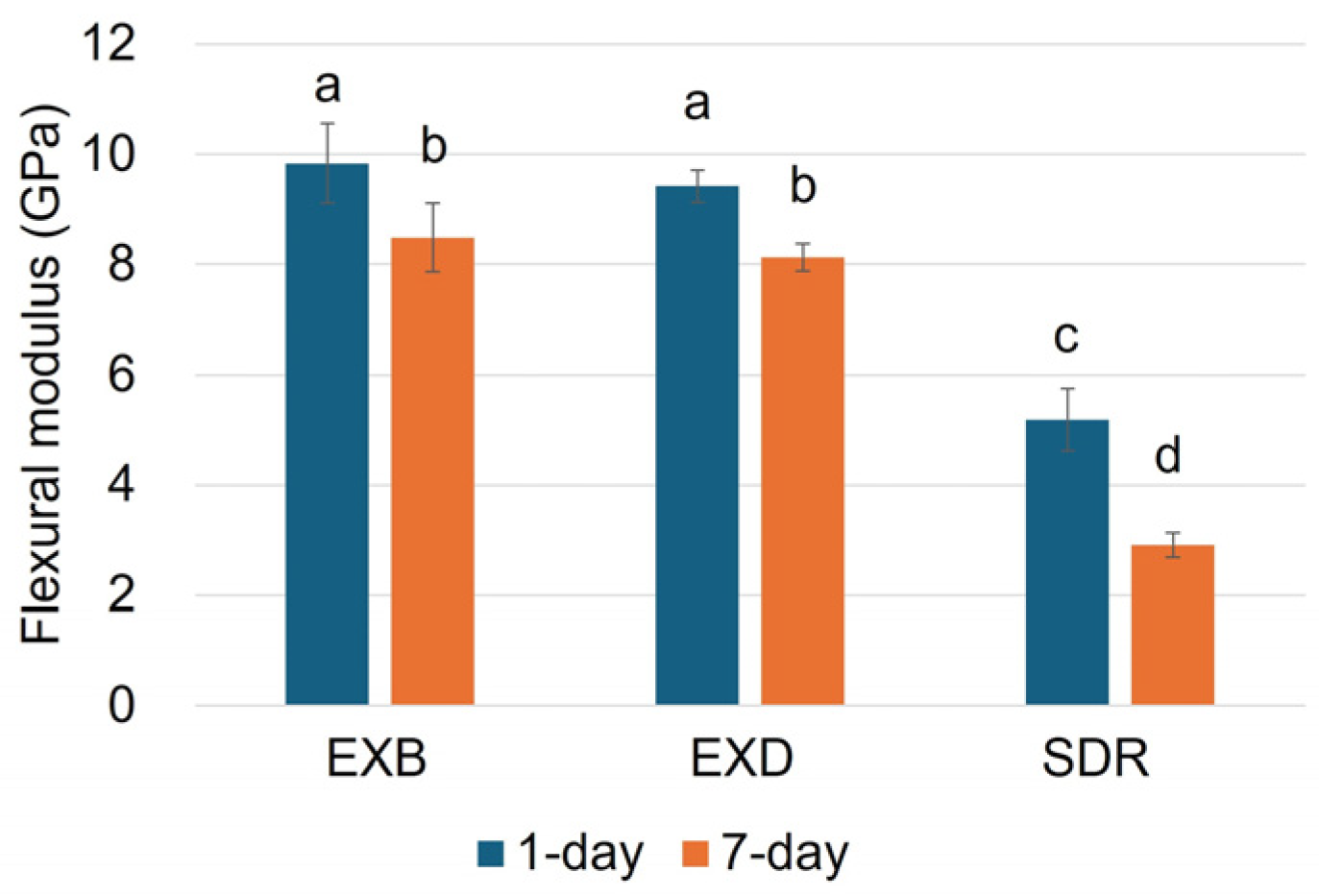
The mean and standard deviation of the flexural modulus are presented in Figure 2. A significant decrease after 7-day water storage was observed in all RCs (p < 0.05). SDR exhibited a significantly lower flexural modulus than EXB and EXD (p < 0.05), whereas no significant difference was found between EXB and EXD (p > 0.05). Quantitatively, SDR showed the most severe percentage reduction (43.9%), whereas the SFRCs exhibited a much lower reduction of 13.7%.
Figure 2.
Mean values of flexural modulus (GPa). Error bars represent standard deviations. Means with the same letter are not significantly different.
3.4. Vickers Hardness
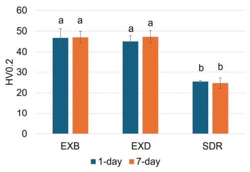
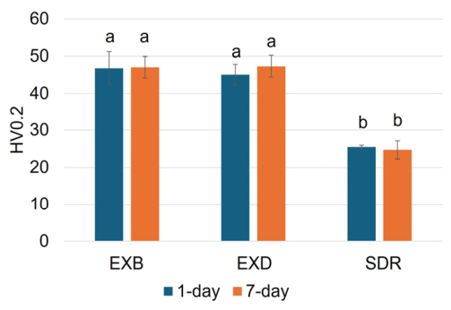
The mean and standard deviation of the Vickers hardness are presented in Figure 3. No significant difference between water storage conditions was found in any of the RCs (p > 0.05). SDR exhibited a significantly lower hardness than EXB and EXD (p < 0.05), whereas there was no significant difference between EXB and EXD (p > 0.05).
Figure 3.
Mean values of Vickers hardness (HV0.2). Error bars represent standard deviations. Means with the same letter are not significantly different.
3.5. Weibull Analysis
The Weibull parameters, including the Weibull modulus (m) and characteristic value (σ), calculated by median rank regression for flexural strength, flexural modulus, and Vickers hardness are summarized in Table 3. Regarding flexural strength and flexural modulus, the SFRCs (EXB and EXD) maintained relatively stable Weibull moduli after 7-day water storage compared to the 1-day condition. In terms of Vickers hardness, SDR initially exhibited a remarkably high Weibull modulus at 1 day, which drastically decreased to 10.6 after 7 days of water storage. In contrast, the Weibull moduli for hardness in EXB and EXD remained stable regardless of the storage period.
Table 3.
Weibull parameters (m: Weibull modulus, σ: characteristic value) for flexural strength, flexural modulus, and Vickers hardness calculated by median rank regression.
3.6. SEM Observation
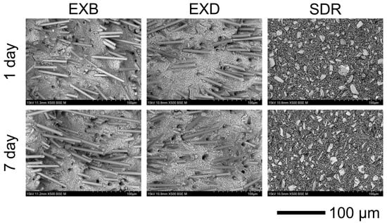
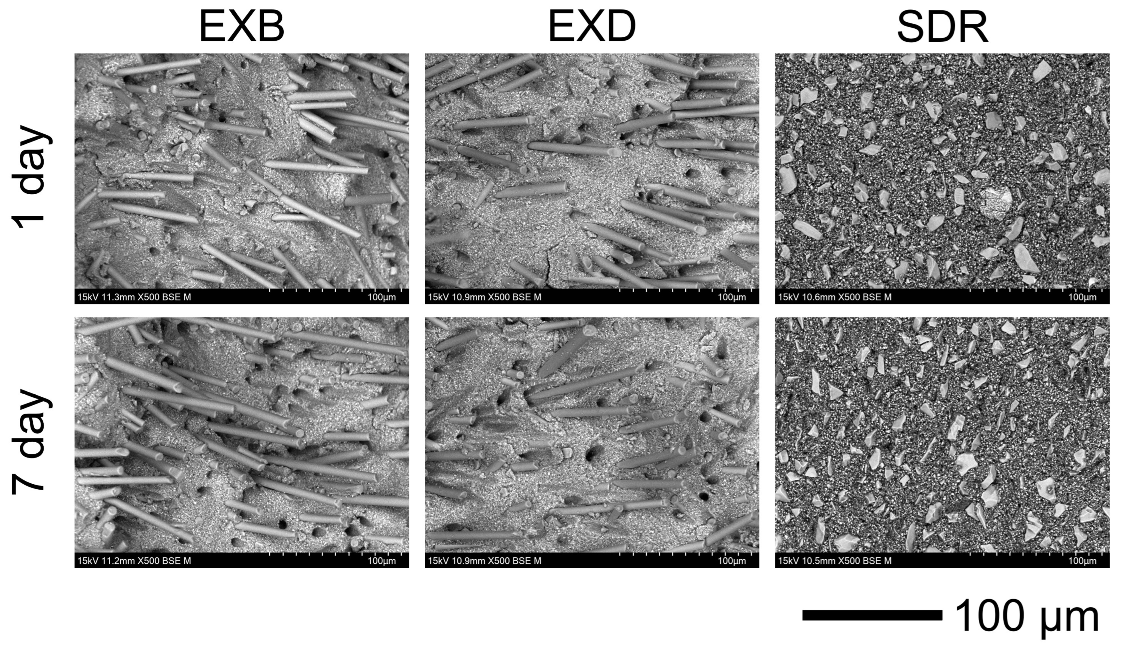
Representative SEM images of the fractured surfaces are shown in Figure 4. In EXB and EXD, fibers with a diameter of approximately 6 μm were clearly observed, accompanied by evidence of fiber pull-out. In contrast, SDR exhibited a resin matrix containing irregularly shaped fillers. No remarkable morphological differences were observed between the storage conditions.
Figure 4.
Scanning electron microscopy images at 500× magnification.
4. Discussion
The purpose of this study was to investigate the effects of water storage as an accelerated aging condition on the mechanical properties of SFRCs. The null hypotheses were partially rejected, as water storage significantly affected the mechanical properties, with the extent of change depending on the property evaluated. Specifically, flexural strength decreased in SDR (the control material), whereas no significant change was observed in SFRCs. In contrast, flexural modulus decreased in all materials, while Vickers hardness was not significantly affected.
SFRCs are considered a promising option for replacing lost dentin in a biomimetic restorative approach due to their superior mechanical properties [17,19]. In such approaches, the enamel is typically restored using other materials, such as conventional RCs, resulting in complete coverage of the SFRC by an enamel-mimicking structure. However, such coverage may not always be maintained under clinical conditions, potentially exposing the underlying SFRC to the aqueous oral environment. To simulate this exposure, a 7-day water immersion period was selected, as previous studies have reported that this duration is sufficient to induce hydrolytic degradation and plasticization in composite resins [20,21]. Interestingly, while our findings reflected this matrix degradation through a significant decrease in flexural modulus, the flexural strength of SFRCs remained unaffected.
Although the flexural modulus results indicated that the 7-day water immersion caused matrix degradation in all tested materials, only SDR exhibited a marked decline in flexural strength. The resistance of EXB and EXD to such strength degradation is primarily driven by fiber-mediated toughening. Specifically, when microcracks initiate within the softened resin matrix, the embedded discontinuous fibers stretch and bridge the crack edges. This bridging action blunts the initially sharp crack and reduces the stress concentration at the crack tip, effectively impeding further crack propagation [6,22]. This mechanism is absent in SDR, which contains only irregularly shaped fillers. Furthermore, in a three-point bending test, the ultimate flexural strength is predominantly governed by the properties of the tension side, where fractures typically initiate. Previous reports concluded that toughness is most effectively enhanced when fibers are located on the tension side of the specimen [23,24]. Therefore, even though the surrounding matrix was plasticized due to water absorption, the intact fibers on the tension side successfully arrested critical crack growth, thereby minimizing the effect of water immersion on the overall flexural strength.
Regarding surface properties, Vickers hardness remained largely unaffected by water immersion across all tested materials. While the flexural modulus reflects the flexibility of the polymer network, surface hardness is fundamentally governed by the inorganic filler content and the localized resistance to deformation [25]. Since the inorganic glass fibers and particulate fillers do not absorb water, they continue to effectively resist indenter penetration even within a plasticized matrix. Moreover, a reduction in surface hardness is directly associated with superficial filler debonding [26], which is typically caused by severe hydrolytic degradation of the silane coupling layer [27]. Therefore, the stable hardness values observed in this study strongly suggest that the 7-day immersion period was insufficient to compromise the integrity of the filler–matrix interface, allowing the highly filled SFRC structure to maintain its surface mechanical properties. Among the evaluated composites, SDR exhibited significantly lower hardness. This can be attributed to its unique stress-relieving monomer system, which is formulated to enhance flexibility and mitigate polymerization shrinkage stress, inherently leading to reduced surface hardness [25,28]. Moreover, the present findings align with prior studies demonstrating that SFRCs generally yield higher hardness values than bulk-fill resin composites [23,29].
When comparing the two SFRCs, no significant differences were found between EXB and EXD across any of the evaluated parameters. Although their fundamental compositions are highly comparable, EXD likely incorporates specific pigments or metal oxides to achieve its dentin shade [30]. A previous investigation highlighted this compositional difference by reporting a greater depth of cure for the bulk shade relative to the dentin shade [31], indicating the presence of minor light-attenuating additives in EXD. Nevertheless, our findings demonstrate that these minor additives do not compromise the baseline mechanical properties or the material’s resistance to hydrolytic degradation.
The Weibull analysis provided valuable insights into the structural reliability of the tested materials (Table 3). Notably, the SFRCs (EXB and EXD) maintained stable Weibull moduli for flexural strength after 7 days of water storage, indicating that fiber-mediated toughening, such as crack bridging [6,22], effectively prevents unpredictable catastrophic failures even when the surrounding matrix is plasticized. In contrast, while the conventional bulk-fill RC (SDR) exhibited a highly uniform surface initially, its reliability drastically dropped after 7-day water storage. This severe decrease implies that water absorption caused uneven local degradation and softening within the non-reinforced matrix. The consistent Weibull moduli of SFRCs across all tested parameters highlight their hydrolytic stability during this short-term aging period, offering predictable initial mechanical performance, which is a significant clinical advantage over conventional particulate-filled composites in aqueous environments.
From a clinical perspective, the surface area of a base material exposed to the oral environment is typically limited to cases where the overlying restoration is lost or fractured. Therefore, the exposed area in vivo is likely much smaller than that of the standardized specimens used in this study, making the current experimental conditions potentially more severe than actual clinical scenarios. A previous study reported a significant decrease in the flexural strength of SFRCs after 30 days of water immersion [6]. Given that our 7-day protocol did not significantly reduce the flexural strength of the SFRCs, it is evident that water-induced degradation progresses over time. Therefore, the findings of this study should be interpreted as an evaluation of the materials’ short-term hydrolytic stability rather than their definitive long-term durability. Clinically, these findings support the current recommendation that SFRC base materials should remain covered by the overlying restoration whenever possible.
However, several limitations of the present study should be noted. First, this was an in vitro study, and the 7-day water immersion protocol cannot fully replicate the complex conditions of the oral environment, which involve thermal fluctuations, pH variations, enzymatic activity, and dynamic masticatory forces. In addition, the present study evaluated only short-term water storage and did not include thermocycling or fatigue loading. Second, the tests were conducted using standardized bar- and disc-shaped specimens, which differ from actual cavity configurations and complex clinical stress distributions. Furthermore, the evaluation was limited to a relatively short aging period and specific mechanical properties. Therefore, further investigations incorporating long-term aging, thermocycling, dynamic fatigue loading, and simulated tooth restorations are necessary to comprehensively evaluate the clinical performance and longevity of these materials when exposed to the oral environment.
5. Conclusions
Based on the results of this study, the following conclusions can be drawn:
- •
- A 7-day water immersion period significantly reduced the flexural strength of the bulk-fill resin composite (SDR), whereas the short fiber-reinforced resin composites (SFRCs; EXB and EXD) maintained their initial flexural strength.
- •
- The flexural modulus of all tested materials decreased significantly after water storage.
- •
- Vickers hardness was not significantly affected by the 7-day water immersion in any of the tested materials.
Author Contributions
Conceptualization, A.S. and Y.I.; methodology, D.M.; software, Y.H.; validation, A.S. and Y.I.; formal analysis, Y.H.; investigation, D.M.; resources, A.S.; data curation, Y.H.; writing—original draft preparation, Y.I.; writing—review and editing, A.S.; visualization, D.M.; supervision, A.S.; project administration, A.S.; funding acquisition, A.S. All authors have read and agreed to the published version of the manuscript.
Funding
This research received no external funding.
Data Availability Statement
The original contributions presented in this study are included in the article. Further inquiries can be directed to the corresponding author.
Conflicts of Interest
The authors declare no conflicts of interest.
Abbreviations
The following abbreviations are used in this manuscript:
| RC | Resin composite |
| SFRC | Short fiber-reinforced composite |
| SEM | Scanning electron microscopy |
| BSE | Backscattered electron |
| ANOVA | Analysis of variance |
References
- Leinonen, J.; Vähänikkilä, H.; Luksepp, R.; Anttonen, V. Five-year survival of class II restorations with and without base bulk-fill composite: A retrospective cohort study. Clin. Oral Investig. 2024, 28, 558. [Google Scholar] [CrossRef] [Scilit]
- Engelhardt, F.; Hahnel, S.; Preis, V.; Rosentritt, M. Comparison of flowable bulk-fill and flowable resin-based composites: An in vitro analysis. Clin. Oral Investig. 2016, 20, 2123–2130. [Google Scholar] [CrossRef] [Scilit]
- Yildiz, E.; Simsek, M.; Pamir, Z. Fracture strength of restorations in proximal cavities of primary molars. Scanning 2016, 38, 43–49. [Google Scholar] [CrossRef] [Scilit]
- Torabzadeh, H.; Ghasemi, A.; Dabestani, A.; Razmavar, S. Fracture resistance of teeth restored with direct and indirect composite restorations. J. Dent. 2013, 10, 417–425. [Google Scholar]
- Van Ende, A.; De Munck, J.; Lise, D.P.; Van Meerbeek, B. Bulk-Fill Composites: A Review of the Current Literature. J. Adhes. Dent. 2017, 19, 95–109. [Google Scholar] [CrossRef] [Scilit] [PubMed]
- Bijelic-Donova, J.; Garoushi, S.; Lassila, L.V.; Keulemans, F.; Vallittu, P.K. Mechanical and structural characterization of discontinuous fiber-reinforced dental resin composite. J. Dent. 2016, 52, 70–78. [Google Scholar] [CrossRef] [Scilit] [PubMed]
- Khan, A.A.; Zafar, M.S.; Fareed, M.A.; AlMufareh, N.A.; Alshehri, F.; AlSunbul, H.; Lassila, L.; Garoushi, S.; Vallittu, P.K. Fiber-reinforced composites in dentistry—An insight into adhesion aspects of the material and the restored tooth construct. Dent. Mater. 2023, 39, 141–151. [Google Scholar] [CrossRef] [Scilit]
- Hazar, A.; Hazar, E. Effect of composite resins with and without fiber-reinforcement on the fracture resistance of teeth with non-carious cervical lesions. J. Appl. Biomater. Funct. Mater. 2024, 22, 22808000241303327. [Google Scholar] [CrossRef] [Scilit] [PubMed]
- Garlapati, T.G.; Krithikadatta, J.; Natanasabapathy, V. Fracture resistance of endodontically treated teeth restored with short fiber composite used as a core material—An in vitro study. J. Prosthodont. Res. 2017, 61, 464–470. [Google Scholar] [CrossRef] [Scilit]
- Wada, K.; Wada, J.; Uctasli, M.B.; Uctasli, S.; Yavuz, Y.; Iwamoto, T.; Vallittu, P.K.; Garoushi, S.; Lassila, L. Impact of restorative material on fracture behaviors of class II restoration in endodontically treated deciduous molars. Dent. Mater. J. 2024, 43, 738–745. [Google Scholar] [CrossRef] [Scilit] [PubMed]
- Lassila, L.; Mangoush, E.; Vallittu, P.K.; Garoushi, S. Fracture behavior of discontinuous fiber-reinforced composite inlay-retained fixed partial denture before and after fatigue aging. J. Prosthodont. Res. 2023, 67, 271–277. [Google Scholar] [CrossRef] [Scilit]
- Safwat, E.M.; Khater, A.G.; Abd-Elsatar, A.G.; Khater, G.A. Glass fiber-reinforced composites in dentistry. Bull. Natl. Res. Cent. 2021, 45, 190. [Google Scholar] [CrossRef] [Scilit]
- Antonucci, J.M.; Dickens, S.H.; Fowler, B.O.; Xu, H.H.; McDonough, W.G. Chemistry of Silanes: Interfaces in Dental Polymers and Composites. J. Res. Natl. Inst. Stand. Technol. 2005, 110, 541–558. [Google Scholar] [CrossRef] [Scilit]
- Zortuk, M.; Kılıc, K.; Uzun, G.; Ozturk, A.; Kesim, B. The effect of different fiber concentrations on the surface roughness of provisional crown and fixed partial denture resin. Eur. J. Dent. 2008, 2, 185–190. [Google Scholar] [CrossRef] [Scilit]
- Akalın-Evren, B.; Kulak-Özkan, Y.; Ozcan, M.; Kadir, T. Candida albicans adhesion on reinforced polymethylmethacrylate denture resin: Effect of fibre architecture and exposure to saliva. Gerodontology 2014, 31, 194–201. [Google Scholar] [CrossRef] [Scilit]
- Bijelic-Donova, J.; Garoushi, S.; Vallittu, P.K.; Lassila, L.V. Mechanical properties, fracture resistance, and fatigue limits of short fiber reinforced dental composite resin. J. Prosthet. Dent. 2016, 115, 95–102. [Google Scholar] [CrossRef] [Scilit]
- Alshabib, A.; Jurado, C.A.; Tsujimoto, A. Short fiber-reinforced resin-based composites (SFRCs); Current status and future perspectives. Dent. Mater. J. 2022, 41, 647–654. [Google Scholar] [CrossRef] [Scilit] [PubMed]
- ISO 4049:2019; Dentistry—Polymer-Based Restorative Materials. International Organization for Standardization: Geneva, Switzerland, 2019.
- Garoushi, S.; Gargoum, A.; Vallittu, P.K.; Lassila, L. Short fiber-reinforced composite restorations: A review of the current literature. J. Investig. Clin. Dent. 2018, 9, e12330. [Google Scholar] [CrossRef] [Scilit]
- Hibino, Y.; Nagasawa, Y.; Eda, Y.; Shigeta, H.; Nakajima, H. Effect of storage conditions on mechanical properties of resin composite blanks for CAD/CAM crowns. Dent. Mater. J. 2020, 39, 742–751. [Google Scholar] [CrossRef] [Scilit] [PubMed]
- Kumar, N.; Sangi, L. Water sorption, solubility, and resultant change in strength among three resin-based dental composites. J. Investig. Clin. Dent. 2014, 5, 144–150. [Google Scholar] [CrossRef] [Scilit] [PubMed]
- Kim, S.H.; Watts, D.C. Effect of glass-fiber reinforcement and water storage on fracture toughness (KIC) of polymer-based provisional crown and FPD materials. Int. J. Prosthodont. 2004, 17, 318–322. [Google Scholar]
- Dyer, S.R.; Lassila, L.V.; Jokinen, M.; Vallittu, P.K. Effect of cross-sectional design on the modulus of elasticity and toughness of fiber-reinforced composite materials. J. Prosthet. Dent. 2005, 94, 219–226. [Google Scholar] [CrossRef] [Scilit]
- Perea-Lowery, L.; Vallittu, P.K. Framework design and pontics of fiber-reinforced composite fixed dental prostheses—An overview. J. Prosthodont. Res. 2018, 62, 281–286. [Google Scholar] [CrossRef] [Scilit] [PubMed]
- Leprince, J.G.; Palin, W.M.; Vanacker, J.; Sabbagh, J.; Devaux, J.; Leloup, G. Physico-mechanical characteristics of commercially available bulk-fill composites. J. Dent. 2014, 42, 993–1000. [Google Scholar] [CrossRef] [Scilit]
- Söderholm, K.J.; Zigan, M.; Ragan, M.; Fischlschweiger, W.; Bergman, M. Hydrolytic degradation of dental composites. J. Dent. Res. 1984, 63, 1248–1254. [Google Scholar] [CrossRef] [Scilit]
- Sideridou, I.; Tserki, V.; Papanastasiou, G. Study of water sorption, solubility and modulus of elasticity of light-cured dimethacrylate-based dental resins. Biomaterials 2003, 24, 655–665. [Google Scholar] [CrossRef] [Scilit]
- Ilie, N.; Hickel, R. Investigations on a methacrylate-based flowable composite based on the SDR™ technology. Dent. Mater. 2011, 27, 348–355. [Google Scholar] [CrossRef] [Scilit] [PubMed]
- Abouelleil, H.; Pradelle, N.; Villat, C.; Attik, N.; Colon, P.; Grosgogeat, B. Comparison of mechanical properties of a new fiber reinforced composite and bulk filling composites. Restor. Dent. Endod. 2015, 40, 262–270. [Google Scholar] [CrossRef] [Scilit]
- Bucuta, S.; Ilie, N. Light transmittance and micro-mechanical properties of bulk fill vs. conventional resin based composites. Clin. Oral Investig. 2014, 18, 1991–2000. [Google Scholar] [CrossRef] [Scilit] [PubMed]
- Szczesio-Wlodarczyk, A.; Garoushi, S.; Vallittu, P.; Bociong, K.; Lassila, L. Polymerization shrinkage of contemporary dental resin composites: Comparison of three measurement methods with correlation analysis. J. Mech. Behav. Biomed. Mater. 2024, 152, 106450. [Google Scholar] [CrossRef] [Scilit]

Disclaimer/Publisher’s Note: The statements, opinions and data contained in all publications are solely those of the individual author(s) and contributor(s) and not of MDPI and/or the editor(s). MDPI and/or the editor(s) disclaim responsibility for any injury to people or property resulting from any ideas, methods, instructions or products referred to in the content. |
© 2026 by the authors. Licensee MDPI, Basel, Switzerland. This article is an open access article distributed under the terms and conditions of the Creative Commons Attribution (CC BY) license.