


Highlights
What are the main findings?
- Lemongrass essential oil (EO) shows strong antialgal activity against terrestrial biofilms.
- Complex mechanisms of action were revealed by physiological and metabolomic analyses.
- Photosynthesis inhibition was accompanied by probable membrane lipid remodeling.
What are the implications of the main findings?
- Biofilms on bricks and renders can be effectively reduced with lemongrass essential oil.
- Treatment causes minimal substrate alterations.
- Lemongrass EO is a promising sustainable and safe component for future protective coatings.
Abstract
Algal biofilms contribute to the biodeterioration of building materials (BMs), while restrictions on conventional algicides stimulate the search for sustainable protection strategies. This study evaluated the antialgal potential of selected essential oils and active compounds, with particular emphasis on lemongrass essential oil (LGC), for the control of microalgal biofilms on brick and render surfaces. Three essential oils (Lavandula angustifolia, Melaleuca alternifolia, and Cymbopogon citratus) and two active compounds, eugenol (EU) and geraniol, were screened against terrestrial microalgal strains associated with biodeterioration. Antialgal activity was assessed based on minimum inhibitory and algicidal concentrations. LGC and EU inhibited microalgal growth, reducing photosynthetic activity, chlorophyll a content, and inducing cellular alterations. Metabolomic analysis revealed a strong stress response involving membrane lipid remodeling, oxidative stress, and disruption of photosynthetic metabolism. LGC and EU caused cellular damage, with LGC inducing faster degradation and EU enabling partial adaptation. LGC effectively reduced microalgal biofilms on brick and render, decreased photosynthetic activity, and partially restored surface coloration, without causing significant changes in substrate pH or water absorptivity. Effectiveness depended on biofilm maturity and substrate characteristics. The integrated microbiological, physiological, metabolomic, and material-based approach confirmed the potential of LGC as a sustainable component of future protective coatings for mineral BMs.
1. Introduction
Biodeterioration of inorganic substrates and building materials (BMs) is a persistent problem affecting both modern and heritage structures. Driven by complex interactions between the substrate and the associated microbial biofilm, colonized surfaces typically undergo a range of undesirable changes, from relatively minor discoloration and aesthetic alterations to the physical weakening of the material matrix and its chemical dissolution [1]. Although the full scale of such phenomena is hard to evaluate, it is estimated that the actual costs of biodeterioration, including infrastructure maintenance and remediation of global impacts, could amount to billions of dollars annually [2]. Considering the prices of conservation strategies and solutions such as biocides, the overall costs of biodeterioration are expected to increase in the coming years [3].
While biodeterioration has historically been attributed mainly to the activity of heterotrophic microorganisms, the role of photoautotrophs in the deterioration of BMs is receiving increasing attention. In fact, this group plays a unique role, acting as pioneer colonizers of inorganic substrates, facilitating microbial succession and thereby initiating biodeterioration processes. Among them, terrestrial microalgae constitute some of the most prevalent colonizers of building façades in temperate regions [1,4]. Apart from acting as pioneering organisms, so far, photoautotrophic biofilms have been also recognized inter alia for the creation of colorful patinas and slimy surfaces, discoloration of colonized materials, increasing water absorptivity, changing pH of the substrate, exerting mechanical stress, and causing surface peeling [5,6,7,8].
Whereas the role of photoautotrophs in chemically mediated biodeterioration remains a subject of ongoing debate, particularly with respect to algae, accumulating evidence suggests that these organisms can contribute to substrate alteration through the uptake of chemical constituents from the substrate and the secretion of metabolites capable of affecting material stability. In addition, photosynthetic carbon fixation may modify the physicochemical conditions at the substrate surface, facilitating mineral deposition and other geochemical transformations [9,10,11]. Importantly, these biodeteriorative processes generally occur simultaneously and in conjunction with environmental stressors, resulting in cumulative effects that become evident over prolonged periods of exposure [1].
To ensure long-term functionality and aesthetic integrity, modern BMs should exhibit sustained resistance to biofouling. Historic structures, however, are often considerably more vulnerable to biodeterioration and present additional challenges for conservation [12,13,14]. In such cases, mitigation strategies must combine high efficacy with material compatibility, ensuring that the treatment itself does not induce undesirable alterations or accelerate substrate degradation. Therefore, safeguarding BMs and cultural heritage monuments from biodeterioration remains a critical objective in conservation science and building material research [15,16]. Conventional biofilm removal strategies encompass a wide range of physical and chemical treatments. Physical approaches include inter alia mechanical cleaning, steam treatment, UV-C irradiation, heat-shock treatment, and infrared (IR) irradiation, while chemical methods involve the application of surfactants, nanoparticles, quaternary ammonium compounds (QACs), hydrogen peroxide, sodium hypochlorite, and various photoactive compounds [1]. In particular, synthetic biocides continue to dominate conservation practice owing to their broad-spectrum efficacy, ease of application, and cost effectiveness [17].
Among the biocidal active substances currently used as film preservatives in protective coatings for BMs are octylisothiazolinone (OIT; 2-octyl-2H-isothiazol-3-one) and 4,5-dichloro-2-octyl-2H-isothiazol-3-one (DCOIT). Historically, façade coatings have also contained compounds such as terbutryn, as well as herbicides including diuron (DCMU) and isoproturon, although these substances have been progressively withdrawn or are no longer approved for use in the European Union due to environmental and human health concerns. These biocides have been widely incorporated into paints and renders to prevent the growth of algae and other microorganisms on coated surfaces. However, their broad-spectrum efficacy is accompanied by considerable environmental persistence, high ecotoxicity, and potential adverse effects on human health. According to the European Chemicals Agency (ECHA), several biocidal active substances are classified as hazardous because they may cause skin sensitization, acute toxicity following ingestion or inhalation, carcinogenicity, or endocrine-disrupting effects, while also posing significant risks to aquatic ecosystems. Furthermore, a life cycle assessment (LCA) conducted by Pedroso et al. [18] demonstrated that although biocides such as DCOIT constitute, on average, only approximately 2.5 wt.% of a coating formulation, they account for more than 68% of its total environmental impact in categories including abiotic depletion potential (ADP) and global warming potential (GWP). Consequently, under the regulatory framework established by the Biocidal Products Regulation [19], the number of approved active substances available for the protection of BMs has steadily declined as hazardous compounds are systematically reassessed or withdrawn (for example, terbutryn, which had been widely used to date, was withdrawn in 2026). In addition, increasingly stringent restrictions on the permitted concentrations of biocidal active substances further limit their use, as these concentrations must balance antimicrobial efficacy with the need to minimize environmental emissions and risks to human health.
To reduce the adverse impact of synthetic biocides on the environment and address reduction of availability, alternative methods of protecting building substrates are rapidly gaining importance. One of the preferred strategies is the use of natural, plant, or microbial compounds exhibiting biocidal properties. Recent studies show that essential oils (EOs) such as oregano, thyme, juniper, rosemary, mint, and yarrow EOs or their active compounds (e.g., eugenol, thymol, geraniol) can be used as promising antimicrobial agents [3,17,20,21,22,23]. Their application has been confirmed in conservation of cultural heritage however only for a narrow spectrum of plant derived compounds. Moreover, most of the previous studies concerned antibacterial and antifungal properties, with antialgal activity still not comprehensively studied. This is especially true for mixed algal biofilms occurring commonly on building substrates in Central Europe.
Taking into consideration that chemical profile of essential oils largely depends on many factors including plant species and their growth conditions, as well as isolation methods, it can be safely assumed that the antialgal potential of EOs in conservation of inorganic materials has not yet been fully discovered. Despite these limitations, the available studies provide valuable insights into the potential mechanisms underlying EO-mediated algal inhibition. Reported effects include impairment of photosystem II (PSII), reduction of chlorophyll a content, and overall inhibition of photosynthetic activity [24,25,26,27]. In addition, EO exposure has been associated with oxidative stress, structural and ultrastructural damage, cellular deformation, and inhibition of key enzymatic activities, ultimately leading to growth suppression or cell death [24,28]. Despite these advances, current knowledge remains fragmented and is derived predominantly from studies on aquatic microalgae or model algal species. Investigations addressing the physiological and metabolic responses of terrestrial green microalgae, particularly those colonizing mineral BMs, are exceedingly scarce. Consequently, the mechanisms underlying EO-induced stress responses in terrestrial algae remain poorly understood, highlighting the need for integrated physiological and metabolomic studies to elucidate their mode of action and support the development of EO-based conservation strategies. Formulations based on essential oils and incorporated into protective coatings represent a promising approach; however, their effectiveness, underlying mechanisms of action, and compatibility with BMs require further systematic and comprehensive evaluation.
The aim of this study was to evaluate selected plant-derived essential oils with biocidal activity, with particular emphasis on the previously unexplored potential of lemongrass essential oil, as sustainable alternatives to conventional biocides used for the prevention and removal of algal biofilms from building material surfaces. The scope of the study included the screening of four essential oils and two reference biocidal active substances against five terrestrial microalgal strains associated with building material biodeterioration. The antialgal efficacy was evaluated based on minimum inhibitory (MIC) and minimum algicidal (MAC) concentration values, while the biological responses of selected treatments were assessed through morphological observations, chlorophyll a analysis, color measurements (CIE L*a*b), and metabolomic profiling (UHPLC- ESI QTOF-MS). The most effective formulations were further tested on algal-colonized ceramic bricks and thin-layer renders to evaluate their performance and impact on substrate properties, including discoloration, chlorophyll fluorescence, pH, and water absorption.
2. Materials and Methods
2.1. Materials
2.1.1. Essential Oils and Bioactive Compounds
The present study investigated the antialgal activity of 6 natural, plant-derived active compounds and mixtures. Four were purchased from Sigma-Aldrich, Inc. (St. Louis, MO, USA) including 2 essential oils, i.e., Bulgarian lavender oil (Lavandula angustifolia), tea tree oil (Melaleuca alternifolia) and 2 pure compounds eugenol and geraniol. Additionally, lemongrass oil (Cymbopogon citratus) and lavender oil (Lavandula angustifolia) acquired through the hydrodistillation process, previously described in [29], were evaluated. Selected isolates and active compounds were described in Table 1.
Table 1.
Selected isolates and active compounds.
The chemical profiles of non-commercial EOs of C. citratus and L. angustifolia were determined using gas chromatography coupled with mass spectrometry (GC-MS; SCION-SQ 456 GC, Bruker Corporation, Billerica, MA, USA). Compound separation was performed using an HP-5MS capillary column (30 m × 0.25 mm i.d. × 0.25 μm film thickness; Agilent J&W, Folsom, CA, USA), with helium used as the carrier gas at a flow rate of 1 mL/min. Prior to analysis, the essential oils were diluted in n-pentane at a ratio of 5:200, and 1 μL of the diluted sample was injected using a split ratio of 1:100. Electron ionization mass spectrometry (EI-MS) was performed at 70 eV. Mass spectra were acquired in positive-ion scan mode over a range of 20–300 m/z, with an acquisition time of 250.0 ms. The initial oven temperature was set to 45 °C and increased at a rate of 3 °C/min to 175 °C. The heating rate was then increased to 15 °C/min until a final temperature of 300 °C was reached, which was maintained for 10 min. The injector and detector temperatures were maintained at 220 °C and 250 °C, respectively. Compound identification was based on comparison of the acquired mass spectra with those in the NIST mass spectral library (NIST Library, version 17.2.3). The resulting chemical profiles are presented in the Supplementary Materials (Tables S1 and S2). The relative abundance of each compound was expressed as the percentage of its peak area relative to the total peak area of all compounds identified in the respective EO sample.
2.1.2. Algal Strains
Antialgal properties of selected compounds were tested against non-axenic mixed algal cultures. For this purpose, previously isolated Bracteacoccus minor PNK015, Stichococcus bacillaris PNK040, Klebsormidium nitens PNK013, Chloroidium saccharophilum PNK010, and Diplosphaera chodatii PNK021 strains were used [4,30,31]. Preparation of algal cultures and inoculation mixtures strictly followed methodology previously described in [1,32]. All algal strains used in this experiment are currently deposited at the Department of Algology and Mycology, University of Lodz. Biological material was cultivated on BG-11 [33] agar plates under artificial light from fluorescent tubes (Osram FLUORA T8 L 36W/77) of 2800 Lux of 22 in a 16 h/8 h day/night period, with a temperature of 22 ± 0.2 °C and air humidity of 50 ± 5%.
2.1.3. Building Materials
In the following study, red bricks and thin-layer renders were used as representative building material (BM) types. Brick samples with average dimensions of 5.0 × 4.0 × 1.5 cm were cut to shape from previously purchased clay- and quartz-sand-based full red bricks (Euroclass A1). For the purpose of this study, render samples were prepared courtesy of ATLAS (Lodz, Poland) by applying biocide-free silicone thin-layer dispersion render on a cardboard and primer scaffold. Samples were standardized to the dimensions of 4.3 × 4.3 × 0.3 cm. Prior to the experiment, specimens were surface-sterilized with UV light.
2.2. Methods
2.2.1. Algal Cultures/Experiment Design
In order to reliably assess the anti-algae potential of plant isolates used in the preservation of BMs, this study was divided into three distinct research stages. Firstly, screening was performed, during which plant isolates and active compounds were subjected to minimal inhibitory (MIC) and minimal algicidal concentration (MAC) assays [34,35]. According to the combined results, acquired after 14 days of incubation, two isolates (i.e., lemongrass essential oil and eugenol) were chosen for subsequent tests. In the following stage, the selected compounds were studied for their mechanism of action (MoA) against mixed algal cultures cultivated in M7, liquid media. The antialgal effect was ascertained based on cell morphology changes, optical density, spectrophotometric color change, fluorescence, and chlorophyll a concentration. Additionally, the stress-response metabolic shift of algal cultures was determined using untargeted mass spectrometry–liquid chromatography (UHPLC-QTOF-MS/MS). Lastly, the isolate with the highest antialgal potential was tested against biofilms cultivated on BMs, and its direct effect on the substrates properties was evaluated.
2.2.2. Minimum Inhibitory (MIC) and Minimum Algicidal (MAC) Concentration Tests
The general antialgal potential of selected plant-derived isolates was initially determined using modified disc-diffusion minimum inhibitory concentration (MIC) and minimum algicidal concentration (MAC) tests [34,35]. Experiments were conducted on Ø85 mm BG-11 agar plates, on to which 100 μL of algal inoculation mixture with optical density set to AU = 1.0 was added. Then, a sterile, cellulose, 6 mm diameter ultra-pure disc with 20 μL of isolate-DMSO mixture was placed centrally on the surface of each sample. For MIC tests, discs were placed immediately after inoculation, while for MAC, the plates were inoculated and incubated for 21 days prior to allow proper biofilm formation. Five different concentrations, i.e., 0:20; 1:19; 5:15; 10:10; 20:0 (μL isolate/μL DMSO), were used. The growth inhibition diameter zones were determined after 7 and 14 days of incubation for MIC and after 3, 7, and 14 days for MAC assessment. The MIC value was defined as the lowest tested concentration of the substance at which a growth inhibition zone >10 mm in diameter was observed. For the determination of the MAC against mature biofilm, two parameters were assessed: the diameter of the mature biofilm removal zone and the diameter of the biofilm discoloration zone. The MAC value was defined as the lowest tested concentration at which both a distinct biofilm removal zone, indicative of the absence of growth, and a discoloration zone >10 mm in diameter were observed. These criteria allowed growth-inhibitory activity (MIC) to be distinguished from activity associated with growth elimination or substantial disruption of the mature biofilm (MAC). To reliably ascertain the antialgal effect of analyzed compounds, inoculated plates not subjected to the isolate/DMSO mixtures were included. Additionally, the effect of pure DMSO was tested.
2.2.3. Biological Effect
Changes in the biological parameters of photoautotrophic biofilms induced by selected active compounds were measured against algal cultures grown in liquid M7 medium purchased from Coimbra Collection of Algae, Portugal (ACOI). First, to sterile Erlenmeyer flasks, filled with 55 mL of M7 media, 10% (v/v) of inoculation mixture with optical density equal to AU = 1,0 was added. Flasks were incubated on Endeavor™ 5000 shaker (130 rpm, OHAUS Europe GmbH, Nanikon, Switzerland). Cultures were grown for 21 days under optimal conditions (see Section 2.1.2), after which they were treated with 0.5 and 1.5% (v/v) plant isolates diluted with DMSO. After 1, 3, and 7 days of exposure, the cultures were subjected to morphological observations, optical density measurement, chlorophyll a content determination, spectrophotometric color change, and metabolomic profiling. As reference samples, three flasks not subjected to the effect of active compounds were used.
2.2.4. Conservation of Technical Materials
The ability of selected isolates to remove existing biofilms without negatively impacting basic properties of BMs was tested in model environment. For that purpose, samples of technical materials were first inoculated with 1 mL of AU = 1.0 algal mixture and incubated in MBF 240 climatic chamber (Binder GmbH, Tuttlingen, Germany). Temperature was set to 22.0 °C. Humidity was kept at a constant level of 75.0%. Day/night light intervals were introduced in the 18 to 8 h ratio under the light intensity of 2800 Lux. After 1-month and 3-month incubation periods, 6 mm cellulose disc containing 40 μL of isolate-DMSO mixture was applied to the surface of each sample. The volume of mixture was increased to account for evaporation/absorption of active compounds and to enable the observation of any undesirable changes in substrate parameters. In regular intervals (after 1, 3, 7, and 18 days of exposure), macroscopic changes, discoloration, and chlorophyll fluorescence were measured. After incubation period, changes in substrate pH and water absorptivity were determined. To compare the effectiveness of isolate and its impact on non-inoculated substrate, 3 types of control samples were used, i.e., (1) non-inoculated samples of BMs to which EOs were applied, (2) samples of BMs on which pure DMSO was applied, and (3) inoculated samples of BMs without application of EOs or DMSO. For each of the types, 3 replicas were used.
2.2.5. Morphological Observations of Algal Cultures
The algal experimental cultures were morphologically examined one day, three days, and seven days after adding the eugenol (EU) and lemongrass (LGC) oil in 0.5% and 1.5% to the mixed culture in M7 liquid medium. The changes in the cells’ structure were investigated using a Nikon Eclipse E600 microscope (Precoptic Co., Warsaw, Poland) equipped with DIC optics, under 1000× magnification. The LM images were taken using an Opta-Tech HDMI digital camera (OPTA-TECH, Warsaw, Poland). The images of taxa in the best visual condition of cells in each culture were compiled into photographic documentation of the effect of essential oils on algal cells.
2.2.6. Cell Density Measurement
Liquid culture cell density was established using optical density measurement. A 3 mL sample taken from the liquid culture was transferred into a glass cuvette, and the absorbance was measured at 680 nm, corresponding to the highest peak recorded for the mixed algal culture prepared in M7 media [31,32]. For each culture, cell density was ascertained before addition of bioactive compounds, after 1, 3, and 7 days of exposure.
2.2.7. Chlorophyll a Measurement
Chlorophyll a concentration was used to evaluate the changes in the density of live cultures under the effect of bioactive compounds. For that reason, 2.5 mL of each culture was transferred into Eppendorf tubes and centrifuged at 6000 rpm for 2 min (MPW-351R, MPW, Warsaw, Poland). Supernatant was discarded, and the remaining biomass was suspended in 100 μL of methanol. Samples were homogenized for 1 min (Mini-homogenizer E0357-01, EURx, Gdańsk, Poland) and filled with methanol to the final volume of 1.5 mL. Then, the samples were vortexed (VM300S, Alchem, Toruń, Poland) and incubated at 4 °C for 24 h. Extracted chlorophyll was centrifuged at 6000 rpm for 2 min, and absorbance was measured at 665 nm and 650 nm. Chlorophyll a concentration (µg/mL) was calculated using Formula (1):
where Chl−a: the chlorophyll a concentration (µg/mL); A665: absorbance measured at 665 nm; A650: absorbance value measured at 650 nm, V: the volume of sample taken from culture, in which the chlorophyll a content was measured.
2.2.8. Fluorescence Measurement
Photosynthetic activity of cultures exposed to the tested compounds was evaluated using chlorophyll a fluorescence parameters measured with a Handy FluorCam FC 1000 H pulse-amplitude modulation (PAM) fluorimeter (Photon Systems Instruments, Drásov, Czech Republic) operated with FluorCam 7 software. The intensities of the measuring flashes, actinic light, saturating pulses, and far-red illumination were adjusted to match the incubation conditions. Prior to fluorescence measurements, all samples were dark-adapted for 60 min.
For the bioactivity assays, 0.5 mL of each culture was evenly spread onto the surface of Ø55 mm BG-11 agar plates before dark adaptation. Photosynthetic performance was assessed based on the maximum quantum yield of photosystem II (Fv/Fm), calculated as the mean value across the entire plate surface.
For the BM assays, fluorescence measurements were performed under sterile conditions by directly scanning the colonized surface. Owing to the high porosity of the substrates and the averaging of fluorescence signals across the entire scanned area (including regions with strong fluorescence and regions lacking strong signal), resulting in signal-to-noise ratio limitations, only the minimum fluorescence (F0) and maximum fluorescence (Fm) parameters were analyzed. The data analysis followed established protocols [32,36].
2.2.9. Color Change Evaluation
Color measurement analysis was performed during biological effect studies conducted with liquid cultures and to ascertain the surface discoloration of BMs. Color components were measured using CIE L*a*b trichromatic color model with CM-700d (Konica Minolta, Warsaw, Poland) spectrophotometer and CM-S100w SpectraMagicTM NX v.2.0 software. For all tests, D65 Illuminant was applied, and measurements were performed in both specular component included (SCI) and specular component excluded (SCE) modes. The change was measured before inoculation, and subsequently, measurements were taken at additional time points. For each sample, 10 individual shots were taken, and the mean value was used for analysis. For liquid cultures, the spectrophotometer was inverted, and the volumetric flask containing the stirred culture was placed directly on the device. For BMs, measurements were performed directly on BM surface in sterile conditions. Due to the aesthetical impact of algal biofouling, data obtained using SCE mode were predominantly used for analysis. Color change was calculated using Formula (2):
where ΔE—value of color change between tested and reference sample; ΔL—difference in lightness (white–black) between tested and reference sample (0 represents completely black sample, and 100 represents maximal brightness); Δa—difference in color between tested and reference sample (green—red; +a = red, −a = green); Δb—difference in color (blue—yellow; +b = yellow,−b = blue) between tested and reference sample.
For the evaluation of color changes, relative scale proposed in [37] was used, where 0 < E < 1—the difference is not noticeable to the inexperienced observer; 1 < E < 2—only a trained observer can see the difference; 2 < E < 3.5—an untrained person can notice a difference; 3.5 < E < 5—a clear color difference is seen; 5 < E—the viewer sees two distinct colors.
2.2.10. Metabolomic Profiling
Algal cultures grown in liquid M7 medium were subjected to metabolomic profiling. Acquired data were used to search for potentially biodeteriorative compounds produced by photoautotrophic biofilm in response to time- and dose-dependent antialgal activity. The analysis was performed using mass spectrometry–liquid chromatography with Bruker Elute UHPLC system (Bruker Daltonik GmbH, Bremen, Germany) operated by Hystar 3.3 software and an ultrahigh resolution (60,000+) mass spectrometer Bruker Impact II (Bruker Daltonik GmbH, Bremen, Germany) ESI QTOF-MS equipped with Data Analysis 4.2 (Bruker Daltonik GmbH, Bremen, Germany) Metaboscape. Bruker Intensity Solo C18 column (2 μm particles of 2.1 × 100 mm dimensions) was used for AutoMSMS measurements. As eluent A, water with 0.1% HCOOH was used, and for eluent B, acetonitrile with 0.1% HCOOH. AutoMSMS measurements flows and percentages were 0 and 2 min—99% A; 17 min—1% A; 20 min—1% A; 20.1, 22 and 30 min—99% A, Flow was 0.25 μLmin−1 from 0 to 20 min and 0.35 from 20.1 to 30 min. Column was held at 40 °C. The column exit was connected to ESI source. Injection volume was 5 μL. Analyses in positive autoMSMS mode m/z range were 50–1200, Collision-induced dissociation (CID) was used with the following settings: absolute area threshold: 5000 counts; active exclusion 2 spectra; release after 0.3 min. Isolation mass for m/z = 100, width was 4; for 300, width was 5; for 500, width was 6; and for 1000, width was 8. Collision energy value was 30 eV. Internal calibration on 10 mM sodium formate (1:1 v/v water:isopropanol) ions was performed automatically in Metaboscape with the use of syringe pump at an infusion flow rate of 0.12 mLh−1, using a high-precision calibration (HPC) mode.
The untargeted annotations were performed in Metaboscape (ver. 2022b) with a criterion of mass deviation (Δm/z) under 2 ppm and mSigma value under 20 as the maximum acceptable deviation of the mass of the compound and the isotopic pattern, respectively. All the molecular formulas were obtained using the Smart Formula tool and the C, H, N, O, P, S, Cl, Br, I and F elements. MSMS spectra were automatically matched against MSMS libraries: Bruker HMDB 2.0 library (spectral data with retention times), MassBank of North America (MoNA) library, and NIST ver. 2020 MSMS library.
2.2.11. pH
For the reliable assessment of the pH change occurring in the surface layers of BMs under the influence of photoautotrophic biofilm and bioactive compounds, the methodology described previously in [38] was adapted. The outmost layers of examined materials were crushed and ground. The crushed material was weighted using laboratory balance scale (Ohaus, Port Melbourne, Australia). Afterwards, it was suspended in deionized water in a ratio of 1:2 for the bricks and 1:4 for the renders and mixed for 5 min in a shaker (Ohaus, Port Melbourne, Australia) at 190 rpm. PH was measured by multifunction meter CX-705 (Elmetron, Zabrze, Poland) with the IJ44AT electrode (Ionode, Brisbane, Australia). Afterwards, each suspension was filtered through SJM 110 Euro 3w 110 mm filters (SLINAP, Łódź, Poland), and the pH measurement was repeated.
2.2.12. Water Absorptivity
For the water absorptivity measurement, the gravimetric method was used. Selected samples were first weighted and then dried at 105 °C with DZ-2BCII drying oven (ChemLand, Stargard, Poland) to a constant weight. Afterwards, each sample was placed individually in a plastic container and gradually soaked using deionized water. To avoid entrapment of air bubbles inside the material structure, water was added every 2 h, and each portion corresponded to ¼ of the sample height. Samples were then soaked overnight. Excess water was removed gently using paper cloth, and the samples were weighed in regular time intervals to confirm constant weight. The absorptivity was calculated using Formula (3):
where M—mass absorptivity (%); Mw—mass of fully wet sample (g), Md—mass of fully dry sample (g).
2.2.13. Statistical Analysis
For each culture, as well as each measurement, three replicas were used. The mean, standard deviation values, error bars, correlation, and one-way analysis of variance (ANOVA) with statistical significance level of 0.05 were calculated in Microsoft Excel 365 version 2211. The mean fluorescence parameters F0 and Fm before and 3, 7, and 21 days after application on brick and render were compared using one-way analysis of variance (ANOVA) at a significance level of α = 0.05. When statistically significant differences were detected (p < 0.05), mean values were further compared using Tukey’s post hoc test at a significance level of 0.05.
3. Results and Discussion
3.1. Initial Screening
In the initial stage of the study, a screening procedure was conducted to identify bioactive compounds with the highest potential for removing photoautotrophic biofilms from BMs. To ensure a reliable selection of the most suitable isolates, the results of disc diffusion assays were complemented with additional data. In the MIC tests, in addition to measuring the inhibition zone diameters against microalgal growth (primary indicator), the inhibition zones formed against the natural accompanying microflora were also considered. Similarly, in the MAC assays, in addition to the diameters of fully cleared zones, the area of discoloration was quantified and used as a supplementary indicator of biofilm growth disruption. The screening results are presented in Figure 1, and the complete dataset is provided in Tables S3 and S4.
Figure 1.
Results of initial screening test. Inhibition zones diameter [mm] registered for MIC test in (A)—only microalgae, (B)—algae and natural bacterial microflora, as well as MAC assay, where (C)—complete removal, (D)—discoloration zone. For a visual representation, see Figure 2.
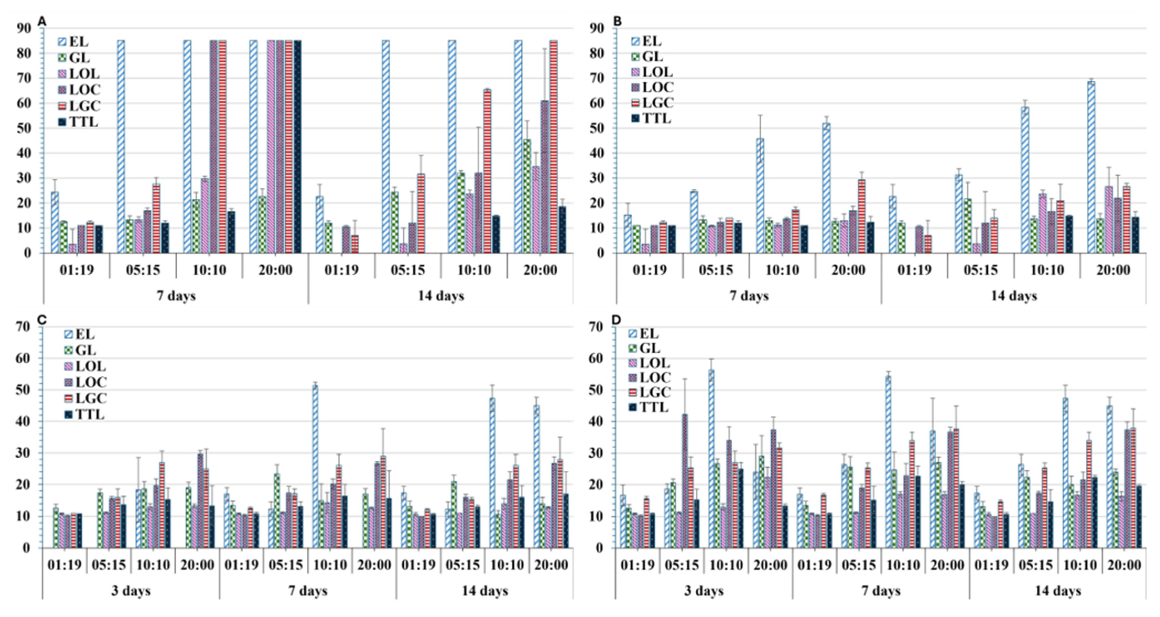
Generally, for all tested compounds, antimicrobial activity increased with concentration, whereas the positive controls (untreated cultures) grew steadily. No antimicrobial effect was observed for pure DMSO, which excludes any solvent-related interference with the test outcomes and confirms that the growth conditions were appropriate. All compounds exhibited inhibitory effects against the mixed algal cultures, although their effectiveness depended on both concentration and incubation time. A 7-day incubation period was sufficient to clearly distinguish between rapidly developing photoautotrophic biofilm and the affected zones.
Figure 2.
Representative examples of the initial screening results. (A) Complete inhibition of the algal biofilm accompanied by partial inhibition of the associated microflora. (B) Presence of both a complete removal zone and a discoloration zone. (C) Presence of a removal zone only.
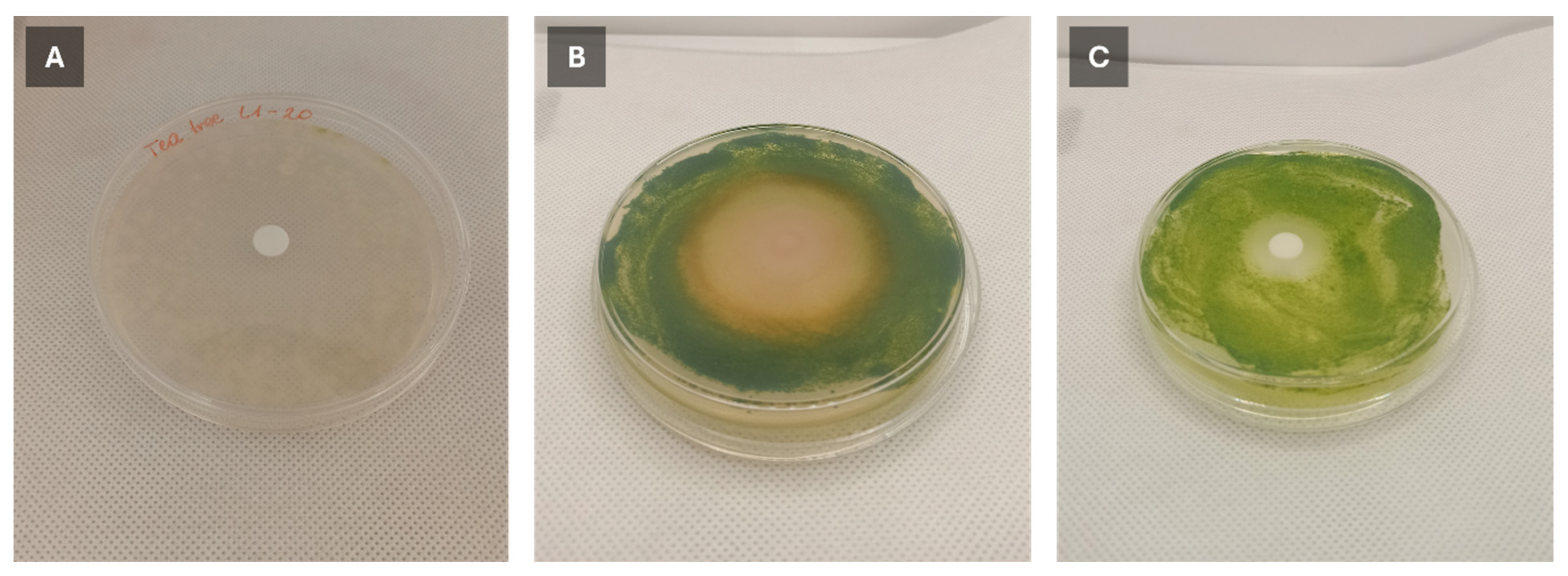
Notably, for most isolates the inhibition zone area decreased significantly between 7 and 14 days of incubation, indicating that although algal growth was initially suppressed and slowed, some regrowth could occur over time. The exception was eugenol, which maintained comparable activity even after 14 days of incubation and showed the highest overall efficacy, achieving a maximum growth inhibition diameter of 85.00 ± 0.00 mm (corresponding to the entire plate) at a 5:15 (μL isolate/μL DMSO) ratio. After 7 days of incubation, LOC and LGC essential oils were the second most effective isolates, reaching the maximum inhibition area at a 10:10 concentration. However, their potency declined with prolonged incubation, and the inhibition zone diameter decreased from 85.00 ± 0.00 mm to 65.33 ± 0.58 mm and 32.00 ± 18.36 mm for LGC and LOC, respectively. In contrast, LOL and TTL exhibited the weakest inhibitory effects against algal cultures. Although slight activity was observed after 7 days at a 1:19 (μL isolate/μL DMSO) concentration of LOL and TTL, no inhibition persisted after 14 days, and all samples were completely overgrown. Only the 10:10 (v/v) concentration resulted in effective algal growth inhibition for these isolates, with tea tree oil showing the lowest potency even at this maximum concentration. It was found that for all tested plant isolates and active compounds, a concentration ratio of 1:19 could be considered the MIC against algal cultures after 7 days of incubation, based on the observed growth inhibition zones of >10 mm in diameter, with the exception of LOL, for which the MIC was determined at 5:15. After 14 days of incubation, the MIC values remained unchanged at 1:19 for EL, GL, and LOC, whereas they increased to 5:15 for LGC and 10:10 for LOL and TTL (Table S3).
In general, the bacterial microflora exhibited higher resistance to the tested bioactive compounds, and no complete growth inhibition was observed (i.e., no sample showed growth inhibition zones equal to the whole plate) regardless of the concentration used. However, the MIC values determined for the individual plant isolates and active compounds were identical to those observed for the algal cultures, both after 7 and 14 days of incubation (Table S3). Similar to the anti-algal activity, eugenol (EL) was the most effective isolate, with mean inhibition zone diameters of 15.17 ± 4.75, 24.67 ± 0.58, 45.67 ± 9.45, and 52.00 ± 2.65 mm at 1:19, 5:15, 10:10, and 20:20 (μL isolate/μL DMSO), respectively, after 7 days. After 14 days, these values increased to 22.67 ± 4.73, 31.33 ± 2.31, 58.33 ± 2.89, and 68.67 ± 1.15 mm for the same concentrations. Notably, in contrast to algal growth inhibition, the antibacterial activity of eugenol increased over time, with larger inhibition zones after 14 days than after 7 days at all concentrations. The lowest inhibitory effects were recorded for low concentrations of LOL and TTL, which showed almost no activity at 1:19 and 5:15 (μL isolate/μL DMSO), although their performance improved at higher doses. Apart from eugenol, the most consistent results were obtained for LGC, with the highest inhibition observed at 10:10 and 20:0 concentrations after 14 days of incubation. Although the bacterial microflora naturally associated with the algal cultures were less sensitive to the tested EOs than the algae, requiring a longer than algae exposure time, their lower susceptibility does not necessarily indicate an absence of effects on non-target microorganisms. Given the complex composition and broad biological activity of essential oils, repeated application may potentially influence microbial community structure and diversity, as well as interspecific interactions. Such exposure may exert selective pressure, favoring less susceptible microorganisms, and thereby alter the composition of the microbial community. Therefore, the ecological consequences of long-term or repeated application of essential oils, particularly with respect to non-target microorganisms, warrant further investigation under environmentally relevant conditions.
In contrast to the growth inhibition assays, the results of the minimal algicidal concentration (MAC) tests were more heterogeneous. Eugenol exhibited the largest discoloration areas across almost all concentrations; however, the diameters of the fully cleared zones were noticeably smaller. Moreover, after 3 days of exposure, no clear algicidal zone was detected for eugenol at 1:19, 5:15, or 20:0 (μL isolate/μL DMSO), contrary to all other isolates. Although distinct algicidal zones formed after 7 days, the 20:0 concentration did not produce a clear zone until 14 days of exposure. Most interestingly, for eugenol, the 10:10 concentration resulted in larger diameters for both discoloration and clearance zones, suggesting that a 1:1 dilution may be more effective than the pure isolate and indicating a potential complementary mechanism. A similar effect was observed for GL, with the 5:15 concentration showing the highest activity after both 7 and 14 days. Overall, although eugenol demonstrated the strongest inhibitory properties, its algicidal action required more time to fully develop when applied to already formed biofilms.
For biofilm removal, lemongrass essential oil (LGC) was relatively the most effective across all incubation times, particularly at 10:10 (27.00 ± 3.61, 26.00 ± 3.61, and 26.00 ± 3.61 mm after 3, 7, and 14 days, respectively) and 20:0 (25.00 ± 6.24, 29.00 ± 8.72, and 28.00 ± 7.00 mm for 3, 7, and 14 days, respectively), followed by LOC. Similar patterns were observed for discoloration zones. No notable recolonization was detected between incubation times; however, the tests were conducted under model conditions that likely limited the evaporation of active compounds. It should also be emphasized that although discoloration is a well-established indicator of stress-related disruption of microalgal activity, from the perspective of building material conservation, complete biofilm removal without the risk of secondary staining is far more important.
It is noteworthy that for all tested isolates, the MAC was determined at a concentration ratio of 1:19 after both 7 and 14 days of incubation. At this concentration, both a distinct biofilm removal zone, indicative of the absence of growth, and a discoloration zone >10 mm in diameter were observed. In the case of eugenol, the MAC was higher after 3 days of exposure, being determined at 10:10, whereas after 7 and 14 days, the MAC decreased to 1:19, indicating that a lower concentration was sufficient to achieve the defined algicidal effect with prolonged exposure.
However, it is worth noting that eugenol and geraniol are single, chemically defined compounds, whereas essential oils (EOs) are complex mixtures of volatile constituents whose biological activity may result from additive, synergistic, or antagonistic interactions among their components. The present study compares the biological effects of pure compounds and EOs at identical nominal volumetric concentrations under the same experimental conditions, rather than their intrinsic efficacy on an equivalent molar or mass basis. This distinction should be considered when interpreting the differences in biological activity observed between the tested treatments.
Based on these findings, EL and LGC were selected as the most suitable candidates for subsequent stages of the study.
3.2. C. citratus Essential Oil Characteristics
Although the chemical composition of essential oils may vary considerably depending on the geographical origin of the plant, season of harvest, condition of the raw material, plant part used, and distillation procedure, the C. citratus EO analyzed in the present study exhibited a typical citral-dominated profile. Geranial (α-citral) was the predominant constituent (38.5%), followed by neral (30.8%), β-myrcene (17.2%), and geraniol (6.2%). Thus, the combined content of the two major citral isomers, geranial and neral, accounted for 69.3% of the identified compounds. This composition is consistent with the chemotype commonly reported for C. citratus EOs, in which citral, expressed as the combined content of geranial and neral, generally constitutes the major fraction of the oil, often accounting for approximately 70%–85%, with β-myrcene frequently representing one of the other quantitatively important constituents [39,40,41,42,43].
The high proportion of citral constituents may contribute to the biological activity observed for the C. citratus EO in the present study. Both geranial and neral have been associated with antimicrobial activity against a range of microorganisms, while β-myrcene has been suggested to contribute to the overall biological effects of C. citratus EO, although its individual antimicrobial activity appears to be more limited [39,44,45]. Previous studies have reported antimicrobial activity of C. citratus EO against, among others, various Candida spp. [46,47,48], as well as activity against established microbial biofilms. In general, the antibacterial activity of C. citratus EO appears to be more pronounced against Gram-positive bacteria, including S. aureus, Bacillus subtilis, and Enterococcus faecalis, than against Gram-negative bacteria [39,49,50,51,52,53,54]. In contrast, Pseudomonas aeruginosa has frequently demonstrated comparatively high resistance to the oil. Nevertheless, inhibitory effects against P. aeruginosa biofilm formation have also been reported, indicating that the activity of the oil may depend on the physiological state of the target microorganism [39,51].
Mechanistic studies conducted primarily in fungal models suggest that the biological activity of C. citratus EO may involve disruption of cellular structures and membranes, impairment of mitochondrial function, increased reactive oxygen species (ROS) generation, DNA damage, and alterations in biofilm structure and adhesion. These observations indicate that the activity of the oil is likely to result from the combined effects of several constituents rather than from a single compound.
Despite the well-documented antimicrobial and antifungal properties of C. citratus EO, information concerning its activity against algae remains comparatively limited, particularly in the context of terrestrial algal biofilms colonizing building materials. Evidence for the involvement of citral in algal inhibition was provided by Ikawa et al. (1992) [55], who reported that citral exhibited the strongest inhibitory activity against Chlorella pyrenoidosa among the terpene alcohols and aldehydes examined in their study. Given the high proportion of geranial and neral in the EO investigated here, the predominance of citral constituents may therefore represent a plausible contributor to the antialgal activity observed in the present study.
3.3. Biological Activity Assays
The biological tests confirmed the antialgal properties of eugenol and lemongrass essential oil regardless of the concentration applied, although their effect on the microalgal cultures varied (Table 2). Both isolates allowed for a significant decrease in the concentration of chlorophyll a. The non-affected cultures maintained chl-a level of 4.86 ± 0.40, 4.47 ± 0.07, 4.66 ± 0.29 µg/mL after 1, 3, and 7 days, respectively, while the addition of eugenol decreased the values up to 1.92 ± 0.42 µg/mL (0.5% EU concentration) and 2.41 ± 0.03 µg/mL (1.5%). LG allowed for almost complete chlorophyll reduction with values of 0.08 ± 0.05 and 0.05 ± 0.01 µg/mL registered for 0.5 and 1.5% concentration already after 3 days of exposure. In contrast, the changes in optical density (OD) followed a distinct pattern. In the control cultures, without LGC essential oil or eugenol, OD values remained relatively stable throughout the 7-day incubation period, ranging from 1.71 to 1.88, with only a slight upward trend. In cultures treated with 0.5% LGC, OD gradually decreased from 1.67 at the beginning of the experiment to 1.32 after 7 days. At 1.5% LGC, OD initially decreased from 1.69 to 1.33 after 1 day, followed by a subsequent increase to 1.66 by day 7. In contrast, eugenol treatment resulted in a progressive increase in OD at both tested concentrations, from an initial value of 1.71 to 2.48 and 2.66 after 7 days at 0.5% and 1.5% eugenol, respectively. This strongly coincided with macroscopic observations that revealed similar differences with lemongrass-affected cultures becoming progressively more translucent, while at the same time, cultures incubated with eugenol became brownish, visibly more dense, and less transparent (see Figure S1 and Table S5). Both types of cultures quicky lost their green hue.
Table 2.
Chlorophyll a (Chl-a) concentration, optical density (OD680), and photosynthetic efficiency (Fv/Fm) changes in mixed microalgal cultures subjected to lemongrass essential oil (LGC) and eugenol (EU).
Exposure to both types of compounds resulted in a marked reduction in photosynthetic activity, as indicated by the maximum quantum yield of photosystem II (Fv/Fm). In the control samples, Fv/Fm remained stable throughout the experiment, ranging from 0.32 to 0.33. In contrast, both LGC and EU caused an almost complete inhibition of photosynthetic activity, with the most pronounced decline observed after the first day of exposure. Initially, EU exerted a slightly stronger inhibitory effect than LGC; however, this trend was reversed after 7 days of exposure. At the end of the experiment, Fv/Fm values were 0.08 ± 0.05, 0.09 ± 0.04, 0.10 ± 0.06, and 0.11 ± 0.02 for 0.5% LGC, 1.5% LGC, 0.5% EU, and 1.5% EU, respectively. The observed decline in photosynthetic activity closely corresponded with the progressive loss of green pigmentation and was consistent with the morphological changes described below. However, the Fv/Fm values recorded for the control samples would generally be considered low and could suggest the presence of environmental stress [56,57].
For microscopic observations, most of the algal taxa in the control culture did not reveal any substantial morphological changes in the structure of cells with time (Figure 3A,B). Only the cells of Klebsormidium showed some variability in the structure of protoplasts. On the first day, they were more hydrated, probably as a result of growing in a liquid medium, then started to accumulate lipids and created short filaments to adjust to the aquatic environment. In all taxa, the shape and appearance of chloroplasts did not differ from physiological plasticity.
Figure 3.
(A)—The changes in algal cells under the effect of Eugenol in 0.5% and 1.5% concentration after 1, 7, and 14 days of experiment to control culture. Images present long filaments of Klebsormidium sp., single rod-shaped cells of Stichococcus bacillaris, oval cells of Chloroidium saccharophilum, circular cells of Bracteacoccus minor, and small coccal cells pointed with an asterisk of Diplosphaera chodatii. (B)—The changes in algal cells under the effect of lemongrass oil in 0.5% and 1.5% concentration after 1, 7, and 14 days of the experiment to control culture. Images present long filaments of Klebsormidium sp., single rod-shaped cells of Stichococcus bacillaris, oval cells of Chloroidium saccharophilum, circular cells of Bracteacoccus minor, and small coccal cells pointed with an asterisk of Diplosphaera chodatii.
The addition of eugenol to the culture after a day of the experiment changed the osmotic pressure in cells. Chloroplasts become flatter and attached to the cell walls. With time, photosynthetic active pigments started to vanish in all taxa except Klebsormidium; however, after 7 days with 0.5% EU, some Chloroidium and Bracteacoccus cells revealed signs of pigment re-synthesis. The eugenol in 1.5% concentration already caused a physiological inactivation of Bracteacoccus, Chloroidium, and Stichococcus after 3 days, and this effect was maintained till the end of the experiment. Cells of Diplosphaera were found in cultures after only 1 day of the experiment; thus, with time they have been quickly decomposed.
In addition, more damages in cell morphology were observed after the use of lemongrass oil. On the first day of the experiment, all cells of Bracteacoccus and Diplosphaera and most of the cells of Chloroidium and Stichococcus revealed signs of physiological inactivation. Cells were pale and transparent; chloroplasts were visible but became highly heterogeneous. Only the Klebsormidium was less sensitive to the short-time effect of LGC. Nevertheless, after 3 days of the experiment, all algal cells from cultures with 0.5% and 1.5% lemongrass oil were either inactive or in a state of cell degradation.
The overall diversity of metabolomic profiling of all analyzed samples is presented in Figure 4 and Figure 5, while the full scope of comparative results is registered in Attachments S1–S3 (Supplementary Materials). The exposure to the eugenol and lemongrass isolates had a profound impact on the changes in metabolomic profile of algal cultures.
Figure 4.
Principal component analysis (PCA) score plot of untargeted metabolomic profiles for all samples from the algal consortium (PL) in liquid medium. The plot includes control cultures and cultures treated with eugenol (EU) or lemongrass essential oil (LGC) across all time points (Day 1, Day 3, and Day 7 of exposure).
Figure 5.
Hierarchical clustering heatmap of untargeted metabolomic profiles for all samples from the algal consortium in liquid medium, including control cultures and cultures treated with eugenol (EU) or lemongrass essential oil (LGC) across all time points (Day 1, Day 3, and Day 7 of exposure). Data were log2-transformed and Pareto-scaled prior to analysis. Clustering was performed using Euclidean distance and Ward linkage.
Most significantly, the presence of bioactive compounds led to the accumulation of lysophosphatidylcholines (LPC, e.g., LPC 16:2, LPC 18:3, LPC 18:2, LPC 16:3), phosphatidylcholines (PC, e.g., PC O-18:3), lysophosphatidylglycerols (LPG, e.g., LPG 14:0, LPG 15:0, LPG 16:0, LPG 18:2, LPG 18:3), sugars (e.g., DL-arabinose, trehalose, D-ribulose), amino acids and their derivatives (e.g., glutathione, asymmetric dimethylarginine, citrulline, tyramine, L-phenylalanine, L-glutamine, L-tryptophan), and fatty acids and their derivatives (e.g., FA 18:1 + 2O, 9(10)-epoxy-12Z-octadecenoic acid, 1-linoleoylglycerol, 9,12-octadecadiynoic acid, heptadecanoic acid). While all those compounds showed notable increase (fold change, FC > 4), the metabolites most strongly differentiating treated microalgal cultures from untreated controls were glycerophospholipids with maximal fold change value reaching 2867.7 for LPC 16:2-SN1. Although compounds such as perillyl alcohol could be introduced to the samples directly with LG essential oil, many of those mentioned above may indicate stress-related microalgal response mostly associated with disruption and structural damage of cellular membranes.
The general comparison of treated cultures and control groups revealed metabolic changes that can be associated with phospholipid membrane remodeling, membrane perturbation, ROS-induced oxidative stress, and degradation of photosynthetic machinery. Elevated levels of lysophosphatidylcholines (LPC), lysophosphatidylethanolamines (LPE), and lysophosphatidylglycerols (LPG) primarily generated through phospholipase-mediated hydrolysis of phospholipids (e.g., PLD, PLC, PLA2) have been known stress indicators. Although it should be noted that the relative amount of studies directly describing the role of lysophospholipids as stress related biomolecules in microalgae is still relatively scarce, analogous studies for plants and cyanobacteria have been found [58,59,60].
Accumulation of the abovementioned groups can also be potentially linked to lipid remodeling and regulation of membrane fluidity, an already recognized stress-driven response [61,62,63,64,65]. Phospholipid remodeling is a fundamental process directly influencing membrane fluidity and functionality [66]. Correspondingly, phospholipids (PLs) are the main constituents of extraplastidial algal cell membranes acting as signaling molecules, which are crucial for maintaining membrane integrity and function, including development or defense mechanisms against external stressful conditions [67]. Since the fluidity and integrity of algal cell membranes is strongly determined by processes such as the Lands cycle (acyl editing), accumulation of metabolites such as LPCs points towards lipid homeostasis disruption. Under physiological conditions, LPCs are generated through the deacylation of phosphatidylcholines (PCs), primarily by phospholipases, and are subsequently reacylated to PCs by lysophosphatidylcholine acyltransferase (LPCAT), thereby maintaining phospholipid composition and membrane integrity [68,69,70]. Noticeably elevated LPC and LPE levels (up to 2867 FC) could suggest active breakdown of structural phospholipids and inhibited reacylation, also pointing towards possible membrane degradation and/or reaction of lipids with reactive oxygen species (ROS) [71]. Similar mechanisms have been observed in plants and the increase in LPC, including linoleoyl-lysoPC 18:2 and deoyl-lysoPC 18:1 (found also in the presented studies), were directly linked with pathophysiology [59,64,72]. Additionally, lysophospholipids have been identified as important lipid second messengers, often associated with plant physiological responses [73]. LPA has also been suggested to be involved in algae osmotic signaling [74], while alteration of microalgal lipid metabolism has also been found during nanoparticle-induced stress [75].
Whereas LPCs and LPEs are primarily associated with extraplastidial membranes, phosphatidylglycerol (PG) and its lysophospholipid derivative, lysophosphatidylglycerol (LPG) are the predominant phospholipids of thylakoid membranes [76,77]. Consequently, the marked accumulation of LPG may reflect degradation of thylakoid membranes and disruption of the photosynthetic apparatus, consistent with the chloroplast deformation observed microscopically and the decline in photosynthetic efficiency (Fv/Fm). The elevated levels of oxylipins (oxygenated metabolites of unsaturated fatty acids), together with their potential precursors, could suggest the involvement of ROS-related oxidative stress and lipid peroxidation in the response of microalgal cultures to essential oils. Oxidative-stress-induced production of oxylipins has been described for algae and higher plants alike [78,79,80]. Similar phospholipid remodeling patterns have been reported in Chlorella vulgaris subjected to Cu-induced oxidative stress, where increases in phosphatidic acid (PA), LPA, LPE, and LPG occurred concomitantly with decreases in PCs and PEs [81]. A similar observation was found in the presented study as with the fold change range of 16.2–20.1.
Naturally, for samples exposed to 0.5 and 1.5% EU dominant, discriminant metabolites included phenylpropanoids and phenylproproponoid derivatives directly related to eugenol biotransformation and oxidation, e.g., dehydrodiisoeugenol, econiferaldehyde, coumarin, isovanillic acid, vanillic and isovanillic acids, and eugenol gentiobioside formed through eugenol glycolysation [82]. Interestingly, pheophorbide A, a direct product of chlorophyll degradation, was also found as a discriminant metabolite with FV > 11 but was registered only after 1 day of exposure, which correlates with morphological observations. Other notable metabolites included 4-guanidinobutyric acid and gamma-glutamylcysteine, which are associated with glutathione biosynthesis, as well as inosine and hypoxanthine; 4-guanidinobutyric acid and gamma-glutamylcysteine, involved in glutathione biosynthesis (GSH) pathway, are also associated with oxidative stress [83], as well as phosphocreatine, inosine, and hypoxanthine. Elevated gluthathione (GSH) levels further indicate ROS-driven response as GSH is one of the most known thiols associated with microalgal and plant oxidative damage response [84,85]. It should be noted, however, that the GSH levels also naturally increased with time in non-treated cultures, probably as a result of their natural growth cycle.
On the other hand, exposure of microalgal cultures to LGC induced terpenoid dominated metabolic signature. The most discriminant metabolites included inter alia monoterpenes (e.g., thymol, perillyl alcohol, eucalyptol), lactones (e.g., jasmine lactone, gamma-caprolactone), retinoid-type apocarotenoid derivatives (e.g., retinoic acid, 4-ketoretinal), aromatic compounds (e.g., phenol, benzaldehyde), and carboxylic acids (e.g., azelaic acid, 6-heptynoic acid, acetic acid). Although apocarotenoids as a broader class are known products of carotenoid oxidation and their elevated levels can be linked to photooxidative stress [86,87,88], no typical stress-markers like β-cyclocitral were found. Unlike for eugenol, no notable fold change increase was observed for pheophorbide in cultures exposed to lemongrass. For LGC-treated samples, oleic acid was also one of the differentiating compounds (FC = 21.5). In the studies previously described by Fal et al. [89], it was implied that the oleic acid is a major molecule upregulated as a result of stress-related metabolomic response within membrane remodeling conducted inter alia by Chlamydomonas reinhardtii.
Time-series analysis revealed that for non-altered microalgal cultures, very strong positive correlation was found predominantly for pheophorbide A, potentially indicating the steady increase in the amount of the compound over time and thus proper development of the cultures. Correlation coefficient equal to 1 was also registered for gluconic acid, chromomoric acid, 12-oxo-5Z,8Z,10E,14Z-eicosatetraenoic acid, 13-HOTrE, (9Z,12Z)-15-hydroxyoctadeca-9,12-dienoic acid, 9-oxo-10(E),12(E)-octadecadienoic acid, oxalic acid, and 9Z,11E,13E-octadecatrienoic acid. Interestingly, relatively high correlation values were also registered for citric (0.9995) and acetic acid (0.9921), compounds of known, previously documented biodeteriorative potential [85,90,91]. Conversely, with microalgal growth and aging, the concentration of dehydrodisoeugenol (−0.9993), 4-chromanone (−0.9985), and l-tryptophan (−0.9971) decreased over time. T-series analysis also clearly indicated accumulation of primary metabolites such as carbohydrates (e.g., L-sorbose, D-fructose, D-ribulose) organic acids, as well as previously mentioned chlorophyll breakdown products.
Additionally, time-series analysis performed for samples exposed to the activity of isolates strongly coincided with the most differentiating metabolytes detected in tested samples and showed accumulation of inter alia perillyl alcohol, trehalose, caffeic acid, methyl 4-hydroxycinnamatane, while at the same time suppressing pheophorbide A and glycerol-2-phosphate. Accumulation of compatible solutes such as proline or trehalose was previously associated with modulation of membrane lipids to alleviate stress and osmotic adjustment and as a way to improve cellular stability (e.g., in Chlorella cells) [92,93]. Negative correlation (−0.9981; t-stat = −39.2) was registered for LGC treatment, signifying initial upregulation followed by a sharp decrease. This observation could suggest potentially strong initial stress-driven response, followed by the loss of activity with protective mechanisms unable to prevent progressive cellular degradation. Conversely, for the EU-treated samples, trehalose accumulated steadily with time (correlation = 1.0), and the increase might be thus associated with partial microalgal adaptation. The abovementioned interpretation would be therefore consistent with microscopic observations including initial signs of pigment re-synthesis for EU treated samples and complete and rapidly progressing breakdown of LGC-affected cultures. These findings are consistent with previous mechanistic studies showing that C. citratus essential oil can induce membrane disruption and increase ROS generation in bacterial and fungal species (see Section 3.2.).
It should be noted that the mechanistic interpretation of the present metabolomics data warrants caution. As this study employed an untargeted metabolomics approach, the observed changes and their integration into metabolic pathways primarily provide indications of potential mechanistic relationships rather than direct proof of specific biochemical mechanisms. Although the detected metabolite patterns strongly suggest perturbations in particular metabolic processes, they do not, by themselves, establish stoichiometric relationships or causal metabolic fluxes. Consequently, the proposed mechanistic interpretations should be regarded as biologically plausible hypotheses that require confirmation through targeted metabolomics, flux analyses, or complementary functional experiments.
3.4. Building Materials Treatment
Prior to the treatment, the development of microalgal biofilm led to a significant discoloration of building substrates with mean ΔE 11.48 ± 0.85 and 5.77 ± 0.27 registered for inoculated bricks and render samples after 28 days of incubation and 22.21 ± 2.95 and 35.77 ± 0.41 after 3 months, respectively (Table 3). Application of LGC allowed for partial but notable reversal of color change, first lowering overall discoloration of bricks to 7.85 ± 2.04 after 3 days, 7.72 ± 2.17 after 7 and 7.77 ± 1.93 after 21 days of exposure for 1-month grown biofilms, and 14.32 ± 0.53, 11.87 ± 1.53, 5.94 ± 2.47 for 3-month biofilms, respectively.
Table 3.
Analysis of CIELAB color parameters of BMs (brick and render) subjected to algal biofilm growth (1 month and 3 months) and treated with LGC essential oil.
For render samples, discoloration was altered to the values of 7.94 ± 1.50, 5.26 ± 0.69, 5.21 ± 0.94 (3, 7, and 21 days for 1-month biofilms) and 16.73 ± 0.43, 15.87 ± 0.44, 12.05 ± 1.20 (3, 7, and 21 days for 3-month biofilms), respectively. All materials were noticeably less green. For bricks, restoration of natural color was noticed, while on renders, a noticeable shift towards yellow was observed. Additionally, no direct color change of both material types (ΔE < 2) was caused by LGC when applied directly to non-inoculated samples, indicating that this isolate can be potentially used without causing secondary, unwanted visual changes even on white renders.
For biofilms growing on bricks for 1 month, application of LGC allowed for almost complete biofilm removal within 21 days of exposure. It should be noted that after treatment, areas with slight white discoloration persisted, explaining ΔE values > 2 for treated samples. However, the same was also observable for control samples, to which only BG-11 medium was applied, indicating that this phenomenon likely resulted from medium interference rather than stemmed from algal activity. For inoculated brick samples incubated for 3 months prior to LGC application, distinct clearance zones without any visible algal presence or secondary discoloration were formed, but no complete microalgae removal was achieved, signifying higher resistance of more mature biofilms (see Figure S2). For cultures grown on render samples, algal biofilm removal resulted in a visible decrease in coverage area and green discoloration. However, noticeably less apparent areas with remaining biofilm turned yellow, indicating the need for treatment reapplication or an additional washing step to remove microalgal residue. It should be noted that render samples were much more porous and more hydrophobic than bricks, which can significantly influence the treatment process. Porosity of material is a well-known factor determining the effectiveness of most conservation methods [1]. Considering that in the presented study, bioactive compounds were applied through disc diffusion, higher porosity of the substrate, more uneven surface, and higher hydrophobicity most likely negatively impacted the LGC diffusion and distribution within the surface layers of render samples and thus lowered its effective activity.
Additionally, in the recently described studies [94], aerophytic, microalgal biofilms have been shown to penetrate into subsequent layers of mineral substrates even when grown in laboratory conditions. This effect was visible after 3 months of biofilm development and is consistent with the macroscopic observations conducted in the presented research. Such growth strategy could potentially elevate the microalgal resistance against applied treatment. Acquired results strongly support that for the plant derived bioactive compounds to be successfully used in field conditions, their further transformation/immobilization is necessary. To overcome the abovementioned limitations, either microencapsulation or hydrogel immobilization could be used. Similar strategies can enhance not only the effectiveness but also improve shelf life, stability, and applicability. Although scarce, some studies already described conservation of historic objects with EOs incorporated within hydrogels [95,96], while other authors report successful microencapsulation [97,98]. It should be noted, however, that the number of available research articles, especially for compounds tested against microalgal biofilms, is relatively limited.
Most importantly, the results of pulse amplitude modulated (PAM) chlorophyll fluorescence imaging showed that the application of LGC significantly decreased microalgal biofilm activity (Table 4).
Table 4.
Fluorescence parameters expressed as minimal fluorescence (F0) and maximum fluorescence (Fm) registered for inoculated, treated, and untreated BMs (bricks and renders).
Within the 3 days of exposure, nearly 65.7% and 77.0% reduction in F0 and Fm was achieved for 1-month biofilms grown on brick samples and, respectively, 54.5%–54.9% on render. With 7 days of exposure on brick, F0 and Fm values dropped to 117.67 (73.2% reduction) and 121.74 (81.8% reduction), respectively; for render, decrease was in the range of 66.2%–66.5%. After 21 days, biofilm activity was almost completely inhibited with F0 and Fm values (reduction on brick: 96.6%–97.4%, on render: 77.7%–78.2%), and values were close to those shown by non-inoculated samples (brick: F0 = 10.41 ± 0.10, Fm = 13.17 ± 0.30; render: F0 = 24.03 ± 0.84, Fm = 29.10 ± 1.03).
Biofilms initially grown on brick samples for 3 months showed higher resistance, and their treatment allowed for 20.44% reduction in F0 and 19.65% in Fm after 3 days, 30.90% and 29.85% after 7, and finally, 65.94% and 65.51% after 21 days. In this case, no complete inhibition was achieved (F0 = 94.43 ± 21.65, Fm = 96.35 ± 1.91), posing the risk of recolonization and reinforcing the need for treatment reapplication or concentration/volume increase. In comparison, non-treated cultures exhibited very high photosynthetic activity even after 7 days of testing (F0 = 400.33 ± 21.01, Fm = 476.42 ± 81.56), with a noticeable natural decrease registered between the 7th and 21st day, finally reaching the values of 161.33 ± 21.34 and 163.28 ± 21.35 for F0 and Fm, respectively. Although lower than initially, the end values of non-treated biofilms show that the biofilms still retained high activity. Considering that these cultures were cultivated for 1-month prior to testing, the abovementioned period might be correlated with the natural growth cycle, as also indicated by comparatively lower initial values recorded for 3-month cultures.
For 3-month-old algal biofilms developed on render samples treated with LGC, the reduction values of the fluorescence parameters F0 and Fm were slightly lower than those observed for 1-month-old biofilms treated with LGC and reached, after 3, 7, and 21 days of exposure, respectively, 48.4%, 57.0%, and 72.9% for F0 and 48.2%, 56.5%, and 72.4% for Fm. These results indicate that the removal effect of algal biofilms, including mature biofilms, was achieved more readily on render substrates compared with brick samples.
Statistical analysis of the Fo and Fm parameters before and after treatment revealed significant temporal changes in the treated brick samples bearing 1- and 3-month-old algal biofilms, as well as in the untreated biofilm controls (Table 4). For the 1-month-old biofilm on brick, statistically significant changes became evident after 3 days, whereas for the 3-month-old biofilm, significant differences were observed after 7 days. In render samples, significant changes in both Fo and Fm were detected as early as 3 days after treatment for both 1- and 3-month-old biofilms. No statistically significant changes in Fo or Fm were observed in the non-inoculated brick and render samples. Although a statistically significant decrease in these parameters was also detected in the untreated biofilm controls, the magnitude of this change was substantially lower than that observed in the treated samples. These results indicate that the changes in Fo and Fm observed following treatment were considerably more pronounced than the natural temporal variation detected in the untreated biofilms.
Similarly to the spectrophotometric measurements, the highest morphological changes were observed for brick samples compared with render samples (Table 5).
Table 5.
Visual representation of photosynthetic activity measured on building material samples based on the F0 chlorophyll fluorescence parameter.
The observed changes confirm the gradual degradation and loss of activity of algal biofilms following LGC application over a 21-day period. The most pronounced effects were observed for 1-month-old biofilms on brick, particularly after 21 days of treatment, indicating progressive inhibition and removal of younger biofilm. In contrast, 3-month-old biofilms growing on brick samples exhibited higher resistance to LGC treatment, suggesting that biofilm maturation enhances tolerance to the applied conservation approach. On render substrates, a visible reduction and removal of algal biofilms was observed for both 1-month-old and 3-month-old cultures, indicating a different response pattern, likely associated with substrate properties and biofilm–material interactions.
Application of lemongrass essential oil (LGC) did not significantly affect the physicochemical properties of the render substrates, including pH and water absorptivity (Table 6). The only significant change observed was a decrease in render pH associated with the development of algal biofilms. This finding is consistent with our previous studies, which demonstrated that acidification resulting from the production of organic acids by terrestrial microalgae represents one of the key mechanisms involved in the biodeterioration of mineral BMs [8,30].
Table 6.
pH and water absorptivity changes registered on untreated and treated (LGC) biofilms formed on BMs after 1 month and 3 months of incubation.
Biofilm development also resulted in a slight increase in the water absorptivity of the render samples, with an approximately 1% increase recorded after three months of incubation (Table 6). This trend is consistent with our previous findings, confirming that microalgal colonization can progressively modify the water absorption properties of BMs [8,30].
For the brick samples, application of LGC to the uninoculated controls resulted in a modest decrease in pH of approximately 0.6 units. A similar tendency was observed in bricks colonized by 1-month-old algal biofilms; however, these changes were not statistically significant. No measurable pH changes were detected for samples colonized by 3-month-old biofilms. Likewise, LGC treatment did not significantly influence the water absorptivity of brick samples, although a slight decreasing trend was observed (Table 6). Overall, these results indicate that LGC treatment does not adversely affect the fundamental physicochemical properties of either render or brick substrates, supporting its potential suitability as a bio-based conservation agent for BMs.
Future studies will focus on improving the stability, retention, and controlled release of essential oils on building material surfaces through the development of suitable delivery systems, including hydrogel-based formulations and microencapsulation. These approaches will be evaluated in terms of their physicochemical stability, release behavior, and effects on the properties of treated materials, including surface morphology, color, water absorption, and surface wettability. Following optimization, the developed systems will be assessed for their long-term anti-algal efficacy and durability under simulated and, subsequently, outdoor environmental conditions, including UV exposure, precipitation, temperature and humidity fluctuations, leaching, volatilization, and repeated application.
4. Conclusions
Among the six tested essential oils and active compounds, eugenol and lemongrass essential oil (LGC) exhibited the highest antialgal activity. Both compounds inhibited microalgal growth by suppressing photosynthetic activity, reducing chlorophyll a content, and inducing morphological alterations, although their efficacy depended on the microalgal species, compound concentration, and exposure time. Metabolomic analysis demonstrated that essential oils induced a pronounced stress response involving membrane lipid remodeling, oxidative stress, and disruption of the photosynthetic apparatus. This study provides the first comprehensive characterization of the metabolomic response and its temporal dynamics in terrestrial microalgae exposed to lemongrass essential oil and eugenol. The marked accumulation of lysophospholipids, particularly LPCs, indicates that destabilization of cellular membranes represents one of the primary mechanisms underlying their antialgal activity. Although eugenol and LGC affected microalgae through partly distinct metabolic pathways, both ultimately resulted in the loss of cellular integrity and inhibition of biological activity. Combined metabolomic and microscopic analyses further demonstrated that LGC caused rapid and progressive cellular degradation, whereas eugenol allowed partial activation of adaptive stress-response mechanisms. The obtained results also suggest that essential oils, as complex mixtures of bioactive compounds, may offer advantages over single active substances due to their multitarget mode of action and potential synergistic effects. Lemongrass essential oil effectively inhibited and removed microalgal biofilms from both brick and render surfaces, reducing surface discoloration and photosynthetic activity. The highest efficacy was achieved against young (1-month-old) biofilms, whereas mature (3-month-old) biofilms exhibited greater resistance. The effectiveness of LGC also depended on the substrate type, most likely due to differences in porosity, hydrophobicity, and surface structure affecting the diffusion and availability of bioactive compounds. Application of lemongrass essential oil did not significantly alter the pH or water absorptivity of the tested BMs. The observed changes in these parameters were primarily associated with microalgal biofilm development, further confirming the contribution of terrestrial microalgae to the biodeterioration of mineral substrates. Overall, the obtained results demonstrate that lemongrass essential oil represents a promising bio-based alternative for the control of algal biofilms on BMS. However, its successful application under field conditions will require the development of controlled-release delivery systems, such as hydrogels or microencapsulation, to improve stability and long-term efficacy.
Supplementary Materials
The following supporting information can be downloaded at https://www.mdpi.com/article/10.3390/coatings16091109/s1, Table S1. Identification, retention time (min) and relative amounts (%) of compounds in the essential oil of C. citratus. Table S2. Identification, retention time (min) and relative amounts (%) of compounds in the essential oil of L. angustifolia. Table S3. Effect of different concentrations of bioactive compounds (1:19, 5:15, 10:10, and 20:0 μL isolate/μL DMSO) on the diameter of growth inhibition zones (mm) recorded for algae and accompanying bacterial microflora after 7 and 14 days of cultivation under optimal conditions. Table S4. Effect of different concentrations of bioactive compounds (1:19, 5:15, 10:10, and 20:0 μL isolate/μL DMSO) on the diameter of biofilm removal zones (mm) and biofilm discoloration zones (mm) observed for algae after 3, 7 and 14 days of growth under optimal conditions. Table S5. Spectrophotometric color change (CIE L*a*b) of microalgal M7 cultures subjected to the activity of selected isolates. Figure S1. Visual changes between cultures exposed to essential oils during bioactivity tests after 1, 3 and 7 days of exposition. EU—eugenol; LGC—Lemon Grass. Figure S2. Representative building material samples colonized by algal biofilms for 3 months and subsequently treated with LGC essential oil. A1–A3: Plaster replicas showing pronounced biofilm inhibition, with residual yellowish discoloration remaining after 21 days on the surface. B1–B3: Brick replicas exhibiting partial restoration of the material surface and residual whitish biofilm remnants following treatment. Attachment S1. Metabolomical analysis.xlsx. Attachment S2. Metabolomics statistical analysis.docx. Attachment S3. Metabolomics time series.docx.
Author Contributions
Conceptualization, M.K., A.P., N.M., T.R. and B.G.; methodology, M.K., T.R. and J.N.; software, T.R. and J.N.; investigation, M.K., A.Z., P.N.-K., A.P.-A., A.D.R. and T.R.; resources, M.K., A.P., N.M., T.R. and B.G.; writing—original draft preparation, M.K.; writing—review and editing, M.K. and B.G.; visualization, M.K.; supervision, B.G. All authors have read and agreed to the published version of the manuscript.
Funding
This research received no external funding.
Institutional Review Board Statement
Not applicable.
Informed Consent Statement
Not applicable.
Data Availability Statement
The original contributions presented in this study are included in the article/Supplementary Materials. Further inquiries can be directed to the corresponding author.
Acknowledgments
The authors gratefully acknowledge Mariana F G Assunção from the Coimbra Collection of Algae (ACOI), Department of Life Sciences, University of Coimbra, 3000-456 Coimbra, Portugal for kindness and valuable scientific guidance.
Conflicts of Interest
The authors declare no conflicts of interest.
Abbreviations
The following abbreviations are used in this manuscript:
| BM | Building material |
| LGC | Lemongrass essential oil |
| EU | Eugenol |
| QAC | Quaternary ammonium compound |
| OIT | Octylisothiazolinone |
| DCOIT | 4,5-dichloro-2-octyl-2H-isothiazol-3-one |
| DCMU | Diuron |
| ECHA | European chemicals Agency |
| LCA | Life cycle assessment |
| ADP | Abiotic depletion potential |
| GWP | Global warming potential |
| PSII | Photosystem II |
| MIC | Minimum inhibitory concentration |
| MAC | Minimum algicidal concentration |
| UHPLC- ESI QTOF-MS | Ultra-High Performance Liquid Chromatography–Electrospray Ionization–Quadrupole Time-of-Flight Mass Spectrometry |
| GL | Geraniol |
| LOL | Bulgarian lavender oil |
| LOC | Lavender oil |
| TTL | Tea tree oil |
| MoA | Mechanism of action |
| AU | Absorbance units |
| ACOI | Coimbra Collection of Algae, Portugal |
| DMSO | Pulse amplitude modulated |
| F0 | Minimum fluorescence |
| Fm | Maximum fluorescence |
| CID | Collision-induced dissociation |
References
- Nowicka-Krawczyk, P.; Komar, M.; Gutarowska, B. Towards understanding the link between the deterioration of building materials and the nature of aerophytic green algae. Sci. Total Environ. 2022, 802, 149856. [Google Scholar] [CrossRef] [Scilit] [PubMed]
- Sanchez-Silva, M.; Rosowsky, D.V. Biodeterioration of Construction Materials: State of the Art and Future Challenges. J. Mater. Civ. Eng. 2008, 20, 352–365. [Google Scholar] [CrossRef] [Scilit]
- Santo, A.P.; Agostini, B.; Cuzman, O.A.; Michelozzi, M.; Salvatici, T.; Perito, B. Essential oils to contrast biodeterioration of the external marble of Florence Cathedral. Sci. Total Environ. 2023, 877, 162913. [Google Scholar] [CrossRef] [Scilit] [PubMed]
- Komar, M.; Nowicka-Krawczyk, P.; Ruman, T.; Nizioł, J.; Konca, P.; Gutarowska, B. Metabolomic analysis of photosynthetic biofilms on building façades in temperate climate zones. Int. Biodeterior. Biodegrad. 2022, 169, 105374. [Google Scholar] [CrossRef] [Scilit]
- Pinna, D. Microbial Growth and its Effects on Inorganic Heritage Materials. In Microorganisms in the Deterioration and Preservation of Cultural Heritage; Joseph, E., Ed.; Springer International Publishing: Cham, Switzerland, 2021; pp. 3–35. [Google Scholar] [CrossRef] [Scilit]
- Reboah, P.; Bolou-Bi, C.B.; Nowak, S.; Verney-Carron, A. Influence of climatic factors on cyanobacteria and green algae development on building surface. PLoS ONE 2023, 18, e0282140. [Google Scholar] [CrossRef] [Scilit] [PubMed]
- De Rosa, A.; Trojsi, G.; Rippa, M.; Di Meo, A.; Borriello, M.; Rossi, P.; Caputo, P.; Cennamo, P. The Complex Life of Stone Heritage: Diagnostics and Metabarcoding on Mosaics from the Archaeological Park of Baia (Bacoli, Italy). Heritage 2025, 8, 470. [Google Scholar] [CrossRef] [Scilit]
- Komar, M.; Nowicka-Krawczyk, P.; Konca, P.; Dyr, T.; Sobczak, G.; Gutarowska, B. Physicochemical and mechanical alterations of brick and render induced by green algae colonisation—Model and field investigations. Int. Biodeterior. Biodegrad. 2026, 212, 106356. [Google Scholar] [CrossRef] [Scilit]
- Samad, L.; Adhikary, S. Diversity of Micro-algae and Cyanobacteria on Building Facades and Monuments in India. Algae 2008, 23, 91–114. [Google Scholar] [CrossRef] [Scilit]
- Macedo, M.; Miller, A.; Dionísio, A.; Saiz-Jimenez, C. Biodiversity of cyanobacteria and green algae on monuments in the Mediterranean Basin: An overview. Microbiology 2009, 155, 3476–3490. [Google Scholar] [CrossRef] [Scilit] [PubMed]
- Grbić, M.; Vukojevic, J.; Subakov Simic, G.; Krizmanic, J.; Stupar, M. Biofilm forming cyanobacteria, algae and fungi on two historic monuments in Belgrade, Serbia. Arch. Biol. Sci. 2010, 62, 625–631. [Google Scholar] [CrossRef] [Scilit]
- Dakal, T.C.; Cameotra, S.S. Microbially induced deterioration of architectural heritages: Routes and mechanisms involved. Environ. Sci. Eur. 2012, 24, 36. [Google Scholar] [CrossRef] [Scilit]
- Cappitelli, F.; Cattò, C.; Villa, F. The Control of Cultural Heritage Microbial Deterioration. Microorganisms 2020, 8, 1542. [Google Scholar] [CrossRef] [Scilit] [PubMed]
- Liu, X.; Koestler, R.J.; Warscheid, T.; Katayama, Y.; Gu, J.-D. Microbial deterioration and sustainable conservation of stone monuments and buildings. Nat. Sustain. 2020, 3, 991–1004. [Google Scholar] [CrossRef] [Scilit]
- Ferrari, C.; Santunione, G.; Libbra, A.; Muscio, A.; Sgarbi, E.; Siligardi, C.; Barozzi, G.S. Review on the influence of biological deterioration on the surface properties of building materials: Organisms, materials, and methods. Int. J. Des. Nat. Ecodynamics 2015, 10, 21–39. [Google Scholar] [CrossRef] [Scilit]
- Xiong, S.; Xu, Y.; Zhou, J. Deterioration of Masonry Cultural Relics by Microbes: Current Remediation Strategies and Prospects. Environ. Microbiol. 2025, 27, e70142. [Google Scholar] [CrossRef] [Scilit] [PubMed]
- Romani, M.; Warscheid, T.; Nicole, L.; Marcon, L.; Di Martino, P.; Suzuki, M.T.; Lebaron, P.; Lami, R. Current and future chemical treatments to fight biodeterioration of outdoor building materials and associated biofilms: Moving away from ecotoxic and towards efficient, sustainable solutions. Sci. Total Environ. 2022, 802, 149846. [Google Scholar] [CrossRef] [Scilit] [PubMed]
- Pedroso, P.F.; Silvestre, J.D.; Borsoi, G.; Flores-Colen, I. Life Cycle Assessment of Protection Products for External Thermal Insulation Composite Systems. Sustainability 2022, 14, 16969. [Google Scholar] [CrossRef] [Scilit]
- No 528/2012 Regulation (EU) of the European Parliament and of the Council of 22 May 2012 Concerning the Making Available on the Market and Use of Biocidal Products. Current Version: 15/06/2026. Available online: https://data.europa.eu/eli/reg/2012/528/oj (accessed on 1 August 2026).
- Fidanza, M.R.; Caneva, G. Natural biocides for the conservation of stone cultural heritage: A review. J. Cult. Herit. 2019, 38, 271–286. [Google Scholar] [CrossRef] [Scilit]
- Corbu, V.M.; Gheorghe-Barbu, I.; Marinas, I.C.; Avramescu, S.M.; Pecete, I.; Geanǎ, E.I.; Chifiriuc, M.C. Eco-Friendly Solution Based on Rosmarinus officinalis Hydro-Alcoholic Extract to Prevent Biodeterioration of Cultural Heritage Objects and Buildings. Int. J. Mol. Sci. 2022, 23, 11463. [Google Scholar] [CrossRef] [Scilit] [PubMed]
- Pinna, D. Essential Oils and Cultural Heritage Conservation: Are They Safe, Environmentally Friendly, Sustainable, and Negligibly Toxic? Gels 2025, 11, 978. [Google Scholar] [CrossRef] [Scilit] [PubMed]
- Komar, M.; De Rosa, A.; Rupp, M.; Ruman, T.; Nizioł, J.; Szymczak, K.; Cennamo, P.; Gutarowska, B. Evaluation of plant-derived extracts for the control of terrestrial algal biofouling on building materials: In vitro and in situ studies. Int. Biodeterior. Biodegrad. 2026, 214, 106411. [Google Scholar] [CrossRef] [Scilit]
- Jiang, Z.; Peiyong, G.; Chang, C.; Gao, L.; Li, S.; Wan, J. Effects of Allelochemicals from Ficus microcarpaon Chlorella pyrenoidosa. Braz. Arch. Biol. Technol. 2014, 57, 595–605. [Google Scholar] [CrossRef] [Scilit]
- Wang, H.; Liang, F.; Zhang, L. Composition and Anti-Cyanobacterial Activity of Essential Oils from Six Different Submerged Macrophytes. Pol. J. Environ. Stud. 2015, 24, 333–338. [Google Scholar] [CrossRef] [Scilit] [PubMed]
- Tebaa, L.; Douma, M.; Tazart, Z.; Manaut, N.; Kh, M.; Loudiki, M. Assessment of the potentially algicidal effects of Thymus satureioides Coss. and Artemisia herba alba L. against Microcystis aeruginosa. Appl. Ecol. Environ. Res. 2018, 16, 903. [Google Scholar] [CrossRef] [Scilit]
- Zhu, X.; Dao, G.; Tao, Y.; Zhan, X.; Hu, H. A review on control of harmful algal blooms by plant-derived allelochemicals. J. Hazard. Mater. 2021, 401, 123403. [Google Scholar] [CrossRef] [Scilit] [PubMed]
- Cui, H.; Zhang, X.; Zhou, H.; Zhao, C.; Lin, L. Antimicrobial activity and mechanisms of Salvia sclarea essential oil. Bot. Stud. 2015, 56, 16. [Google Scholar] [CrossRef] [Scilit] [PubMed]
- Fernandes, L.; Paiva, D.S.; Pereira, E.; Rufino, A.C.; Landim, E.; Marques, M.P.; Cabral, C.; Portugal, A.; Mesquita, N. Evaluating the Antifungal Activity of Volatilized Essential Oils on Fungi Contaminating Artifacts from a Museum Collection. Appl. Sci. 2025, 15, 2378. [Google Scholar] [CrossRef] [Scilit]
- Komar, M.; Nowicka-Krawczyk, P.; Ruman, T.; Nizioł, J.; Dudek, M.; Gutarowska, B. Biodeterioration potential of algae on building materials—Model study. Int. Biodeterior. Biodegrad. 2023, 180, 105593. [Google Scholar] [CrossRef] [Scilit]
- Komar, M.; Szulc, J.; Kata, I.; Szafran, K.; Gutarowska, B. Development of a Method for Assessing the Resistance of Building Coatings to Phoatoautotrophic Biofouling. Appl. Sci. 2023, 13, 8009. [Google Scholar] [CrossRef] [Scilit]
- Komar, M.; Derese, N.; Szymczak, K.; Nowicka-Krawczyk, P.; Gutarowska, B. Natural Plant Oils as Anti-Algae Biocides for Sustainable Application in Cultural Heritage Protection. Sustainability 2025, 17, 6996. [Google Scholar] [CrossRef] [Scilit]
- Andersen, R.A. Algal Culturing Techniques; Academic Press: Burlington, MA, USA, 2005. [Google Scholar]
- Dahms, H.-U.; Hellio, C. Laboratory bioassays for screening marine antifouling compounds. In Advances in Marine Antifouling Coatings and Technologies; Hellio, C., Yebra, D., Eds.; Woodhead Publishing Series in Metals and Surface Engineering; Woodhead Publishing: Cambridge, UK, 2009; pp. 275–307. [Google Scholar] [CrossRef] [Scilit]
- Desbois, A.; Smith, V. Disk Diffusion Assay to Assess the Antimicrobial Activity of Marine Algal Extracts. Methods Mol. Biol. 2015, 1308, 403–410. [Google Scholar] [CrossRef] [Scilit] [PubMed]
- Eggert, A.; Häubner, N.; Klausch, S.; Karsten, U.; Schumann, R. Quantification of algal biofilms colonising building materials: Chlorophyll α measured by PAM-fluorometry as a biomass parameter. Biofouling 2006, 22, 79–90. [Google Scholar] [PubMed]
- Mokrzycki, W.; Tatol, M. Color difference Delta E—A survey. Mach. Graph. Vis. 2011, 20, 383–411. [Google Scholar]
- Wang, W.-C.; Huang, W.-H.; Lee, M.-Y.; Duong, H.T.; Chang, Y.-H. Standardized Procedure of Measuring the pH Value of Cement Matrix Material by Ex-Situ Leaching Method (ESL). Crystals 2021, 11, 436. [Google Scholar] [CrossRef] [Scilit]
- Bassolé, I.H.N.; Lamien-Meda, A.; Bayala, B.; Obame, L.C.; Ilboudo, A.J.; Franz, C.; Novak, J.; Nebié, R.C.; Dicko, M.H. Chemical Composition and Antimicrobial Activity of Cymbopogon citratus and Cymbopogon giganteus Essential Oils Alone and in Combination. Phytomedicine 2011, 18, 1070–1074. [Google Scholar] [CrossRef] [Scilit] [PubMed]
- Luang-In, V.; Saengha, W.; Karirat, T.; Senakun, C.; Siriamornpun, S. Phytochemical Profile of Cymbopogon citratus (DC.) Stapf Lemongrass Essential Oil from Northeastern Thailand and Its Antioxidant and Antimicrobial Attributes and Cytotoxic Effects on HT-29 Human Colorectal Adenocarcinoma Cells. Foods 2024, 13, 2928. [Google Scholar] [CrossRef] [Scilit] [PubMed]
- Aćimović, M.; Čabarkapa, I.; Cvetković, M.; Stanković, J.; Kiprovski, B.; Gvozdenac, S.; Puvača, N. Cymbopogon citratus (DC.) Staph: Chemical Composition, Antimicrobial and Antioxidant Activities, Use in Medicinal and Cosmetic Purpose. J. Agron. Technol. Eng. Manag. 2019, 2, 344–360. [Google Scholar]
- Elmi, A.; Abdoul-Latif, F.M.; Pasc, A.; Risler, A.; Philippot, S.; Gil-Ortiz, R.; Laurain-Mattar, D.; Spina, R. Exploring the Antibacterial Potency of Cymbopogon Essential Oils: Liposome Encapsulation and Phytochemical Insights. Antibiotics 2025, 14, 510. [Google Scholar] [CrossRef] [Scilit] [PubMed]
- Valková, V.; Ďúranová, H.; Galovičová, L.; Borotová, P.; Vukovic, N.L.; Vukic, M.; Kačániová, M. Cymbopogon citratus Essential Oil: Its Application as an Antimicrobial Agent in Food Preservation. Agronomy 2022, 12, 155. [Google Scholar] [CrossRef] [Scilit]
- Tchinda, E.S.; Jazet, P.M.D.; Tatsadjieu, L.N.; Ndongson, B.D.; Amvam, P.H.Z.; Menut, C. Antifungal Activity of the Essential Oil of Cymbopogon citratus (Poaceae) Against Phaeoramularia angolensis. J. Essent. Oil Bear. Plants 2009, 12, 218–224. [Google Scholar] [CrossRef] [Scilit]
- Onawunmi, G.O.; Yisak, W.-A.; Ogunlana, E.O. Antibacterial Constituents in the Essential Oil of Cymbopogon citratus (DC.) Stapf. J. Ethnopharmacol. 1984, 12, 279–286. [Google Scholar] [CrossRef] [Scilit] [PubMed]
- Jain, N.; Sharma, M. Phytochemical Screening and Antidermatophytic Activity of Cymbopogon citratus Leaves Essential Oil and Their Fractions. J. Essent. Oil Bear. Plants 2017, 20, 1107–1116. [Google Scholar] [CrossRef] [Scilit]
- Boukhatem, M.N.; Ferhat, M.A.; Kameli, A.; Saidi, F.; Kebir, H.T. Lemon Grass (Cymbopogon citratus) Essential Oil as a Potent Anti-Inflammatory and Antifungal Drugs. Libyan J. Med. 2014, 9, 25431. [Google Scholar] [CrossRef] [Scilit] [PubMed]
- Rhimi, W.; Mohammed, M.A.; Zarea, A.A.K.; Greco, G.; Tempesta, M.; Otranto, D.; Cafarchia, C. Antifungal, Antioxidant and Antibiofilm Activities of Essential Oils of Cymbopogon spp. Antibiotics 2022, 11, 829. [Google Scholar] [CrossRef] [Scilit] [PubMed]
- Gurung, M.; Yadav, R.K.; Sah, R.K.; Shekh, M.U.; Joshi, K.R.; Panta, S. GC-MS Analysis and Evaluation of Antibacterial Activities of Essential Oils from Cymbopogon citratus and Artemisia dubia Using in Vitro and in Silico Approaches. Discov. Chem. 2026, 3, 181. [Google Scholar] [CrossRef] [Scilit]
- Dallali, I.; Sriti, J.; Mohamed, S.N.; Djebali, H.M.; Brinsi, C.; Dhaou, M.B.; Vioque, J.; Selmi, H. Screening the Phytochemical Analysis, Biological Activity and in Vitro Gas Production Parameters of Tunisian Lemongrass (Cymbopogon citratus) Essential Oils. Int. J. Environ. Health Res. 2026, 36, 1702–1709. [Google Scholar] [CrossRef] [Scilit] [PubMed]
- Isyaka, S.M.; Usman, A.; Anyam, J.V.; Labaran, H.S.; Bamidele, M.I.; Abdullahi, A.M. Antibacterial studies of essential oil from the fresh leaf of lemon grass Cymbopogon citratus. Fudma J. Sci. 2024, 8, 242–246. [Google Scholar] [CrossRef] [Scilit]
- Wahdan, M.O.; Saber, F.R.; Hassan, M.; Hassan, M.E.; Farag, M.A. Headspace GC–MS Volatiles Profiling in Leaves of 4 Cymbopogon Species and Their Vapor-Phase Antibacterial Effects. Sci. Rep. 2026, 16, 12718. [Google Scholar] [CrossRef] [Scilit] [PubMed]
- Oliveira, J.B.; Teixeira, M.A.; de Paiva, L.F.; de Oliveira, R.F.; Mendonça, A.R.d.A.; de Brito, M.J.A. In Vitro and In Vivo Antimicrobial Activity of Cymbopogon citratus (DC.) Stapf. Against Staphylococcus spp. Isolated from Newborn Babies in an Intensive Care Unit. Microb. Drug Resist. 2019, 25, 1490–1496. [Google Scholar] [CrossRef] [Scilit] [PubMed]
- Hasan, Z.Y.M.; Al-Halbosiy, M.M.F.; Al-Lihaibi, R.K.; Al-Nauimi, E.H. Short Communication: Antimicrobial of Lemongrass (Cymbopogon citratus L.) Volatile Oil and Cytotoxic Effects against L20B and MCF-7cell Lines. Biodiversitas 2022, 23, 5298–5301. [Google Scholar] [CrossRef] [Scilit]
- Ikawa, M.; Mosley, S.P.; Barbero, L.J. Inhibitory Effects of Terpene Alcohols and Aldehydes on Growth of Green Alga Chlorella pyrenoidosa. J. Chem. Ecol. 1992, 18, 1755–1760. [Google Scholar] [CrossRef] [Scilit] [PubMed]
- Rincon, S.M.; Urrego, N.F.; Avila, K.J.; Romero, H.M.; Beyenal, H. Photosynthetic activity assessment in mixotrophically cultured Chlorella vulgaris biofilms at various developmental stages. Algal Res. 2019, 38, 101408. [Google Scholar] [CrossRef] [Scilit]
- Gao, Y.; Bernard, O.; Fanesi, A.; Perré, P.; Lopes, F. The effect of light intensity on microalgae biofilm structures and physiology under continuous illumination. Sci. Rep. 2024, 14, 1151. [Google Scholar] [CrossRef] [Scilit] [PubMed]
- Welti, R.; Li, W.; Li, M.; Sang, Y.; Biesiada, H.; Zhou, H.E.; Rajashekar, C.B.; Williams, T.D.; Wang, X. Profiling membrane lipids in plant stress responses: Role of phospholipase Dα in freezing-induced lipid changes in arabidopsis. J. Biol. Chem. 2002, 277, 31994–32002. [Google Scholar] [PubMed]
- Zhu, H.; Wu, Y.; Zheng, Y. Effects of heat shock on photosynthesis-related characteristics and lipid profile of Cycas multipinnata and C. panzhihuaensis. BMC Plant Biol. 2022, 22, 442. [Google Scholar] [CrossRef] [Scilit] [PubMed]
- Wang, M.; Zhu, Q.; Li, X.; Hu, J.; Song, F.; Liang, W.; Ma, X.; Wang, L.; Liang, W. Effect of Drought Stress on Degradation and Remodeling of Membrane Lipids in Nostoc flagelliforme. Foods 2022, 11, 1798. [Google Scholar] [CrossRef] [Scilit] [PubMed]
- Martin, G.J.O.; Hill, D.R.A.; Olmstead, I.L.D.; Bergamin, A.; Shears, M.J.; Dias, D.A.; Kentish, S.E.; Scales, P.J.; Botté, C.Y.; Callahan, D.L. Lipid profile remodeling in response to nitrogen deprivation in the microalgae Chlorella sp. (Trebouxiophyceae) and Nannochloropsis sp. (Eustigmatophyceae). PLoS ONE 2014, 9, e103959. [Google Scholar] [CrossRef] [Scilit] [PubMed]
- Chen, J.; Li, M.; Yang, R.; Luo, Q.; Xu, J.; Ye, Y.; Yan, X. Profiling lipidome changes of Pyropia haitanensis in short-term response to high-temperature stress. J. Appl. Phycol. 2016, 28, 1903–1913. [Google Scholar] [CrossRef] [Scilit]
- Légeret, B.; Schulz-Raffelt, M.; Nguyen, H.M.; Auroy, P.; Beisson, F.; Peltier, G.; Blanc, G.; Li-Beisson, Y. Lipidomic and transcriptomic analyses of Chlamydomonas reinhardtii under heat stress unveil a direct route for the conversion of membrane lipids into storage lipids. Plant Cell Environ. 2016, 39, 834–847. [Google Scholar] [CrossRef] [Scilit] [PubMed]
- Zhu, S.; Liang, C.; Tian, J.; Xue, Y. Advances in Plant Lipid Metabolism Responses to Phosphate Scarcity. Plants 2022, 11, 2238. [Google Scholar] [CrossRef] [Scilit] [PubMed]
- Chadova, K. Algal Adaptation to Environmental Stresses: Lipidomics Research. Int. J. Plant Biol. 2024, 15, 719–732. [Google Scholar] [CrossRef] [Scilit]
- Roggero, A.; Toyama, M.H.; Sousa, S.F. Lands’ Cycle at the Crossroads: Phospholipid Remodelling, Oxidative Stress, Cellular Toxicity, and Therapeutic Targeting. ACS Pharmacol. Transl. Sci. 2025, 8, 3729–3756. [Google Scholar] [CrossRef] [Scilit] [PubMed]
- Jouhet, J.; Alves, E.; Boutté, Y.; Darnet, S.; Domergue, F.; Durand, T.; Fischer, P.; Fouillen, L.; Grube, M.; Joubès, J.; et al. Plant and algal lipidomes: Analysis, composition, and their societal significance. Prog. Lipid Res. 2024, 96, 101290. [Google Scholar] [CrossRef] [Scilit] [PubMed]
- Shindou, H.; Hishikawa, D.; Harayama, T.; Yuki, K.; Shimizu, T. Recent progress on acyl CoA: Lysophospholipid acyltransferase research. J. Lipid Res. 2009, 50, S46–S51. [Google Scholar] [CrossRef] [Scilit] [PubMed]
- Shindou, H.; Shimizu, T. Acyl-CoA:Lysophospholipid Acyltransferases. J. Biol. Chem. 2009, 284, 1–5. [Google Scholar] [CrossRef] [Scilit] [PubMed]
- Hoffmann, D.Y.; Shachar-Hill, Y. Do betaine lipids replace phosphatidylcholine as fatty acid editing hubs in microalgae? Front. Plant Sci. 2023, 14, 1227985. [Google Scholar] [CrossRef] [Scilit] [PubMed]
- Prabutzki, P.; Schiller, J.; Engel, K.M. Phospholipid-derived lysophospholipids in (patho)physiology. Atherosclerosis 2024, 395, 117609. [Google Scholar] [CrossRef] [Scilit] [PubMed]
- Wi, S.J.; Seo, S.Y.; Cho, K.; Nam, M.H.; Park, K.Y. Lysophosphatidylcholine enhances susceptibility in signaling pathway against pathogen infection through biphasic production of reactive oxygen species and ethylene in tobacco plants. Phytochemistry 2014, 104, 48–59. [Google Scholar] [CrossRef] [Scilit] [PubMed]
- Rodas-Junco, B.A.; Racagni-Di-Palma, G.E.; Canul-Chan, M.; Usorach, J.; Hernández-Sotomayor, S.M.T. Link between lipid second messengers and osmotic stress in plants. Int. J. Mol. Sci. 2021, 22, 2658. [Google Scholar] [CrossRef] [Scilit] [PubMed]
- Meijer, H.J.G.; Berrie, C.P.; Iurisci, C.; Divecha, N.; Musgrave, A.; Munnik, T. Identification of a new polyphosphoinositide in plants, phosphatidylinositol 5-monophosphate (PtdIns5P), and its accumulation upon osmotic stress. Biochem. J. 2001, 360, 491–498. [Google Scholar] [CrossRef]
- Kaliamurthi, S.; Selvaraj, G.; Cakmak, Z.E.; Korkmaz, A.D.; Cakmak, T. The relationship between Chlorella sp. and zinc oxide nanoparticles: Changes in biochemical, oxygen evolution, and lipid production ability. Process Biochem 2019, 85, 43–50. [Google Scholar] [CrossRef] [Scilit]
- Svenning, J.B.; Vasskog, T.; Campbell, K.; Bæverud, A.H.; Myhre, T.N.; Dalheim, L.; Forgereau, Z.L.; Osanen, J.E.; Hansen, E.H.; Bernstein, H.C. Lipidome Plasticity Enables Unusual Photosynthetic Flexibility in Arctic vs. Temperate Diatoms. Mar. Drugs 2024, 22, 67. [Google Scholar] [CrossRef] [Scilit] [PubMed]
- Hu, X.; Zhang, L.; Fu, H.; Ma, X.; Liu, J.; Zhang, C.; Duan, B.; Guan, H.; Wang, P.; Zhou, Q. Effects of phosphorus deficiency on lipid composition and photosynthesis process in Zygophyllum xanthoxylum. BMC Plant Biol. 2025, 25, 1404. [Google Scholar] [CrossRef] [Scilit] [PubMed]
- Knieper, M.; Viehhauser, A.; Dietz, K.J. Oxylipins and Reactive Carbonyls as Regulators of the Plant Redox and Reactive Oxygen Species Network under Stress. Antioxidants 2023, 12, 814. [Google Scholar] [CrossRef] [Scilit] [PubMed]
- Conde, T.; Lopes, D.; Pais, R.; Batista, J.; Maurício, T.; Rey, F.; Melo, T.; Domingues, P.; Domingues, R. Discovering oxidized polar lipids in microalgae lipidome using liquid chromatography mass spectrometry approaches. Algal Res. 2024, 84, 103764. [Google Scholar] [CrossRef] [Scilit]
- Linares-Maurizi, A.; Awad, R.; Durbec, A.; Reversat, G.; Gros, V.; Galano, J.M.; Bertrand-Michel, J.; Durand, T.; Pradelles, R.; Oger, C.; et al. Stress-Induced Production of Bioactive Oxylipins in Marine Microalgae. Mar. Drugs 2024, 22, 406. [Google Scholar] [CrossRef] [Scilit] [PubMed]
- Wang, L.; Huang, X.; Sun, W.; Too, H.Z.; Laserna, A.K.C.; Li, S.F.Y. A global metabolomic insight into the oxidative stress and membrane damage of copper oxide nanoparticles and microparticles on microalga Chlorella vulgaris. Environ. Pollut. 2020, 258, 113647. [Google Scholar] [CrossRef] [Scilit] [PubMed]
- Chen, P.-C.; Zhang, H.; Zheng, P. Direct biodegradation of eugenol to coniferyl aldehyde and other higher value-added products by Gibberella fujikuroi ZH-34. Electron. J. Biotechnol. 2019, 38, 32–39. [Google Scholar] [CrossRef] [Scilit]
- Noctor, G.; Mhamdi, A.; Chaouch, S.; Han, Y.; Neukermans, J.; Marquez-Garcia, B.; Queval, G.; Foyer, C.H. Glutathione in plants: An integrated overview. Plant Cell Environ. 2012, 35, 454–484. [Google Scholar] [CrossRef] [Scilit] [PubMed]
- Zhu, J.; Cai, Y.; Wakisaka, M.; Yang, Z.; Yin, Y.; Fang, W.; Xu, Y.; Omura, T.; Yu, R.; Zheng, A.L.T. Mitigation of oxidative stress damage caused by abiotic stress to improve biomass yield of microalgae: A review. Sci. Total Environ. 2023, 896, 165200. [Google Scholar] [CrossRef] [Scilit] [PubMed]
- Larreur-Cayol, S.; Bertron, A.; Escadeillas, G. Degradation of cement-based materials by various organic acids in agro-industrial waste-waters. Cem. Concr. Res. 2011, 41, 882–892. [Google Scholar] [CrossRef] [Scilit]
- Dmitrieva, V.A.; Tyutereva, E.V.; Voitsekhovskaja, O.V. Singlet Oxygen in Plants: Generation, Detection, and Signaling Roles. Int. J. Mol. Sci. 2020, 21, 3237. [Google Scholar] [CrossRef] [Scilit] [PubMed]
- Havaux, M. β-Cyclocitral and derivatives: Emerging molecular signals serving multiple biological functions. Plant Physiol. Biochem. 2020, 155, 35–41. [Google Scholar] [CrossRef] [Scilit] [PubMed]
- Moreno, J.C.; Mi, J.; Alagoz, Y.; Al-Babili, S. Plant apocarotenoids: From retrograde signaling to interspecific communication. Plant J. 2021, 105, 351–375. [Google Scholar] [CrossRef] [Scilit] [PubMed]
- Fal, S.; Aasfar, A.; Rabie, R.; Smouni, A.; El Arroussi, H. Salt induced oxidative stress alters physiological, biochemical and metabolomic responses of green microalga Chlamydomonas reinhardtii. Heliyon 2022, 8, e12372. [Google Scholar] [CrossRef] [Scilit] [PubMed]
- Cwalina, B. Biodeterioration of concrete, brick and other mineral-based building materials. In Understanding Biocorrosion: Fundamentals and Applications; Liengen, T., Feron, D., Basseguy, R., Beech, I.B., Sunner, J., Eds.; Woodhead Publishing (Elsevier): Cambridge, UK, 2014; pp. 281–312. [Google Scholar] [CrossRef] [Scilit]
- Löher, M.; Ukrainczyk, N.; Bogner, A.; Hirsch, A.; Dehn, F.; Koenders, E. Reactive transport modeling of acetic acid-induced degradation in portland cement paste. Cem. Concr. Res. 2025, 187, 107704. [Google Scholar] [CrossRef] [Scilit]
- Barati, B.; Gan, S.Y.; Lim, P.E.; Beardall, J.; Phang, S.M. Green algal molecular responses to temperature stress. Acta Physiol. Plant. 2019, 41, 26. [Google Scholar] [CrossRef] [Scilit]
- Yisimayi, N.; Yang, L.; Sun, M.; Tang, X.; Chen, L.; Tuheti, A.; Ai, S.; Chen, Y. Study on the Short-Term High-Temperature Response Mechanisms and Ethanolamine Metabolic Regulation in Desert Chlorella. Phycology 2026, 6, 13. [Google Scholar] [CrossRef] [Scilit]
- Nowicka-Krawczyk, P.; Komar, M.; Gutarowska, B.; Żelazna-Wieczorek, J.; Jesionowska, I.A.; Glińska, S.; Balcerzak, Ł. Growth strategy of aerial green algae on building materials in the temperate climate zone and its relevance to substrate biodeterioration. Sci. Rep. 2026, 16, 2167. [Google Scholar] [CrossRef] [Scilit] [PubMed]
- Chelu, M. Hydrogels with Essential Oils: Recent Advances in Designs and Applications. Gels 2024, 10, 636. [Google Scholar] [CrossRef] [Scilit] [PubMed]
- Aliasghari Veshareh, A.; Mohammadi, P.; Ahmadi, S. Hydrogels containing Thymus vulgaris essential oil as a novel approach for cleaning monument glazed tiles. Herit. Sci. 2025, 13, 130. [Google Scholar] [CrossRef] [Scilit]
- Sousa, V.I.; Parente, J.F.; Marques, J.F.; Forte, M.A.; Tavares, C.J. Microencapsulation of Essential Oils: A Review. Polymers 2022, 14, 1730. [Google Scholar] [CrossRef] [Scilit] [PubMed]
- Paolino, B.; Sorrentino, M.C.; Lahoz, E.; Ramos, M.; Garrigós Selva, M.C.; Pacifico, S. New multi-functional encapsulation systems based on HP-β-cyclodextrin, chitosan and essential oils for conservation of organic-media cultural heritage. J. Cult. Herit. 2026, 78, 154–164. [Google Scholar] [CrossRef] [Scilit]
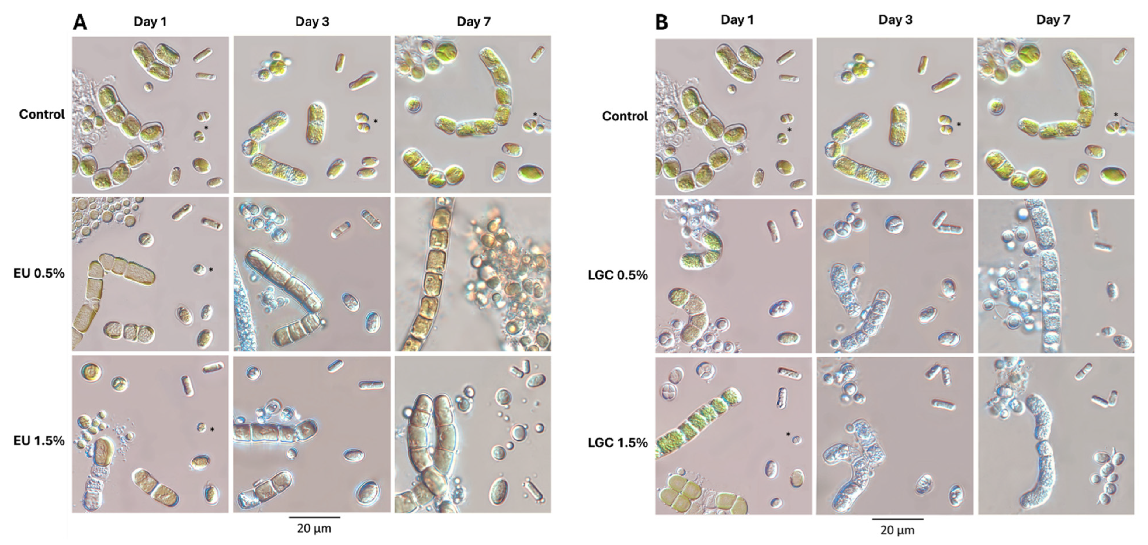
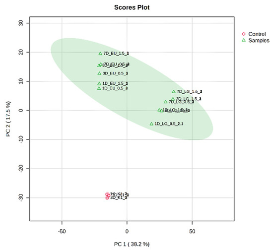
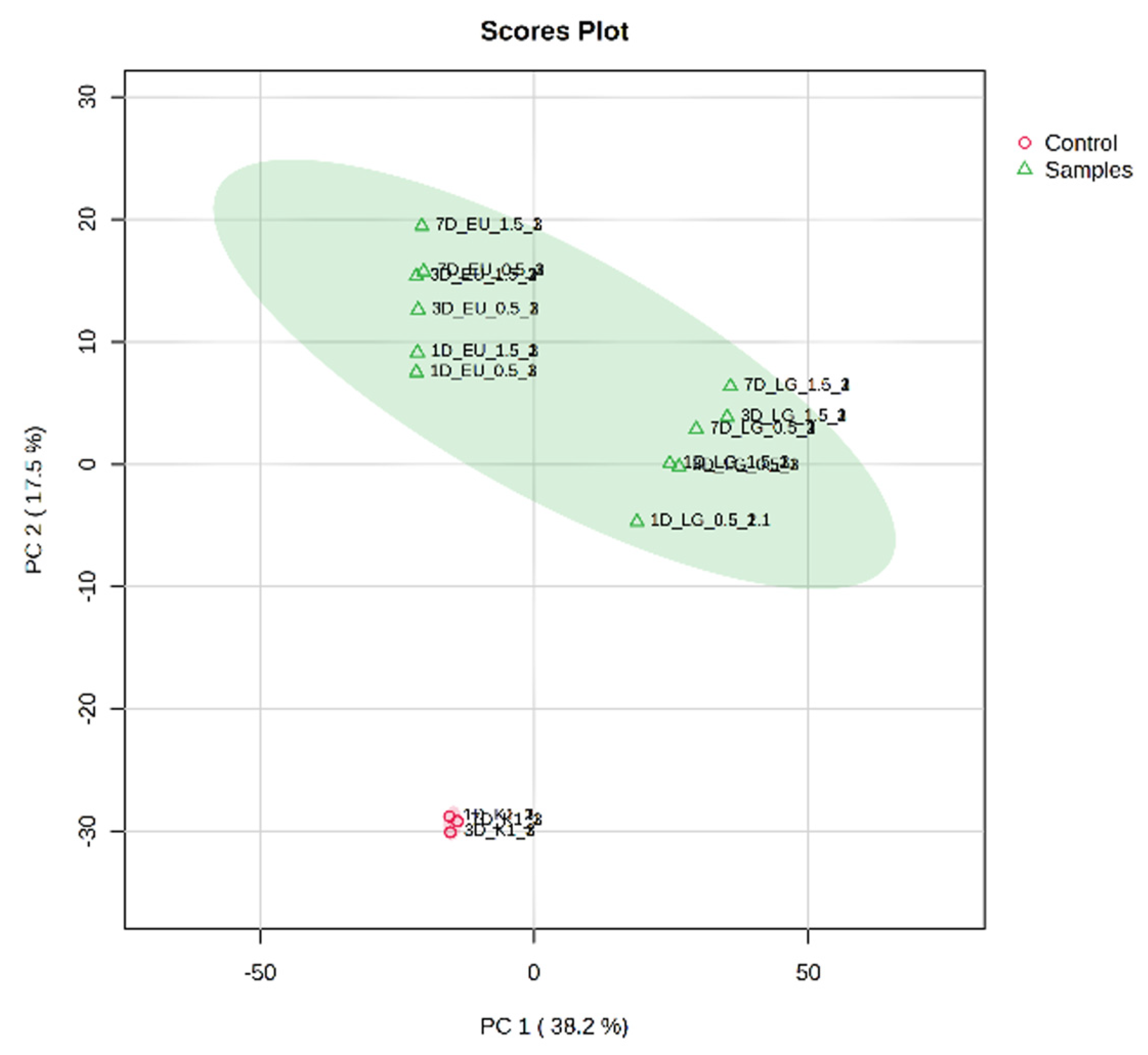
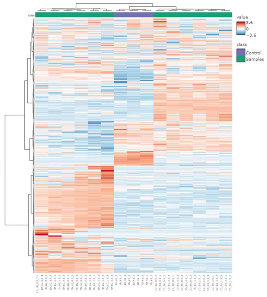
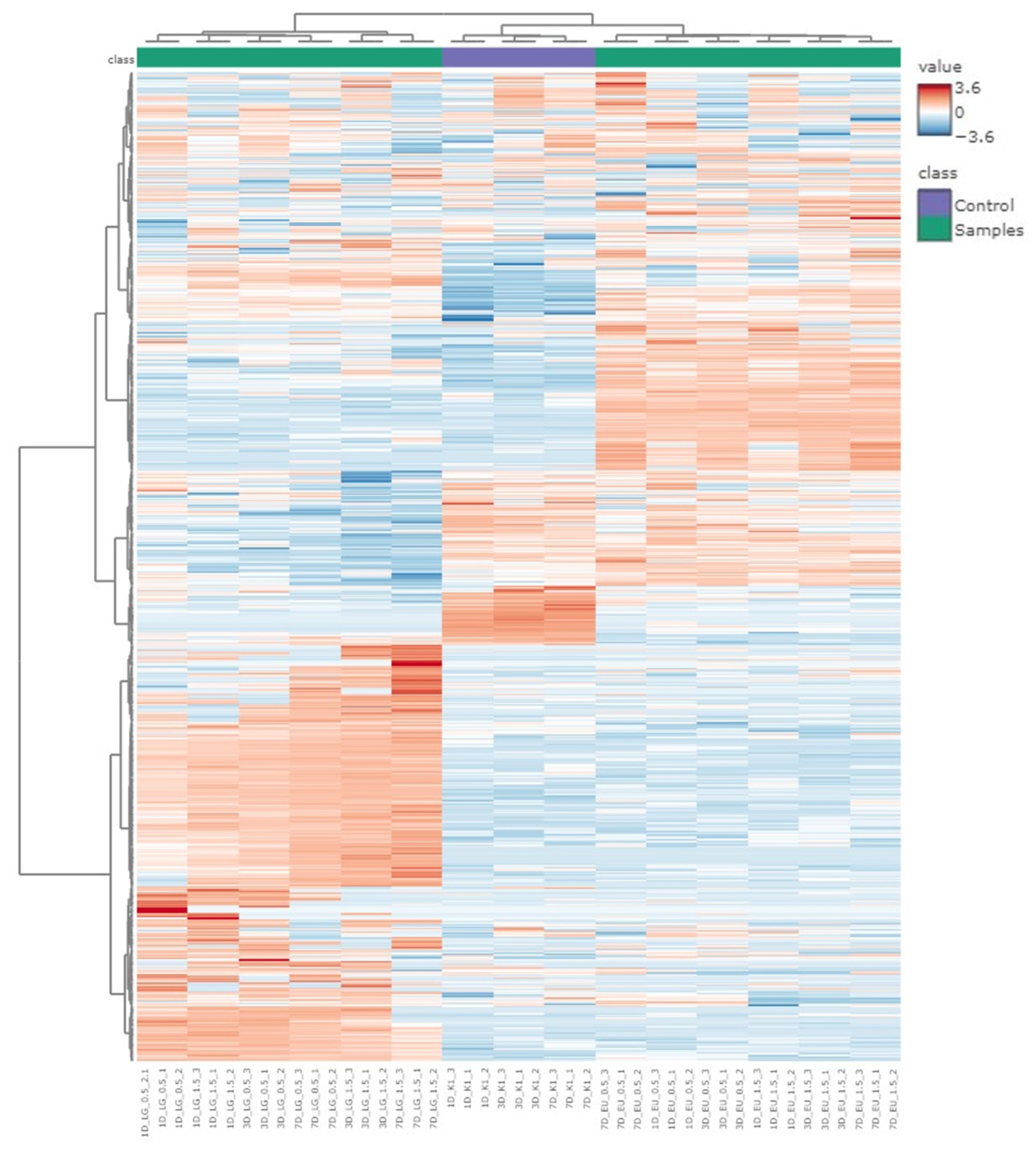

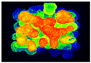
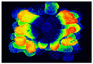
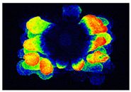
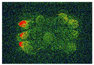
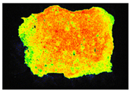
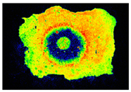
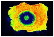
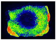
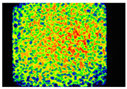
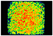
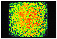
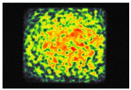
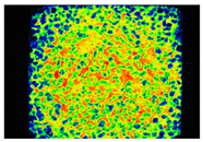
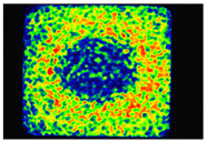
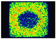
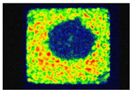
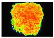
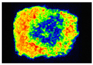
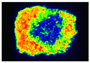
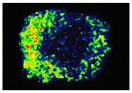
Disclaimer/Publisher’s Note: The statements, opinions and data contained in all publications are solely those of the individual author(s) and contributor(s) and not of MDPI and/or the editor(s). MDPI and/or the editor(s) disclaim responsibility for any injury to people or property resulting from any ideas, methods, instructions or products referred to in the content. |
© 2026 by the authors. Licensee MDPI, Basel, Switzerland. This article is an open access article distributed under the terms and conditions of the Creative Commons Attribution (CC BY) license.