


Highlights
What are the main findings?
- Electrospun bio-based PLA nanofibrous coatings were fabricated.
- Drug release followed biphasic burst–diffusion behavior.
- Fickian diffusion was the dominant release mechanism.
- Peppas–Sahlin best described release kinetics.
- Coatings showed biocompatible and antioxidant properties.
What are the implications of the main findings?
- Enables tunable drug-eluting biodegradable coatings.
- Supports design of diffusion-controlled PLA systems.
- Provides mechanistic insight into nanofibrous release behavior.
- Advances sustainable bio-based polymer coatings.
- Suitable for biomedical surface engineering applications.
Abstract
The development of sustainable and functional surface coatings has become a central theme in materials science due to increasing environmental concerns and expanding biomedical needs. This study reports the preparation and characterization of bio-based polylactic acid (PLA) nanofibrous coatings fabricated by electrospinning, aimed at delivering biodegradable and bioactive surfaces with controlled release functionality. Electrospinning enabled the formation of uniform nanofibrous coatings with high surface area and morphology reminiscent of extracellular matrices, presenting potential as multifunctional coating platforms. The obtained coatings were systematically evaluated for their physicochemical and biological properties, including antioxidant activity, cytocompatibility, and the controlled release of acetylsalicylic acid as a model bioactive agent. Release behavior was analyzed to elucidate the dominant transport mechanisms, revealing a biphasic profile characterized by an initial burst followed by sustained diffusion-controlled release. These results demonstrate that the electrospun PLA coatings successfully combine biodegradability, bioactivity, and tunable release properties within a bio-based polymer coating format. The findings highlight the potential of such bio-based polymer coatings for biomedical applications where biodegradable and functional surface solutions are required, and contribute to the broader advancement of eco-friendly and multifunctional coating technologies.
1. Introduction
The development of advanced biomaterials capable of mimicking the natural extracellular matrix (ECM) has become a key research area in regenerative medicine and biomedical engineering [1,2,3,4,5]. In recent years, increasing attention has also been directed toward bio-based and biodegradable polymer coatings that not only protect surfaces but actively interact with the biological environment. Such coatings are designed to support cell adhesion, modulate biological responses, and enable localized delivery of therapeutic agents, thereby expanding their role beyond conventional passive surface layers.
Among the available fabrication techniques, electrospinning has gained particular interest due to its ability to produce nanofibrous polymer coatings with high specific surface area, interconnected porosity, and architecture closely resembling native tissues such as cardiac muscle [1,2,3,4,5]. The electrospinning process involves the application of a high electric field to a polymer solution or melt, leading to the formation of a Taylor cone and the ejection of ultrafine fibers that solidify with diameters typically in the nanometer range [1,2,3,4,5]. When deposited onto a collector or substrate, these fibers form uniform nanofibrous coatings that can serve as functional biomedical surfaces.
Electrospun coatings offer significant advantages for biomedical applications, as their ECM-like morphology promotes cell adhesion, proliferation, and differentiation [2,3,4,5]. By adjusting electrospinning parameters such as applied voltage, flow rate, collector rotation speed, and solution concentration, it is possible to precisely tailor fiber diameter, pore size, and coating thickness, thereby influencing biological performance and mass transport properties. Such control is particularly important for coatings intended for cardiovascular-related applications, where surface–cell interactions and controlled bioactivity play a critical role [6,7,8,9,10].
Numerous electrospinning techniques have been developed to accommodate different polymers and functional requirements. These include melt electrospinning, which relies on the extrusion and rapid solidification of thermoplastic polymers; solution electrospinning, widely used for thermally sensitive and biodegradable polymers and based on solvent evaporation; and emulsion electrospinning, which enables the fabrication of composite nanofibers with enhanced functionality, including drug delivery capability [8,9,10,11,12]. Each method offers distinct advantages depending on the physicochemical properties of the polymer system and the intended coating application [10,11].
A typical electrospinning setup consists of a high-voltage power supply, a syringe or capillary spinneret, and a grounded collector [13,14,15]. Process parameters such as applied voltage, needle-to-collector distance, and solution flow rate strongly influence fiber continuity, uniformity, and porosity [10,14,15]. For instance, lower flow rates generally favor sufficient solvent evaporation and the formation of defect-free fibers, whereas excessively high flow rates may lead to bead formation due to incomplete drying [15]. In addition, the physicochemical properties of the polymer solution, including viscosity, concentration, surface tension, and molecular weight, must be carefully optimized to ensure stable jet formation and reproducible coating morphology [7,10,16].
A wide range of natural and synthetic polymers has been explored for electrospun biomedical coatings. Among them, polylactic acid (PLA), polycaprolactone (PCL), and poly(lactic-co-glycolic acid) (PLGA) are the most frequently used synthetic polymers due to their biodegradability, biocompatibility, and favorable mechanical properties [5]. These polymers are often combined with natural biopolymers such as collagen, gelatin, or chitosan to enhance biological performance. In particular, PLA-based nanofibrous coatings exhibit promising degradation behavior and mechanical stability, making them suitable for temporary biomedical applications, including vascular grafts, wound dressings, and coatings for cardiac tissue regeneration [5].
Electrospun nanofibrous coatings can be fabricated under mild conditions at room temperature and atmospheric pressure, possess a high surface-to-volume ratio, and enable efficient incorporation of bioactive compounds [5,17,18]. These features make them attractive platforms for controlled and localized drug delivery, which is especially beneficial for reducing inflammation and preventing infection at implantation sites [7]. Consequently, electrospun coatings have been investigated not only for tissue engineering scaffolds but also for biosensors, filtration membranes, protective materials, and advanced biomedical surface coatings [5,7].
Alongside electrospinning, 3D bioprinting technologies have been developed to fabricate patient-specific tissue constructs, including vascularized cardiac patches [6,19]. Although bioprinting offers precise spatial control, reproducing the fibrous microenvironment characteristic of native ECM remains challenging. In this context, electrospun nanofibrous coatings can provide complementary structural cues that guide cellular behavior and enhance biological integration when combined with printed constructs or bioactive components.
An additional strategy to enhance the functionality of electrospun coatings involves the incorporation of nanoparticles within the nanofibrous matrix. Due to their high surface area and tunable surface chemistry, nanoparticles can impart antibacterial, antioxidant, imaging, or targeted drug delivery properties to the coating system [10]. However, nanoparticles intended for biomedical use must exhibit appropriate biocompatibility and biodegradability while minimizing the risk of inflammatory or cytotoxic responses. Their size, shape, and surface modification play a decisive role in cellular interactions and biodistribution [20].
Rare earth metal oxide nanoparticles, such as gadolinium oxide (Gd2O3), are widely used as contrast agents in magnetic resonance imaging, although their potential toxicity necessitates careful optimization and surface modification [21,22]. Bismuth oxide nanoparticles (Bi2O3), on the other hand, demonstrate high electrochemical stability and favorable biocompatibility, making them attractive for biosensing and targeted therapeutic applications [23]. In the context of cardiovascular disease treatment, nanotechnology-based systems offer new opportunities for localized drug delivery and tissue regeneration with reduced systemic side effects [24,25,26]. Nevertheless, nanoparticle cytotoxicity remains strongly dependent on particle size and surface characteristics, underscoring the importance of thorough biological evaluation prior to clinical translation [10,27,28].
Although electrospun PLA-based fibrous systems and drug-eluting coatings have been widely investigated, the combined use of plant-derived acetone extracts of Kalanchoe pinnata and Sideritis together with bismuth oxide (Bi2O3) and gadolinium oxide (Gd2O3) within a single bio-based electrospun PLA coating platform has not been systematically explored in the context of biodegradable, bioactive surface layers with short-term cardioprotective drug-release functionality. In particular, limited attention has been given to how such hybrid organic–inorganic modifications influence not only antioxidant and cytocompatible behavior, but also the mechanism of acetylsalicylic acid release from electrospun PLA coatings. The specific problem addressed in this study is therefore whether the incorporation of novel plant extracts and metal-oxide additives into electrospun PLA coatings can generate multifunctional biodegradable surfaces with tunable release behavior and preserved biological performance. We hypothesized that the simultaneous introduction of bioactive plant extracts and oxide nanoparticles, especially Bi2O3, would modify the internal structure and interfacial properties of the coating, resulting in measurable changes in antioxidant activity, cytocompatibility, and the balance between burst release and diffusion-controlled transport. Accordingly, the distinct contribution of this work lies in the development and characterization of a hybrid bio-based PLA coating system enriched with previously uncombined extract–nanoparticle additives, together with a comparative kinetic analysis demonstrating how composition affects the release profile and confirms the predominance of diffusion-driven aspirin release.
2. Materials and Methods
2.1. Materials Preparation
2.1.1. Chemicals and Reagents
A range of reagents and chemical compounds was employed in this study. Granulated polylactide (PLA) was supplied by Goodfellow Cambridge Ltd. (Huntingdon, UK). Acetone (analytical grade), sodium hydroxide, sulfuric acid solution (95%), acetic anhydride, and nitric acid solution (65%) were obtained from Warchem (Warsaw, Poland). Bismuth(III) nitrate monohydrate was purchased from Polskie Odczynniki Chemiczne (Gliwice, Poland). Polyethylene glycol with an average molecular weight of 2000 (PEG 2000) was supplied by RBI (Warszawa, Poland). Isopropanol was obtained from Chempur (Piekary Śląskie, Poland), while gadolinium(III) oxide was purchased from OnyxMet (Olsztyn, Poland). 1,4-Dioxane and distilled water were used for solution preparation, dilution, and washing procedures.
Plant-derived acetone extracts of Kalanchoe pinnata leaves and Sideritis were prepared at a fixed ratio of 10 g of plant material per 250 mL of acetone. Prior to extraction, the plant materials were visually inspected and dried under controlled conditions. The extracts were prepared under identical conditions to ensure reproducibility of the extraction procedure. After extraction, the solutions were filtered to remove solid residues and stored in sealed containers at room temperature until further use.
Simulated body fluid (SBF) solution was used for selected degradation studies. A DPPH solution with a concentration of 25 mg/L was prepared for antioxidant activity evaluation. Acetylsalicylic acid was synthesized manually according to the procedure described in a subsequent section of this work and was used as a model bioactive compound in the release studies. The extraction ratio (10 g plant material per 250 mL acetone) and extraction time were kept constant for all preparations. The obtained extracts were filtered before use to remove solid residues and stored in sealed containers at room temperature until further use.
2.1.2. Preparation of Polymer Solutions
Three polymer solutions with different compositions were prepared, as summarized in Table 1. Each formulation contained 5 g of polylactide (PLA) granules, 10 g of 1,4-dioxane, and 20 g of acetone. Depending on the variant, an additional 20 g of either an acetone extract of Kalanchoe pinnata, an acetone extract of Sideritis, or pure acetone was added. The mixtures were stirred and heated using a magnetic stirrer (MS-H-Pro, Chemland, Poland) at 82 °C for 2 h until complete polymer dissolution was achieved. The resulting homogeneous solutions contained 10 wt% PLA.
Table 1.
Composition of the prepared polymer solutions.
2.1.3. Synthesis of Oxide Nanoparticles
To synthesize bismuth oxide nanoparticles, 1.6 g of bismuth(III) nitrate [Bi(NO3)3] was dissolved in 10 mL of distilled water. The resulting suspension was subjected to ultrasonic treatment in a pressure ultrasonic bath (Emmi-20HC, EMAG Technologies, Salach, Germany) while maintaining the temperature at 80 °C. Subsequently, 0.9 mL of 65% nitric acid (HNO3) was added dropwise. After complete dissolution of the Bi(NO3)3 precipitate and formation of a clear solution, 0.07 g of polyethylene glycol (PEG) dissolved in 7 mL of distilled water was added as a stabilizing agent. After 30 min of stirring, 17 mL of 4 M sodium hydroxide (NaOH) solution was slowly introduced, resulting in the formation of a homogeneous light-yellow suspension. The reaction was continued in the ultrasonic bath at 80 °C for an additional 2 h. The resulting precipitate was then separated from the supernatant by repeated filtration using a sintered glass filter funnel. In the case of gadolinium oxide, commercially available Gd2O3 nanoparticles were used directly without additional synthesis.
2.1.4. Preparation of Aspirin Solution
Aspirin was synthesized by the acetylation of salicylic acid. Briefly, 4.0 g of salicylic acid was weighed and transferred into a 100 mL Erlenmeyer flask. Subsequently, 6.0 mL of acetic anhydride and two drops of concentrated sulfuric acid were added. The reaction mixture was gently swirled to ensure homogenization and then heated in a water bath at 60–70 °C for 20 min, with intermittent mixing. After heating, the flask was removed from the bath and 40 mL of distilled water was added with thorough stirring. The mixture was then cooled in an ice-water bath for 10 min to promote crystallization. The resulting precipitate was isolated by vacuum filtration using a Büchner funnel and washed twice with cold distilled water under reduced pressure. The collected solid was dried at 60 °C for 1 h and subsequently at 105 °C for an additional 30 min.
2.2. Methods
2.2.1. Electrospinning Membrane Fabrication Procedure
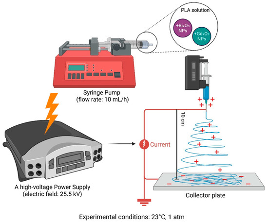
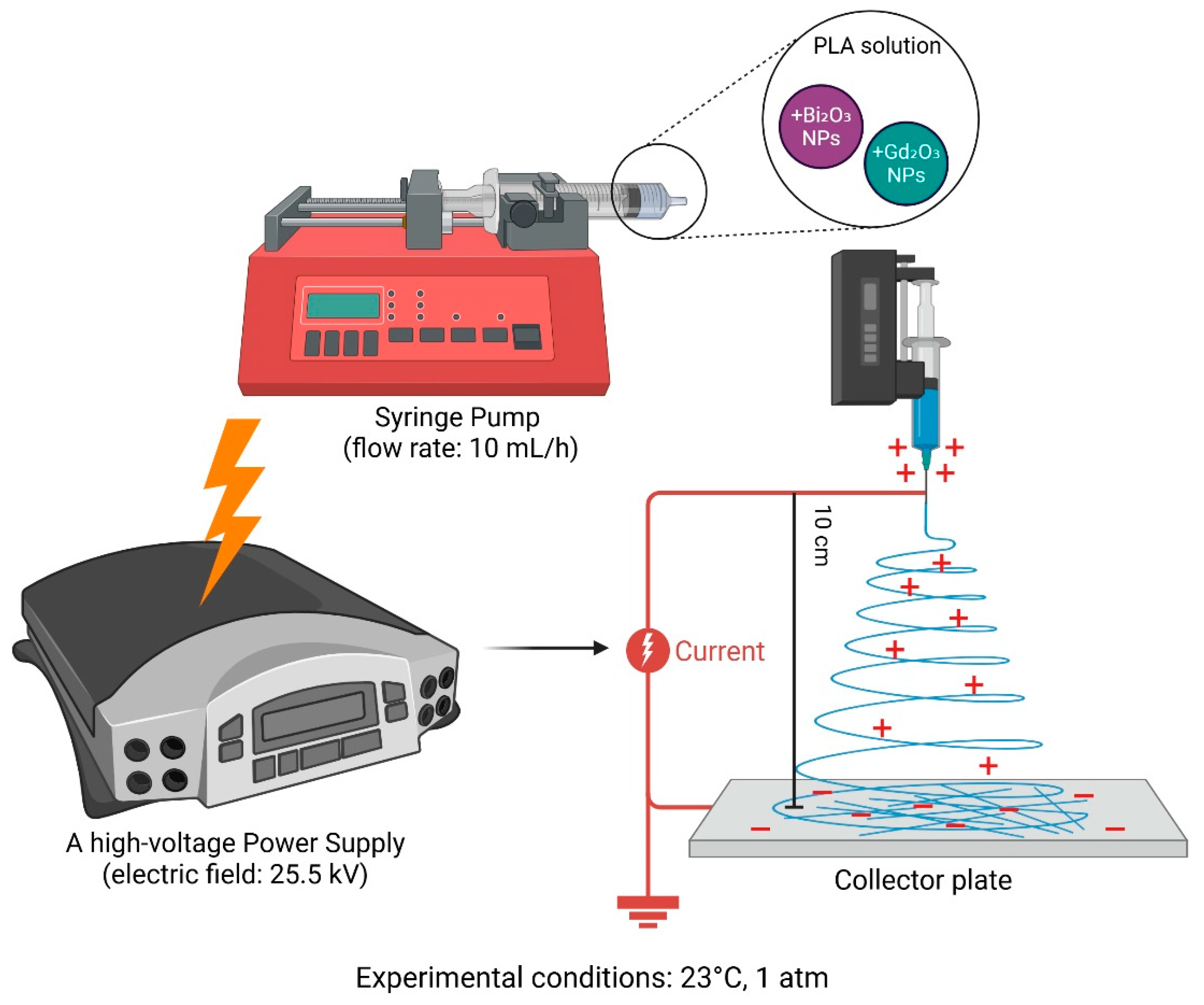
Electrospinning was carried out using a programmable syringe pump NE-8000 (Lab-Partner, Warsaw, Poland) operated at a constant flow rate of 10 mL/h. A high-voltage power supply (LEYBOLD, Köln, Germany) set to 25.5 kV was used to generate the electric field required for fiber formation. The polymer solution was loaded into a 10 mL plastic syringe fitted with a metallic needle. The fibers were deposited onto a grounded collector covered with aluminum foil to facilitate removal of the obtained coating. Each syringe was filled with a polymer solution containing either bismuth oxide or gadolinium oxide nanoparticles at the concentrations listed in Table 2. The distance between the needle tip and the collector was maintained at 10 cm. All electrospinning experiments were conducted under ambient conditions (23 °C, 1 atm). The electrospinning time was approximately 10 min per sample, which allowed coatings of comparable surface coverage to be obtained. A schematic representation of the electrospinning setup and the composition of the polymer solutions is presented in Figure 1.
Table 2.
Compositions of polymer solutions with added nanoparticles.
Figure 1.
Schematic representation of the electrospinning setup and polymer solution compositions.
2.2.2. Scanning Electron Microscopy and Energy Dispersive Spectroscopy (SEM-EDS)
The structure of the bismuth (III) oxide and the morphological characteristics of the electrospun membrane fibers were analyzed using Scanning Electron Microscopy (SEM) by Oxford Instruments, Buckinghamshire, UK. Additionally, Energy Dispersive Spectroscopy (EDS) was performed on the bismuth (III) oxide nanoparticles to determine their elemental composition. Fiber diameters were determined based on SEM images. All measurements were conducted at room temperature. Fiber diameters were determined from SEM micrographs using image analysis software Fiji J (version 2023) by measuring representative fibers from randomly selected regions of each sample.
2.2.3. In Vitro Biodegradation Study
In vitro biodegradation of the membranes was carried out in a Simulated Body Fluid (SBF) solution. Pre-weighed membranes were placed in containers with 20 mL of SBF. The pH of the solution was measured immediately after immersion and again after 20 h. Subsequently, the membranes were removed, weighed, and air-dried at room temperature (22 °C) for one week. After drying, the membranes were re-immersed in fresh SBF solution, and the pH was measured. A second pH measurement was performed after two weeks, followed by drying the membranes in an oven at 60 °C for 45 min.
2.2.4. Swelling Determination
The weighed membranes were placed in individual wells of a multi-well plate, and 4 mL of distilled water was added to each well. The membranes were left to swell in the water for 20 h. After the swelling period, the membranes were removed, and their swollen mass was weighed again.
2.2.5. Determination of the Wetting Angle
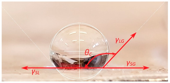
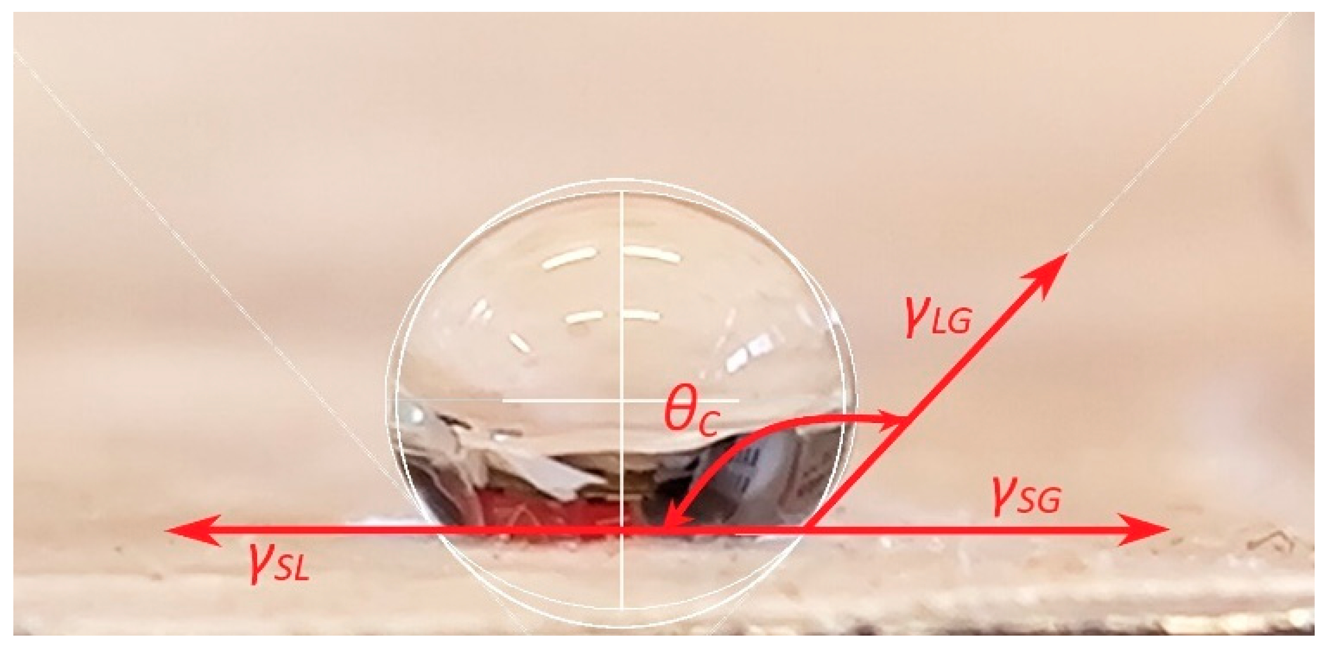
The surface wettability of the polymer membranes was evaluated using the sessile drop method. A drop of distilled water was placed on the surface of each membrane, and the static contact angle (θ) was measured by determining the angle formed between the baseline of the droplet and the tangent at the liquid–solid–air interface. All measurements were performed under ambient conditions. Droplet images were captured using a camera positioned perpendicular to the membrane surface (Nikon D3100, Warsaw, Poland). Below in Figure 2 is an example image of measurement procedure. Contact angle values were analyzed using ImageJ software (version 2023) equipped with the Contact Angle plugin. To allow the droplet to stabilize, measurements were taken 10 s after deposition. For each membrane sample, ten initial measurements were recorded, from which three representative values were selected. The average of these three measurements was reported as the final contact angle (θ) for each sample, and these values were also used to estimate the associated measurement uncertainty.
Figure 2.
Example image of wetting angle measurement of membrane samples.
2.2.6. Measurement of Antioxidant Capacity by the DPPH Radical Method
The antioxidant activity of the polymer membranes was evaluated using a modified version of the Brand-Williams method with the synthetic radical DPPH (1,1-diphenyl-2-picrylhydrazyl). A DPPH solution with a concentration of 25 mg/L was prepared for the assay. Membrane samples were immersed in the DPPH solution, and the decrease in absorbance was monitored. Measurements were carried out at a wavelength of 517 nm using an Agilent 8453 UV–Visible spectrophotometer (Agilent Technologies, Santa Clara, CA, USA). The reduction in absorbance values was used to assess the radical scavenging ability of the membranes.
2.2.7. Aspirin Release
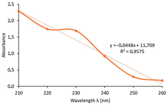
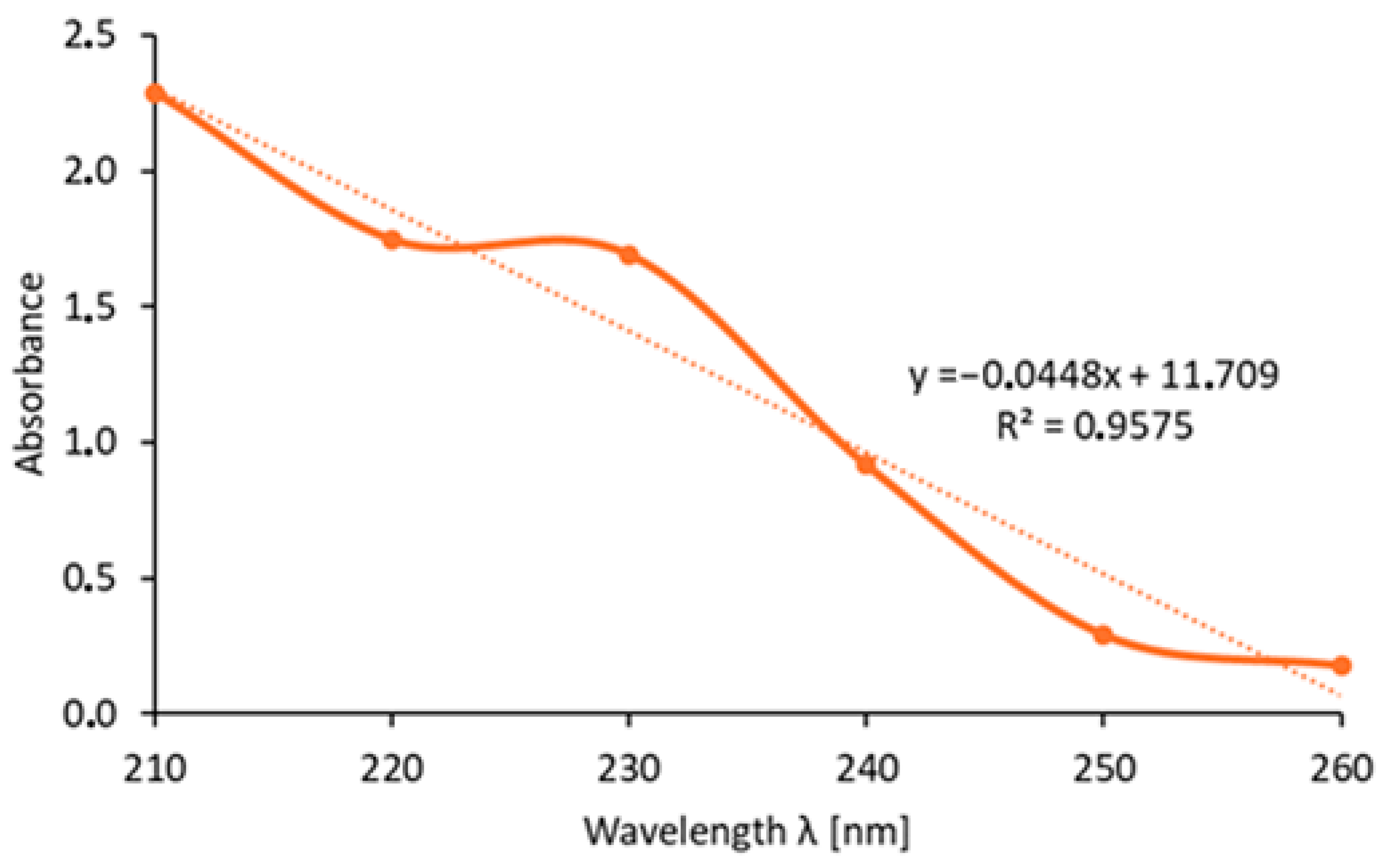
Aspirin release studies were performed using 50 mL beakers, each filled with exactly 50 mL of phosphate-buffered saline (PBS, pH 7.4). The beakers were placed on magnetic stirrers ENVAG to maintain continuous mixing. Membrane samples containing immobilized aspirin were then immersed in the PBS solutions. At predetermined time intervals (5, 10, 30, 60, and 120 min), 1 mL aliquots of the PBS solution were collected and transferred into separate test tubes for analysis. The absorbance of the collected samples was measured using an Agilent 8453 UV–Visible spectrophotometer. A calibration solution of aspirin was first prepared by dissolving 50 mg of aspirin in 10 mL of 96% ethanol, followed by a 100-fold dilution to achieve a final concentration of 50 mg/L. The absorbance spectrum of this standard solution was recorded over the wavelength range of 210–260 nm in order to determine the wavelength of maximum absorption. Figure 3 presents the dependence of the absorbance of the 50 mg/L standard aspirin solution on the wavelength (λ), which was used to select the optimal detection wavelength for the release analysis. Absorbance measurements of the PBS samples obtained during the release experiment were subsequently conducted to quantify the amount of aspirin released from the membranes.
Figure 3.
Dependence of the absorbance of a standard aspirin solution at a concentration of 50 mg/L on the wavelength (λ).
2.2.8. Porosity and Density Testing
To assess the density and porosity of the polymer membranes, measurements were performed based on volume changes of isopropanol before and after membrane immersion. Prior to the experiment, membrane samples were weighed with an accuracy of four decimal places. An initial volume of 8 mL of isopropanol (V1) was poured into a graduated cylinder. Each membrane sample was then individually immersed into the liquid, and the resulting volume (V2) was recorded. After 15 min of immersion, the membrane was removed, and the final volume (V3) of the isopropanol was noted. Following each measurement, the isopropanol volume in the cylinder was replenished to the original 8 mL to maintain consistent conditions across all samples.
2.2.9. Cytotoxicity Test
The cytotoxicity assessment was performed using a quantitative XTT assay (Roche, Basel, Switzerland), following the manufacturer’s protocol and in accordance with ISO 10993-5 [29], which specifies the evaluation of cytotoxicity for medical devices. The osteosarcoma MG-63 cell line (Merck, Darmstadt, Germany), commonly used in biological assessments, was utilized for the test. Cells were cultured under standard conditions: in a humidified atmosphere with 5% CO2 at 37 °C in a Biosan incubator (DanLab, Białystok, Poland). The culture medium used was DMEM supplemented with 10% fetal bovine serum (FBS), 1% antibiotic/antimycotic solution, PBS, and trypsin (all from Merck). The medium was replaced every 48 h. The XTT assay is based on the measurement of cell viability through mitochondrial activity, which reduces the tetrazolium salt XTT to a water-soluble formazan dye. The intensity of the resulting color was measured spectrophotometrically, enabling the determination of the cytotoxic potential of the tested sample.
All experiments were performed in triplicate (n = 3). Results are presented as mean ± standard deviation (SD). Statistical differences were evaluated using one-way ANOVA followed by Tukey’s post hoc test. A p-value < 0.05 was considered statistically significant.
3. Results and Discussion
3.1. EDS Analysis of Elemental Composition
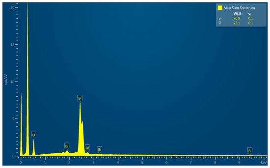
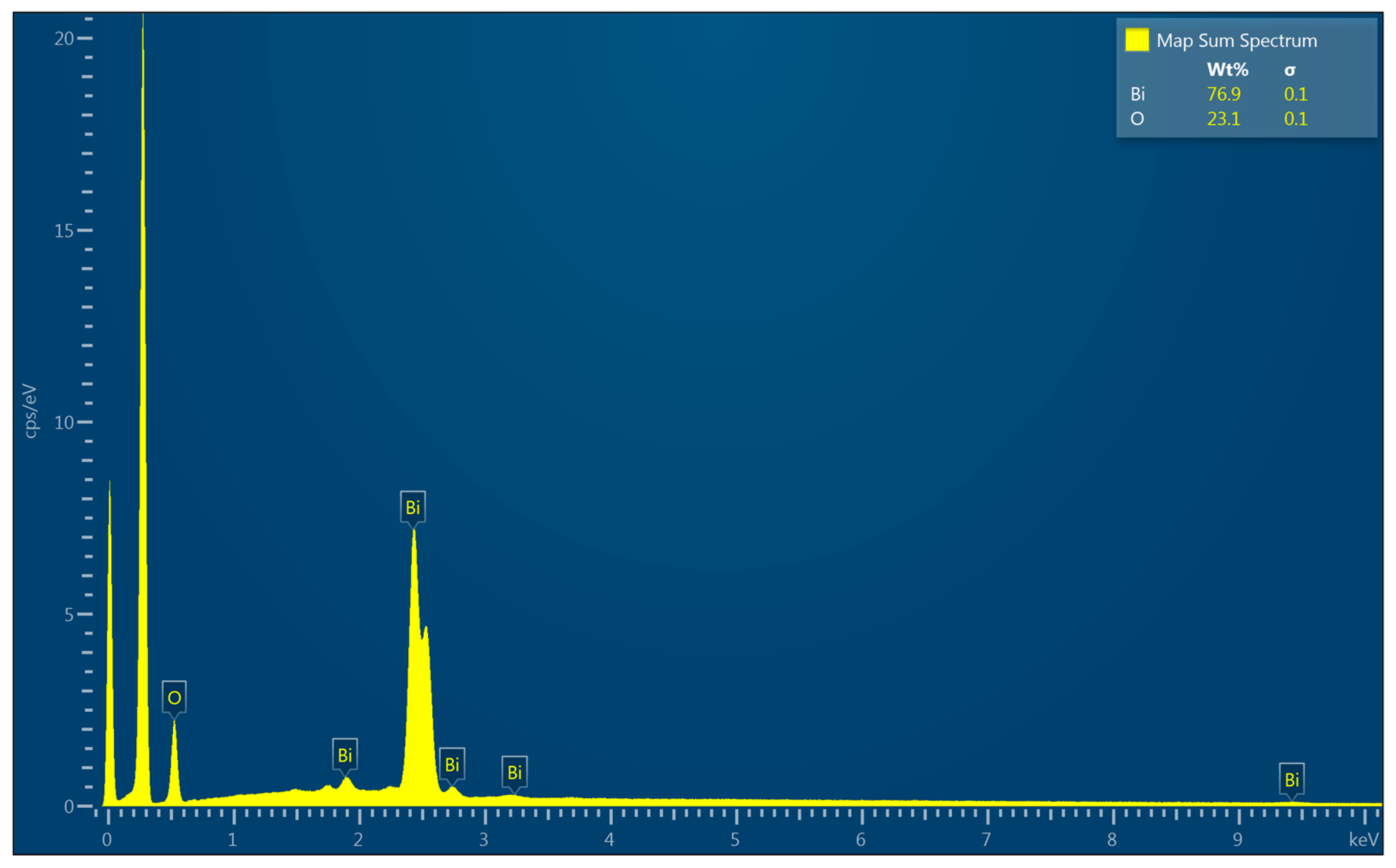
The elemental composition of bismuth (III) oxide, as presented in Figure 4. The EDS spectrum confirmed the presence of bismuth and oxygen, validating the successful synthesis of the material. The quantitative analysis indicated a composition consistent close the theoretical stoichiometry. Two dominant peaks observed in the low-energy region (0–0.4 keV) correspond to oxygen (O) and carbon (C), which originate from the oxide structure and the carbon coating applied during SEM sample preparation.
Figure 4.
EDS graph showing the analysis of bismuth (III) oxide nanoparticle samples.
3.2. SEM Analysis of Surface Morphology and Composition
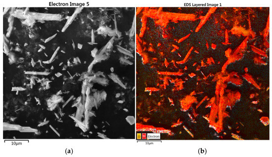
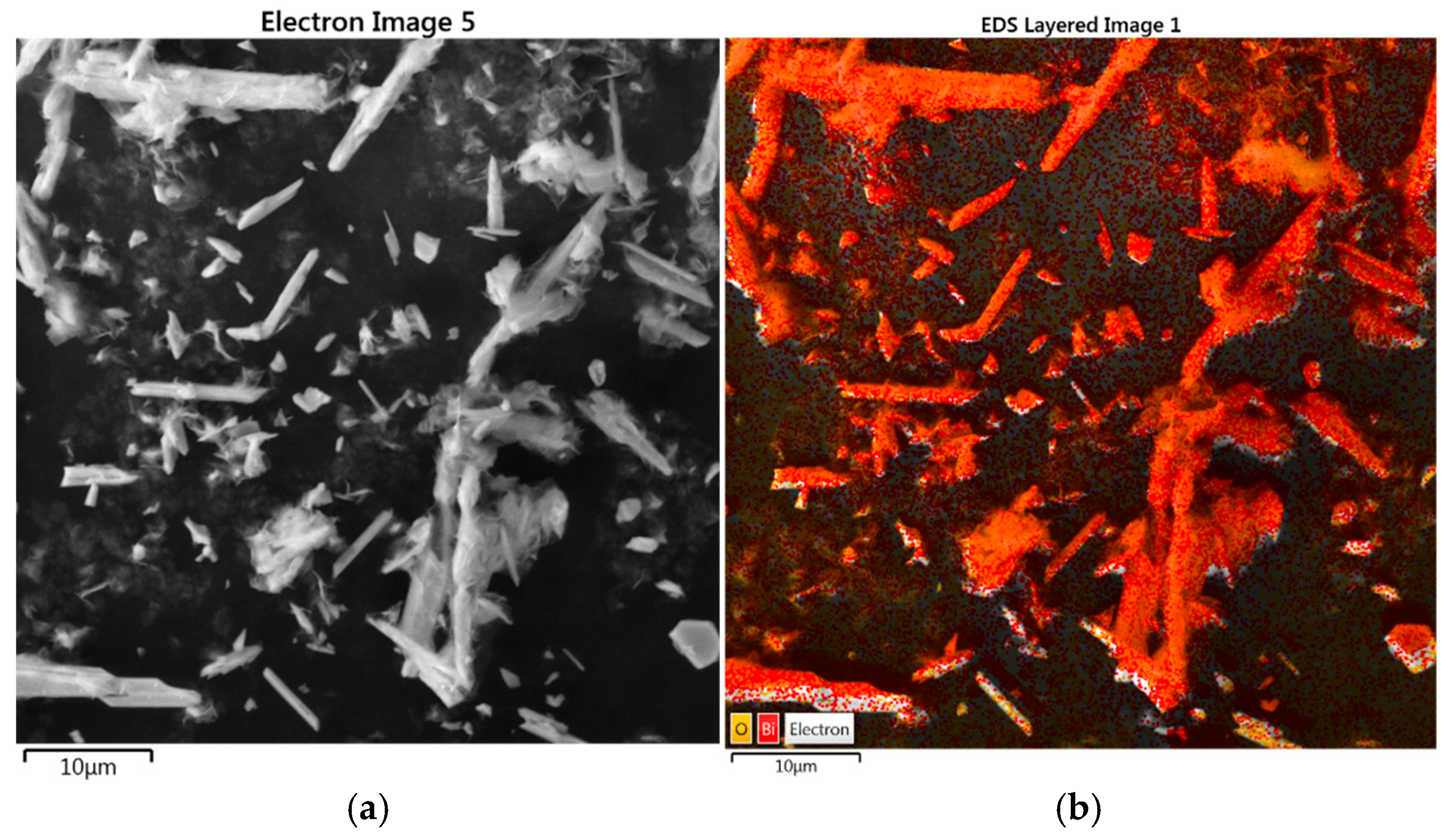
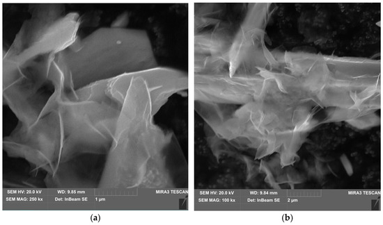
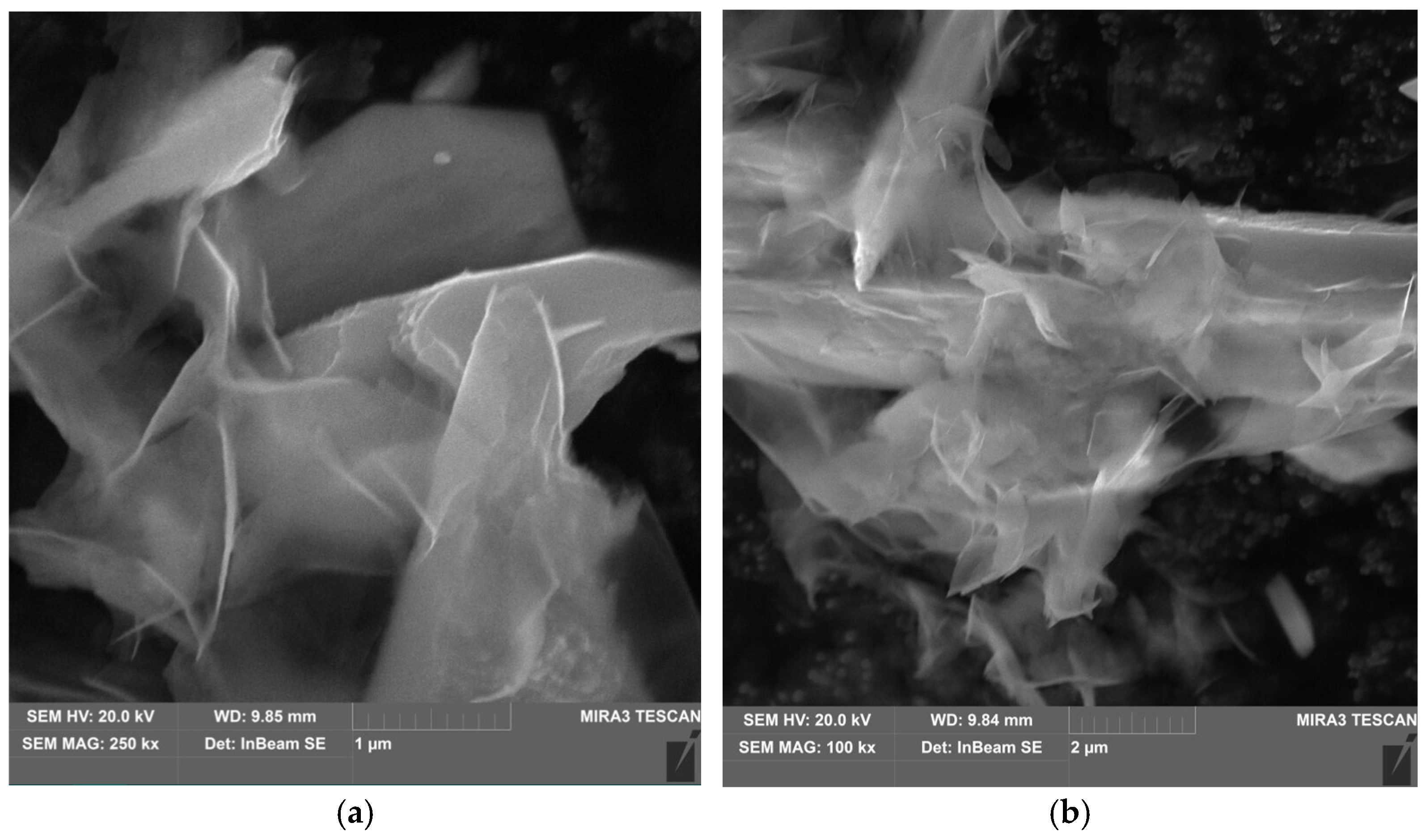
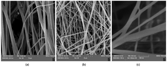
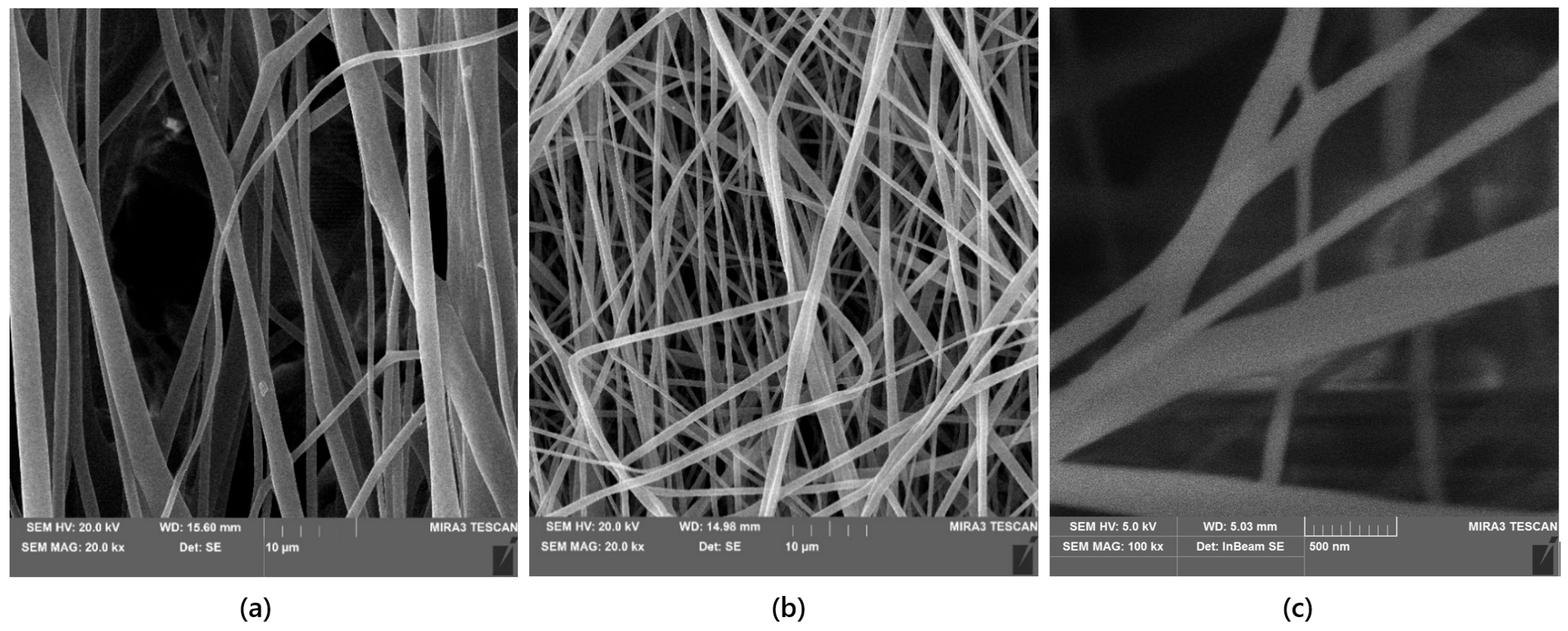
Scanning Electron Microscopy (SEM) was used to evaluate the morphology of the synthesized bismuth(III) oxide nanoparticles and the fibrous structure of the electrospun PLA-based coatings. As shown in Figure 5 and Figure 6, the Bi2O3 particles exhibited a platelet-like morphology. SEM images of the electrospun coatings (Figure 7) revealed a well-developed fibrous architecture without visible bead formation or major structural defects. The coating prepared from the PLA solution containing Kalanchoe pinnata extract (sample 1) showed a more ordered and smoother fibrous arrangement, whereas the pure PLA coating (sample 2) exhibited a denser and more randomly distributed fibrous network. Based on measurements from representative SEM micrographs, the average fiber diameters were 15.60 µm for sample 1 and 14.98 µm for sample 2, indicating that the main structural framework of the coatings was formed by micrometer-scale fibers. However, higher-magnification SEM imaging additionally revealed finer fibrous features in the submicron and nanoscale range, suggesting that the electrospun coatings exhibited a hierarchical micro-/nanofibrous morphology. This multi-scale organization may be beneficial for biomedical applications, as it combines the structural stability of thicker fibers with the high surface area associated with finer fibrous elements.
Figure 5.
Image of: (a) structure of bismuth (III) oxide nanoparticles; (b) elemental composition (EDS analysis).
Figure 6.
SEM images of bismuth (III) oxide nanoparticles at: (a) 250× magnification and (b) 100× magnification.
Figure 7.
SEM images of electrospun PLA-based coatings: (a) sample 1 (PLA solution with Kalanchoe pinnata extract), showing a more ordered fibrous structure; (b) sample 2 (pure PLA solution), showing a denser fibrous network; (c) higher-magnification image of sample 2, revealing finer nanoscale fibrous features within the coating. The SEM observations indicate a hierarchical micro-/nanofibrous morphology of the electrospun layers.
3.3. In Vitro Biodegradation Study
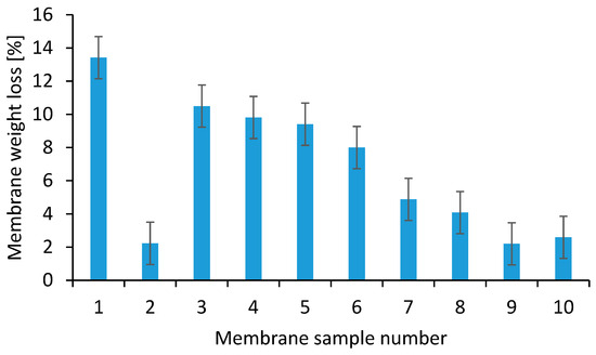
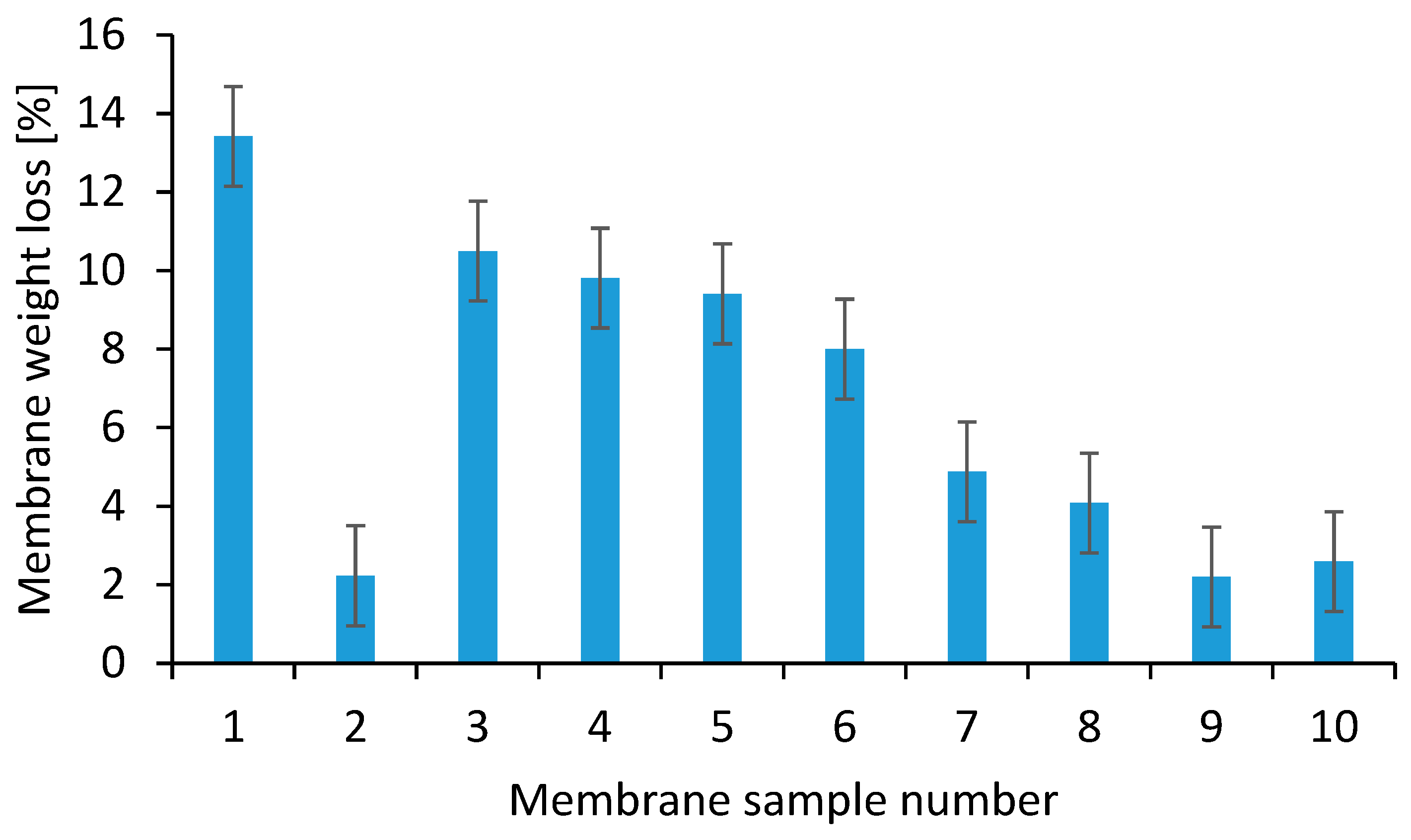
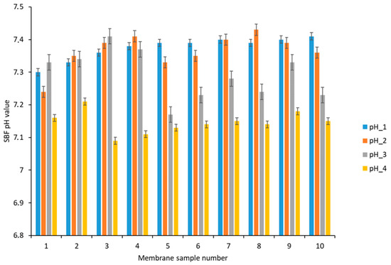
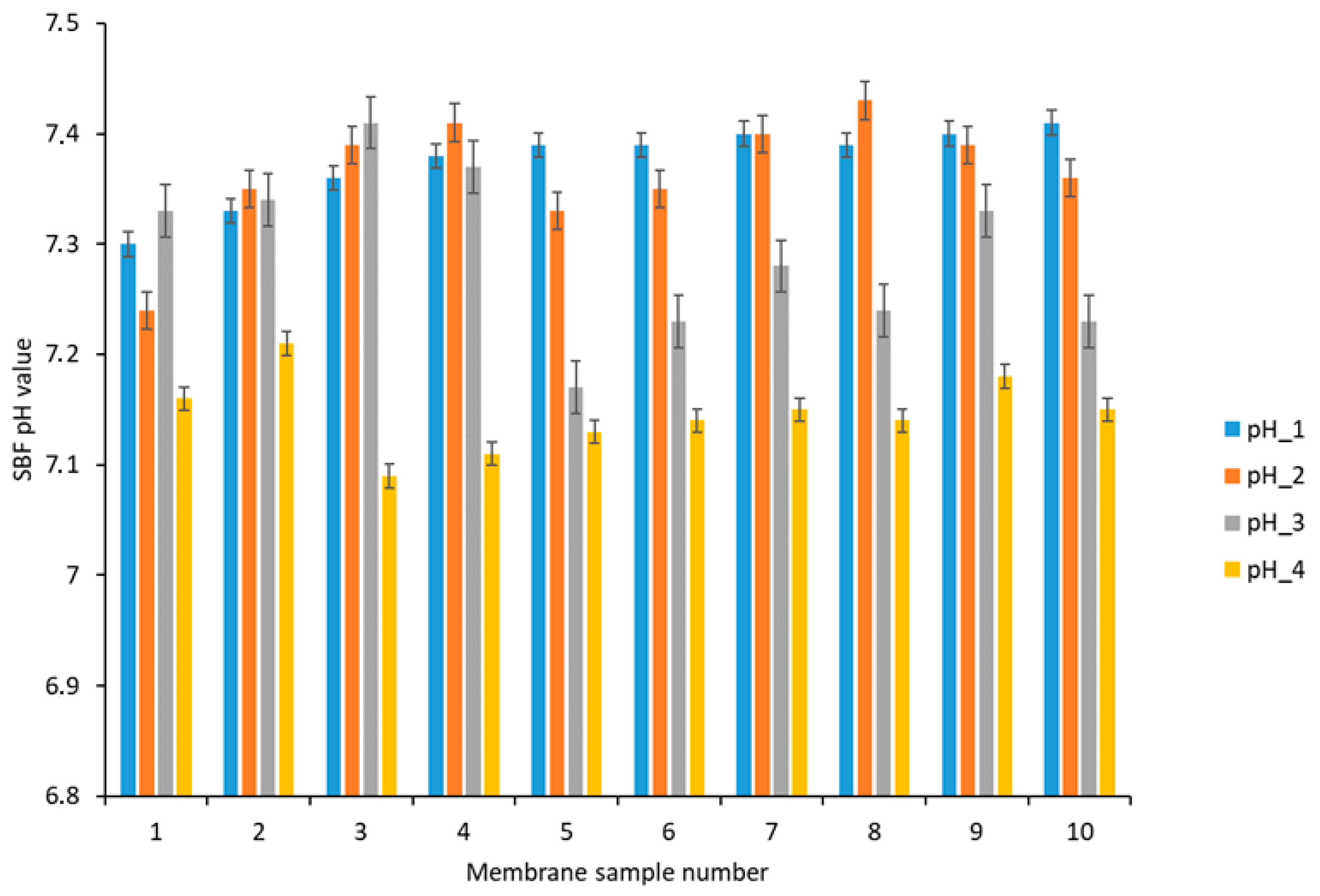
Biodegradability is a crucial factor for nanofibrous scaffolds, as they should be absorbed by the surrounding tissues without the need for surgical removal. The degradation rate should align with the speed of tissue formation as closely as possible. This way, as cells form their own extracellular matrix, the scaffold provides structural integrity within the organism and ultimately degrades, leaving behind the newly formed tissue, which will take on mechanical load responsibilities. In vitro biodegradation studies were conducted to assess the behavior of polylactic acid membranes in a medium with ion concentrations similar to those of human plasma. Degradation was determined based on the percentage loss of membrane mass as shown in Figure 8. After two weeks, the greatest mass loss (ranging from 4.08% to 13.42%) was observed in membranes 1 and 3–8, which contained a solution of Kalanchoe pinnata. In contrast, the lowest mass loss was found in sample 2 (pure polymer solution) and samples 9 and 10, which included an eanwort extract. A similar degradation behavior was observed for samples 9 and 10, which may be attributed to the reduced swelling capacity and lower porosity of these membranes compared with samples containing Kalanchoe extract. Lower swelling limits water penetration into the polymer matrix, which in turn slows hydrolytic degradation of PLA. Additionally, both after 15 h and two weeks (336 h) of immersing the membranes in the SBF solution, a decrease in pH was observed, indicating partial degradation of the polylactic acid membranes as shown in Figure 9.
Figure 8.
Percentage weight loss of membranes after two weeks.
Figure 9.
Changes in the pH of the SBF solution with membranes during degradation, successively: pH_1—Initial pH; pH_2—pH after 15 h; pH_3—pH value after membranes have dried; pH_4—pH after 336 h.
3.4. Swelling Determination
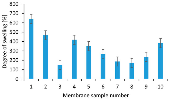
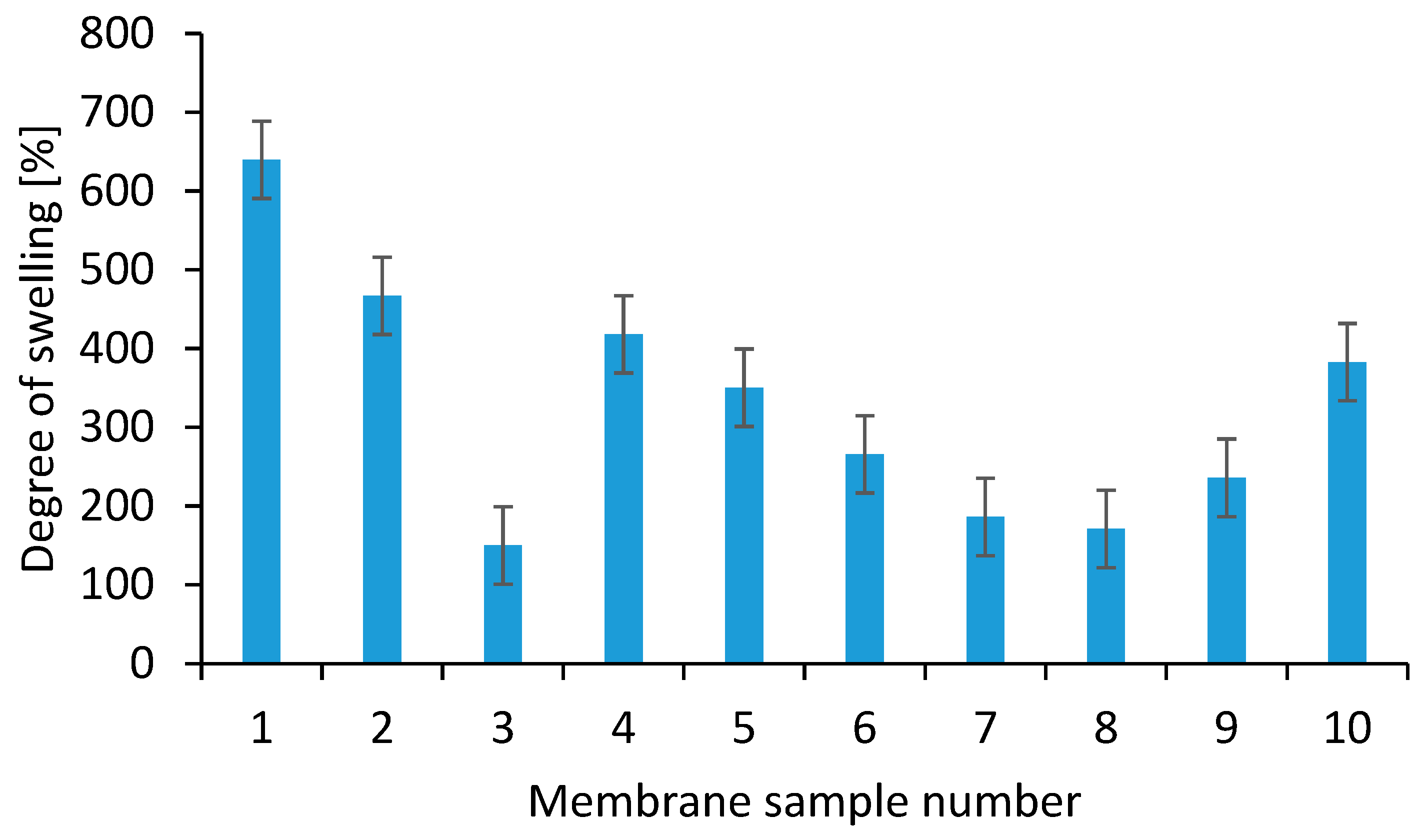
The swelling process is significantly influenced by temperature, pH, and the concentration of the substance. Generally, as temperature increases, so does the swelling of the material. The pH can also impact the swelling process since changes in pH alter the charges on the molecules, which in turn affects their arrangement. Higher concentrations of the substance result in greater swelling. In Figure 10, all nanofibrous membrane samples exhibited swelling after 20 h of incubation. Sample 1 (PLA solution + Kalanchoe pinnata extract) showed the greatest swelling ratio (639.7%), while the lowest swelling ratio (150.2%) was seen in sample 3 (PLA solution + Kalanchoe pinnata extract + 0.006 g of bismuth (III) oxide). Membranes made from polymer solutions (Samples 1 and 2) that did not contain nanoparticles displayed the highest swelling levels among all the samples. The obtained porosity and density values were consistent with the swelling behavior of the membranes. In general, samples with higher porosity exhibited lower apparent density and greater swelling capacity, which may be attributed to easier penetration of water into the fibrous structure. Conversely, membranes containing inorganic additives tended to show reduced porosity and lower swelling, suggesting partial densification of the fibrous network. These relationships confirm that the internal architecture of the electrospun coatings strongly influences both liquid uptake and physicochemical performance.
Figure 10.
Percentage of degrees of swelling in water of individual membrane samples.
3.5. Determination of the Wetting Angle
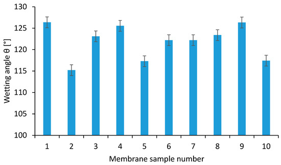
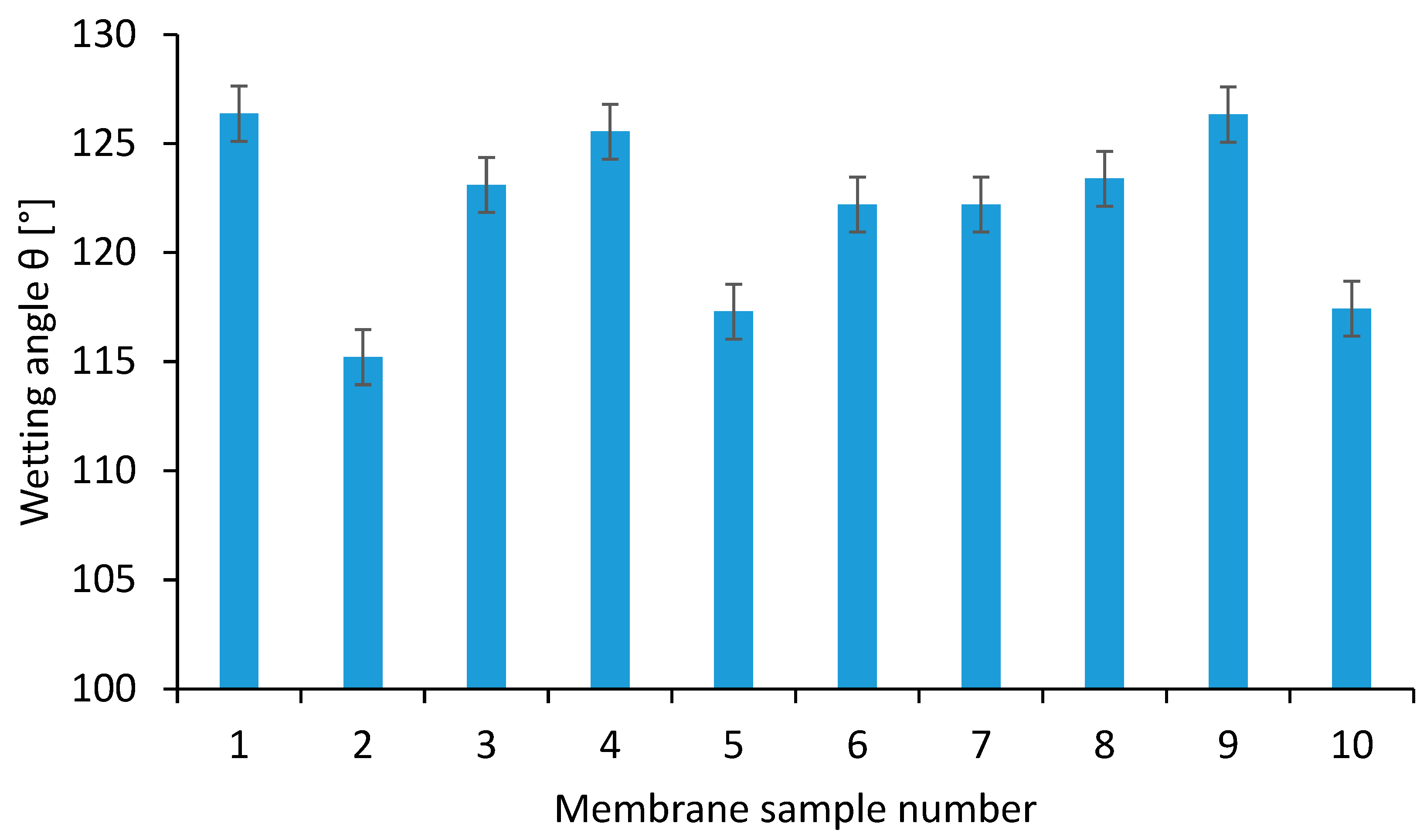
The contact angle is a method used to measure the wettability of a surface or material. Wettability is determined by measuring the contact angle formed between a liquid and a solid surface. The tendency for wetting increases as the contact angle or surface tension decreases. A hydrophilic material forms a contact angle of less than 90° with a liquid, while a hydrophobic material has a contact angle between 90° and 180°. According to the traditional definition, the contact angle (θ °C) is the angle formed between a liquid and a solid or liquid surface when the liquid is deposited on it. The contact angle values for the membrane samples as shown in Figure 11 range from 115.21° to 126.33°, indicating that all membrane surfaces exhibit hydrophobic properties. Although all coatings exhibited hydrophobic surface character, as reflected by contact angle values above 115°, the cytotoxicity results indicated that the materials remained cytocompatible. This may be explained by the fibrous surface topography, which can support cell attachment despite limited wettability, as well as by the presence of bioactive additives influencing the cell–material interaction. Therefore, in the present system, high contact angle values did not preclude biological compatibility.
Figure 11.
Summary values of wetting angles θ for individual membranes samples.
3.6. Measurement of Antioxidant Capacity by the DPPH Radical Method
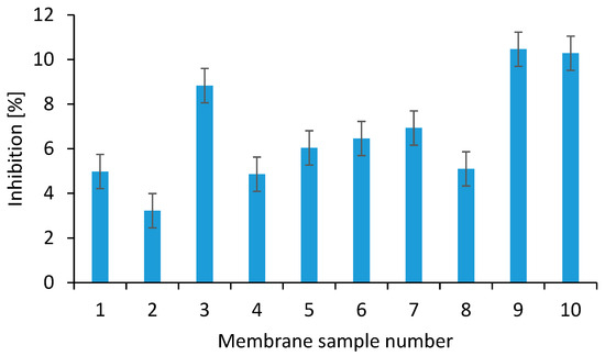
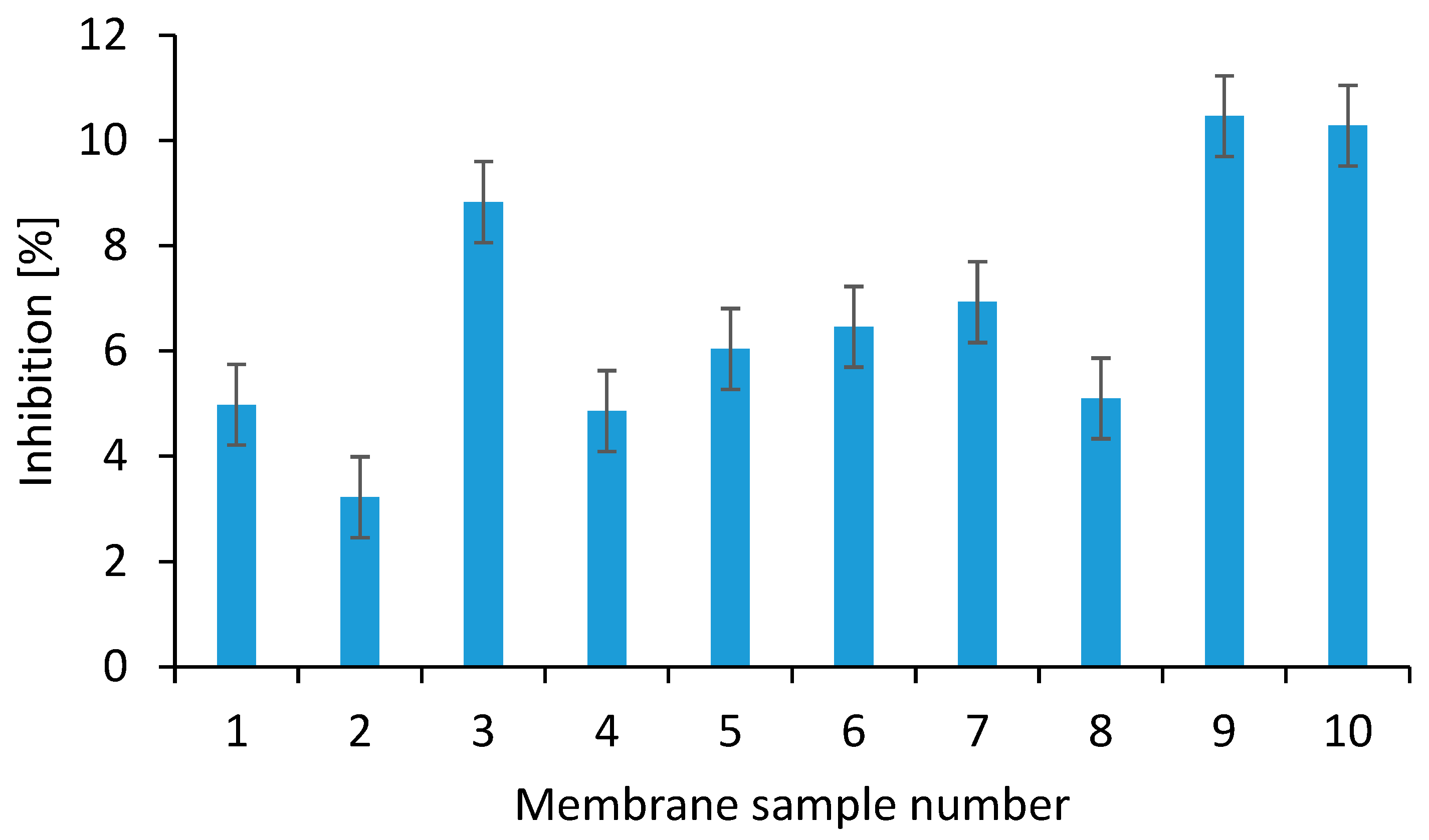
The DPPH reagent (1,1-diphenyl-2-picrylhydrazyl) forms a stable radical cation. Its solution has a dark violet color with a maximum absorbance in ethanolic solution at a wavelength of λ = 517 nm. When reacting with a substance capable of donating a hydrogen atom, it forms the reduced DPPH, leading to the disappearance of the violet color. The decrease in absorbance is proportional to the amount of oxidized DPPH remaining in the solution. Sample 2, which does not contain Kalanchoe pinnata or Sideritis extract, exhibited the lowest average inhibition percentage of 3.22% as shown in Figure 12. The inhibition percentages for the membrane samples 1, 3, 4, 5, 7, and 8, which included Kalanchoe pinnata extract, ranged from 4.86% to 8.83%. The highest inhibition percentage, and thus the greatest oxidative capacity, was observed in samples 9 and 10, which contained Sideritis extract. The pure polylactic acid solution, without the addition of Kalanchoe pinnata or Sideritis extract, demonstrated the lowest antioxidant activity.
Figure 12.
Summary results of average percentage inhibition of individual membrane samples.
3.7. Drug Release
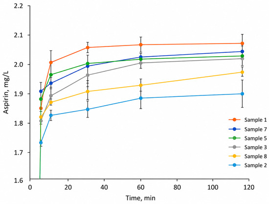
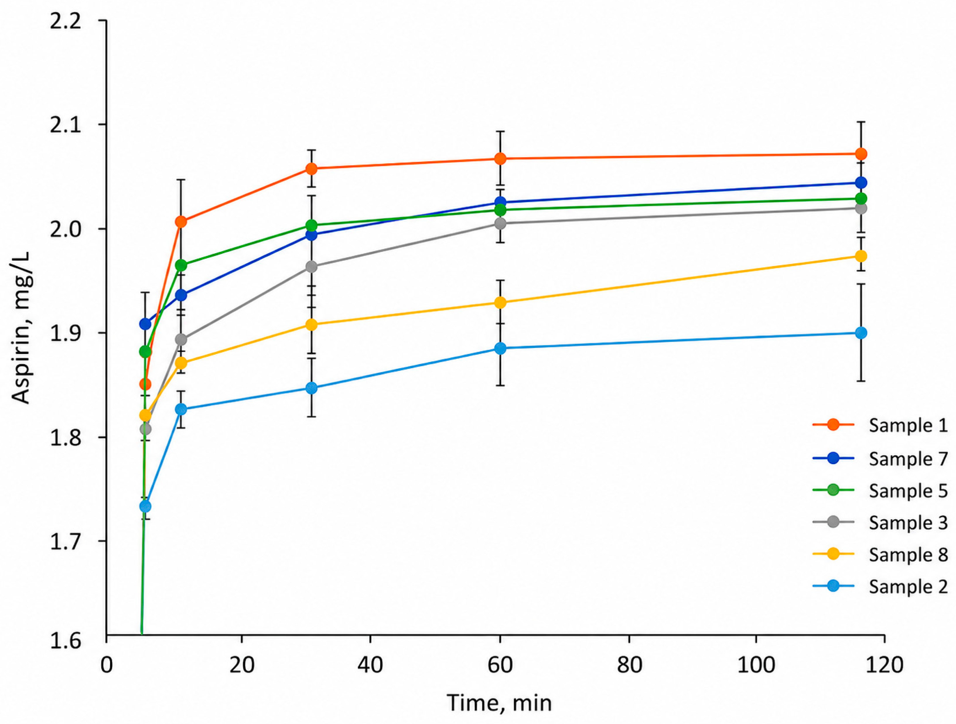
The initial concentration of the acetylsalicylic acid solution used for immobilization was 5 mg/mL. As shown in Figure 13, the concentration of the drug in the release medium, measured by UV absorbance at 230 nm, remained relatively stable between 1.8 and 2.0 mg/mL in most cases after the first 10 min. This indicates that the majority of the drug was rapidly released from the membranes within the initial 5–10 min of the test. An exception was observed for Sample 4, where a prolonged release profile was noted. After 120 min, the concentration of acetylsalicylic acid reached 2.87 mg/mL, suggesting a more sustained retention of the drug within the matrix in this particular formulation.
Figure 13.
Release profile of acetylsalicylic acid from membranes over 120 min.
3.8. Drug Release Kinetics Analysis
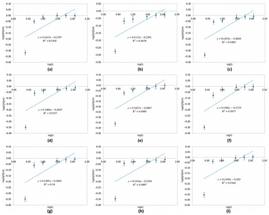
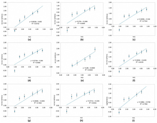
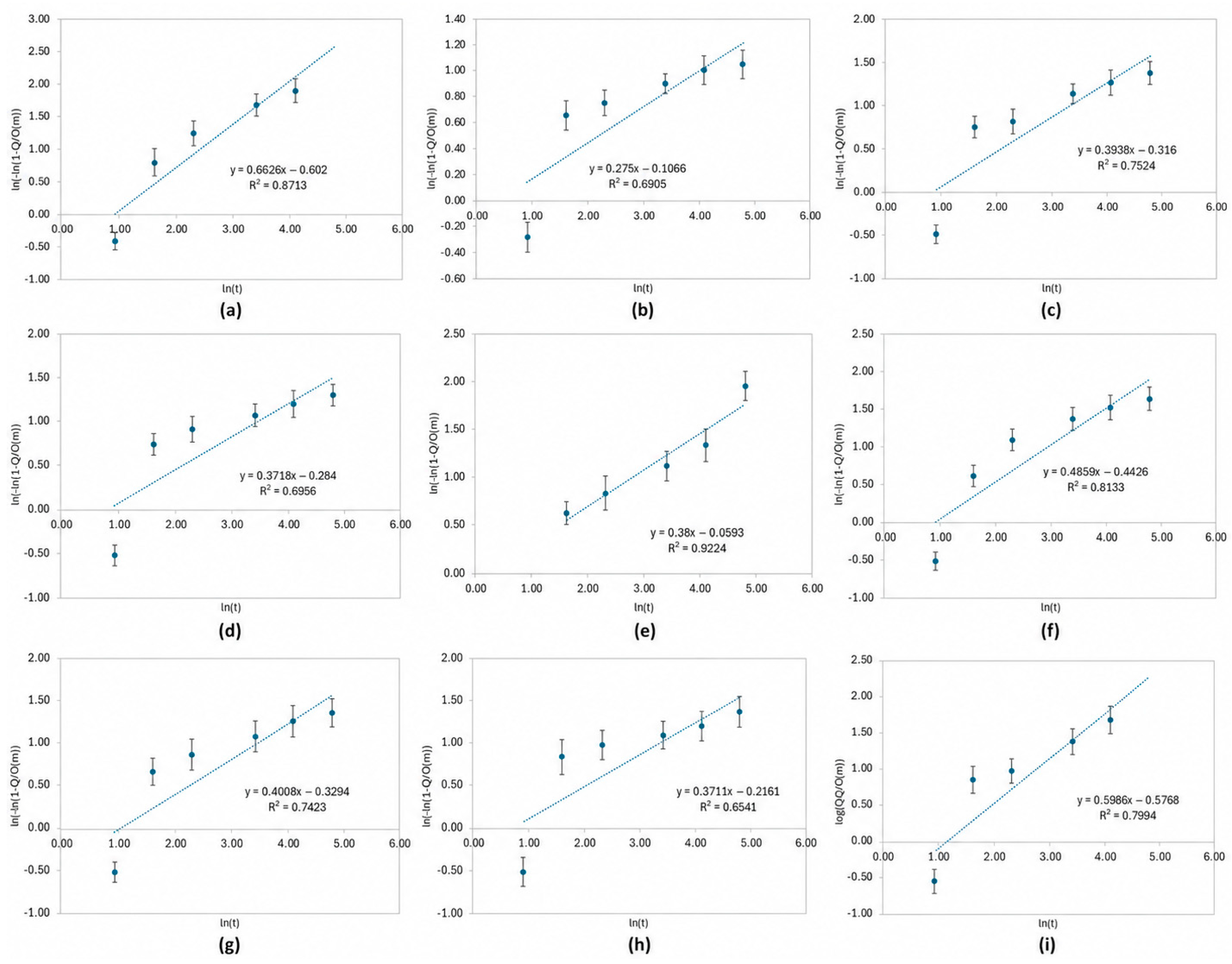
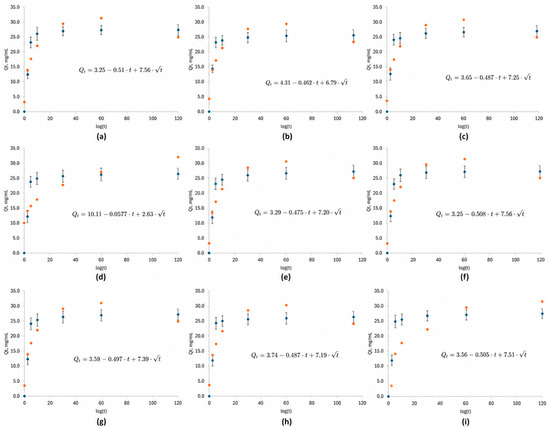
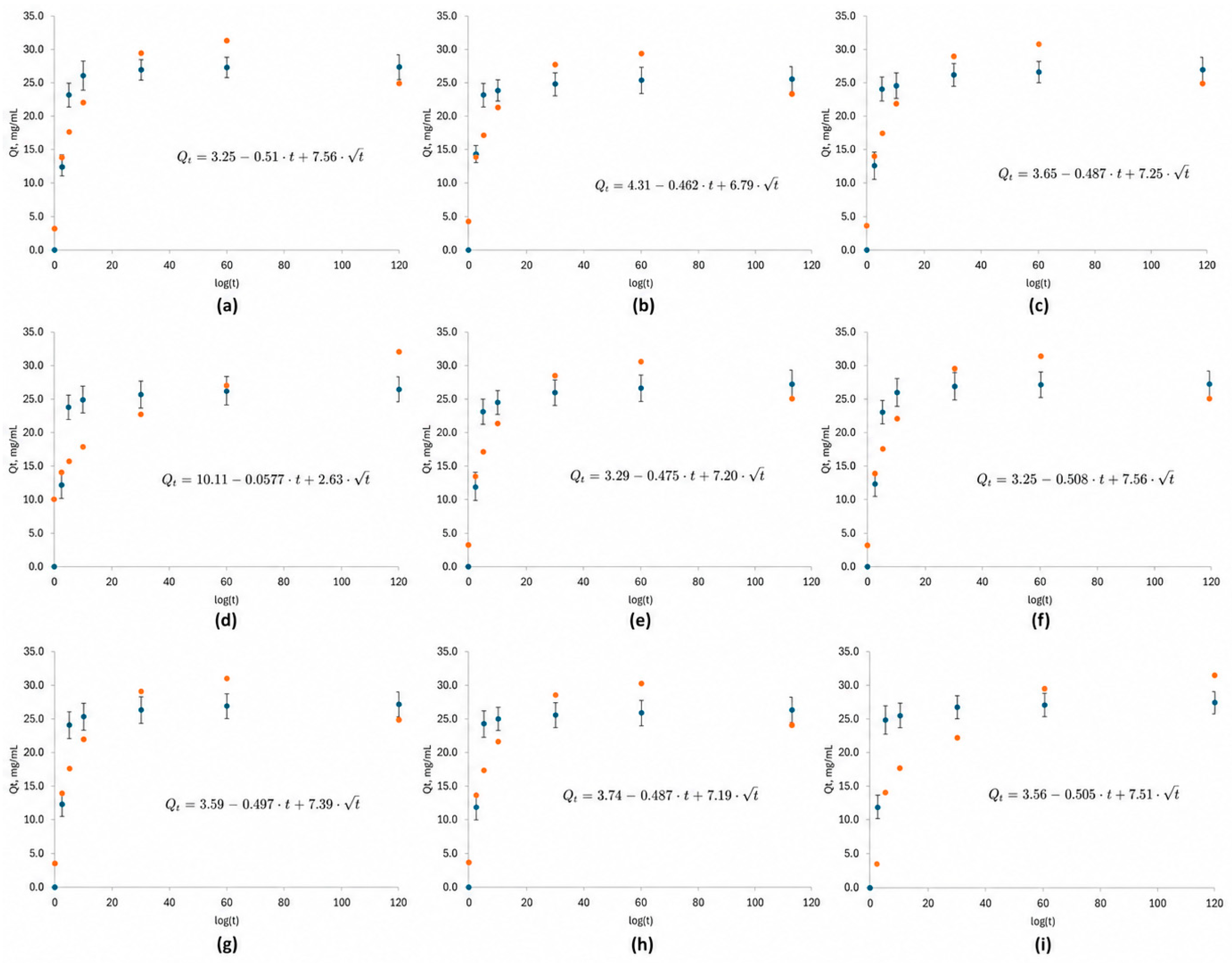
The release kinetics of acetylsalicylic acid from electrospun PLA-based nanofibrous coatings were analyzed using three commonly applied mathematical models: Korsmeyer–Peppas (Figure 14), Weibull (Figure 15), and Peppas–Sahlin (Figure 16). These models were selected to evaluate the dominant release mechanisms and to distinguish between diffusional transport, burst release, and potential erosion-related contributions.
Figure 14.
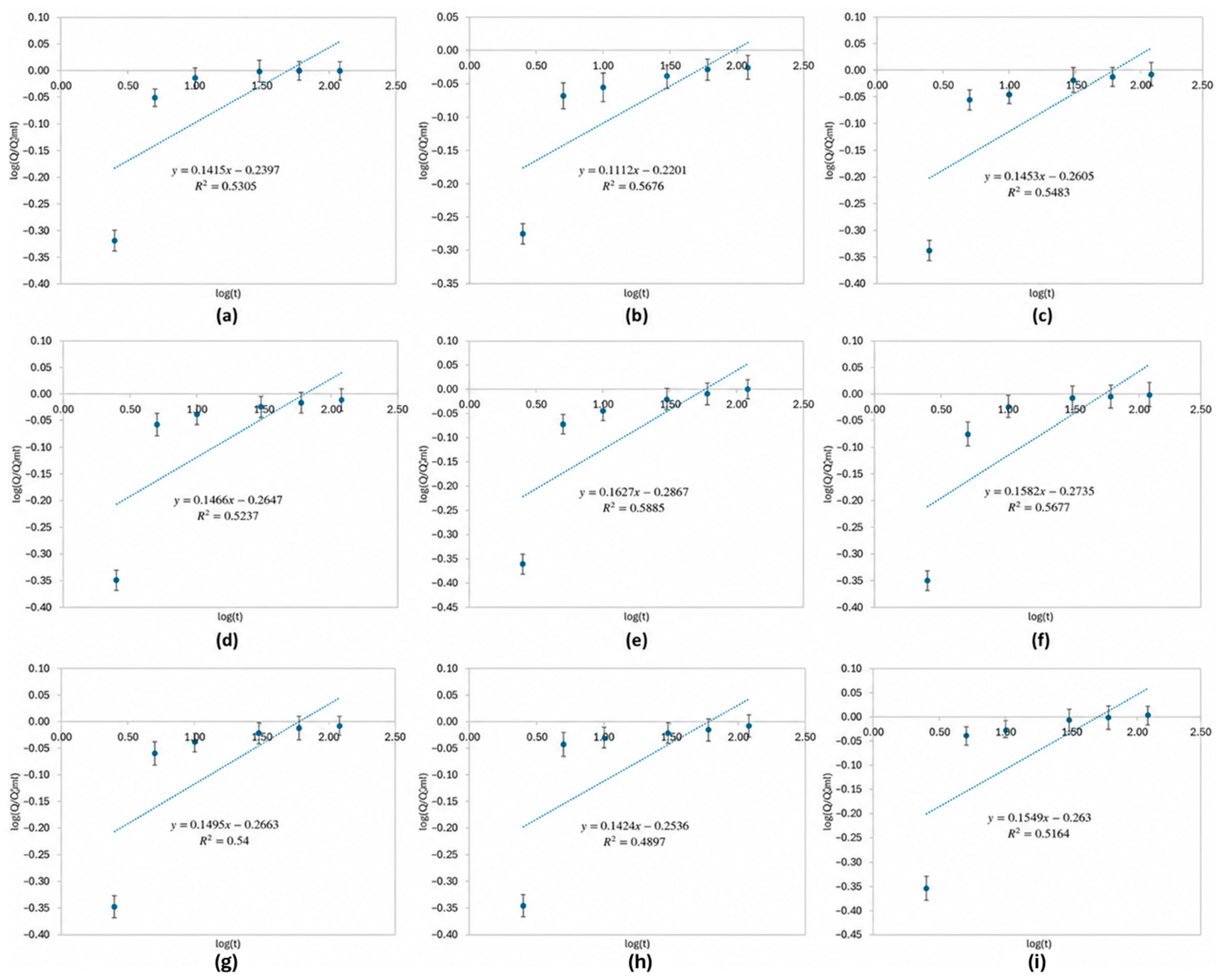
Korsmeyer–Peppas Model (Power Law Model): (a)—sample 1; (b)—sample 2; (c)—sample 3; (d)—sample 4; (e)—sample 5; (f)—sample 6; (g)—sample 7; (h)—sample 8; (i)—sample 9.
Figure 15.
Weibull Model: (a)—sample 1; (b)—sample 2; (c)—sample 3; (d)—sample 4; (e)—sample 5; (f)—sample 6; (g)—sample 7; (h)—sample 8; (i)—sample 9.
Figure 16.
Peppas–Sahlin Model: (a)—sample 1; (b)—sample 2; (c)—sample 3; (d)—sample 4; (e)—sample 5; (f)—sample 6; (g)—sample 7; (h)—sample 8; (i)—sample 9; blue dots—measurement points; orange dots model fit to the measurement points.
These three models were selected because they represent complementary approaches commonly used in drug release analysis. The Korsmeyer–Peppas model is widely applied to identify the dominant transport mechanism during the initial stage of release, particularly to determine whether diffusion or anomalous transport occurs. The Weibull model provides a flexible empirical description capable of fitting the entire release profile and identifying curve shape characteristics. In contrast, the Peppas–Sahlin model allows separation of diffusion-controlled and erosion-related contributions, enabling a mechanistic interpretation of the release process. The combined use of these models therefore enables a more comprehensive evaluation of the drug release behavior from electrospun coatings.
The Korsmeyer–Peppas model was applied to describe the initial stage of drug release according to Equation (1):
where Qt is the amount of drug released at time t, Q∞ is the total amount of drug released at infinite time, k is the kinetic constant, and n is the diffusion exponent indicating the release mechanism.
Qt/Q∞ = k × tn
The Weibull model was used to describe the entire release profile using Equation (2):
where Qt is the amount of drug released at time t, Q∞ is the maximum amount of drug released, Ti is the lag time (assumed to be zero), α is the scale parameter related to the release rate, and β is the shape parameter describing the release curve profile.
Qt/Q∞ = 1 − exp [−((t − Ti)/α)β]
To separate the contributions of burst release, diffusion, and erosion-related processes, the Peppas–Sahlin model was applied according to Equation (3):
where Q0 represents the initial burst release, k1 is the kinetic constant associated with erosion or desorption processes, and k2 corresponds to the Fickian diffusion contribution.
Qt/Q∞ = Q0 + k1 × t + k2 × t1/2
The drug release profiles from electrospun PLA-based nanofibrous coatings were first analyzed using the Korsmeyer–Peppas model (Figure 14). The obtained diffusion exponent values ranged from 0.111 to 0.163 for all samples, which is well below the critical value of 0.45 for cylindrical systems. These results clearly indicate that the release process was governed by Fickian diffusion. The kinetic constant k exhibited a narrow range (0.517–0.602), suggesting comparable release rates across the investigated coatings. However, the coefficients of determination (R2 = 0.490–0.589) were relatively low, demonstrating that the Korsmeyer–Peppas model was insufficient to describe the complete release profiles, likely due to its limitation to the early release stage.
To further evaluate the release behavior over the entire time range, the experimental data were fitted to the Weibull model (Figure 15). The shape parameter β ranged from 0.275 to 0.663, indicating concave release profiles characteristic of diffusion-controlled transport. No values of β approaching or exceeding unity were observed, confirming the absence of erosion- or degradation-driven release mechanisms. The scale parameter α varied between 1.168 and 2.613, reflecting differences in release rates among the coatings. The quality of fit was moderate to high (R2 = 0.654–0.924), suggesting that the Weibull model adequately described the release kinetics for selected samples but failed to consistently capture the pronounced burst release and plateau behavior.
The most comprehensive description of the release process was obtained using the Peppas–Sahlin model. This approach enabled the separation of the initial burst release, diffusional transport, and erosion-related contributions. For most samples, the burst release parameter Q0 ranged from 3.25 to 4.31 mg/L, indicating a moderate initial release. Sample 4 exhibited a markedly higher Q0 value (10.11 mg/L), suggesting increased drug availability at the coating surface or structural heterogeneity within the nanofibrous layer. The erosion-related constant k1 remained low and negative for all samples, indicating that erosion or desorption processes played a negligible role in drug release. In contrast, the diffusion-related constant k2 showed consistently high values (6.79–7.56), confirming that Fickian diffusion was the dominant release mechanism. The Peppas–Sahlin model provided good agreement with the experimental data (R2 = 0.820–0.867), except for sample 4, which exhibited atypical behavior.
Overall, the comparative analysis of the three kinetic models demonstrated that drug release from electrospun PLA-based nanofibrous coatings follows a biphasic mechanism, consisting of an initial burst release followed by sustained diffusion-controlled transport. Among the applied models, the Peppas–Sahlin equation most accurately captured both the quantitative trends and mechanistic aspects of the release process. These findings highlight the suitability of electrospun PLA coatings as controlled drug delivery platforms, where release behavior can be effectively tuned through coating morphology and composition. It should be emphasized that the present experiment was designed to evaluate the initial stage of release, particularly burst release and early diffusion behavior. Therefore, the obtained results should be interpreted as a short-term release profile rather than long-term controlled release.
3.9. Porosity and Density Testing
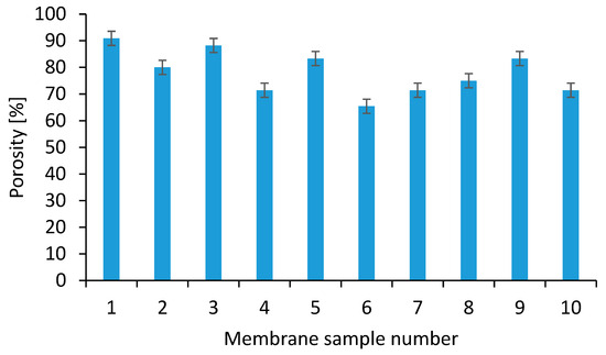
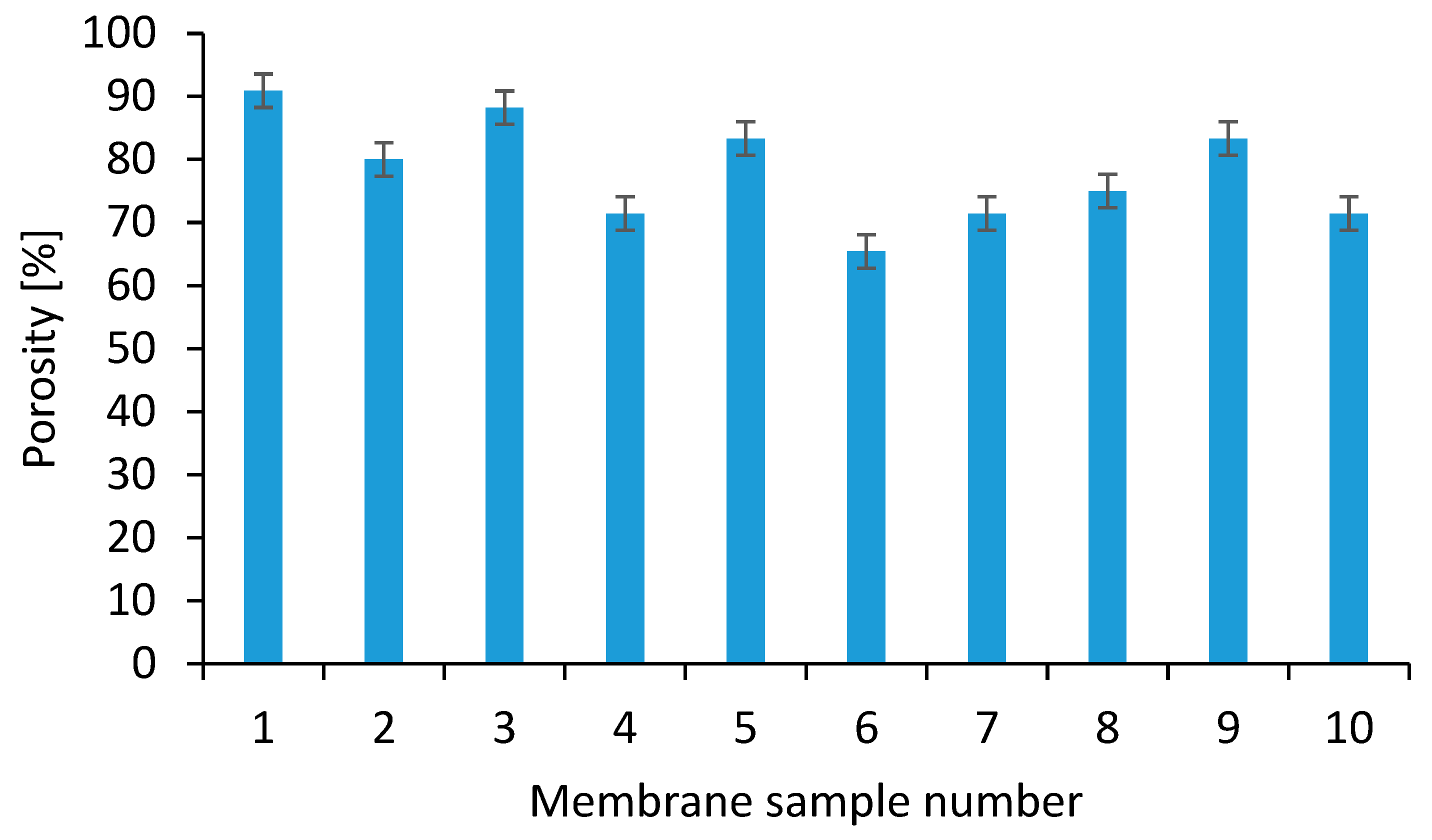
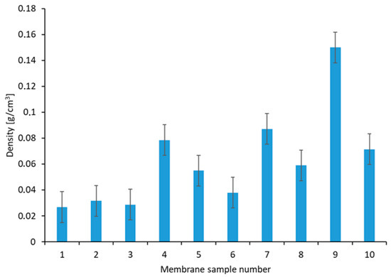
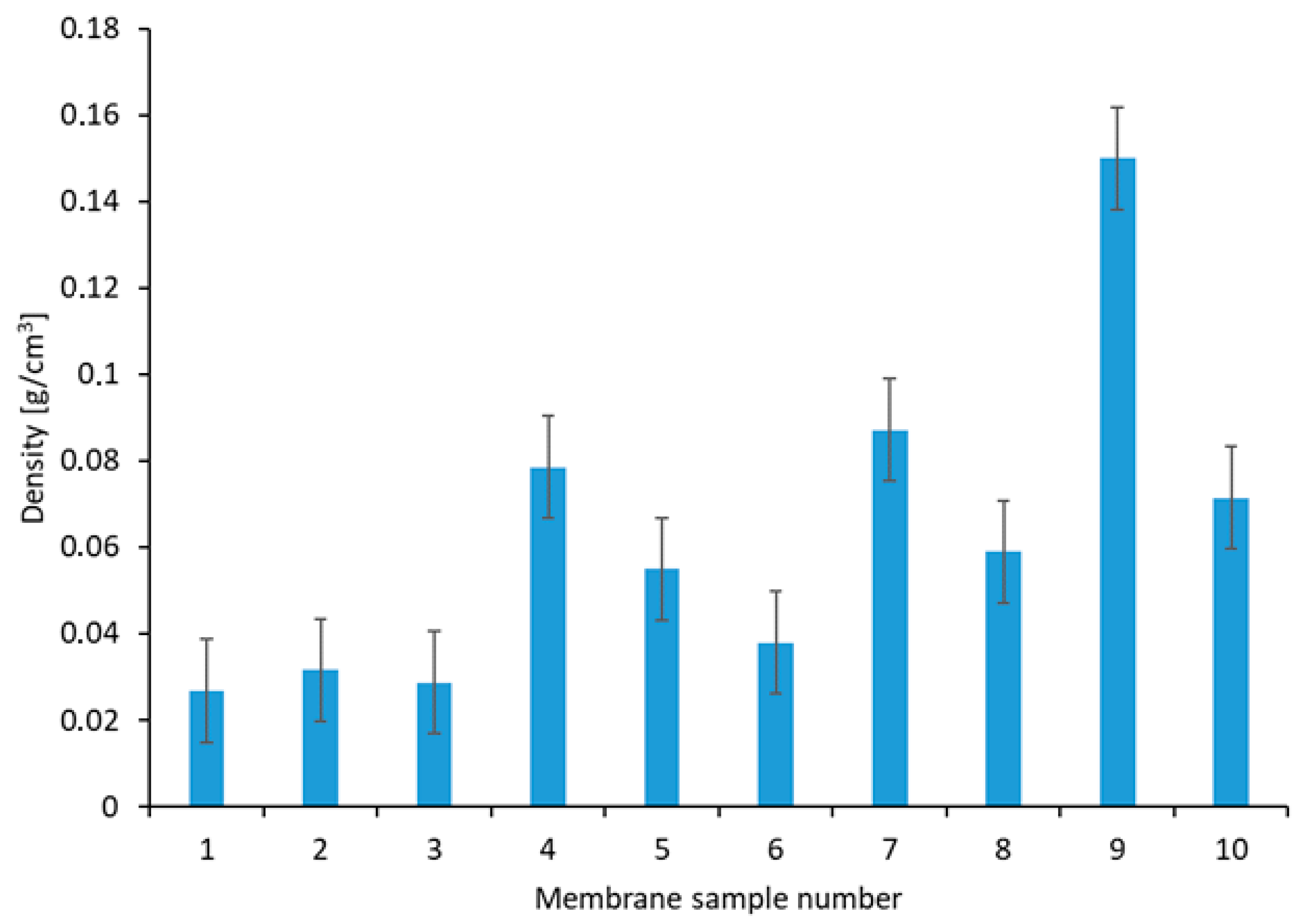
An essential requirement for nanofibrous scaffolds intended for tissue engineering applications is high porosity, along with well-connected pore structures. The presence of small pores and a high specific surface area not only contributes to the inhibition of microbial growth but also facilitates effective fluid drainage control. Increased porosity has been positively correlated with improved wound healing outcomes. Moreover, highly porous membranes mimic the structure of the extracellular matrix (ECM), enabling cell attachment, inward migration, and interaction with the scaffold. Therefore, scaffolds with micro- and nanoscale porosity are particularly well-suited to support nutrient and gas exchange, both critical for cell proliferation and tissue regeneration. The optimal porosity range for such applications is generally considered to be between 60% and 90% [30,31,32]. In this study, all fabricated scaffolds exhibited porosity values above 71%, placing them within the preferred range for nanofibrous membranes, as shown in Figure 17. The highest porosity (90.91%) was observed for Sample 1, which contained PLA enriched with Kalanchoe pinnata extract. In contrast, the lowest porosity values were recorded for Samples 4, 7, and 8 (combinations of Kalanchoe pinnata extract with bismuth (III) oxide or gadolinium (III) oxide nanoparticles), as well as for Sample 10, which included Sideritis extract and gadolinium (III) oxide nanoparticles. Nonetheless, all samples met the porosity criteria desirable for scaffold-based regenerative applications. The highest density of nanofibrous membranes was observed for Sample 9, as illustrated in Figure 18.
Figure 17.
Percentage porosity values of nanofibrous membranes.
Figure 18.
Percentage density analysis of nanofibrous membranes.
3.10. Cytotoxicity Assay
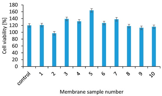
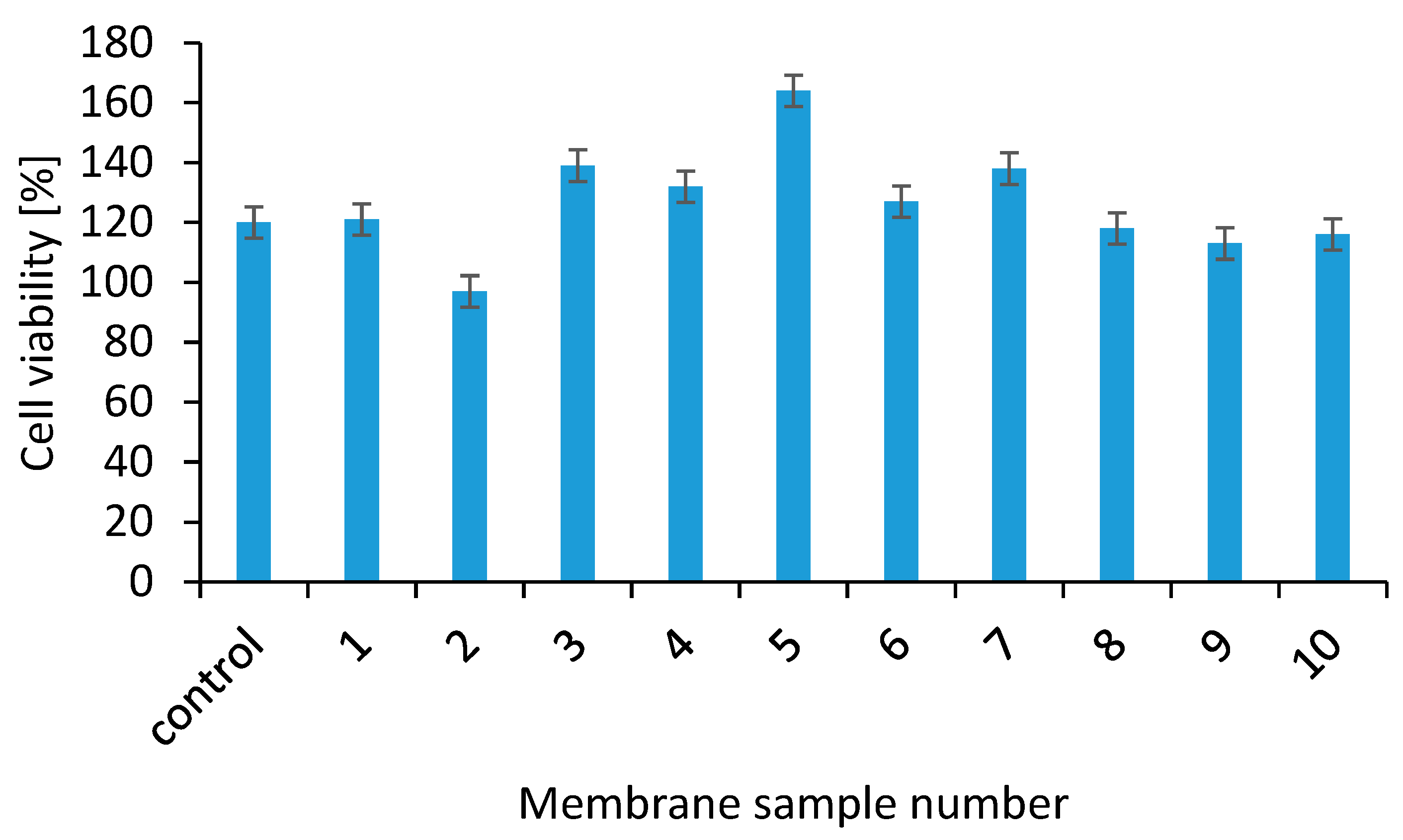
In addition to fulfilling a range of physicochemical requirements, biomaterials intended for biomedical applications must also demonstrate a lack of cytotoxicity [33,34,35,36,37,38,39,40,41,42,43,44,45,46,47,48]. The cytocompatibility of the fabricated membranes was quantitatively assessed using the XTT assay, as shown in Figure 19, where materials maintaining at least 70% cell viability are considered non-toxic. All tested membranes exhibited no detrimental effects on cell proliferation, confirming their biocompatibility. Notably, several formulations even enhanced cellular growth, with the highest metabolic activity observed for membranes 3, 5, and 7, which were enriched with Kalanchoe pinnata extract. The neat PLA membrane showed a neutral effect, with cell viability reaching 97%. However, a slight reduction in cell viability was noted in samples with higher concentrations of metal oxide nanoparticles, suggesting a concentration-dependent response. Furthermore, the Kalanchoe pinnata extract demonstrated superior biological performance compared to the extract derived from Sideritis, indicating its greater potential for enhancing cell proliferation.
Figure 19.
Cell viability of fibroblasts cultured on nanofibrous membranes, assessed by XTT assay.
4. Conclusions
In this study, bio-based polylactide (PLA) nanofibrous coatings were successfully fabricated by electrospinning and evaluated as biodegradable, multifunctional surfaces with controlled drug release capability. The applied electrospinning approach enabled the formation of uniform nanofibrous coatings with high surface area and morphology resembling the extracellular matrix, making them suitable for biomedical coating applications.
The release behavior of acetylsalicylic acid from the electrospun PLA coatings was systematically analyzed using three kinetic models. The Korsmeyer–Peppas model confirmed that drug release was governed by a Fickian diffusion mechanism; however, its limited fitting quality indicated that it was not sufficient to describe the complete release profiles. The Weibull model provided a better overall description of the experimental data and indicated diffusion-controlled release behavior, although its applicability varied among samples. The Peppas–Sahlin model offered the most comprehensive interpretation of the release process, allowing the separation of burst release, diffusional transport, and erosion-related contributions.
The kinetic analysis demonstrated that drug release from the electrospun PLA coatings followed a biphasic pattern, consisting of an initial burst release followed by sustained diffusion-controlled transport, with negligible contribution from matrix erosion. Differences observed between individual samples, particularly the pronounced burst release in one formulation, highlight the strong influence of coating structure and composition on release behavior.
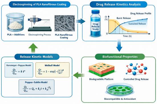
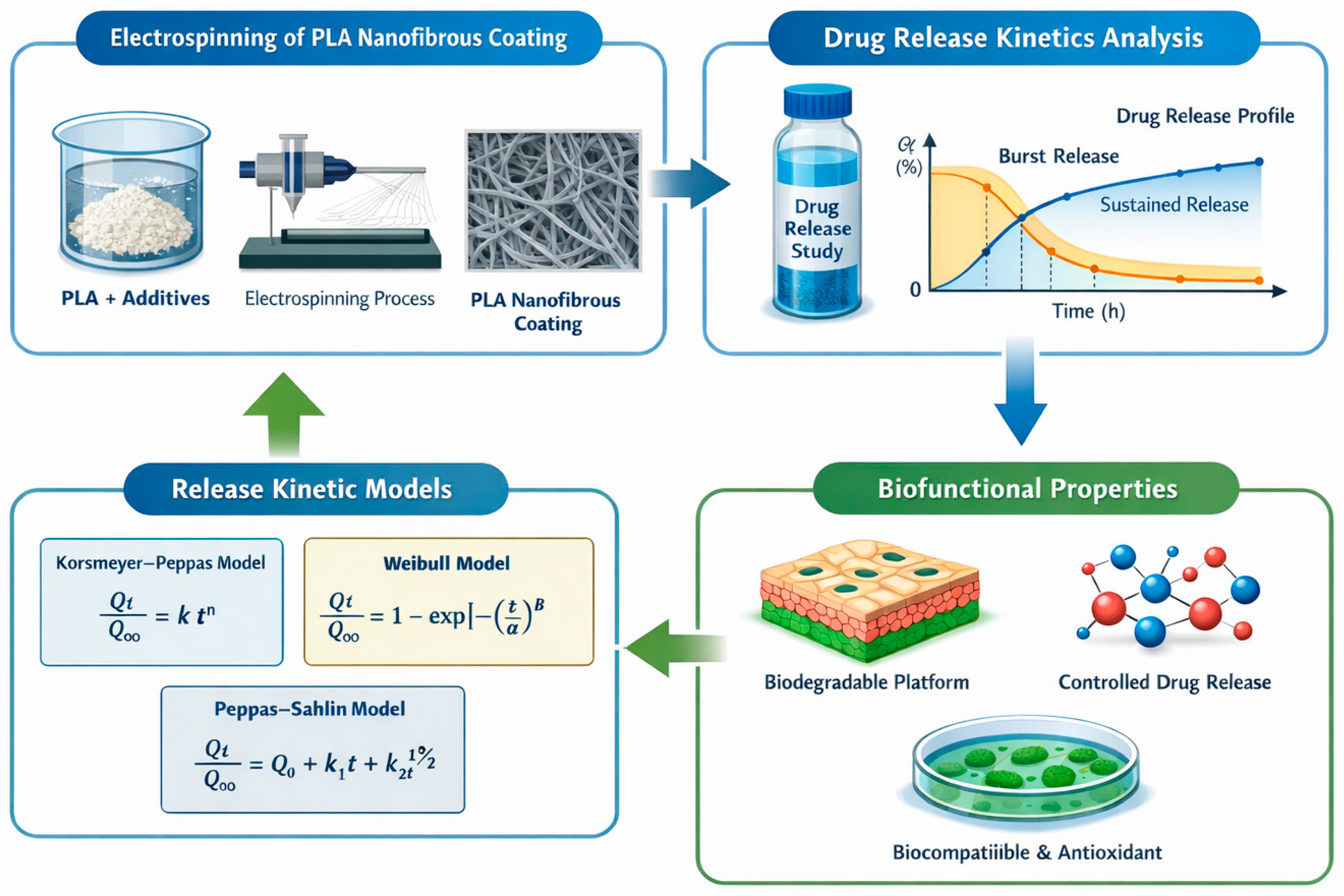
Overall, the results confirm that electrospun PLA-based coatings combine biodegradability, tunable release characteristics, and bioactivity, supporting their potential use as bio-based polymer coatings for biomedical applications. The findings provide valuable insights into the design of electrospun coatings with controlled drug delivery functionality and may contribute to the further development of sustainable and multifunctional coating systems for advanced biomedical surface engineering (Figure 20).
Figure 20.
Results summary.
Author Contributions
Conceptualization, J.R.-P. and Ł.J.; methodology, J.R.-P. and Ł.J.; validation, A.S.-B., J.R.-P. and Ł.J.; investigation, M.N., A.S.-B., T.G., K.K. and J.R.-P.; data curation, Ł.J., M.N. and J.R.-P.; writing—original draft preparation, M.N. and J.R.-P.; resources, M.T., Ł.J. and J.R.-P.; supervision, J.R.-P. and M.T. All authors have read and agreed to the published version of the manuscript.
Funding
This research received no external funding.
Institutional Review Board Statement
Not applicable.
Informed Consent Statement
Not applicable.
Data Availability Statement
The data presented in this study are available on request from the corresponding author.
Acknowledgments
Conflicts of Interest
The authors declare no conflicts of interest.
References
- Christodoulou, E.; Papadopoulos, A.; Tzetzis, D.; Bikiaris, D. PLA-based electrospun nanofibrous mats for drug delivery applications. Pharmaceutics 2025, 17, 589. [Google Scholar] [CrossRef] [Scilit] [PubMed]
- Das, A.; Chatterjee, S.; Mukherjee, S.; Roy, S. Electrospun nanofibers: Transformative innovations in fabrication and biomedical applications. Cogent Eng. 2024, 11, 2433147. [Google Scholar] [CrossRef] [Scilit]
- Jose, M.S.; Sumathi, S. A review of electrospun polymeric fibers as potential drug delivery systems for tunable release kinetics. J. Sci. Adv. Mater. Devices 2025, 10, 100933. [Google Scholar] [CrossRef] [Scilit]
- Concha, V.O.C.; Bahú, J.O.; Crivellin, S.; Khouri, N.G.; Munoz, F.L.; Souza, S.D.A.; Yoshida, M.C.P.; Severino, P.; Concha, L.S.C.; Lopes, M.S. Harnessing electrospinning for improvement of polymeric drug delivery systems. Polym. Bull. 2025, 82, 5909–5943. [Google Scholar] [CrossRef] [Scilit]
- Miętus, M.; Kolankowski, K.; Gołofit, T.; Denis, P.; Bandzerewicz, A.; Spychalski, M.; Mąkosa-Szczygieł, M.; Pilarek, M.; Wierzchowski, K.; Gadomska-Gajadhur, A. From poly(glycerol itaconate) gels to novel nonwoven materials for biomedical applications. Gels 2023, 9, 788. [Google Scholar] [CrossRef] [Scilit] [PubMed]
- Li, M.; Wu, H.; Yuan, Y.; Hu, B.; Gu, N. Recent fabrications and applications of cardiac patch in myocardial infarction treatment. View 2022, 3, e20200153. [Google Scholar] [CrossRef] [Scilit]
- Bhardwaj, N.; Kundu, S.C. Electrospinning: A fascinating fiber fabrication technique. Biotechnol. Adv. 2010, 28, 325–347. [Google Scholar] [CrossRef] [Scilit] [PubMed]
- Gupta, B.; Revagade, N.; Hilborn, J. Poly(lactic acid) fiber: An overview. Prog. Polym. Sci. 2007, 32, 455–482. [Google Scholar] [CrossRef] [Scilit]
- Sill, T.J.; von Recum, H.A. Electrospinning: Applications in drug delivery and tissue engineering. Biomaterials 2008, 29, 1989–2006. [Google Scholar] [CrossRef] [Scilit] [PubMed]
- Bhattarai, R.S.; Bachu, R.D.; Boddu, S.H.S.; Bhaduri, S. Biomedical applications of electrospun nanofibers: Drug and nanoparticle delivery. Pharmaceutics 2019, 11, 5. [Google Scholar] [CrossRef] [Scilit] [PubMed]
- Gogolewski, S.; Pennings, A.J. Resorbable materials of poly(L-lactide). II. Fibers spun from solutions of poly(L-lactide) in good solvents. J. Appl. Polym. Sci. 1983, 28, 1045–1061. [Google Scholar] [CrossRef] [Scilit]
- Castaño, O.; Eltohamy, M.; Kim, H.W. Electrospinning technology in tissue regeneration. Methods Mol. Biol. 2012, 811, 127–140. [Google Scholar] [CrossRef] [Scilit] [PubMed]
- Zhang, C.L.; Yu, S.H. Nanoparticles meet electrospinning: Recent advances and future prospects. Chem. Soc. Rev. 2014, 43, 4423–4448. [Google Scholar] [CrossRef] [Scilit] [PubMed]
- Zhang, C.; Yuan, X.; Wu, L.; Han, Y.; Sheng, J. Study on morphology of electrospun poly(vinyl alcohol) mats. Eur. Polym. J. 2005, 41, 423–432. [Google Scholar] [CrossRef] [Scilit]
- Zargham, S.; Bazgir, S.; Tavakoli, A.; Rashidi, A.S.; Damerchely, R. The effect of flow rate on morphology and deposition area of electrospun nylon-6 nanofiber. J. Eng. Fibers Fabr. 2012, 7, 42–49. [Google Scholar] [CrossRef] [Scilit]
- Sukigara, S.; Gandhi, M.; Ayutsede, J.; Micklus, M.; Ko, F. Regeneration of Bombyx mori silk by electrospinning—Part 1: Processing parameters and geometric properties. Polymer 2003, 44, 5721–5727. [Google Scholar] [CrossRef] [Scilit]
- Rahmati, M.; Mills, D.K.; Urbanska, A.M.; Saeb, M.R.; Venugopal, J.R.; Ramakrishna, S.; Mozafari, M. Electrospinning for tissue engineering applications. Prog. Mater. Sci. 2021, 117, 100721. [Google Scholar] [CrossRef] [Scilit]
- Agarwal, S.; Wendorff, J.H.; Greiner, A. Use of electrospinning technique for biomedical applications. Polymer 2008, 49, 5603–5621. [Google Scholar] [CrossRef] [Scilit]
- Brovold, M.; Almeida, J.I.; Pla-Palacín, I.; Sainz-Arnal, P.; Sánchez-Romero, N.; Rivas, J.J.; Almeida, H.; Royo Dachary, P.; Serrano-Aulló, T.; Soker, S.; et al. Naturally-derived biomaterials for tissue engineering applications. In Novel Biomaterials for Regenerative Medicine; Baptista, P.M., Ed.; Advances in Experimental Medicine and Biology; Springer: Cham, Switzerland, 2018; Volume 1077, pp. 421–449. [Google Scholar] [CrossRef] [Scilit] [PubMed]
- Dormont, F.; Varna, M.; Couvreur, P. Nanoplumbers: Biomaterials to fight cardiovascular diseases. Mater. Today 2018, 21, 122–143. [Google Scholar] [CrossRef] [Scilit]
- Heng, B.C.; Das, G.K.; Zhao, X.; Ma, L.-L.; Tan, T.T.-Y.; Ng, K.W.; Loo, J.S.-C. Comparative cytotoxicity evaluation of lanthanide nanomaterials on mouse and human cell lines with metabolic and DNA-quantification assays. Biointerphases 2010, 5, FA88–FA97. [Google Scholar] [CrossRef] [Scilit] [PubMed]
- Setyawati, M.I.; Khoo, P.K.S.; Eng, B.H.; Xiong, S.; Zhao, X.; Das, G.K.; Tan, T.T.-Y.; Loo, J.S.-C.; Leong, D.T.; Ng, K.W. Cytotoxic and genotoxic characterization of titanium dioxide, gadolinium oxide, and poly(lactic-co-glycolic acid) nanoparticles in human fibroblasts. J. Biomed. Mater. Res. A 2013, 101A, 633–640. [Google Scholar] [CrossRef] [Scilit] [PubMed]
- Shahbazi, M.-A.; Faghfouri, L.; Ferreira, M.P.A.; Figueiredo, P.; Maleki, H.; Sefat, F.; Hirvonen, J.; Santos, H.A. The versatile biomedical applications of bismuth-based nanoparticles and composites: Therapeutic, diagnostic, biosensing, and regenerative properties. Chem. Soc. Rev. 2020, 49, 1253–1321. [Google Scholar] [CrossRef] [Scilit] [PubMed]
- Zhao, G.; Zhang, X.; Lu, T.J.; Xu, F. Recent Advances in Electrospun Nanofibrous Scaffolds for Cardiac Tissue Engineering. Adv. Funct. Mater. 2015, 25, 5726–5738. [Google Scholar] [CrossRef] [Scilit]
- Iqbal, J.; Iqbal, A.; Mukhtar, H.; Jahangir, K.; Mashkoor, Y.; Zeeshan, M.H.; Nadeem, A.; Ashraf, A.; Maqbool, S.; Sadiq, S.M.; et al. Cardioprotective effects of nanoparticles in cardiovascular diseases: A state-of-the-art review. Curr. Probl. Cardiol. 2023, 48, 101713. [Google Scholar] [CrossRef] [Scilit] [PubMed]
- Pala, R.; Anju, V.T.; Dyavaiah, M.; Busi, S.; Nauli, S.M. Nanoparticle-mediated drug delivery for the treatment of cardiovascular diseases. Int. J. Nanomed. 2020, 15, 3741–3769. [Google Scholar] [CrossRef] [Scilit] [PubMed]
- Pretorius, D.; Serpooshan, V.; Zhang, J. Nanomedicine in the cardiovascular system. Front. Pharmacol. 2021, 12, 640182. [Google Scholar] [CrossRef] [Scilit] [PubMed]
- Hesari, M.; Mohammadi, P.; Khademi, F.; Shackebaei, D.; Momtaz, S.; Moasefi, N.; Farzaei, M.H.; Abdollahi, M. Current advances in the use of nanophytomedicine therapies for human cardiovascular diseases. Int. J. Nanomed. 2021, 16, 3293–3315. [Google Scholar] [CrossRef] [Scilit] [PubMed]
- ISO 10993-5:2009; Biological evaluation of medical devices—Part 5: Tests for in vitro cytotoxicity. International Organization for Standardization: Geneva, Switzerland, 2009.
- Lee, S.J.; Heo, D.N.; Park, J.S.; Kwon, S.K.; Lee, J.H.; Lee, J.H.; Kim, W.D.; Kwon, I.K.; Park, S.A. Electrospun chitosan nanofibers with controlled levels of silver nanoparticles: Preparation, characterization and antibacterial activity. Carbohydr. Polym. 2014, 111, 530–537. [Google Scholar] [CrossRef] [Scilit] [PubMed]
- Lewinski, N.; Colvin, V.; Drezek, R. Cytotoxicity of nanoparticles. Small 2008, 4, 26–49. [Google Scholar] [CrossRef] [Scilit] [PubMed]
- Geelen, T.; Paulis, L.E.; Coolen, B.F.; Nicolay, K.; Strijkers, G.J. Passive targeting of lipid-based nanoparticles to mouse cardiac ischemia–reperfusion injury. Contrast Media Mol. Imaging 2013, 8, 117–126. [Google Scholar] [CrossRef] [Scilit] [PubMed]
- Chong, E.J.; Phan, T.T.; Lim, I.J.; Zhang, Y.Z.; Bay, B.H.; Ramakrishna, S.; Lim, C.T. Evaluation of electrospun PCL/gelatin nanofibrous scaffold for wound healing and layered dermal reconstitution. Acta Biomater. 2007, 3, 321–330. [Google Scholar] [CrossRef] [Scilit] [PubMed]
- Ratner, B.D.; Bryant, S.J. Biomaterials: Where we have been and where we are going. Annu. Rev. Biomed. Eng. 2004, 6, 41–75. [Google Scholar] [CrossRef] [Scilit] [PubMed]
- Sell, S.A.; McClure, M.J.; Garg, K.; Wolfe, P.S.; Bowlin, G.L. Electrospinning of collagen/biopolymers for regenerative medicine and cardiovascular tissue engineering. Adv. Drug Deliv. Rev. 2009, 61, 1007–1019. [Google Scholar] [CrossRef] [Scilit] [PubMed]
- Leung, V.; Ko, F. Biomedical applications of nanofibers. Polym. Adv. Technol. 2011, 22, 350–365. [Google Scholar] [CrossRef] [Scilit]
- Ramakrishna, S.; Fujihara, K.; Teo, W.E.; Lim, T.C.; Ma, Z. An Introduction to Electrospinning and Nanofibers; World Scientific: Singapore, 2005. [Google Scholar] [CrossRef]
- Teo, W.E.; Ramakrishna, S. A review on electrospinning design and nanofibre assemblies. Nanotechnology 2006, 17, R89–R106. [Google Scholar] [CrossRef] [Scilit] [PubMed]
- Farahani, A.; Zarei-Hanzaki, A.; Abedi, H.R.; Tayebi, L.; Mostafavi, E. Polylactic acid piezo-biopolymers: Chemistry, fabrication methods, and tissue engineering applications. J. Funct. Biomater. 2021, 12, 71. [Google Scholar] [CrossRef] [Scilit] [PubMed]
- Li, D.; Xia, Y. Electrospinning of nanofibers: Reinventing the wheel? Adv. Mater. 2004, 16, 1151–1170. [Google Scholar] [CrossRef] [Scilit]
- Nasr, S.M.; Rabiee, N.; Hajebi, S.; Ahmadi, S.; Fatahi, Y.; Hosseini, M.; Bagherzadeh, M.; Ghadiri, A.M.; Rabiee, M.; Jajarmi, V.; et al. Biodegradable nanopolymers in cardiac tissue engineering: From concept towards nanomedicine. Int. J. Nanomed. 2020, 15, 4205–4224. [Google Scholar] [CrossRef] [Scilit] [PubMed]
- Smith, S.; Goodge, K.; Delaney, M.; Struzyk, A.; Tansey, N.; Frey, M. A Comprehensive Review of the Covalent Immobilization of Biomolecules onto Electrospun Nanofibers. Nanomaterials 2020, 10, 2142. [Google Scholar] [CrossRef] [Scilit] [PubMed]
- Wei, Y.; Liu, Z.; Zhu, X.; Jiang, L.; Shi, W.; Wang, Y.; Xu, N.; Gang, F.; Wang, X.; Zhao, L.; et al. Dual directions to address the problem of aseptic loosening via electrospun PLGA@aspirin nanofiber coatings on titanium. Biomaterials 2020, 257, 120237. [Google Scholar] [CrossRef] [Scilit] [PubMed]
- Pascual-Gil, S.; Garbayo, E.; Díaz-Herráez, P.; Prosper, F.; Blanco-Prieto, M.J. Heart regeneration using synthetic biomaterials. J. Control. Release 2015, 203, 23–38. [Google Scholar] [CrossRef] [Scilit] [PubMed]
- Ning, Y.; Shen, W.; Ao, F. Immobilization of electrospun fibers in biomedical applications. RSC Adv. 2020, 10, 37246–37265. [Google Scholar] [CrossRef] [Scilit] [PubMed]
- Veeman, D.; Sai, M.S.; Sureshkumar, P.; Jagadeesha, T.; Natrayan, L.; Ravichandran, M.; Mammo, W.D. Additive manufacturing of biopolymers for tissue engineering and regenerative medicine: An overview, potential applications, advancements, and trends. Int. J. Polym. Sci. 2021, 2021, 4907027. [Google Scholar] [CrossRef] [Scilit]
- Lam, M.T.; Wu, J.C. Biomaterial applications in cardiovascular tissue repair and regeneration. Expert Rev. Cardiovasc. Ther. 2012, 10, 1039–1049. [Google Scholar] [CrossRef] [Scilit] [PubMed]
- Entcheva, E.; Bien, H.; Yin, L.; Chung, C.-Y.; Farrell, M.; Kostov, Y. Functional cardiac cell constructs on cellulose-based scaffolding. Biomaterials 2004, 25, 5753–5762. [Google Scholar] [CrossRef] [PubMed]

Disclaimer/Publisher’s Note: The statements, opinions and data contained in all publications are solely those of the individual author(s) and contributor(s) and not of MDPI and/or the editor(s). MDPI and/or the editor(s) disclaim responsibility for any injury to people or property resulting from any ideas, methods, instructions or products referred to in the content. |
© 2026 by the authors. Licensee MDPI, Basel, Switzerland. This article is an open access article distributed under the terms and conditions of the Creative Commons Attribution (CC BY) license.