


Abstract
This study addressed the poor wear resistance of Ti-7.5Nb-4Mo-2Sn shape memory alloy through the development of Ti-xSn (x = 6, 8, 9, 10, 20 at.%) coatings and laser cladding technology. This β-type titanium alloy is a promising biomaterial for artificial joints and bone fixation implants, and laser cladding is a superior surface modification technology for fabricating metallurgically bonded high-performance coatings. Microstructural characterization revealed that increasing Sn content from 6% to 10% progressively suppressed β-phase formation while enhancing microhardness (peak value: 430.06 HV1) and wear resistance. Conversely, further Sn addition of 20% degraded these properties. The optimal Ti-10Sn alloy was subsequently laser cladded onto a Ti-7.5Nb-4Mo-2Sn substrate in the form of pre-placed thin sheets under varying laser scanning speeds (7–13 mm/s). The results indicated that processing at 10 mm/s produced superior coating features, including complete metallurgical bonding (20 μm transition layer), the maximum surface hardness (494 HV1, 93% increase), and superior wear resistance. Microscopic analysis confirmed a wear mechanism transition from mixed adhesive–abrasive wear (7.5Nb-4Mo-2Sn substrate) to pure abrasive wear (Ti-10Sn coating), resulting in the enhanced wear resistance of the substrate. This study demonstrated that synergistic alloy design combined with a laser cladding approach can significantly enhance biomedical alloy performance.
1. Introduction
Ti-7.5Nb-4Mo-2Sn, a typical third-generation β-type biomedical titanium alloy, exhibits excellent comprehensive performance, including low elastic modulus matching human bone, superior biocompatibility, and unique shape memory and super-elastic properties, making it a promising candidate for bone fixation implants and artificial joints [1,2]. However, its inherent poor surface wear resistance limits long-term clinical application: frictional wear during service generates debris that triggers adverse biological responses such as inflammation and tissue damage [3,4]. Surface modification is thus a critical and necessary strategy to enhance the tribological performance of this alloy, and laser cladding has emerged as a superior surface engineering technology for this purpose, enabling the fabrication of metallurgically bonded, high-performance coatings with precise process control [5,6].
The selection of cladding materials is pivotal to the success of laser cladding for biomedical titanium alloys, requiring simultaneous satisfaction of thermodynamic compatibility with the substrate, excellent biocompatibility, and enhanced mechanical properties [7,8]. Ti-Sn alloys have become an ideal coating material for Ti-based biomedical alloys due to their favorable properties: they form stable Ti-Sn intermetallic compounds at room temperature that improve hardness and wear resistance, possess excellent biocompatibility suitable for clinical applications, and feature a favorable atomic size mismatch that induces a significant strengthening effect [9,10,11,12].
The current study presents a comprehensive investigation of the Ti-xSn (x = 6, 8, 9, 10, 20 at.%) alloy coatings that are deposited on Ti-7.5Nb-4Mo-2Sn substrates via laser cladding. Through systematic variation in Sn content in the coating alloy and laser scanning speed during cladding, we establish clear composition–microstructure–property relationships, identify the optimal coating composition and processing parameters, and elucidate the underlying interfacial bonding and wear mechanisms. The results demonstrate that carefully engineered Ti-Sn coatings can significantly enhance the surface wear resistance of Ti-7.5Nb-4Mo-2Sn while maintaining its excellent bulk biomedical properties, offering a practical surface modification solution to improve the longevity and reliability of medical titanium alloy implants in clinical applications. This study shows that laser cladding, along with proper alloy design, can effectively enhance the surface wear resistance of biomedical alloys. Notably, the synergistic combination of optimized Ti-Sn alloy coating design and laser cladding outperforms conventional surface modification methods for biomedical titanium alloys: it overcomes the drawbacks of poor interfacial bonding (thermal spraying), low hardness stability (electroplating) and narrow modification layer (ion implantation), and realizes the integration of strong metallurgical bonding, high wear resistance and excellent biocompatibility. This integrated surface engineering strategy thus provides a more practical and reliable technical solution for the surface modification of biomedical titanium alloy implants.
2. Experimental Materials and Methods
2.1. Substrate and Coating Materials
The substrate material employed in this study was a β-type titanium shape memory alloy (Ti-7.5Nb-4Mo-2Sn) developed by Zhang et al., which was based on d-electron orbital theory [1,2]. This innovative alloy exhibited several advantageous characteristics for biomedical applications, including a single β-phase structure at room temperature with an exceptionally low elastic modulus (~60 GPa) closely matching that of human bone. The absence of toxic or allergenic elements ensures excellent biocompatibility, while its unique combination of shape memory effect, superelasticity, and room temperature stability makes it particularly suitable for implant applications. For the coating material, a series of Ti-xSn alloys (x = 6, 8, 9, 10, 20 at.%) was designed and prepared as potential coating materials, with the optimal composition selected for laser cladding experiments. Notably, the cladding material was applied as pre-placed thin sheets rather than conventional powder feedstock, offering potential advantages in processing control and coating uniformity. High-purity raw materials (Ti, Nb, Mo, Sn, ≥99.9 wt.%) were purchased from Beijing Lichengxin Material Technology Co., Ltd., Beijing, China.
2.2. Experimental Methods
2.2.1. Material Preparation Methods
Both substrate and coating alloys were prepared using a JVAM-1C non-consumable vacuum arc furnace (Shenyang 7Q Precision Technology Co., Ltd., Shenyang, China) with high-purity argon as the protective atmosphere. To ensure compositional homogeneity, each ingot was subjected to multiple remelting cycles (minimum six times) with intermediate flipping. The molten alloys were then cast into plates (70 mm × 12 mm × 2 mm) using a JVAM-1C vacuum suction casting machine (Shenyang 7Q Precision Technology Co., Ltd., Shenyang, China). The substrate plates were subsequently sectioned into smaller specimens (20 mm × 6 mm × 2 mm) using a DK7720 precision wire electrical discharge machining machine (Taizhou Zhongde CNC Machine Tool Co., Ltd., Taizhou, China). For laser cladding, the selected Ti-10Sn alloy was prepared as thin sheets (20 mm × 4 mm × 0.5 mm) and pre-placed on the substrate surface. The cladding process was performed using a TFL-H6000 CO2 laser system (Wuhan Hans GS Laser System Co., Ltd., Wuhan, China) with the following parameters: constant laser power of 1300 W, focused beam diameter of 4 mm, and variable scanning speeds of 7, 10, and 13 mm/s. This one-step cladding approach was conducted under controlled quenching conditions to optimize the coating microstructure.
2.2.2. Performance Characterization
Microhardness measurements were conducted using an HVS-1000 digital microhardness tester (Shanghai Aolong Xingdi Testing Equipment Co., Ltd., Shanghai, China) with standardized testing parameters (9.8 N load, 15 s dwell time). Vickers microhardness results were reported as HV1, corresponding to a test load of 1 kgf (9.8 N). Each specimen underwent 5–7 indentations to ensure statistical reliability. Tribological properties were evaluated using an HRS-2M high-speed reciprocating wear tester (Lanzhou Zhongke Kaihua Technology Development Co., Ltd., Lanzhou, China) under dry sliding conditions at room temperature. The wear tests employed a 4 mm diameter quenched and tempered steel ball counterface with a 5 N normal load, 300 rpm rotational speed, 5 mm stroke length, and 600 s test duration. Prior to testing, all specimens were mechanically polished, ground with SiC abrasive paper (400# → 800# → 1200# → 2000#), and polished with diamond paste (1.0 μm followed by 0.5 μm) to a mirror surface finish (Ra < 0.2 μm). To achieve a uniform surface finish. Microstructural characterization was performed using an EVO MA10 scanning electron microscope (SEM) (Carl Zeiss AG, Oberkochen, Germany) operating at an acceleration voltage of 20 kV, a spot size of 5.0, and in a secondary electron mode; energy dispersive spectroscopy (EDS) was integrated with the SEM for elemental analysis. Phase identification was carried out by a D/max-r X-ray diffractometer (XRD) (Rigaku Corporation, Tokyo, Japan) with Cu Kα radiation (λ = 1.5406 Å), a scanning speed of 8°/min, a 2θ scanning range of 20–90°, and a step size of 0.02°.
3. Results
3.1. Preparation of Ti-xSn Alloy Coating Alloys as Potential Coating Materials
Prior to laser cladding, multiple Ti-Sn alloy compositions were designed and prepared as candidate coatings. The optimal alloy, exhibiting superior performance, was subsequently selected for use as the cladding coating. According to the Ti-Sn binary phase diagram [13], Ti-Sn alloys with Sn contents ranging from 6% to 20% (atomic percentage) primarily consist of an α-Ti matrix that is reinforced with a suitable amount of the intermetallic compound Ti3Sn. Based on this, a series of Ti-xSn (x = 6, 8, 9, 10, 20) alloys was fabricated to systematically investigate the influence of Sn content on phase composition, microhardness, and wear resistance.
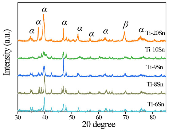
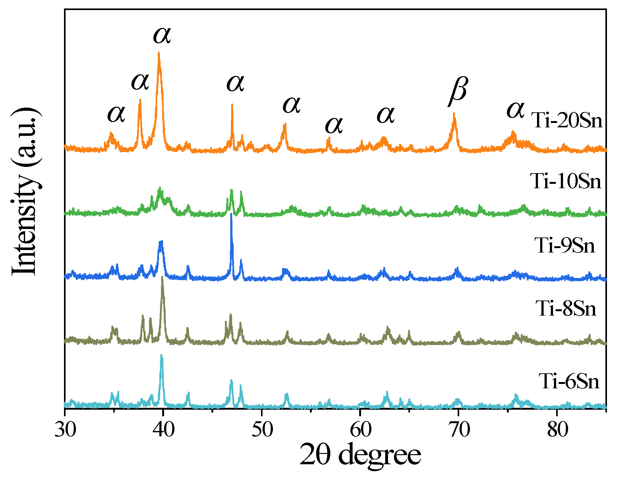
Figure 1 shows the XRD pattern of Ti-xSn alloys (x = 6, 8, 9, 10, 20). It can be seen that the microstructure of Ti-Sn alloys was composed of both α and β phases, and an appropriate amount of intermetallic compound Ti3Sn (in the form of α phase) can also be found, which is consistent with the expected results. There is a peak of β phase near 2θ = 70°, indicating that the high-temperature β phase was retained through rapid cooling [14]. The content of the β phase decreased with the increase in Sn content from 6% to 10%, while it increased with the increase in Sn content from 10% to 20%. The suppression of the soft β-phase effectively reduces the plastic deformability of the alloy matrix, and the concomitant formation of hard Ti3Sn intermetallic compounds further reinforces the α-Ti matrix; this synergistic effect significantly enhances the microhardness and resistance to plastic flow under shear stress, thereby improving the wear resistance of Ti-xSn alloys. However, when Sn content reached 20 at.%, excessive brittle Ti-Sn intermetallic compounds were formed and were severely segregated at grain boundaries, resulting in microcracks and reduced structural integrity. The excessive brittleness and interior defects led to easier particle detachment during friction, thus deteriorating both mechanical and tribological properties [15]. This phase evolution directly influenced mechanical properties, as the β-phase’s superior deformability (attributed to its 12 or 24 slip systems) compared to the α-phase’s limited slip systems (3 or 6) results in reduced hardness and altered tribological behavior [16]. The observed phase composition correlates well with the measured mechanical property variations shown in subsequent results.
Figure 1.
XRD pattern of Ti-xSn alloys (x = 6, 8, 9, 10, 20).
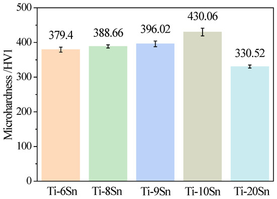
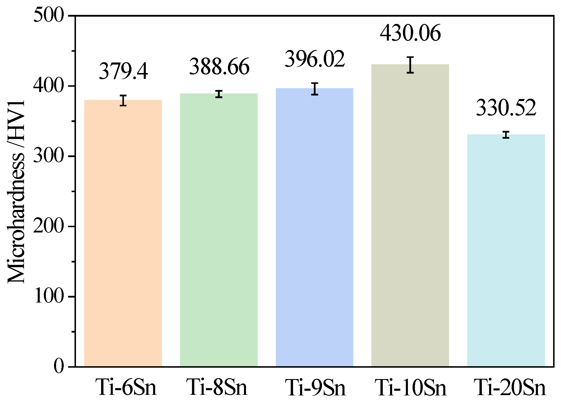
Figure 2 presents the microhardness evolution of Ti-Sn alloys with varying Sn content. The hardness initially increased from 6% to 10% Sn (peaking at 430.06 HV1 for Ti-10Sn), then decreased with further Sn addition to 20%. This non-monotonic trend could correlate with the β-phase suppression at optimal Sn concentrations.
Figure 2.
Microhardness of Ti-xSn alloys (x = 6, 8, 9, 10, 20).
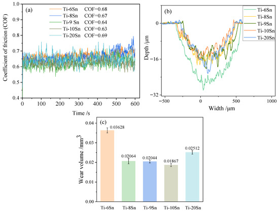
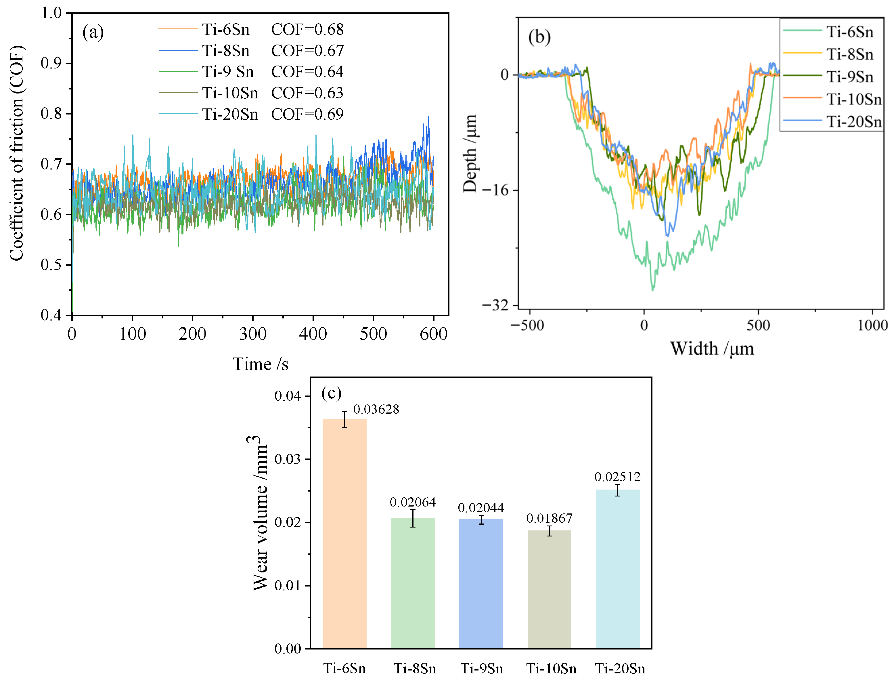
The wear resistances of Ti-xSn alloys were evaluated using indicators of the friction curve, wear volume, and wear scar morphology. Generally, under the same experimental conditions, the smaller the friction coefficient of the friction curve and the smaller the wear volume, the better the wear resistance of the alloy. As can be seen from Figure 3a, the friction coefficients of Ti-xSn alloys (x = 6, 8, 9, 10, 20) remain between 0.6 and 0.7, with average friction coefficients of 0.68, 0.67, 0.64, 0.63, and 0.69, respectively. Ti-10Sn revealed superior tribological properties for its lowest average friction coefficient (0.63) and more stable sliding behavior compared to other alloys. The wear scar contour of Ti-xSn alloy was further measured using the surface profilometer, as shown in Figure 3b, which was serrated and exhibited obvious plowing grooves, indicating that the wear mechanism of Ti-xSn alloys was abrasive wear [17]. According to the contour area (the cross-sectional area of the wear scar) and the sliding distance (S) in the friction experiment, the wear volumes of Ti-xSn alloys were calculated, which characterized the volume change in the alloys before and after wear. As shown in Figure 3c, Ti-10Sn has the smallest wear volume, and Ti-6Sn has the largest wear volume.
Figure 3.
(a) Friction curve, (b) wear scar contour, and (c) wear volume of Ti-xSn alloys (x = 6, 8, 9, 10, 20).
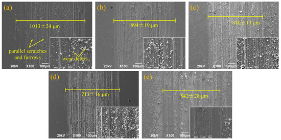
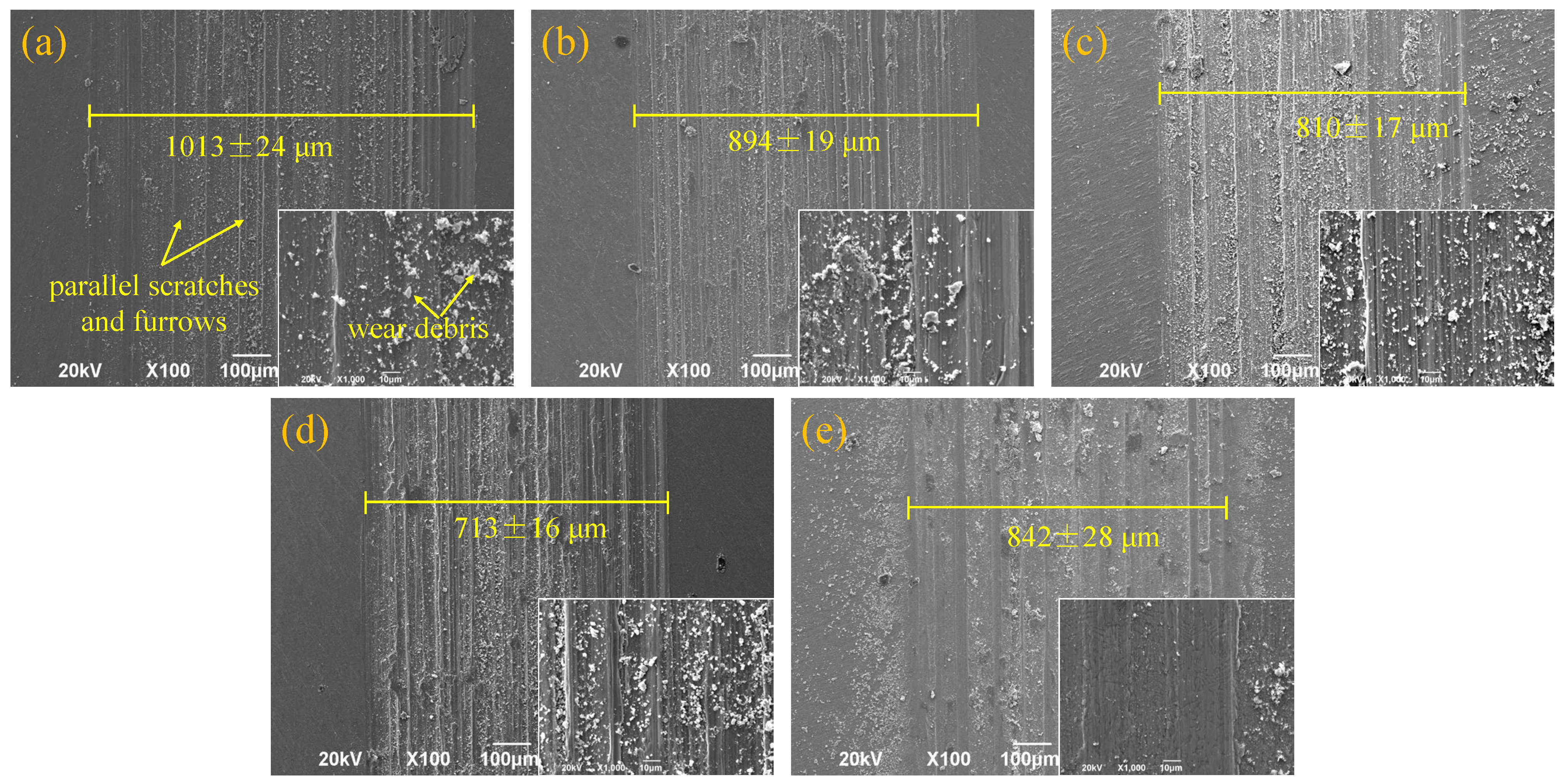
To further analyze the wear mechanism of the Ti-xSn alloys, the surface morphologies of the wear scar were observed by the SEM after friction experiments. As shown in Figure 4, parallel scratches or furrows on the worn surface of the alloys can be obviously observed, and the debris was in the form of particles, strips, or chips. This further confirmed that the wear mechanisms of Ti-xSn alloys were abrasive wear [18,19]. Additionally, the wear scar widths of Ti-xSn alloys (x = 6, 8, 9, 10, 20) were measured, which were 1013 μm, 894 μm, 810 μm, 713 μm, and 842 μm, respectively. Among them, Ti-10Sn had the smallest wear scar width.
Figure 4.
Wear scar morphology of Ti-Sn alloys: (a) Ti-6Sn; (b) Ti-8Sn; (c) Ti-9Sn; (d) Ti-10Sn; and (e) Ti-20Sn. The single yellow line in each image marks the wear scar width, with the labeled value representing the average width and standard deviation measured from five different positions of each wear scar.
In summary, as the Sn content increased, the microhardness of the Ti-Sn alloys initially rose and then declined, peaking at 430.06 HV1 in the Ti-10Sn alloy. Similarly, the friction coefficient first decreased before increasing, attaining its lowest value of 0.63 for Ti-10Sn. Furthermore, Ti-10Sn demonstrated the narrowest average scratch width (713 μm) and the smallest wear volume (0.01867 mm3) among all tested compositions. These results confirm that Ti-10Sn possesses the highest microhardness and superior wear resistance compared to the other Ti-xSn alloys.
3.2. Laser Cladding Ti-10Sn Alloy onto Ti-7.5Nb-4Mo-2Sn Shape Memory Alloy with Different Laser Scan Speed
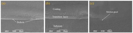
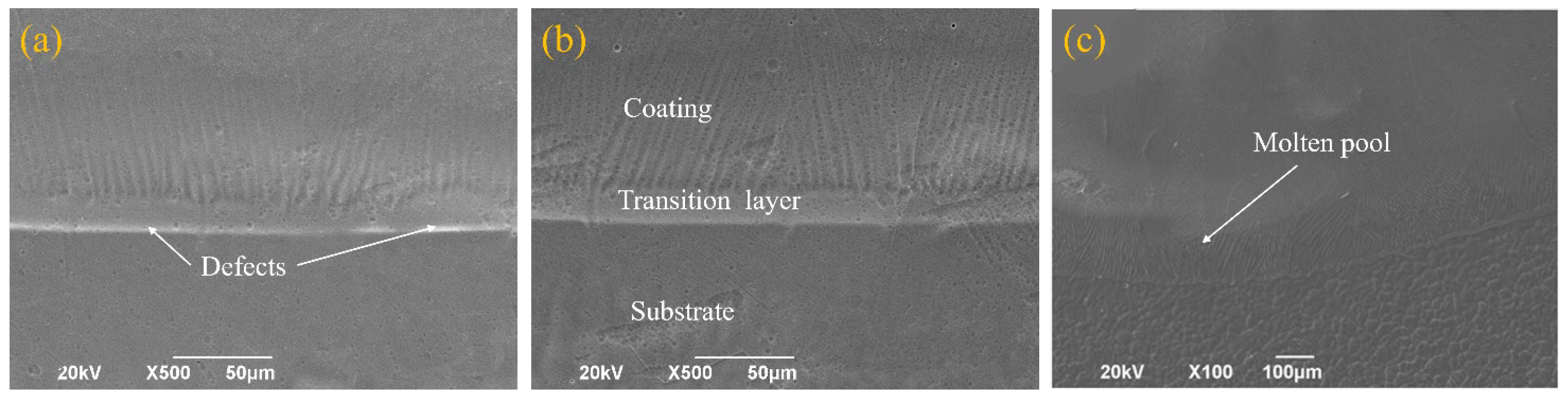
Figure 5 presents SEM micrographs of the coating cross-sections at different scanning speeds. At 13 mm/s (Figure 5a), the transition layer appeared narrow and irregular, with distinct white boundaries near the substrate interface, suggesting potential incomplete bonding and a defective formation [20]. The optimal condition was observed at 10 mm/s (Figure 5b), where a well-defined ~20 μm transition layer formed between the coating and substrate without detectable pores or cracks, indicating excellent metallurgical bonding. This moderate ~20 μm transition layer enables a sufficient mutual interdiffusion of alloying elements between the coating and substrate to form a compositionally graded zone, which achieves robust metallurgical bonding while avoiding excessive substrate dilution; meanwhile, the graded structure buffers interfacial thermal and mechanical stresses, improves load transfer efficiency at the interface, and prevents crack initiation and propagation at the coating–substrate boundary [21]. However, at the reduced scanning speed of 7 mm/s (Figure 5c), the excessive heat input resulted in a large molten pool with high dilution rates, eliminating the distinct transition layer and compromising both coating and substrate integrity. These observations can be explained by the thermal dynamics of laser cladding. Lower scanning speeds increase melt pool temperatures while decreasing cooling rates, significantly affecting interfacial characteristics. As the scanning speed decreased from 13 to 7 mm/s, the elevated interface temperature and reduced cooling rate produced markedly different bonding morphologies in the coated alloys [22].
Figure 5.
SEM images of the cross-section of the coated alloy at different scanning speeds: (a) 13 mm/s; (b) 10 mm/s; and (c) 7 mm/s.
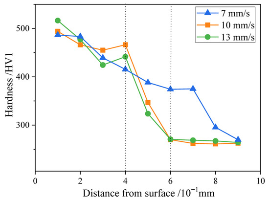
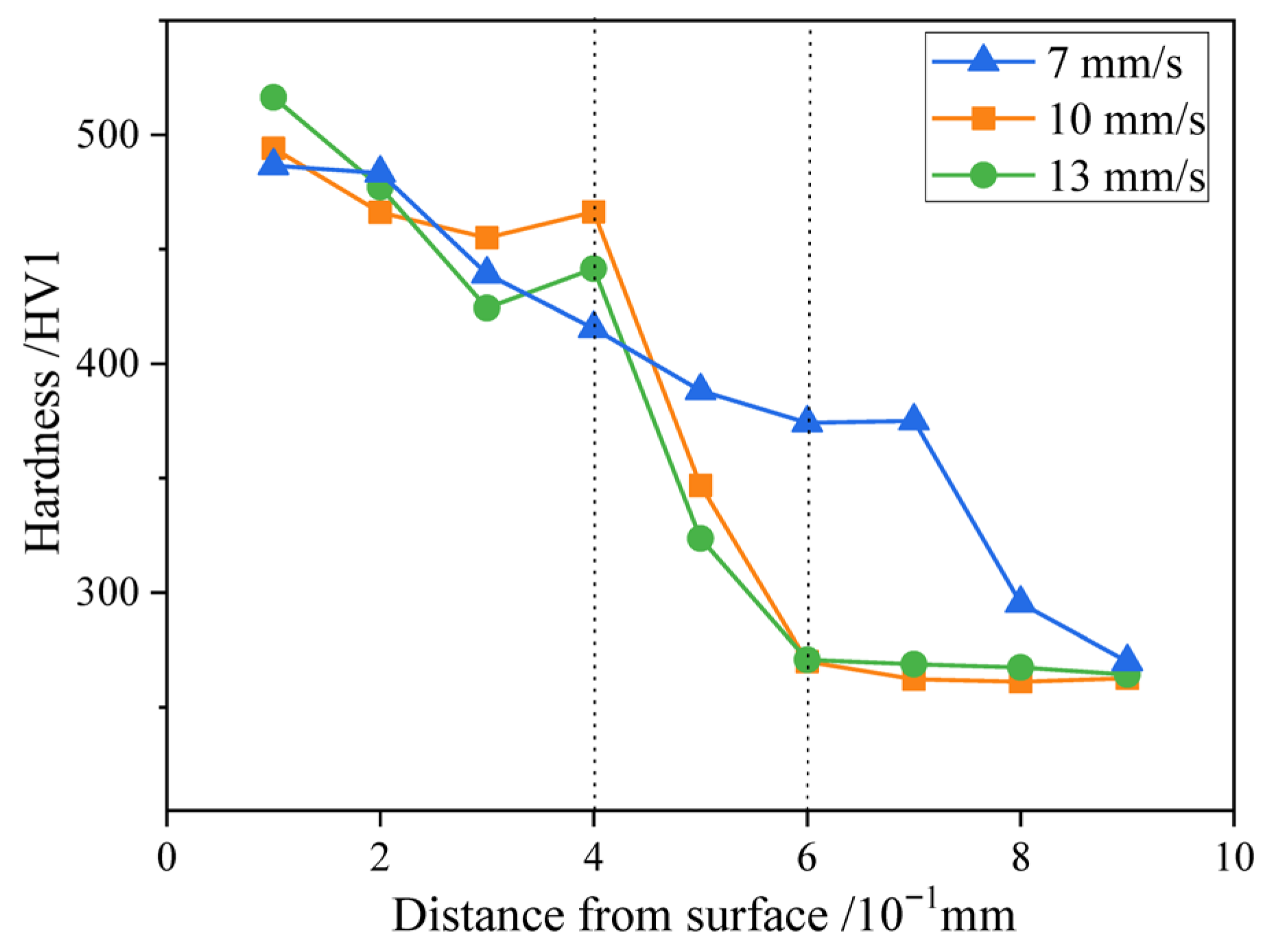
Microhardness profiles across the alloy cross-sections were measured at different scanning speeds, as presented in Figure 6, where the horizontal axis represents the distance from the test point to the surface of the coating, and microhardness was measured at intervals of 0.2 mm. As can be seen, the hardness increased from the substrate to the coating, and the highest microhardness is located on the surface of the coating. When the scanning speeds were 13 mm/s, 10 mm/s, and 7 mm/s, the maximum microhardness reached 516 HV1, 494 HV1, and 486 HV1, respectively, indicating the surface hardness of the Ti-based alloy substrate was significantly improved after laser cladding, with about twice the value of the substrate (256 HV1).
Figure 6.
Microhardness distribution of the coated alloy cross-section at different scanning speeds.
The microhardness distribution across the coated alloy cross-sections revealed significant variations with scanning speed. At 13 mm/s (green line), the hardness decreased gradually from the surface, reaching a minimum at 0.3 mm depth, likely due to interfacial defects such as micropores [23]. The 10 mm/s condition (origin line) exhibited three distinct zones: a high-hardness coating region (0–0.4 mm, ~480 HV1), a transitional zone (0.4–0.6 mm), and the unaffected substrate (>0.6 mm, ~256 HV1). This graded structure—particularly the intermediate transition layer, the effectively buffered interfacial stresses, and the prevention of crack formation—resulted in the metallurgical bonding between the coating and the substrate [21]. In contrast, the 7 mm/s sample (blue line) showed a near-linear hardness decrease with depth, with values exceeding other conditions beyond 0.5 mm, which are attributable to excessive dilution from the enlarged molten pool [24]. Notably, all laser-clad coatings demonstrated enhanced hardness compared to as-cast Ti-10Sn (430 HV1), with maximum values of 516 HV1 (13 mm/s), 494 HV1 (10 mm/s), and 486 HV1 (7 mm/s). This improvement could stem from rapid solidification effects, including grain refinement and increased dislocation density during laser quenching [25].
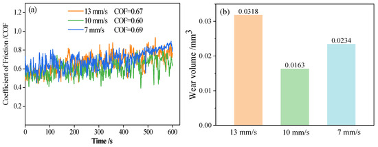
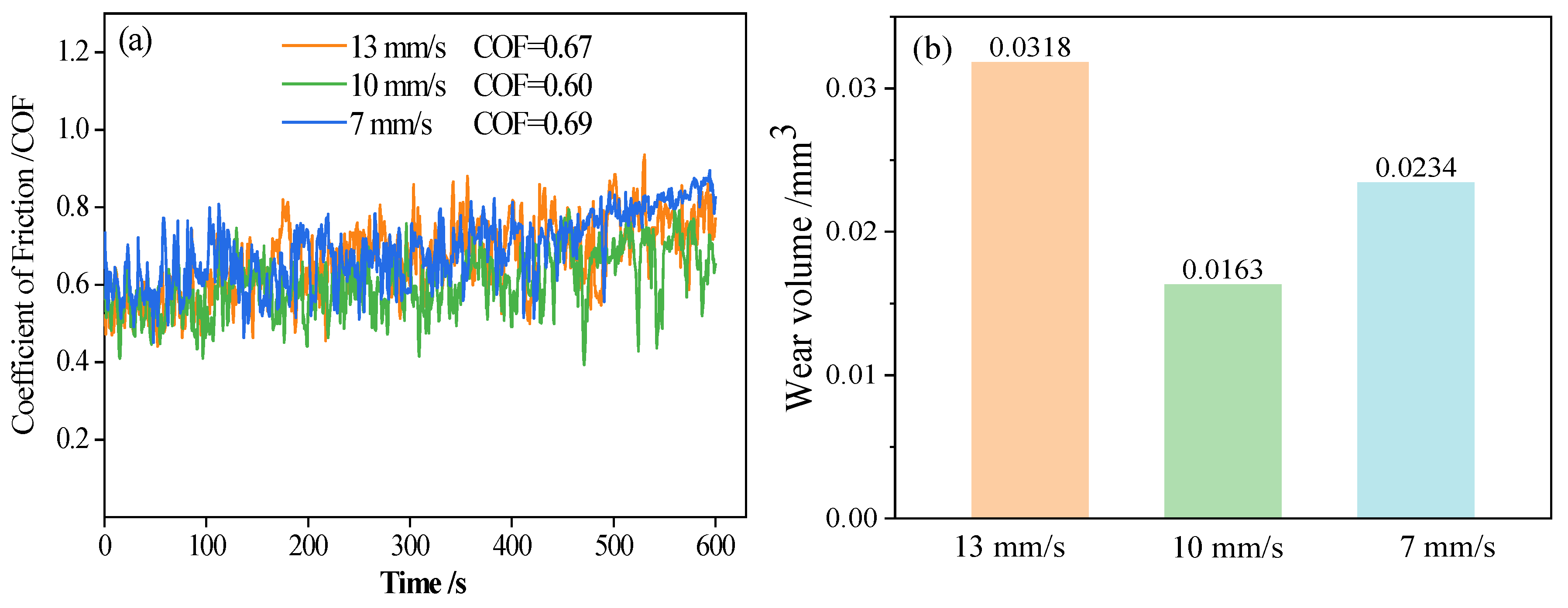
Wear testing results (Figure 7) demonstrated superior tribological performance of the clad coatings compared to the substrate. The 10 mm/s condition exhibited optimal characteristics with the lowest average friction coefficient (0.60 vs. substrate’s 0.81) and the minimal wear volume (0.0163 mm3 vs. substrate’s 0.0685 mm3). This performance advantage correlates with its well-defined hardness gradient and defect-free interface [26]. While all clad samples showed improved wear resistance over the substrate, the 13 mm/s and 7 mm/s conditions displayed higher friction coefficients (0.67 and 0.69, respectively) and greater wear volumes (0.0318 mm3 and 0.0234 mm3), which were consistent with their respective microstructural characteristics. The exceptional performance of the 10 mm/s coating underscores the importance of optimized laser parameters in achieving balanced mechanical and tribological properties through controlled microstructure development.
Figure 7.
(a) Friction curves and (b) wear volumes of the coated alloys at different scanning speeds.
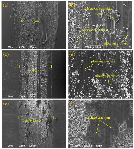
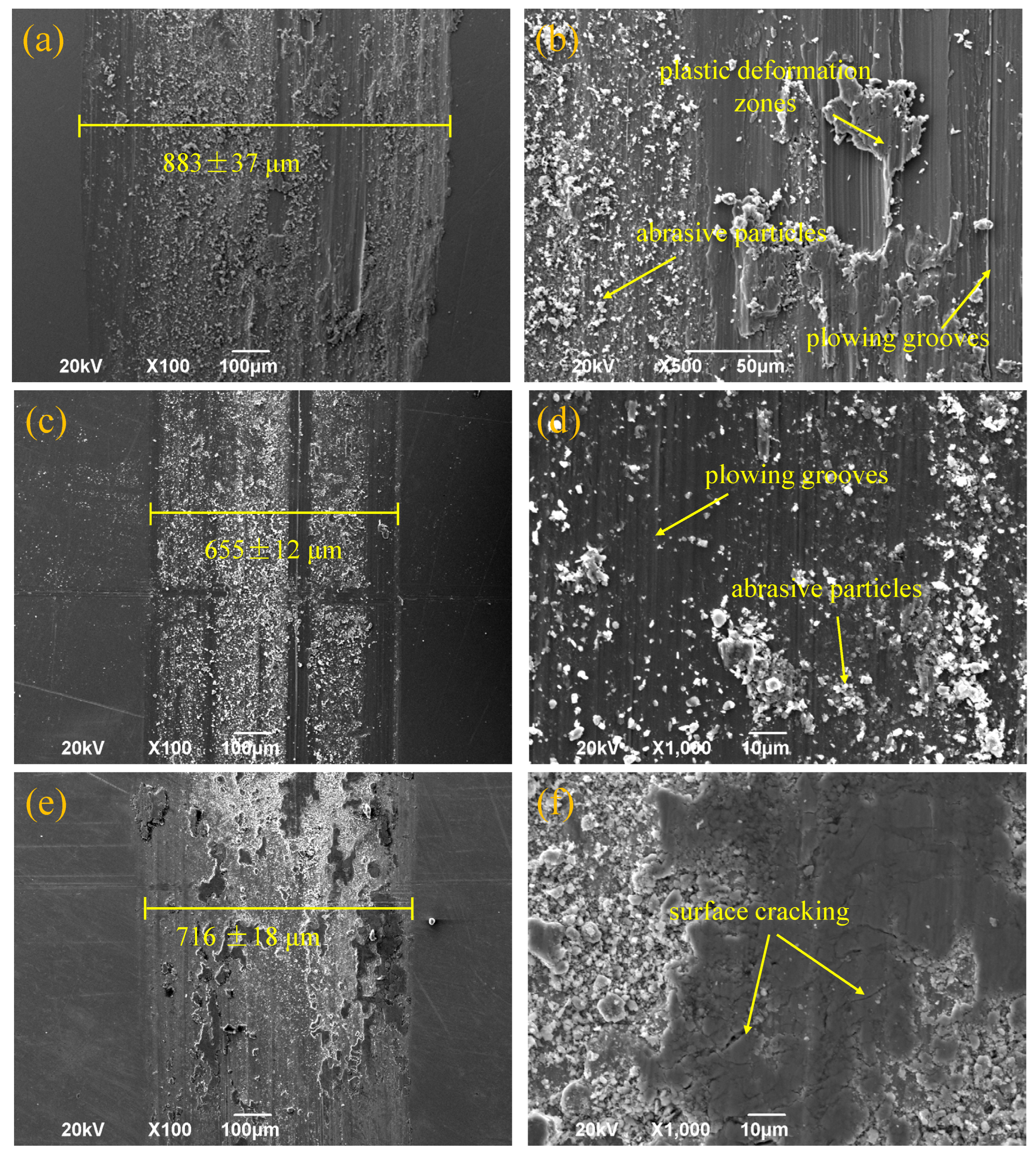
Surface wear morphology analysis revealed distinct wear mechanisms corresponding to different laser scanning speeds (Figure 8). The 13 mm/s condition (Figure 8a,b) exhibited characteristic adhesive wear features, including significant plastic deformation zones and adhered white abrasive particles, accompanied by minor plowing grooves. The adhered materials on the surface of the 13 mm/s coating are mainly formed by the smearing of wear debris under shear stress, and the insufficient interfacial bonding strength leads to the accumulation of smeared debris and the formation of obvious adhesive junctions [27]. This morphology indicates a predominant adhesive wear mechanism with secondary abrasive wear contributions [28]. In contrast, the 10 mm/s samples (Figure 8c,d) displayed typical abrasive wear characteristics, featuring shallow parallel grooves and numerous detached white particles aligned with the sliding direction. The absence of severe plastic deformation suggests that the enhanced coating hardness (494 HV1) inhibited excessive plastic flow and promoted a transition in wear mechanism [29]. During friction, stress concentration at the surface promotes defect-initiated microcracks, leading to particle detachment. These liberated hard particles subsequently participated in a three-body abrasion, generating the observed groove patterns [30]. The 7 mm/s condition (Figure 8e,f) demonstrated a fatigue-dominated wear mechanism, evidenced by extensive surface cracking. This behavior suggests that reduced cooling rates at lower scanning speeds compromised the coating’s fatigue resistance. Quantitative analysis of wear scar widths (883 μm, 655 μm, and 716 μm for 13, 10, and 7 mm/s, respectively) confirmed the superior wear resistance of the 10 mm/s coating, correlating well with its minimal wear volume (0.0163 mm3). The 655 μm wear scar width observed for this condition further validates its optimal tribological performance among the tested parameters.
Figure 8.
The wear scar morphologies and an enlarged image of the coated alloys at different scanning speeds: (a,b) for 13 mm/s; (c,d) for 10 mm/s; and (e,f) for 7 mm/s.
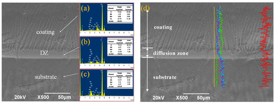
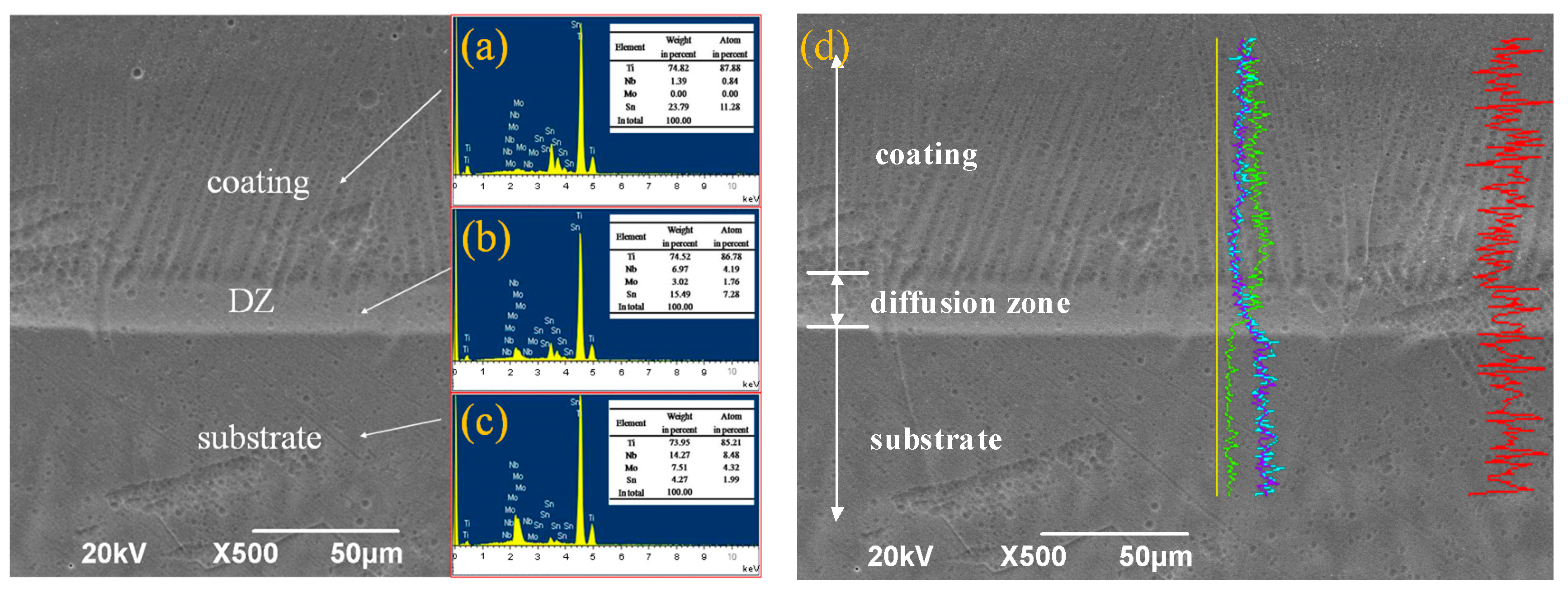
To elucidate the interfacial bonding mechanism at the optimal scanning speed of 10 mm/s, cross-sectional elemental distribution was characterized by EDS analysis (Figure 9). The elemental mapping results (Figure 9a–c) revealed distinct interdiffusion phenomena between the Ti-10Sn coating and the Ti-7.5Nb-4Mo-2Sn substrate. Notably, substrate elements (Nb and Mo) were detected in the coating layer, while Sn content exhibited a gradual increase from substrate to coating. The transition layer (20 μm width) showed intermediate concentrations of all alloying elements, confirming mutual diffusion across the interface [31].
Figure 9.
Diagram of EDS spectra with elemental analysis: (a) coating; (b) diffusion zone; (c) substrate; and (d) EDS line scan result of alloy cross-section, where the red, blue, purple and green curve represent the elements of Ti, Mo, Nb and Sn, respectively, and the yellow straight line is the zero line.
The EDS line scan profile (Figure 9d) quantitatively demonstrates this interdiffusion behavior. The Ti (red) and Sn (yellow–green) concentration gradients show complementary trends, while Mo (blue) and Nb (purple) profiles exhibit diffusion tails extending approximately 15 μm into the coating region. These concentration gradients provide the thermodynamic driving force for elemental interdiffusion [25], with the high thermal energy input during laser cladding (1300 W, 10 mm/s) providing the necessary kinetic conditions [26]. The observed elemental redistribution confirms the formation of a metallurgical bond through: (a) mutual solubility of alloying elements across the interface, (b) formation of compositionally graded transition zones, and (c) absence of abrupt concentration changes that are characteristic of mechanical bonding [32]. This diffusion-controlled bonding mechanism explains the superior interfacial integrity that is observed in microstructural analysis (Figure 5b), where the compositionally graded transition layer effectively accommodates thermal expansion mismatches and prevents stress concentration at the coating–substrate interface. The mutual interdiffusion of Nb, Mo and Sn atoms forms a continuous compositional gradient at the interface, which enhances the atomic bonding force between the coating and the substrate to improve interfacial bonding strength; meanwhile, the gradient structure reduces the abrupt change in mechanical properties at the interface, effectively accommodating interfacial thermal and mechanical stresses and avoiding stress concentration-induced crack initiation.
3.3. Comparison of Properties Between Ti-10Sn Coating and Ti-7.5Nb-4Mo-2Sn Substrate
A comparative analysis of the mechanical and tribological properties between the Ti-10Sn coating and the uncoated Ti-based substrate reveals significant improvements through laser cladding surface modification. As summarized in Table 1, the optimal processing parameters (1300 W laser power, 4 mm beam diameter, and 10 mm/s scanning speed) produced a coated alloy with substantially enhanced performance characteristics. The microhardness of the coated surface (494 HV1) demonstrated a 93% increase over the substrate material (256 HV1), while wear testing showed a 60% reduction in the friction coefficient (0.60 vs. 0.81) and an 80% decrease in wear volume (0.0163 mm3 vs. 0.083 mm3). These improvements are directly attributable to the microstructural modifications induced by optimized Ti-Sn alloy design and laser cladding.
Table 1.
Comparison of alloy properties between the Ti-7.5Nb-4Mo-2Sn substrate and the Ti-10Sn coating that was fabricated at 10 mm/s.
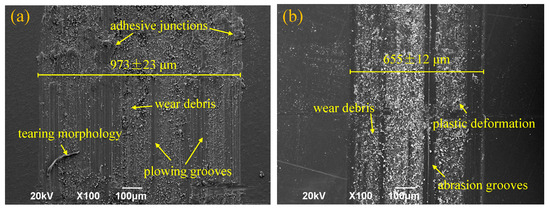
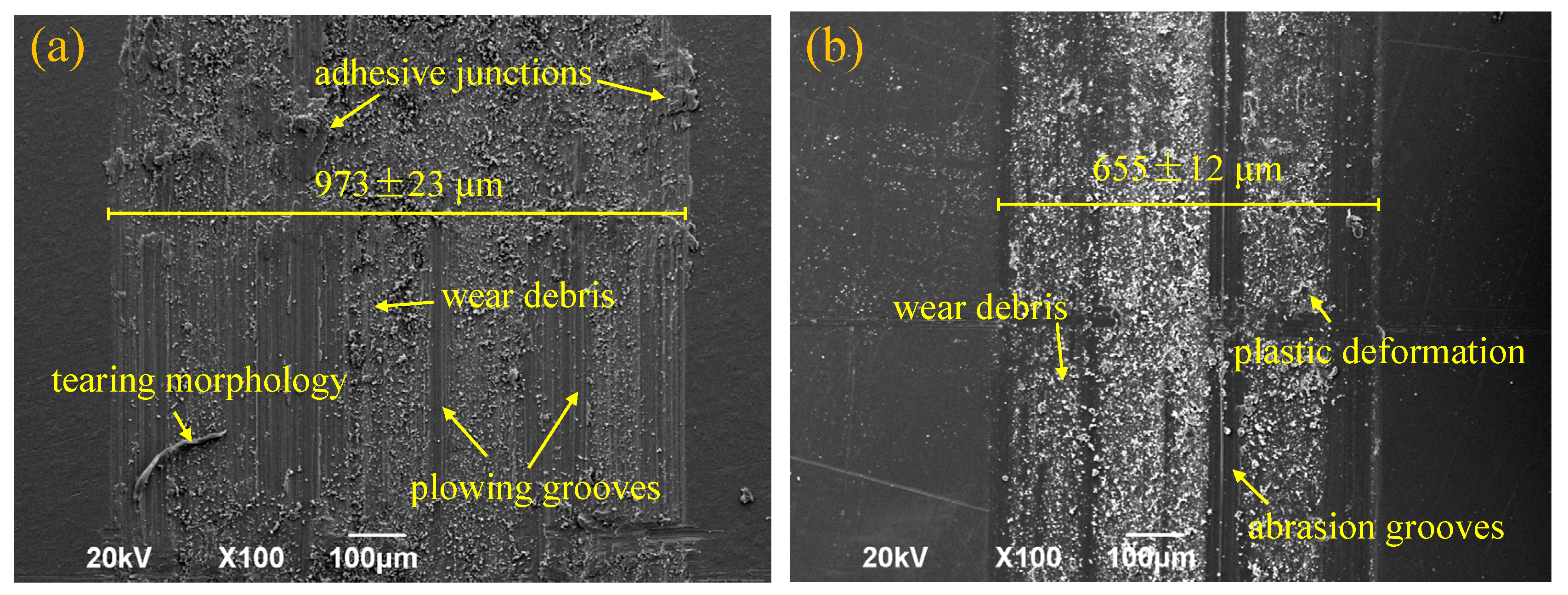
Distinct wear mechanisms were identified through surface morphology analysis (Figure 10). The substrate surface (Figure 10a) exhibited severe plastic deformation features that are characteristic of predominant adhesive wear, including: (i) deep plowing grooves (20–50 μm width), (ii) material transfer regions with adhesive junctions, (iii) surface tearing morphology, and (iv) accumulated wear debris particles. This damage pattern results from the substrate’s lower hardness and higher ductility, which promotes extensive plastic flow under the applied shear stresses from the steel counterface [29,33]. In contrast, the Ti-10Sn coated surface (Figure 10b) displayed different characteristics, including: (i) uniform shallow abrasion grooves (5–10 μm depth), (ii) minimal adhesive features, (iii) limited plastic deformation, and (iv) well-distributed fine wear debris. The transition from severe adhesive–abrasive wear (substrate) to mild abrasive wear (coating) confirms the effectiveness of laser cladding in surface property enhancement. This mechanism shift could be attributed to an increased surface hardness that reduced plastic deformation, refined the microstructure that resisted crack initiation, strengthened metallurgical bonding that prevented delamination, and optimized the hardness gradient that minimized stress concentration [34]. The high hardness of the Ti-10Sn coating fundamentally inhibits the plastic flow and material transfer that are the core causes of adhesive wear, thus the wear mechanism is dominated by mild abrasive wear instead of the mixed adhesive–abrasive wear of the substrate. These results demonstrated that effective coating design combined with laser cladding technology fundamentally alters the wear behavior of Ti-based alloys through microstructural modification and mechanical property enhancement, providing a viable surface engineering solution for tribological applications.
Figure 10.
Comparison in wear morphology between (a) Ti-7.5Nb-4Mo-2Sn substrate and (b) Ti-10Sn coating.
4. Conclusions
This study investigated the surface modification of Ti-7.5Nb-4Mo-2Sn β-type titanium shape memory alloy, which exhibits excellent biomechanical compatibility but suffers from inadequate wear resistance for biomedical implant applications. Through systematic alloy design and laser cladding processing optimization, the following key conclusions were drawn:
- (1)
- A series of Ti-xSn (x = 6, 8, 9, 10, 20 at.%) alloys was designed and fabricated as potential coating materials. Microstructural characterization and mechanical testing revealed that as the Sn content increased from 6% to 10%, the β phase content decreased, resulting in the improvement of microhardness and wear resistance of the Ti-xSn alloy. When the Sn content increased from 10% to 20%, the result was the opposite. Ti-10Sn exhibited the highest microhardness and superior wear resistance.
- (2)
- Ti-10Sn coatings were successfully fabricated on Ti-7.5Nb-4Mo-2Sn substrates via laser cladding. Process optimization revealed that increasing the scanning speed enhanced cooling rates while reducing the interfacial temperatures significantly affected wear mechanisms: 13 mm/s produced mixed adhesive-abrasive wear, 10 mm/s yielded optimal abrasive wear characteristics, and 7 mm/s led to fatigue-dominated wear due to excessive heat input.
- (3)
- The coating fabricated at 10 mm/s exhibited the best wear resistance, with metallurgical bonding between the substrates. While the substrate’s wear mechanism was a combination of adhesive and abrasive wear, the coating’s wear mechanism shifted to abrasive wear. This led to a marked increase in surface microhardness and wear resistance. This study shows that laser cladding, along with proper alloy design, can effectively enhance the surface wear resistance of biomedical alloys.
Author Contributions
Z.L.: Conceptualization, methodology, writing—original draft. Y.G.: Methodology, data curation, formal analysis. S.L.: Resources, methodology, formal analysis. X.Y.: Resources, writing—review and editing and supervision. All authors have read and agreed to the published version of the manuscript.
Funding
This work was supported by the Hunan Provincial Natural Science Foundation of China (2026JJ70139) and the Undergraduate Innovation and Entrepreneurship Training Program of Hunan Province (S202411527051).
Institutional Review Board Statement
Not applicable.
Informed Consent Statement
Not applicable.
Data Availability Statement
Data is contained within the article.
Conflicts of Interest
The authors declare that they have no known competing financial interests or personal relationships that could have appeared to influence the work reported in this paper.
References
- Lei, C.; Zhu, C.; Lin, J.; Zhang, D. Room temperature creep behavior of Ti–Nb–Mo–Sn based shape memory alloy for biomedical application. Smart Mater. Manuf. 2023, 1, 100019. [Google Scholar]
- Zhang, D.; Wang, Y.; Lin, J.; Wen, C. Microstructure and superelasticity of a biomedical β-type titanium alloy under various processing routes. Appl. Mater. Today 2016, 5, 41–51. [Google Scholar] [CrossRef] [Scilit]
- Couto, M.; Vasconcelos, D.P.; Sousa, D.M.; Sousa, B.; Conceição, F.; Neto, E.; Lamghari, M.; Alves, C.J. The Mechanisms Underlying the Biological Response to Wear Debris in Periprosthetic Inflammation. Front. Mater. 2020, 7, 274. [Google Scholar] [CrossRef] [Scilit]
- Hodges, N.A.; Sussman, E.M.; Stegemann, J.P. Aseptic and septic prosthetic joint loosening: Impact of biomaterial wear on immune cell function, inflammation, and infection. Biomaterials 2021, 278, 121127. [Google Scholar] [CrossRef] [Scilit]
- Zhu, L.; Xue, P.; Lan, Q.; Meng, G.; Ren, Y.; Yang, Z.; Xu, P.; Liu, Z. Recent research and development status of laser cladding: A review. Opt. Laser Technol. 2021, 138, 106915. [Google Scholar] [CrossRef] [Scilit]
- Liu, Y.; Ding, Y.; Yang, L.; Sun, R.; Zhang, T.; Yang, X. Research and progress of laser cladding on engineering alloys: A review. J. Manuf. Process. 2021, 66, 341–363. [Google Scholar] [CrossRef] [Scilit]
- Singh, N.; Mehta, A.; Vasudev, H.; Samra, P.S. A review on the design and analysis for the application of Wear and corrosion resistance coatings. Int. J. Interact. Des. Manuf. (IJIDeM) 2024, 18, 5381–5405. [Google Scholar]
- Zhai, W.; Bai, L.; Zhou, R.; Fan, X.; Kang, G.; Liu, Y.; Zhou, K. Recent Progress on Wear-Resistant Materials: Designs, Properties, and Applications. Adv. Sci. 2021, 8, 2003739. [Google Scholar]
- Wang, X.F.; Li, W.; Fang, G.P.; Wu, C.W.; Lin, J.G. First-principles calculations on the electronic structure and cohesive properties of titanium stannides. Intermetallics 2009, 17, 768–773. [Google Scholar] [CrossRef] [Scilit]
- Azmat, A.; Tufail, M.; Chandio, A.D. Synthesis and Characterization of Ti-Sn Alloy for Orthopedic Application. Materials 2021, 14, 7660. [Google Scholar] [CrossRef] [Scilit] [PubMed]
- Batibay, A.; Erci, F.; Kotan, H.; Karavelioglu, Z.; Koc, R.C.; Oral, I.; Evcin, A. Investigation and production of non-cytotoxic TixNbxSn (x = 5, 10, 15, 20) alloys by high-energy mechanical milling with antibacterial activity. Mater. Today Commun. 2023, 37, 106912. [Google Scholar] [CrossRef] [Scilit]
- Song, Y.-H.; Kim, M.-K.; Park, E.-J.; Song, H.-J.; Anusavice, K.J.; Park, Y.-J. Cytotoxicity of alloying elements and experimental titanium alloys by WST-1 and agar overlay tests. Dent. Mater. 2014, 30, 977–983. [Google Scholar] [CrossRef] [Scilit]
- Okamoto, H. Sn-Ti (Tin-Titanium). J. Phase Equilib. Diffus. 2010, 31, 202–203. [Google Scholar]
- Xu, J.; Zeng, W.; Zhao, Y.; Sun, X.; Du, Z. Influence of cooling rate following heat treatment on microstructure and phase transformation for a two-phase alloy. J. Alloys Compd. 2016, 688, 301–309. [Google Scholar]
- Colinet, C.; Tedenac, J.-C.; Fries, S.G. Structural stability of intermetallic phases in the Sn–Ti system. Calphad 2009, 33, 250–259. [Google Scholar] [CrossRef] [Scilit]
- Lewis, A.C.; Qidwai, S.M.; Geltmacher, A.B. Slip systems and initiation of plasticity in a body-centered-cubic titanium alloy. Metall. Mater. Trans. A 2010, 41, 2522–2531. [Google Scholar]
- Jiang, X.; Lu, J.; Zhao, N.; Chen, Z.; Zhao, Z. A Review of Wear in Additive Manufacturing: Wear Mechanism, Materials, and Process. Lubricants 2024, 12, 321. [Google Scholar] [CrossRef] [Scilit]
- Da Silva, W.M.; De Mello, J.D.B. Using parallel scratches to simulate abrasive wear. Wear 2009, 267, 1987–1997. [Google Scholar] [CrossRef] [Scilit]
- Nagarjuna, C.; You, H.J.; Ahn, S.; Song, J.W.; Jeong, K.Y.; Madavali, B.; Song, G.; Na, Y.S.; Won, J.W.; Kim, H.S.; et al. Worn surface and subsurface layer structure formation behavior on wear mechanism of CoCrFeMnNi high entropy alloy in different sliding conditions. Appl. Surf. Sci. 2021, 549, 149202. [Google Scholar]
- Cheng, J.; Xing, Y.; Dong, E.; Zhao, L.; Liu, H.; Chang, T.; Chen, M.; Wang, J.; Lu, J.; Wan, J. An overview of laser metal deposition for cladding: Defect formation mechanisms, defect suppression methods and performance improvements of laser-cladded layers. Materials 2022, 15, 5522. [Google Scholar] [CrossRef] [Scilit]
- Liu, Z.; Zhang, H.; Tang, L.; Guo, J.; Zhang, Y.; Li, Y.; You, L.; Wang, J.; Dong, X.; Liu, Q.; et al. Effect of diffusion interlayer and graded multi-interlayers on the reduction of thermal stress in tritium penetration barrier coating system. Fusion Eng. Des. 2024, 205, 114568. [Google Scholar] [CrossRef] [Scilit]
- Jiao, X.; Wang, J.; Wang, C.; Gong, Z.; Pang, X.; Xiong, S. Effect of laser scanning speed on microstructure and wear properties of T15M cladding coating fabricated by laser cladding technology. Opt. Lasers Eng. 2018, 110, 163–171. [Google Scholar] [CrossRef] [Scilit]
- Park, C.W.; Adomako, N.K.; Lee, M.G.; Kim, J.H.; Kim, J.H. Interfacial structure and pore formation mechanism during laser cladding of pure vanadium on Ti-6Al-4V alloy. Int. J. Refract. Met. Hard Mater. 2021, 101, 105671. [Google Scholar] [CrossRef] [Scilit]
- Jeong, W.; Yang, J.; Choi, J.P.; Hwang, J.Y.; Kim, Y.W.; Park, S.J.; Choi, J.W.; Heogh, W.; Lee, H.; Park, J.; et al. Al-based functionally graded super-intermetallic compounds for the turbine blade of a high-performance jet engine. Adv. Compos. Hybrid Mater. 2025, 8, 420. [Google Scholar] [PubMed]
- Wang, G.; Luo, S.; Guo, Y.; Huang, R.; Wang, C.; Qu, Z. Investigating the correlation between mechanical properties and gradient microstructures in laser shock peened CrCoNi alloy. Int. J. Plast. 2025, 189, 104331. [Google Scholar] [CrossRef] [Scilit]
- Zhang, B.; He, B.; Wang, H. Investigation on laser cladding Fe-based gradient coatings with hierarchical enhanced wear resistance on U75V railway steel. Wear 2024, 552–553, 205444. [Google Scholar]
- Aghababaei, R.; Warner, D.H.; Molinari, J.-F. On the debris-level origins of adhesive wear. Proc. Natl. Acad. Sci. USA 2017, 114, 7935–7940. [Google Scholar]
- Wan, J.; Van Aken, D.C.; Qing, J.; Xu, M. A comparison of adhesive wear with three-body abrasive wear characteristics of graphitic white irons designed for metal-to-metal wear systems. Int. J. Met. 2021, 15, 447–458. [Google Scholar] [CrossRef] [Scilit]
- Podgornik, B. Adhesive Wear Failures. J. Fail. Anal. Prev. 2022, 22, 113–138. [Google Scholar] [CrossRef] [Scilit]
- Liu, Z.; Zhou, K.; Zhu, J.; Liao, Z.; Zou, L.; Xu, D. Fabricating ultra wear-resistant surfaces on titanium alloy by combining laser-induced modification with abrasive belt grinding. Tribol. Int. 2024, 200, 110160. [Google Scholar]
- Fu, K.; Zhong, C.; Xue, Y.; Ren, X.; Luan, B. Interfacial phase transformation and element diffusion mechanism of high-speed laser cladding Inconel 625 in high-temperature environment. Mater. Charact. 2024, 211, 113879. [Google Scholar] [CrossRef] [Scilit]
- Liu, Y.; Guo, C.; Zhang, Z.; Lu, D.; Zhang, H.; Guo, B.; Zhou, S.; Li, W.; Yin, S.; Liu, J. Enhanced tribological performance of Cu matrix composites reinforced with high-V wear-resistant steel particles by laser cladding-based additive manufacturing. J. Mater. Res. Technol. 2026, 41, 6020–6029. [Google Scholar] [CrossRef] [Scilit]
- Zhao, S.; Meng, F.; Fan, B.; Dong, Y.; Wang, J.; Qi, X. Evaluation of wear mechanism between TC4 titanium alloys and self-lubricating fabrics. Wear 2022, 512–513, 204532. [Google Scholar] [CrossRef] [Scilit]
- Dearnaley, G. Adhesive and abrasive wear mechanisms in ion implanted metals. Nucl. Instrum. Methods Phys. Res. Sect. B Beam Interact. Mater. At. 1985, 7, 158–165. [Google Scholar] [CrossRef] [Scilit]

Disclaimer/Publisher’s Note: The statements, opinions and data contained in all publications are solely those of the individual author(s) and contributor(s) and not of MDPI and/or the editor(s). MDPI and/or the editor(s) disclaim responsibility for any injury to people or property resulting from any ideas, methods, instructions or products referred to in the content. |
© 2026 by the authors. Licensee MDPI, Basel, Switzerland. This article is an open access article distributed under the terms and conditions of the Creative Commons Attribution (CC BY) license.