


Abstract
Tetracycline (TC), a widely used broad-spectrum antibiotic, was experimentally and theoretically investigated for its potential effect on thyroid hormone biosynthesis through interaction with thyroid peroxidase (TPO). Horseradish peroxidase (HRP) was used as a model of TPO, while FeTPPCl was employed as a model of the active site of the enzyme. The inhibitory effect of TC on the catalytic oxidation of iodide to active iodine was studied spectrophotometrically through the formation of triiodide (I3−), and the IC50 value was determined. Kinetic studies using Michaelis–Menten and Lineweaver–Burk analysis demonstrated that TC acts as a reversible non-competitive inhibitor of HRP, leading to decreased Km and Vmax values and the formation of the ESI complex. Molecular docking calculations further supported the experimental findings, indicating that TC binds at a site different from the active center of the enzyme. The results suggest that tetracycline may interfere with thyroid hormone biosynthesis by inhibiting TPO-mediated iodide oxidation, revealing a possible mechanism associated with thyroid-related side effects during prolonged antibiotic administration.
1. Introduction
The thyroid gland plays a fundamental role in the regulation of metabolism, growth, and cellular homeostasis through the biosynthesis of the thyroid hormones thyroxine (T4) and triiodothyronine (T3) [1,2,3,4,5,6,7,8,9,10,11]. The biosynthesis of these hormones is initiated by thyroid peroxidase (TPO), a heme-containing enzyme that catalyzes the oxidation of iodide to active iodine in the presence of hydrogen peroxide, followed by iodination of tyrosyl residues of thyroglobulin [12,13,14]. TPO-mediated reactions are therefore essential for the formation of mono- and diiodotyrosine intermediates and ultimately for the production of T3 and T4 [14,15]. Several compounds are known to interfere with this pathway, including clinically used antithyroid drugs such as methimazole (MMI) and propylthiouracil (PTU), which inhibit thyroid hormone biosynthesis through interaction with TPO catalytic pathways and active iodine intermediates [8,16,17].
Tetracycline (TC) is a broad-spectrum antibiotic extensively used for the treatment of Gram-positive and Gram-negative bacterial infections through inhibition of bacterial protein synthesis [18,19]. Besides its antimicrobial properties, tetracycline exhibits strong metal-binding ability and can interact with metalloproteins and redox-active biological systems because of its polydentate coordination environment [20]. Such interactions raise the possibility that tetracycline may also affect heme-containing enzymes involved in endocrine processes, including thyroid peroxidase. Despite the extensive clinical use of tetracycline antibiotics, limited information is available regarding their possible involvement in thyroid hormone biosynthesis and thyroid-related side effects.
Horseradish peroxidase (HRP) is frequently used as a biomimetic model of TPO [11,21]. Both HRP and TPO are heme-containing peroxidases that use H2O2 as an oxidizing agent and are able to catalyze the oxidation of iodide. This common catalytic activity makes HRP a useful and well-characterized model for studying reactions related to TPO-mediated iodide oxidation. However, important structural differences exist between the two enzymes. HRP is a plant peroxidase, whereas TPO is a mammalian membrane-associated peroxidase with a more complex domain organization. In addition, differences exist in the environment of the heme group and in the substrate-binding regions of the two enzymes. Therefore, HRP does not represent a complete structural model of TPO but provides a useful functional model for investigating peroxidase-mediated iodide oxidation and its possible inhibition [11,21]. In addition, iron tetraphenylporphyrin chloride (FeTPPCl) has been employed as a model of the active site of TPO for mechanistic studies associated with iodide oxidation and thyroid hormone formation [10,22].
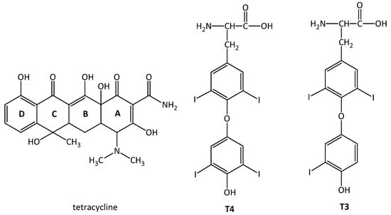
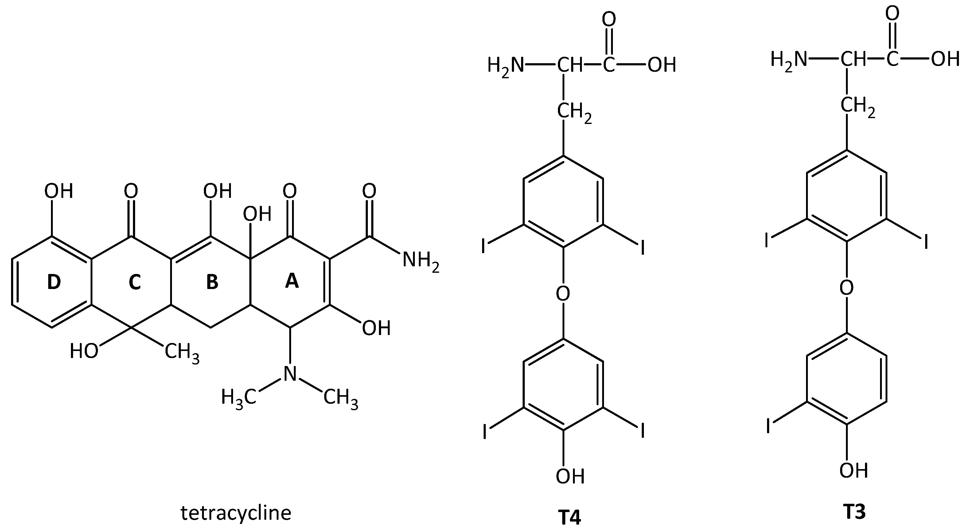
In the course of our studies on the mechanism of action of antithyroid drugs and the side effects that other pharmaceuticals may exert on thyroid hormone biosynthesis [1,2,3,4,5,6,7,8,9,10,11], the interaction of tetracycline (Scheme 1) with HRP and FeTPPCl was investigated both experimentally and theoretically in the present work, in order to elucidate its possible role in the biosynthesis of the thyroid hormones T4 and T3 (Scheme 1). Spectrophotometric kinetic studies, inhibition assays, and molecular docking calculations were performed and compared with the corresponding behavior of methimazole, a clinically established antithyroid drug.
Scheme 1.
The structure of tetracycline (TC) and thyroid hormones Thyroxine (T4) and Thyronine (T3).
2. Results and Discussion
2.1. Effect of Tetracycline on Horseradish Peroxidase (HRP) Activity
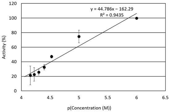
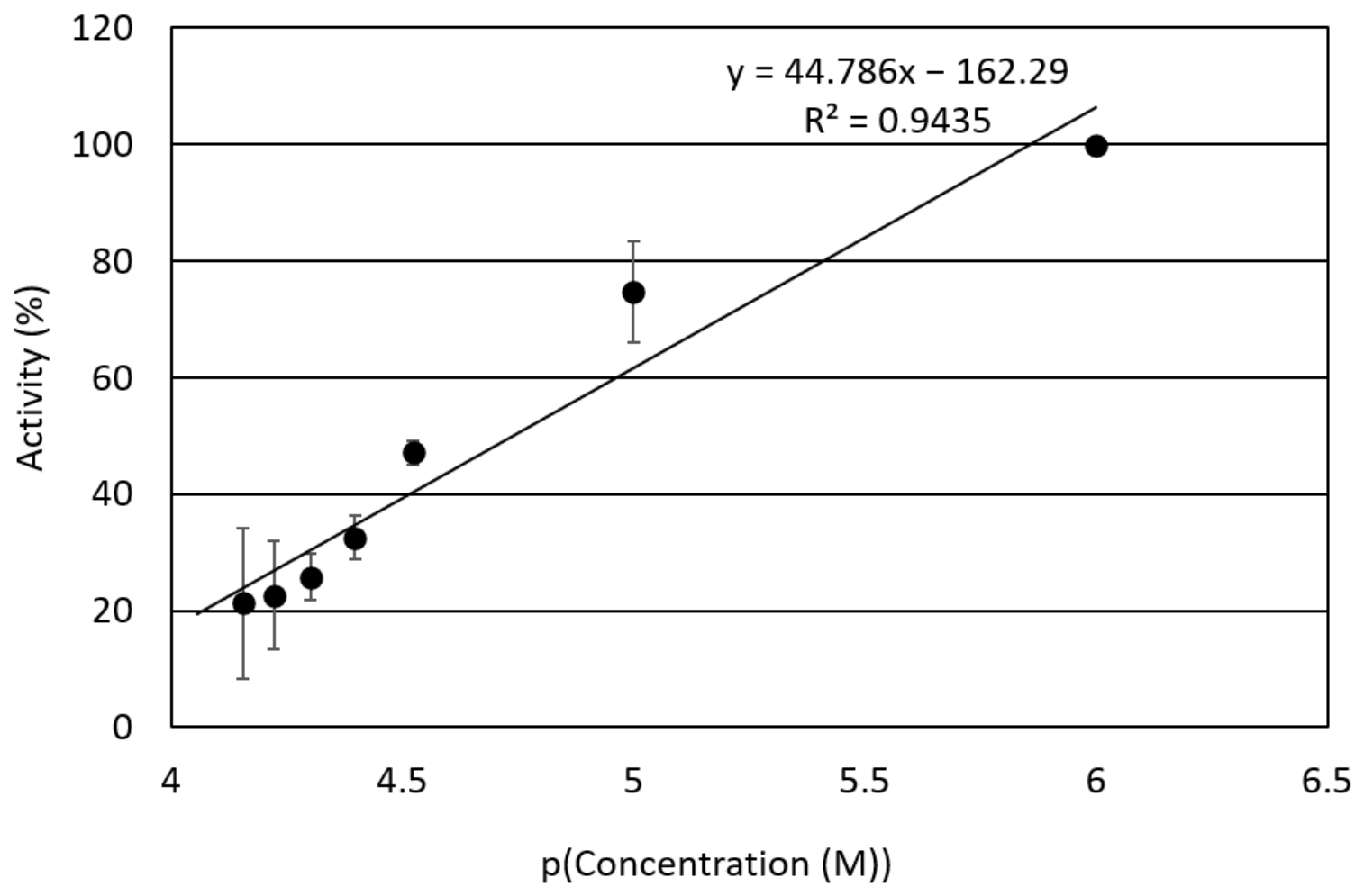
UV–Vis Spectroscopic Study: The effect of TC on the catalytic activity of HRP, used as a model of TPO, was investigated spectrophotometrically by monitoring the oxidation of iodide to triiodide (I3−). The enzymatic activity (A%) was determined from the initial reaction rates measured in the absence and presence of increasing concentrations of TC. Figure 1 presents the percent inhibition curve of HRP enzymatic activity vs. −Log(concentration) of TC.
Figure 1.
HRP activity (A%) as a function of tetracycline concentration, expressed as pC, where pC = −log C and C is the molar concentration of TC. Data are expressed as mean ± SD from three independent experiments (n = 3), and the error bars represent the standard deviation (SD). The pC representation was used for the determination of the IC50 value.
The IC50 is defined as the concentration of tetracycline that inhibits 50% of the enzymatic activity. Therefore, from the plot of enzymatic activity (A%) versus pC, the pIC50 corresponds to the pC value at which the residual enzymatic activity is 50% of the initial activity. The corresponding IC50 value is then calculated according to: IC50 = 10−pIC50 (M). The IC50 value of TC toward HRP is determined to be 36.3 ± 3.2 μM. The reference antithyroid drugs methimazole (MMI) and propylthiouracil (PTU) exhibited greater inhibitory potency, with IC50 values of 18.9 μM and 27.8 μM, respectively [11]. Thus, TC interacts with HRP, reducing its catalytic activity and suggesting a potential interference with thyroid peroxidase (TPO)-like enzymatic function. To further elucidate the inhibitory mechanism of TC, enzyme kinetic studies, molecular docking analyses, and catalytic activity assays using the iron–porphyrin model system FeTPPCl, which mimics the heme-containing active site of HRP, were performed.
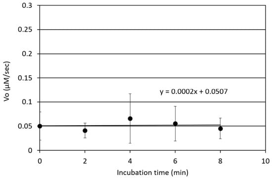
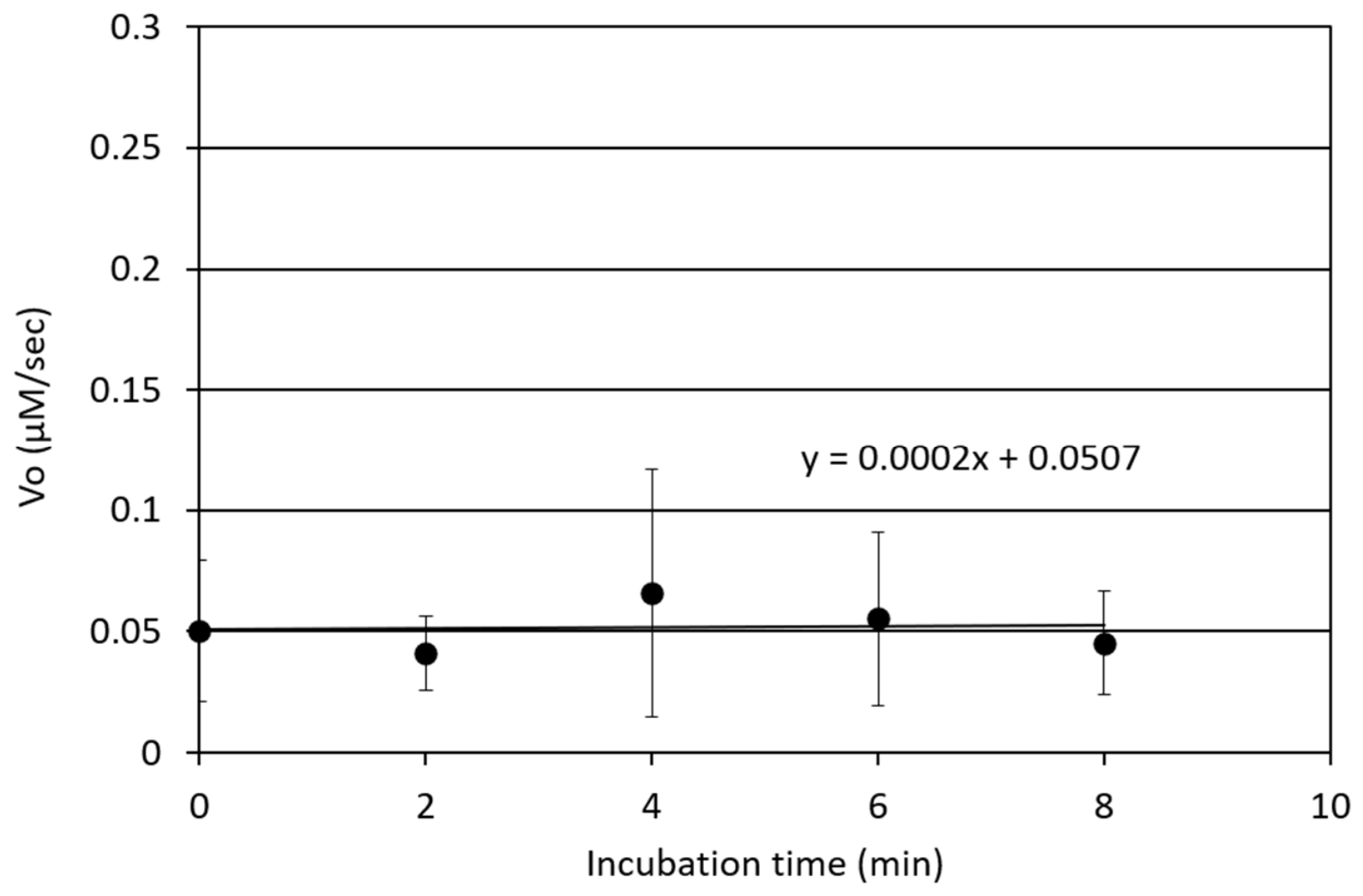
To further investigate the mechanism of inhibition, the enzyme HRP was pre-incubated with TC for different time intervals (Figure 2). The Vo values at all incubation time intervals remained unchanged, suggesting that TC is a reversible inhibitor, similar to MMI and PTU [11].
Figure 2.
Vo (μΜ/s) vs. difference in incubation interval time (min) upon tetracycline.
2.2. Type of Inhibition Mechanism
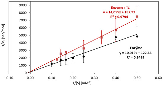
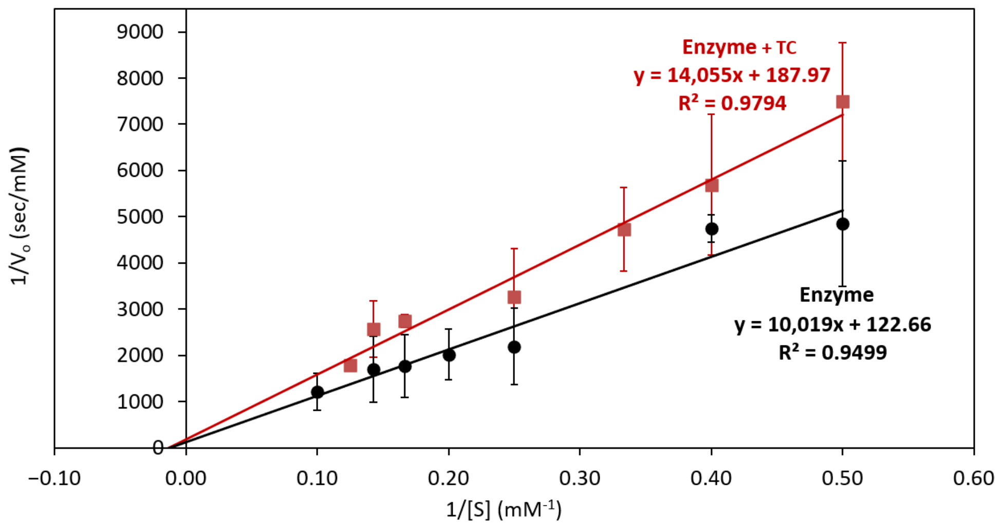
The inhibitory mechanism of TC was further investigated by steady-state kinetic analysis over a range of substrate concentrations (0.5–5 mM for the control and 0.5–6 mM in the presence of TC). Enzyme kinetics were evaluated in both the absence and presence of the inhibitor, and the experimental data were analyzed using Lineweaver–Burk double-reciprocal plots (Figure 3). The kinetic parameters, KM and Vmax, were determined from the slope and intercept of the corresponding linear regression plots from the Lineweaver–Burk Equation (1):
Figure 3.
Lineweaver–Burk plots for the determination of KM and Vmax of HRP in the absence and presence of TC. Data are expressed as mean ± SD from three independent experiments. Error bars represent the standard deviation (SD). The corresponding linear regression equations and R2 values are shown in the graph.
The Lineweaver–Burk double-reciprocal plots (Equation (1) and Figure 3) yielded the following linear regression equations: y = 10,019x + 122.66 (R2 = 0.95) for the control experiment and y = 14,055x + 187.97 (R2 = 0.98) in the presence of TC. The kinetic parameters obtained from three independent experiments were KM = 81.7 mM and Vmax = 8.2 × 10⁻³ mMs−1 for HRP in the absence of TC, and KM = 74.8 mM and Vmax = 5.3 × 10⁻³ mMs−1 in the presence of TC. Thus, in the presence of TC, the Vmax value decreased by 35.4%, whereas the KM value decreased by only 8.4% compared with the control experiment. The much higher reduction in Vmax value than KM indicates that TC primarily affects the catalytic activity of HRP, while having only a limited effect on substrate binding. These results are consistent with a predominantly non-competitive inhibition mechanism. In a non-competitive inhibition mechanism, TC binds to both the free enzyme (E) and the enzyme–substrate complex (ES), leading to the formation of EI and ESI complexes. The larger reduction in Vmax than in KM indicates that TC mainly affects the catalytic activity of HRP rather than substrate binding.
2.3. In Silico Studies
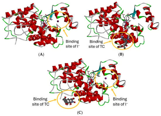
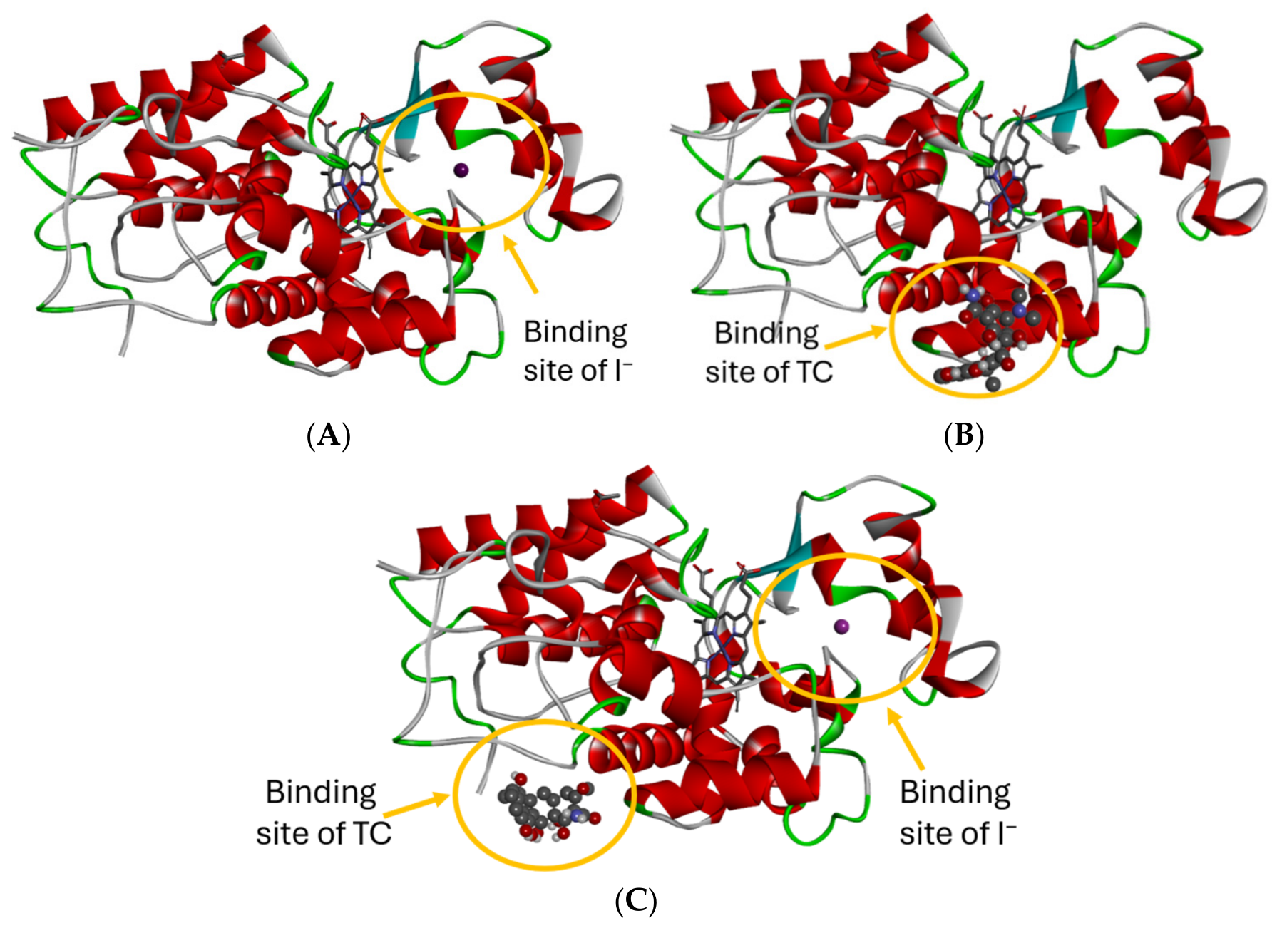
In order to investigate the binding mode of TC to HRP, molecular docking studies were performed (Figure 4). The crystal structure of the unbound enzyme (PDB ID: 1H5G) was used as the receptor. The crystal structure of the unbound enzyme (PDB ID: 1H5G) was used as the receptor. This structure was selected because the unbound form of HRP allows the investigation of ligand binding without assuming a specific binding site. The docking search space included the entire enzyme, allowing TC to explore different possible binding sites. This approach was considered appropriate because the kinetic studies suggested a predominantly non-competitive inhibition mechanism, in which TC may bind at a site different from the substrate-binding site. Therefore, the docking studies were performed to investigate the possible binding site of TC and to support the experimental kinetic results. In addition, the use of PDB ID 1H5G is consistent with our previous computational studies using HRP [11].
Figure 4.
Binding sites of ES (A), EI (B), and ESI (C).
Table 1 summarizes the ES, EI, and ESI binding energies, as well as the amino acid (AA) residues that form the docking cavity of the substrate and/or the inhibitor and the distance (d) of the substrate and/or the inhibitor from the close amino acid residues of the cavity (AA). The binding energy of HRP with triiodide (I3−) was calculated to be −1.04 kcal mol−1, whereas the binding energy of the HRP-TC (EI) complex was −5.96 kcal mol−1. The binding mode of TC was further investigated using the enzyme–substrate–inhibitor (ESI) complex. The calculated binding energy of the HRP-I3−-TC (ESI) complex was −6.50 kcal mol−1, which was lower than that of the HRP-TC (EI) complex (−5.96 kcal mol−1), indicating a more stable interaction. In both the EI and ESI complexes, TC binds at a site distinct from the substrate-binding site, indicating that substrate binding is not prevented (Table 1).
Table 1.
Binding energy values when the substrate (I3−) binds to the enzyme HRP with or without the presence of TC ES, as well as the amino acid (AA) residues forming the docking cavity of the substrate and/or the inhibitor and the distance (d) of the binder (substrate and/or the inhibitor) from the close amino acid residues of the cavity (AA).
These results are consistent with a non-competitive inhibition mechanism and are in good agreement with the steady-state kinetic analysis. In a non-competitive inhibition mechanism, the inhibitor binds to both the free enzyme (E) and the enzyme-substrate complex (ES), leading to the formation of the EI and ESI complexes. A non-competitive inhibitor binds to a site different from the catalytic site of the enzyme. Therefore, it does not compete with the substrate for binding. Instead, it changes the conformation of the enzyme and reduces its catalytic activity, while substrate binding remains largely unaffected [10,11,23,24,25].
2.4. Inhibition of the Catalytic Activity of FeTPPCl
Since TPO contains an iron–porphyrin active site that catalyzes the oxidation of iodide to active iodine in the presence of hydrogen peroxide, followed by the iodination of tyrosyl residues in thyroglobulin, we extended our studies to investigate the reactivity of tetracycline toward iodide oxidation in the presence of FeTPPCl, which serves as a model of the TPO active site. The iodide anion (I−) is readily oxidized by H2O2 through a two-electron transfer process, either non-enzymatically or enzymatically in the presence of peroxidases, according to the following equation [10]:
Oxidation reactions of iodide anion by hydrogen peroxide.
The inhibition activity of TC was measured in the presence of FeTPPCl (a model of the active site of TPO) and H2O2, yielding I3− resulting from the oxidation of I− according to reaction (2).
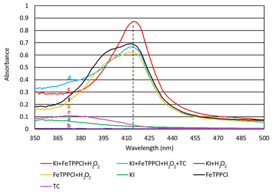
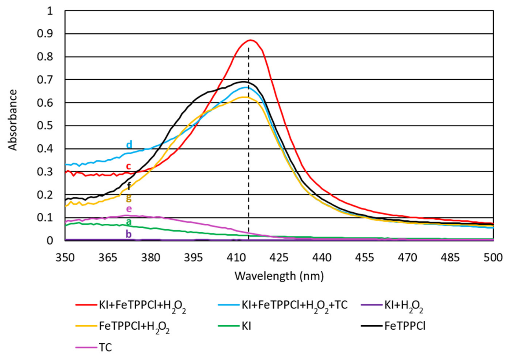
The final rSeaction product monitored was triiodide (I3−), which exhibits a characteristic UV-Vis absorption band at 415 nm. Figure 5 presents the UV-Vis spectra of DMSO solutions of: (a) KI (10 mM); (b) KI (10 mM) and H2O2 (50 μM); (c) KI (10 mM), H2O2 (50 μM), and FeTTPCl (5 μM); and (d) KI (10 mM), H2O2 (50 μM), FeTTPCl (5 μM), and TC (5 μM). For comparison, the spectra of (e) TC (5 μM), (f) FeTTPCl (5 μM), and (g) FeTTPCl (5 μM) in the presence of H2O2 (50 μM) are also included.
Figure 5.
UV spectra of the effect caused by TC on the catalytic activity of FeTPPCl.
The UV-Vis spectra indicate that the FeTPPCl/H2O2/KI system catalyzes the oxidation of iodide, leading to the formation of triiodide (I3−), as evidenced by the presence of an absorption band at 415 nm. Upon addition of TC, the absorbance at 415 nm decreased, indicating a reduced formation of I3− and inhibition of the catalytic oxidation of iodide. Thus, TC suppresses the catalytic activity of the FeTPPCl/H2O2 system.
However, the UV-Vis spectra alone do not provide direct evidence that tetracycline binds to the catalytic heme center (active site) of the enzyme model. The reduced I3− formation may arise from several mechanisms, including inhibition of the catalytic cycle, interaction with reactive intermediates (Compound I or Compound II), or scavenging of oxidizing species. Therefore, these spectral data should be regarded as evidence of reduced catalytic activity, rather than definitive proof of active-site binding.
3. Conclusions
The present study provides experimental and computational evidence that tetracycline can interfere with peroxidase-mediated iodide oxidation. TC inhibited HRP activity in a concentration-dependent and reversible manner, while the kinetic studies indicated a predominantly non-competitive inhibition mechanism. Molecular docking studies further supported the experimental findings, showing that TC binds to HRP at a site different from the substrate-binding site. In addition, TC reduced the catalytic activity of the FeTPPCl model system; however, these results do not support a direct interaction of TC with the heme iron center.
Therefore, the present findings reveal a previously unexplored interaction of tetracycline with peroxidase systems and suggest a possible mechanism by which this widely used antibiotic may interfere with processes involved in thyroid hormone biosynthesis.
4. Experimental Section
Materials and instruments: All solvents were of reagent grade and were used as received. Horseradish peroxidase, tetracycline, ferric tetraphenylporphyrin chloride, potassium iodide, and hydrogen peroxide (Sigma-Aldrich, Merck, Burlington, MA, USA) were also used without further purification. Electronic absorption spectra were recorded using a UV-1600 PC series VWR spectrophotometer (Darmstadt, Germany).
4.1. Horse Radish Peroxidase (HRP) Inhibition Assay and Molecular Docking Studies
HRP inhibition was evaluated spectrophotometrically following a modified literature protocol. All experiments were performed in 0.050 M sodium acetate buffer (pH = 5.5), prepared by dissolving sodium acetate in double-distilled water, adjusting the pH with 50% (w/v) NaOH and 37% (v/v) hydrochloric acid, and diluting to the desired final volume. The substrate solution was freshly prepared by dissolving 1.7 mM KI in dd water. Next, 50 nM horseradish peroxidase (HRP) in PBS buffer and 10 mM H2O2 in 50 mM ice-cold sodium acetate buffer (pH 5.5) were freshly prepared and maintained on ice throughout the experiments to preserve enzymatic activity. The formation of triiodide was monitored spectrophotometrically at 353 nm (ε = 25,500 M−1 cm−1) using a UV–Vis spectrophotometer thermostated at 25 °C, in the presence or absence of tetracycline (TC), with a final reaction volume of 2 mL. TC was evaluated over a concentration range of 1–70 μM in DMSO solutions.
The inhibitory mechanism of TC was investigated by steady-state kinetic analysis using KI concentrations ranging from 0.5 to 6 mM, while maintaining constant concentrations of HRP (50 nM) and H2O2 (10 mM), in the absence and presence of TC at its IC50 concentration. The kinetic parameters, Km and Vmax, were determined from the slope and intercept of the Lineweaver–Burk double-reciprocal plots.
All experiments concerning HRP inhibition and the investigation of the inhibition mechanism were performed independently in triplicate, and the kinetic parameters are expressed as mean ± SD.
4.2. Docking Studies
AutodockVina was used for the molecular docking studies [26]. The graphical interface AutoDockTools (ADT, version 1.5.7) was used for receptor and ligand preparation [27]. The enzyme structure was treated as rigid, while the ligands were allowed full conformational flexibility without the application of torsional restraints. Prior to docking, all water molecules and crystallographic solvent molecules were removed from the protein structure. The docking search space was defined using a grid box large enough to encompass the entire enzyme, allowing unrestricted exploration of potential binding sites by the ligands. All docking calculations were performed using the default software parameters. The crystal structure of the enzyme (PDB ID: 1H5G) was used, following the procedure previously described in Ref [11]. The output graphics, pocket amino acid residues, and the binder-enzyme distances were drawn by Discovery Studio 2001.
4.3. Inhibition of the Catalytic Activity of FeTPPCl
The reaction mixture consisted of 1 mM KI, 1 mM H2O2, 5 μM FeTPPCl, and 5 μM TC in DMSO. The formation of I3− was monitored spectrophotometrically by measuring the absorbance at 415 nm.
Author Contributions
Conceptualization: S.K.H.; supervision: S.K.H.; investigation: C.N.B. and K.G.; methodology: C.N.B. and S.K.H.; validation: S.K.H.; writing—original draft: C.N.B. and S.K.H.; writing—review and editing: C.N.B. and S.K.H. All authors have read and agreed to the published version of the manuscript.
Funding
The program “Supporting the Research Activities of the Laboratory of Biological Chemistry (MaRePBIC)” (University of Ioannina Research Committee program No 83718) is acknowledged.
Data Availability Statement
The data supporting the findings of this study are available from the corresponding authors upon request.
Conflicts of Interest
The authors declare no conflicts of interest.
References
- Antoniadis, C.D.; Corban, G.; Hadjikakou, S.K.; Hadjiliadis, N.; Kubicki, M.; Warner, S.; Butler, I.S. Synthesis and characterization of (PTU)I2 (PTU=6-n-propyl-thiouracil) and (CMBZT)I2 (CMBZT= 5-chloro-2-mercapto-benzothiazole) and possible implications with the mechanism of action of anti-thyroid drugs. Eur. J. Inorg. Chem. 2003, 2003, 1635–1640. [Google Scholar] [CrossRef] [Scilit]
- Antoniadis, C.D.; Hadjikakou, S.K.; Hadjiliadis, N.; Kubicki, M.; Butler, I.S. Synthesis, X-ray characterization and study of a new ionic complex of 2-mercaptopyridine, obtained by oxidation with I2. Implications in the mechanism of action of anti-thyroid drugs. Eur. J. Inorg. Chem. 2004, 2004, 4324–4329. [Google Scholar]
- Antoniadis, C.D.; Hadjikakou, S.K.; Hadjiliadis, N.; Kubicki, M.; Butler, I.S. Synthesis, X-ray characterization and study of new ionic complexes of 2-pyridone, obtained by oxidation with I2. New J. Chem. 2005, 29, 714–720. [Google Scholar] [CrossRef] [Scilit]
- Antoniadis, C.D.; Hadjikakou, S.K.; Hadjiliadis, N.; Papakyriakou, A.; Baril, M.; Butler, I.S. Synthesis and Structures of Se Analogues of the Antithyroid Drug 6-n-Propyl-2-thiouracil and Its Alkyl Derivatives: Formation of Dimeric Se–Se Compounds and Deselenation Reactions of Charge-Transfer Adducts of Diiodine. Chem. Eur. J. 2006, 12, 6888–6897. [Google Scholar] [CrossRef] [Scilit]
- Antoniadis, C.D.; Blake, A.J.; Hadjikakou, S.K.; Hadjiliadis, N.; Hubberstey, P.; Schröder, M.; Wilson, C. Structural characterization of selenium and selenium-diiodine analogues of the antithyroid drug 6-n-propyl-2-thiouracil and its alkyl derivatives. Acta Crystallogr. B 2006, 62, 580–591. [Google Scholar] [CrossRef] [Scilit]
- Corban, G.J.; Antoniadis, C.D.; Hadjikakou, S.K.; Kourkoumelis, N.; Tyurin, V.Y.; Dolgano, A.; Milaeva, E.R.; Kubicki, M.; Bernhardt, P.V.; Tiekink, E.R.T.; et al. Reactivity of di-iodine toward thiol: Desulfuration reaction of 5-nitro-2-mercapto-benzimidazole upon reaction with di-iodine. Heteroat. Chem. 2012, 23, 498–511. [Google Scholar] [CrossRef] [Scilit]
- Daga, V.; Hadjikakou, S.K.; Hadjiliadis, N.; Kubicki, M.; Santos, J.H.Z.; Butler, I.S. Synthesis, Spectroscopic and Structural Characterization of Novel Diiodine Adducts with the Heterocyclic Thioamides, Thiazolidine-2-thione (tzdtH), Benzothiazole-2-thione (bztzdtH) and Benzimidazole-2-thione (bzimtH). Eur. J. Inorg. Chem. 2002, 2022, 1718–1728. [Google Scholar] [CrossRef] [Scilit]
- Corban, G.J.; Hadjikakou, S.K.; Hadjiliadis, N.; Kubicki, M.; Tiekink, E.R.T.; Butler, I.S.; Drougas, E.; Kosmas, A.M. Synthesis, structural characterization, and computational studies of novel di-iodine adducts with the heterocyclic thioamides N-methyl-benzothiazole-2-thione and benzimidazole-2-thione. Implications with the mechanism of action of anti-thyroid drugs. Inorg. Chem. 2005, 44, 8617–8627. [Google Scholar]
- Corban, G.J.; Antoniodis, C.; Hadjikakou, S.K.; Hadjiliadis, N.; Meng, J.-F.; Butler, I.S. Pressure-Tuning Raman Spectra of Diiodine Thioamide Compounds: Models for Antithyroid Drug Activity. Bioinorg. Chem. Appl. 2006, 2006, 68542. [Google Scholar] [CrossRef] [Scilit]
- Corban, G.J.; Hadjikakou, S.K.; Tsipis, A.C.; Kubicki, M.; Bakas, T.; Hadjiliadis, N. Inhibition of peroxidase-catalyzed iodination by thioamides: Experimental and theoretical study of the antithyroid activity of thioamides. New J. Chem. 2011, 35, 213–224. [Google Scholar] [CrossRef] [Scilit]
- Banti, C.N.; Kourkoumelis, N.; Raptopoulou, C.P.; Psycharis, V.; Hadjikakou, S.K. N-(4-hydroxyphenyl)acetamide against di-iodine towards polyiodide dianion. New J. Chem. 2017, 41, 5555–5564. [Google Scholar] [CrossRef] [Scilit]
- Ruf, J.; Carayon, P. Structural and functional aspects of thyroid peroxidase. Arch. Biochem. Biophys. 2006, 445, 269–277. [Google Scholar] [CrossRef] [Scilit]
- Taurog, A. Hormone synthesis. In Endocrinology; DeGroot, L.J., Cahill, G.F., Jr., Odell, W.D., Martini, L., Potts, J.T., Jr., Nelson, D.H., Steinberger, E., Winegrad, A.I., Eds.; Grune and Stratton: New York, NY, USA, 1979; Volume 1, pp. 331–342. [Google Scholar]
- Carvalho, D.P.; Dupuy, C. Thyroid hormone biosynthesis and release. Mol. Cell. Endocrinol. 2017, 458, 6–15. [Google Scholar] [CrossRef] [Scilit]
- Soundarrajan, M.; Kopp, P.A. Thyroid Hormone Biosynthesis and Physiology. In Thyroid Disease and Reproduction: A Clinical Guide to Diagnosis and Management; Springer International Publishing: Cham, Switzerland, 2019; pp. 1–17. [Google Scholar]
- Cooper, D.S. Antithyroid drugs. N. Engl. J. Med. 2005, 352, 905–917. [Google Scholar] [CrossRef] [Scilit]
- Burch, H.B.; Cooper, D.S. Management of Graves disease. JAMA 2015, 314, 2544–2554. [Google Scholar] [CrossRef] [Scilit]
- Tsigara, A.S.; Banti, C.N.; Hatzidimitriou, A.; Hadjikakou, S.K. Tetracycline: Structural characterization and antimicrobial properties of its water-soluble di-anionic bi-sodium salt. Dalton Trans. 2024, 53, 12080. [Google Scholar] [CrossRef] [Scilit]
- Chopra, I.; Roberts, M. Tetracycline antibiotics: Mode of action, applications, molecular biology, and epidemiology of bacterial resistance. Microbiol. Mol. Biol. Rev. 2001, 65, 232–260. [Google Scholar] [CrossRef] [Scilit]
- Nelson, M.L.; Levy, S.B. The history of the tetracyclines. Ann. N. Y. Acad. Sci. 2011, 1241, 17–32. [Google Scholar] [CrossRef] [Scilit]
- Veitch, N.C. Horseradish peroxidase: A modern view of a classic enzyme. Phytochemistry 2004, 65, 249–259. [Google Scholar] [CrossRef] [Scilit]
- Meunier, B. Metalloporphyrins as versatile catalysts for oxidation reactions and oxidative DNA cleavage. Chem. Rev. 1992, 92, 1411–1456. [Google Scholar] [CrossRef] [Scilit]
- Xanthopoulou, M.N.; Hadjikakou, S.K.; Hadjiliadis, N.; Kubicki, M.; Karkabounas, S.; Charalabopoulos, K.; Kourkoumelis, N.; Bakas, T. Synthesis and characterization of a new chloro-di-phenyltin(IV) complex with 2-mercapto-nicotinic acid: Study of its influence upon the catalytic oxidation of linoleic acid to hydroperoxylinoleic acid by the enzyme lipoxygenase. J. Organomet. Chem. 2006, 691, 1780–1789. [Google Scholar] [CrossRef] [Scilit]
- Xanthopoulou, M.N.; Hadjikakou, S.K.; Hadjiliadis, N.; Milaeva, E.R.; Gracheva, J.A.; Tyurin, V.-Y.; Kourkoumelis, N.; Christoforidis, K.C.; Metsios, A.K.; Karkabounas, S.; et al. Biological Studies of New Organotin(IV) Complexes of Thioamide Ligands. Eur. J. Med. Chem. 2008, 43, 327–335. [Google Scholar] [CrossRef] [Scilit]
- Arsalan, A.; Younus, H. Enzymes and nanoparticles: Modulation of enzymatic activity via nanoparticles. Int. J. Biol. Macromol. 2018, 118, 1833–1847. [Google Scholar] [CrossRef] [Scilit]
- Trott, O.; Olson, A.J. AutoDock Vina: Improving the speed and accuracy of docking with a new scoring function, efficient optimization, and multithreading. J. Comput. Chem. 2010, 31, 455–461. [Google Scholar] [CrossRef] [Scilit] [PubMed]
- Morris, G.M.; Huey, R.; Lindstrom, W.; Sanner, M.F.; Belew, R.K.; Goodsell, D.S.; Olson, A.J. AutoDock4 and AutoDockTools4: Automated docking with selective receptor flexibility. J. Comput. Chem. 2009, 16, 2785–2791. [Google Scholar] [CrossRef] [Scilit]

Disclaimer/Publisher’s Note: The statements, opinions and data contained in all publications are solely those of the individual author(s) and contributor(s) and not of MDPI and/or the editor(s). MDPI and/or the editor(s) disclaim responsibility for any injury to people or property resulting from any ideas, methods, instructions or products referred to in the content. |
© 2026 by the authors. Licensee MDPI, Basel, Switzerland. This article is an open access article distributed under the terms and conditions of the Creative Commons Attribution (CC BY) license.