


Abstract
Background: While equine patient-mediated introduction of A. baumannii into hospital settings has been documented, its environmental dissemination and the risk of hospital-acquired surgical site infection remain poorly understood. Objective: Therefore, this descriptive observational study examined (a) the environmental distribution of Acinetobacter spp. in an equine university hospital, (b) the impact of the implementation of new hygiene protocols, (c) the specification of resistance patterns, and (d) the evaluation of the presence of Acinetobacter spp. in hospital-acquired wound infections. Methods: During three sampling periods, environmental samples of the stables, the treatment, and surgery areas were collected before and after cleaning and disinfection. After sampling period 1 (December 2021), the cleaning routines were optimized by reviewing the cleaning and disinfection process, as well as including further surfaces in the cleaning schedule for January 2022). This was followed by a second (February 2022) and a third (June 2022) sampling period. During sampling periods 1 and 2, 76 surfaces were sampled; in sampling period 3, only 21 critical surfaces were examined. Samples were cultured on selective agar plates and incubated at 37 °C, with bacterial growth evaluated after 24–48 h. Wound swabs were enriched in broth before culturing. Bacteria were identified using MALDI-TOF mass spectrometry. During the first sampling period, antibiotic susceptibility testing was performed using broth microdilution according to CLSI-vet standards. Results: During each sampling period, Acinetobacter spp. was detected in at least one sample in each of the different areas; however, there was a reduced detection rate from sampling period 1 throughout sampling period 3. The isolates were highly resistant against beta-lactam and macrolide antibiotics but mostly sensitive to fluroquinolones (enrofloxacin, 2.2% resistance; marbofloxacin, 0.0% resistance), aminoglycosides (gentamicin, 6.5% resistance; kanamycin, 8.7% resistance), and tetracyclines. Acinetobacter spp. was not detected in surgical site infections. Conclusions: Environmental persistence of Acinetobacter spp. in an equine clinical setting does not necessarily translate into surgical site infections. Through prudent antibiotic use, the antibiotic susceptibility of the isolates may be perceived.
1. Introduction
Hospital-acquired or nosocomial infections are not only an emerging threat concerning human medicine—increased reports of nosocomial infections, especially in large animal hospitals, are a major health concern [1,2,3,4,5,6,7], and pathogens such as Methicillin-resistant Staphylococcus aureus (MRSA) may exert zoonotic potential [1].
Acinetobacter spp. poses a significant challenge, since they may be part of the human skin and pharyngeal flora [8,9,10,11] and may be persistent in a hospital environment [12]. Furthermore, animals can serve as a potential reservoir for multidrug-resistant (MDR) Acinetobacter spp. [13,14,15].
In humans, Acinetobacter spp. is implicated in bacteremia, pulmonary infections, meningitis, diarrheal illness, and problematic nosocomial outbreaks, which are associated with mortality rates ranging from 20% to 60% [12]. Also, in small animals, a variety of infections with Acinetobacter spp. have been reported, including urinary tract, wound, and skin infections [16,17,18]. In horses, in addition to colonization of the nostrils and the gastrointestinal tract [14,18], detection of Acinetobacter spp., mostly A. baumannii, was reported from intravenous catheter tips, including those showing signs of thrombophlebitis [13,19], from tracheal washes from horses with respiratory diseases or “poor performance” [20] and foals with bronchopneumonia [21], as well as in the blood culture of a septic foal [22].
Particularly in equine clinics, challenging conditions prevail due to species-specific factors (animal size, bedding in stalls, feed, and stable ventilation). These species-specific factors complicate the development of targeted hygiene measurements, which adds up to difficulties in preventing nosocomial infections [23].
Previous studies focusing on injured horses or horses presenting for colic surgery admitted to our clinic identified a 0.9–2.9% A. baumannii carrier rate (nasal vestibule, fecal samples) [14,24,25]; its environmental persistence and relevance as a nosocomial pathogen have not yet been assessed in our clinic.
Therefore, the objective of the study was to (a) determine the environmental distribution of Acinetobacter spp. in our equine university hospital, (b) to examine the impact of the implementation of new hygiene protocols on the environmental distribution of Acinetobacter spp., (c) to specify the resistance patterns of Acinetobacter spp. in an equine university hospital, and (d) to evaluate the presence of Acinetobacter spp. in hospital-acquired wound infections.
2. Material and Methods
2.1. Surface Samples
For this descriptive observational study, surface samples for microbiological examination were collected from surface areas of different parts of the clinic. Surfaces were selected based on a risk assessment and on clinical criteria such as frequent contact with hands and/or patients, proximity to patients, and relevance to clinical workflows. The aim was to include surfaces that may serve as potential reservoirs for microorganisms in routine clinical practice and that are simultaneously relevant for transmission between humans, animals, and the environment. This targeted selection enabled a focused assessment of hygienically critical contact surfaces.
The samples were taken as impressions smears (DIN 10113-1) with agar plates provided by Laboklin GmbH & Co. KG (Bad Kissingen, Germany). The samples were collected directly from the respective surfaces by applying even pressure with the agar surface onto the area to be examined. After sampling, the agar plates were stored under refrigerated conditions (+2 °C to +8 °C) within the clinic during the day in order to minimize uncontrolled microbial growth prior to shipment. In the evening of the same day, the plates were sealed airtight with Parafilm, packaged for protection from impact, and placed into insulated Styrofoam boxes with cool packs. The subsequent shipment to the microbiological laboratory of Laboklin GmbH & Co. KG was carried out via Laboklin’s internal courier service from the Berlin site. Transport of the samples was conducted in compliance with the cooling conditions prescribed for microbiological specimens, within a temperature range of +2 °C to +8 °C.
The first sampling period was performed in November 2021. The surfaces were sampled before and after cleaning and disinfection, regardless of macroscopic contamination. The interval that passed between cleaning and disinfection of the surfaces and sample collection varied according to the specific material properties and the practical implementation of the cleaning schedule. Sampling only took place after the measures were fully completed as required and after the surfaces had dried sufficiently.
After the first sampling period, the cleaning schedule was reviewed and revised. Furthermore, additional surfaces were included in the examination scheme. The changes in the examination scheme were implemented in January 2022. Furthermore, the janitors and the clinical staff were informed about the cleaning deficiencies detected in sampling period 1, such as insufficient contact time and concentration of the effective agent, use of the same cleaning cloth for the same areas, skipping important surfaces such as walls or shelves, and stocks), and use of a dirty scrubber dryer machine. Both cleaning schedules are available in Supplementary File S1.
A second sampling period was performed in February 2022. Samples were taken both before and after cleaning in this instance as well. In sampling period 1 and 2, 76 surfaces were sampled, so that in total, 152 samples were examined per sampling period. The third sampling period in June 2022 focused on 21 critical surfaces out of the originally tested 76 areas, leading to 42 samples in total.
Within sampling period 2, few additional surfaces were sampled while few surfaces were excluded from the sampling. Sampling period 3 focused on critical surfaces exclusively. Critical surfaces were defined as surfaces in close contact with the patient or as surfaces that are often touched but easily forgotten in the cleaning process. All in all, 17 areas were sampled three times, 45 areas were checked twice, and 29 areas were checked only once. Details on the sampled surfaces are given in Supplementary File S2.
2.2. Samples from Wounds
To identify hospital-acquired surgical site infections with Acinetobacter spp., samples of infected wounds were collected. The samples were collected routinely to identify the pathogens involved and to select the appropriate antibiotic based on antimicrobial susceptibility testing. Upon admission of their horses, owners consented to the anonymized use of their horses’ data for scientific purposes. Included in the analysis were exclusively swabs from infected surgical sites either during the initial hospitalization or a readmission to the hospital due to surgical site infection.
The samples were collected by clinical staff, such as veterinarians or veterinary nurses, between October 2021 and June 2022, so that the wound sample collection started one month prior to the first surface sampling period.
2.3. Microbiological Examinations
After arrival at the microbiology laboratory of Laboklin GmbH & Co. KG (Bad Kissingen, Germany), agar plates (RODAC contact plates for surface examination and Columbia blood and Endo agar for wound swabs) were inoculated and incubated at +37 °C with macroscopic examination after 24 and additionally after 48 h under aerobic conditions. Additionally, the swabs originating from wounds were incubated in enrichment broth (trypticase soy broth, Becton Dickinson, Heidelberg, Germany) for 16–24 h at 37 °C with subsequent subculture on Columbia blood and Endo agar (Becton Dickinson, Heidelberg, Germany) for 16–24 h at 37 °C and evaluation. Matrix-assisted laser desorption/ionization time-of-flight mass spectrometry (MALDI-TOF-MS, Bruker, Ettlingen, Germany) was used to identify the isolated bacteria. This method allowed for the unambiguous identification of each bacterial species using the current version for the time (MALDI Biotyper Reference Library MBT Compass Library Revision L (2020) 9607 MSP covering 3239 species/entries (9607 MSP); beginning in December 2021, MBT Compass Library Revision H (2021) covering 3893 species/entries (10833 MSP), Bruker Daltonics, Bremen, Germany). According to the software used (MALDI Biotyper Reference Library), genus and species determination was provided when the score was 2 or higher, genus was applied when the score was within 1.7 and 2, and results of 1.7 and below were considered to be unreliable. Confirmation by molecular methods was not carried out, and thus, there was no classification done concerning independent or clonal origin of the isolates.
In sampling period 1, antibiograms of the isolates were also generated by using broth microdilution (CLSI-vet, MIC-based testing, human-derived breakpoints, as there are currently no breakpoints for samples from horses available, with reporting as susceptible, intermediate, or resistant according to the CLSI database). Additionally, internal controls and biannual external controls were implemented. Minimum inhibitory concentrations (MIC) were categorized as sensitive (S), intermediate (I), or resistant (R; according to CLSI M100 Edition 36, 2006). These categories were used to calculate resistance proportions for isolated Acinetobacter spp. across several active substances. For presentation, antibiotics used in the clinic were selected. These include amoxicillin, ampicillin, ceftiofur, cefquinome, doxycycline, enrofloxacin, gentamicin, marbofloxacin, penicillin, and tetracycline, as well as trimethoprim and sulfamethoxazole in combination. The panel is not specific for Acinetobacter spp. but is designed to aid treatment of all bacterial isolates found in equine practice and thus does not consider intrinsic resistances.
A statistical analysis was omitted, since only few Acinetobacter spp. were detected, and there was a reduction in sample size.
3. Results
3.1. Prevalence of Acinetobacter spp.
During the first sampling period, Acinetobacter spp. was detected in at least one sample in each of the different areas. This included detection in both environmental areas, such as the floors and the walls, and in equipment directly in contact with the horses, such as endotracheal tubes.
After revising the cleaning schedule, fewer surfaces tested positive for Acinetobacter spp. in sampling period 2, but surfaces in critical areas such as the surgical theatre still tested positive for Acinetobacter spp. The reduced detection rate remained consistent months after the implementation of the revised cleaning schedule in sampling period 3. In sampling period 1, 32.9% (25/76) of samples tested positive for Acinetobacter spp. before cleaning and disinfection compared to 26.3% (20/76) afterwards. In sampling periods 2 and 3, positivity rates were 14.5% (11/76) and 14.2% (3/21) before cleaning and disinfection and 2.6% (2/76) and 9.5% (3/21) afterwards.
An overview of the surfaces and Acinetobacter spp. detection is provided in the three tables below (Table 1, Table 2 and Table 3).
Table 1.
Detection of Acinetobacter spp. in the surgical area during the three sampling periods.
Table 2.
Detection of Acinetobacter spp. in the examination area during the three sampling periods.
Table 3.
Detection of Acinetobacter spp. in the stabling area during the three sampling periods.
During the study period, samples of surgical site infections were submitted for bacterial culture. Of these, 38 were regarded as nosocomial infections. Acinetobacter spp. was not detected in any of these 38 samples.
3.2. Resistance Profiles
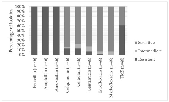
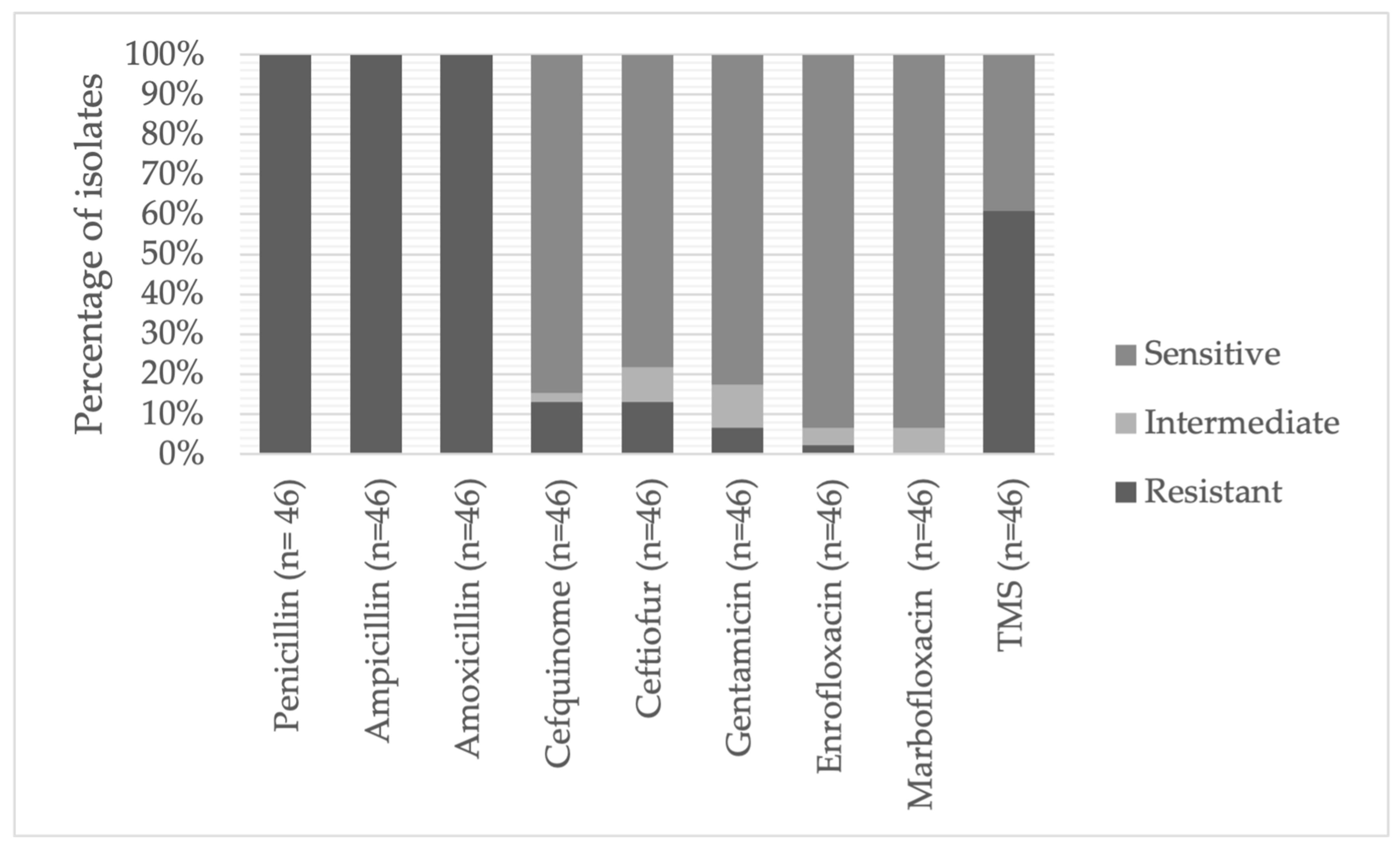
Most isolates were highly resistant against beta-lactam antibiotics such as penicillin, against aminopenicillins such as ampicillin (97.9%) and amoxicillin (100%), and against macrolide antibiotics (100% resistance) such as azithromycin, erythromycin, spiramycin, and clarithromycin, as well as against the lincosamide antibiotic clindamycin. The isolates were mostly sensitive to the fluoroquinolones enrofloxacin (2.2% resistance) and marbofloxacin (0.0% resistance), as well as aminoglycosides (gentamicin, 6.5% resistance; kanamycin, 8.7% resistance). Figure 1 shows resistance data for commonly used antibiotics in equine medicine.
Figure 1.
Resistance rates of the Acinetobacter isolates against commonly used antibiotics in equine medicine. TMS: trimethoprim and sulfamethoxazol.
The Acinetobacter baumannii isolated from the manger in the isolation stable during sampling period 1 was sensitive to cefquinome, ceftiofur, gentamicin, enrofloxacin, tetracycline, and doxycycline when only looking at the commonly used antibiotics in equine medicine.
4. Discussion
Environmental sampling of our equine hospital yielded recovery of non-baumannii Acinetobacter spp., including A. lwoffii, A. johnsonii, A. pseudolwoffii, and A. beijerinckii, with only one single detection of A. baumannii. Of the genus Acinetobacter, A. baumannii is the most common species associated with nosocomial infections, even though involvement of other Acinetobacter species in hospital-acquired infections has been reported previously, especially in humans [26,27,28,29,30,31,32]. The reduction of environmental Acinetobacter spp. underlines the effectiveness of the revision of the cleaning schedule and staff training. The effects of training and monitoring were shown in previous studies and are now, in addition to regular reminders, incorporated in the WHO Guidelines on Hand Hygiene in Health Care [31,32,33]. The results also show that clinical facilities might benefit from repeated training, since the percentage of positive samples increased in sampling period 3 after cleaning and disinfection when compared to sampling period 2. Possible effects of season, patient density, case distribution, and staff changes on the recovery rates of Acinetobacter spp. cannot be completely excluded. Additionally, it is worrying that some surfaces tested positive for Acinetobacter spp. both before and after cleaning and disinfection or only afterwards. This may be attributed to pathogen persistence on the surfaces or by the redistribution of microorganisms during cleaning procedures, which was described during cleaning with detergent wipes [34] or by using contaminated cleaning equipment [35].
In human hospitals, A. baumannii has emerged as a major cause of healthcare-associated infections, including ventilator-associated pneumonia, bloodstream infections, and postoperative/wound infections, frequently in immunocompromised or critically ill patients [12,28,36,37]. Also, the descriptions of wound infections, postoperative soft-tissue infections, and urinary tract infections linked to multidrug-resistant Acinetobacter spp. in companion animal reports emphasize the therapeutic challenges posed by resistance to multiple antimicrobial classes [16,17,18]. The involvement of Acinetobacter spp. in equine infections, such as thrombophlebitis [13,19], respiratory infections [20,21], and neonatal sepsis [22], underlines the susceptibility of equines to this bacterial species. In the present study, Acinetobacter spp. was detected in several areas of the equine clinic environment during the examination period, including surfaces frequently contacted by horses, personnel, or equipment. Despite this environmental presence, no surgical site infections associated with Acinetobacter spp. were documented during the study period. The limited clinical relevance of Acinetobacter spp. in surgical site infections in our clinic during the sampling period indicates that the environmental contamination of the surgery and orthopedic treatment area did not translate into wound infection. The predominance of low-virulence, non-baumannii Acinetobacter species in the environment, effective perioperative asepsis, and preoperative preparation of the surgeon may be the reason for this circumstance. In addition, the absence of Acinetobacter spp. in surgical site infections of clean surgical wounds suggests that established hygiene and biosecurity practices during the postoperative care are effective and prevent environmental microorganisms from gaining access to surgical sites. Furthermore, these findings emphasize the distinction between mere environmental presence and true nosocomial pathogenicity, highlighting that environmental detection alone should not be equated with clinical relevance. The presence of Acinetobacter spp. in infections other than surgical site infections was not evaluated with this study; therefore, it may be possible that Acinetobacter spp. played a role in infections other than those of the surgical site.
The revision of the cleaning and disinfection protocol resulted in a descriptive decrease in the detection of Acinetobacter spp. This further limited the risk of contracting a surgical site infection with Acinetobacter spp. during the study period. However, it must be kept in mind that the sample size decreased during the study, since sampling period 3 only focused on sensitive surfaces, and therefore, only 21 surfaces were examined. If all 76 (or more) surfaces were sampled during sampling period 3, the recovery rate of Acinetobacter spp., and especially of A. baumannii, may have been higher.
Acinetobacter spp. commonly exhibits high rates of bacterial resistance, including resistance to aminoglycosides, fluroquinolones, β-lactams, and cephalosporins, as well as carbapenems, with Acinetobacter baumannii commonly being the most resistant species [38,39,40,41,42,43,44]. The challenge of multidrug-resistant Acinetobacter baumannii in equine medicine is emphasized by isolates from horses—including samples from the urogenital tract and wounds/abscesses—exhibiting high resistance rates against commonly used antibiotics. This also emphasizes the challenges of their multidrug resistance, including resistance against gentamicin, enrofloxacin, marbofloxacin, tetracycline, and trimethoprim/sulfamethoxazole [45], which are commonly used in horses. An important finding of this study is that the environmental Acinetobacter spp. in the examined clinical environment were susceptible to several antibiotic classes, including the single A. baumannii isolate from the manger in the isolation stable during sampling period 1. This contrasts with the pronounced multidrug resistance and carbapenem resistance increasingly observed in human, companion animal, and equine nosocomial A. baumannii isolates [16,17,18,28,45,46]. The absence of highly resistant isolates in this equine setting may reflect differences in antimicrobial selection pressure or species composition within the Acinetobacter genus.
The predominance of non-baumannii Acinetobacter spp. that are susceptible to a variety of antibiotics in our clinic is in line with findings from a study evaluating non-hospital environmental Acinetobacter spp. loads [47], indicating a lower selective pressure for resistance, potentially resulting from stewardship-compliant antimicrobial prescribing. Since the recovered isolates are mostly sensitive to the combination of trimethoprim-sulfamethoxazol, tetracycline, doxycycline, and the fluoroquinolones enrofloxacin and marbofloxacin, as well as the aminoglycoside gentamicin, it should be possible to effectively treat infections with the recovered isolates.
Despite the lack of clinically apparent infections, the detection of Acinetobacter spp. in the clinic environment remains noteworthy. Environmental contamination of the clinic, especially the stables, may act as a reservoir for opportunistic pathogens and could contribute to infection risk under certain conditions, for example, untreated pituitary pars intermedia dysfunction in aged horses [48]. Furthermore, Acinetobacter spp. epidemiology in humans has shifted over time towards greater antimicrobial resistance and more virulent lineages [49,50] which may also happen in our facility. Continuous surveillance and routine environmental hygiene protocols in equine clinics therefore remain justified, particularly in high-risk units such as surgical theatres and intensive care stables.
When interpreting these study results, the limitations of the study should be taken into account. An insufficient number of isolates were detected on the same surfaces to allow for a meaningful statistical evaluation of differences between the sampling periods. Since there were also no nosocomial surgical site infections caused by Acinetobacter, a statistical analysis was not meaningful in this case either. Therefore, the presented data remain descriptive and should thus be interpreted with sufficient care. Furthermore, the sampled surfaces did not remain precisely the same during the three sampling periods, which does not allow exact comparison between the three sampling periods.
5. Conclusions
Overall, these findings indicate that despite environmental persistence, Acinetobacter spp. held a low clinical relevance regarding surgical site infections and did not show high rates of resistance in the equine clinic of the Freie Universität Berlin during the study period. Nevertheless, longitudinal surveillance is warranted to assess whether environmental strains in equine clinics acquire resistance traits or pathogenic potential over time. Adherence to antimicrobial stewardship guidelines may help to preserve the antibiotic susceptibility of Acinetobacter spp., which highlights the importance of bacteriological analysis and resistance testing.
Supplementary Materials
The following supporting information can be downloaded at: https://www.mdpi.com/article/10.3390/antibiotics15060558/s1, Supplementary File S1: Cleaning Schedules, Supplementary File S2: Sampled Surfaces.
Author Contributions
Conceptualization, H.G., E.M., A.S., and S.D.S.; methodology, H.G.; validation, S.D.S. and A.S.; formal analysis, S.D.S. and A.S.; investigation, A.S. data curation, S.D.S. and A.S.; writing—original draft, S.D.S.; writing—review and editing, S.D.S., A.S., E.M., and H.G.; supervision, H.G.; project administration, H.G. All authors have read and agreed to the published version of the manuscript.
Funding
The bacteriological examinations were sponsored by Laboklin, Bad Kissingen, Germany. Supported by Open Access funds of Freie Universität Berlin.
Institutional Review Board Statement
The wound samples were collected as a part of the clinical routine examination of horses with surgical site infections and, therefore, did not require ethical review.
Informed Consent Statement
Upon admission of their horses, owners consented to the anonymized use of their horses’ data for scientific purposes.
Data Availability Statement
The data are available upon request from the authors.
Acknowledgments
The samples were collected and analyzed as a part of a dissertation project (A. Sauerwein).
Conflicts of Interest
E. Mueller is employed at Laboklin GmbH & Co. KG. The remaining authors declare that the research was conducted in the absence of any commercial or financial relationships that could be construed as a potential conflict of interest.
References
- Seguin, J.C.; Walker, R.D.; Caron, J.P.; Kloos, W.E.; George, C.G.; Hollis, R.J.; Jones, R.N.; Pfaller, M.A. Methicillin-Resistant Staphylococcus aureus Outbreak in a Veterinary Teaching Hospital: Potential Human-to-Animal Transmission. J. Clin. Microbiol. 1999, 37, 1459–1463. [Google Scholar] [CrossRef] [Scilit]
- Boerlin, P.; Eugster, S.; Gaschen, F.; Straub, R.; Schawalder, P. Transmission of Opportunistic Pathogens in a Veterinary Teaching Hospital. Vet. Microbiol. 2001, 82, 347–359. [Google Scholar] [CrossRef] [Scilit] [PubMed]
- Schott, H.C., 2nd; Ewart, S.L.; Walker, R.D.; Dwyer, R.M.; Dietrich, S.; Eberhart, S.W.; Kusey, J.; Stick, J.A.; Derksen, F.J. An Outbreak of Salmonellosis among Horses at a Veterinary Teaching Hospital. J. Am. Vet. Med. Assoc. 2001, 218, 1152–1159. [Google Scholar] [CrossRef] [Scilit]
- Cummings, K.J.; Divers, T.J.; McDonough, P.L.; Switt, A.M.; Wiedmann, M.; Warnick, L.D. Temporal Clusters of Bovine Salmonella Cases at a Veterinary Medical Teaching Hospital, 1996-2007. Vector Borne Zoonotic Dis. 2010, 10, 471–479. [Google Scholar] [CrossRef] [Scilit] [PubMed]
- Dallap Schaer, B.L.; Aceto, H.; Rankin, S.C. Outbreak of Salmonellosis Caused by Salmonella enterica Serovar Newport MDR-AmpC in a Large Animal Veterinary Teaching Hospital. J. Vet. Intern. Med. 2010, 24, 1138–1146. [Google Scholar] [CrossRef] [Scilit]
- Steneroden, K.K.; Van Metre, D.C.; Jackson, C.; Morley, P.S. Detection and Control of a Nosocomial Outbreak Caused by Salmonella Newport at a Large Animal Hospital. J. Vet. Intern. Med. 2010, 24, 606–616. [Google Scholar] [CrossRef] [Scilit]
- Burgess, B.A.; Tokateloff, N.; Manning, S.; Lohmann, K.; Lunn, D.P.; Hussey, S.B.; Morley, P.S. Nasal Shedding of Equine Herpesvirus-1 from Horses in an Outbreak of Equine Herpes Myeloencephalopathy in Western Canada. J. Vet. Intern. Med. 2012, 26, 384–392. [Google Scholar] [CrossRef] [Scilit]
- Glew, R.H.; Moellering, R.C., Jr.; Kunz, L.J. Infections with Acinetobacter calcoaceticus (Herellea vaginicola): Clinical and laboratory studies. Medicine 1977, 56, 79–97. [Google Scholar] [CrossRef] [Scilit] [PubMed]
- Rosenthal, S.; Tager, I.B. Prevalence of Gram Negative Rods in the Normal Pharyngeal Flora. Ann. Intern. Med. 1975, 83, 355–357. [Google Scholar] [CrossRef] [Scilit]
- Taplin, D.; Rebell, G.; Zaias, N. The Human Skin as a Source of Mima-Herellea Infections. JAMA 1963, 186, 952–955. [Google Scholar] [CrossRef] [Scilit]
- Somerville, D.; Noble, W.C. A Note on the Gram Negative Bacilli of Human Skin. Rev. Eur. Etud. Clin. Biol. 1970, 15, 669–671. [Google Scholar] [PubMed]
- Bergogne-Bérézin, E.; Towner, K.J. Acinetobacter spp. as Nosocomial Pathogens: Microbiological, Clinical, and Epidemiological Features. Clin. Microbiol. Rev. 1996, 9, 148–165. [Google Scholar] [CrossRef]
- Endimiani, A.; Hujer, K.M.; Hujer, A.M.; Bertschy, I.; Rossano, A.; Koch, C.; Gerber, V.; Francey, T.; Bonomo, R.A.; Perreten, V. Acinetobacter baumannii Isolates from Pets and Horses in Switzerland: Molecular Characterization and Clinical Data. J. Antimicrob. Chemother. 2011, 66, 2248–2254. [Google Scholar] [CrossRef] [Scilit]
- Walther, B.; Klein, K.S.; Barton, A.K.; Semmler, T.; Huber, C.; Wolf, S.A.; Tedin, K.; Merle, R.; Mitrach, F.; Guenther, S.; et al. Extended-Spectrum Beta-Lactamase (ESBL)-Producing Escherichia coli and Acinetobacter baumannii among Horses Entering a Veterinary Teaching Hospital: The Contemporary “Trojan Horse”. PLoS ONE 2018, 13, e0191873. [Google Scholar] [CrossRef] [Scilit]
- van der Kolk, J.H.; Endimiani, A.; Graubner, C.; Gerber, V.; Perreten, V. Acinetobacter in Veterinary Medicine, with an Emphasis on Acinetobacter baumannii. J. Glob. Antimicrob. Resist. 2019, 16, 59–71. [Google Scholar] [CrossRef] [Scilit] [PubMed]
- Kristensen, S.; Krogh, H.V. A Study of Skin Diseases in Dogs and Cats. III. III. Microflora of the Skin of Dogs with Chronic Eczema. Nord. Vet. Med. 1978, 30, 223–230. [Google Scholar]
- Lupo, A.; Châtre, P.; Ponsin, C.; Saras, E.; Boulouis, H.-J.; Keck, N.; Haenni, M.; Madec, J.-Y. Clonal Spread of Acinetobacter baumannii Sequence Type 25 Carrying BlaOXA-23 in Companion Animals in France. Antimicrob. Agents Chemother. 2016, 61, e01881-16. [Google Scholar] [CrossRef] [Scilit] [PubMed]
- Ewers, C.; Klotz, P.; Leidner, U.; Stamm, I.; Prenger-Berninghoff, E.; Göttig, S.; Semmler, T.; Scheufen, S. OXA-23 and ISAba1–OXA-66 Class D β-Lactamases in Acinetobacter baumannii Isolates from Companion Animals. Int. J. Antimicrob. Agents 2017, 49, 37–44. [Google Scholar] [CrossRef] [Scilit]
- Vaneechoutte, M.; Devriese, L.A.; Dijkshoorn, L.; Lamote, B.; Deprez, P.; Verschraegen, G.; Haesebrouck, F. Acinetobacter baumannii-Infected Vascular Catheters Collected from Horses in an Equine Clinic. J. Clin. Microbiol. 2000, 38, 4280–4281. [Google Scholar] [CrossRef] [Scilit]
- Wood, J.L.N.; Burrell, M.H.; Roberts, C.A.; Chanter, N.; Shaw, Y. Streptococci and Pasteurella spp. Associated with Disease of the Equine Lower Respiratory Tract. Equine Vet. J. 1993, 25, 314–318. [Google Scholar] [CrossRef] [Scilit]
- Jokisalo, J.; Bryan, J.; Legget, B.; Abbott, Y.; Katz, L.M. Multiple-drug Resistant Acinetobacter baumannii Bronchopneumonia in a Colt Following Intensive Care Treatment. Equine Vet. Educ. 2010, 22, 281–286. [Google Scholar] [CrossRef] [Scilit]
- Bentz, A.I.; Wilkins, P.A.; MacGillivray, K.C.; Barr, B.S.; Palmer, J.E. Severe Thrombocytopenia in 2 Thoroughbred Foals with Sepsis and Neonatal Encephalopathy. J. Vet. Intern. Med. 2002, 16, 494–497. [Google Scholar] [CrossRef]
- Gehlen, H. Der Neue Hygienemanagement-Leitfaden 2020. Pferde Spieg. 2020, 23, 48–49. [Google Scholar] [CrossRef] [Scilit]
- Gehlen, H.; Klein, K.S.; Merle, R.; Lübke-Becker, A.; Stoeckle, S.D. Does Colonization with MRSA, ESBL—Producing Enterobacteriaceae, and/or Acinetobacter baumannii—Increase the Risk for Postoperative Surgical Site Infection? Vet. Med. Sci. 2023, 9, 729–737. [Google Scholar] [CrossRef] [Scilit] [PubMed]
- Stöckle, S.D.; Kannapin, D.A.; Merle, R.; Lübke-Becker, A.; Gehlen, H. Comparison of a Single-Shot Antibiotic Protocol Compared to a Conventional 5-Day Antibiotic Protocol in Equine Diagnostic Laparotomy Regarding Pre- and Postoperative Colonization with Multi-Drug-Resistant Indicator Pathogens. Antibiotics 2026, 15, 106. [Google Scholar] [CrossRef] [Scilit] [PubMed]
- Nemec, A.; De Baere, T.; Tjernberg, I.; Vaneechoutte, M.; Van der Reijden, T.J.K.; Dijkshoorn, L. Acinetobacter ursingii sp. Nov. and Acinetobacter schindleri sp. Nov., Isolated from Human Clinical Specimens. Int. J. Syst. Evol. Microbiol. 2001, 51, 1891–1899. [Google Scholar] [CrossRef] [Scilit]
- Nemec, A.; Dijkshoorn, L.; Cleenwerck, I.; De Baere, T.; Janssens, D.; van der Reijden, T.J.K.; Ježek, P.; Vaneechoutte, M. Acinetobacter parvus sp. Nov., a Small-Colony-Forming Species Isolated from Human Clinical Specimens. Int. J. Syst. Evol. Microbiol. 2003, 53, 1563–1567. [Google Scholar] [CrossRef] [Scilit]
- Peleg, A.Y.; Seifert, H.; Paterson, D.L. Acinetobacter baumannii: Emergence of a Successful Pathogen. Clin. Microbiol. Rev. 2008, 21, 538–582. [Google Scholar] [CrossRef] [Scilit]
- Turton, J.F.; Shah, J.; Ozongwu, C.; Pike, R. Incidence of Acinetobacter Species Other than A. Baumannii among Clinical Isolates of Acinetobacter: Evidence for Emerging Species. J. Clin. Microbiol. 2010, 48, 1445–1449. [Google Scholar] [CrossRef] [Scilit]
- Karah, N.; Haldorsen, B.; Hegstad, K.; Simonsen, G.S.; Sundsfjord, A.; Samuelsen, Ø. Species Identification and Molecular Characterization of Acinetobacter spp. Blood Culture Isolates from Norway. J. Antimicrob. Chemother. 2011, 66, 738–744. [Google Scholar] [CrossRef] [Scilit]
- Van Den Broek, P.J.; Van Der Reijden, T.J.K.; Van Strijen, E.; Helmig-Schurter, A.V.; Bernards, A.T.; Dijkshoorn, L. Endemic and Epidemic Acinetobacter Species in a University Hospital: An 8-Year Survey. J. Clin. Microbiol. 2009, 47, 3593–3599. [Google Scholar] [CrossRef] [Scilit]
- Isogami, H.; Sugeno, M.; Imaizumi, K.; Fukuda, T.; Kamo, N.; Yasuda, S.; Yamaguchi, A.; Fujimori, K. Maternal Septic Shock Due to Acinetobacter lwoffii Infection:A Case Report. Fukushima J. Med. Sci. 2023, 69, 191–196. [Google Scholar] [CrossRef] [Scilit]
- WHO. WHO Guidelines on Hand Hygiene in Health Care First Global Patient Safety Challenge Clean Care Is Safer Care; WHO: Geneva, Switzerland, 2009. [Google Scholar]
- Ramm, L.; Siani, H.; Wesgate, R.; Maillard, J.Y. Pathogen Transfer and High Variability in Pathogen Removal by Detergent Wipes. Am. J. Infect. Control 2015, 43, 724–728. [Google Scholar] [CrossRef] [Scilit] [PubMed]
- Engelhart, S.T.; Krizek, L.; Glasmacher, A.; Fischnaller, E.; Marklein, G.; Exner, M. Pseudomonas Aeruginosa Outbreak in a Haematology–Oncology Unit Associated with Contaminated Surface Cleaning Equipment. J. Hosp. Infect. 2002, 52, 93–98. [Google Scholar] [CrossRef] [Scilit] [PubMed]
- Howard, A.; O’Donoghue, M.; Feeney, A.; Sleator, R.D. Acinetobacter baumannii: An Emerging Opportunistic Pathogen. Virulence 2012, 3, 243–250. [Google Scholar] [CrossRef] [Scilit] [PubMed]
- Dijkshoorn, L.; Nemec, A.; Seifert, H. An Increasing Threat in Hospitals: Multidrug-Resistant Acinetobacter baumannii. Nat. Rev. Microbiol. 2007, 5, 939–951. [Google Scholar] [CrossRef] [Scilit]
- Lyytikäinen, O.; Köljalg, S.; Härmä, M.; Vuopio-Varkila, J. Outbreak Caused by Two Multi-Resistant Acinetobacter baumannii Clones in a Burns Unit: Emergence of Resistance to Imipenem. J. Hosp. Infect. 1995, 31, 41–54. [Google Scholar] [CrossRef] [Scilit]
- Paton, R.; Miles, R.S.; Hood, J.; Amyes, S.G. ARI 1: β-Lactamase-Mediated Imipenem Resistance in Acinetobacter baumannii. Int. J. Antimicrob. Agents 1993, 2, 81–87. [Google Scholar] [CrossRef] [Scilit]
- Seifert, H.; Baginski, R.; Schulze, A.; Pulverer, G. Antimicrobial Susceptibility of Acinetobacter Species. Antimicrob. Agents Chemother. 1993, 37, 750–753. [Google Scholar] [CrossRef] [Scilit]
- Vila, J.; Marcos, A.; Marco, F.; Abdalla, S.; Vergara, Y.; Reig, R.; Gomez-Lus, R.; Jimenez De Anta, T. In Vitro Antimicrobial Production of β-Lactamases, Aminoglycoside-Modifying Enzymes, and Chloramphenicol Acetyltransferase by and Susceptibility of Clinical Isolates of Acinetobacter baumannii. Antimicrob. Agents Chemother. 1993, 37, 138–141. [Google Scholar] [CrossRef] [Scilit]
- Traub, W.H.; Spohr, M. Antimicrobial Drug Susceptibility of Clinical Isolates of Acinetobacter Species (A. Baumannii, A. Haemolyticus, Genospecies 3, and Genospecies 6). Antimicrob. Agents Chemother. 1989, 33, 1617–1619. [Google Scholar] [CrossRef] [Scilit]
- Kinross, P.; Gagliotti, C.; Merk, H.; Plachouras, D.; Monnet, D.L.; Högberg, L.D.; Strauss, R.; Mertens, K.; Sabtcheva, S.; Andrasevic, A.T.; et al. Large Increase in Bloodstream Infections with Carbapenem-Resistant Acinetobacter Species during the First 2 Years of the COVID-19 Pandemic, EU/EEA, 2020 and 2021. Euro Surveill. 2022, 27, 2200845. [Google Scholar] [CrossRef] [Scilit]
- Thomsen, J.; Abdulrazzaq, N.M.; Alrand, H.; Everett, D.B.; Senok, A.; Menezes, G.A.; Moubareck, C.A. Epidemiology and Antimicrobial Resistance Trends of Acinetobacter Species in the United Arab Emirates: A Retrospective Analysis of 12 Years of National AMR Surveillance Data. Front. Public Health 2024, 11, 1245131. [Google Scholar] [CrossRef] [Scilit] [PubMed]
- Rühl-Teichner, J.; Jacobmeyer, L.; Leidner, U.; Semmler, T.; Ewers, C. Genomic Diversity, Antimicrobial Susceptibility, and Biofilm Formation of Clinical Acinetobacter baumannii Isolates from Horses. Microorganisms 2023, 11, 556. [Google Scholar] [CrossRef] [Scilit]
- Antunes, L.C.S.; Visca, P.; Towner, K.J. Acinetobacter baumannii: Evolution of a Global Pathogen. Pathog. Dis. 2014, 71, 292–301. [Google Scholar] [CrossRef] [Scilit] [PubMed]
- Al Atrouni, A.; Hamze, M.; Rafei, R.; Eveillard, M.; Joly-Guillou, M.L.; Kempf, M. Diversity of Acinetobacter Species Isolated from Different Environments in Lebanon: A Nationwide Study. Future Microbiol. 2016, 11, 1147–1156. [Google Scholar] [CrossRef] [Scilit] [PubMed]
- Hart, K.; Durham, A.; Frank, N.; Schott, H.; Stewart, A.; Andrews, F.; Kritchevsky, J.; McFarlane, D. Recommendations for the Diagnosis and Treatment of Pituitary Pars Intermedia Dysfunction (PPID). Equine Endocrinology Group. 2019, pp. 1–13. Available online: https://espace.library.uq.edu.au/view/UQ:1c93c04 (accessed on 25 May 2026).
- Doi, Y.; Murray, G.L.; Peleg, A.Y. Acinetobacter baumannii: Evolution of Antimicrobial Resistance—Treatment Options. Semin. Respir. Crit. Care Med. 2015, 36, 85–98. [Google Scholar] [CrossRef] [Scilit]
- Scoffone, V.C.; Trespidi, G.; Barbieri, G.; Arshad, A.; Israyilova, A.; Buroni, S. The Evolution of Antimicrobial Resistance in Acinetobacter baumannii and New Strategies to Fight It. Antibiotics 2025, 14, 85. [Google Scholar] [CrossRef] [Scilit]

Disclaimer/Publisher’s Note: The statements, opinions and data contained in all publications are solely those of the individual author(s) and contributor(s) and not of MDPI and/or the editor(s). MDPI and/or the editor(s) disclaim responsibility for any injury to people or property resulting from any ideas, methods, instructions or products referred to in the content. |
© 2026 by the authors. Licensee MDPI, Basel, Switzerland. This article is an open access article distributed under the terms and conditions of the Creative Commons Attribution (CC BY) license.