


Abstract
Background/Objectives: Enterococcus spp. are important indicators of AMR and potential opportunistic pathogens. Urban green spaces, frequented by dogs and humans, may serve as reservoirs for resistant bacteria. This study assessed the occurrence, AMR profiles, and virulence traits of Enterococcus spp. in dog feces from urban green spaces in Porto (Portugal). Methods: In December 2023 and May 2024, 240 dog fecal samples were collected from 12 urban green spaces across Porto. Enterococcus spp. were isolated using selective culture, identified to species level, and tested for antimicrobial susceptibility following CLSI guidelines. PCR screening was performed for resistance genes (vanA, vanB, erm(A/B/C), vatD/E, tet(M/O/L/K)) and virulence genes (gelE, ace). Environmental and socioeconomic features, including vegetation density (NDVI), presence of water features, and neighborhood deprivation (EDI), were recorded to explore associations with bacterial occurrence and traits. Results: Thirty-two isolates were recovered, mainly E. faecium (n = 9) and E. faecalis (n = 7). High resistance rates were observed to tetracycline (56.3%) and quinupristin/dalfopristin (37.5%), with lower rates for vancomycin, teicoplanin, and ciprofloxacin (3.1%), and imipenem (6.3%). Tet(M) was the most prevalent resistance gene (40.6%), and gelE and ace were frequently detected, often co-occurring with resistance determinants. Distribution of resistance and virulence genes varied across green spaces, with widely used parks showing more isolates. Vegetation density and water features were not directly associated with bacterial recovery. Conclusions: Dog feces in urban green spaces contribute to localized AMR hotspots, acting as potential reservoirs of resistant and potentially pathogenic Enterococcus spp. These findings highlight the importance of One Health strategies for urban sanitation and AMR surveillance.
1. Introduction
Enterococcus spp. are Gram-positive bacteria that naturally inhabit the gastrointestinal tract of humans and animals and are widely used as indicators of fecal contamination in environmental matrices [1,2]. Among the different species, Enterococcus faecalis and Enterococcus faecium are the most clinically relevant, being responsible for a variety of nosocomial infections [3]. Other species, such as Enterococcus hirae and Enterococcus gallinarum, are commonly associated with animals and environmental sources, though they are increasingly being identified as causes of human infections [4,5,6,7,8]. The ability of Enterococcus to survive under harsh environmental conditions, combined with their intrinsic and acquired resistance mechanisms, makes them efficient reservoirs and disseminators of antimicrobial resistance (AMR) genes in both clinical and non-clinical settings [9].
The rapid emergence of drug-resistant Enterococcus strains, particularly those resistant to vancomycin, macrolides, and tetracyclines, represents a significant challenge to public health, as these are employed for human therapies and used in animal production [10]. Resistance to these antimicrobials is often mediated by transferable genetic determinants, such as the vanA and vanB genes (vancomycin resistance), erm genes (macrolide resistance), and tet genes (tetracycline resistance) [11,12]. Moreover, virulence factors such as gelatinase (gelE) and collagen adhesin (ace) contribute to colonization and persistence in the environment and host tissues [13]. The coexistence of these resistance and virulence genes enhances the potential of Enterococcus spp. to act as reservoirs and vectors of AMR within the One Health framework, linking human, animal, and environmental health [14,15].
Urban green spaces are increasingly recognized as essential Nature-Based Solutions (NBS) that play a vital role in promoting public health and community well-being [16]. A growing body of evidence links exposure to green spaces to healthier behaviors and improved mental and physical health across the life course—from children to adults—and to their role in buffering the effects of adverse events [17,18,19]. However, for these areas to effectively serve their salutogenic purpose, it is fundamental to ensure they are safe and free from biological hazards. The contamination of urban green spaces by dog feces is not merely an aesthetic issue but a significant public health challenge, as it can transform these recreational areas into reservoirs for the environmental dissemination of antimicrobial-resistant bacteria. In this context, domestic dogs emerge as key agents in the urban ecosystem that contribute to this spread through fecal contamination [20]. Their waste can contaminate soil, water, and vegetation, facilitating the dissemination of antimicrobial-resistant bacteria to humans and other animals through direct or indirect contact [21,22]. Despite growing awareness of this issue, data on antimicrobial resistance in Enterococcus spp. of canine origin, particularly from urban environments, remain limited [23].
This study aimed to characterize Enterococcus spp. isolated from dog feces collected in urban green spaces of Porto (Portugal), focusing on their antimicrobial susceptibility profiles and the presence of selected resistance (tet, erm, van, vat) and virulence (gelE, ace) genes. In addition to microbiological characterization, the study explored the environmental and urban context of the sampled green spaces by considering ecological and socioeconomic descriptors, allowing a broader interpretation of the factors that may influence the persistence and dissemination of antimicrobial-resistant enterococci in public recreational areas. By integrating microbiological, environmental, and urban dimensions, this work provides insight into the potential role of canine fecal contamination in shaping AMR dynamics in urban green spaces and reinforces the relevance of One Health-based surveillance strategies.
2. Results and Discussion
2.1. Prevalence of Enterococcus spp.
A total of 240 dog fecal samples were collected from 12 urban green spaces in Porto, with 10 samples obtained per garden in each of the two sampling periods (December 2023 and May 2024). From these samples, 32 Enterococcus spp. isolates were recovered, corresponding to an overall isolation rate of 13.3%. While this isolation rate is lower than might be expected based on the natural canine enteric flora, it may reflect natural variability in Enterococcus abundance among individual dogs, environmental factors affecting bacterial viability in urban green spaces, and the strict criteria used for selecting only fresh, uncontaminated fecal samples [24]. Care was also taken to minimize the time between collection and laboratory processing. These results indicate that Enterococcus spp. are present in dog feces deposited in urban green spaces, supporting their role as indicators of fecal contamination and potential environmental reservoirs of antimicrobial-resistant bacteria in public settings.
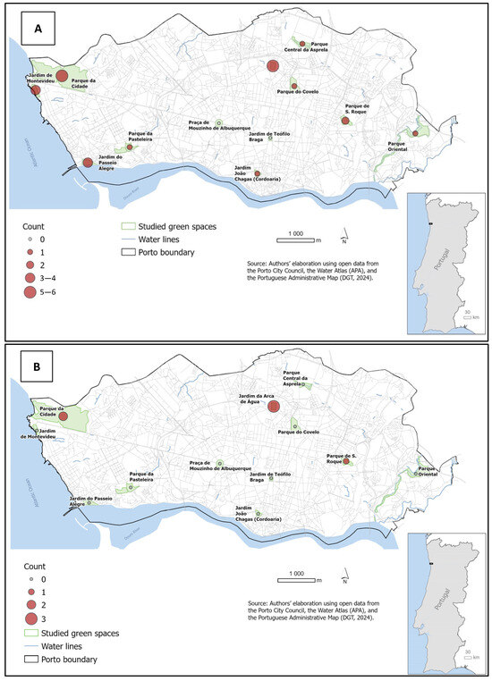
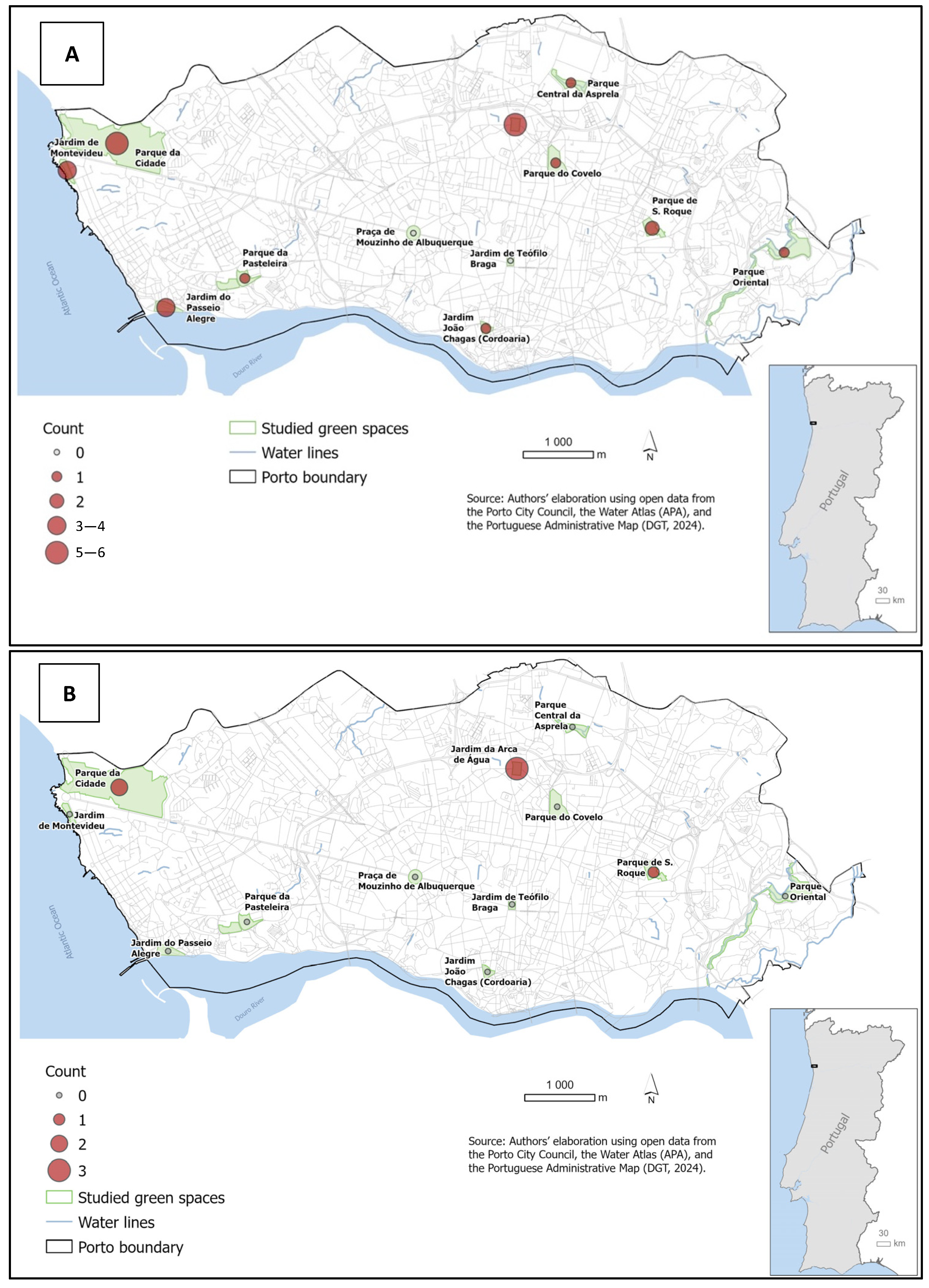
The spatial distribution of Enterococcus spp. isolates, as illustrated in Figure 1, reveals a widespread occurrence of fecal contamination across the urban landscape of Porto. Considering the sampling effort (10 samples per site and period), recovery rates varied among locations. Higher proportions of positive samples were observed in Jardim da Arca de Água (n = 6 in December and n = 3 in May), Parque da Cidade (n = 5 in December and n = 2 in May), and Parque de S. Roque (n = 2 in December and n = 1 in May). These differences should be interpreted with caution, as recovery rates may be influenced by factors such as sample preservation conditions and environmental exposure. This generalized presence highlights that dog feces serve as a consistent source of environmental dissemination of Enterococcus strains throughout the city [25]. Furthermore, the identification of Enterococcus spp. in diverse recreational areas highlights the importance of maintaining hygienic practices, such as the proper removal of dog feces, to minimize potential exposure to a range of environmental bacteria [26,27].
Figure 1.
Spatial distribution and prevalence of Enterococcus spp. in Porto’s urban green spaces during December 2023 (A) and May 2024 (B).
Regarding the sampling periods, a relevant variation in the prevalence and diversity of Enterococcus species was observed (Table 1). In December, a total of 26 isolates were recovered, with E. faecium being the most frequent species (30.8%), followed by E. faecalis (11.5%). In contrast, the sampling in May yielded only 6 isolates; however, a shift in species dominance was noted, with E. faecalis representing 66.7% of the isolates, while E. faecium accounted for only 16.7%. The remaining isolates from both periods (57.7% in December and 16.7% in May) were identified as Enterococcus spp., following negative PCR results for E. hirae and E. gallinarum. The environmental conditions typical of December in Porto, characterized by high humidity levels and moderate temperatures, may enhance the environmental persistence of a wider variety of enterococci in dog feces. These factors protect the bacteria from desiccation, thereby maintaining a higher bacterial load in urban spaces [28]. Conversely, the higher temperatures and increased UV radiation exposure in May might selectively favor more resilient species, such as E. faecalis, which represented 66.7% of the isolates in that period, or lead to a faster degradation of fecal matter, resulting in the recovery of fewer but potentially more robust multidrug-resistant strains [29]. This may be explained by the fact that certain antimicrobial resistance mechanisms, such as efflux pumps and stress response systems, can also contribute to enhanced tolerance to environmental stressors, thereby supporting bacterial persistence under adverse conditions [30]. These findings underscore the importance of year-round monitoring and a One Health approach to fully understand how seasonal dynamics influence the role of canine fecal contamination as a persistent source of resistant bacteria in shared public environments.
Table 1.
Distribution of Enterococcus species by sampling period (December 2023 and May 2024).
2.2. Antimicrobial Resistance Profiles
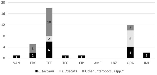
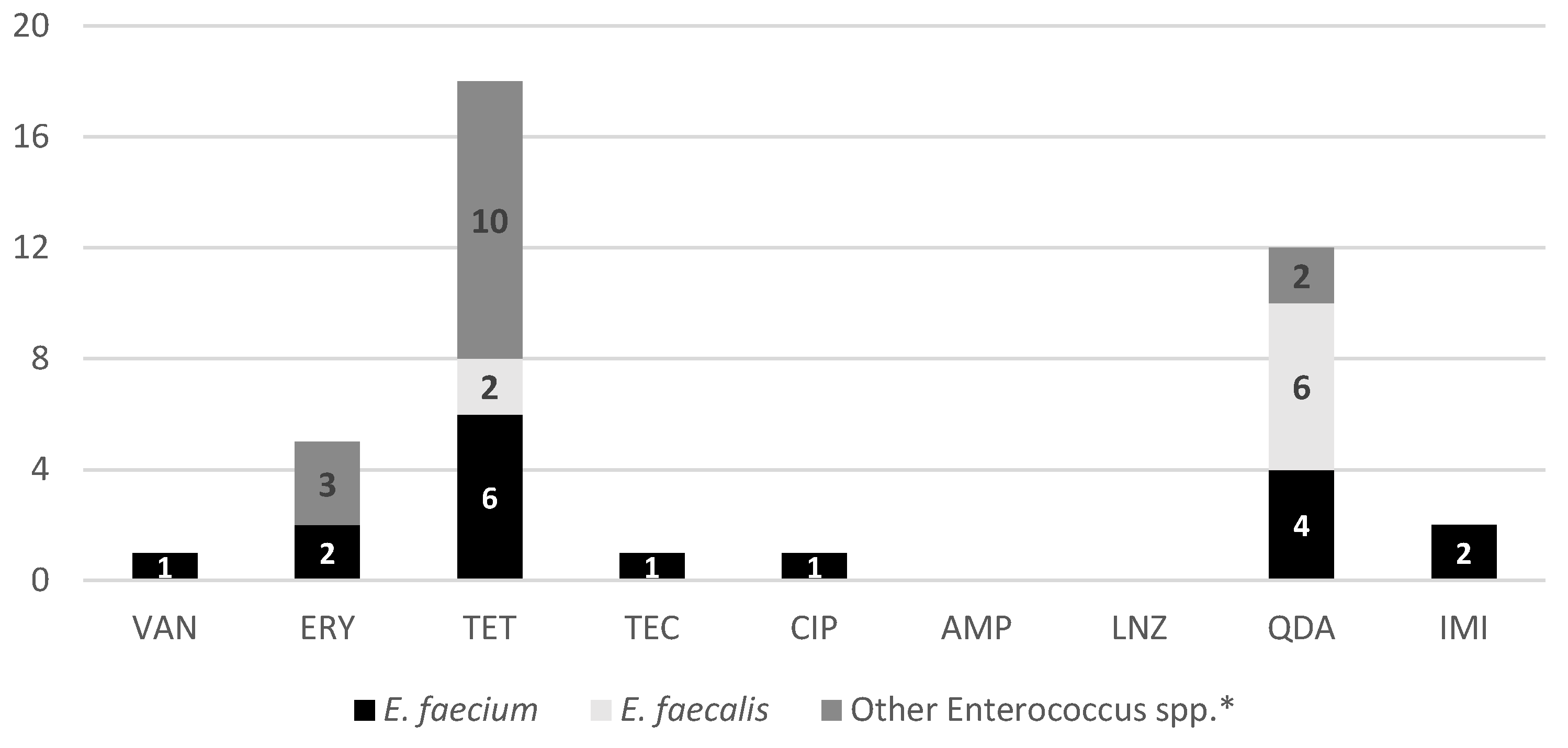
The phenotypic analysis of the 32 Enterococcus spp. isolates reveal a concerning distribution of AMR within Porto’s urban green spaces (Figure 2). The most striking finding is the high prevalence of resistance to tetracycline, which was observed in 56.3% (n = 18) of the isolates, affecting E. faecium, E. faecalis, and other species alike. Another significant observation is the resistance to quinupristin/dalfopristin, detected in 37.5% (n = 12) of the total isolates, with a particularly high occurrence in E. faecalis (n = 6) and E. faecium (n = 4). While some resistance in E. faecalis can be expected, the presence of these phenotypes in a public space is a matter of public health concern [31]. Furthermore, the study identified resistance to last-resort antibiotics, including a 3.1% (n = 1) resistance rate for vancomycin, teicoplanin, and ciprofloxacin, as well as a 6.3% (n = 2) resistance rate for imipenem among the isolates. The presence of these resistant phenotypes in dog feces collected from recreational areas suggests that urban green spaces are acting as environmental reservoirs for resistant bacteria. This poses a potential risk of transmission to humans and other animals through contact with contaminated soil or vegetation [20].
Figure 2.
Phenotypic AMR profiles of the 32 Enterococcus spp. isolates. The bars represent the number of isolates resistant to each tested antibiotic, categorized by species: E. faecium (n = 9), E. faecalis (n = 7), and other Enterococcus spp. (n = 16). Tested antibiotics include VAN (vancomycin), ERY (erythromycin), TET (tetracycline), TEC (teicoplanin), CIP (ciprofloxacin), AMP (ampicillin), LNZ (linezolid), QDA (quinupristin/dalfopristin), and IMI (imipenem). * Isolates negative for E. hirae and E. gallinarum by PCR.
The analysis of phenotypic resistance across the two sampling periods suggests potential seasonal differences in the AMR profiles within the urban environment. During the December sampling period, which yielded a higher number of isolates (n = 26), a diverse range of resistance profiles was observed, including resistance to tetracycline (53.8%), erythromycin (15.4%), and quinupristin/dalfopristin (26.9%), alongside sporadic resistance to vancomycin, ciprofloxacin, and imipenem (3.8% each). In contrast, although the May collection resulted in fewer isolates (n = 6), relatively high proportions of resistance were also observed, with four out of six (66.7%) isolates showing resistance to tetracycline and five (83.3%) exhibiting resistance to quinupristin/dalfopristin. Teicoplanin resistance was detected only once, during the May sampling. These seasonal differences may suggest that while the colder and more humid conditions in December facilitate the environmental persistence of Enterococcus strains, the warmer conditions in May might favor the survival of specific, highly resilient multidrug-resistant isolates [32]. However, these observations should be interpreted with caution due to the limited number of isolates, particularly in May, and potential influences of sample condition on isolation success. Therefore, no definitive conclusions regarding seasonal variation can be drawn.
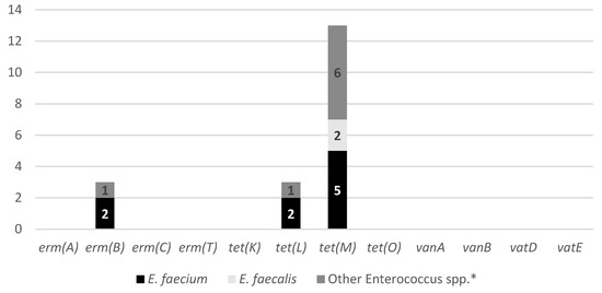
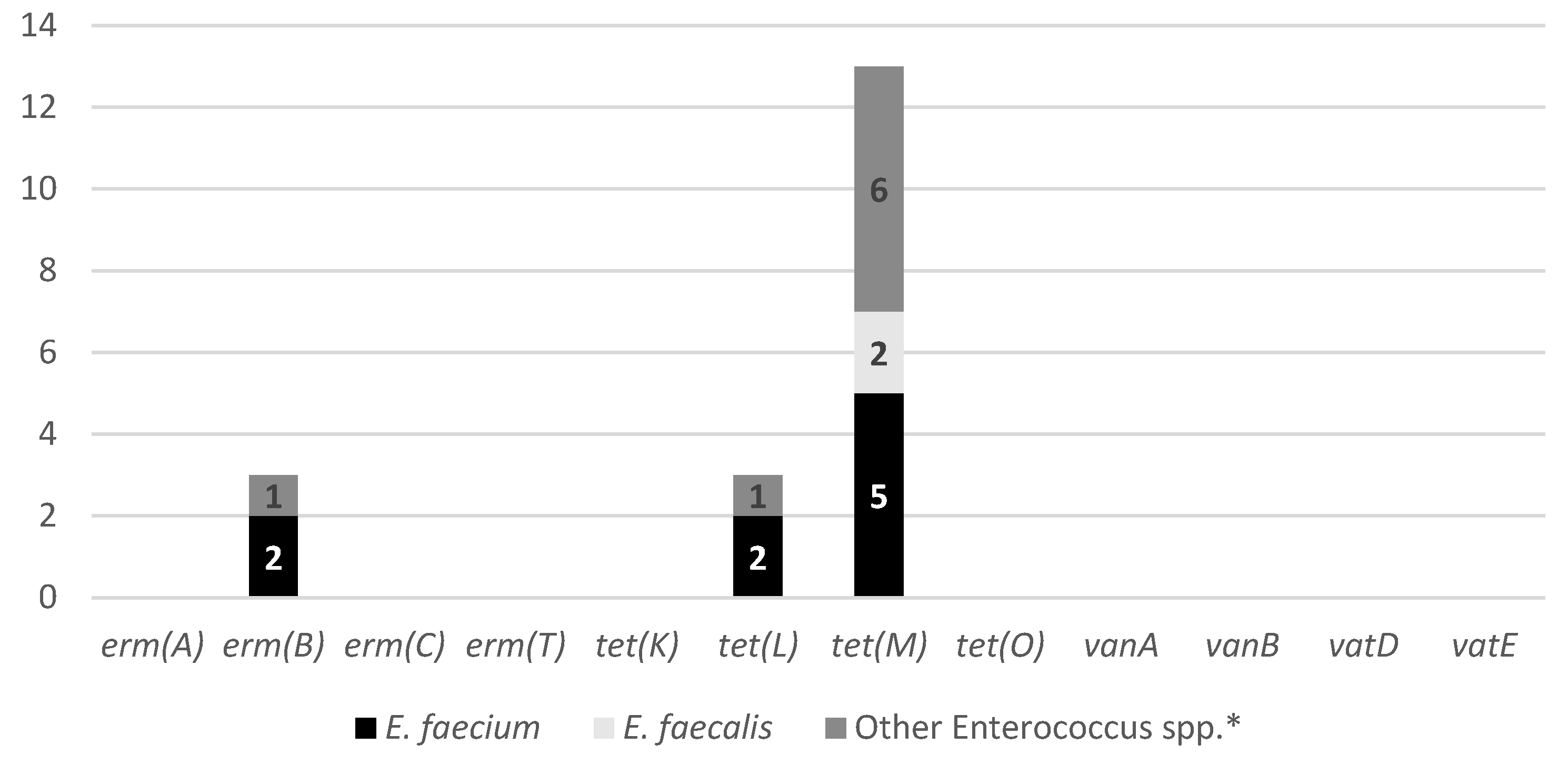
The distribution of AMR genes among the Enterococcus spp., providing insight into the genetic basis of the resistance phenotypes observed, is presented in Figure 3. The most prevalent resistance determinant was tet(M), identified in 40.6% of the isolates, making it the dominant gene across all Enterococcus species analyzed. The predominance of the tet(M) gene is particularly concerning as it is frequently associated with conjugative transposons of the Tn916/Tn1545 family, which play a major role in horizontal gene transfer between bacteria from animal, human, and environmental origins [33,34]. The detection of this gene in isolates derived from dog feces deposited in urban green spaces suggests that these environments may function as reservoirs for transferable tetracycline resistance genes. The additional presence of tet(L) and tet(K) further reflects the diversity of tetracycline resistance mechanisms, including efflux-mediated pathways, circulating in the urban environment. Eflux pumps are transporter proteins that extrude several toxic substances, including antibiotics, from within a cell to its external environment [35]. Macrolide resistance genes were detected at lower frequencies, with erm(B) being the only identified (9.3%) erm determinant. The coexistence of tetracycline and macrolide resistance genes in some isolates may facilitate co-selection, particularly in environments exposed to multiple antimicrobial pressures [36]. Importantly, no streptogramin resistance genes (vatD or vatE) were detected, despite the phenotypic resistance to quinupristin–dalfopristin observed in several isolates. Similarly, vancomycin resistance genes (vanA and vanB) were not identified, even though one isolate exhibited a vancomycin-resistant phenotype. The lack of full correlation between phenotypic and genotypic profiles is a well-documented phenomenon in Enterococcus spp. These discrepancies suggest that resistance may be mediated by alternative mechanisms, such as intrinsic resistance traits, chromosomal mutations, or resistance genes not targeted by the PCR assays used in this study [25,37]. Regarding the antibiotics for which no specific resistance genes were screened, such as imipenem and ciprofloxacin, resistance in Enterococcus spp. is frequently associated with chromosomal mutations or non-specific mechanisms like efflux pumps, rather than easily detectable mobile genetic elements [38]. Indeed, previous studies have shown that imipenem resistance in E. faecalis depends on the presence of a low-affinity PBP4, supporting the rationale for not targeting specific resistance genes for this antibiotic [39]. Additionally, teicoplanin resistance was indirectly assessed through the screening of vanA and vanB genes, which represent the primary genetic determinants for glycopeptide resistance in this genus [37].
Figure 3.
Genotypic AMR profiles of the 32 Enterococcus spp. isolates. The bars represent the absolute number of resistance genes detected in each species: E. faecium (n = 9), E. faecalis (n = 7), and other Enterococcus spp. (n = 16). The genetic screening targeted determinants associated with resistance to erythromycin (erm(A), erm(B), erm(C), erm(T)), tetracycline (tet(K), tet(L), tet(M), tet(O)), vancomycin (vanA, vanB), and quinupristin-dalfopristin (vatE, vatD). * Isolates negative for E. hirae and E. gallinarum by PCR.
The occurrence of AMR genes demonstrated distinct patterns between the two sampling periods. During the winter sampling (December 2023), the most frequently detected determinant was tet(M) (34.6%), followed by erm(B) (12.6%) and tet(L) (6.3%). In contrast, isolates recovered in the spring sampling (May 2024) exhibited a higher relative contribution of tetracycline resistance genes, with tet(M) detected in 66.7% of the isolates and tet(L) in 12.6%, while erm(B) was identified in only 6.3% of the isolates. Once more, the increase in the proportion of tet(M)-positive isolates during the spring period suggests a potential seasonal influence on the environmental burden of tetracycline-resistant Enterococcus spp. in Porto’s urban green spaces, possibly related to differences in environmental conditions that favor the persistence or selection of resistant strains [32].
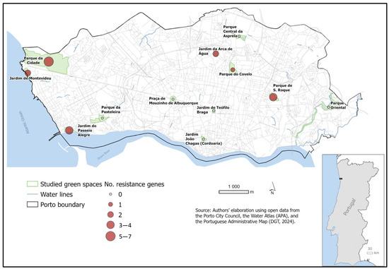
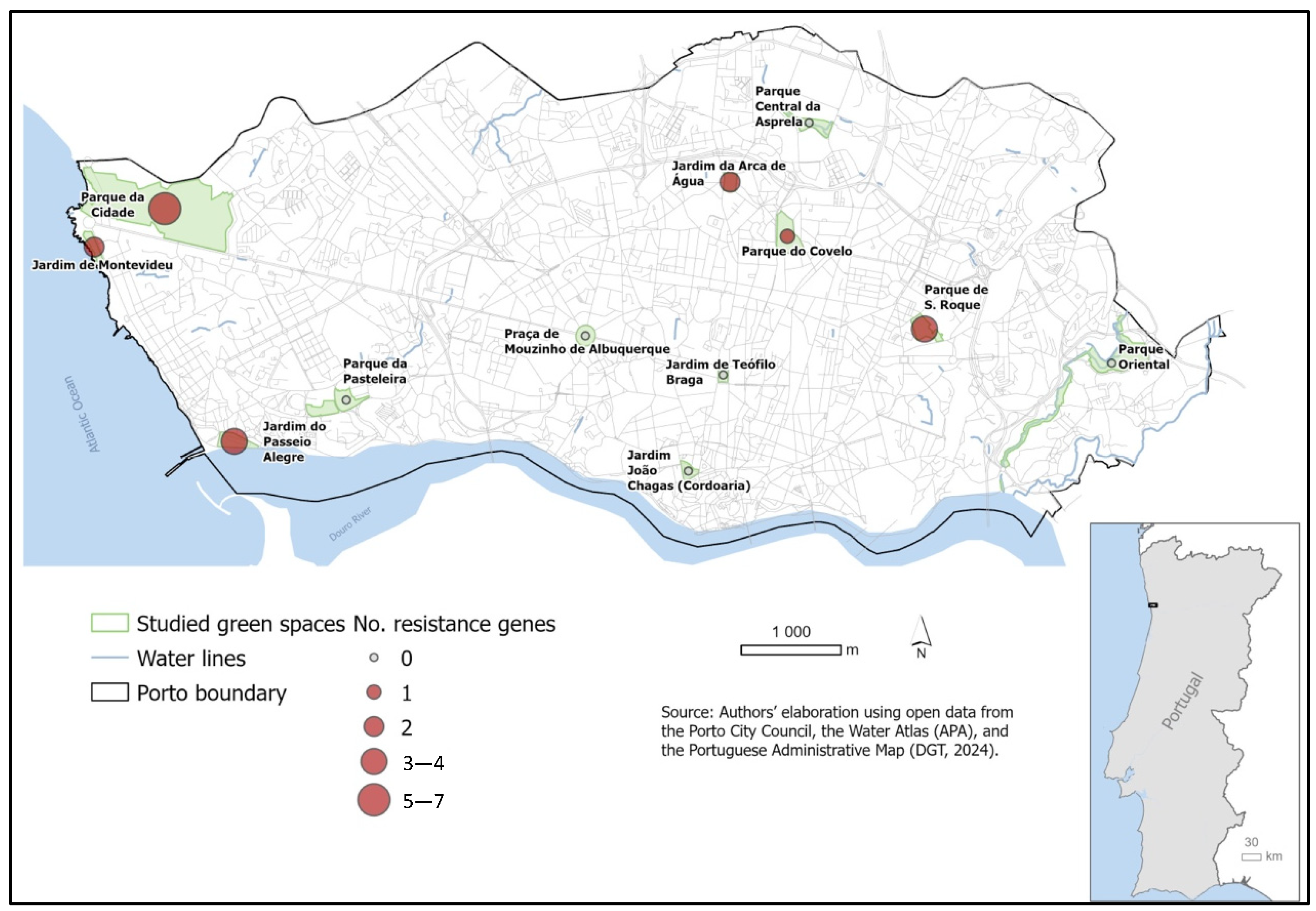
The geographic distribution of AMR genes (Figure 4) indicates areas with relatively higher genetic diversity, although some regions show very low counts. Locations such as Parque da Cidade, Parque de S. Roque, and Jardim do Passeio Alegre exhibited the highest variety of resistance determinants, harboring variants of tet and erm. AMR genes in specific green spaces indicate that canine fecal contamination is not only spreading resistant bacteria but also contributing to the accumulation of a complex ‘resistome’ in areas of high human–animal interaction [40].
Figure 4.
Geographic distribution of AMR genes detected in the 32 Enterococcus spp. isolates. The size and color of the markers indicate the total number of resistance genes (including tet, erm, van, and vat variants) identified per sampling location.
2.3. Virulence Profiles
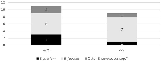
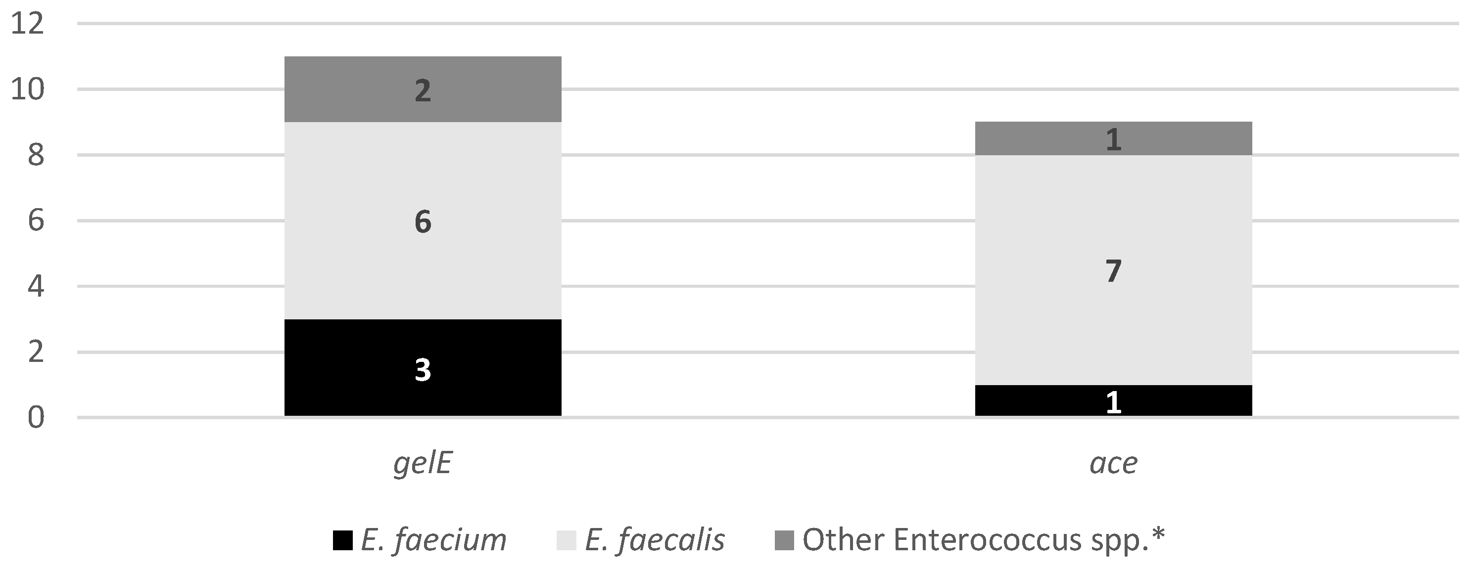
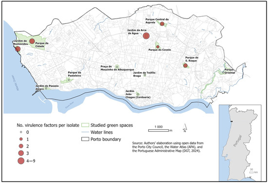
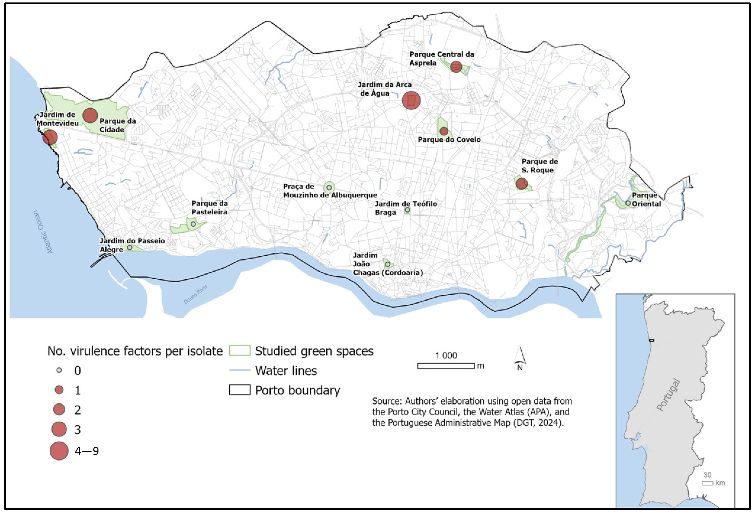
The analysis of virulence-associated genes in Enterococcus spp. isolated from dog feces (Figure 5) revealed the presence of clinically relevant determinants in a considerable proportion of isolates. The gelE gene was the most frequently detected virulence factor (34.4%), followed by ace (28.1%), and both genes were identified across different Enterococcus species, including E. faecium and E. faecalis. The detection of gelE, a gene previously associated with tissue degradation, biofilm formation, and environmental persistence, suggests that these isolates may harbor traits that could enhance survival in urban environments and facilitate dissemination [41]. Similarly, the presence of ace, a collagen-binding adhesin linked to host colonization and invasive infections, highlights the potential pathogenic relevance of enterococci circulating in urban green spaces [42]. Notably, some isolates harbored more than one virulence determinant, indicating the coexistence of traits that may increase both environmental fitness and pathogenic potential. The occurrence of these virulence genes in Enterococcus spp. from dog feces reinforces concerns regarding urban green spaces as reservoirs of opportunistic pathogens. Taken together, the virulence profiles observed in Figure 6, particularly when combined with the AMR data, support the role of canine fecal contamination in the environmental dissemination of enterococci with clinically relevant characteristics, underscoring the importance of surveillance within a One Health framework [43].
Figure 5.
Virulence profiles of the 32 Enterococcus spp. isolates. The bars represent the absolute number of virulence factors detected in each species: E. faecium (n = 9), E. faecalis (n = 7), and other Enterococcus spp. (n = 16). The genetic screening targeted determinants associated with gelE and ace. * Isolates negative for E. hirae and E. gallinarum by PCR.
Figure 6.
Spatial occurrence and density of virulence-associated genes (gelE and ace) in Enterococcus spp. isolates. Symbols represent the cumulative number of virulence factors per isolate at each sampled urban green space. These findings underscore the potential of canine fecal contamination as a source of clinically relevant strains in shared human–animal environments.
A temporal variation in the distribution of virulence-associated genes was observed between the two sampling periods. In December, gelE was detected in 7 out of 26 Enterococcus spp. isolates (26.9%), while ace was identified in four isolates (15.4%), all of which also carried gelE. In contrast, despite the lower number of isolates recovered in May (n = 6), a higher relative frequency of virulence determinants was observed, with gelE detected in four isolates (66.7%) and ace in five isolates (83.3%). Notably, all gelE-positive isolates recovered in May also harbored ace, indicating a predominance of strains carrying combined virulence traits during this period. Again, these findings may indicate that while winter conditions could favor the environmental persistence of a broader diversity of Enterococcus spp., spring conditions could selectively allow the persistence of strains with enhanced adhesion and colonization potential [32]. However, these observations should be interpreted with caution due to the limited number of isolates and the potential influence of sample condition on isolation success. Overall, the detection of virulence determinants in both sampling periods highlights dog fecal contamination as a continuous source of potentially pathogenic enterococci in urban green spaces, reinforcing the relevance of year-round monitoring within a One Health framework.
Figure 6 illustrates the spatial distribution and density of virulence-associated genes (gelE and ace) detected in the 32 Enterococcus spp. isolates. Virulence determinants were not restricted to a single location but were detected in multiple green spaces, indicating a widespread dissemination of potentially pathogenic enterococci throughout the urban environment. The co-occurrence of gelE and ace in several locations further highlights the presence of strains with combined adhesion and tissue-degrading capabilities, which may enhance environmental persistence and increase the likelihood of host colonization following exposure. The spatial overlap between virulence gene hotspots and areas previously identified as harboring AMR determinants reinforces the concern that certain urban green spaces may function as reservoirs of enterococci with both virulence and resistance traits. This convergence increases the potential public health risk, as it facilitates the circulation of strains with enhanced fitness and pathogenic potential at the human–animal–environment interface.
2.4. Ecological and Urban Drivers of Antimicrobial-Resistant Enterococcus spp.
The analysis of vegetation density across the sampled urban green spaces, assessed using the mean Normalized Difference Vegetation Index (NDVI), revealed some heterogeneity among the public green spaces of Porto. Mean NDVI values ranged from 0.229 in Parque de S. Roque to 0.495 in Jardim João Chagas (Cordoaria), reflecting differences in vegetation coverage, structure, and vigor across the studied locations. However, when NDVI values were examined in relation to the spatial distribution of Enterococcus spp. isolates and their associated AMR and virulence determinants, no direct positive association between higher vegetation density and increased bacterial recovery was observed. Among the green spaces with higher NDVI values (mean NDVI > 0.40), such as Jardim de Montevideu, Praça de Mouzinho de Albuquerque, Jardim de Teófilo Braga, and Jardim João Chagas (Cordoaria), only Jardim de Montevideu yielded Enterococcus spp. isolates and corresponding resistance and virulence genes. In contrast, the highest numbers of isolates were recovered from Jardim da Arca de Água (n = 6 in December; n = 3 in May), Parque da Cidade (n = 5 in December; n = 2 in May), and Parque de S. Roque (n = 2 in December; n = 1 in May), which are not among the sites with the highest NDVI values. From an ecological perspective, highly vegetated green spaces are typically associated with shaded microhabitats, increased soil moisture, and reduced exposure to ultraviolet radiation, which are environmental conditions known to favor bacterial survival in soil and on vegetation surfaces [44,45]. Such conditions may enhance the persistence of fecal material deposited by companion animals and potentially prolong the environmental survival of antimicrobial-resistant Enterococcus spp. Nevertheless, the present findings indicate that vegetation density does not constitute a direct risk factor for bacterial occurrence per se but may instead act as an ecological facilitator by modulating microenvironmental conditions (such as moisture retention, organic matter availability, and host or vector presence) whose influence is strongly context dependent. Factors such as the intensity of human and animal activity, repeated fecal inputs, soil disturbance, and local management practices are likely to modulate the extent to which favorable microclimatic conditions translate into detectable bacterial persistence [46].
In addition to vegetation density, other characteristics of the sampled green spaces, including the park size and presence of water features, were considered to contextualize the environmental distribution of Enterococcus spp. and their associated resistance and virulence determinants. Large and highly frequented green spaces, such as Parque da Cidade, combine extensive surface area, diverse vegetation structures, and multiple water features, creating heterogeneous microhabitats that may support the persistence of antimicrobial-resistant enterococci. Conversely, smaller and more compact green spaces, including Jardim João Chagas (Cordoaria) and Praça de Mouzinho de Albuquerque, are subject to a higher intensity of use per unit area, which may increase fecal deposition pressure and contribute to the detection of Enterococcus spp. despite their limited spatial extent. Among the sites where Enterococcus spp. were recovered, Parque da Cidade and Parque de S. Roque are characterized by the presence of lakes or fountains within their boundaries. However, bacterial isolates were also recovered from Parque do Covelo during the December sampling period, despite the absence of both natural watercourses and artificial water features at this site. Conversely, Jardim João Chagas (Cordoaria), which includes artificial water elements, did not yield Enterococcus spp. isolates during the May sampling campaign. These contrasting observations suggest that the presence or absence of water bodies alone is insufficient to explain the spatial and temporal distribution of Enterococcus spp. in urban green spaces.
To further contextualize the environmental distribution of Enterococcus spp., the socioeconomic characteristics of the areas surrounding each green space were assessed using the European Deprivation Index (EDI). Based on the distribution of EDI values across the sampled locations, sites were categorized into low, medium, and high deprivation using empirically defined thresholds (EDI < −3, −3 EDI 2, EDI > 2, respectively). A socioeconomic heterogeneity was observed among the sampled green spaces. Jardim de Montevideu, Parque da Cidade, Praça de Mouzinho de Albuquerque, and Jardim do Passeio Alegre were classified as low-deprivation areas; Parque do Covelo, Parque de S. Roque, Jardim da Arca de Água, Jardim de Teódilo Braga, Parque Central da Asprela, and Parque da Pasteleira as medium-deprivation áreas; and Jardim João Chagas (Cordoaria) and Parque Oriental as high-deprivation areas. Most Enterococcus spp. isolates were recovered from green spaces located in low- and medium-deprivation neighborhoods, whereas only a single isolate was obtained from each of the two high-deprivation sites, Parque João Chagas (Cordoaria) and Parque Oriental, during the December sampling. Importantly, these isolates did not carry AMR or virulence-associated genes, indicating that socioeconomic deprivation alone does not necessarily favor the persistence of resistant or virulent enterococci in urban green spaces. A possible explanation is that lower access to veterinary care in these high-deprivation areas may result in less frequent antibiotic use in pets, reducing selective pressure for the emergence and dissemination of resistant strains [47].
Although the exploratory nature of this study and the limited number of sampling sites preclude causal inference, integrating socioeconomic indicators with ecological and microbiological data provides a more comprehensive understanding of AMR dissemination in urban environments [48]. The combined assessment of vegetation density, water features, and spatial dimensions suggests that certain urban green spaces create ecological conditions favorable to the environmental persistence of Enterococcus spp. carrying resistance and virulence determinants. Rather than acting as isolated risk factors, these environmental characteristics likely interact to form localized “hotspots” where resistant bacteria may persist and accumulate [49]. Overall, these findings highlight the importance of incorporating biophysical and ecological descriptors into AMR surveillance, as the integration of remotely sensed vegetation indices, spatial metrics, and water feature information allows for a nuanced understanding of how urban green spaces, designed to promote health and well-being, may inadvertently serve as reservoirs for clinically relevant bacteria when pet waste management is insufficient. These results also underscore the value of including social context in One Health surveillance frameworks, recognizing that AMR in public spaces emerges from the dynamic interplay between environmental structure, animal behavior, and broader urban inequalities.
3. Materials and Methods
3.1. Sample Collection
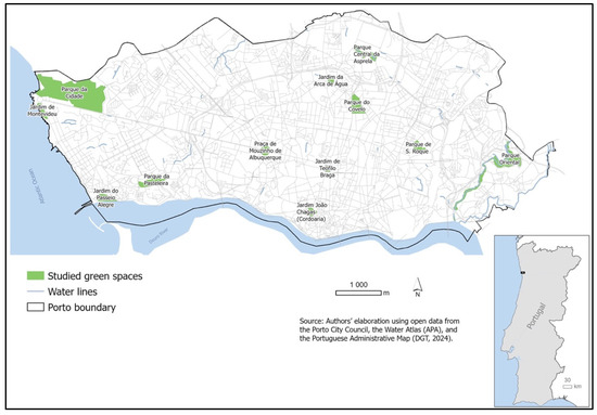
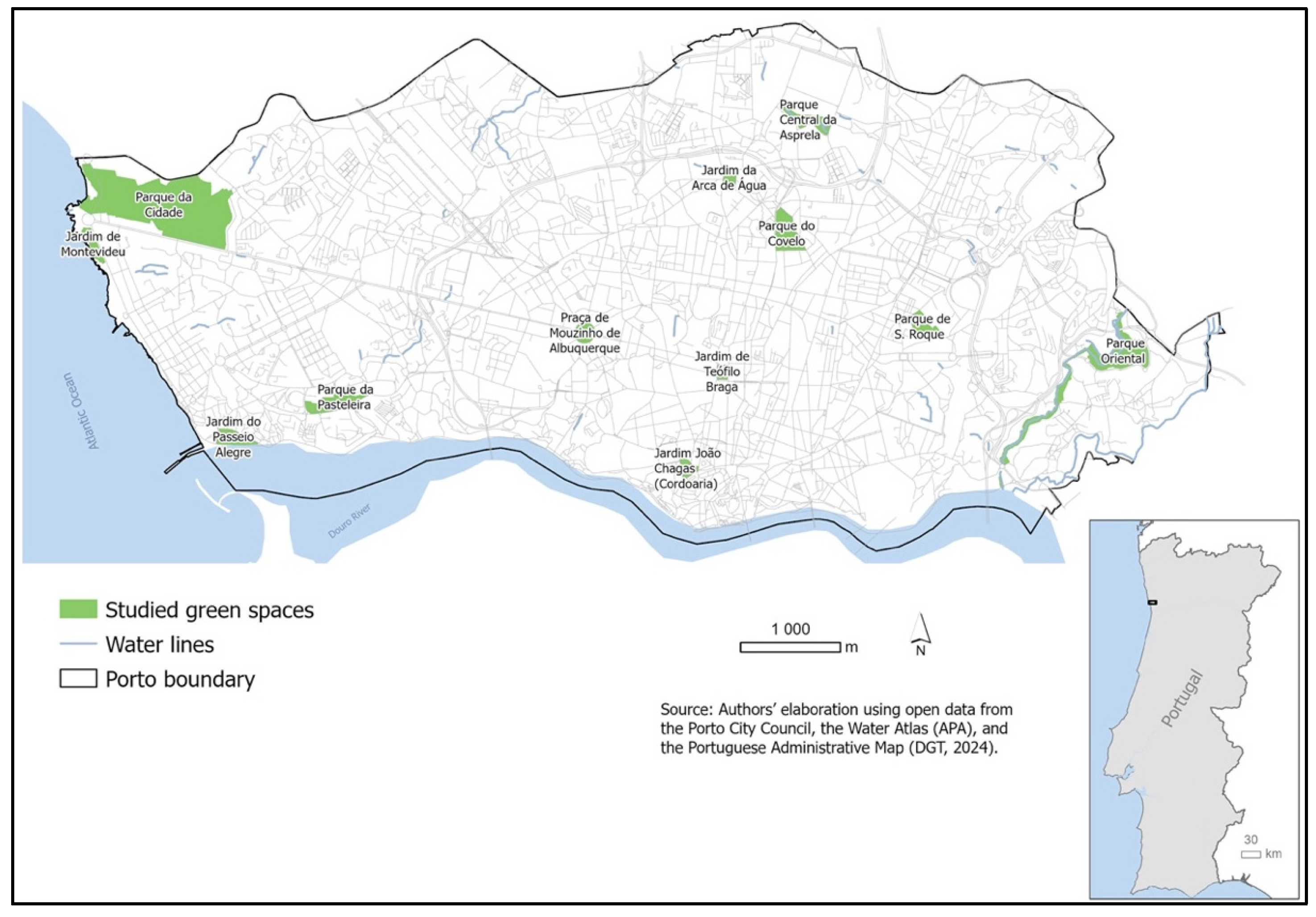
Fecal samples were collected in December 2023 and May 2024 from 12 urban green spaces located across the city of Porto (Portugal): Jardim de Montevideu, Parque da Cidade, Jardim do Passeio Alegre, Parque da Pasteleira, Praça de Mouzinho de Albuquerque, Jardim de Teófilo Braga, Jardim João Chagas (Cordoaria), Parque de S. Roque, Parque Oriental, Parque do Covelo, Parque Central da Asprela, and Jardim da Arca de Água (Figure 7). Sampling was conducted in two different periods to capture potential seasonal variation. In each garden, ten fresh fecal samples were collected per sampling period, resulting in a total of 240 samples. Only fresh samples, showing no signs of degradation or environmental contamination, were collected to avoid overgrowth of environmental bacteria or cross-contamination. Samples were collected directly from the ground using sterile spatulas and placed into sterile containers. Each sample was labeled according to the collection site and period and transported to the laboratory under refrigerated conditions (4 °C) within 24 h. Upon arrival, samples were processed immediately or stored at 4 °C for a maximum of 48 h before microbiological analysis.
Figure 7.
Geographic location of the 12 urban green spaces sampled in Porto (Portugal). Sampling sites were selected to represent different urban areas for the collection of dog fecal samples (n = 240) during the two study periods (December 2023 and May 2024).
3.2. Isolation and Identification of Enterococcus spp.
Approximately 2–3 g of each fecal sample were inoculated into Brain Heart Infusion (BHI) broth (Oxoid Ltd., Basingstoke, UK) and incubated at 37 °C for 24 h to allow bacterial enrichment. After incubation, aliquots from each culture were streaked onto Slanetz–Bartley agar (Oxoid Ltd., Basingstoke, UK) plates and incubated at 37 °C for 48 h. Colonies displaying the typical pink to red coloration characteristic of Enterococcus spp. were selected and subsequently subcultured on Kanamycin Aesculin Azide (KAA) agar (Oxoid Ltd., Basingstoke, UK). Plates were incubated at 37 °C for 24 h, and colonies that produced blackening of the medium, indicative of aesculin hydrolysis, were presumptively identified as Enterococcus spp. Representative isolates were preserved in skim milk medium at −80 °C for further analyses, including antimicrobial susceptibility testing and PCR detection of selected resistance and virulence genes.
3.3. Antimicrobial Susceptibility Testing
Antimicrobial susceptibility was determined by the disk diffusion method on Mueller–Hinton agar (Oxoid Ltd., Basingstoke, UK). The following antibiotics disks (Oxoid Ltd., Basingstoke, UK) were tested (µg per disk): vancomycin (30), teicoplanin (30), erythromycin (15), tetracycline (30), ciprofloxacin (5), ampicillin (10), linezolid (30), quinupristin-dalfopristin (5), and imipenem (10), according to Clinical and Laboratory Standards Institute (CLSI) guidelines [50], except for teicoplanin, quinupristin-dalfopristin, and imipenem, which were included based on the EUCAST guidelines [51]. Plates were incubated at 37 °C for 24 h, and inhibition zone diameters were measured and interpreted according to CLSI and EUCAST breakpoints for Enterococcus spp. (Table 2). Isolates exhibiting resistance to at least one antibiotic in three or more antimicrobial classes were classified as multidrug-resistant.
Table 2.
Cut-off values for disk diffusion testing of Enterococcus spp. isolates.
3.4. Detection of Antimicrobial Resistance and Virulence Genes
Genomic DNA was isolated from overnight BHI broth cultures using the Insta Gene™ Matrix (Bio-Rad Laboratories, Hercules, CA, USA). DNA concentration and purity were determined with a NanoDrop ND-100 spectrophotometer (Thermo Fisher Scientific, Waltham, MA, USA). Gene detection was then conducted via polymerase chain reaction (PCR). PCR was performed on a ProFlex™ PCR System (Applied Biosystems, Waltham, MA, USA) in a total volume of 50 µL, containing 30.2 µL ultrapure water (Milli-Q®, Merck Millipore, Burlington, MA, USA), 5 µL complete buffer (Bioron GmbH, Römerberg, Germany), 1.5 µL 100 mM MgCl2 (100 mM, Bioron GmbH, Römerberg, Germany), (10 mM, Bioron GmbH, Römerberg, Germany), 1 µL of each primer (50 µM, Eurofins Genomics, Ebersberg, Germany), 0.3 µL DFS-Taq DNA polymerase (5 U/µL, Bioron GmbH, Römerberg, Germany), and 10 µL of template DNA (10 ng). Positive controls consisted of strains from the MicroART collection, while Milli-Q water was used as the negative control. The analysis focused on four Enterococcus species: E. faecium (ddlE. faecium), E. faecalis (ddlE. faecalis), E. hirae (murG), and E. gallinarum (vanC1). Twelve primers were employed to identify genes conferring resistance to five antibiotic classes, including glycopeptides (vanA, vanB), macrolides (erm(A), erm(B), erm(C)) streptogramins (vatD, vatE), and tetracyclines (tet(M), tet(O), tet(L), tet(K)). In addition, isolates were screened for the virulence-associated genes gelE (encoding gelatinase) and ace (encoding a collagen-binding adhesin). Details on primer sequences, PCR conditions, and expected amplicon sizes are presented in Table 3.
Table 3.
List of primers employed for PCR-based detection of Enterococcus species, antibiotic resistance determinants, and virulence-associated genes. The table provides the target genes, corresponding primer sequences, PCR cycling parameters, and expected product sizes.
3.5. Data Analysis
The frequencies of resistance phenotypes and genotypes were calculated and expressed as percentages. The distribution of resistant isolates and detected genes was analyzed according to Enterococcus species and garden location.
Spatial data visualization was performed using ArcGIS Pro 3.5.0. Graduated symbol maps (Esri Inc., Redlands, CA, USA) (natural breaks classification) were produced to depict the prevalence of Enterococcus spp. and the distribution of specific AMR and virulence genes across the sampled public urban green spaces in Porto.
In addition, we characterized the features of each green space, namely the presence of water features, vegetation levels, and the socioeconomic deprivation of the surrounding areas. Spatial datasets and analytical procedures were employed.
To assess the presence of water features, we used official cartographic data on surface water bodies provided by the Porto City Council [59]. Spatial overlay operations were conducted to identify whether water bodies intersected each urban green space. The presence of artificial water features (e.g., ponds, fountains, lakes) was further assessed through visual inspection of high-resolution aerial imagery in Google Earth. Vegetation was characterized using the NDVI, computed from Sentinel-2 imagery with less than 5% cloud cover, following the methodology adopted in previous studies [60,61] for the years 2023 and 2024. NDVI was calculated as:
where NIR corresponds to the near-infrared band and Red to the red band of the electromagnetic spectrum. NDVI values range from −1 to +1, with higher values indicating greater vegetation density and vigor, and values close to zero or negative indicating sparse or absent vegetation.
NDVI = (NIR + Red)/(NIR − Red),
Finally, socioeconomic deprivation in the surroundings of each green space was assessed using the EDI, computed for Portuguese small areas, following the methodology fully described elsewhere [62]. A 300-m street-network buffer, corresponding to the distance recommended by the World Health Organization (WHO) as an indicator of adequate accessibility to urban green spaces [63], was generated around each green space to represent its local catchment area. The weighted average EDI was then calculated based on the population of each census block group intersecting the buffer, allowing for a population-adjusted estimate of neighborhood deprivation.
4. Conclusions
This study demonstrates that dog feces deposited in urban green spaces represent a relevant environmental reservoir of antimicrobial-resistant and potentially virulent Enterococcus spp. in Porto. Although the overall isolation rate was moderate, the recovery of clinically significant species, particularly E. faecium and E. faecalis, together with the detection of resistance to important antimicrobials, highlights the public health relevance of canine fecal contamination in shared recreational environments.
The high prevalence of tetracycline resistance, largely associated with the widespread detection of the mobile gene tet(M), underscores the role of urban environments as reservoirs and potential dissemination centers for transferable AMR determinants. The presence of virulence-associated genes (gelE and ace), often co-occurring with resistance traits, further emphasizes the pathogenic potential and environmental fitness of enterococci circulating in public green spaces.
By integrating microbiological findings with ecological and socioeconomic descriptors, this work provides novel insight into the complex drivers shaping the environmental persistence of antimicrobial-resistant bacteria in urban settings. Rather than acting as isolated risk factors, vegetation structure, water features, intensity of human–animal interaction, and neighborhood socioeconomic context appear to co-occur in specific urban green spaces, potentially contributing to localized conditions that may favor the persistence and accumulation of resistant and virulent enterococci. Notably, green spaces located in low- and medium-deprivation areas exhibited a higher diversity of resistance and virulence determinants, suggesting that patterns of park use, pet ownership, and maintenance practices may play a more decisive role than deprivation alone.
Overall, these findings reinforce the concept that urban green spaces, while essential for promoting health and well-being, may inadvertently function as reservoirs for antimicrobial-resistant bacteria when pet waste management is inadequate. This study highlights the importance of adopting integrated One Health surveillance strategies that combine microbiological monitoring with environmental and social indicators. Strengthening responsible pet ownership, improving sanitation practices, and implementing spatially informed monitoring approaches are essential steps to mitigate the environmental dissemination of antimicrobial-resistant and virulent bacteria in urban public spaces.
Author Contributions
Conceptualization, J.R. and T.L.M.; methodology, J.R., R.L., F.C.N. and A.I.R.; validation, V.S. and P.P.; formal analysis, V.S.; investigation, J.R.; resources, G.I., T.L.M. and P.P. data curation, A.I.R.; writing—original draft preparation, J.R.; writing—review and editing, J.R.; visualization, J.R., T.L.M. and A.I.R.; supervision, P.P. All authors have read and agreed to the published version of the manuscript.
Funding
This work was supported by the Portuguese Foundation for Science and Technology (FCT) through projects UIDB/CVT/00772/2020 (DOI:10.54499/UIDB/00772/2020) and LA/P/0059/2020. Also, it was supported by national funds through FCT/MCTES (PIDDAC): CIMO, UID/00690/2025 (DOI:10.54499/UID/00690/2025) and UID/PRR/00690/2025 (DOI:10.54499/UID/PRR/00690/2025); and the SusTEC, LA/P/0007/2020 (DOI:10.54499/LA/P/0007/2020). Jessica Ribeiro acknowledges financial support from FCT for her doctoral fellowship (2023.00592.BD). The participation of Teresa Letra Mateus was funded by national funds through FCT (https://doi.org/10.54499/UID/05937/2025). Ana Isabel Ribeiro was funded by the Centre of Studies in Geography and Spatial Planning (CEGOT), funded by national funds through the FCT under the reference UIDB/04084/2025, as well as through the projects with references UIDB/04750/2020 and LA/P/0064/2020 and DOI identifiers https://doi.org/10.54499/UIDB/04750/2020 and https://doi.org/10.54499/LA/P/0064/2020.
Institutional Review Board Statement
Not applicable.
Informed Consent Statement
Not applicable.
Data Availability Statement
The original contributions presented in this study are included in the article. Further inquiries can be directed to the corresponding author.
Conflicts of Interest
The authors declare no conflicts of interest.
Abbreviations
The following abbreviations are used in this manuscript:
| AMP | Ampicillin |
| AMR | Antimicrobial Resistance |
| BHI | Brain Heart Infusion |
| CIP | Ciprofloxacin |
| CLSI | Clinical and Laboratory Standards Institute |
| DNA | Deoxyribonucleic Acid |
| EDI | European Deprivation Index |
| ERY | Erythromycin |
| IMI | Imipenem |
| KAA | Kanamycin Aesculin Azide |
| LNZ | Linezolid |
| NBS | Nature-Based Solutions |
| NDVI | Normalized Difference Vegetation Index |
| PCR | Polymerase Chain Reaction |
| QDA | Quinupristin-dalfopristin |
| SB | Stanetz-Bartley |
| TEC | Teicoplanin |
| TET | Tetracycline |
| UV | Ultra-Violet |
| VAN | Vancomycin |
| WHO | World Health Organization |
References
- Novais, C.; Freitas, A.R.; Silveira, E.; Antunes, P.; Silva, R.; Coque, T.M.; Peixe, L. Spread of Multidrug-Resistant Enterococcus to Animals and Humans: An Underestimated Role for the Pig Farm Environment. J. Antimicrob. Chemother. 2013, 68, 2746–2754. [Google Scholar] [CrossRef] [Scilit] [PubMed]
- Krawczyk, B.; Wityk, P.; Gałęcka, M.; Michalik, M. The Many Faces of Enterococcus spp.—Commensal, Probiotic and Opportunistic Pathogen. Microorganisms 2021, 9, 1900. [Google Scholar] [CrossRef] [Scilit] [PubMed]
- Ayobami, O.; Willrich, N.; Reuss, A.; Eckmanns, T.; Markwart, R. The Ongoing Challenge of Vancomycin-Resistant Enterococcus faecium and Enterococcus faecalis in Europe: An Epidemiological Analysis of Bloodstream Infections. Emerg. Microbes Infect. 2020, 9, 1180–1193. [Google Scholar] [CrossRef] [Scilit] [PubMed]
- Avberšek, J.; Mićunović, J.; Šemrov, N.; Ocepek, M. Surveillance of the Source of Poultry Infections with Enterococcus hirae and Enterococcus cecorum in Slovenia and E. hirae Antibiotic Resistance Patterns. New Microbiol. 2021, 44, 210–216. [Google Scholar]
- Coccitto, S.N.; Cinthi, M.; Fioriti, S.; Morroni, G.; Simoni, S.; Vignaroli, C.; Garofalo, C.; Mingoia, M.; Brenciani, A.; Giovanetti, E. Linezolid-Resistant Enterococcus gallinarum Isolate of Swine Origin Carrying cfr, optrA and poxtA Genes. J. Antimicrob. Chemother. 2021, 77, 331–337. [Google Scholar] [CrossRef] [Scilit]
- Soares, R.; Miranda, C.; Cunha, S.; Ferreira, L.; Martins, Â.; Igrejas, G.; Poeta, P. Antibiotic Resistance of Enterococcus Species in Ornamental Animal Feed. Animals 2023, 13, 1761. [Google Scholar] [CrossRef] [Scilit]
- Zhao, B.; Ye, M.S.; Zheng, R. Enterococcus gallinarum Meningitis: A Case Report and Literature Review. BMC Infect. Dis. 2018, 18, 231. [Google Scholar] [CrossRef] [Scilit]
- Piccinini, D.; Bernasconi, E.; Di Benedetto, C.; Martinetti Lucchini, G.; Bongiovanni, M. Enterococcus hirae Infections in the Clinical Practice. Infect. Dis. 2023, 55, 71–73. [Google Scholar] [CrossRef] [Scilit]
- Geraldes, C.; Tavares, L.; Gil, S.; Oliveira, M. Enterococcus Virulence and Resistant Traits Associated with Its Permanence in the Hospital Environment. Antibiotics 2022, 11, 857. [Google Scholar] [CrossRef] [Scilit]
- Sparo, M.; Delpech, G.; Allende, N.G. Impact on Public Health of the Spread of High-Level Resistance to Gentamicin and Vancomycin in Enterococci. Front. Microbiol. 2018, 9, 3073. [Google Scholar] [CrossRef] [Scilit]
- Maleki, D.; Manouchehrifar, M.; Kheljan, M.N.; Mossavi, S.H.; Jannati, E.; Doghaheh, H.P.; Teimourpour, R.; Khademi, F.; Arzanlou, M. Vancomycin-Resistant Enterococcus Species: Antimicrobial Resistance and Virulence Genes Profile. Gene Rep. 2021, 25, 101338. [Google Scholar] [CrossRef] [Scilit]
- Fatoba, D.O.; Amoako, D.G.; Akebe, A.L.K.; Ismail, A.; Essack, S.Y. Genomic Analysis of Antibiotic-Resistant Enterococcus spp. Reveals Novel Enterococci Strains and the Spread of Plasmid-Borne tet(M), tet(L) and erm(B) Genes from Chicken Litter to Agricultural Soil in South Africa. J. Environ. Manag. 2022, 302, 114101. [Google Scholar] [CrossRef] [Scilit] [PubMed]
- Too, E.; Masila, E. The Interconnection between Virulence Factors, Biofilm Formation, and Horizontal Gene Transfer in Enterococcus: A Review. In Enterococcus—Unveiling the Emergence of a Potent Pathogen; Téllez-Isaías, G., Graham, D., El-Ashram, S., Eds.; IntechOpen: London, UK, 2024. [Google Scholar]
- Marques, J.M.; Coelho, M.; Santana, A.R.; Pinto, D.; Semedo-Lemsaddek, T. Dissemination of Enterococcal Genetic Lineages: A One Health Perspective. Antibiotics 2023, 12, 1140. [Google Scholar] [CrossRef] [Scilit] [PubMed]
- Zaheer, R.; Cook, S.R.; Barbieri, R.; Goji, N.; Cameron, A.; Petkau, A.; Polo, R.O.; Tymensen, L.; Stamm, C.; Song, J.; et al. Surveillance of Enterococcus spp. Reveals Distinct Species and Antimicrobial Resistance Diversity across a One-Health Continuum. Sci. Rep. 2020, 10, 3937. [Google Scholar] [CrossRef] [Scilit]
- Pinto, L.V.; Inácio, M.; Pereira, P. Green and Blue Infrastructure (GBI) and Urban Nature-Based Solutions (NbS) Contribution to Human and Ecological Well-Being and Health. Oxf. Open Infrastruct. Health 2023, 1, ouad004. [Google Scholar] [CrossRef] [Scilit]
- Ribeiro, A.I.; Triguero-Mas, M.; Jardim Santos, C.; Gómez-Nieto, A.; Cole, H.; Anguelovski, I.; Silva, F.M.; Baró, F. Exposure to Nature and Mental Health Outcomes during COVID-19 Lockdown. A Comparison between Portugal and Spain. Environ. Int. 2021, 154, 106664. [Google Scholar] [CrossRef] [Scilit]
- Ribeiro, A.I.; Behlen, M.; Henriques, A.; Severo, M.; Jardim Santos, C.; Barros, H. Exposure to Green and Blue Spaces and Depression among Older Adults from the EPIPorto Cohort: Examining Environmental, Social, and Behavioral Mediators and Varied Space Types. Cities Health 2024, 1–14. [Google Scholar] [CrossRef] [Scilit]
- Marcilla-Toribio, I.; Bizzozero-Peroni, B.; Notario-Pacheco, B.; Ribeiro, A.I.; Santos, M.P.; Fernandez-Perez, M.; Cekrezi, S.; la Cruz, L.L.d.; Martinez-Andres, M. The Role of Surrounding Residential Greenness in Healthy Behaviours: A Systematic Review and Meta-Analysis. Discov. Public Health 2025, 22, 253. [Google Scholar] [CrossRef] [Scilit]
- Vassallo, A.; Kett, S.; Purchase, D.; Marvasi, M. The Bacterial Urban Resistome: Recent Advances. Antibiotics 2022, 11, 512. [Google Scholar] [CrossRef] [Scilit]
- Gwenzi, W.; Chaukura, N.; Muisa-Zikali, N.; Teta, C.; Musvuugwa, T.; Rzymski, P.; Abia, A.L.K. Insects, Rodents, and Pets as Reservoirs, Vectors, and Sentinels of Antimicrobial Resistance. Antibiotics 2021, 10, 68. [Google Scholar] [CrossRef] [Scilit]
- Cinquepalmi, V.; Monno, R.; Fumarola, L.; Ventrella, G.; Calia, C.; Greco, M.F.; De Vito, D.; Soleo, L. Environmental Contamination by Dog’s Faeces: A Public Health Problem? Int. J. Environ. Res. Public Health 2013, 10, 72–84. [Google Scholar] [CrossRef] [Scilit]
- Miranda, C.; Silva, V.; Igrejas, G.; Poeta, P. Impact of European Pet Antibiotic Use on Enterococci and Staphylococci Antimicrobial Resistance and Human Health. Future Microbiol. 2021, 16, 185–203. [Google Scholar] [CrossRef] [Scilit] [PubMed]
- Lebreton, F.; Willems, R.J.L.; Gilmore, M.S. Enterococcus Diversity, Origins in Nature, and Gut Colonization. In Enterococci: From Commensals to Leading Causes of Drug Resistant Infection; Gilmore, M.S., Clewell, D.B., Ike, Y., Shankar, N., Eds.; Massachusetts Eye and Ear Infirmary: Boston, MA, USA, 2014. [Google Scholar]
- Stępień-Pyśniak, D.; Bertelloni, F.; Dec, M.; Cagnoli, G.; Pietras-Ożga, D.; Urban-Chmiel, R.; Ebani, V.V. Characterization and Comparison of Enterococcus spp. Isolates from Feces of Healthy Dogs and Urine of Dogs with UTIs. Animals 2021, 11, 2845. [Google Scholar] [CrossRef] [Scilit]
- Selway, C.A.; Mills, J.G.; Weinstein, P.; Skelly, C.; Yadav, S.; Lowe, A.; Breed, M.F.; Weyrich, L.S. Transfer of Environmental Microbes to the Skin and Respiratory Tract of Humans after Urban Green Space Exposure. Environ. Int. 2020, 145, 106084. [Google Scholar] [CrossRef] [Scilit] [PubMed]
- Ibekwe, A.M.; Obayiuwana, A.C.; Murinda, S.E. Enterococcus Species and Their Antimicrobial Resistance in an Urban Watershed Affected by Different Anthropogenic Sources. Water. 2024, 16, 116. [Google Scholar] [CrossRef] [Scilit]
- Byappanahalli, M.N.; Nevers, M.B.; Korajkic, A.; Staley, Z.R.; Harwood, V.J. Enterococci in the Environment. Microbiol. Mol. Biol. Rev. 2012, 76, 685–706. [Google Scholar] [CrossRef] [Scilit]
- Raza, S.; Matuła, K.; Karoń, S.; Paczesny, J. Resistance and Adaptation of Bacteria to Non-antibiotic Antibacterial Agents: Physical Stressors, Nanoparticles, and Bacteriophages. Antibiotics 2021, 10, 435. [Google Scholar] [CrossRef] [Scilit]
- Dawan, J.; Ahn, J. Bacterial Stress Responses as Potential Targets in Overcoming Antibiotic Resistance. Microorganisms 2022, 10, 1385. [Google Scholar] [CrossRef] [Scilit]
- Namaki Kheljan, M.; Teymorpour, R.; Peeri Doghaheh, H.; Arzanlou, M. Antimicrobial Biocides Susceptibility and Tolerance-Associated Genes in Enterococcus faecalis and Enterococcus faecium Isolates Collected from Human and Environmental Sources. Curr. Microbiol. 2022, 79, 170. [Google Scholar] [CrossRef] [Scilit]
- MacFadden, D.R.; McGough, S.F.; Fisman, D.; Santillana, M.; Brownstein, J.S. Antibiotic Resistance Increases with Local Temperature. Nat. Clim. Change 2018, 8, 510–514. [Google Scholar] [CrossRef] [Scilit]
- Agersø, Y.; Pedersen, A.G.; Aarestrup, F.M. Identification of Tn5397-like and Tn916-like Transposons and Diversity of the Tetracycline Resistance Gene tet(M) in Enterococci from Humans, Pigs and Poultry. J. Antimicrob. Chemother. 2006, 57, 832–839. [Google Scholar] [PubMed]
- Rizzotti, L.; Gioia, F.; Dellaglio, F.; Torriani, S. Molecular Diversity and Transferability of the Tetracycline Resistance Gene tet(M), Carried on Tn916-1545 Family Transposons, in Enterococci from a Total Food Chain. Antonie Leeuwenhoek Int. J. Gen. Mol. Microbiol. 2009, 96, 43–52. [Google Scholar] [CrossRef] [Scilit] [PubMed]
- Molale, L.G.; Bezuidenhout, C.C. Antibiotic Resistance, Efflux Pump Genes and Virulence Determinants in Enterococcus spp. from Surface Water Systems. Environ. Sci. Pollut. Res. 2016, 23, 21501–21510. [Google Scholar] [CrossRef] [Scilit] [PubMed]
- Maurya, A.P.; Rajkumari, J.; Bhattacharjee, A.; Pandey, P. Development, Spread and Persistence of Antibiotic Resistance Genes (ARGs) in the Soil Microbiomes through Co-Selection. Rev. Environ. Health 2020, 35, 371–378. [Google Scholar]
- Torres, C.; Alonso, C.A.; Ruiz-Ripa, L.; León-Sampedro, R.; Del Campo, R.; Coque, T.M. Antimicrobial Resistance in Enterococcus spp. of Animal Origin. Microbiol. Spectr. 2018, 6. [Google Scholar] [CrossRef] [Scilit]
- Esfahani, S.; Ahmadrajabi, R.; Mollaei, H.; Saffari, F. Co-Incidence of Type II Topoisomerase Mutations and Efflux Expression in High Fluoroquinolone Resistant Enterococcus faecalis Isolated from Urinary Tract Infections. Infect. Drug Resist. 2020, 13, 553–559. [Google Scholar] [CrossRef] [Scilit]
- Ono, S.; Muratani, T.; Matsumoto, T. Mechanisms of Resistance to Imipenem and Ampicillin in Enterococcus faecalis. Antimicrob. Agents Chemother. 2005, 49, 2954–2958. [Google Scholar] [CrossRef] [Scilit]
- Kumari, P.; Tripathi, B.M.; Eo, K.Y.; Kimura, J.; Yamamoto, N. Spatioseasonal Comparison of Fecal Resistome and Pathogenome of Raccoon Dogs in Korea. Ecohealth 2025, 23, 71–83. [Google Scholar] [CrossRef] [Scilit]
- Soares, R.O.; Fedi, A.C.; Reiter, K.C.; Caierão, J.; D’Azevedo, P.A. Correlation between Biofilm Formation and gelE, esp, and agg Genes in Enterococcus spp. Clinical Isolates. Virulence 2014, 5, 634–637. [Google Scholar]
- Madani, W.A.M.; Ramos, Y.; Cubillos-Ruiz, J.R.; Morales, D.K. Enterococcal-Host Interactions in the Gastrointestinal Tract and Beyond. FEMS Microbes 2024, 5, xtae027. [Google Scholar] [CrossRef] [Scilit]
- Monteiro Marques, J.; Pita, B.; Pinto, D.; Barreto-Crespo, M.T.; Mato, R.; Semedo-Lemsaddek, T. One Health Insights into Enterococcus: Antimicrobial Resistance and Virulence in Companion Animals and Their Tutors. Int. J. Mol. Sci. 2026, 27, 654. [Google Scholar] [CrossRef] [Scilit] [PubMed]
- Erell, E. Urban Greening and Microclimate Modification. In Advances in 21st Century Human Settlements; Springer: Berlin/Heidelberg, Germany, 2017; pp. 73–93. [Google Scholar]
- Silva, I.; Alves, M.; Malheiro, C.; Silva, A.R.R.; Loureiro, S.; Henriques, I.; González-Alcaraz, M.N. Short-Term Responses of Soil Microbial Communities to Changes in Air Temperature, Soil Moisture and UV Radiation. Genes 2022, 13, 850. [Google Scholar] [CrossRef] [Scilit] [PubMed]
- Hellberg, R.S.; Chu, E. Effects of Climate Change on the Persistence and Dispersal of Foodborne Bacterial Pathogens in the Outdoor Environment: A Review. Crit. Rev. Microbiol. 2016, 42, 548–572. [Google Scholar] [CrossRef] [Scilit] [PubMed]
- Thomson, K.; Berry, R.; Robinson, T.; Brown, H.; Bambra, C.; Todd, A. An Examination of Trends in Antibiotic Prescribing in Primary Care and the Association with Area-Level Deprivation in England. BMC Public Health 2020, 20, 1148. [Google Scholar] [CrossRef] [Scilit]
- Singer, R.; Sandfort, M.; Reichert, F.; Dörre, A.; Hoebel, J.; Klingeberg, A.; Haller, S.; Michalski, N. Socioeconomic Position and Urban Environments as Drivers of Antimicrobial Resistance? An Ecological Study in Germany, 2010 to 2019. Eurosurveillance 2025, 30, 2400723. [Google Scholar] [CrossRef] [Scilit]
- Duan, Y.; Cole, J.; Mkrtchyan, H.V.; Xu, Z. The Impact of Green Spaces, Urban Settings, Seasonal Changes, and Pollutants on Dissemination of Antimicrobial Genes in Air. Sci. Rep. 2025, 15, 20818. [Google Scholar] [CrossRef] [Scilit]
- CLSI. Performance Standards for Antimicrobial Susceptibility Testing, 33rd ed.; CLSI Supplement M100; Clinical and Laboratory Standards Institute: Wayne, PA, USA, 2023. [Google Scholar]
- EUCAST. Breakpoint Tables for Interpretation of MICs and Zone Diameters, Version 13.0; The European Committee on Antimicrobial Susceptibility Testing: Växjö, Sweden, 2023. [Google Scholar]
- Dutka-Malen, S.; Evers, S.; Courvalin, P. Detection of Glycopeptide Resistance Genotypes and Identification to the Species Level of Clinically Relevant Enterococci by PCR. J. Clin. Microbiol. 1995, 33, 24–27. [Google Scholar] [CrossRef] [Scilit]
- Arias, C.A.; Robredo, B.; Singh, K.V.; Torres, C.; Panesso, D.; Murray, B.E. Rapid Identification of Enterococcus hirae and Enterococcus durans by PCR and Detection of a Homologue of the E. hirae mur-2 Gene in E. durans. J. Clin. Microbiol. 2006, 44, 1567–1570. [Google Scholar] [CrossRef] [Scilit]
- Sutcliffe, J.; Tait-Kamradt, A.; Wondrack, L. Streptococcus pneumoniae and Streptococcus pyogenes Resistant to Macrolides but Sensitive to Clindamycin: A Common Resistance Pattern Mediated by an Efflux System. Antimicrob. Agents Chemother. 1996, 40, 1817–1824. [Google Scholar] [CrossRef] [Scilit]
- Robredo, B.; Singh, K.V.; Baquero, F.; Murray, B.E.; Torres, C. Vancomycin-Resistant Enterococci Isolated from Animals and Food. Int. J. Food Microbiol. 2000, 54, 197–204. [Google Scholar] [CrossRef] [Scilit]
- Aarestrup, F.M.; Agerso, Y.; Gerner-Smidt, P.; Madsen, M.; Jensen, L.B. Comparison of Antimicrobial Resistance Phenotypes and Resistance Genes in Enterococcus faecalis and Enterococcus faecium from Humans in the Community, Broilers, and Pigs in Denmark. Diagn. Microbiol. Infect. Dis. 2000, 37, 127–137. [Google Scholar] [CrossRef] [Scilit]
- Aminov, R.I.; Garrigues-Jeanjean, N.; Mackie, R.I. Molecular Ecology of Tetracycline Resistance: Development and Validation of Primers for Detection of Tetracycline Resistance Genes Encoding Ribosomal Protection Proteins. Appl. Environ. Microbiol. 2001, 67, 22–32. [Google Scholar] [CrossRef] [Scilit]
- Mannu, L.; Paba, A.; Daga, E.; Comunian, R.; Zanetti, S.; Duprè, I.; Sechi, L.A. Comparison of the Incidence of Virulence Determinants and Antibiotic Resistance between Enterococcus faecium Strains of Dairy, Animal and Clinical Origin. Int. J. Food Microbiol. 2003, 88, 291–304. [Google Scholar] [CrossRef] [Scilit]
- Câmara Municipal do Porto: Portal de Dados. Available online: https://opendata.porto.digital/ (accessed on 10 January 2026).
- Castro, M.R.; Alves, H.T.; Moreira, C.; Astell-Burt, T.; Cancela, M.; Guilherme, F.; Ribeiro, A.I. Exposure to Green, Blue, and Biodiverse Spaces and Their Associations with Loneliness in Urban Adults: Findings from the EPIPorto Cohort. Health Place 2026, 97, 103610. [Google Scholar] [CrossRef] [Scilit]
- Paciência, I.; Moreira, A.; Moreira, C.; Cavaleiro Rufo, J.; Sokhatska, O.; Rama, T.; Hoffimann, E.; Santos, A.C.; Barros, H.; Ribeiro, A.I. Neighbourhood Green and Blue Spaces and Allergic Sensitization in Children: A Longitudinal Study Based on Repeated Measures from the Generation XXI Cohort. Sci. Total Environ. 2021, 772, 145394. [Google Scholar] [CrossRef] [Scilit]
- Ribeiro, A.I.; Launay, L.; Guillaume, E.; Launoy, G.; Barros, H. The Portuguese Version of the European Deprivation Index: Development and Association with All-Cause Mortality. PLoS ONE 2018, 13, e0208320. [Google Scholar] [CrossRef] [Scilit]
- WHO. Urban Green Spaces and Health; WHO: Copenhagen, Denmark, 2016. [Google Scholar]

Disclaimer/Publisher’s Note: The statements, opinions and data contained in all publications are solely those of the individual author(s) and contributor(s) and not of MDPI and/or the editor(s). MDPI and/or the editor(s) disclaim responsibility for any injury to people or property resulting from any ideas, methods, instructions or products referred to in the content. |
© 2026 by the authors. Licensee MDPI, Basel, Switzerland. This article is an open access article distributed under the terms and conditions of the Creative Commons Attribution (CC BY) license.